Prion Protein Misfolding Affects Calcium Homeostasis and Sensitizes Cells to Endoplasmic Reticulum Stress Mauricio Torres 1,2 , Karen Castillo 1,2 , Ricardo Armise ´n 1 , Andre ´ s Stutzin 1 , Claudio Soto 3 *, Claudio Hetz 1,2,4,5 * 1 Center for Molecular Studies of the Cell, Institute of Biomedical Sciences, Faculty of Medicine, University of Chile, Santiago, Chile, 2 Biomedical Neuroscience Institute, Faculty of Medicine, University of Chile, Santiago, Chile, 3 Mitchell Center for Alzheimer’s Disease and Related Brain Disorders, Department of Neurology, University of Texas Houston Medical School, Houston, Texas, United States of America, 4 Neurounion Biomedical Foundation, Santiago, Chile, 5 Harvard School of Public Health, Boston, Massachusetts, United States of America Abstract Prion-related disorders (PrDs) are fatal neurodegenerative disorders characterized by progressive neuronal impairment as well as the accumulation of an abnormally folded and protease resistant form of the cellular prion protein, termed PrP RES . Altered endoplasmic reticulum (ER) homeostasis is associated with the occurrence of neurodegeneration in sporadic, infectious and familial forms of PrDs. The ER operates as a major intracellular calcium store, playing a crucial role in pathological events related to neuronal dysfunction and death. Here we investigated the possible impact of PrP misfolding on ER calcium homeostasis in infectious and familial models of PrDs. Neuro2A cells chronically infected with scrapie prions showed decreased ER-calcium content that correlated with a stronger upregulation of UPR-inducible chaperones, and a higher sensitivity to ER stress-induced cell death. Overexpression of the calcium pump SERCA stimulated calcium release and increased the neurotoxicity observed after exposure of cells to brain-derived infectious PrP RES . Furthermore, expression of PrP mutants that cause hereditary Creutzfeldt-Jakob disease or fatal familial insomnia led to accumulation of PrP RES and their partial retention at the ER, associated with a drastic decrease of ER calcium content and higher susceptibility to ER stress. Finally, similar results were observed when a transmembrane form of PrP was expressed, which is proposed as a neurotoxic intermediate. Our results suggest that alterations in calcium homeostasis and increased susceptibility to ER stress are common pathological features of both infectious and familial PrD models. Citation: Torres M, Castillo K, Armise ´n R, Stutzin A, Soto C, et al. (2010) Prion Protein Misfolding Affects Calcium Homeostasis and Sensitizes Cells to Endoplasmic Reticulum Stress. PLoS ONE 5(12): e15658. doi:10.1371/journal.pone.0015658 Editor: Maria A. Deli, Biological Research Center of the Hungarian Academy of Sciences, Hungary Received September 1, 2010; Accepted November 18, 2010; Published December 29, 2010 Copyright: ß 2010 Torres et al. This is an open-access article distributed under the terms of the Creative Commons Attribution License, which permits unrestricted use, distribution, and reproduction in any medium, provided the original author and source are credited. Funding: This work was supported by FONDECYT no. 1070444, Millennium Nucleus no. P07-048-F, Michael J. Fox Foundation for Parkinson’s Research, and ICGEB, Alzheimer’s Disease Foundation (to CH), FONDAP grant no. 15010006 (to AS and CH), FONDECYT no. 3100112 (KC), CONICYT PhD fellowship (MT); and the NIH grant R01 NS05349 (CS). The funders had no role in study design, data collection and analysis, decision to publish, or preparation of the manuscript. Competing Interests: The authors have declared that no competing interests exist. * E-mail: [email protected] (CH); [email protected] (CS) Introduction Most neurodegenerative disorders, including amyotrophic lateral sclerosis, Alzheimer’s, Parkinson’s, Huntington’s disease, and Prion-related disorders (PrDs), share common pathology features, highlighted by the accumulation of abnormal protein aggregates containing disease-specific misfolded proteins [1]. PrDs, also known as transmissible spongiform encephalopathies, are fatal neurodegenerative diseases affecting humans and other animals. Primary symptoms include rapid and progressive dementia, and ataxia [2]. Prion diseases are characterized by the spongiform degeneration of the brain accompanied by the accumulation of a misfolded and protease-resistant form of the cellular prion protein (PrP C ), termed PrP RES [2,3]. The etiology of PrDs can be divided into three categories including hereditary, sporadic and infectious forms. Familial prion diseases, including Creutzfeldt-Jakob disease (CJD), fatal familial insomnia (FFI), and Gerstmann-Stra ¨ ussler-Scheinker syndrome (GSS), are all linked to mutations in the gene encoding PrP C , PRNP, where at least 20 different mutations which trigger PrP misfolding and the generation of different levels and conformers of PrP RES [2]. Infectious PrDs have an unusual mechanism of transmission and include scrapie in goat and sheep, chronic wasting disease in elk and deer, and bovine spongiform encephalopathy in cattle. The ‘‘protein-only’’ hypothesis postulates that infectious prion patho- genicity results from a conformational change of natively folded PrP C from its primarily a-helical structure to an insoluble b sheet conformation, initiated by a direct interaction with PrP RES present in the infectious agent. Then, PrP misfolding replicates in a cyclic manner where newly generated PrP RES catalyzes the generation of more pathological prions at the expense of endogenous PrP C [2,4]. Like other secretory proteins, PrP C undergoes extensive post- translational processing in the endoplasmic reticulum (ER) and Golgi [5]. After trafficking through the secretory pathway, fully matured PrP C localizes to cholesterol-rich lipid rafts, and cycles through the endocytic pathway (review in [5]). During the folding process at the ER, around 10% of PrP C is naturally misfolded and eliminated by the proteasome through the ER-associated degra- dation (ERAD) pathway [6]. The rate of ERAD-mediated degradation is substantially increased for familial PrP mutant forms [7,8,9,10,11]. Upon synthesis, most familial mutant PrP variants are retained and aggregated in the ER and Golgi, where they may exert their pathological effects (review in [12]). For instance, the neurotoxic mutants PrP D178N/Met129 , linked to FFI, PLoS ONE | www.plosone.org 1 December 2010 | Volume 5 | Issue 12 | e15658

Welcome message from author
This document is posted to help you gain knowledge. Please leave a comment to let me know what you think about it! Share it to your friends and learn new things together.
Transcript
Prion Protein Misfolding Affects Calcium Homeostasisand Sensitizes Cells to Endoplasmic Reticulum StressMauricio Torres1,2, Karen Castillo1,2, Ricardo Armisen1, Andres Stutzin1, Claudio Soto3*, Claudio
Hetz1,2,4,5*
1 Center for Molecular Studies of the Cell, Institute of Biomedical Sciences, Faculty of Medicine, University of Chile, Santiago, Chile, 2 Biomedical Neuroscience Institute,
Faculty of Medicine, University of Chile, Santiago, Chile, 3 Mitchell Center for Alzheimer’s Disease and Related Brain Disorders, Department of Neurology, University of
Texas Houston Medical School, Houston, Texas, United States of America, 4 Neurounion Biomedical Foundation, Santiago, Chile, 5 Harvard School of Public Health,
Boston, Massachusetts, United States of America
Abstract
Prion-related disorders (PrDs) are fatal neurodegenerative disorders characterized by progressive neuronal impairment aswell as the accumulation of an abnormally folded and protease resistant form of the cellular prion protein, termed PrPRES.Altered endoplasmic reticulum (ER) homeostasis is associated with the occurrence of neurodegeneration in sporadic,infectious and familial forms of PrDs. The ER operates as a major intracellular calcium store, playing a crucial role inpathological events related to neuronal dysfunction and death. Here we investigated the possible impact of PrP misfoldingon ER calcium homeostasis in infectious and familial models of PrDs. Neuro2A cells chronically infected with scrapie prionsshowed decreased ER-calcium content that correlated with a stronger upregulation of UPR-inducible chaperones, and ahigher sensitivity to ER stress-induced cell death. Overexpression of the calcium pump SERCA stimulated calcium releaseand increased the neurotoxicity observed after exposure of cells to brain-derived infectious PrPRES. Furthermore, expressionof PrP mutants that cause hereditary Creutzfeldt-Jakob disease or fatal familial insomnia led to accumulation of PrPRES andtheir partial retention at the ER, associated with a drastic decrease of ER calcium content and higher susceptibility to ERstress. Finally, similar results were observed when a transmembrane form of PrP was expressed, which is proposed as aneurotoxic intermediate. Our results suggest that alterations in calcium homeostasis and increased susceptibility to ERstress are common pathological features of both infectious and familial PrD models.
Citation: Torres M, Castillo K, Armisen R, Stutzin A, Soto C, et al. (2010) Prion Protein Misfolding Affects Calcium Homeostasis and Sensitizes Cells to EndoplasmicReticulum Stress. PLoS ONE 5(12): e15658. doi:10.1371/journal.pone.0015658
Editor: Maria A. Deli, Biological Research Center of the Hungarian Academy of Sciences, Hungary
Received September 1, 2010; Accepted November 18, 2010; Published December 29, 2010
Copyright: � 2010 Torres et al. This is an open-access article distributed under the terms of the Creative Commons Attribution License, which permitsunrestricted use, distribution, and reproduction in any medium, provided the original author and source are credited.
Funding: This work was supported by FONDECYT no. 1070444, Millennium Nucleus no. P07-048-F, Michael J. Fox Foundation for Parkinson’s Research, andICGEB, Alzheimer’s Disease Foundation (to CH), FONDAP grant no. 15010006 (to AS and CH), FONDECYT no. 3100112 (KC), CONICYT PhD fellowship (MT); and theNIH grant R01 NS05349 (CS). The funders had no role in study design, data collection and analysis, decision to publish, or preparation of the manuscript.
Competing Interests: The authors have declared that no competing interests exist.
* E-mail: [email protected] (CH); [email protected] (CS)
Introduction
Most neurodegenerative disorders, including amyotrophic
lateral sclerosis, Alzheimer’s, Parkinson’s, Huntington’s disease,
and Prion-related disorders (PrDs), share common pathology
features, highlighted by the accumulation of abnormal protein
aggregates containing disease-specific misfolded proteins [1].
PrDs, also known as transmissible spongiform encephalopathies,
are fatal neurodegenerative diseases affecting humans and other
animals. Primary symptoms include rapid and progressive
dementia, and ataxia [2]. Prion diseases are characterized by the
spongiform degeneration of the brain accompanied by the
accumulation of a misfolded and protease-resistant form of the
cellular prion protein (PrPC), termed PrPRES [2,3]. The etiology of
PrDs can be divided into three categories including hereditary,
sporadic and infectious forms. Familial prion diseases, including
Creutzfeldt-Jakob disease (CJD), fatal familial insomnia (FFI), and
Gerstmann-Straussler-Scheinker syndrome (GSS), are all linked to
mutations in the gene encoding PrPC, PRNP, where at least 20
different mutations which trigger PrP misfolding and the
generation of different levels and conformers of PrPRES [2].
Infectious PrDs have an unusual mechanism of transmission and
include scrapie in goat and sheep, chronic wasting disease in elk and
deer, and bovine spongiform encephalopathy in cattle. The
‘‘protein-only’’ hypothesis postulates that infectious prion patho-
genicity results from a conformational change of natively folded
PrPC from its primarily a-helical structure to an insoluble b sheet
conformation, initiated by a direct interaction with PrPRES present
in the infectious agent. Then, PrP misfolding replicates in a cyclic
manner where newly generated PrPRES catalyzes the generation of
more pathological prions at the expense of endogenous PrPC [2,4].
Like other secretory proteins, PrPC undergoes extensive post-
translational processing in the endoplasmic reticulum (ER) and
Golgi [5]. After trafficking through the secretory pathway, fully
matured PrPC localizes to cholesterol-rich lipid rafts, and cycles
through the endocytic pathway (review in [5]). During the folding
process at the ER, around 10% of PrPC is naturally misfolded and
eliminated by the proteasome through the ER-associated degra-
dation (ERAD) pathway [6]. The rate of ERAD-mediated
degradation is substantially increased for familial PrP mutant
forms [7,8,9,10,11]. Upon synthesis, most familial mutant PrP
variants are retained and aggregated in the ER and Golgi, where
they may exert their pathological effects (review in [12]). For
instance, the neurotoxic mutants PrPD178N/Met129, linked to FFI,
PLoS ONE | www.plosone.org 1 December 2010 | Volume 5 | Issue 12 | e15658
and PrPPG14 (nine-octapeptide insertion), linked to CJD, are
partially retained in their transit through the secretory pathway
[13]. The mutant PrPQ217R linked to GSS is also retained at the
ER and strongly interacts with the ER chaperone BiP/Grp78
[7,14]. In addition, the experimental point mutation PrPL9R/3AV,
leads to expression of an abnormal form of PrP called PrPCTM,
exclusively located at the ER/Golgi as a transmembrane protein
[9,15,16,17]. PrPCTM is proposed to be an intermediate species in
PrPRES formation, mediating prion neurotoxicity. In contrast to
familial PrDs, the generation of infectious PrPRES is proposed to
occur at the plasma membrane and during its cycling through the
endocytic pathway [18,19,20]. However, many studies in
infectious PrDs models have shown the trafficking and accumu-
lation of PrPRES at the ER and cytosol [21,22,23,24,25,26,27].
Although the mechanism of PrPRES pathogenesis is still highly
controversial, accumulating data suggests that perturbations in ER
homeostasis may contribute to neurodegeneration in PrDs. ER stress
is triggered by a number of conditions that interfere with oxidative
protein folding processes in the ER which lead to accumulation of
intralumenal misfolded proteins (reviewed in [28]). The unfolded
protein response (UPR) is an integrated signal transduction pathway
activated by ER stress that transduces information about the protein
folding status in its lumen to the cytosol and nucleus to increase
protein folding capacity and decrease unfolded protein load [28].
Conversely, under chronic ER stress cells undergo apoptosis [29].
Chronic ER stress is associated with the pathogenicity of several
neurodegenerative diseases linked to protein misfolding [1].
Upregulation of UPR-responsive chaperones, such as Grp78/BiP,
Grp94, and Grp58/ERp57, and other ER stress markers is observed
in the brain of patients affected with sporadic and new variant CJD
[30,31] and in different mouse models of scrapie [22,30,32,33,
34,35]. Besides, a recent report suggests that the expression of a
GSS-linked PrP mutant triggers ER stress in a cellular model [36]. In
addition, ER stress conditions induce PrPC aggregation in neuronal
cultures [29,37] and in yeast models of PrD [38]. In vitro studies
revealed that PrPRES purified from the brain of scrapie-infected mice
[30] or synthetic PrP-derived peptides induces ER stress [39,40].
However, a direct link between PrPRES and perturbations of ER
homeostasis is still missing.
Different conditions alter the protein folding process at the ER
lumen. Among them, sustained calcium release from the ER has
been shown to affect the normal function of different ER-resident
chaperones (review in [41,42]). Suboptimal activity of ER
chaperones triggers stress due to deficiency of protein folding,
activating the UPR [43]. This mechanism has been recently
proposed to operate in diseases such as lysosomal storage disorders
and diabetes [44,45]. It has been suggested that brain-derived
infectious PrPRES [30] and synthetic PrP peptides may affect
calcium homeostasis [46,47,48,49,50,51], but its possible contri-
bution to PrPRES neurotoxicity and ER stress has not been
addressed directly. Here we have evaluated the possible impact of
PrPRES on ER stress responses and calcium homeostasis using both
acute and chronic models of infectious PrDs, in addition to two
familial PrDs models. Our data suggest that alteration of ER
calcium homeostasis is a common and key neuropathological
event in different models of PrDs, playing an important role in
neurodegeneration.
Results
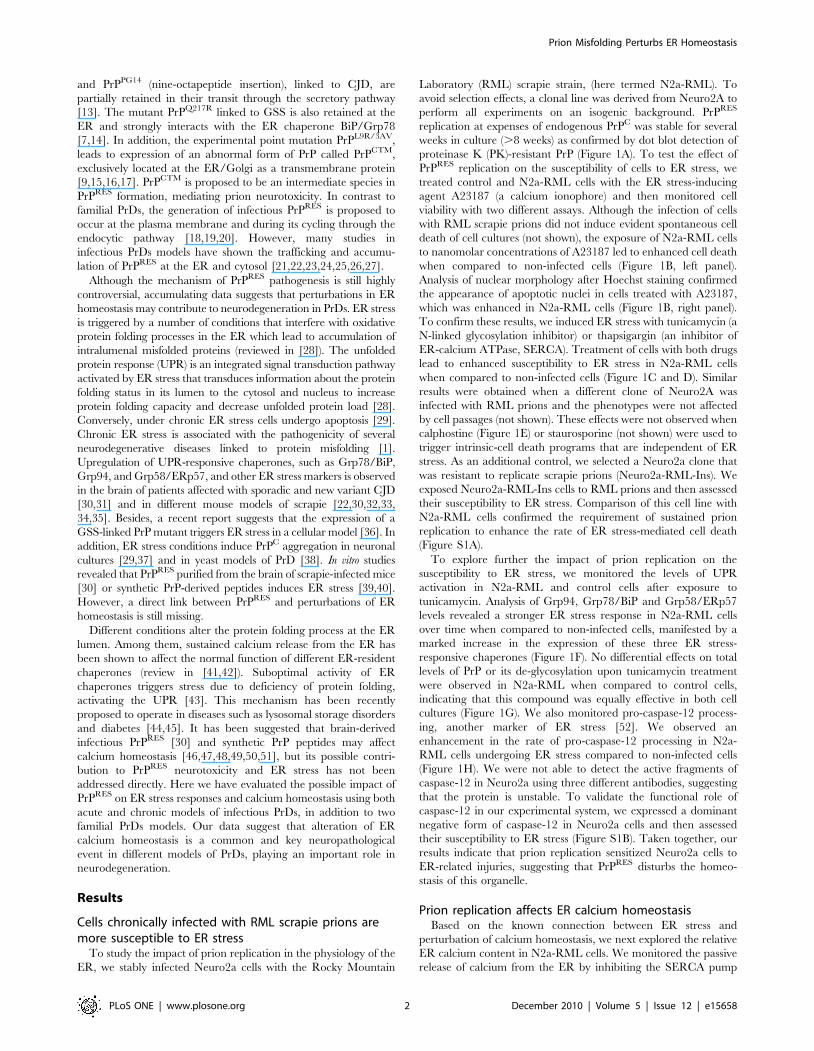
Cells chronically infected with RML scrapie prions aremore susceptible to ER stress
To study the impact of prion replication in the physiology of the
ER, we stably infected Neuro2a cells with the Rocky Mountain
Laboratory (RML) scrapie strain, (here termed N2a-RML). To
avoid selection effects, a clonal line was derived from Neuro2A to
perform all experiments on an isogenic background. PrPRES
replication at expenses of endogenous PrPC was stable for several
weeks in culture (.8 weeks) as confirmed by dot blot detection of
proteinase K (PK)-resistant PrP (Figure 1A). To test the effect of
PrPRES replication on the susceptibility of cells to ER stress, we
treated control and N2a-RML cells with the ER stress-inducing
agent A23187 (a calcium ionophore) and then monitored cell
viability with two different assays. Although the infection of cells
with RML scrapie prions did not induce evident spontaneous cell
death of cell cultures (not shown), the exposure of N2a-RML cells
to nanomolar concentrations of A23187 led to enhanced cell death
when compared to non-infected cells (Figure 1B, left panel).
Analysis of nuclear morphology after Hoechst staining confirmed
the appearance of apoptotic nuclei in cells treated with A23187,
which was enhanced in N2a-RML cells (Figure 1B, right panel).
To confirm these results, we induced ER stress with tunicamycin (a
N-linked glycosylation inhibitor) or thapsigargin (an inhibitor of
ER-calcium ATPase, SERCA). Treatment of cells with both drugs
lead to enhanced susceptibility to ER stress in N2a-RML cells
when compared to non-infected cells (Figure 1C and D). Similar
results were obtained when a different clone of Neuro2A was
infected with RML prions and the phenotypes were not affected
by cell passages (not shown). These effects were not observed when
calphostine (Figure 1E) or staurosporine (not shown) were used to
trigger intrinsic-cell death programs that are independent of ER
stress. As an additional control, we selected a Neuro2a clone that
was resistant to replicate scrapie prions (Neuro2a-RML-Ins). We
exposed Neuro2a-RML-Ins cells to RML prions and then assessed
their susceptibility to ER stress. Comparison of this cell line with
N2a-RML cells confirmed the requirement of sustained prion
replication to enhance the rate of ER stress-mediated cell death
(Figure S1A).
To explore further the impact of prion replication on the
susceptibility to ER stress, we monitored the levels of UPR
activation in N2a-RML and control cells after exposure to
tunicamycin. Analysis of Grp94, Grp78/BiP and Grp58/ERp57
levels revealed a stronger ER stress response in N2a-RML cells
over time when compared to non-infected cells, manifested by a
marked increase in the expression of these three ER stress-
responsive chaperones (Figure 1F). No differential effects on total
levels of PrP or its de-glycosylation upon tunicamycin treatment
were observed in N2a-RML when compared to control cells,
indicating that this compound was equally effective in both cell
cultures (Figure 1G). We also monitored pro-caspase-12 process-
ing, another marker of ER stress [52]. We observed an
enhancement in the rate of pro-caspase-12 processing in N2a-
RML cells undergoing ER stress compared to non-infected cells
(Figure 1H). We were not able to detect the active fragments of
caspase-12 in Neuro2a using three different antibodies, suggesting
that the protein is unstable. To validate the functional role of
caspase-12 in our experimental system, we expressed a dominant
negative form of caspase-12 in Neuro2a cells and then assessed
their susceptibility to ER stress (Figure S1B). Taken together, our
results indicate that prion replication sensitized Neuro2a cells to
ER-related injuries, suggesting that PrPRES disturbs the homeo-
stasis of this organelle.
Prion replication affects ER calcium homeostasisBased on the known connection between ER stress and
perturbation of calcium homeostasis, we next explored the relative
ER calcium content in N2a-RML cells. We monitored the passive
release of calcium from the ER by inhibiting the SERCA pump
Prion Misfolding Perturbs ER Homeostasis
PLoS ONE | www.plosone.org 2 December 2010 | Volume 5 | Issue 12 | e15658
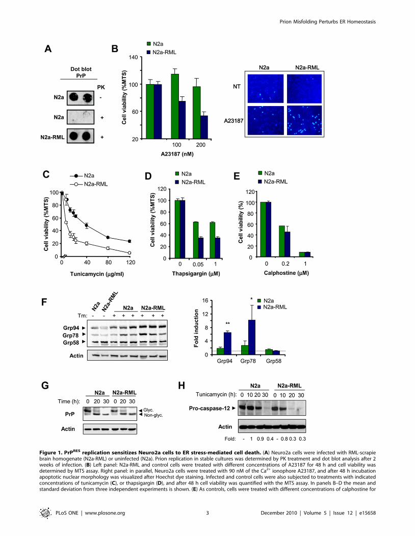
Figure 1. PrPRES replication sensitizes Neuro2a cells to ER stress-mediated cell death. (A) Neuro2a cells were infected with RML-scrapiebrain homogenate (N2a-RML) or uninfected (N2a). Prion replication in stable cultures was determined by PK treatment and dot blot analysis after 2weeks of infection. (B) Left panel: N2a-RML and control cells were treated with different concentrations of A23187 for 48 h and cell viability wasdetermined by MTS assay. Right panel: in parallel, Neuro2a cells were treated with 90 nM of the Ca2+ ionophore A23187, and after 48 h incubationapoptotic nuclear morphology was visualized after Hoechst dye staining. Infected and control cells were also subjected to treatments with indicatedconcentrations of tunicamycin (C), or thapsigargin (D), and after 48 h cell viability was quantified with the MTS assay. In panels B–D the mean andstandard deviation from three independent experiments is shown. (E) As controls, cells were treated with different concentrations of calphostine for
Prion Misfolding Perturbs ER Homeostasis
PLoS ONE | www.plosone.org 3 December 2010 | Volume 5 | Issue 12 | e15658
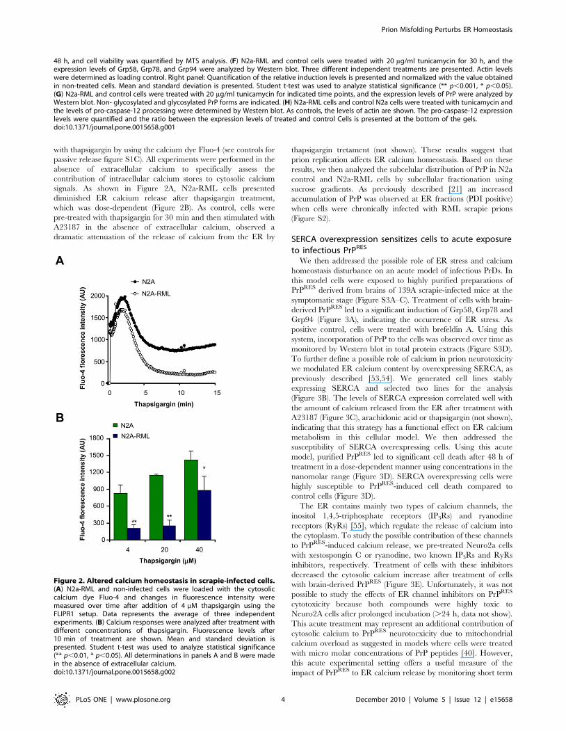
with thapsigargin by using the calcium dye Fluo-4 (see controls for
passive release figure S1C). All experiments were performed in the
absence of extracellular calcium to specifically assess the
contribution of intracellular calcium stores to cytosolic calcium
signals. As shown in Figure 2A, N2a-RML cells presented
diminished ER calcium release after thapsigargin treatment,
which was dose-dependent (Figure 2B). As control, cells were
pre-treated with thapsigargin for 30 min and then stimulated with
A23187 in the absence of extracellular calcium, observed a
dramatic attenuation of the release of calcium from the ER by
thapsigargin tretament (not shown). These results suggest that
prion replication affects ER calcium homeostasis. Based on these
results, we then analyzed the subcelular distribution of PrP in N2a
control and N2a-RML cells by subcellular fractionation using
sucrose gradients. As previously described [21] an increased
accumulation of PrP was observed at ER fractions (PDI positive)
when cells were chronically infected with RML scrapie prions
(Figure S2).
SERCA overexpression sensitizes cells to acute exposureto infectious PrPRES
We then addressed the possible role of ER stress and calcium
homeostasis disturbance on an acute model of infectious PrDs. In
this model cells were exposed to highly purified preparations of
PrPRES derived from brains of 139A scrapie-infected mice at the
symptomatic stage (Figure S3A–C). Treatment of cells with brain-
derived PrPRES led to a significant induction of Grp58, Grp78 and
Grp94 (Figure 3A), indicating the occurrence of ER stress. As
positive control, cells were treated with brefeldin A. Using this
system, incorporation of PrP to the cells was observed over time as
monitored by Western blot in total protein extracts (Figure S3D).
To further define a possible role of calcium in prion neurotoxicity
we modulated ER calcium content by overexpressing SERCA, as
previously described [53,54]. We generated cell lines stably
expressing SERCA and selected two lines for the analysis
(Figure 3B). The levels of SERCA expression correlated well with
the amount of calcium released from the ER after treatment with
A23187 (Figure 3C), arachidonic acid or thapsigargin (not shown),
indicating that this strategy has a functional effect on ER calcium
metabolism in this cellular model. We then addressed the
susceptibility of SERCA overexpressing cells. Using this acute
model, purified PrPRES led to significant cell death after 48 h of
treatment in a dose-dependent manner using concentrations in the
nanomolar range (Figure 3D). SERCA overexpressing cells were
highly susceptible to PrPRES-induced cell death compared to
control cells (Figure 3D).
The ER contains mainly two types of calcium channels, the
inositol 1,4,5-triphosphate receptors (IP3Rs) and ryanodine
receptors (RyRs) [55], which regulate the release of calcium into
the cytoplasm. To study the possible contribution of these channels
to PrPRES-induced calcium release, we pre-treated Neuro2a cells
with xestospongin C or ryanodine, two known IP3Rs and RyRs
inhibitors, respectively. Treatment of cells with these inhibitors
decreased the cytosolic calcium increase after treatment of cells
with brain-derived PrPRES (Figure 3E). Unfortunately, it was not
possible to study the effects of ER channel inhibitors on PrPRES
cytotoxicity because both compounds were highly toxic to
Neuro2A cells after prolonged incubation (.24 h, data not show).
This acute treatment may represent an additional contribution of
cytosolic calcium to PrPRES neurotocxicity due to mitochondrial
calcium overload as suggested in models where cells were treated
with micro molar concentrations of PrP peptides [40]. However,
this acute experimental setting offers a useful measure of the
impact of PrPRES to ER calcium release by monitoring short term
Figure 2. Altered calcium homeostasis in scrapie-infected cells.(A) N2a-RML and non-infected cells were loaded with the cytosoliccalcium dye Fluo-4 and changes in fluorescence intensity weremeasured over time after addition of 4 mM thapsigargin using theFLIPR1 setup. Data represents the average of three independentexperiments. (B) Calcium responses were analyzed after treatment withdifferent concentrations of thapsigargin. Fluorescence levels after10 min of treatment are shown. Mean and standard deviation ispresented. Student t-test was used to analyze statistical significance(** p,0.01, * p,0.05). All determinations in panels A and B were madein the absence of extracellular calcium.doi:10.1371/journal.pone.0015658.g002
48 h, and cell viability was quantified by MTS analysis. (F) N2a-RML and control cells were treated with 20 mg/ml tunicamycin for 30 h, and theexpression levels of Grp58, Grp78, and Grp94 were analyzed by Western blot. Three different independent treatments are presented. Actin levelswere determined as loading control. Right panel: Quantification of the relative induction levels is presented and normalized with the value obtainedin non-treated cells. Mean and standard deviation is presented. Student t-test was used to analyze statistical significance (** p,0.001, * p,0.05).(G) N2a-RML and control cells were treated with 20 mg/ml tunicamycin for indicated time points, and the expression levels of PrP were analyzed byWestern blot. Non- glycosylated and glycosylated PrP forms are indicated. (H) N2a-RML cells and control N2a cells were treated with tunicamycin andthe levels of pro-caspase-12 processing were determined by Western blot. As controls, the levels of actin are shown. The pro-caspase-12 expressionlevels were quantified and the ratio between the expression levels of treated and control Cells is presented at the bottom of the gels.doi:10.1371/journal.pone.0015658.g001
Prion Misfolding Perturbs ER Homeostasis
PLoS ONE | www.plosone.org 4 December 2010 | Volume 5 | Issue 12 | e15658
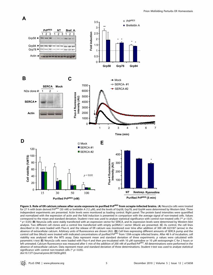
Figure 3. Role of ER calcium release after acute exposure to purified PrPRES from scrapie-infected brains. (A) Neuro2a cells were treatedfor 27 h with brain derived PrPRES (50 nM) or brefeldin A (12 mM), and the levels of Grp58, Grp78, and Grp94 were determined by Western blot. Threeindependent experiments are presented. Actin levels were monitored as loading control. Right panel: The protein band intensities were quantifiedand normalized with the expression of actin and the fold induction is presented in comparison with the average signal of non-treated cells. Valuescorrespond to the mean and standard deviation. Student t-test was used to analyze statistical significance with control non-treated cells (** p,0.01,* p,0.05) (B) Neuro2a cells were stably transfected with an expression vector for SERCA, and its expression levels were determined by Western blotanalysis. Two different cell clones and a control line transfected with empty pcDNA3.1 vector (Mock) are presented. (C) As control, the cell linesdescribed in (A) were loaded with Fluo-4, and the release of ER calcium was monitored over time after addition of 300 nM A23187 (arrow) in theabsence of extracellular calcium. Arbitrary units of fluorescence are shown (AU). (D) Cell lines expressing different amounts of SERCA pump and thecontrol cell line (Mock) were treated with indicated concentrations of purified PrPRES from 139A-scrapie infected brains. After 48 h of incubation, cellviability was analyzed with the MTS assay. Data represent mean and standard deviation of three experiments. p values were calculated withparametric t-test (E) Neuro2a cells were loaded with Fluo-4 and then pre-incubated with 10 mM ryanodine or 10 mM xestospongin C for 2 hours orleft untreated. Calcium fluorescence was measured after 5 min of the addition of 200 nM of purified PrPRES. All determinations were performed in theabsence of extracellular calcium. Data represent mean and standard deviation of three determinations. Student t-test was used to analyze statisticalsignificance with control non-treated cells (* p,0.05).doi:10.1371/journal.pone.0015658.g003
Prion Misfolding Perturbs ER Homeostasis
PLoS ONE | www.plosone.org 5 December 2010 | Volume 5 | Issue 12 | e15658
toxicity. Taken together, these results suggest that infectious
PrPRES alters ER calcium homeostasis.
Expression of PrP mutants linked to CJD and FFI increasethe susceptibility of cells to ER stress
To investigate the role of ER stress in familial forms of PrD, we
expressed two PrP mutant forms in Neuro2a cells, PrPPG14 and
PrPD177N/Met128, which are linked to familial CJD and FFI
respectively [2,56]. In addition, we employed the neurotoxic
mutant PrPCTM. We transiently transfected expression vectors for
these PrP mutants and PrPC as a control using EGFP fusion
proteins, and then visualized their subcellular distribution by
confocal microscopy and resistance to PK. As predicted, PrPC was
mainly located at the plasma membrane (Figure 4A and B). We
also confirmed the partial retention of PrPD177N and PrPPG14 at
the ER after co-expression of PrP with the ER marker KDEL-
dsRED (Figure 4A and B). Similarly, PrPCTM predominantly
accumulated at the ER (Figure 4A) and Golgi in our experimental
system (co-stained with anti-GM130 antibodies, not shown). To
monitor the possible generation of PrPRES, we transiently
expressed 3F4-epitope tagged mutants in 293T cells. The addition
of a 3F4 tag allowed us specifically detecting overexpressed PrP
and not the endogenous protein. Total protein extracts were
treated with two concentrations of PK and analyzed by Western
blot. As shown in Figure 4C, expression of the PrP mutants lead to
significant accumulation of PK-resistant PrP species. PrPD177N
displayed higher expression and increased PK-resistance
(Figure 4C). Changes in the electrophoresis pattern of the mutants
were observed as previously described [13,16], corresponding to
changes in the glycosylation pattern for PrPCTM and PrPD177N, or
a higher molecular weight for PrPPG14 due to the insertional
mutation. After characterizing our cellular model, we generated
Neuro2a cell lines stably expressing PrPC or the three PrD-related
mutants. Exposure of these cell lines to tunicamycin revealed that
the expression of PrPD177N increased the susceptibility to ER
stress-induced cell death (Figure 4D). Expression of PrPCTM or
PrPPG14 expressing cells showed an intermediate phenotype,
slightly enhancing their susceptibility to lower concentrations of
tunicamycin treatment (Figure 4D). Treating PrPD177N expressing
cells with thapsigargin also confirmed an enhanced susceptibility
to ER stress (Figure 4E). The changes in cell viability were small
for PrPCTM or PrPPG14 possibly because the stable selection of
these neurotoxic mutants may lead to the elimination of a
subpopulation of cells that are more susceptible to the pathological
effects of the PrP mutants. In fact, the transient transfection of PrP
mutants lead to ,40% of cell death after 72 h of transfection as
monitored by the MTS assay (not shown). Moreover, when we
analyzed the levels of the different PrP constructs over time, we
observed a decreased expression of PrPCTM or PrPPG14 after stable
selection, whereas higher PrPD177N expression levels were
maintained over time (Figure S4). Thus, these results suggest that
PrP mutants related to familial PrDs, in addition to PrPCTM, alters
the homeostasis of the ER to different extents, increasing the
susceptibility to ER stress.
Expression of mutant PrP forms alters ER calciumhomeostasis
To address the possible impact of familial PrD mutant PrP on
ER calcium homeostasis, we measured the relative ER calcium
content in cells stably expressing three PrP mutants or wild type
PrPC after thapsigargin treatment in the absence of extracellular
calcium. Treatment of Neuro2a cells expressing PrPCTM or
PrPD177N with 10 mM thapsigargin led to a decreased release of
ER calcium toward the cytosol compared to control PrPC
expressing cells (Figure 5A and B). Similar results were observed
when cells were exposed to higher thapsigargin concentrations
(40 mM; Figure 5C and D). These results were also recapitulated in
cells expressing the PrPPG14 mutant (Figure 5C and D).
To further confirm that cells expressing mutant PrPs have
alterations in ER homeostasis, we studied the effect of other ER
calcium agonists. Treatment of PrPCTM, PrPD177N, or PrPPG14
expressing cells with A23187 in absence of extracellular calcium
showed a substantial decrease on ER calcium release as compared
with PrPC expressing cells (Figure 5E). Similarly, treatment of all
mutant expressing cells with arachidonic acid (Figure 1F), which
induces IP3R-mediated calcium release, led to a lower cytosolic
calcium increase. Together these results suggest that accumulation
of PrPRES at the ER in familial PrD models results in decreased
ER calcium content.
Discussion
The protein folding capacity of the ER is directly affected by
different environmental factors including the redox status of the
cell, ER calcium content and ATP levels. ER stress responses are
observed in PrD models, and in human post mortem brain
samples from patients affected with sporadic and new variant CJD
[30,31,32,33,34,35,36,39]. Remarkably, chronic ER stress is
observed in many pathological conditions affecting the nervous
system where the disease is etiologically linked to protein
misfolding and abnormal protein aggregation [1].
The cellular events involved in perturbations of ER homeostasis
in PrDs are not well understood. The main issue is rooted in the
fact that prion replication mostly occurs at the plasma membrane
(in lipids rafts) and during its trafficking and recycling through the
endosomal pathway [5]. In addition, the majority of misfolded PrP
in infectious PrD models accumulates in the brain extracellular
space. Interestingly, different groups have shown that PrPRES is
also observed in intracellular compartments, where it could be
poly-ubiquitinated and accumulated into cytosolic aggresomes and
autophagosomes [25,26]. In addition, there is evidence that PrP
trafficking from the Golgi to the ER also contributes to the
generation of PrPRES in scrapie-infected cells [21]. We have
reported the co-inmunoprecipitation of PrPRES with ER-resident
chaperones in scrapie-infected cells [34], suggesting that a
subpopulation of infectious PrPRES may actually target the ER
lumen. Moreover, several studies have described the accumulation
of mutant PrP molecules associated with hereditary PrDs at the
ER and Golgi [7,10,11,14,15,16], suggesting that some prion
mutants may exert their pathological effects by affecting these
organelles. Alternatively, different observations suggest that
extracellular PrPRES may lead to activation of intracellular
signaling pathways that could indirectly impact the ER homeo-
stasis and contribute to neuronal dysfunction [12]. However, the
actual mechanism mediating PrP neurotoxicity in PrDs is not well
understood.
In the present study we investigated the possible contribution of
ER perturbations to prion pathogenesis in distinct PrD models
resembling infectious and familial forms of the disease. Our data
indicates that PrP misfolding leads to a higher susceptibility to ER
stress and abnormal ER calcium homeostasis in chronically
infected scrapie cells and in cells expressing PrP mutant forms
associated with FFI and familial CJD. Similar results were
observed when the neurotoxic mutant PrPCTM was expressed in
Neuro2a cells. In addition, we studied the impact of PrPRES on
calcium homeostasis using an acute PrD infectious model where
exogenous PrPRES was added to the cells at nanomolar
Prion Misfolding Perturbs ER Homeostasis
PLoS ONE | www.plosone.org 6 December 2010 | Volume 5 | Issue 12 | e15658
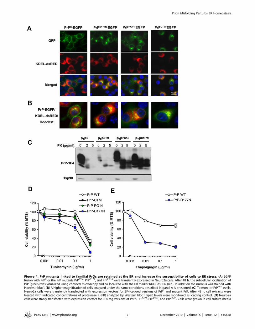
Figure 4. PrP mutants linked to familial PrDs are retained at the ER and increase the susceptibility of cells to ER stress. (A) EGFPfusion with PrPC or the PrP mutants PrPCTM, PrPD177, and PrPPG14 were transiently expressed in Neuro2a cells. After 48 h, the subcellular localization ofPrP (green) was visualized using confocal microscopy and co-localized with the ER-marker KDEL-dsRED (red). In addition the nucleus was stained withHoechst (blue). (B) A higher magnification of cells analyzed under the same conditions described in panel A is presented. (C) To monitor PrPRES levels,Neuro2a cells were transiently transfected with expression vectors for 3F4-tagged versions of PrPC and mutant PrP. After 48 h, cell extracts weretreated with indicated concentrations of proteinase K (PK) analyzed by Western blot. Hsp90 levels were monitored as loading control. (D) Neuro2acells were stably transfected with expression vectors for 3F4-tag versions of PrPC, PrPCTM, PrPD177, and PrPPG14. Cells were grown in cell culture media
Prion Misfolding Perturbs ER Homeostasis
PLoS ONE | www.plosone.org 7 December 2010 | Volume 5 | Issue 12 | e15658
concentrations. Using this system, we functionally addressed the
impact of ER calcium by monitoring cell toxicity after expression
of the ER calcium pump SERCA. In this acute setting, it is
predicted that drastic and fast changes in cytosolic calcium will
lead to mitochondrial calcium overload and apoptosis. In a
chronic conditions, this release of calcium may occur slowly (i.e. in
scrapie infected cells or neurons expressing familial PrP mutants),
generating in the long term a decrease in the ER steady state
calcium levels and ER stress. It may be feasibly that both cytosolic
increased of calcium together with decreased steady state ER
calcium content may synergise in the toxicity of misfolded PrP.
We speculate that a progressive and sustained release of ER
calcium in neurons expressing misfolded PrP species may affect the
protein folding status in this organelle, leading to basal ER stress,
resulting in organelle failure and neuronal dysfunction. At the
same time, increased levels of calcium in the cytoplasm may
perturb several signaling pathways implicated in controlling
neuronal function and survival. Indeed, our recent results suggest
that PrPRES formation leads to an hyperactivation of the calcium-
dependent phosphatase calcineurin in vivo, leading to dephosphor-
ylation of CREB and BAD [58]. Strikingly, treatment of prion
infected mice at the clinical phase of the disease with the FDA-
approved calcineurin inhibitor FK506 reduced neurodegenera-
tion, leading to improvement on behavioral alterations and
increase animal survival [58], suggesting a functional role of
calcium in synaptic dysfunction and neuronal death.
The ER is a primary compartment for PrPC folding and also
plays a crucial role in the generation of pathogenic PrP protein
conformations in familial PrDs. Therefore, it is essential to
understand the folding pathways of PrPC because this information
may give clues about the mechanism underlying sporadic forms of
CJD (the most common PrD in humans) where alteration in the
folding/quality control process or the ER environment may be a
key event in initiating the pathology. In support of this hypothesis,
post-mortem studies of sporadic and new variant CJD brain
samples demonstrated upregulation of ER stress-inducible foldases
[5,30,31]. Several studies indicate that alterations of the ER
homeostasis or inhibition of the proteasome, which blocks ERAD
of misfolded PrPC, lead to accumulation of abnormally folded
PrPC that is partially resistant to proteases and insoluble in non-
denaturing detergents [6,22,27,37,57]. These findings together
with the results described in the current study suggest that ER
factors and/or the ER environment, including ER calcium
concentrations, are key determinants of PrP misfolding and the
generation of neurotoxic species. The early signaling events
mediating ER calcium abnormalities by the accumulation of
PrPRES remain to be determined. Interactions between misfolded
PrP and the proteins modulating calcium transport (i.e. SERCA,
IP3Rs and/or RyRs) may explain part of these phenotypes as
suggested in Huntington’s disease models [59,60]. The findings
presented here may also contribute to a better understanding of
the pathogenesis of other diseases affecting the nervous system
related to protein misfolding and ER stress.
Materials and Methods
MaterialsTunicamycin, A23187, thapsigargin, calphostine, xestospongin
C, and ryanodine were purchased from Calbiochem EMB
Bioscience Inc. (Darmstadt, Germany). Cell medium, fetal calf
serum and antibiotics were obtained from Life Technologies
(Maryland, USA). Arachidonic acid and dantrolene were obtained
from Sigma (Basel, Switzerland). Fluo-4 was purchased from
Molecular Probes. Superfect and plasmid purification kits were
pursed from Qiagene (HiSpeed Plasmid Midi Kit).
Cell culture and viability assaysNeuro2A cells were obtained from ATCC and were cultured in
DMEM supplemented with 10% fetal calf serum and antibiotics
(109000 U/ml Penicillin, 10 mg/ml streptomycin), at 37uC and
5% CO2. For cell viability analysis, cells were grown in collagen IV
coated 96-well plates for 24 h in cell culture medium in 1% serum
before addition of the agonist. Cell viability was quantified using 3-
(4, 5-dimethylthazol-2-yl)-5-3-carboxymethoxy-phenyl)-2-(4-sulfo-
phenyl)-2H-tetrazolium (MTS) according to the recommendations
of the supplier (Promega, CellTiter96H Aqueous, Madison, WI). In
addition, cellular death by apoptosis was quantified by nuclear
staining with Hoechst33342.
PrPRES purification from the brain of scrapie infectedmice.
PrPRES was purified from mice infected with 139A scrapie as
previously described [61]. Experimental animal protocols for
animal use has been reviewed and approved by the Institutional
Review Board’s Animal Care and Use Committee of the Faculty of
Medicine of the University of Chile (approved protocol CBA #0232 FMUCH). Brain tissue (approximately 12 brains per
preparation) was homogenized with a manual potter of 20 ml in
PBS (final concentration 50% weight/volume) containing a
protease inhibitor cocktail. After homogenization, an equivalent
volume of a solution containing 20% salkosyl and 0.05% octanol
was added. The brain extract was incubated for 15 min at room
temperature with constant agitation in a wheel rotor. After this step,
non-disrupted tissue was eliminated by centrifugation at 7.0006 g
for 15 min. The supernatant was collected and 1/3 volume of 0.1%
SB3-14 was added to the brain homogenate, mixed and centrifuged
at 50.000 r.p.m. in a Ti60 rotor (Beckman) for 2 h at 4uC. After
centrifugation, the pellets were collected and resuspended by
sonication in 10% NaCl, 0.1% SB3-14. The homogenized pellet
was loaded over a sucrose solution (20% sucrose, 0.1% SB-314) and
centrifuged at 80.000 r.p.m. for 2 h at 4uC in a TL100 rotor
(Beckman). The pellet was collected, washed in PBS and
resuspended by sonication in PBS containing 0.1% SB-314.
Thereafter, samples were treated with PK (50 mg/ml) for 2 h
followed by another sucrose step separation after centrifugation at
80.000 r.p.m. for 2 h. The pellet was washed four times with sterile
PBS and resuspended in 400 ml of PBS by sonication. After this step,
purity was estimated to be higher than 90% as estimated by silver
staining and mass spectrometric analysis. PrPRES concentration was
estimated by western blot analysis, comparing in the same blot the
signal intensity of different dilutions of the purified protein with
known concentrations of the recombinant mouse PrPC, purchased
from Prionics Inc (Zurich, Switzerland).
Plasmids and cell transfectionsExpression vector containing SERCA from rabbit was kindly
provided by Frederica Del Monte (University of Toronto,
containing 2% serum for 16 h and then exposed to different concentrations of tunicamycin. After 24 h, cell viability was determined with the MTSassay. Data represent mean and standard deviation of three determinations that are representative of three independent experiments. (E) In parallel,PrPC and PrPD177 expressing cells were treated with indicated concentrations of thapsigargin and analyzed as described in D.doi:10.1371/journal.pone.0015658.g004
Prion Misfolding Perturbs ER Homeostasis
PLoS ONE | www.plosone.org 8 December 2010 | Volume 5 | Issue 12 | e15658
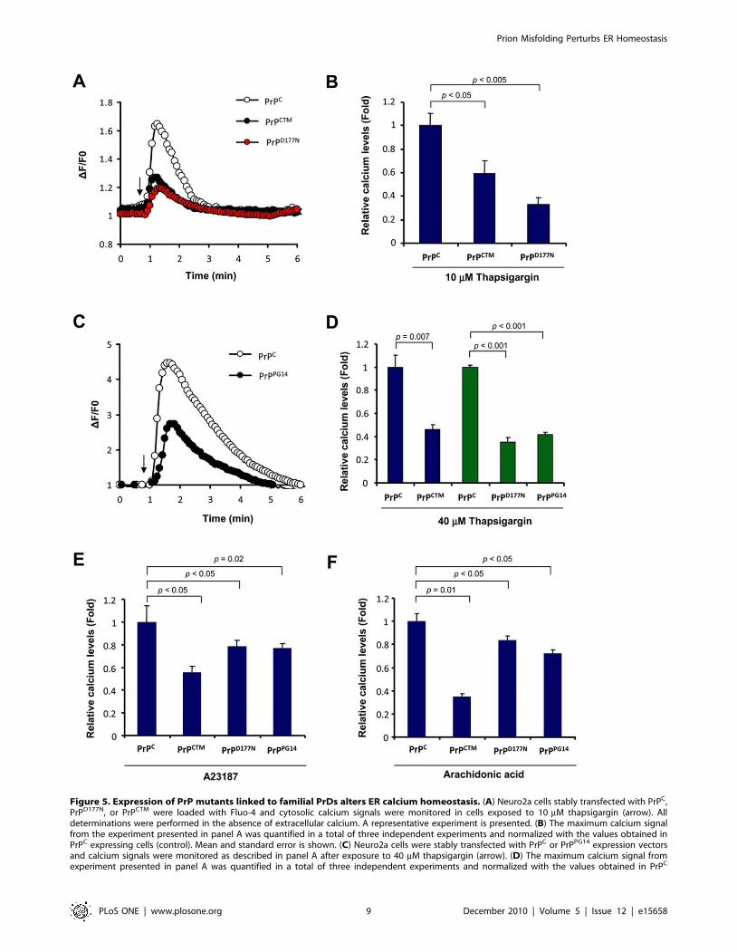
Figure 5. Expression of PrP mutants linked to familial PrDs alters ER calcium homeostasis. (A) Neuro2a cells stably transfected with PrPC,PrPD177N, or PrPCTM were loaded with Fluo-4 and cytosolic calcium signals were monitored in cells exposed to 10 mM thapsigargin (arrow). Alldeterminations were performed in the absence of extracellular calcium. A representative experiment is presented. (B) The maximum calcium signalfrom the experiment presented in panel A was quantified in a total of three independent experiments and normalized with the values obtained inPrPC expressing cells (control). Mean and standard error is shown. (C) Neuro2a cells were stably transfected with PrPC or PrPPG14 expression vectorsand calcium signals were monitored as described in panel A after exposure to 40 mM thapsigargin (arrow). (D) The maximum calcium signal fromexperiment presented in panel A was quantified in a total of three independent experiments and normalized with the values obtained in PrPC
Prion Misfolding Perturbs ER Homeostasis
PLoS ONE | www.plosone.org 9 December 2010 | Volume 5 | Issue 12 | e15658
Canada). Expression plasmid containing catalytically inactive
caspase-12 mutant lacking its N-terminal pro-domain (amino
acids 1-94, pC12DN) was provided by R. Rao (California).
Expression vectors of 3F4 tagged PrP mutants (PrPCTM, PrPPG14,
and PrPD177N) and GFP fusion proteins were provided by David
Harris (Washington University). The generation of PrPC-3F4 and
PrPC-EGFP constructs was previously described [57]. Co-
localization of PrP with ER was assessed by co-transfection with
pDsRed-ER (Clontech Laboratories, cat. 6982-1). Stably-express-
ing Neuro2a cells were produced by transfection using SuperFect
kit (Qiagene, Valencia, CA) following the manufacturer’s instruc-
tions. After 48 h of transfection, cells were selected using
hygromicin (1.5 mg/ml) or G418 (1.3 mg/ml). Individual clones
of Neuro2a cells expressing different levels of SERCA, were
obtained by limiting dilution as described before [30].
Generation of Neuro2A cells chronically infected withRML scrapie prions
Neuro2a cells can be infected with PrPRES although the
response and the stability of the infected cells has proved variable,
suggesting heterogeneity may exist in the original cell line [62,63].
In order to establish a stable chronically infected cell, we separated
the original culture in subclones by limiting dilution. A growing
culture was diluted to a density of 5 cells/ml and 100 ml was
transferred to individual wells of a 96 well plate and cultured for 1
week. The individual cultures were examined microscopically to
determine those wells which contained a single focus of growing
cells. The single cell derived cultures were then transferred to 24
well plates and serially passaged every 3–4 days at 1:15 dilution to
maintain stocks. Single clone cultures were tested for sensitivity to
infection by the RML strain of PrPRES. To do this, 4 ml of a 10%
late stage infected brain extract was added per well of newly
passaged cells, and the cultures were left for a further 4 days to
reach confluence. Cells were serially passaged thereafter in the
absence of PrPRES. Tests showed that all trace of the initial
inoculum disappeared by passage 4. At this and later passages
individual cultures were tested for the presence of PrPRES by dot
blotting as previously described [62].
Measurement of intracellular calciumFor biosecurity reasons, in experiment with RML infected cells
or cells treated with purified PrPRES, the changes in intracellular
calcium levels were measured using the automatized FLIPR1
machine (Molecular devices, Sunnyvale, CA) by the use of the
fluorescent dye Fluo-4, which shows increased fluorescence at
515–535 nm after calcium binding. 105 cells per well were grown
on 96-black wells plate coated with collagen IV for 24 h and
serum was decreased to 1% for a further 24 h. Cells were loaded
with Fluo-4 at a final concentration of 10 mg/mL. After 120 min
of incubation at 37uC, the loaded cells were washed twice with
FLIPR buffer (150 mM NaCl, 5 mM KCl, 1 mM MgCl2, 10 mM
Hepes, 10 mM glucose) in the absence of extracellular calcium
and plates were mounted on the FLIPR1 setup. Fluorescence
emission was quantified every 5 seconds for a total time of 30 min.
The basal fluorescence of the dye was usually determined before
the addition of the samples and was assigned a value of zero. The
agonists and inhibitor were added automatically by the FLIPR1
setup, and were prepared in an independent 96-well plate. To
determine the origin of intracellular calcium, cells were pre-treated
with thapsigargin (5 mM), a ryanodine mix (10 mM) or xestospon-
gin C for 15 min before addition of the agonists.
In experiment with PrP mutants, cells were grown in coverslips
and loaded with Fluo-4 AM (3 mM) for 30 min at room
temperature. Coverslips were mounted in a 1 ml capacity
chamber and washed three times with calcium-free buffer
(150 mM NaCl, 5 mM KCl, 1 mM MgCl2, 10 mM Hepes,
10 mM glucose and 5 mM EGTA, pH 7.4). Calcium signals were
recorded using an IX-81 inverted microscope for fluorescence
measurements (DSU, Olympus), equipped with a 150-W xenon
lamp (Olympus MT-20). Fluo-4 fluorescence was excited and
detected with a FITC filter cube, using a 40x/1.4 NA oil
immersion objective. Changes in [Ca2+]i were measured in a field-
of-view consisting of 15–30 cells. Images were acquired every 5
seconds and analyzed using CellR (Olympus) and NIH ImageJ
software. The mean intensities of small cellular areas of interest
were collected as F(t) and the background intensity was subtracted,
using a same-size region of interest outside the cell, yielding F(t)s.
The final signal was normalized to baseline fluorescence F(0), as
[F(t)s 2 F(0)]/F(0) [64,65].
SDS-PAGE and Western Blot AnalysisCells were homogenized on ice in RIPA buffer (20 mM Tris
pH 8.0, 150 mM NaCl, 0.1% SDS, 0.5% DOC, 0.5% triton X-
100) containing a protease inhibitor cocktail (Roche, Basel,
Germany) as previously described [66]. Protein concentration
was determined by micro-BCA assay (Pierce, Rockford, IL). The
equivalent of 30–50 mg of total protein was generally loaded onto
10% SDS-PAGE minigels (Novex NuPage, Invitrogen Life
Technologies, Basel, Switzerland) and analysed by Western
blotting as described. The following antibodies and dilutions were
used: 6H4 anti-PrP, 1:10,000 (Prionics, Zurich, Switzerland), anti-
Caspase-12, 1:2,000 (Exalpha, Watertown, USA); anti-GRP78/
Bip, anti-Grp58/ERp57 and anti-Grp94 1:2,000 (StressGene, San
Diego, CA); anti-actin, 1:2,000 and Hsp90, 1:3000 (Santa Cruz),
anti-3F4 antibody 1:5000 (Abcam). After incubation with the
primary antibody, membranes were incubated for 1 h at room
temperature with horseradish peroxidase-coupled second antibod-
ies diluted 1:10,000 in washing buffer. After washing, specifically
bound antibodies were detected by enhanced chemiluminescence
assay (Amersham Biosciences, Cardiff, UK).
Subcellular FractionationTo separate and enrich ER membranes, Neuro2a cells were
homogenized by using a stainless steel ball-bearing homogenizer in
0.25 M sucrose, 10 mM Tris-HCl, pH 7.4, 1 mM magnesium
acetate, and a protease inhibitor mixture in a final concentration
of 1 volume of cell pellet per 5 volumes of homogenizing medium.
Sucrose gradients were performed as described in [21]. 1-ml
fractions were collected from the top of each gradient, assayed for
protein content, and methanol-precipitated. After centrifugation at
14,000 rpm for 20 min, the pellets were resuspended in SDS
loading buffer.
expressing cells (control). Mean and standard error is shown. Similar experiments were performed in cells expressing PrPD177N, or PrPCTM. (E) Inparallel, cells expressing PrPC, PrPCTM, PrPD177N, or PrPPG14 were exposed to 10 mM A23187., or (F) 15 mM arachidonic acid and analyzed usingconditions described in panel A. The maximum calcium signal from the experiment presented in panel A was quantified in a total of threeindependent experiments and normalized with the values obtained in PrPC expressing cells (control). Mean and standard error is shown. In B, D, E andF, indicated p values were calculated with parametric t-test.doi:10.1371/journal.pone.0015658.g005
Prion Misfolding Perturbs ER Homeostasis
PLoS ONE | www.plosone.org 10 December 2010 | Volume 5 | Issue 12 | e15658
Statistical analysisData was analyzed by parametric t-test (two-tailed) and
significance was expressed as follow: * P,0.05; ** P,0.01;
*** P,0.005. For the analysis the program SigmaPlot and
GraphPad were employed.
Supporting Information
Figure S1 Control experiments. (A) Replication ofPrPRES at expenses ofendogenous PrPC is required toincrease the susceptibility to ER stress. Twodifferent
Neuro2a clones were selected by their property to sustain
replication of RMLprions (N2a-RML) or that are resistant to
replication (N2a-RML-Ins). Cell were exposedto RML scrapie
prion and after several weeks in culture, they were treated with 12
mMbrefeldin A (Bref. A) or 40 nM A23187. After 48h cell viability
was monitored using theMTS assay. Mean and standard deviation
is presented of three determinations. (B) Expression of acaspase-12 dominant negative mutant form protectagainst ERstress. Left panel: Neuro2 cells were stably
transfected with empty pCDNA.3 vector oran expression vector
for a caspase-12 dominant negative (C289A) construct. Then,cell
viability was monitored after exposure of cells to 12 mM brefeldin
A or 5 mMthapsigargin for 48h using the MTS assay. Data
represent mean and standarddeviation of three determinations.
Right panel: Expression levels of caspase-12 andactin are
presented as controls. (C) Thapsigargin treatment triggerspassive relatedof ER calcium, not affected by inhibitionof IP3R. Neuro2a cells were loaded withFluo-4 and cytosolic
calcium signals were monitored in cells exposed to 10 mMthapsi-
gargin (arrow). Cells were pretreated or not with 1 mM
Xestospongine B (IP3Rinhibitor) for 1h or 50 mM dantrolen
(RYR inhibitor) for 30 min. All determinations wereperformed in
the absence of extracellular calcium. A representative experiment
ispresented.
(PDF)
Figure S2 Increased accumulation PrP at ER fractionsin Neuro2a cells infectedwith RML scrapie prions. Post-
nuclear cell extracts from Neuro2a control and RMLinfectedcells
were fractionated on a sucrose gradient to separate ER fractions
asdescribed in material and methods. Total proteins present in
fractions of 1 ml wereprecipitated and analyzed by Western blot.
Total PrP levels were monitored in eachfraction. As control to
identify ER-enriched fractions, the distribution of PDI wasassessed
by Western blot.
(PDF)
Figure S3 Purification of PrPRES from 139A-scrapieinfected brains. (A) Schematicrepresentation of the preparation
steps used to purify PrPRES from 139A-scrapieinfected brains
(described in material and methods). (B) Qualitative analysis of
theenrichment on PrPRES during purification procedure. Equiv-
alent samples from differentsteps of the purification process were
analyzed by western blot or by silver staining ofthe total proteins
presented in each sample. Samples were loaded in the followin-
gorder: 1: 10% brain homogenate in PBS. 2: Sarcosyl
solubilization. 3: Sarcosylextraction, pellet. 4: Pellet obtained
after sucrose gradient before PK treatment. 5: Pellet obtained after
sucrose gradient after PK treatment. 6: 500 ng of recombi-
nantPrPC, used as a positive control. (C) Quantification of
PrPRES concentration. Knownamounts of recombinant PrPC
were compared with different dilutions of purified PrPRESby
western blot analysis (left panel). The band intensity was quantified
to estimate theconcentration of PrPRES by comparison to the
values obtained with the calibration curveof recombinant PrPC
(right panel). R2 corresponds to the linear regression coefficient.
(D) Neuro2a cells were treated with 1 mg/ml brain derived
PrPRES for indicated timepoints and then washed extensively
with PBS. Then cells were collected bytripsinization and further
washed in PBS by centrifugation. PrP levels were monitoredby
Western blot in total protein extracts. The molecular weight of PrP
corresponds tothe PK-resistant core, indicating the detection of the
exogenously added brain-derivedPrP. In this assay, PrPRES
oligomers are also observed (*). Actin levels were monitoredas
control.
(PDF)
Figure S4 Stable expression of prion mutants. Neuro2A
cells were transfected withindicated PrP expressing vectors
containing the 3F4 tag epitope, and then selectedwith G418 (1.3
mg/ml). The expression levels for each PrP version was assessed
overtime by Western blot analysis during early selection (~1 week,
upper panel) or afterstable selection (3 week, middle panel; 4
weeks, bottom panel). As loading control anon specific band is
presented from the 3F4 Western blot. Cells presented in the
rightpanel were used to the viability assays.
(PDF)
Acknowledgments
We thank Drs. Kinsey Maundrell and Milene Ruselakis for advice on
experiments and Drs. Ute Woehlbier and Soledad Matus for helpful
discussion. We thank Federica Del-Monte for providing SERCA
expression vectors. We thank David Harris for providing PrP expression
vectors. We thank Yves Cambet and Yves Humbert for advices in the use
of the FLIPR1 setup.
Author Contributions
Conceived and designed the experiments: CH CS AS KC MT RA.
Performed the experiments: MT KC CH. Analyzed the data: MT CH RA
KC. Contributed reagents/materials/analysis tools: CH CS AS. Wrote the
paper: CH MT CS.
References
1. Matus S, Lisbona F, Torres M, Leon C, Thielen P, et al. (2008) The stress
rheostat: an interplay between the unfolded protein response (UPR) and
autophagy in neurodegeneration. Curr Mol Med 8: 157–172.
2. Prusiner SB (1998) Prions. Proc Natl Acad Sci U S A 95: 13363–13383.
3. Hetz C, Soto C (2003) Protein misfolding and disease: the case of prion
disorders. Cell Mol Life Sci 60: 133–143.
4. Soto C, Saborio GP (2001) Prions: disease propagation and disease therapy by
conformational transmission. Trends Mol Med 7: 109–114.
5. Hegde RS, Rane NS (2003) Prion protein trafficking and the development of
neurodegeneration. Trends Neurosci 26: 337–339.
6. Yedidia Y, Horonchik L, Tzaban S, Yanai A, Taraboulos A (2001) Proteasomes
and ubiquitin are involved in the turnover of the wild-type prion protein. Embo J
20: 5383–5391.
7. Jin T, Gu Y, Zanusso G, Sy M, Kumar A, et al. (2000) The chaperone protein
BiP binds to a mutant prion protein and mediates its degradation by the
proteasome. J Biol Chem 275: 38699–38704.
8. Ma J, Lindquist S (2001) Wild-type PrP and a mutant associated with prion
disease are subject to retrograde transport and proteasome degradation. Proc
Natl Acad Sci U S A 98: 14955–14960.
9. Stewart RS, Drisaldi B, Harris DA (2001) A transmembrane form of the prion
protein contains an uncleaved signal peptide and is retained in the endoplasmic
Reticulum. Mol Biol Cell 12: 881–889.
10. Gu Y, Verghese S, Bose S, Mohan M, Singh N (2007) Mutant prion
protein D202N associated with familial prion disease is retained in the
endoplasmic reticulum and forms ‘curly’ intracellular aggregates. J Mol Neurosci
32: 90–96.
Prion Misfolding Perturbs ER Homeostasis
PLoS ONE | www.plosone.org 11 December 2010 | Volume 5 | Issue 12 | e15658
11. Zanusso G, Petersen RB, Jin T, Jing Y, Kanoush R, et al. (1999) Proteasomal
degradation and N-terminal protease resistance of the codon 145 mutant prionprotein. J Biol Chem 274: 23396–23404.
12. Hetz CA, Soto C (2006) Stressing out the ER: a role of the unfolded protein
response in prion-related disorders. Curr Mol Med 6: 37–43.13. Drisaldi B, Stewart RS, Adles C, Stewart LR, Quaglio E, et al. (2003) Mutant
PrP is delayed in its exit from the endoplasmic reticulum, but neither wild-typenor mutant PrP undergoes retrotranslocation prior to proteasomal degradation.
J Biol Chem 278: 21732–21743.
14. Singh N, Zanusso G, Chen SG, Fujioka H, Richardson S, et al. (1997) Prionprotein aggregation reverted by low temperature in transfected cells carrying a
prion protein gene mutation. J Biol Chem 272: 28461–28470.15. Ivanova L, Barmada S, Kummer T, Harris DA (2001) Mutant prion proteins are
partially retained in the endoplasmic reticulum. J Biol Chem 276: 42409–42421.16. Stewart RS, Piccardo P, Ghetti B, Harris DA (2005) Neurodegenerative illness in
transgenic mice expressing a transmembrane form of the prion protein.
J Neurosci 25: 3469–3477.17. Stewart RS, Harris DA (2005) A transmembrane form of the prion protein is
localized in the Golgi apparatus of neurons. J Biol Chem 280: 15855–15864.18. Vey M, Pilkuhn S, Wille H, Nixon R, DeArmond SJ, et al. (1996) Subcellular
colocalization of the cellular and scrapie prion proteins in caveolae-like
membranous domains. Proc Natl Acad Sci U S A 93: 14945–14949.19. Naslavsky N, Stein R, Yanai A, Friedlander G, Taraboulos A (1997)
Characterization of detergent-insoluble complexes containing the cellular prionprotein and its scrapie isoform. J Biol Chem 272: 6324–6331.
20. Marella M, Lehmann S, Grassi J, Chabry J (2002) Filipin prevents pathologicalprion protein accumulation by reducing endocytosis and inducing cellular PrP
release. J Biol Chem 277: 25457–25464.
21. Beranger F, Mange A, Goud B, Lehmann S (2002) Stimulation of PrP(C)retrograde transport toward the endoplasmic reticulum increases accumulation
of PrP(Sc) in prion-infected cells. J Biol Chem 277: 38972–38977.22. Rane NS, Kang SW, Chakrabarti O, Feigenbaum L, Hegde RS (2008) Reduced
translocation of nascent prion protein during ER stress contributes to
neurodegeneration. Dev Cell 15: 359–370.23. Taraboulos A, Serban D, Prusiner SB (1990) Scrapie prion proteins accumulate
in the cytoplasm of persistently infected cultured cells. J Cell Biol 110:2117–2132.
24. Kristiansen M, Deriziotis P, Dimcheff DE, Jackson GS, Ovaa H, et al. (2007)Disease-associated prion protein oligomers inhibit the 26S proteasome. Mol Cell
26: 175–188.
25. Grenier C, Bissonnette C, Volkov L, Roucou X (2006) Molecular morphologyand toxicity of cytoplasmic prion protein aggregates in neuronal and non-
neuronal cells. J Neurochem 97: 1456–1466.26. Kristiansen M, Messenger MJ, Klohn PC, Brandner S, Wadsworth JD, et al.
(2005) Disease-related prion protein forms aggresomes in neuronal cells leading
to caspase activation and apoptosis. J Biol Chem 280: 38851–38861.27. Ma J, Lindquist S (2002) Conversion of PrP to a self-perpetuating PrPSc-like
conformation in the cytosol. Science 298: 1785–1788.28. Hetz C, Glimcher LH (2009) Fine-tuning of the unfolded protein response:
Assembling the IRE1alpha interactome. Mol Cell 35: 551–561.29. Hetz CA (2007) ER stress signaling and the BCL-2 family of proteins:
from adaptation to irreversible cellular damage. Antioxid Redox Signal 9:
2345–2355.30. Hetz C, Russelakis-Carneiro M, Maundrell K, Castilla J, Soto C (2003) Caspase-
12 and endoplasmic reticulum stress mediate neurotoxicity of pathological prionprotein. Embo J 22: 5435–5445.
31. Yoo BC, Krapfenbauer K, Cairns N, Belay G, Bajo M, et al. (2002)
Overexpressed protein disulfide isomerase in brains of patients with sporadicCreutzfeldt-Jakob disease. Neurosci Lett 334: 196–200.
32. Brown AR, Rebus S, McKimmie CS, Robertson K, Williams A, et al. (2005)Gene expression profiling of the preclinical scrapie-infected hippocampus.
Biochem Biophys Res Commun 334: 86–95.
33. Hetz C, Lee AH, Gonzalez-Romero D, Thielen P, Castilla J, et al. (2008)Unfolded protein response transcription factor XBP-1 does not influence prion
replication or pathogenesis. Proc Natl Acad Sci U S A 105: 757–762.34. Hetz C, Russelakis-Carneiro M, Walchli S, Carboni S, Vial-Knecht E, et al.
(2005) The disulfide isomerase Grp58 is a protective factor against prionneurotoxicity. J Neurosci 25: 2793–2802.
35. Steele AD, Hetz C, Yi CH, Jackson WS, Borkowski AW, et al. (2007) Prion
pathogenesis is independent of caspase-12. Prion 1: 243–247.36. Xu K, Wang X, Shi Q, Chen C, Tian C, et al. Human Prion Protein Mutants
with Deleted and Inserted Octarepeats Undergo Different Pathways to TriggerCell Apoptosis. J Mol Neurosci.
37. Orsi A, Fioriti L, Chiesa R, Sitia R (2006) Conditions of endoplasmic reticulum
stress favor the accumulation of cytosolic prion protein. J Biol Chem 281:30431–30438.
38. Apodaca J, Kim I, Rao H (2006) Cellular tolerance of prion protein PrP in yeastinvolves proteolysis and the unfolded protein response. Biochem Biophys Res
Commun 347: 319–326.39. Ferreiro E, Resende R, Costa R, Oliveira CR, Pereira CM (2006) An
endoplasmic-reticulum-specific apoptotic pathway is involved in prion and
amyloid-beta peptides neurotoxicity. Neurobiol Dis 23: 669–678.
40. Ferreiro E, Costa R, Marques S, Cardoso SM, Oliveira CR, et al. (2008)
Involvement of mitochondria in endoplasmic reticulum stress-induced apoptotic
cell death pathway triggered by the prion peptide PrP(106-126). J Neurochem
104: 766–776.
41. Gorlach A, Klappa P, Kietzmann T (2006) The endoplasmic reticulum: folding,
calcium homeostasis, signaling, and redox control. Antioxid Redox Signal 8:
1391–1418.
42. Puzianowska-Kuznicka M, Kuznicki J (2009) The ER and ageing II: calcium
homeostasis. Ageing Res Rev 8: 160–172.
43. Michalak M, Robert Parker JM, Opas M (2002) Ca2+ signaling and calcium
binding chaperones of the endoplasmic reticulum. Cell Calcium 32: 269–278.
44. Ong DS, Mu TW, Palmer AE, Kelly JW. Endoplasmic reticulum Ca2+increases enhance mutant glucocerebrosidase proteostasis. Nat Chem Biol 6:
424–432.
45. Park SW, Zhou Y, Lee J, Lee J, Ozcan U. Sarco(endo)plasmic reticulum Ca2+-
ATPase 2b is a major regulator of endoplasmic reticulum stress and glucose
homeostasis in obesity. Proc Natl Acad Sci U S A.
46. Agostinho P, Oliveira CR (2003) Involvement of calcineurin in the neurotoxic
effects induced by amyloid-beta and prion peptides. Eur J Neurosci 17:
1189–1196.
47. Florio T, Grimaldi M, Scorziello A, Salmona M, Bugiani O, et al. (1996)
Intracellular calcium rise through L-type calcium channels, as molecular
mechanism for prion protein fragment 106-126-induced astroglial proliferation.
Biochem Biophys Res Commun 228: 397–405.
48. Kawahara M, Kuroda Y, Arispe N, Rojas E (2000) Alzheimer’s beta-amyloid,
human islet amylin, and prion protein fragment evoke intracellular free calcium
elevations by a common mechanism in a hypothalamic GnRH neuronal cell
line. J Biol Chem 275: 14077–14083.
49. O’Donovan CN, Tobin D, Cotter TG (2001) Prion protein fragment PrP-(106-
126) induces apoptosis via mitochondrial disruption in human neuronal SH-
SY5Y cells. J Biol Chem 276: 43516–43523.
50. Thellung S, Florio T, Villa V, Corsaro A, Arena S, et al. (2000) Apoptotic cell
death and impairment of L-type voltage-sensitive calcium channel activity in rat
cerebellar granule cells treated with the prion protein fragment 106-126.
Neurobiol Dis 7: 299–309.
51. Ferreiro E, Oliveira CR, Pereira CM (2008) The release of calcium from the
endoplasmic reticulum induced by amyloid-beta and prion peptides activates the
mitochondrial apoptotic pathway. Neurobiol Dis 30: 331–342.
52. Nakagawa T, Zhu H, Morishima N, Li E, Xu J, et al. (2000) Caspase-12
mediates endoplasmic-reticulum-specific apoptosis and cytotoxicity by amyloid-
beta. Nature 403: 98–103.
53. Scorrano L, Oakes SA, Opferman JT, Cheng EH, Sorcinelli MD, et al. (2003)
BAX and BAK regulation of endoplasmic reticulum Ca2+: a control point for
apoptosis. Science 300: 135–139.
54. Xu C, Xu W, Palmer AE, Reed JC (2008) BI-1 regulates endoplasmic reticulum
Ca2+ homeostasis downstream of Bcl-2 family proteins. J Biol Chem 283:
11477–11484.
55. Berridge MJ (2002) The endoplasmic reticulum: a multifunctional signaling
organelle. Cell Calcium 32: 235–249.
56. Harris DA (1999) Cellular biology of prion diseases. Clin Microbiol Rev 12:
429–444.
57. Hetz C, Castilla J, Soto C (2007) Perturbation of endoplasmic reticulum
homeostasis facilitates prion replication. J Biol Chem 282: 12725–12733.
58. Barria MA, Mukherjee A, Gonzalez-Romero D, Morales R, Soto C (2009) De
novo generation of infectious prions in vitro produces a new disease phenotype.
PLoS Pathog 5: e1000421.
59. Tang TS, Tu H, Chan EY, Maximov A, Wang Z, et al. (2003) Huntingtin and
huntingtin-associated protein 1 influence neuronal calcium signaling mediated
by inositol-(1,4,5) triphosphate receptor type 1. Neuron 39: 227–239.
60. Vidal R, Caballero B, Couve A, Hetz C (2011) Converging pathways in the
occurrence of endoplasmic reticulum (ER) stress in Huntington’s disease. Curr.
Mol. Med, In press.
61. Soto C, Kascsak RJ, Saborio GP, Aucouturier P, Wisniewski T, et al. (2000)
Reversion of prion protein conformational changes by synthetic beta-sheet
breaker peptides. Lancet 355: 192–197.
62. Bosque PJ, Prusiner SB (2000) Cultured cell sublines highly susceptible to prion
infection. J Virol 74: 4377–4386.
63. Enari M, Flechsig E, Weissmann C (2001) Scrapie prion protein accumulation
by scrapie-infected neuroblastoma cells abrogated by exposure to a prion protein
antibody. Proc Natl Acad Sci U S A 98: 9295–9299.
64. Gleason MR, Armisen R, Verdecia MA, Sirotkin H, Brehm P, et al. (2004) A
mutation in serca underlies motility dysfunction in accordion zebrafish. Dev Biol
276: 441–451.
65. Varela D, Simon F, Olivero P, Armisen R, Leiva-Salcedo E, et al. (2007)
Activation of H2O2-induced VSOR Cl- currents in HTC cells require
phospholipase Cgamma1 phosphorylation and Ca2+ mobilisation. Cell Physiol
Biochem 20: 773–780.
66. Lisbona F, Rojas-Rivera D, Thielen P, Zamorano S, Todd D, et al. (2009) BAX
inhibitor-1 is a negative regulator of the ER stress sensor IRE1alpha. Mol Cell
33: 679–691.
Prion Misfolding Perturbs ER Homeostasis
PLoS ONE | www.plosone.org 12 December 2010 | Volume 5 | Issue 12 | e15658
Related Documents