Please cite this article in press as: Tomás-Juan J, et al. Corneal Regeneration After Photorefractive Keratectomy: A Review. J Optom. (2014), http://dx.doi.org/10.1016/j.optom.2014.09.001 ARTICLE IN PRESS +Model OPTOM-123; No. of Pages 21 Journal of Optometry (2014) xxx, xxx---xxx www.journalofoptometry.org REVIEW Corneal Regeneration After Photorefractive Keratectomy: A Review Javier Tomás-Juan a,b,∗ , Ane Murueta-Goyena Larra˜ naga c , Ludger Hanneken a a Department of Visual Science, VallmedicVision International Eye Clinic, Andorra b School of Health Sciences, La Salle University, Bogotá, Colombia c Collaborator Researcher at Department of Neuroscience, University of Basque Country, Leioa, Spain Received 28 June 2014 ; received in revised form 1 August 2014 KEYWORDS Photorefractive keratectomy; Cornea; Wound healing; Contact lenses Abstract Photorefractive keratectomy (PRK) remodels corneal stroma to compensate refrac- tive errors. The removal of epithelium and the ablation of stroma provoke the disruption of corneal nerves and a release of several peptides from tears, epithelium, stroma and nerves. A myriad of cytokines, growth factors, and matrix metalloproteases participate in the process of corneal wound healing. Their balance will determine if reepithelization and stromal remodeling are appropriate. The final aim is to achieve corneal transparency for restoring corneal function, and a proper visual quality. Therefore, wound-healing response is critical for a successful refrac- tive surgery. Our goal is to provide an overview into how corneal wounding develops following PRK. We will also review the influence of intraoperative application of mitomycin C, bandage contact lenses, anti-inflammatory and other drugs in preventing corneal haze and post-PRK pain. © 2014 Spanish General Council of Optometry. Published by Elsevier España, S.L.U. All rights reserved. PALABRAS CLAVE Queratectomía fotorrefractiva; Córnea; Curación de heridas; Lentes de contacto Regeneración de la córnea tras queratectomía fotorreactiva: revisión bibliográfica Resumen La queratectomía fotorrefractiva (PRK) remodela el estroma de la córnea para com- pensar los errores refractivos. La eliminación del epitelio y la ablación del estroma provoca la alteración de los nervios corneales y la liberación de diversos péptidos de la lágrima, epite- lio, estroma y nervios. Innumerables citoquinas, factores de crecimiento y metaloproteasas de la matriz participan en el proceso de regeneración y cicatrización corneal. Su equilibrio determinará si la re-epitelización y la remodelación del estroma son adecuados. El objetivo final The authors have not proprietary or commercial interest in the medical devices that are involved in this manuscript. ∗ Corresponding author at: VallmedicVision Andorra Avinguda Nacions Unides 17. AD700, Escaldes-Engordany, Andorra. E-mail address: [email protected] (J. Tomás-Juan). http://dx.doi.org/10.1016/j.optom.2014.09.001 1888-4296/© 2014 Spanish General Council of Optometry. Published by Elsevier España, S.L.U. All rights reserved.

Welcome message from author
This document is posted to help you gain knowledge. Please leave a comment to let me know what you think about it! Share it to your friends and learn new things together.
Transcript
Document down ccepted and posted on the Web before copyediting.
ARTICLE IN PRESS+ModelOPTOM-123; No. of Pages 21
Journal of Optometry (2014) xxx, xxx---xxx
www.journalofoptometry.org
REVIEW
Corneal Regeneration After PhotorefractiveKeratectomy: A Review�
Javier Tomás-Juana,b,∗, Ane Murueta-Goyena Larranagac, Ludger Hannekena
a Department of Visual Science, VallmedicVision International Eye Clinic, Andorrab School of Health Sciences, La Salle University, Bogotá, Colombiac Collaborator Researcher at Department of Neuroscience, University of Basque Country, Leioa, Spain
Received 28 June 2014 ; received in revised form 1 August 2014
KEYWORDSPhotorefractivekeratectomy;Cornea;Wound healing;Contact lenses
Abstract Photorefractive keratectomy (PRK) remodels corneal stroma to compensate refrac-tive errors. The removal of epithelium and the ablation of stroma provoke the disruption ofcorneal nerves and a release of several peptides from tears, epithelium, stroma and nerves. Amyriad of cytokines, growth factors, and matrix metalloproteases participate in the process ofcorneal wound healing. Their balance will determine if reepithelization and stromal remodelingare appropriate. The final aim is to achieve corneal transparency for restoring corneal function,and a proper visual quality. Therefore, wound-healing response is critical for a successful refrac-tive surgery. Our goal is to provide an overview into how corneal wounding develops followingPRK. We will also review the influence of intraoperative application of mitomycin C, bandagecontact lenses, anti-inflammatory and other drugs in preventing corneal haze and post-PRKpain.© 2014 Spanish General Council of Optometry. Published by Elsevier España, S.L.U. All rightsreserved.
PALABRAS CLAVEQueratectomía
Regeneración de la córnea tras queratectomía fotorreactiva: revisión bibliográfica
loaded from http://www.journalofoptometry.org, day 13/03/2015. This copy is for personal use. Any transmission of this document by any media or format is strictly prohibited. This early online article has been reviewed, a
fotorrefractiva; Resumen La queratectomía fotorrefractiva (PRK) remodela el estroma de la córnea para com-pensar los errores refractivos. La eliminación del epitelio y la ablación del estroma provoca la
Córnea;Please cite this article in press as: Tomás-Juan J, et al. Corneal Regeneration After Photorefractive Keratectomy: A Review.J Optom. (2014), http://dx.doi.org/10.1016/j.optom.2014.09.001
Curación de heridas;Lentes de contacto
alteración de los nervios corneales y la liberación de diversos péptidos de la lágrima, epite-lio, estroma y nervios. Innumerables citoquinas, factores de crecimiento y metaloproteasasde la matriz participan en el proceso de regeneración y cicatrización corneal. Su equilibriodeterminará si la re-epitelización y la remodelación del estroma son adecuados. El objetivo final
� The authors have not proprietary or commercial interest in the medical devices that are involved in this manuscript.∗ Corresponding author at: VallmedicVision Andorra Avinguda Nacions Unides 17. AD700, Escaldes-Engordany, Andorra.
E-mail address: [email protected] (J. Tomás-Juan).
http://dx.doi.org/10.1016/j.optom.2014.09.0011888-4296/© 2014 Spanish General Council of Optometry. Published by Elsevier España, S.L.U. All rights reserved.

ARTICLE IN PRESS+ModelOPTOM-123; No. of Pages 21
2 J. Tomás-Juan et al.
es el logro de la transparencia corneal para restablecer la función de la córnea, así como lacalidad visual adecuada. Por tanto, la respuesta de regeneración y cicatrización corneal esesencial para el éxito de la cirugía refractiva. Nuestro objetivo es aportar una visión generalsobre el modo en que se desarrolla dicho proceso tras la PRK. Revisaremos también la influenciade la aplicación intraoperatoria de mitomicina C, lentes de contacto terapéuticas, y otrosfármacos para prevenir el haze y el dolor tras la PRK.© 2014 Spanish General Council of Optometry. Publicado por Elsevier España, S.L.U. Todos losderechos reservados.
roAPauAuaedceobLvcmptoapttuwi
cesOch
clqcf
C
Cm
wmArsea(acaehtcwnsAtabm
E
Tbioeralaiaintoomb
Document downloaded from http://www.journalofoptometry.org, day 13/03/2015. This copy is for personal use. Any transmission of this document by any media or format is strictly prohibited. This early online article has been reviewed, accepted and posted on the Web before copyediting.
The ablation surgery of the corneal surface for the cor-ection of refractive errors began with the developmentf the excimer laser. The acronym laser means ‘‘Lightmplification by the Stimulated Emission of Radiation’’.hotorefractive keratectomy (PRK), developed by Trokelnd colleagues in 1983, uses an excimer laser that emitsltraviolet light of 193 nanometers (nm), a combination ofrgon and Fluor (ArF) to remodel the corneal.1---6 It was notntil 1996 when the Food and Drug Administration (FDA)proved PRK as a refractive surgery technique.7 In PRK thexcimer laser acts on the anterior corneal stroma,2,8,9 pro-ucing a stromal remodeling, and, consequently, inducing ahange in corneal refraction.10,11 It corrects mild to mod-rate myopia, hyperopia and astigmatism, with high levelf safety and efficacy.3,11---20 However, the use of PRK haseen reduced over the past years by the introduction of theaser In Situ Keratomileusis (LASIK).12,21 Although LASIK pro-ides less postoperative pain, less inflammation, and fasterorneal wound healing and visual recovery,8,17,19,22---25 PRKay be a useful alternative in post-radial keratotomy,26---28
ost-penetrating keratoplasty,29 in thin corneas, irregularopographies, alterations of the basal membrane, treatmentf some LASIK flap complications or residual refractive errorsfter LASIK.11,12,19,30---32 It is also indicated in military pilots,rofessional athletes, or patients that have a high risk forraumatic postoperative flap dislocation.12,31 In addition tohe above-mentioned advantages, the PRK has gained pop-larity with the recent wave front guided laser ablation,hich reduces postoperative high order aberrations (HOA),
mproving the optical quality.30
The visual quality might not be optimal if someomplications take place, like subepithelial corneal haze,pithelial hypertrophy, regression of refractive error, depo-ition of subepithelial extracellular matrix or fibrosis.ther adverse effects include postoperative pain, abnormalorneal nerve regeneration, and night vision symptoms likealos and glare.3,10,11,14,18,22,33---40
The purpose of this review is to explain the main cellularhanges and complications that occur in different cornealayers after PRK, and to explain how they affect the visualuality. We discuss the role of mytomicin C and bandageontact lenses in corneal regeneration, and the role of dif-erent drugs in postoperative corneal pain management.
Please cite this article in press as: Tomás-Juan J, et al. Corneal RJ Optom. (2014), http://dx.doi.org/10.1016/j.optom.2014.09.
orneal Wound Healing
orneal wound healing is a complex process that, in nor-al conditions, culminates in the restoration of the tissue,
deto
ithout scar formation or vascularization. The aim is toaintain transparency to recover a proper visual function.fter epithelial injury, the corneal healing starts with theemoval of necrotic cells.41 Fibronectin provides a tran-ient matrix for the adhesion of migrating cells, until anpithelial monolayer covers the injured area.42 Fibronectinlso stimulates the production of plasminogen activatorPAA), and by a cascade of events, cell-subepithelial matrixdhesions break down.42 In the next step, limbal stemells undergo mitosis to reestablish lost cells, and with thenchoring of hemidesmosomes to the underlying stroma, thepithelial regeneration process completes.41 Stromal woundealing depends on epithelial cells, and on their interac-ion with keratocytes.43 Following stromal injury, releasedytokines induce the apoptosis of keratocytes under theound, and stimulate the proliferation and migration ofeighboring keratocytes.44 These active keratocytes synthe-ize matrix metalloproteases (MMP) to remodel the stroma.t later stages, a number of them take the repair pheno-ype, the so-called myofibroblasts,45 and produce collagennd extracellular matrix (ECM), until the basement mem-rane prevents the inflow of cytokines in the stroma, andyofibroblast, presumably, commit apoptosis (Fig. 1).46,47
pithelial Wound Healing Following PRK
he corneal epithelium is formed by superficial, wing andasal cells.48,49 In order to facilitate the stromal ablationn PRK, the corneal epithelium is removed. The absencef the epithelium will condition corneal repair. Cornealpithelial cells are the first cells involved in the cornealegeneration process after PRK.50 Epithelial cells prolifer-te and migrate from the limbus and the basal epithelialayer to reestablish corneal layers.8,51 Corneal regenerationfter PRK can be better understood using current, non-nvasive, confocal microscopy. It has been used on animalsnd on humans for corneal cellular structure visualizationn real time.2,5,10,22,25,40,48 Esquenazi et al.22 proved using aew generation high-resolution in vivo confocal microscopehat environmental conditions influenced the regenerationf the corneal epithelium. They showed that the numberf the superficial cells was reduced in desiccating environ-ents compared with normal conditions, and the number ofasal epithelial cells was increased. Histological studies con-
egeneration After Photorefractive Keratectomy: A Review.001
ucted in animals and in humans, have found that cornealpithelium is thicker after PRK,2,52 caused by an elonga-ion of the basal epithelial cells and an increased numberf superficial cell layers.25 The corneal flattening in myopic
ARTICLE IN PRESS+ModelOPTOM-123; No. of Pages 21
Corneal Regeneration After Photorefractive Keratectomy 3
4 3
2
5
1
Laser excimer
Fibronectin matrix
Keratocytes
Migrating cells
1. Photoablation of anterior stromaalters corneal integrity
2. Anterior keratocytes commitapoptosis
3. Migrating epithelial cells form a monolayer on a fibronectin matrix
4. Limbal stem cells activate to produce post-mitotic cells
5. Keratocytes become active and synthesize new extracellular matrix
rst st
itca(gsI1dfinet
arkdapcgmbmobbdccl(gtgc
Document downloaded from http://www.journalofoptometry.org, day 13/03/2015. This copy is for personal use. Any transmission of this document by any media or format is strictly prohibited. This early online article has been reviewed, accepted and posted on the Web before copyediting.
Figure 1 Corneal alterations and fi
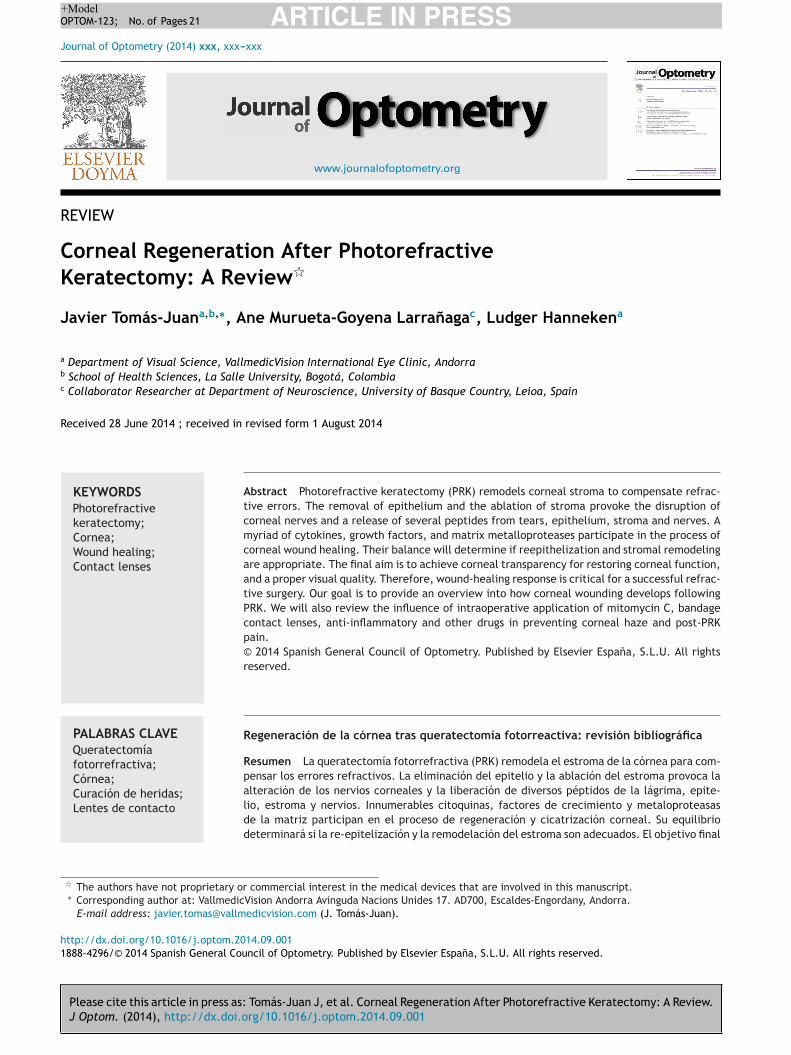
PRK may result in postoperative epithelial thickening due tothe lack of mechanical influences of the upper eyelid thatpolishes the corneal surface with blinking.2 Epithelial hyper-plasia in PRK is associated with deep stromal ablation depthsand with small ablation zones (4.00---4.5 mm) because thereis a marked curvature change in the edges of the ablatedarea. When ablation zones are large (6.00 mm), they haveless demarcated contours, and thus, the change in epithe-lial thickness is minimal.53---56 Table 1 shows the variationof central corneal thickness with different surgery tech-niques published in the scientific literature. Erie2 provedthat, after PRK, the central epithelial thickness returnedto preoperative levels at 1 month. However, it continuedto progressively increase during the first year, being 21%thicker at that time. This result is similar to the 22% thick-ness increase seen in LASIK by Erie et al.57 However, thetime required for thickness stabilization differs between thetwo techniques, due to the complex interaction of epithe-lial cells and activated keratocytes in PRK.2 According toPatel et al.,25 central corneal epithelium in LASIK increased24% during the first year after surgery and remained stableduring the next 7 years. In PRK, corneal thickness contin-ued to increase at 1 month, 1 year and 7 years (442±39 �m,464±44 �m, 471±45 �m; respectively).25 Recently, Ivarsenet al.52 have concluded that in PRK and LASIK, the epithelialthickness increases 15%---20% after surgery, but the epithe-lial changes in LASIK occur during the first week and remainunaltered during the following 3 years. It has been suggestedthat epithelial hyperplasia can induce a reduction of post-operative refractive effect. Erie showed myopic regressionsignificantly associated with epithelial thickness increase.2
Nevertheless, Ivarsen et al.52 did not found any correla-tion between changes in epithelial thickness and changesin refraction after PRK or LASIK, probably because of thesmall size of their sample.
Stromal Wound Healing Following PRK
Please cite this article in press as: Tomás-Juan J, et al. Corneal RJ Optom. (2014), http://dx.doi.org/10.1016/j.optom.2014.09.
Stroma occupies approximately the 90% of cornealthickness,58 and it can be subdivided into three continuouslayers: anterior, middle and posterior.49 The corneal stroma
ucdb
eps of wound healing following PRK.
s built up from collagen fibers, ground substance, kera-ocytes and nerve fibers.5,49 Keratocytes --- corneal stromalells --- play a major role in maintaining corneal transparency,nd synthesizing the components of the extracellular matrixECM).58 Active keratocytes produce collagen and proteo-lycans to form the ECM after stromal injury. The humantromal cornea contains collagen type I, V and VI.59,60 Type
is predominant (75%), followed by type VI (approximately,7%).60 Type III collagen appears in inflammatory events oruring wound healing. Proteoglycans participate in collagenbrillogenesis and matrix assembly.61 After corneal injury,ewly produced collagen fibers tend to have larger diam-ters, as they contain high levels of dermatan sulphate (aype of proteoglycan) that lasts up to 6 months.62
Stromal keratocytes are normally quiescent or inactive,nd are the second cells involved in the process of cornealegeneration, just after corneal epithelial cells. After PRK,eratocytes underlying the wound disappear by apoptosisue to a stress exposure.2,24,37,40,50,63 During the first 24 hfter injury, macrophages, monocytes, T cells and polymor-honuclear cells infiltrate the area and remove damagedells.44,64 Metallonoproteinases (MMPs) and the plasmino-en activator system remove the affected extracellularatrix.9,65---67 The MMPs are proteolytic enzymes secretedy active keratocytes or fibroblasts, and degrade complexolecules of the extracellular matrix. Although nine types
f MMPs exist, in the cornea only four MMPs are important,eing MMP-1 the most relevant.67 MMP-8 concentration haseen observed to be significantly elevated in the seconday after PRK (P=.001).68 The remaining keratocytes, adja-ent to wound borders, are activated in response to variousytokines released by cells in upper layers, such as inter-eukin (IL)-1, and growth factors like tumor necrosis factorTNF), fibroblast growth factor (FGF), platelet-derivedrowth factor (PDGF), epithelial growth factor (EGF), andransforming growth factor (TGF).2,8,14,22,24,35,51,58,69---74 Theserowth factors are normal components of the tear andorneal cells, produced by the lacrimal gland,35 and reg-
egeneration After Photorefractive Keratectomy: A Review.001
late a variety of processes involved in homeostasis andorneal wound healing, including migration, mitosis and cellifferentiation.75 Particularly, transformation growth factoreta (TGF-beta) seems to transform active keratocytes into
Please cite this article in press as: Tomás-Juan J, et al. Corneal RJ Optom. (2014), http://dx.doi.org/10.1016/j.optom.2014.09.
ARTICLE IN+ModelOPTOM-123; No. of Pages 21
4
Tabl
e
1
Vari
atio
n
of
Cent
ral C
orne
al
Thic
knes
s,
Mea
n±SD
or
Rang
e
(�m
).
Stud
y
Tech
niqu
ePr
eop
1
wee
k
1
mon
th
3
mon
ths
6
mon
ths
1
year
2
year
s
3
year
s
5
year
s
7 ye
ars
Ivar
sen
et
al.
(200
9)52
LASI
K
529±
4946
0
±
4747
7±43
PRK
522±
3242
3
±
3546
3±32
Wal
lau
and
Cam
pos
(200
8)15
9
LASI
K
543±
2547
3±28
473
±
2747
3
±
27PR
K
wit
hM
MCa
545±
2645
6±30
458
±
3046
0
±
29
Pate
l et
al.
(200
7)25
LASI
K
514±
37(4
37---
576)
452±
38(4
11---
563)
456±
42(3
86---
576)
456±
39(4
02---
564)
462±
37(4
11---
577)
456±
30(3
88---
524)
455±
31(3
71---
509)
PRK
493±
36(4
27---
560)
442±
40(3
88---
526)
464±
44(3
96---
534)
469±
44(4
11---
544)
472±
59(3
93---
573)
468±
48(4
06---
555)
471±
45(4
00---
557)
Koza
k
et
al.
(200
3)56
LASI
K
549±
37(5
15---
620)
467
±
29
474
±
30
481
±
23
PRK
552±
34(5
04---
602)
473
±
39
477
±
35
482
±
35
LASI
K,
lase
r
in
situ
kera
tom
ileus
is;
PRK,
phot
oref
ract
ive
kera
tect
omy;
MM
C,
mit
omyc
in
C.a
0.00
2%,
1
min
.
mio
epcawtfap(m(CtaddaioPtia
egdvtkbmaachviEestwrbgsdbe1(tdc3a
Document downloaded from http://www.journalofoptometry.org, day 13/03/2015. This copy is for personal use. Any transmission of this document by any media or format is strictly prohibited. This early online article has been reviewed, accepted and posted on the Web before copyediting.
PRESSJ. Tomás-Juan et al.
yofibroblasts that appear at later stages of stromal heal-ng. Myofibroblasts can be identified through the expressionf �-smooth muscle actin (SMA).45
PRK produces oxygen free radicals, secondary to thexposure of ultraviolet radiation, thermal increase, andolymorphonuclear cell infiltration.76,77 Free oxygen radi-als may interact with lipid components, nucleotic acids,nd sulphur contained in enzymes,72,78 and particularlyith reactive oxygen species (ROE) that are considered
o produce the most reactive and cytotoxic damage. Inact, they have been described as a partial cause of ker-tocyte apoptosis.77 Among the antioxidant enzymes thatrotect the cornea from radicals, superoxide dismutaseSOD), glutathione peroxidase (Gpx) and catalase are theost relevants.75,78 Ascorbic acid and dl-alpha-tocopherol
Vitamin E) also prevents from the effects of free radicals.78
orneal epithelial ascorbic acid absorbs ultraviolet radia-ion, protecting keratocytes, but high or altered cornealscorbate levels in the human cornea after PRK, may pro-uce accelerated keratocyte death.5 In rabbit corneas,ecreased activity of SOD and Gpx enzymes has been provedfter refractive surgery.78 For this reason, additional antiox-dant enzymes seem to be involved in reducing cornealxidative stress following PRK. 1-cys peroxiredoxin (1-cysrx) may be an important enzyme involved in the differentia-ion, migration and proliferation of epithelial cells. 1-cysPrxncreases 4 h after PRK and remains in high levels until 7 daysfter PRK.75
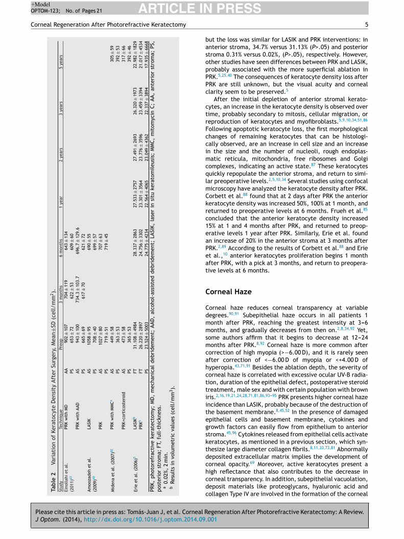
The density of keratocytes varies across the stroma. It isstimated that in the anterior stroma the density is 5%---10%reater than in middle and posterior stroma.79,80 It has beenocumented that a corneal stroma rich in keratocytes pre-ents the epithelial corneal infection or, at least, minimizeshe extension of the infection.2,5,10 After PRK, the anterioreratocyte population drastically diminishes, and the distri-ution and shape is greatly altered.2,10,34,40,81,82 In confocalicroscopy, high reflectance, hyperplasticity, hypertrophy
nd a decrease in the contrast of the anterior stromal ker-tocytes can be observed.34,63,83 Human histological studiesonfirm that the decrease of anterior stromal keratocytes inumans and animal respond similarly.84,85 Table 2 shows theariation of anterior, posterior and total keratocyte densityn the different studies published in the scientific literature.rie et al.5 confirmed that after 5 years of PRK there werevidences of keratocyte density loss in middle and posteriortroma. They observed a reduction of 20%---24% in the pos-erior stroma (P<.05) although they claimed that this lossas not completely evident. Keratocyte density in the ante-
ior 10% of the stroma continues to decrease 5% per yearetween 1 and 3 years after PRK.2 Erie2 reported a pro-ressive decline in anterior stromal keratocytes, becomingignificant at 36 months after PRK (P=.02). In contrast, mid-le and posterior keratocyte densities remained unchangedetween 1 and 3 years after PRK.2 In another study, Eriet al.10 proved that the keratocyte density in the anterior0% of the stroma, decreased at 6, 12, 24 and 36 months41%, 40%, 43%, 45%; respectively) after PRK, comparedo pre-PRK. In a posterior longer-term study, Erie et al.5
egeneration After Photorefractive Keratectomy: A Review.001
emonstrated a similar decreasing pace in anterior kerato-yte density: 40%, 42%, 45%, and 47% at 6 months, 2 years,
years, and 5 years (P<.001). Amoozadeh et al.40 found reduction in keratocyte density 6 months after surgery,
Please cite this article in press as: Tomás-Juan J, et al. Corneal RJ Optom. (2014), http://dx.doi.org/10.1016/j.optom.2014.09.
ARTICLE IN+ModelOPTOM-123; No. of Pages 21
Corneal Regeneration After Photorefractive Keratectomy
Tabl
e
2
Vari
atio
n
of
Kera
tocy
te
Den
sity
Afte
r
Surg
ery,
Mea
n±SD
(cel
l/m
m2).
Stud
y
Tech
niqu
ePr
eop
3
mon
ths
6
mon
ths
1
year
2
year
s3
year
s5
year
sEi
nolla
hi
et
al.
(201
1)63
PRK
wit
h
MD
AA
902
±
107
704
±
119
643
±
134
PS
653
±
7262
2
±
5360
9
±
60PR
K
wit
h
AAD
AS
943
±
100
734.
3
±
103.
769
6.7
±
129.
6PS
665
±
6961
7
±
7062
1
±
72Am
ooza
deh
et
al.
(200
9)40
LASI
K
AS
1058
±
9569
0
±
55PS
708
±
4069
9
±
57PR
K
AS
1027
±
8070
7
±
63PS
719
±
5171
9
±
45M
iden
a
et
al.
(200
7)82
PRK
wit
h
MM
CaAS
449
±
5830
5
±
59PS
363
±
5339
2
±
53PR
K+co
rtic
oste
roid
AS
473
±
5831
7
±
66PS
365
±
5339
2
±
46Er
ie
et
al.
(200
6)5
LASI
KbFT
31.1
08
±
4984
28.3
37
±
2863
27.5
33
±
2757
27.4
91
± 26
9326
.320
±
1973
22.9
82
±
1829
PRKb
FT
26.2
20
±
2897
24.7
56
±
3302
23.3
01
±
3564
23.7
76
± 35
9623
.459
±
3394
21.0
17
±
4534
PS
23.5
24
±
5003
24.7
75
±
4234
22.5
64
±
4065
23.0
49
± 43
6222
.337
±
3894
17.9
35
±
6668
PRK,
phot
oref
ract
ive
kera
tect
omy;
MD
,
mec
hani
cal
debr
idem
ent;
AAD
,
alco
hol-
assi
sted
debr
idem
ent;
LASI
K,
lase
r
in
situ
kera
tom
ileus
is;
MM
C,
mit
omyc
in
C;
AA,
ante
rior
stro
ma;
PS,
post
erio
r
stro
ma;
FT,
full-
thic
knes
s.a
0.02
%,
2
min
.b
Resu
lts
in
volu
met
ric
valu
es
(cel
l/m
m3).
basopPPc
ctrFccimcqlmCkrc1eaPeat
C
Cdmmsmcahcttiitegsktdchcdc
Document downloaded from http://www.journalofoptometry.org, day 13/03/2015. This copy is for personal use. Any transmission of this document by any media or format is strictly prohibited. This early online article has been reviewed, accepted and posted on the Web before copyediting.
PRESS5
ut the loss was similar for LASIK and PRK interventions: innterior stroma, 34.7% versus 31.13% (P>.05) and posteriortroma 0.31% versus 0.02%, (P>.05), respectively. However,ther studies have seen differences between PRK and LASIK,robably associated with the more superficial ablation inRK.5,25,40 The consequences of keratocyte density loss afterRK are still unknown, but the visual acuity and corneallarity seem to be preserved.5
After the initial depletion of anterior stromal kerato-ytes, an increase in the keratocyte density is observed overime, probably secondary to mitosis, cellular migration, oreproduction of keratocytes and myofibroblasts.5,9,10,34,51,86
ollowing apoptotic keratocyte loss, the first morphologicalhanges of remaining keratocytes that can be histologi-ally observed, are an increase in cell size and an increasen the size and the number of nucleoli, rough endoplas-atic reticula, mitochondria, free ribosomes and Golgi
omplexes, indicating an active state.87 These keratocytesuickly repopulate the anterior stroma, and return to simi-ar preoperative levels.2,5,10,34 Several studies using confocalicroscopy have analyzed the keratocyte density after PRK.orbett et al.88 found that at 2 days after PRK the anterioreratocyte density was increased 50%, 100% at 1 month, andeturned to preoperative levels at 6 months. Frueh et al.85
oncluded that the anterior keratocyte density increased5% at 1 and 4 months after PRK, and returned to preop-rative levels 1 year after PRK. Similarly, Erie et al. foundn increase of 20% in the anterior stroma at 3 months afterRK.2,89 According to the results of Corbett et al.88 and Eriet al.,10 anterior keratocytes proliferation begins 1 monthfter PRK, with a pick at 3 months, and return to preopera-ive levels at 6 months.
orneal Haze
orneal haze reduces corneal transparency at variableegrees.90,91 Subepithelial haze occurs in all patients 1onth after PRK, reaching the greatest intensity at 3---6onths, and gradually decreases from then on.2,8,34,92 Yet,
ome authors affirm that it begins to decrease at 12---24onths after PRK.8,92 Corneal haze is more common after
orrection of high myopia (>−6.00 D), and it is rarely seenfter correction of <−6.00 D of myopia or <+4.00 D ofyperopia.43,71,91 Besides the ablation depth, the severity oforneal haze is correlated with excessive ocular UV-B radia-ion, duration of the epithelial defect, postoperative steroidreatment, male sex and with certain population with brownris.2,16,19,21,24,28,71,81,86,93---95 PRK presents higher corneal hazencidence than LASIK, probably because of the destruction ofhe basement membrane.8,45,52 In the presence of damagedpithelial cells and basement membrane, cytokines androwth factors can easily flow from epithelium to anteriortroma.45,96 Cytokines released from epithelial cells activateeratocytes, as mentioned in a previous section, which syn-hesize large diameter collagen fibrils.8,11,33,73,81 Abnormallyeposited extracellular matrix implies the development oforneal opacity.69 Moreover, active keratocytes present a
egeneration After Photorefractive Keratectomy: A Review.001
igh reflectance that also contributes to the decrease inorneal transparency. In addition, subepithelial vacuolation,eposit materials like proteoglycans, hyaluronic acid andollagen Type IV are involved in the formation of the corneal
IN+ModelO
6
hsefgaemefa
bbfitatsitwabtlbhrprr6fcpim
lTfiucastsSbh
V
Tashoa
sacoahctclat
bs7teifiincetTiyitsataspowtmtawst
R
Tbtsdrten
Document downloaded from http://www.journalofoptometry.org, day 13/03/2015. This copy is for personal use. Any transmission of this document by any media or format is strictly prohibited. This early online article has been reviewed, accepted and posted on the Web before copyediting.
ARTICLEPTOM-123; No. of Pages 21
aze in advanced stages.33,77 Plasminogen activator---plasminystem degrades the damaged ECM, and extended low lev-ls beyond the third day after PRK causes corneal hazeormation.97 Guerriero et al.58 affirm that the loss of colla-en type IV is related to the activation of keratocytes in vivond in vitro, and Winkler et al.33 and Mohrenfels et al.98
mphasize on the role of type IV collagen in the develop-ent of corneal cloudiness. Secondary ultraviolet B (UV-B)
xposure, originating from sun or solarium is a causal factoror aforementioned abnormal proteoglycan deposition andssociated augmented corneal thickness.99
On the other hand, myofibroblast, derivatives of TGF-eta responding keratocytes, are thought to be the firstiological event for corneal haze formation.51,93,94 Myo-broblasts play an essential role in the recovery ofhe corneal integrity after penetrating injury, mainly indvanced stages.22 They secrete extracellular matrix, con-ract wounds and have the ability to generate adhesiontructures with the surrounding substrate.71 TGF-beta alsonduces the expression of connective tissue growth fac-or (CTGF), which mediates collagen synthesis, and alongith myofibroblasts regulates the corneal wound healing,nd may promote scar formation.100 After PRK, myofibro-lasts appear as a pathological response to injury,71 andheir decreased transparency roots in the low intracel-ular content of crystalline.101 Irregular surface has alsoeen related to high incidence of corneal haze,94,102 andigher irregularity is seen with increasing dioptric cor-ections in PRK.103 Interestingly, surface irregularity isositively correlated with myofibroblast density in the ante-ior stroma.43 In normal corneal wound healing, completeegeneration of the basal membrane after PRK occurs within---8 weeks in rabbits,104 which limits the access of growthactors to the stroma69 and, consequently, myofibroblastsommit apoptosis46 modulated by IL-1.47 Therefore, theresence of myofibroblast, and subsequent corneal haze,s largely dependent upon the restoration of the basementembrane.43,105
Corneal haze has been traditionally measured in the slit-amp, and graded with diverse scales, like Hanna’s scale.he new technology leads us to use automated instrumentsor corneal haze measurement. In vivo confocal microscopys a reliable tool, as far as standardized methods aresed.106 It is the most widely used objective method inlinical setting for haze measurement. In the last years,lternative techniques have come out. Confocal imaging ofecond harmonic-generated (SHG) signals has been showno be sensitive in measuring corneal fibrosis after refractiveurgery.107 Recently, the densiometry program of Pentacamcheimpflug imaging system (Oculus Optikgeräte GmbH) haseen proved to be a useful method for measuring cornealaze.108
isual Disturbances of Corneal Haze
he corneal haze produces a reduction of low contrast visualcuity and night vision symptoms that, in the vast majority of
Please cite this article in press as: Tomás-Juan J, et al. Corneal RJ Optom. (2014), http://dx.doi.org/10.1016/j.optom.2014.09.
ituations, improve with time.67 It is possible to see cornealaze formation after PRK by means of confocal microscopy,bserved as a decrease in the contrast of the image andn increase in reflectivity.81 Böhnke et al.81 using a Tandem
msoC
PRESSJ. Tomás-Juan et al.
canning confocal microscopy, correlated corneal haze andnterior stromal reflectivity. However, the tandem scanningonfocal microscopy is not able to detect acellular regionsf the anterior stroma early after PRK when epitheliumnd sub-basal plexus are not formed.10 Although cornealaze in humans is less pronunced than in animal models, iforneal haze persists and affects significantly to the cornealransparency, it causes light scatter.4,94,109 For this reason,orneal haze may be described and analyzed through backight scattering (backscatter).81,110 It also causes irregularstigmatisms,2,34,93 and subsequent loss of corrected dis-ance visual acuity (CDVA).86
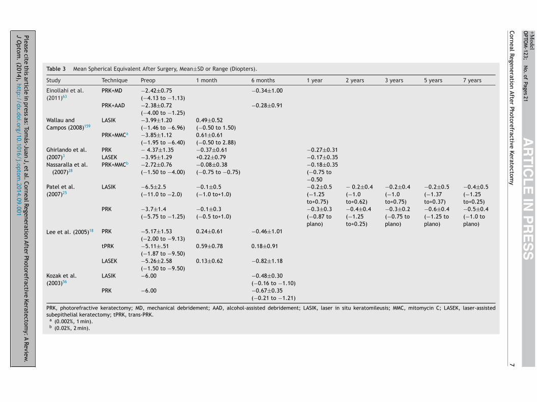
The regression of the refractive error may be producedy epithelial irregularity, alterations in the keratocyte den-ity or subepithelial deposits. Myopic regression occurs in8% of eyes in the first 12 months after PRK.2 Table 3 showshe mean spherical equivalent changes reported in differ-nt scientific studies. In the first week after PRK, epithelialrregularity causes a reduction in visual quality.88 During therst month, altered keratocytes decrease contrast sensitiv-
ty, mainly in high frequencies, and cause glare. During theext 2 months, subepithelial deposits produce a decrease inontrast sensitivity, especially in low frequencies.4,88 Ginist al.4 reported that subepithelial deposits are the first fac-or that contributes to the development of corneal scatter.he visual quality is affected temporarily, although there
s evidence that in some cases it persists for more than 1ear.43,91 In order to avoid a decrease in the visual qual-ty, all postoperative efforts must go oriented to controlhe subepithelial matter.88 The corneal epithelium does noteem to contribute significantly to the refractive changefter PRK, although some studies suggest that epithelialhickening may produce myopic regression,2 even 5 yearsfter PRK.90 Moller-Pedersen et al.55 and Cua and Pepose92
uggested that new keratocytes growth in central cornea orostoperative corneal scarring is likely to be the main causesf myopic regression in ablations of 6 mm. In agreementith this hypothesis, Moller-Pedersen et al.55 demonstrated
hat hyperopic changes were the direct result of a stro-al thinning. Erie2 found an increase of 12 �m of epithelial
hickness at 12 months after PRK that was associated with myopic regression of −0.41 diopters but no correlationas found between stromal thickening and myopic regres-
ion; however, the combined effect of epithelial and stromalhickening was correlated with myopic regression.
egeneration of Corneal Innervation
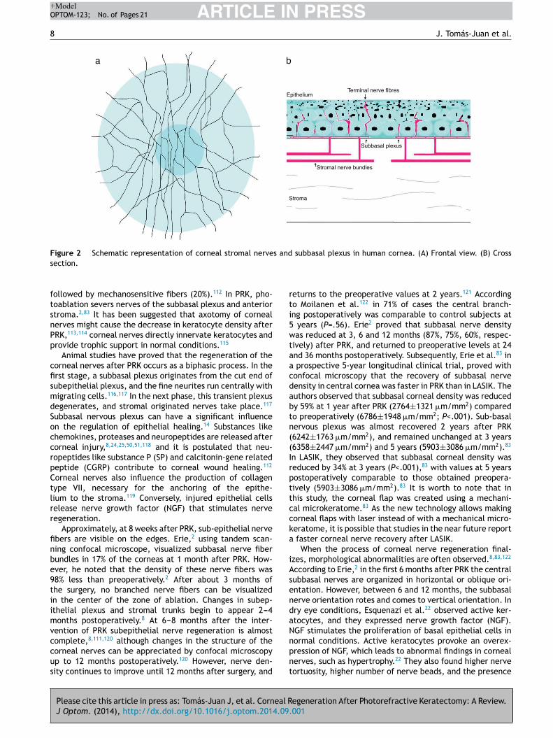
he cornea is the most innervated tissue of the humanody,7 and these sensory nerves are derived from the oph-halmic branch of the trigeminal nerve fibers.2,111 Cornealensory nerves penetrate the limbus and form nerve bun-les in the anterior third of the stroma. Once there, theyun perpendicularly to cross Bowman’s membrane, and formhe sub-basal nerve plexus as a network between the basalpithelial cells and Bowman’s layer (Fig. 2).49,111 Cornealerve fibers, if visualized using confocal microscopy in nor-
egeneration After Photorefractive Keratectomy: A Review.001
al conditions, show high reflectivity across the cornealtroma with a rectilinear pattern. Subepithelial nerve fibers,n the other hand, are thinner than stromal nerve fibers.orneal fibers are considered primarily nociceptive (70%),
Please cite
this article
in press
as: Tom
ás-Juan J,
et al.
Corneal Regeneration
After Photorefractive
Keratectomy:
A Review
.J
Optom
. (2014),
http://dx.doi.org/10.1016/j.optom.2014.09.001
AR
TIC
LE
IN P
RE
SS
+Model
OPTO
M-123;
N
o. of
Pages 21
Corneal Regeneration
After Photorefractive
Keratectomy
7
Table 3 Mean Spherical Equivalent After Surgery, Mean±SD or Range (Diopters).
Study Technique Preop 1 month 6 months 1 year 2 years 3 years 5 years 7 years
Einollahi et al.(2011)63
PRK+MD −2.42±0.75(−4.13 to −1.13)
−0.34±1.00
PRK+AAD −2.38±0.72(−4.00 to −1.25)
−0.28±0.91
Wallau andCampos (2008)159
LASIK −3.99±1.20(−1.46 to −6.96)
0.49±0.52(−0.50 to 1.50)
PRK+MMCa −3.85±1.12(−1.95 to −6.40)
0.61±0.61(−0.50 to 2.88)
Ghirlando et al.(2007)3
PRK − 4.37±1.35 −0.37±0.61 −0.27±0.31LASEK −3.95±1.29 +0.22±0.79 −0.17±0.35
Nassaralla et al.(2007)28
PRK+MMCb −2.72±0.76(−1.50 to −4.00)
−0.08±0.38(−0.75 to −0.75)
−0.18±0.35(−0.75 to−0.50
Patel et al.(2007)25
LASIK −6.5±2.5(−11.0 to −2.0)
−0.1±0.5(−1.0 to+1.0)
−0.2±0.5(−1.25to+0.75)
− 0.2±0.4(−1.0to+0.62)
−0.2±0.4(−1.0to+0.75)
−0.2±0.5(−1.37to+0.37)
−0.4±0.5(−1.25to+0.25)
PRK −3.7±1.4(−5.75 to −1.25)
−0.1±0.3(−0.5 to+1.0)
−0.3±0.3(−0.87 toplano)
−0.4±0.4(−1.25to+0.25)
−0.3±0.2(−0.75 toplano)
−0.6±0.4(−1.25 toplano)
−0.5±0.4(−1.0 toplano)
Lee et al. (2005)18 PRK −5.17±1.53(−2.00 to −9.13)
0.24±0.61 −0.46±1.01
tPRK −5.11±.51(−1.87 to −9.50)
0.59±0.78 0.18±0.91
LASEK −5.26±2.58(−1.50 to −9.50)
0.13±0.62 −0.82±1.18
Kozak et al.(2003)56
LASIK −6.00 −0.48±0.30(−0.16 to −1.10)
PRK −6.00 −0.67±0.35(−0.21 to −1.21)
PRK, photorefractive keratectomy; MD, mechanical debridement; AAD, alcohol-assisted debridement; LASIK, laser in situ keratomileusis; MMC, mitomycin C; LASEK, laser-assistedsubepithelial keratectomy; tPRK, trans-PRK.
a (0.002%, 1 min).b (0.02%, 2 min).
Document downloaded from http://www.journalofoptometry.org, day 13/03/2015. This copy is for personal use. Any transmission of this document by any media or format is strictly prohibited. This early online article has been reviewed, accepted and posted on the Web before copyediting.
ARTICLE IN PRESS+ModelOPTOM-123; No. of Pages 21
8 J. Tomás-Juan et al.
EpitheliumTerminal nerve fibres
Subbasal plexus
Stromal nerve bundles
Stroma
a b
F s ands
ftsnPp
cfismdSoccrpCtlrr
finbe9tiimvccus
rti5wtaacdabtn((Irpttccka
iAsendaN
Document downloaded from http://www.journalofoptometry.org, day 13/03/2015. This copy is for personal use. Any transmission of this document by any media or format is strictly prohibited. This early online article has been reviewed, accepted and posted on the Web before copyediting.
igure 2 Schematic representation of corneal stromal nerveection.
ollowed by mechanosensitive fibers (20%).112 In PRK, pho-oablation severs nerves of the subbasal plexus and anteriortroma.2,83 It has been suggested that axotomy of cornealerves might cause the decrease in keratocyte density afterRK,113,114 corneal nerves directly innervate keratocytes androvide trophic support in normal conditions.115
Animal studies have proved that the regeneration of theorneal nerves after PRK occurs as a biphasic process. In therst stage, a subbasal plexus originates from the cut end ofubepithelial plexus, and the fine neurites run centrally withigrating cells.116,117 In the next phase, this transient plexusegenerates, and stromal originated nerves take place.117
ubbasal nervous plexus can have a significant influencen the regulation of epithelial healing.14 Substances likehemokines, proteases and neuropeptides are released afterorneal injury,8,24,25,50,51,118 and it is postulated that neu-opeptides like substance P (SP) and calcitonin-gene relatedeptide (CGRP) contribute to corneal wound healing.112
orneal nerves also influence the production of collagenype VII, necessary for the anchoring of the epithe-ium to the stroma.119 Conversely, injured epithelial cellselease nerve growth factor (NGF) that stimulates nerveegeneration.
Approximately, at 8 weeks after PRK, sub-epithelial nervebers are visible on the edges. Erie,2 using tandem scan-ing confocal microscope, visualized subbasal nerve fiberundles in 17% of the corneas at 1 month after PRK. How-ver, he noted that the density of these nerve fibers was8% less than preoperatively.2 After about 3 months ofhe surgery, no branched nerve fibers can be visualizedn the center of the zone of ablation. Changes in subep-thelial plexus and stromal trunks begin to appear 2---4onths postoperatively.8 At 6---8 months after the inter-
ention of PRK subepithelial nerve regeneration is almost8,111,120
Please cite this article in press as: Tomás-Juan J, et al. Corneal RJ Optom. (2014), http://dx.doi.org/10.1016/j.optom.2014.09.
omplete, although changes in the structure of theorneal nerves can be appreciated by confocal microscopyp to 12 months postoperatively.120 However, nerve den-ity continues to improve until 12 months after surgery, and
npnt
subbasal plexus in human cornea. (A) Frontal view. (B) Cross
eturns to the preoperative values at 2 years.121 Accordingo Moilanen et al.122 in 71% of cases the central branch-ng postoperatively was comparable to control subjects at
years (P=.56). Erie2 proved that subbasal nerve densityas reduced at 3, 6 and 12 months (87%, 75%, 60%, respec-
ively) after PRK, and returned to preoperative levels at 24nd 36 months postoperatively. Subsequently, Erie et al.83 in
prospective 5-year longitudinal clinical trial, proved withonfocal microscopy that the recovery of subbasal nerveensity in central cornea was faster in PRK than in LASIK. Theuthors observed that subbasal corneal density was reducedy 59% at 1 year after PRK (2764±1321 �m/mm2) comparedo preoperatively (6786±1948 �m/mm2; P<.001). Sub-basalervous plexus was almost recovered 2 years after PRK6242±1763 �m/mm2), and remained unchanged at 3 years6358±2447 �m/mm2) and 5 years (5903±3086 �m/mm2).83
n LASIK, they observed that subbasal corneal density waseduced by 34% at 3 years (P<.001),83 with values at 5 yearsostoperatively comparable to those obtained preopera-ively (5903±3086 �m/mm2).83 It is worth to note that inhis study, the corneal flap was created using a mechani-al microkeratome.83 As the new technology allows makingorneal flaps with laser instead of with a mechanical micro-eratome, it is possible that studies in the near future report
faster corneal nerve recovery after LASIK.When the process of corneal nerve regeneration final-
zes, morphological abnormalities are often observed.8,83,122
ccording to Erie,2 in the first 6 months after PRK the centralubbasal nerves are organized in horizontal or oblique ori-ntation. However, between 6 and 12 months, the subbasalerve orientation rotes and comes to vertical orientation. Inry eye conditions, Esquenazi et al.22 observed active ker-tocytes, and they expressed nerve growth factor (NGF).GF stimulates the proliferation of basal epithelial cells in
egeneration After Photorefractive Keratectomy: A Review.001
ormal conditions. Active keratocytes provoke an overex-ression of NGF, which leads to abnormal findings in cornealerves, such as hypertrophy.22 They also found higher nerveortuosity, higher number of nerve beads, and the presence
IN+Model
E
Icrwlu(octacmcralpnasddltaTtniaihfg3rg(P6Betpdowsdd
dtEe
Document downloaded from http://www.journalofoptometry.org, day 13/03/2015. This copy is for personal use. Any transmission of this document by any media or format is strictly prohibited. This early online article has been reviewed, accepted and posted on the Web before copyediting.
ARTICLEOPTOM-123; No. of Pages 21
Corneal Regeneration After Photorefractive Keratectomy
of nerve sprouts in desiccating environment group,22 whichmeans there is a high metabolic activity to repair the alter-ations in the corneal epithelium.
Corneal Pain and Sensitivity
Photoablation severs corneal nerves, disrupting the lacrimalfunctional unit (LFU). LFU is constituted by the lachry-mal gland, ocular surface and innervation. It regulates tearsecretion, and affects its composition.123 Thereby, photoab-lation produces transitory dry eye, deterioration of cornealbarrier function and alteration in corneal sensitivity.83,111,124
A reduction of the tear flow after PRK has been provedusing Schirmer test.124 According to Erie et al.,83 LASIKpresents higher prevalence of postoperative dry eye, alteredcorneal epithelium and tear film than PRK. Dry eye hasbeen associated with low corneal sensitivity.125,126 Differ-ent devices are available to measure corneal sensitivity,as Cochet---Bonnet esthesiometry or non-contact gas esthe-siometer. Cochet---Bonnet esthesiometer only stimulatesmechanosensory fibers, whereas non-contact gas esthe-siometer measures activation thresholds of nociceptorsusing controlled chemical, thermal and mechanical pulses.Non-contact gas esthesiometer is, therefore, a more sensi-tive device for measuring alterations in corneal sensitivity.Still, Coche---Bonnet esthesiometry is more widely used,and controversy remains about the time course of thecorneal sensitivity recovery after PRK with this device.Kauffmann et al.120 affirm that the recovery of cornealsensitivity usually starts at 4---6 weeks, completing approx-imately within 6---12 months following PRK. However, Erieet al.83 claim that the recovery of corneal sensitivity iscompleted from 3 months to 1 year after PRK. Hypoes-thesia is often expected until 3 months after surgery, dueto the loss of corneal nerves.8 On the other hand, Gallaret al.127 measured corneal mechanical and chemical sensi-tivity following PRK with non-contact gas esthesiometer, andfound that both types of sensitivities were reduced even 5years postoperatively, achieving normal values in 10 years.Despite the diminished corneal sensitivity, intense pain isusually present hours after PRK.128 Gallar et al.129 attributedcorneal pain and discomfort sensations to the altered func-tionality of corneal nerves. They recorded spontaneousactivity and modified responsiveness in corneal fibers ofcats that underwent PRK.129 Experimental evidences sup-port the idea that ongoing activity evokes spontaneous painsensations.130,131
Acceleration of Corneal Regeneration Process,Reduction of Corneal Haze and Corneal PainManagement
Nowadays there are several alternatives to speed up theprocess of epithelial regeneration, like epithelial removaltechniques, amniotic membrane, or bandage contact lenses.
Please cite this article in press as: Tomás-Juan J, et al. Corneal RJ Optom. (2014), http://dx.doi.org/10.1016/j.optom.2014.09.
In PRK, agents like mytomicin-C (MMC) or fluoroquinonesthat reduce the corneal haze formation are used, anddrugs to reduce the corneal pain and inflammation arealso prescribed.
srifi
PRESS9
pithelial Removal Techniques
n PRK, previous to the impact of laser energy over theornea, the corneal epithelium has to be removed. Theemoval of the corneal epithelium is carried out mainlyith epithelial mechanical scraping using chemical agents
ike diluted ethanol solution,9 through a rotary brush orsing the laser itself --- known as transepithelial ablationFig. 3).18,21,22,39,63,128,132,133 The epithelial scraping has post-perative adverse effects like pain, myopic regression ororneal haze. Some modification in PRK technique can alterhe wound healing response with the aim of minimizing thedverse effects.25 The exposition to agents such as ethanolan produce an increase in the inflammatory response andore damage to the anterior stromal keratocytes that
ould increase the haze formation.21,94 Yet, controversyemains in the scientific literature because other authorsffirm that alcohol-assisted epithelial removal producesess inflammation, favoring epithelial regeneration andreventing corneal haze or keratoyce apoptosis.9,63 Esque-azi et al.22 proved that the epithelial scraping might bessociated with an increase in the number of reflectivetructures in the stroma, mainly in corneas with ocularryness after PRK. The laser-scrape epithelial removalecreases the degree of keratocyte apoptosis, producing aess pronounced loss of superficial keratocytes.2 However,he irrigation with cold balanced salt solution (BSS) maylter the keratocyte apoptosis in the retroablation zone.2
he time necessary for mechanical debridement is greaterhan the time required for laser or alcohol scrape tech-iques, even for expert surgeons.18 Mechanical debridements related to stromal dehydration and disappearance ofnterior stromal keratocytes.18,63 This loss provokes anncrease of cells in the underlying stroma, causing stromalyperplasia and haze formation.134 Einollahi et al.63 foundaster mean epithelial healing time in the alcohol-assistedroup than in the mechanical group (3.0±0.3 versus.2±0.4 days, P=.001). They observed greater anterioretroablation stromal keratocyte density in the mechanicalroup than in the alcohol-assisted groups at 3 months704.3±119.9 cells/mm2 versus 743.3±103.7 cells/mm2,=.05) and at 6 months (643.8±134.4 cells/mm2 versus96.7±129.6 cells/mm2, P=.02).63 In the same study,ahram et al. did not found statistically significant differ-nces in middle and posterior keratocyte density betweenhe mechanical and alcohol-assisted groups.63 They alsoroved that mechanical and alcohol-assisted epithelialebridement after PRK present similar visual and refractiveutcomes in patients with mild myopia,63 in agreementith the results of Goreishi et al.135 They reported similar
afety and efficacy with alcohol-assisted and mechanicalebridement in a 1250 eye sample, but anterior keratocyteensity was not assessed in this study.135
Laser-assisted subepithelial keratomileusis (LASEK) waseveloped in order to reduce corneal pain and haze forma-ion associated with PRK, and to accelerate visual recovery.pithelial delamination with diluted alcohol showed in anlectron microscope study that was able to leave a smooth
egeneration After Photorefractive Keratectomy: A Review.001
urface, ideal for LASEK intervention.136 It seems that aegular surface before laser application helps corneal heal-ng and prevents haze.137 Chen et al.138 contrasted thesendings in a later study, and showed a high variability
ARTICLE IN PRESS+ModelOPTOM-123; No. of Pages 21
10 J. Tomás-Juan et al.
F my)
o
ida2icneatlrisuhptclgTseP
triplott
mip1tasaev(es
A
AbppcPssiPcpmnpresses TGF-beta1, collagen III and fibronectin.150 Taken
Document downloaded from http://www.journalofoptometry.org, day 13/03/2015. This copy is for personal use. Any transmission of this document by any media or format is strictly prohibited. This early online article has been reviewed, accepted and posted on the Web before copyediting.
igure 3 TransPRK (transepithelial photorefractive keratectof Schwind Eye-Tech-Solutions).
n morphological changes after diluted alcohol treatment,ependent upon concentration and time. Cell viability wasffected when alcohol exceeded its concentration by 25% or5-s exposure.138 Yet, these studies have been conductedn vitro, and the complex interactions of tear film andorneal surface were not considered. In vivo studies doot show any difference between LASEK and PRK.139 Leet al.18 evaluated epithelial healing, postoperative painnd visual outcomes using epithelial mechanical (conven-ional PRK), transepithelial PRK and 20% diluted alcoholaser-assisted subepithelial keratomileusis (LASEK) with flapepositioning. After 6 months, they found little differencesn clinical outcomes between the 3 techniques, noting alight overcorrection in the transepithelial PRK and slightndercorrection in LASEK. Corneal pain and subepithelialaze results were similar.18 Subsequently, Ghanem et al.139
roved in a prospective randomized double-masked studyhat the reepithelialization was faster in a PRK groupompared with a butterfly LASEK group, even though epithe-ial semi-discs were repositioned intraoperatively in LASEKroup. (4.35±0.48 days versus. 4.75±0.72 days, P=.002).hey also found lower pain level in PRK group, but paincores and ocular discomfort were not statistically differ-nt from butterfly LASEK (3.31±4.09 versus. 4.43±4.27;=.18).139
It has been proven in animal studies that transepi-helial ablation produces a uniform surface for cornealegeneration, and prevents keratocyte apoptosis,36 reduc-ng the risk of corneal haze.21,72 Wang et al.140 presentedromising preliminary results of SCHWIND-ESIRIS excimeraser for transepithelial ablation, but the flawed design
Please cite this article in press as: Tomás-Juan J, et al. Corneal RJ Optom. (2014), http://dx.doi.org/10.1016/j.optom.2014.09.
f the study makes difficult to assess the real value ofhis technique. Later, Aslanides et al.21 proved in humanshat transepithelial ablation was safer than the epithelial
tes
on the Schwind Amaris 1050 RS laser platform (with permission
echanical scraping using chemical agents as alcohol, ast provides a faster epithelial healing, less postoperativeain and less corneal haze at 1 week (P=.07), and at, 3, and 6 months after surgery (P<.05). In addition,hey observed an improvement of 3 Snellen lines in visualcuity on day 3 in the modified transepithelial PRK (all-urface laser ablation) group compared to conventionallcohol-assisted PRK group (0.4 versus 0.2; P<.05).21 Trans-pithelial ablation also resulted in better corrected distanceisual acuity (DCVA) than conventional alcohol-assisted PRK33% versus 13%, respectively, P>.05),21 although differ-nces in higher order aberrations were not statisticallyignificant.21
mniotic Membrane Transplantation
part from the above mentioned techniques, amniotic mem-rane transplantation reduces the inflammation after PRK,revents polymorphonuclear cell infiltration, produces lesseroxidation, avoids keratocyte apoptosis and stimulatesorneal epithelialization.37,77 It is usually combined withRK to treat corneal dystrophies, corneal degenerations,cars, keratopathies,141,142 or even to treat corneal hazeecondary to PRK.143 The amniotic membrane restricts thenflux of polymorphonuclear cells (PMC) to the patch.144,145
MCs adhere to the amniotic membrane and eventuallyommit apoptosis.146 This is a physiological way of sup-ressing corneal inflammation.147 In addition, amnioticembrane has intrinsic keratocyte growth factors, EGF and
eurotrophins that promote epithelization.148,149 It also sup-
egeneration After Photorefractive Keratectomy: A Review.001
ogether, amniotic membrane has a potent anti-scarringffect that reduces corneal haze formation, as demon-trated in animal studies.144,151
IN+Model
rmghombMm
dwcetbwcdarsretaaMMfna
EaoEaAullbioe31Dteastm2ad
Document downloaded from http://www.journalofoptometry.org, day 13/03/2015. This copy is for personal use. Any transmission of this document by any media or format is strictly prohibited. This early online article has been reviewed, accepted and posted on the Web before copyediting.
ARTICLEOPTOM-123; No. of Pages 21
Corneal Regeneration After Photorefractive Keratectomy
Agents to Enhance Wound Healing
The wound healing response may be altered by the prophy-lactic application of a topical solution of mitomycin-C (MMC)immediately after the laser ablation,27 in order to avoidor minimize myofibroblast activation.8,25,40,71,86,90,133,152---154
MMC is an antineoplastic antibiotic agent of the familyof anti-tumor quinolones and derived from Streptomycescaespitosus. It is a potent DNA crosslinker: it inhibits thereplication of deoxyribonucleic acid (DNA).28,32,91,93,155---159
Thereby, MMC inhibit cell mitosis, including epithelial andstromal cells.8,34,86,93,133,155,157,160,161 Mitomycin-C decreasescorneal haze compared to corticosteroid treatment,82 and,consequently, improves visual acuity.152 Its use is speciallyindicated in high myopia (≥−6.00 D) and deeper abla-tion depths (≥75 �m).82,86,90,133,155,160 Wallau and Campos162
obtained better UCVA and BSCVA with the combination ofPRK with MMC, than with LASIK (P=.027 and P<.001, respec-tively) at 3, 6 and 12 months after surgery. Goreishi et al.163
reported an incidence of 4% of corneal haze at 1 year post-operatively with intraoperative application of 0.02% MMC, ina sample with a mean refractive error of −5D. Fazel et al.164
found that two-step administration of 0.02% MMC (45 s, fol-lowed by 15 s) further decreased corneal haze formation inhigh myopia, compared to a single dose of 45 s. The ben-efits of MMC have also been described once the haze hasbeen established, where mechanical epithelial scraping andinstillation of MMC restores corneal transparency.165
Although the application of the mitomycin C is helpfulfor corneal recovery, it is necessary to control the doses andthe time of exposure.20 According to Thornton et al.133 theconcentration is a more important factor than the durationof MMC exposure in corneal haze prevention. Rajan et al.34
analyzed the effects of MMC after correction of −9.00diopters by PRK in 3 groups of human corneas: without MMCapplication, with MMC (0.2 mg/mL) application for 1 min andwith MMC (0.2 mg/mL) application for 2 min. The 2 min MMCgroup (0.2 mg/mL) had thinner epithelium than the 1 minand without MMC application groups (P<.0001). The applica-tion of the intraoperative MMC lasts between 10 s and 120 s,depending on the surgeon.11 According to Khoury et al.156 theapplication of intraoperative MMC vary from 12 s to 5 min.Shojaei et al.153 affirm that short-time MMC exposure pre-vents low-grade haze in low ablation depths. The MMC dosesoscillate between 0.002% and 0.06%.156 The intraoperativeapplication of 0.02% MMC solution is the most recommended,as it produces less corneal haze, and provides better uncor-rected visual acuity (UCVA) and best spectacle-correctedvisual acuity (BSCVA).124,166,167 Still, Ramjoo et al.90 foundsimilar refractive and haze outcomes with 0.01% and 0.02%MMC for mild myopia, recommending the use of 0.01%. Thelowest dose available is recommended to avoid side effects.Rajan et al.34 observed a delay in keratocyte regenerationafter MMC application (P<.0005). Midena et al.82 proved bymeans of confocal microscopy that the application of 0.02%MMC produced a considerable decrease of anterior stromalkeratocytes, but there is no evidence of this decline inthe posterior stromal keratocytes. Subsequently, Thornton
Please cite this article in press as: Tomás-Juan J, et al. Corneal RJ Optom. (2014), http://dx.doi.org/10.1016/j.optom.2014.09.
et al.133 observed a keratocyte loss in the anterior stroma 1month and 6 months after PRK with standard MMC concen-trations (0.02%). Razmjoo et al.90 did not found significant
eia
PRESS11
eduction in keratocyte density after application of 0.02%itomycin C (MMC). The dose of MMC is associated with the
rade of refractive error. Thornton et al.133 believe that forigh myopia corrections (>−6.00 D) standard concentrationf topical MMC (0.02%) may be used, whereas for moderateyopia (−3.00 to −5.90 D) low dose of MMC (0.002%) maye considered, although it seems that intermediate dose ofMC (0.02%) is more effective than 0.002% for moderateyopia.The cytotoxicity of MMC increases with cumulative
oses,161 and when MMC is combined with ethanol,hich increments the apoptosis of keratocytes.20 Fewomplications have been associated with its use with thexception of a decrease in the short term of the kera-ocyte density.71,155,157 However, some complications haveeen documented at the time of instillation or after someeeks. Although unusual, scleral ulceration, non-healingonjunctivas and complications associated with high MMCoses (0.04%) or prolonged postoperative topical use mayppear,86 because high doses of MMC suppress cellular RNAeplication and protein synthesis.93 As MMC is applied in thetromal bed, it seems that it might penetrate into the ante-ior chamber, because cytotoxic effects on the ciliary bodypithelium have been reported.11,161 There is controversy inhe scientific literature, but MMC does not seem to cause anylteration in the ciliary body or intraocular pressure (IOP)fter PRK.161 Kymionis et al.161 investigated the effects ofMC after PRK in 40 eyes of 20 rabbits. They applied 0.02%MC for 2 min in one eye, and balanced salt solution (BSS)
or 2 min in the contralateral eye. After 3 months, they didot found differences in the morphology of the ciliary body,nd tonometric measurements remained stable (P=.075).
The endothelium is the inner layer of the cornea.ndothelial cells have a hexagonal or polygonal shape,48
nd they are homogeneously distributed, without signsf polymegatism and pleomorphism in normal conditions.ndothelial cells are not able to regenerate,40,157 and
reduction in the number of cells is seen with age.fter PRK, endothelial structure, shape and density remainnaltered.81,85,168 Table 4 shows the variation of endothe-ial cells in the different studies published in the scientificiterature. Polymegatism or pleomorphism, if present, maye secondary to still unknown corneal metabolism.169 Theres also controversy about the toxic effect of MMC in theverall morphology of the endothelium.8,11,86,153 Moralest al.158 proved that intraoperative 0.02% MMC during0 s after PRK induced corneal endothelial cell loss at
month and 3 months (P=.0006, P=.002; respectively).iakonis et al.11 applied Mitomycin C (MMC) for 15 s andhe density of endothelial cells was not affected. Zaret al.170 obtained similar results when 0.02% MMC waspplied for 45 s. Subsequently, Shojaei et al.153 foundignificant differences of mean endothelial cell densi-ies in the MMC group and in the control group at 6onths after surgery (2878.79±283.04 cells/mm2 versus
826.19±286.25 cells/mm2, P=.25). Undoubtedly, after thepplication of MMC the DNA of endothelial cells getsamaged.171 It remains to be determined the long-term
egeneration After Photorefractive Keratectomy: A Review.001
ffects of such event. According to Wilson,71 long-term stud-es (more than 10 years) are necessary to determine thedverse effects of MMC.

ARTICLE IN PRESS+ModelOPTOM-123; No. of Pages 21
12 J. Tomás-Juan et al.
Table 4 Variation of Endothelial cell density After Surgery, Mean±SD or Range (cell/mm2).
Study Technique Preoperatively 1 month 3 months 6 months 12 months
Shojaei et al.(2013)153
PRK with MMC 2879±298 2849±296 2878 ± 283
PRK with BSS 2819±303 2825±283 2826 ± 286Einollahi et al.
(2011)63PRK with MD 3102±281
(2498---3823)2996±259 2795 ± 764
PRK with AAD 3125±299(2610---4276)
3011±240 2946 ± 240
Amoozadeh et al.(2009)40
LASIK 3022±224 3030 ± 186
PRK 2983±293 3025 ± 404Wallau and
Campos(2008)159
LASIK 2709±242 2667 ± 277
PRK with MMCb 2709±246 2686 ± 253Diakonis et al.
(2007)11PRK+MMCe 2757±117 2736 ± 144 2729±131 2716 ± 136 2721±113
Epi-LASIK 2769±158 2727 ± 179 2741±177 2758 ± 176 2760±102Nassaralla et al.
(2007)28PRK withMMCa,c
2150±180(1800---2650)
2100±205(1680---2540)
2200±210(1680---2500)
Morales et al.(2006)158
PRK+MMCd 2835±395 2416 ± 291 2357±404
PRK+BSS 2779±492 2711 ± 555 2746±526
PRK, photorefractive keratectomy; MMC, mitomycin C; BSS, balanced saline solution; MD, mechanical debridement; AAD, alcohol-assisteddebridement; LASIK, laser in situ keratomileusis; Epi-LASIK, epipolis laser in situ keratomileusis.
a After radial keratotomy.b 0.002%, 1 minc 0.02%, 2 min.d 0.02%, 30 s.
ccetIotdo
B
AdesaiIat4tft
e3ipcctetlvi
loS5twFcaL
Document downloaded from http://www.journalofoptometry.org, day 13/03/2015. This copy is for personal use. Any transmission of this document by any media or format is strictly prohibited. This early online article has been reviewed, accepted and posted on the Web before copyediting.
e 0.02%, 15 s.
New generation quinolones, instead of preventingorneal haze, are used as prophylactic antibiotics to avoidorneal infections after refractive surgery.172 They alsonhance the rate of corneal recovery. Fourth genera-ion fluoroquinolones like gatifloxacin (Zymar, Allergan,rvine, California) and moxifloxacin (Vigamox, Alcon Lab-ratories, Fort Worth, Texas) have been demonstratedo mediate faster corneal healing,172 without evidentifferences between both of them in terms of visualutcomes.118
andage Contact Lenses
fter PRK, the corneal surface needs between 2 and 4ays to regenerate,2 and the vision may fluctuate for sev-ral weeks to months. If epithelial regeneration delays, theubepithelial haze increases; for this reason, an appropri-te corneal reepithelization is crucial.8,105 Reepithelizations the first step during corneal regeneration after PRK.51
f the reepithelialization is facilitated with the appropri-te contact lenses, visual acuity improves.30,173 Althoughherapeutic contact lenses have been used for more than
Please cite this article in press as: Tomás-Juan J, et al. Corneal RJ Optom. (2014), http://dx.doi.org/10.1016/j.optom.2014.09.
0 years, PRK has increased their popularity.23,30 One ofhe major disadvantages of PRK is the pain and discom-ort during 1---3 days after intervention.7,15,174 To ease offhe postoperative pain and discomfort, and to promote
OCac
pithelial healing, bandage contact lenses are fitted for---5 days after surgery.12,23,30,31 Other techniques and med-cations has been proposed in order to reduce cornealain like occlusive pressure patching, but the bandageontact lenses are still the gold standard.173 Bandageontact lenses are used to protect the epithelium fromhe eyelid, to reduce the haze formation,31,173 to enhancepithelial healing, to control the sensation of pain, ando prevent epithelial erosions.12,23,30,128 Faster reepithe-ialization produces a reduction of discomfort, facilitatesisual recovery, and restores the corneal barrier to preventnfections.12
Because of the prolonged use of therapeutic contactenses, and to assure the proper corneal metabolism, a highxygen permeability (Dk/t) contact lens are used.23,30,31,173
ilicone hydrogel contact lenses have a Dk/t coefficient---10-fold greater than conventional hydrogel lenses.12 Forhis reason, silicone hydrogel bandage contact lenses areidely fitted,7,12,30,31 and are the ones approved by theDA for prolonged use after PRK. Currently, a variety ofontact lenses are used as therapeutic soft contact lensesfter PRK like Lotrafilcon A (Focus Night & Day, Ciba Vision),otrafilcon B (O 2 Optix, Ciba Vision), Senofilcon A (Acuvue
egeneration After Photorefractive Keratectomy: A Review.001
asys, Vistakon Inc.), Balafilcon A, Omafilcon A (Proclear,ooper Vision) and Senofilcon A.12,23,30,31 Lotrafilcon B ispproved by FDA for 6 days of continuous wear and Senofil-on A for 1 week of continuous wear, while Lotrafilcon A

IN+Model
IoaichcAcadpeon
cmitimtneotid2tptAnorencepia2nb
iiaflPukbaF
Document downloaded from http://www.journalofoptometry.org, day 13/03/2015. This copy is for personal use. Any transmission of this document by any media or format is strictly prohibited. This early online article has been reviewed, accepted and posted on the Web before copyediting.
ARTICLEOPTOM-123; No. of Pages 21
Corneal Regeneration After Photorefractive Keratectomy
is approved for 30 days of continuous wear and therapeu-tic use.12,23 The therapeutic efficacy of the Lotrafilcon Aafter PRK has been intensively studied,12,23,30,31 and reduc-tion of discomfort and faster corneal reepithelialization in48 h have been described.12,23 Edwards et al.31 proved thatLotrafilcon A showed better best spectacle-correction visualacuity (BSCVA) than Omafilcon A, without statistically sig-nificant differences in contrast sensitivity or uncorrectedvisual acuity (UVA). Omafilcon A reduced the BSCVA in 40.4%of patients at 1 month, whereas Lotrafilcon A reduced theBSCVA in 18.6% of the patients (P=.002). The corneal painwas greater with Omafilcon A than with Lotrafilcon A at 1day (P=.000) and 4 days postoperatively (P=.027).31 In con-trast, an increase in corneal infiltrates with Lotrafilcon Awas observed compared to Omafilcon A, and there was nota statistically significant difference in reepithelialization.31
The authors suggested that corneal infiltrates might be aconsequence of Lotraficon A’s rigidity due to its reducedwater content (24%) versus 59% of Omafilcon A.31 Subse-quently, Razmjoo et al.30 in a comparative study, found thatthe 58.3% of the eyes with Senofilcon A and 41.7% of theeyes with Lotrafilcon A completed the reepithelialization atday 5 (P>.05). Although there were not statistically signif-icant differences in the rate of corneal reepithelializationbetween both contact lenses (P>.05), and the postopera-tive pain and discomfort index was significantly lower inSenofilcon A group (P<.05).30 They also compared the visualacuity between Senofilcon A and Lotrafilcon A after PRK, andproved that in both groups the UCVA was worse at 3 days thanat day 1. However, the UCVA improved at day 5, with 97.7%reaching UCVA of 20/40. A feasible explanation is that, atday 3, the epithelial healing process is located in the centerof the cornea.30 As only 44 patients were included, in futurestudies a larger size sample would be recommendable.
Bandage contact lenses also minimize corneal haze.Edwards et al.31 showed a minimum tendency to a highlevel of corneal haze with Omafilcon A compared withLotrafilcon A (P=.0064). However, all efforts are madeto minimize the corneal haze intraoperatively, using coldbalanced saline (BSS) and MMC. Application of BSS inthe stromal body reduces the corneal pain and cornealhaze;128 yet, the application of mitomycin-C (MMC) is morewidely used.
Although bandage contact lenses have various advan-tages, the presence of silicone may produce irritation,increased protein and lipid deposits, and reduced wettabil-ity because of its hydrophibicity.31 A plasma treatment isgiven to enhance the hydrophilicity of Lotrafilcon A surface,but this technique is not completely effective.31 Bacterialkeratitis and subepithelial infiltrates have been describedwith bandage contact lenses after PRK.17 The risk of infec-tious keratitis of soft contact lenses fitted for approximately3 days is low, and antibiotics are prescribed to further min-imize the risk.175
Corticosteroids and Non-steroidalAnti-inflammatory Agents (NSAIDs) Therapy
Please cite this article in press as: Tomás-Juan J, et al. Corneal RJ Optom. (2014), http://dx.doi.org/10.1016/j.optom.2014.09.
It is necessary to distinguish between corneal haze thatappears in the first weeks or months after PRK and pathologi-cal corneal haze that appears as a result of myofibroblasts.71
potn
PRESS13
f the corneal haze persists over time, it may cause a cornealpacity and the thickening of the tissue that would result in
regression of the refractive error, decreased visual acu-ty and irregular astigmatism.19,34,67,71 Clinically significantorneal haze occurs in 0.5%---5% of the cases.109 Cornealaze that most commonly occurs after PRK is not clini-ally significant, and is not attributed to myofibroblasts.71,121
ccording to Wilson,71 in human corneas that develop lateorneal haze after PRK, the resolution of the opacity is slow,nd the restauration of the refractive correction is pro-uced between 1 and 3 years postoperatively. It has beenostulated that the extinction of corneal haze can be influ-nced by the disappearance of myofibroblasts, reabsortionf abnormal extracellular matrix (ECM) and restoration oformal corneal structure.71
After surgery, a variety of drugs are prescribed to avoidorneal haze, for instance, corticosteroids --- antiinflam-atories to avoid the pain and inflammation-, plasmin
nhibitors, growth factors or antimetabolites.13,176 Topicalherapy after PRK prevents complications like keratitis,nfections or corneal haze.177 The most common treat-ent after PRK to avoid the corneal inflammation is
he application of corticosteroids.109,158 Corticosteroids areot recommended for long periods because of their sideffects, like intraocular pressure (IOP) rise and the riskf cataracts.109,178,179 Javadi et al.180 reported a rise inhe IOP using 0.1% betamethasone at 2 weeks post-PRKn a minority of patients. Furthermore, corticosteroidselay epithelial healing.179 When corneal haze appears---3 months after PRK, the clinical observations confirmhat haze is ‘‘corticosteroid-responsive’’ in 10%---15% ofatients.71 Researchers disagree about the benefit of cor-icosteroids to reduce the corneal haze after PRK.2,177
ccording to Wilson,71 the topical administration of 1% pred-isolone acetate (Pred Forte) quickly removes the cornealpacity and produces a change in refractive error. In theemaining 85% or 90% of cases, the corticosteroids do notxert any change.71 Corticosteroids could be replaced byon-steroidal anti-inflammatory agents (NSAIDs), tranilast,ysteine or antioxidants like Vitamine E.109,177 NSAIDs areffective in reducing corneal pain, postoperative photo-hobia and inflammation.7,128 The inflammatory responses mediated by prostaglandins synthesized from arachidoniccid by cyclooxygenase 1 (COX-1) or cyclooxygenase 2 (COX-).128 The antiinflammatory and analgesic properties of theonsteroidal anti-inflammatory drugs (NSAIDs) are achievedy the inhibition of COXs activity.7,128
The use of certain steroidal and non-steroidal anti-nflammatory drugs (NSAID) delay reepithelialization andncrease the risk of haze formation,7 although the resultsre still contradictory. Vetrugno et al.177 proved that 0.1%uorometholone acetate administered in the first day afterRK reduced corneal haze and myopic regression, partic-larly in high myopic patients. NSAIDs like diclofenac andetorolac have shown reduction in the pain sensation,7,128
ut also a significant delay in corneal reepithelializationfter PRK.181 Nepafenac (Nevanac; Alcon Laboratories Inc.,t Worth, Tex) is a new topical NSAID with greater corneal
egeneration After Photorefractive Keratectomy: A Review.001
ermeability that has been approved for the treatmentf inflammation after surgery.7,181 Jalali et al.13 foundhat 0.1% Nepafenac did not increase haze formation,either hamper corneal epithelial healing, but they did
IN+ModelO
1
nrgmso2(aI
scfiiCpPdttff0iTwsfi
A
ThtekmbtttaiklwtthptjcdbaIp�d
tcTpelPTkiigtPdfPmTF7r
ccttaatoae(atta7el
CC
Tafrtntmoacb
Document downloaded from http://www.journalofoptometry.org, day 13/03/2015. This copy is for personal use. Any transmission of this document by any media or format is strictly prohibited. This early online article has been reviewed, accepted and posted on the Web before copyediting.
ARTICLEPTOM-123; No. of Pages 21
4
ot found statistically significant differences in cornealeepithelization between nepafenac and non-nepafenacroups (P=.61). Caldwell et al.,181 in a randomized double-asked study, demonstrated that 0.1% nepafenac was
afe for corneal reepithelialization, and reduced the post-perative pain at day 1 (0.76 versus 1.68) and day
(1.26 versus 2.23) compared with the placebo groupP<.0005). Other NSAIDs for corneal pain reduction arelso available, like Bromfenac, Flurbiprofen sodium andndomethacin.7
Despite the presence of complications is low, non-teroidal anti-inflammatory drugs (NSAIDs) may produceonjunctival hyperemia, transient burning, stinging, super-cial punctate keratitis, epithelial defects, subepithelial
nfiltrates, corneal melting and perforation.7,17 However,aldwell et al.,181 in a randomized double-masked study,roved that 0.1% nepafenac did not had adverse effects.ostoperative oral analgesics, like NSAIDs, are able to pro-uce gastrointestinal, cardiovascular, respiratory and cen-ral nervous system complications.17,181 Another treatmenthat is widespread for the inhibition of inflammation andor treatment of dry eye is the Cyclosporine A, with dosesrom 0.05% to 2.00%.109 Nien et al.109 used Cyclosporine A.05% and prednisolone acetate 0.01% to compare the effectn corneal haze prevention in rabbit corneas following PRK.hey concluded that Cyclosporine A did not have any effect,hereas prednisolone acetate was effective in reducing
hort-term corneal haze, but did not prevent cornealbrosis.109
lternative Therapies for Corneal Haze Prevention
he use of drugs does not completely suppress cornealaze formation after PRK. Research has focused on newherapies that could prevent corneal haze, like geneticvaluation of type IV collages synthesis.33 Lumican anderatocan genes have also been evaluated for manage-ent of subepithelial persistent corneal haze after PRK,ut without a consistent finding.182 It has been postulatedhat vitamin E, probulcol or heparin may inhibit collagenype IV synthesis, but they have not been approved foropic use because of their adverse effects.33 Vitamin End amino acids play an important role in corneal reep-thelialization and in the prevention of corneal haze anderatocyte apoptosis, especially in high myopia.9,77 A pre-iminary clinical trial concluded that oral supplementationith vitamin A and vitamin E accelerated the reepitheliza-
ion, and reduced corneal haze formation,183 but it seemshat the topical administration of vitamin A alone do notave any effect.184 Alternative treatments to MMC thatrevent corneal haze formation, but produce less damageo keratocyte are bevacizumab and rapamycin.185 Subcon-unctival injection of PRM-151 could presumably preventorneal haze, as it inhibits the pro-fibrotic myofibroblastifferentation.186 Trichostatin A, similarly, prevents myofi-roblast formation by inhibiting TGF-beta1.187 As cytokinesnd growth factors control the synthesis of collagen type
Please cite this article in press as: Tomás-Juan J, et al. Corneal RJ Optom. (2014), http://dx.doi.org/10.1016/j.optom.2014.09.
V, they might be also useful treatments for corneal hazerevention.33 PRK increases the release of leukocytes, TGF-1, TNF-� and PDGF-BB in human tears during the firstays of wound healing.14,50 TGF is a cytokine released by
eced
PRESSJ. Tomás-Juan et al.
he lacrimal gland, corneal epithelium and conjunctivalells.179 Three forms of TGF-� exist (TGF-�1, TGF-�2 andGF-�3) and each one is involved in the wound healingrocess in a different way. TGF-�1 is increased in earlypithelial healing, and exerts an influence in the subepithe-ial fibrosis formation and activation of keratocytes afterRK.73 Bühren et al.179 proved that the application of anti-GF-� in felines reduced the differentiation in vitro oferatocytes into myofibroblast, and corneal haze dimin-shed. They suggested that this reduction in differentiationmproved optical quality. The combination of the nerverowth factor (NGF) and decosahexanoic acid stimulatedhe regeneration of basal epithelial cells in rabbits afterRK,22 which is imperative for a proper wound healing. Med-uri et al.35 studied the effect of basic fibroblast growthactor (b-FGF) in circumstances of delayed healing afterRK.9 50 patients were enrolled in b-FGF eye drop treat-ent group and 50 patients in saline drops (placebo) group.hey observed greater corneal epithelial healing in the b-GF group than in the placebo group at 4 days (98% versus2%, respectively) and 5 days after surgery (100% versus 92%,espectively).35
Artificial tears are the most widely used solution fororneal lubrication. However, they do not have biologicalomponents that promote corneal regeneration. In fact,hey contain stabilizers, preservatives, or other additiveshat may induce toxic or allergic reactions.51 Blood derivatess plasma rich in growth factors are an alternative tortificial tears, and have not possibility of rejection.51 Ani-ua et al.51 proved that plasma rich in growth factorsbtained from patient’s blood enhanced corneal healing,nd reduced the formation of corneal haze. The differ-nce between the plasma rich in growth factors groupPRGF-Endoret treatment) and control group was negligiblet day 3. They attributed it to the increase of prolifera-ive cells (Ki-67 þ) in the control group. They suggestedhat the increase in proliferative cells could be associ-ted with epithelial hyperplasia observed at day 3 and
after PRK in control group.51 They also found that thepithelium of the PRGF-Endoret group was formed by 5---6ayers.51
orneal Nerve Regeneration and Neuropathicorneal Pain Management
he regeneration of the corneal nerves after PRK is associ-ted with the improvement of cellular integrity.22 To date,ew therapeutic treatments have been developed for nerveegeneration. Javaloy et al.188 investigated the benefits ofopical platelet-rich plasma, but subbasal nerve density didot improve after 3 months of treatment compared to con-rols (P=.66). Studies in animal models have demonstratedore encouraging results. Esquenazi et al.22 studied the
utcomes of the combination of nerve growth factor (NGF)nd docosahexaenoic acid (DHA) in rabbits in promotingorneal nerve regeneration. They observed that this com-ination increased corneal nerve regeneration, as well as
egeneration After Photorefractive Keratectomy: A Review.001
pithelial proliferation and decreased rose bengal stainingompared to the application of NGF or DHA alone. Cortinat al.189 showed similar results with pigment epithelial-erived factor (PEDF) plus docosahexaenoic acid (DHA).
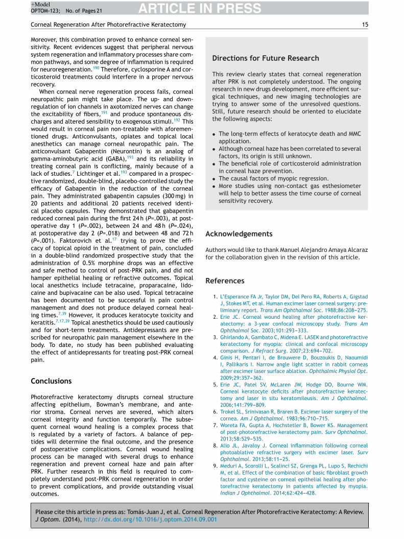
IN PRESS+Model
15
Directions for Future Research
This review clearly states that corneal regenerationafter PRK is not completely understood. The ongoingresearch in new drugs development, more efficient sur-gical techniques, and new imaging technologies aretrying to answer some of the unresolved questions.Still, future research should be oriented to elucidatethe following aspects:
• The long-term effects of keratocyte death and MMCapplication.
• Although corneal haze has been correlated to severalfactors, its origin is still unknown.
• The beneficial role of corticosteroid administrationin corneal haze prevention.
• The causal factors of myopic regression.• More studies using non-contact gas esthesiometer
will help to better assess the time course of cornealsensitivity recovery.
A
Af
R
Document downloaded from http://www.journalofoptometry.org, day 13/03/2015. This copy is for personal use. Any transmission of this document by any media or format is strictly prohibited. This early online article has been reviewed, accepted and posted on the Web before copyediting.
ARTICLEOPTOM-123; No. of Pages 21
Corneal Regeneration After Photorefractive Keratectomy
Moreover, this combination proved to enhance corneal sen-sitivity. Recent evidences suggest that peripheral nervoussystem regeneration and inflammatory processes share com-mon pathways, and some degree of inflammation is requiredfor neuroregeneration.190 Therefore, cyclosporine A and cor-ticosteroid treatments could interfere in a proper nervousrecovery.
When corneal nerve regeneration process fails, cornealneuropathic pain might take place. The up- and down-regulation of ion channels in axotomized nerves can changethe excitability of fibers,191 and produce spontaneous dis-charges and altered sensibility to exogenous stimuli.192 Thiswould result in corneal pain non-treatable with aforemen-tioned drugs. Anticonvulsants, opiates and topical localanesthetics can manage corneal neuropathic pain. Theanticonvulsant Gabapentin (Neurontin) is an analog ofgamma-aminobutyric acid (GABA),193 and its reliability intreating corneal pain is conflicting, mainly because of alack of studies.7 Lichtinger et al.193 compared in a prospec-tive randomized, double-blind, placebo-controlled study theefficacy of Gabapentin in the reduction of the cornealpain. They administrated gabapentin capsules (300 mg) in20 patients and additional 20 patients received identi-cal placebo capsules. They demonstrated that gabapentinreduced corneal pain during the first 24 h (P=.003), at post-operative day 1 (P=.002), between 24 and 48 h (P=.024),at postoperative day 2 (P=.018) and between 48 and 72 h(P=.001). Faktorovich et al.17 trying to prove the effi-cacy of topical opioid in the treatment of pain, concludedin a double-blind randomized prospective study that theadministration of 0.5% morphine drops was an effectiveand safe method to control of post-PRK pain, and did nothamper epithelial healing or refractive outcomes. Topicallocal anesthetics include tetracaine, proparacaine, lido-caine and bupivacaine can be also used. Topical tetracainehas been documented to be successful in pain controlmanagement and does not produce delayed corneal heal-ing times.7,39 However, it produces keratocyte toxicity andkeratitis.7,17,39 Topical anesthetics should be used cautiouslyand for short-term treatments. Antidepressants are pre-scribed for neuropathic pain management elsewhere in thebody. To date, no study has been published evaluatingthe effect of antidepressants for treating post-PRK cornealpain.
Conclusions
Photorefractive keratectomy disrupts corneal structureaffecting epithelium, Bowman’s membrane, and ante-rior stroma. Corneal nerves are severed, which alterscorneal integrity and function temporarily. The subse-quent corneal wound healing is a complex process thatis regulated by a variety of factors. A balance of pep-tides will determine the final outcome, and the presenceof postoperative complications. Corneal wound healingprocess can be managed with several drugs to enhanceregeneration and prevent corneal haze and pain after
Please cite this article in press as: Tomás-Juan J, et al. Corneal RJ Optom. (2014), http://dx.doi.org/10.1016/j.optom.2014.09.
PRK. Further research in this field is required to com-pletely understand post-PRK corneal regeneration in orderto prevent complications, and provide outstanding visualoutcomes.
cknowledgements
uthors would like to thank Manuel Alejandro Amaya Alcarazor the collaboration given in the revision of this article.
eferences
1. L’Esperance FA Jr, Taylor DM, Del Pero RA, Roberts A, GigstadJ, Stokes MT, et al. Human excimer laser corneal surgery: pre-liminary report. Trans Am Ophthalmol Soc. 1988;86:208---275.
2. Erie JC. Corneal wound healing after photorefractive ker-atectomy: a 3-year confocal microscopy study. Trans AmOphthalmol Soc. 2003;101:293---333.
3. Ghirlando A, Gambato C, Midena E. LASEK and photorefractivekeratectomy for myopia: clinical and confocal microscopycomparison. J Refract Surg. 2007;23:694---702.
4. Ginis H, Pentari I, de Brouwere D, Bouzoukis D, NaoumidiI, Pallikaris I. Narrow angle light scatter in rabbit corneasafter excimer laser surface ablation. Ophthalmic Physiol Opt.2009;29:357---362.
5. Erie JC, Patel SV, McLaren JW, Hodge DO, Bourne WM.Corneal keratocyte deficits after photorefractive keratec-tomy and laser in situ keratomileusis. Am J Ophthalmol.2006;141:799---809.
6. Trokel SL, Srinivasan R, Braren B. Excimer laser surgery of thecornea. Am J Ophthalmol. 1983;96:710---715.
7. Woreta FA, Gupta A, Hochstetler B, Bower KS. Managementof post-photorefractive keratectomy pain. Surv Ophthalmol.2013;58:529---535.
8. Alio JL, Javaloy J. Corneal inflammation following cornealphotoablative refractive surgery with excimer laser. SurvOphthalmol. 2013;58:11---25.
9. Meduri A, Scorolli L, Scalinci SZ, Grenga PL, Lupo S, RechichiM, et al. Effect of the combination of basic fibroblast growth
egeneration After Photorefractive Keratectomy: A Review.001
factor and cysteine on corneal epithelial healing after pho-torefractive keratectomy in patients affected by myopia.Indian J Ophthalmol. 2014;62:424---428.
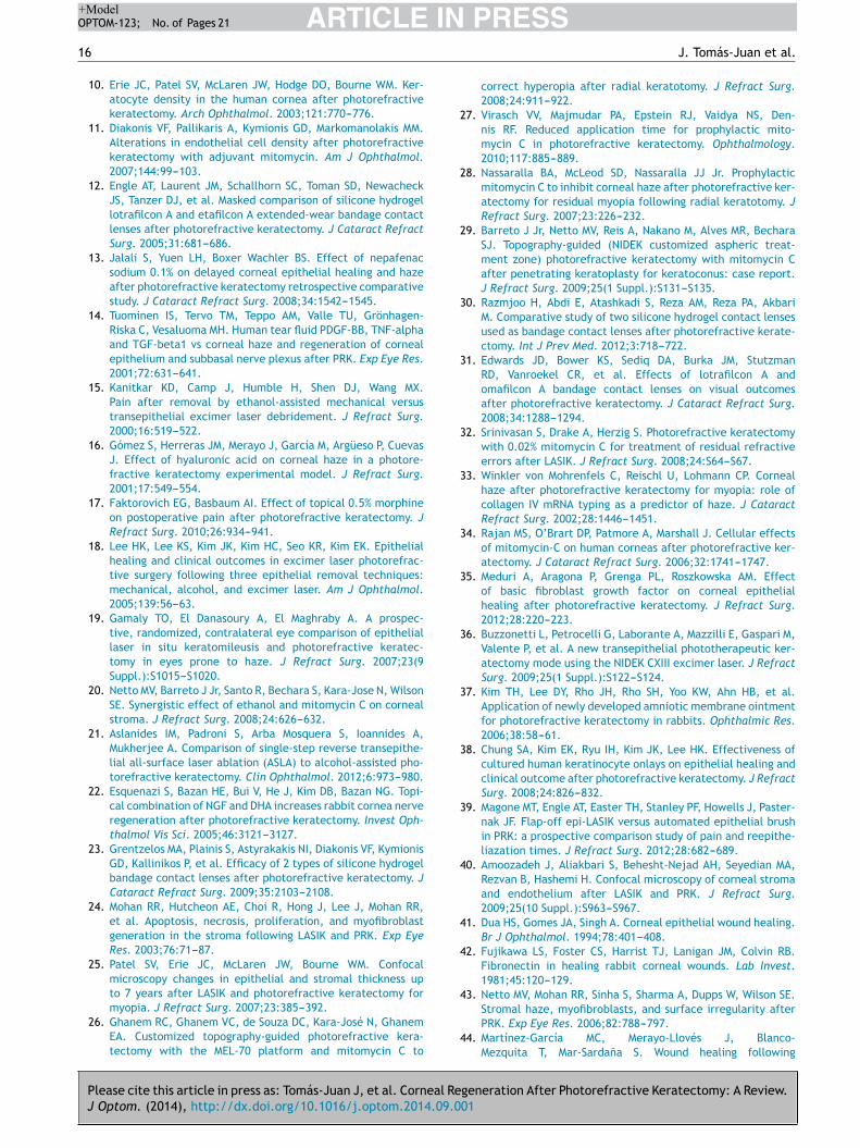
IN+ModelO
1
Document downloaded from http://www.journalofoptometry.org, day 13/03/2015. This copy is for personal use. Any transmission of this document by any media or format is strictly prohibited. This early online article has been reviewed, accepted and posted on the Web before copyediting.
ARTICLEPTOM-123; No. of Pages 21
6
10. Erie JC, Patel SV, McLaren JW, Hodge DO, Bourne WM. Ker-atocyte density in the human cornea after photorefractivekeratectomy. Arch Ophthalmol. 2003;121:770---776.
11. Diakonis VF, Pallikaris A, Kymionis GD, Markomanolakis MM.Alterations in endothelial cell density after photorefractivekeratectomy with adjuvant mitomycin. Am J Ophthalmol.2007;144:99---103.
12. Engle AT, Laurent JM, Schallhorn SC, Toman SD, NewacheckJS, Tanzer DJ, et al. Masked comparison of silicone hydrogellotrafilcon A and etafilcon A extended-wear bandage contactlenses after photorefractive keratectomy. J Cataract RefractSurg. 2005;31:681---686.
13. Jalali S, Yuen LH, Boxer Wachler BS. Effect of nepafenacsodium 0.1% on delayed corneal epithelial healing and hazeafter photorefractive keratectomy retrospective comparativestudy. J Cataract Refract Surg. 2008;34:1542---1545.
14. Tuominen IS, Tervo TM, Teppo AM, Valle TU, Grönhagen-Riska C, Vesaluoma MH. Human tear fluid PDGF-BB, TNF-alphaand TGF-beta1 vs corneal haze and regeneration of cornealepithelium and subbasal nerve plexus after PRK. Exp Eye Res.2001;72:631---641.
15. Kanitkar KD, Camp J, Humble H, Shen DJ, Wang MX.Pain after removal by ethanol-assisted mechanical versustransepithelial excimer laser debridement. J Refract Surg.2000;16:519---522.
16. Gómez S, Herreras JM, Merayo J, García M, Argüeso P, CuevasJ. Effect of hyaluronic acid on corneal haze in a photore-fractive keratectomy experimental model. J Refract Surg.2001;17:549---554.
17. Faktorovich EG, Basbaum AI. Effect of topical 0.5% morphineon postoperative pain after photorefractive keratectomy. JRefract Surg. 2010;26:934---941.
18. Lee HK, Lee KS, Kim JK, Kim HC, Seo KR, Kim EK. Epithelialhealing and clinical outcomes in excimer laser photorefrac-tive surgery following three epithelial removal techniques:mechanical, alcohol, and excimer laser. Am J Ophthalmol.2005;139:56---63.
19. Gamaly TO, El Danasoury A, El Maghraby A. A prospec-tive, randomized, contralateral eye comparison of epitheliallaser in situ keratomileusis and photorefractive keratec-tomy in eyes prone to haze. J Refract Surg. 2007;23(9Suppl.):S1015---S1020.
20. Netto MV, Barreto J Jr, Santo R, Bechara S, Kara-Jose N, WilsonSE. Synergistic effect of ethanol and mitomycin C on cornealstroma. J Refract Surg. 2008;24:626---632.
21. Aslanides IM, Padroni S, Arba Mosquera S, Ioannides A,Mukherjee A. Comparison of single-step reverse transepithe-lial all-surface laser ablation (ASLA) to alcohol-assisted pho-torefractive keratectomy. Clin Ophthalmol. 2012;6:973---980.
22. Esquenazi S, Bazan HE, Bui V, He J, Kim DB, Bazan NG. Topi-cal combination of NGF and DHA increases rabbit cornea nerveregeneration after photorefractive keratectomy. Invest Oph-thalmol Vis Sci. 2005;46:3121---3127.
23. Grentzelos MA, Plainis S, Astyrakakis NI, Diakonis VF, KymionisGD, Kallinikos P, et al. Efficacy of 2 types of silicone hydrogelbandage contact lenses after photorefractive keratectomy. JCataract Refract Surg. 2009;35:2103---2108.
24. Mohan RR, Hutcheon AE, Choi R, Hong J, Lee J, Mohan RR,et al. Apoptosis, necrosis, proliferation, and myofibroblastgeneration in the stroma following LASIK and PRK. Exp EyeRes. 2003;76:71---87.
25. Patel SV, Erie JC, McLaren JW, Bourne WM. Confocalmicroscopy changes in epithelial and stromal thickness upto 7 years after LASIK and photorefractive keratectomy for
Please cite this article in press as: Tomás-Juan J, et al. Corneal RJ Optom. (2014), http://dx.doi.org/10.1016/j.optom.2014.09.
myopia. J Refract Surg. 2007;23:385---392.26. Ghanem RC, Ghanem VC, de Souza DC, Kara-José N, Ghanem
EA. Customized topography-guided photorefractive kera-tectomy with the MEL-70 platform and mitomycin C to
PRESSJ. Tomás-Juan et al.
correct hyperopia after radial keratotomy. J Refract Surg.2008;24:911---922.
27. Virasch VV, Majmudar PA, Epstein RJ, Vaidya NS, Den-nis RF. Reduced application time for prophylactic mito-mycin C in photorefractive keratectomy. Ophthalmology.2010;117:885---889.
28. Nassaralla BA, McLeod SD, Nassaralla JJ Jr. Prophylacticmitomycin C to inhibit corneal haze after photorefractive ker-atectomy for residual myopia following radial keratotomy. JRefract Surg. 2007;23:226---232.
29. Barreto J Jr, Netto MV, Reis A, Nakano M, Alves MR, BecharaSJ. Topography-guided (NIDEK customized aspheric treat-ment zone) photorefractive keratectomy with mitomycin Cafter penetrating keratoplasty for keratoconus: case report.J Refract Surg. 2009;25(1 Suppl.):S131---S135.
30. Razmjoo H, Abdi E, Atashkadi S, Reza AM, Reza PA, AkbariM. Comparative study of two silicone hydrogel contact lensesused as bandage contact lenses after photorefractive kerate-ctomy. Int J Prev Med. 2012;3:718---722.
31. Edwards JD, Bower KS, Sediq DA, Burka JM, StutzmanRD, Vanroekel CR, et al. Effects of lotrafilcon A andomafilcon A bandage contact lenses on visual outcomesafter photorefractive keratectomy. J Cataract Refract Surg.2008;34:1288---1294.
32. Srinivasan S, Drake A, Herzig S. Photorefractive keratectomywith 0.02% mitomycin C for treatment of residual refractiveerrors after LASIK. J Refract Surg. 2008;24:S64---S67.
33. Winkler von Mohrenfels C, Reischl U, Lohmann CP. Cornealhaze after photorefractive keratectomy for myopia: role ofcollagen IV mRNA typing as a predictor of haze. J CataractRefract Surg. 2002;28:1446---1451.
34. Rajan MS, O’Brart DP, Patmore A, Marshall J. Cellular effectsof mitomycin-C on human corneas after photorefractive ker-atectomy. J Cataract Refract Surg. 2006;32:1741---1747.
35. Meduri A, Aragona P, Grenga PL, Roszkowska AM. Effectof basic fibroblast growth factor on corneal epithelialhealing after photorefractive keratectomy. J Refract Surg.2012;28:220---223.
36. Buzzonetti L, Petrocelli G, Laborante A, Mazzilli E, Gaspari M,Valente P, et al. A new transepithelial phototherapeutic ker-atectomy mode using the NIDEK CXIII excimer laser. J RefractSurg. 2009;25(1 Suppl.):S122---S124.
37. Kim TH, Lee DY, Rho JH, Rho SH, Yoo KW, Ahn HB, et al.Application of newly developed amniotic membrane ointmentfor photorefractive keratectomy in rabbits. Ophthalmic Res.2006;38:58---61.
38. Chung SA, Kim EK, Ryu IH, Kim JK, Lee HK. Effectiveness ofcultured human keratinocyte onlays on epithelial healing andclinical outcome after photorefractive keratectomy. J RefractSurg. 2008;24:826---832.
39. Magone MT, Engle AT, Easter TH, Stanley PF, Howells J, Paster-nak JF. Flap-off epi-LASIK versus automated epithelial brushin PRK: a prospective comparison study of pain and reepithe-liazation times. J Refract Surg. 2012;28:682---689.
40. Amoozadeh J, Aliakbari S, Behesht-Nejad AH, Seyedian MA,Rezvan B, Hashemi H. Confocal microscopy of corneal stromaand endothelium after LASIK and PRK. J Refract Surg.2009;25(10 Suppl.):S963---S967.
41. Dua HS, Gomes JA, Singh A. Corneal epithelial wound healing.Br J Ophthalmol. 1994;78:401---408.
42. Fujikawa LS, Foster CS, Harrist TJ, Lanigan JM, Colvin RB.Fibronectin in healing rabbit corneal wounds. Lab Invest.1981;45:120---129.
43. Netto MV, Mohan RR, Sinha S, Sharma A, Dupps W, Wilson SE.
egeneration After Photorefractive Keratectomy: A Review.001
Stromal haze, myofibroblasts, and surface irregularity afterPRK. Exp Eye Res. 2006;82:788---797.
44. Martínez-García MC, Merayo-Llovés J, Blanco-Mezquita T, Mar-Sardana S. Wound healing following
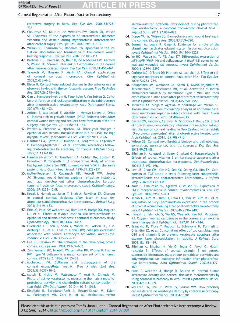
IN+Model
Document downloaded from http://www.journalofoptometry.org, day 13/03/2015. This copy is for personal use. Any transmission of this document by any media or format is strictly prohibited. This early online article has been reviewed, accepted and posted on the Web before copyediting.
ARTICLEOPTOM-123; No. of Pages 21
Corneal Regeneration After Photorefractive Keratectomy
refractive surgery in hens. Exp Eye Res. 2006;83:728---735.
45. Chaurasia SS, Kaur H, de Medeiros FW, Smith SD, WilsonSE. Dynamics of the expression of intermediate filamentsvimentin and desmin during myofibroblast differentiationafter corneal injury. Exp Eye Res. 2009;89:133---139.
46. Wilson SE, Chaurasia SS, Madeiros FW. Apoptosis in the ini-tiation. Modulation and termination of the corneal woundhealing response. Exp Eye Res. 2007;85:305---311.
47. Barbosa FL, Chaurasia SS, Kaur H, de Medeiros FW, AgrawalV, Wilson SE. Stromal interleukin-1 expression in the corneaafter haze-associated injury. Exp Eye Res. 2010;91:456---461.
48. Tavakoli M, Hossain P, Malik RA. Clinical applicationsof corneal confocal microscopy. Clin Ophthalmol.2008;2:435---445.
49. Efron N. Contact lens-induced changes in the anterior eye asobserved in vivo with the confocal microscope. Prog Retin EyeRes. 2007;26:398---436.
50. Gan L, Hamberg-Nyström H, Fagerholm P, Van Setten G. Cellu-lar proliferation and leukocyte infiltration in the rabbit corneaafter photorefractive keratectomy. Acta Ophthalmol Scand.2001;79:488---492.
51. Anitua E, Muruzabal F, Alcalde I, Merayo-Lloves J, OriveG. Plasma rich in growth factors (PRGF-Endoret) stimulatescorneal wound healing and reduces haze formation after PRKsurgery. Exp Eye Res. 2013;115:153---161.
52. Ivarsen A, Fledelius W, Hjortdal JØ. Three-year changes inepithelial and stromal thickness after PRK or LASIK for highmyopia. Invest Ophthalmol Vis Sci. 2009;50:2061---2066.
53. Gauthier CA, Epstein D, Holden BA, Tengroth B, FagerholmP, Hamberg-Nyström H, et al. Epithelial alterations follow-ing photorefractive keratectomy for myopia. J Refract Surg.1995;11:113---118.
54. Hamberg-Nyström H, Gauthier CA, Holden BA, Epstein D,Fagerholm P, Tengroth B. A comparative study of epithe-lial hypertrophy after PRK: summit versus VISX in the samepatient. Acta Ophthalmol Scand. 1996;74:228---231.
55. Moller-Pedersen T, Cavanagh HD, Petroll WM, JesterJV. Stromal wound healing explains refractive instabilityand haze development after photorefractive keratec-tomy: a 1-year confocal microscopic study. Ophthalmology.2000;107:1235---1245.
56. Kozak I, Hornak M, Juhas T, Shah A, Rawlings EF. Changesin central corneal thickness after laser in situ ker-atomileusis and photorefractive keratectomy. J Refract Surg.2003;19:149---153.
57. Erie JC, Patel SV, McLaren JW, Ramirez M, Hodge DO, MaguireLJ, et al. Effect of myopic laser in situ keratomileusis onepithelial and stromal thickness: a confocal microscopy study.Ophthalmology. 2002;109:1447---1452.
58. Guerriero E, Chen J, Sado Y, Mohan RR, Wilson SE, Fun-derburgh JL, et al. Loss of alpha3 (IV) collagen expressionassociated with corneal keratocyte activation. Invest Oph-thalmol Vis Sci. 2007;48:627---635.
59. Lee RE, Davison PF. The collagens of the developing bovinecornea. Exp Eye Res. 1984;39:639---652.
60. Zimmermann DR, Trueb B, Winterhalter KH, Witmer R, FischerRW. Type VI collagen is a major component of the humancornea. FEBS Lett. 1986;197:55---58.
61. Michelacci YM. Collagens and proteoglycans of thecorneal extracellular matrix. Braz J Med Biol Res.2003;36:1037---1046.
62. Mutoh T, Nishio M, Matsumoto Y, Arai K, Chikuda M.Photorefractive keratectomy: measuring the matrix metallo-
Please cite this article in press as: Tomás-Juan J, et al. Corneal RJ Optom. (2014), http://dx.doi.org/10.1016/j.optom.2014.09.
proteinase activity and chondroitin sulfate concentration intear fluid. Clin Ophthalmol. 2010;4:1015---1018.
63. Einollahi B, Baradaran-Rafii A, Rezaei-Kanavi M, EslaniM, Parchegani MR, Zare M, et al. Mechanical versus
PRESS17
alcohol-assisted epithelial debridement during photorefrac-tive keratectomy: a confocal microscopic clinical trial. JRefract Surg. 2011;27:887---893.
64. Dupps WJ Jr, Wilson SE. Biomechanics and wound healing inthe cornea. Exp Eye Res. 2006;83:709---720.
65. Berman M, Leary R, Gage J. Evidence for a role of theplasminogen activator---plasmin system in corneal ulceration.Invest Ophthalmol Vis Sci. 1980;19:1204---1221.
66. Ye HQ, Maeda M, Yu FS, Azar DT. Differential expression ofMT1-MMP (MMP-14) and collagenase III (MMP-13) genes in nor-mal and wounded rat corneas. Invest Ophthalmol Vis Sci.2000;41:2894---2899.
67. Corbett MC, O’Brart DP, Patmore AL, Marshall J. Effect of col-lagenase inhibitors on corneal haze after PRK. Exp Eye Res.2001;72:253---259.
68. Holopainen JM, Moilanen JA, Sorsa T, Kivelä-Rajamäki M,Tervahartiala T, Vesaluoma MH, et al. Activation of matrixmetalloproteinase-8 by membrane type 1-MMP and theirexpression in human tears after photorefractive keratectomy.Invest Ophthalmol Vis Sci. 2003;44:2550---2556.
69. Torricelli AA, Singh V, Agrawal V, Santhiago MR, Wilson SE.Transmission electron microscopy analysis of epithelial base-ment membrane repair in rabbit corneas with haze. InvestOphthalmol Vis Sci. 2013;54:4026---4033.
70. Davies BW, Panday V, Caldwell M, Scribbick F, Reilly CD. Effectof topical immunomodulatory interleukin 1 receptor antago-nist therapy on corneal healing in New Zealand white rabbits(Oryctolagus cunniculus) after photorefractive keratectomy.Arch Ophthalmol. 2011;129:909---913.
71. Wilson SE. Corneal myofibroblast biology and pathobiology:generation, persistence, and transparency. Exp Eye Res.2012;99:78---88.
72. Bilgihan K, Adiguzel U, Sezer C, Akyol G, Hasanreisoglu B.Effects of topical vitamin E on keratocyte apoptosis aftertraditional photorefractive keratectomy. Ophthalmologica.2001;215:192---196.
73. Lee JB, Choe CM, Kim HS, Seo KY, Seong GJ, Kim EK. Com-parison of TGF-beta1 in tears following laser subepithelialkeratomileusis and photorefractive keratectomy. J RefractSurg. 2002;18:130---134.
74. Kaur H, Chaurasia SS, Agrawal V, Wilson SE. Expression ofPDGF receptor-alpha in corneal myofibroblasts in situ. ExpEye Res. 2009;89:432---434.
75. Tchah H, Kim MJ, Kim TI, Choi HJ, Kim JY, Kim MJ, et al.Regulation of 1-cys peroxiredoxin expression in the processof stromal wound healing after photorefractive keratectomy.Invest Ophthalmol Vis Sci. 2005;46:2396---2403.
76. Hayashi S, Ishimoto S, Wu GS, Wee WR, Rao NA, McDonnellPJ. Oxygen free radical damage in the cornea after excimerlaser therapy. Br J Ophthalmol. 1997;81:141---144.
77. Brancato R, Fiore T, Papucci L, Schiavone N, Formigli L,Orlandini SZ, et al. Concomitant effect of topical ubiquinoneQ10 and vitamin E to prevent keratocyte apoptosis afterexcimer laser photoablation in rabbits. J Refract Surg.2002;18:135---139.
78. Bilgihan A, Bilgihan K, Yis O, Sezer C, Akyol G, Hasan-reisoglu B. Effects of topical vitamin E on cornealsuperoxide dismutase, glutathione peroxidase activities andpolymorphonuclear leucocyte infiltration after photorefrac-tive keratectomy. Acta Ophthalmol Scand. 2003;81:177---180.
79. Patel S, McLaren J, Hodge D, Bourne W. Normal humankeratocyte density and corneal thickness measurements byusing confocal microscopy in vivo. Invest Ophthalmol Vis Sci.
egeneration After Photorefractive Keratectomy: A Review.001
2001;42:333---339.80. McLaren JW, Nau CB, Patel SV, Bourne WM. How precisely
can we determine keratocyte density by confocal microscopy?Invest Ophthalmol Vis Sci. 2001;42:S281.
IN+ModelO
1
Document downloaded from http://www.journalofoptometry.org, day 13/03/2015. This copy is for personal use. Any transmission of this document by any media or format is strictly prohibited. This early online article has been reviewed, accepted and posted on the Web before copyediting.
ARTICLEPTOM-123; No. of Pages 21
8
81. Böhnke M, Thaer A, Schipper I. Confocal microscopy revealspersisting stromal changes after myopic photorefractivekeratectomy in zero haze corneas. Br J Ophthalmol.1998;82:1393---1400.
82. Midena E, Gambato C, Miotto S, Cortese M, Salvi R, GhirlandoA. Long-term effects on corneal keratocytes of mitomycin Cduring photorefractive keratectomy: a randomized contralat-eral eye confocal microscopy study. J Refract Surg. 2007;23(9Suppl.):S1011---S1014.
83. Erie JC, McLaren JW, Hodge DO, Bourne WM. Recovery ofcorneal subbasal nerve density after PRK and LASIK. Am JOphthalmol. 2005;140:1059---1064.
84. Ambrósio R Jr, Kalina R, Mohan RR, Mohan RR, Possin DD,Huang J, et al. Early wound healing response to epithelialscrape injury in the human cornea. Invest Ophthalmol Vis Sci.2002;43. E-Abstract 4206.
85. Frueh BE, Cadez R, Böhnke M. In vivo confocal microscopyafter photorefractive keratectomy in humans. A prospec-tive, long-term study. Arch Ophthalmol. 1998;116:1425---1431.
86. Hofmeister EM, Bishop FM, Kaupp SE, Schallhorn SC. Ran-domized dose---response analysis of mitomycin-C to preventhaze after photorefractive keratectomy for high myopia. JCataract Refract Surg. 2013;39:1358---1365.
87. Fini EM. Keratocyte and fibroblast phenotypes in the repairingcornea. Prog Retin Eye Res. 1999;18:529---551.
88. Corbett MC, Prydal JI, Verma S, Oliver KM, Pande M,Marshall J. An in vivo investigation of the structuresresponsible for corneal haze after photorefractive kerate-ctomy and their effect on visual function. Ophthalmology.1996;103:1366---1380.
89. Erie JC, Patel SV, McLaren JW, Maguire LJ, Ramirez M, BourneWM. Keratocyte density in vivo after photorefractive kerate-ctomy in humans. Trans Am Ophthalmol Soc. 1999;97:221---236.
90. Razmjoo H, Kooshanmehr MR, Peyman A, Kor Z, Mohammades-maeil E. Comparison of standard and low dose intraoperativemitomycin C in prevention of corneal haze after photorefrac-tive keratectomy. Int J Prev Med. 2013;4:204---207.
91. Netto MV, Mohan RR, Sinha S, Sharma A, Gupta PC, Wil-son SE. Effect of prophylactic and therapeutic mitomycinC on corneal apoptosis, cellular proliferation, haze, andlong-term keratocyte density in rabbits. J Refract Surg.2006;22:562---574.
92. Cua IY, Pepose JS. Late corneal scarring after photorefractivekeratectomy concurrent with development of systemic lupuserythematosus. J Refract Surg. 2002;18:750---752.
93. Kremer I, Ehrenberg M, Levinger S. Delayed epithelial heal-ing following photorefractive keratectomy with mitomycin Ctreatment. Acta Ophthalmol. 2012;90:271---276.
94. de Medeiros FW, Mohan RR, Suto C, Sinhá S, Bonilha VL,Chaurasia SS, et al. Haze development after photorefrac-tive keratectomy: mechanical vs ethanol epithelial removalin rabbits. J Refract Surg. 2008;24:923---927.
95. Resch MD, Nagy ZZ, Szentmáry N, Máthé M, Kovalszky I,Süveges I. Spatial distribution of keratin sulfate in the rab-bit cornea following photorefractive keratectomy. J RefractSurg. 2005;21:485---493.
96. Meltendorf C, Burbach GJ, Bühren J, Bug R, Ohrloff C, DellerT. Corneal femtosecond laser keratotomy results in isolatedstromal injury and favorable wound-healing response. InvestOphthalmol Vis Sci. 2007;48:2068---2075.
97. Csutak A, Tözsér J, Békési L, Hassan Z, Berta A, SilverDM. Plasminogen activator activity in tears after excimer
Please cite this article in press as: Tomás-Juan J, et al. Corneal RJ Optom. (2014), http://dx.doi.org/10.1016/j.optom.2014.09.
laser photorefractive keratectomy. Invest Ophthalmol Vis Sci.2000;41:3743---3747.
98. von Mohrenfels CW, Reischl U, Gabler B, Lohmann CP. Cornealhaze after photorefractive keratectomy. Role of individual
PRESSJ. Tomás-Juan et al.
collagen type IV synthesis on postoperative corneal opacity.Ophthalmologe. 2002;99:532---537.
99. Nagy ZZ, Hiscott P, Seitz B, Shlötzer-Schrehardt U, SimonM Jr, Süveges I, et al. Ultraviolet-B enhances stromalresponse to 193-nm excimer laser treatment. Ophthalmology.1997;104:375---380.
100. Blalock TD, Duncan MR, Varela JC, Goldstein MH, Tuli SS, Gro-tendorst GR, et al. Connective tissue growth factor expressionand action in human corneal fibroblast cultures and ratcorneas after photorefractive keratectomy. Invest Ophthal-mol Vis Sci. 2003;44:1879---1887.
101. Jester JV, Petroll WM, Cavanagh HD. Corneal stromal woundhealing in refractive surgery: the role of myofibroblasts. ProgRetin Eye Res. 1999;18:311---356.
102. Vinciguerra P, Azzolini M, Airaghi P, Radice P, De MolfettaV. Effect of decreasing surface and interface irregularitiesafter photorefractive keratectomy and laser in situ ker-atomileusis on optical and functional outcomes. J RefractSurg. 1998;14:S199---S203.
103. Taylor SM, Fields CR, Barker FM, Sanzo CJ. Effect of depthupon the smoothness of excimer laser corneal ablation.Optom Vis Sci. 1994;71:104---108.
104. Javier JA, Lee JB, Oliveira HB, Chang JH, Azar DT. Basementmembrane and collagen deposition after laser subepithe-lial keratomileusis and photorefractive keratectomy inthe leghorn chick eye. Arch Ophthalmol. 2006;124:703---709.
105. Nakamura K, Kurosaka D, Bissen-Miyajima H, Tsubota K. Intactcorneal epithelium is essential for the prevention of stromalhaze after laser assisted in situ keratomileusis. Br J Ophthal-mol. 2001;85:209---213.
106. McLaren JW, Bourne WM, Patel SV. Standardization of cornealhaze measurement in confocal microscopy. Invest OphthalmolVis Sci. 2010;51:5610---5616.
107. Farid M, Morishige N, Lam L, Wahlert A, Steinert RF, JesterJV. Detection of corneal fibrosis by imaging second harmonic-generated signals in rabbit corneas treated with mitomycin Cafter excimer laser surface ablation. Invest Ophthalmol VisSci. 2008;49:4377---4383.
108. Takacs AI, Mihaltz K, Nagy ZZ. Corneal density with the Pen-tacam after photorefractive keratectomy. J Refract Surg.2011;27:269---277.
109. Nien CJ, Flynn KJ, Chang M, Brown D, Jester JV. Reducing peakcorneal haze after photorefractive keratectomy in rabbits:prednisolone acetate 1.00% versus cyclosporine A 0.05%. JCataract Refract Surg. 2011;37:937---944.
110. Møller-Pedersen T, Cavanagh HD, Petroll WM, Jester JV.Corneal haze development after PRK is regulated by volumeof stromal tissue removal. Cornea. 1998;17:627---639.
111. Nejima R, Miyata K, Tanabe T, Okamoto F, Hiraoka T, KiuchiT, et al. Corneal barrier function, tear film stability, andcorneal sensation after photorefractive keratectomy andlaser in situ keratomileusis. Am J Ophthalmol. 2005;139:64---71.
112. Belmonte C, Acosta MC, Gallar J. Neural basis of sensation inintact and injured corneas. Exp Eye Res. 2004;78:513---525.
113. Prydal JI, Kerr Muir MG, Dilly PN, Corbett MC, Verma S,Marshall J. Confocal microscopy using oblique sections formeasurement of corneal epithelial thickness in conscioushumans. Acta Ophthalmol Scand. 1997;75:624---628.
114. Vesaluoma M, Perez-Santonja J, Petroll WM, Linna T, Alió J,Tervo T. Corneal stromal changes induced by myopic LASIK.Invest Ophthalmol Vis Sci. 2000;41:369---376.
115. Müller LJ, Pels L, Vrensen GF. Ultrastructural organization of
egeneration After Photorefractive Keratectomy: A Review.001
human corneal nerves. Ophthalmol Vis Sci. 1996;37:476---488.116. Tervo K, Latvala TM, Tervo TM. Recovery of corneal
innervation following photorefractive keratoablation. ArchOphthalmol. 1994;112:1466---1470.
IN+Model
Document downloaded from http://www.journalofoptometry.org, day 13/03/2015. This copy is for personal use. Any transmission of this document by any media or format is strictly prohibited. This early online article has been reviewed, accepted and posted on the Web before copyediting.
ARTICLEOPTOM-123; No. of Pages 21
Corneal Regeneration After Photorefractive Keratectomy
117. Rozsa AJ, Guss RB, Beuerman RW. Neural remodeling followingexperimental surgery of the rabbit cornea. Invest OphthalmolVis Sci. 1983;24:1033---1051.
118. Burka JM, Bower KS, Vanroekel RC, Stutzman RD, KuzmowychCP, Howard RS. The effect of moxifloxacion and gatifloxacinon long-term visual outcomes following photorefractive ker-atectomy. J Refract Surg. 2007;23:414---417.
119. Baker KS, Anderson SC, Romanowski EG, Thoft RA, Sundar-Raj N. Trigeminal ganglion neurons affect corneal epithelialphenotype. Influence on type VII collagen expression in vitro.Invest Ophthalmol Vis Sci. 1993;34:137---144.
120. Kauffmann T, Bodanowitz S, Hesse L, Kroll P. Corneal rein-nervation after photorefractive keratectomy and laser in situkeratomileusis: an in vivo study with a confocal videomicro-scope. Ger J Ophthalmol. 1996;5:508---512.
121. Neira-Zalentein W, Moilanen JA, Tuisku IS, Holopainen JM,Tervo TM. Photorefractive keratectomy retreatment afterLASIK. J Refract Surg. 2008;24:710---712.
122. Moilanen JA, Vesaluoma MH, Müller LJ, Tervo TM. Long-termcorneal morphology after PRK by in vivo confocal microscopy.Invest Ophthalmol Vis Sci. 2003;44:1064---1069.
123. Stern ME, Gao J, Siemasko KF, Beuerman RW, Pflugfelder SC.The role of the lacrimal functional unit in the pathophysiologyof dry eye. Exp Eye Res. 2004;78:409---416.
124. Kymionis GD, Tsiklis NS, Ginis H, Diakonis VF, Pallikaris I. Dryeye after photorefractive keratectomy with adjuvant mito-mycin C. J Refract Surg. 2006;22:511---513.
125. Hosal BM, Ornek N, Zilelioglu G, Elhan AH. Morphology ofcorneal nerves and corneal sensation in dry eye: a preliminarystudy. Eye (Lond). 2005;19:1276---1279.
126. Bourcier T, Acosta MC, Borderie V, Borrás F, Gallar J, Bury T,et al. Decreased corneal sensitivity in patients with dry eye.Invest Ophthalmol Vis Sci. 2005;46:2341---2345.
127. Gallar J, Moilanen J, Acosta C, Holopainen J, Belmonte C,Tervo T, et al. Long-term corneal sensitivity after PRK deter-mined by non-contact gas esthesiometry. Invest OphthalmolVis Sci. 2013;54 [E-Abstract 904].
128. Sher NA, Golben MR, Bond W, Trattler WB, Tauber S, Voirin TG.Topical bromfenac 0.09% vs. ketorolac 0.4% for the control ofpain, photophobia, and discomfort following PRK. J RefractSurg. 2009;25:214---220.
129. Gallar J, Acosta MC, Gutiérrez AR, Belmonte C. Impulseactivity in corneal sensory nerve fibers after photorefractivekeratectomy. Invest Ophthalmol Vis Sci. 2007;48:4033---4037.
130. Campero M, Serra J, Marchettini P, Ochoa JL. Ectopic impulsegeneration and autoexcitation in single myelinated afferentfibers in patients with peripheral neuropathy and positive sen-sory symptoms. Muscle Nerve. 1998;21:1661---1667.
131. Ji RR, Kohno T, Moore KA, Woolf CJ. Central sensitization andLTP: do pain and memory share similar mechanisms? TrendsNeurosci. 2003;26:696---705.
132. Sia RK, Ryan DS, Stutzman RD, Psolka M, Mines MJ, Wagner ME,et al. Alcohol versus brush PRK: visual outcomes and adverseeffects. Lasers Surg Med. 2012;44:475---481.
133. Thornton I, Xu M, Krueger RR. Comparison of standard (0.02%)and low dose (0.002%) mitomycin C in the prevention ofcorneal haze following surface ablation for myopia. J RefractSurg. 2008;24:S68---S76.
134. Hanna KD, Pouliquen YM, Waring GO III, Savoldelli M, FantesF, Thompson KP. Corneal wound healing in monkeys afterrepeated excimer laser photorefractive keratectomy. ArchOphthalmol. 1992;110:1286---1291.
135. Ghoreishi M, Attarzadeh H, Tavakoli M, Moini HA, Zandi A,Masjedi A. Alcohol-assisted versus mechanical epithelium
Please cite this article in press as: Tomás-Juan J, et al. Corneal RJ Optom. (2014), http://dx.doi.org/10.1016/j.optom.2014.09.
removal in photorefractive keratectomy. J Ophthalmic VisRes. 2010;5:223---227.
136. Browning AC, Shah S, Dua HS, Maharajan SV, Gray T, BragheethMA. Alcohol debridement of the corneal epithelium in PRK and
PRESS19
LASEK: an electron microscopic study. Invest Ophthalmol VisSci. 2003;44:510---513.
137. Shah S, Sebai Sarhan AR, Doyle SJ, Pillai CT, Dua HS. Theepithelial flap for photorefractive keratectomy. Br J Ophthal-mol. 2001;85:393---396.
138. Chen CC, Chang JH, Lee JB, Javier J, Azar DT. Human cornealepithelial cell viability and morphology after dilute alcoholexposure. Invest Ophthalmol Vis Sci. 2002;43:2593---2602.
139. Ghanem VC, Souza GC, Souza DC, Viese JM, Weber SL, Kara-José N. PRK and butterfly LASEK: prospective, randomized,contralateral eye comparison of epithelial healing and oculardiscomfort. J Refract Surg. 2008;24:591---599.
140. Wang DM, Du Y, Chen GS, Tang LS, He JF. Transepithe-lial photorefractive keratectomy mode using SCHWIND-ESIRISexcimer laser: initial clinical results. Int J Ophthalmol.2012;5:334---337.
141. Mannan R, Pruthi A, Rampal U. Combined phototherapeutickeratectomy and amniotic membrane grafts for symptomaticbullous keratopathy. Cornea. 2010;29:1207---1208.
142. Rathi VM, Vyas SP, Sangwan VS. Phototherapeutic keratec-tomy. Indian J Ophthalmol. 2012;60:5---14.
143. Lee HK, Kim JK, Kim EK, Kim GO, Lee IS. Phototherapeutic ker-atectomy with amniotic membrane for severe subepithelialfibrosis following excimer laser refractive surgery. J CataractRefract Surg. 2003;29:1430---1435.
144. Choi YS, Kim JY, Wee WR, Lee JH. Effect of the applicationof human amniotic membrane on rabbit corneal wound heal-ing after excimer laser photorefractive keratectomy. Cornea.1998;17:389---395.
145. Wang MX, Gray T, Parks WC, Prabhasawat P, Culbertson W,Forster R, et al. Reduction in corneal haze and apoptosis byamniotic membrane matrix in excimer laser photoablation inrabbits. J Cataract Refract Surg. 2001;27:310---319.
146. Park WC, Tseng SC. Modulation of acute inflammation and ker-atocyte death by suturing, blood, and amniotic membrane inPRK. Invest Ophthalmol Vis Sci. 2000;41:2906---2914.
147. Savill J, Haslett C. Granulocyte clearance by apoptosis inthe resolution of inflammation. Semin Cell Biol. 1995;6:385---393.
148. Khokhar S, Natung T, Sony P, Sharma N, Agarwal N, VajpayeeRB. Amniotic membrane transplantation in refractory neu-rotrophic corneal ulcers: a randomized, controlled clinicaltrial. Cornea. 2005;24:654---660.
149. Meller D, Pauklin M, Thomasen H, Westekemper H, Steuhl KP.Amniotic membrane transplantation in the human eye. DtschArztebl Int. 2011;108:243e8.
150. Zhong Y, Zhai Z, Zhou Y, Ye W, Wang K. Effect of amnioticmembrane on expressions of TGF-beta 1, collagens I, III andfibronectin in rabbit corneal healing after photorefractivekeratectomy. Yan Ke Xue Bao. 2000;16:239---242.
151. Woo HM, Kim MS, Kweon OK, Kim DY, Nam TC, Kim JH. Effectsof amniotic membrane on epithelial wound healing and stro-mal remodelling after excimer laser keratectomy in rabbitcornea. Br J Ophthalmol. 2001;85:345---349.
152. Farahi A, Hashemi H, Mehravaran S. The effects ofmitomycin C on tear function after photorefractive kerate-ctomy: a contralateral comparative study. J Refract Surg.2013;29:260---264.
153. Shojaei A, Ramezanzadeh M, Soleyman-Jahi S, Almasi-Nasrabadi M, Rezazadeh P, Eslani M. Short-time mitomycin-Capplication during photorefractive keratectomy in patientswith low myopia. J Cataract Refract Surg. 2013;39:197---203.
154. Dawson DG, Grossniklaus HE, McCarey BE, Edelhauser HF.
egeneration After Photorefractive Keratectomy: A Review.001
Biomechanical and wound healing characteristics of corneasafter excimer laser keratorefractive surgery: is there a differ-ence between advanced surface ablation and subBowman’skeratomileusis? J Refract Surg. 2008;24:S90---S96.
IN+ModelO
2
Document downloaded from http://www.journalofoptometry.org, day 13/03/2015. This copy is for personal use. Any transmission of this document by any media or format is strictly prohibited. This early online article has been reviewed, accepted and posted on the Web before copyediting.
ARTICLEPTOM-123; No. of Pages 21
0
155. Netto MV, Chalita MR, Krueger RR. Corneal haze followingPRK with mitomycin C as a retreatment versus prophylac-tic use in the contralateral eye. J Refract Surg. 2007;23:96---98.
156. Khoury JM, Farah T, El-Haibi CP, Noureddin BN. Corneal lightshield as a delivery system for standardized application ofmitomycin C in excimer surface ablation. J Refract Surg.2007;23:716---719.
157. Goldsberry DH, Epstein RJ, Majmudar PA, Epstein RH, Den-nis RF, Holley G, et al. Effect of mitomycin C on the cornealendothelium when used for corneal subepithelial haze pro-phylaxis following photorefractive keratectomy. J RefractSurg. 2007;23:724---727.
158. Morales AJ, Zadok D, Mora-Retana R, Martínez-Gama E, Rob-ledo NE, Chayet AS. Intraoperative mitomycin and cornealendothelium after photorefractive keratectomy. Am J Oph-thalmol. 2006;142:400---404.
159. Wallau AD, Campos M. Photorefractive keratectomy with mit-omycin C versus LASIK in custom surgeries for myopia: abilateral prospective randomized clinical trial. J Refract Surg.2008;24:326---336.
160. Sia RK, Ryan DS, Edwards JD, Stutzman RD, Bower KS. TheU.S. Army Surface Ablation Study: comparison o PRK, MMC-PRK, and LASEK in moderate to high myopia. J Refract Surg.2014;30:256---264.
161. Kymionis GD, Diakonis VF, Charisis S, Pallikaris AI, BouzoukisDI, Yoo SH, et al. Effects of topical mitomycin C on the cil-iary body and intraocular pressure after PRK: an experimentalstudy. J Refract Surg. 2008;24:633---638.
162. Wallau AD, Campos M. One-year outcomes of a bilateralrandomised prospective clinical trial comparing PRK withmitomycin C and LASIK. Br J Ophthalmol. 2009;93:1634---1638.
163. Ghoreishi M, Attarzadeh H, Zandi A, Moini HA, TavakoliM, Fesharaki H. Outcomes of photorefractive keratectomywith intraoperative mitomycin-C. J Ophthalmic Vis Res.2009;4:142---146.
164. Fazel F, Roshani L, Rezaei L. Two-step versus single applica-tion of mitomycin-C in photorefractive keratectomy for highmyopia. J Ophthalmic Vis Res. 2012;7:17---23.
165. Spadea L, Verrecchia V. Effectiveness of scraping andmitomycin C to treat haze after myopic photorefractive ker-atectomy. Open Ophthalmol J. 2011;5:63---65.
166. Majmudar PA, Forstot SL, Dennis RF, Nirankari VS, DamianoRE, Brenart R, et al. Topical mitomycin-C for subepithe-lial fibrosis after refractive corneal surgery. Ophthalmology.2000;107:89---94.
167. Xu H, Liu S, Xia X, Huang P, Wang P, Wu X. Mitomycin C reduceshaze formation in rabbits after excimer laser photorefractivekeratectomy. J Refract Surg. 2001;17:342---349.
168. Patel SV, Bourne WM. Corneal endothelial cell loss 9 yearsafter excimer laser keratorefractive surgery. Arch Ophthal-mol. 2009;127:1423---1427.
169. Mardelli PG, Piebenga LW, Matta CS, Hyde LL, GiraJ. Corneal endothelial status 12 to 55 months afterexcimer laser photorefractive keratectomy. Ophthalmology.1995;102:544---549.
170. Zare M, Jafarinasab MR, Feizi S, Zamani M. The effect ofmitomycin-C on corneal endothelial cells after photorefrac-tive keratectomy. J Ophthalmic Vis Res. 2011;6:8---12.
171. Roh DS, Funderburgh JL. Impact on the corneal endotheliumof mitomycin C during photorefractive keratectomy. J RefractSurg. 2009;25:894---897.
172. Burka JM, Bower KS, Vanroekel RC, Stutzman RD, Kuz-
Please cite this article in press as: Tomás-Juan J, et al. Corneal RJ Optom. (2014), http://dx.doi.org/10.1016/j.optom.2014.09.
mowych CP, Howard RS. The effect of fourth-generationfluoroquinolones gatifloxacin and moxifloxacin on epithelialhealing following photorefractive keratectomy. Am J Oph-thalmol. 2005;140:83---87.
PRESSJ. Tomás-Juan et al.
173. Plaka A, Grentzelos MA, Astyrakakis NI, Kymionis GD, Pal-likaris IG, Plainis S. Efficacy of two silicone-hydrogel contactlenses for bandage use after photorefractive keratectomy.Cont Lens Anterior Eye. 2013;36:243---246.
174. Brilakis HS, Deutsch TA. Topical tetracaine with bandage softcontact lens pain control after photorefractive keratectomy.J Refract Surg. 2000;16:444---447.
175. Dantas PE, Nishiwaki-Dantas MC, Ojeda VH, Holzchuh N, Mim-ica LJ. Microbiological study of disposable soft contact lensesafter photorefractive keratectomy. CLAO J. 2000;26:26---29.
176. Cherry PM. The treatment of pain following excimerlaser photorefractive keratectomy: additive effect of localanesthetic drops, topical diclofenac, and bandage softcontact. Ophthalmic Surg Lasers. 1996;27(5 Suppl.):S477---S480.
177. Vetrugno M, Maino A, Quaranta GM, Cardia L. The effect ofearly steroid treatment after PRK on clinical and refractiveoutcomes. Acta Ophthalmol Scand. 2001;79:23---27.
178. Vigo L, Scandola E, Carones F. Scraping and mitomycin C totreat haze and regression after photorefractive keratectomyfor myopia. J Refract Surg. 2003;19:449---454.
179. Bühren J, Nagy L, Swanton JN, Kenner S, MacRae S, Phipps RP,et al. Optical effects of anti-TGFbeta treatment after pho-torefractive keratectomy in a cat model. Invest OphthalmolVis Sci. 2009;50:634---643.
180. Javadi MA, Mirbabaei-Ghafghazi F, Mirzade M, Yazdani S,Yaseri M. Steroid induced ocular hypertension followingmyopic photorefractive keratectomy. J Ophthalmic Vis Res.2008;3:42---46.
181. Caldwell M, Reilly C. Effects of topical nepafenac on cornealepithelial healing time and postoperative pain after PRK: abilateral, prospective, randomized, masked trial. J RefractSurg. 2008;24:377---382.
182. Szabó V, Balogh K, Süveges I, Rácz K, Hunyady L, Nagy ZZ.The role of lumican and keratocan genes in persistent subep-ithelial corneal haze following excimer laser photorefractivekeratectomy. Mol Vis. 2006;12:597---605.
183. Vetrugno M, Maino A, Cardia G, Quaranta GM, Cardia L. Arandomised, double masked, clinical trial of high dose vita-min A and vitamin E supplementation after photorefractivekeratectomy. Br J Ophthalmol. 2001;85:537---539.
184. Chelala E, Dirani A, Fadlallah A, Fahd S. The role of top-ical vitamin A in promoting healing in surface refractiveprocedures: a prospective randomized controlled study. ClinOphthalmol. 2013;7:1913---1918.
185. Lee KS, Ko DA, Kim ES, Kim MJ, Tchah H, Kim JY. Bevacizumaband rapamycin can decrease corneal opacity and apoptotickeratocyte number following photorefractive keratectomy.Invest Ophthalmol Vis Sci. 2012;53:7645---7653.
186. Santhiago MR, Singh V, Barbosa FL, Agrawal V, WilsonSE. Monocyte development inhibitor PRM-151 decreasescorneal myofibroblast generation in rabbits. Exp Eye Res.2011;93:786---789.
187. Sharma A, Mehan MM, Sinha S, Cowden JW, Mohan RR. Tri-chostatin a inhibits corneal haze in vitro and in vivo. InvestOphthalmol Vis Sci. 2009;50:2695---2701.
188. Javaloy J, Alió JL, Rodriguez AE, Vega A, Munoz G. Effectof platelet-rich plasma in nerve regeneration after LASIK. JRefract Surg. 2013;29:213---219.
189. Cortina MS, He J, Li N, Bazan NG, Bazan HE. Recovery ofcorneal sensitivity, calcitonin gene-related peptide-positivenerves, and increased wound healing induced by pigmentepithelial-derived factor plus docosahexaenoic acid afterexperimental surgery. Arch Ophthalmol. 2012;130:76---83.
egeneration After Photorefractive Keratectomy: A Review.001
190. Shaheen BS, Bakir M, Jain S. Corneal nerves in health anddisease. Surv Ophthalmol. 2014;59:263---285.
191. Devor M. Sodium channels and mechanisms of neuropathicpain. J Pain. 2006;7(Suppl. 1):S3---S12.

IN+Model
Document downloaded from http://www.journalofoptometry.org, day 13/03/2015. This copy is for personal use. Any transmission of this document by any media or format is strictly prohibited. This early online article has been reviewed, accepted and posted on the Web before copyediting.
ARTICLEOPTOM-123; No. of Pages 21
Corneal Regeneration After Photorefractive Keratectomy
Please cite this article in press as: Tomás-Juan J, et al. Corneal RJ Optom. (2014), http://dx.doi.org/10.1016/j.optom.2014.09.
192. Rivera L, Gallar J, Pozo MA, Belmonte C. Responses of nervefibres of the rat saphenous nerve neuroma to mechani-cal and chemical stimulation: an in vitro study. J Physiol.2000;527:305---313.
PRESS21
egeneration After Photorefractive Keratectomy: A Review.001
193. Lichtinger A, Purcell TL, Schanzlin DJ, Chayet AS. Gabapentinfor postoperative pain after photorefractive keratectomy:a prospective, randomized, double-blind, placebo-controlledtrial. J Refract Surg. 2011;27:613---617.
Related Documents