Corneal Biomechanical Changes after Collagen Cross- Linking from Porcine Eye Inflation Experiments Sabine Kling, 1 Laura Remon, 1 Alfonso Pe ´rez-Escudero 1 , Jesus Merayo-Lloves, 2 and Susana Marcos 1 PURPOSE. Understanding corneal biomechanics is important to refractive or therapeutic corneal treatments. The authors stud- ied the corneal response to variable intraocular pressure (IOP) in porcines eyes after UV collagen cross-linking (CXL), in com- parison with untreated eyes. METHODS. Twenty-three enucleated eyes were treated with standard CXL conditions (365 nm, 3 mW, 30 minutes), and 15 contralateral eyes served as control. Eyes (within a humidity- and temperature-monitored wet chamber) were measured by Scheimpflug corneal three-dimensional topographer. Images were obtained automatically while IOP either remained con- stant (14 eyes) or increased (24 eyes) by 40 mm Hg and then decreased (4-mm Hg steps). Measurements were performed immediately after treatment and 24 hours later. Corneal geom- etry was analyzed as a function of IOP, and whole globe stress-strain curves were calculated. RESULTS. Instillation of riboflavin-dextran solution reduced cor- neal thickness (by 281 5 m). Cross-linking produced a 1.54 reduction in corneal thinning and 2.8 reduction in corneal apical rise with increased IOP. Anterior and posterior cornea flattened with increased IOP (less flattening in CXL eyes) and became steeper with decreased IOP. The horizontal meridian flattened significantly (P 0.01) more than the ver- tical meridian. Young’s modulus was higher in cross-linked eyes (1.096 0.30 kN/m 2 ) than in non– cross-linked eyes (0.692 0.30 kN/m 2 ). Hysteresis in nontreated eyes was also larger than in cross-linked eyes. CONCLUSIONS. Cross-linking stiffened porcine corneas signifi- cantly. Both experimental data and stress-strain analysis are valuable for finite element models to improve understanding of CXL and its predictability. Although differences are expected between human corneas in vivo and porcine corneas ex vivo, the results are consistent with clinical data found in patients. The apparent biomechanical anisotropy of pig corneas must be confirmed in humans. (Invest Ophthalmol Vis Sci. 2010;51: 3961–3968) DOI:10.1167/iovs.09-4536 C orneal collagen cross-linking [CXL] is an emerging tech- nique aimed at strengthening corneal tissue. It has been successful in slowing down the progression of keratoconus and preventing ectasia. The technique works through the ap- plication of riboflavin (vitamin B2) as a photosensitizer (after epithelium removal) and irradiation of the cornea with UV-A to increase the formation of intrafibrillar and interfibrillar cova- lent bonds by photosensitized oxidation. 1,2 After it was first proposed by Seiler 3 in 1998, several studies have examined corneal CXL with regard to dosage parameters of riboflavin, safety of UV-A radiation, 4–6 penetration depth, 7 and efficacy of the treatment in keratoconus 8 or of the combined effect of corneal surgical treatments (i.e., intracorneal rings, laser refrac- tive surgery, or orthokeratoplasty). 9,10 The formation of additional cross-links between individual collagen molecules leads to an increase in corneal rigidity. Biomechanical changes have been demonstrated in human, 8,11 porcine 11 and rabbit 12,13 corneas. To a large extent, these studies rely on static stress-strain experiments on corneal strips. A recent study reported a significant increase in corneal rigidity after cross-linking, indicated by a rise in stress in treated porcine corneas (by 71.9%) and human corneas (by 328.9%) and in Young’s modulus by a factor of 1.8 in porcine corneas and of 4.5 in human corneas 14 using extensometry methods. Previous studies have stressed the limitations of corneal strip extensometry measurements to measure biomechanical properties. 15 Some limitations are that strip specimens origi- nate from a curved sample, that the corneal structure is dis- rupted because the lamellae are cut, and that several crucial constraints are ignored (real pachymetry or meridional differ- ences, among others). Although some models have been de- veloped to improve the accuracy of extensometry tests, 15 it has been suggested that the resultant material stress-strain relation- ship from strip extensometry is considerably stiffer than that derived using inflation tests, and the differences between the reported estimates of Young’s modulus using one or the other technique can vary by an order of magnitude. Whole eye globe inflation testing overcomes many of the problems of extensometry, although care must be taken to control tissue hydration if the results aim to mimic in vivo biomechanical responses. Inflation testing methods have been used for many years to derive ocular rigidity 16 –18 and to deter- mine pressure-displacement response relationships in the cor- nea. 15,18 –20 Some studies 20,21 use only the anterior portion of the globe (cornea and a ring of sclera) attached to a chamber with a complex fixture scheme to separate corneal deforma- tion from scleral deformation. Although this technique has the advantage of avoiding the potential influence of the supporting sclera in the measurements, the use of the intact eye represents From the 1 Instituto de Optica, Daza de Valde ´s, Consejo Superior de Investigaciones Científicas, Madrid, Spain; and the 2 Instituto Uni- versitario de Oftalmobiología Aplicada, Universidad de Valladolid, Val- ladolid, Spain. Presented in part at the annual meeting of the Association for Research in Vision and Ophthalmology, Fort Lauderdale, Florida, May 2009. Supported by Ministerio de Ciencia e Innovacio ´n Grant FIS2008 – 02065 and EUROHORCS-ESF Grant EURYI-05–102-ES (SM); Ministerio de Ciencia e Innovacio ´n FPU predoctoral fellowship (APE); CSIC JAE- Program (LR); Ministerio de Ciencia e Innovacio ´n Erasmus Program and FPI predoctoral fellowship (SK); Unidad Asociada Instituto de Optica-CSIC/Instituto de Oftalmobiología Aplicada-Universidad de Val- ladolid; and IROC Medical (SK). Submitted for publication August 25, 2009; revised November 25, 2009, and January 4, 2010; accepted March 7, 2010. Disclosure: S. Kling, None; L. Remon, None; A. Pe ´rez-Escud- ero, None; J. Merayo-Lloves, None; S. Marcos, None Corresponding author: Susana Marcos, Instituto de O ´ ptica, Daza de Valde ´s, Consejo Superior de Investigaciones Científicas, Serrano 121, 28006 Madrid, Spain; [email protected]. Cornea Investigative Ophthalmology & Visual Science, August 2010, Vol. 51, No. 8 Copyright © Association for Research in Vision and Ophthalmology 3961

Welcome message from author
This document is posted to help you gain knowledge. Please leave a comment to let me know what you think about it! Share it to your friends and learn new things together.
Transcript
Corneal Biomechanical Changes after Collagen Cross-Linking from Porcine Eye Inflation Experiments
Sabine Kling,1 Laura Remon,1 Alfonso Perez-Escudero1, Jesus Merayo-Lloves,2 andSusana Marcos1
PURPOSE. Understanding corneal biomechanics is important torefractive or therapeutic corneal treatments. The authors stud-ied the corneal response to variable intraocular pressure (IOP)in porcines eyes after UV collagen cross-linking (CXL), in com-parison with untreated eyes.
METHODS. Twenty-three enucleated eyes were treated withstandard CXL conditions (365 nm, 3 mW, 30 minutes), and 15contralateral eyes served as control. Eyes (within a humidity-and temperature-monitored wet chamber) were measured byScheimpflug corneal three-dimensional topographer. Imageswere obtained automatically while IOP either remained con-stant (14 eyes) or increased (24 eyes) by 40 mm Hg and thendecreased (4-mm Hg steps). Measurements were performedimmediately after treatment and 24 hours later. Corneal geom-etry was analyzed as a function of IOP, and whole globestress-strain curves were calculated.
RESULTS. Instillation of riboflavin-dextran solution reduced cor-neal thickness (by 281 � 5 �m). Cross-linking produced a1.54� reduction in corneal thinning and 2.8� reduction incorneal apical rise with increased IOP. Anterior and posteriorcornea flattened with increased IOP (less flattening in CXLeyes) and became steeper with decreased IOP. The horizontalmeridian flattened significantly (P � 0.01) more than the ver-tical meridian. Young’s modulus was higher in cross-linkedeyes (1.096 � 0.30 kN/m2) than in non–cross-linked eyes(0.692 � 0.30 kN/m2). Hysteresis in nontreated eyes was alsolarger than in cross-linked eyes.
CONCLUSIONS. Cross-linking stiffened porcine corneas signifi-cantly. Both experimental data and stress-strain analysis arevaluable for finite element models to improve understanding ofCXL and its predictability. Although differences are expectedbetween human corneas in vivo and porcine corneas ex vivo,
the results are consistent with clinical data found in patients.The apparent biomechanical anisotropy of pig corneas must beconfirmed in humans. (Invest Ophthalmol Vis Sci. 2010;51:3961–3968) DOI:10.1167/iovs.09-4536
Corneal collagen cross-linking [CXL] is an emerging tech-nique aimed at strengthening corneal tissue. It has been
successful in slowing down the progression of keratoconusand preventing ectasia. The technique works through the ap-plication of riboflavin (vitamin B2) as a photosensitizer (afterepithelium removal) and irradiation of the cornea with UV-A toincrease the formation of intrafibrillar and interfibrillar cova-lent bonds by photosensitized oxidation.1,2 After it was firstproposed by Seiler3 in 1998, several studies have examinedcorneal CXL with regard to dosage parameters of riboflavin,safety of UV-A radiation,4–6 penetration depth,7 and efficacy ofthe treatment in keratoconus8 or of the combined effect ofcorneal surgical treatments (i.e., intracorneal rings, laser refrac-tive surgery, or orthokeratoplasty).9,10
The formation of additional cross-links between individualcollagen molecules leads to an increase in corneal rigidity.Biomechanical changes have been demonstrated in human,8,11
porcine11 and rabbit12,13 corneas. To a large extent, thesestudies rely on static stress-strain experiments on cornealstrips. A recent study reported a significant increase in cornealrigidity after cross-linking, indicated by a rise in stress in treatedporcine corneas (by 71.9%) and human corneas (by 328.9%)and in Young’s modulus by a factor of 1.8 in porcine corneasand of 4.5 in human corneas14 using extensometry methods.
Previous studies have stressed the limitations of cornealstrip extensometry measurements to measure biomechanicalproperties.15 Some limitations are that strip specimens origi-nate from a curved sample, that the corneal structure is dis-rupted because the lamellae are cut, and that several crucialconstraints are ignored (real pachymetry or meridional differ-ences, among others). Although some models have been de-veloped to improve the accuracy of extensometry tests,15 it hasbeen suggested that the resultant material stress-strain relation-ship from strip extensometry is considerably stiffer than thatderived using inflation tests, and the differences between thereported estimates of Young’s modulus using one or the othertechnique can vary by an order of magnitude.
Whole eye globe inflation testing overcomes many of theproblems of extensometry, although care must be taken tocontrol tissue hydration if the results aim to mimic in vivobiomechanical responses. Inflation testing methods have beenused for many years to derive ocular rigidity16–18 and to deter-mine pressure-displacement response relationships in the cor-nea.15,18–20 Some studies20,21 use only the anterior portion ofthe globe (cornea and a ring of sclera) attached to a chamberwith a complex fixture scheme to separate corneal deforma-tion from scleral deformation. Although this technique has theadvantage of avoiding the potential influence of the supportingsclera in the measurements, the use of the intact eye represents
From the 1Instituto de Optica, Daza de Valdes, Consejo Superiorde Investigaciones Científicas, Madrid, Spain; and the 2Instituto Uni-versitario de Oftalmobiología Aplicada, Universidad de Valladolid, Val-ladolid, Spain.
Presented in part at the annual meeting of the Association forResearch in Vision and Ophthalmology, Fort Lauderdale, Florida, May2009.
Supported by Ministerio de Ciencia e Innovacion Grant FIS2008–02065 and EUROHORCS-ESF Grant EURYI-05–102-ES (SM); Ministeriode Ciencia e Innovacion FPU predoctoral fellowship (APE); CSIC JAE-Program (LR); Ministerio de Ciencia e Innovacion Erasmus Programand FPI predoctoral fellowship (SK); Unidad Asociada Instituto deOptica-CSIC/Instituto de Oftalmobiología Aplicada-Universidad de Val-ladolid; and IROC Medical (SK).
Submitted for publication August 25, 2009; revised November 25,2009, and January 4, 2010; accepted March 7, 2010.
Disclosure: S. Kling, None; L. Remon, None; A. Perez-Escud-ero, None; J. Merayo-Lloves, None; S. Marcos, None
Corresponding author: Susana Marcos, Instituto de Optica, Dazade Valdes, Consejo Superior de Investigaciones Científicas, Serrano121, 28006 Madrid, Spain; [email protected].
Cornea
Investigative Ophthalmology & Visual Science, August 2010, Vol. 51, No. 8Copyright © Association for Research in Vision and Ophthalmology 3961
a closer situation of the in vivo response than a mechanicalfixation of the sclera, because fixation at the limbus couldintroduce a nonphysiological boundary condition that couldaffect central corneal strains.22 Given that the Young modulusof the sclera is 3 to 3.5 times higher than the cornea,17 theimpact of the scleral biomechanical properties on the cornealdeformation measurements is potentially small. In the wholeeye model, the limbal junction is allowed to move, which isthought to be crucial to establish a biomechanical model mim-icking the optical function of a real eye.18,23–25
Porcine models are frequently used to assess biomechanicalproperties that may be extrapolated to human corneas. Despitedifferences in corneal thickness (the average porcine cornea is1.6� thicker than the human cornea) and stiffness (the porcinecornea is more elastic than the human cornea),26,27 it is ac-cepted that the porcine cornea is a suitable model for thehuman cornea in mechanical properties. They both exhibitsimilar stress-strain behavior under short- and long-term load-ing and react similarly to sustained loading, although the vis-coelastic creep is higher in human than in porcine corneas.Corneal strip extensometry experiments on human and por-cine cross-linked corneas reveal a larger stiffening effect onhuman than on porcine corneas, which has been attributed tothe relatively larger corneal cross-section irradiated in the over-all thinner human cornea.14
To our knowledge, this study presents for the first timeinflation tests on porcine corneas that had undergone CXLtreatment compared with non–cross-linked corneas. In addi-tion, though most inflation experiments only report the shift ofthe corneal apex in response to the variation of intraocularpressure, we also report measurements of anterior and poste-rior corneal shape and thickness as a function of increased anddecreased pressure. These results allow us to accomplish di-rect comparisons with clinical refractive changes measured invivo. In addition, these measurements can be used to deriveestimates of stress-strain behavior and Young’s modulus aftercross-linking (and to estimate the changes of corneal stiffnessand biomechanical properties induced by the treatment). Thefull parametric characterization of corneal deformation as afunction of pressure will be valuable to improve the predict-ability of finite element modeling of the cornea and ultimatelythe predictability of the procedure.
MATERIALS AND METHODS
Eyes
Measurements were performed on fresh enucleated porcine eyes froma local slaughterhouse. A total of 38 eyes were used in the study.Twenty-three eyes were treated with cross-linking. Control eyes (15contralateral eyes from the treated eye) were not irradiated with UVlight but underwent epithelium removal and riboflavin instillation.Sixteen cross-linked eyes and eight control eyes were measured undervarying intraocular pressure, and seven cross-linked and seven controleyes were measured under constant pressure.
Measurements were performed within 4 hours of death (immedi-ately after treatment) and were repeated 24 hours later. Betweenmeasurements the eyes were kept in a refrigerator at 4°C in a chamberwith cotton soaked in physiological saline (sodium chloride 0.9%).
Cross-linking Treatment
CXL treatment was performed (UV-X radiation system; IROC, Zurich,Switzerland) on 23 enucleated porcine eyes in accordance with stan-dard clinical procedures.1
First, the whole epithelium was removed with a scalpel to allow thephotosensitizer solution to diffuse into the stroma and to facilitate UVlight exposure. Then a solution containing 0.125% riboflavin (vitaminB12) and 20% dextran was instilled every 3 minutes for 30 minutes
before irradiation to increase the efficiency of the irradiation and toavoid light damage to the retina. The corneas were then irradiated withUV-A (365 nm) for 30 minutes (while riboflavin was instilled every 3minutes during irradiation) with an irradiance of 3 mW/cm2. The UV-Adelivery system was located 60 mm from the cornea. We used anirradiation area of 10-mm diameter (large) and 6-mm diameter (small).
Scheimpflug Imaging
We used a Scheimpflug-based camera (Pentacam; Oculus GmbH, Wet-zlar, Germany) anterior segment imaging system based on the Sche-impflug principle. The Scheimpflug camera is a modification of aslit-lamp camera, with a modified geometry to improve depth of focus.The slit rotates around the optical axis of the instrument, allowing thecapture of 25 anterior segment diametral sections at different anglesand resulting in three-dimensional elevation maps of the anterior andposterior corneal surfaces. In a previous study, we demonstrated quan-titative anterior segment biometry and cornea and crystalline lensgeometry after application of custom-developed optical and geometricdistortion-correction algorithms on Scheimpflug camera (Pentacam;Oculus GmbH) images.28 In addition, we validated the accuracy of themeasurements of the anterior and posterior corneal surface geometryprovided by the commercial system with the use of a new hybridporcine/plastic eye model. We demonstrated that measurements of theposterior corneal radius of curvature and asphericity are not affectedby changes in the anterior corneal surface,29 and we found that theused Scheimpflug system (Pentacam) provides a good posterior radius.Therefore, the system can be used reliably to estimate anterior andposterior corneal deformation by intraocular pressure changes.
Measurements of vertical and horizontal radii of curvature andasphericity were obtained by fitting the raw elevation maps providedby the software to biconic surfaces, using custom routines written incomputing software (MatLab; MathWorks, Natick, MA).29,30 Centralpachymetry was obtained directly from the commercial software. Theaxial shift of the corneal apex was estimated by measuring horizontaland diagonal distance from the apex to a constant reference point inScheimpflug images (white dot in the upper left corner). These datawere used to calculate the apical displacement on a subset of eyes(four untreated eyes and four cross-linked eyes), which were notmoved at all during the measurements.
Experimental Setup
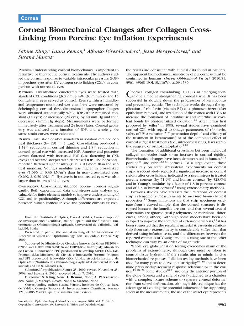
Figure 1 shows a diagram of the experimental setup used to measurecorneal deformation on application of variable intraocular pressure.Intraocular pressure was monitored by means of a water column anda pressure transducer and was modified by infusion of saline solution
FIGURE 1. Schematic setup of the ocular inflation test.
3962 Kling et al. IOVS, August 2010, Vol. 51, No. 8
into the eye through a needle automatically controlled by a pumpingsystem (NE-500; New Era Pump Systems, Inc., Wantagh, NY). Thehumidity and temperature in the moist chamber in which the eyeswere placed during the measurements were also continuously moni-tored and recorded.
Sinusoidal alternating voltage was achieved by constant playing ofa 440-Hz tone from a personal computer sound card amplified to serveas voltage supply for a Wheatstone bridge setup. The pressure sensorwas taken as the variable resistance that measured the current pressureof the water column. Finally, the difference of voltage was measured,amplified again, and fed back onto the microphone entrance of thecomputer. In addition, the Scheimpflug system data acquisition wasautomatized (by automatic control of the keyboard) so that three-dimensional anterior segment images were also recorded without theneed of manual control. The system was controlled by dedicatedsoftware written in computing software (MatLab; MathWorks).
Intraocular pressure was increased from 18.5 to 58.5 mm Hg andthen was decreased to 18.5 mm Hg. Scheimpflug measurements weretaken at 4-mm Hg increments of the IOP increases, as controlled by thepressure monitoring system or every 5 minutes (in the experiments inwhich the IOP was kept constant at 19 mm Hg). Temperature andhumidity were recorded every 2 seconds throughout the experiment.
Experimental Protocols
The enucleated porcine eyes were placed in a custom-made holder toguarantee proper centration during the application of the treatmentand during the measurements. A view of the holder can be seen inFigure 1. The holder consists of two rectangular plastic parts, eachwith an inwrought half-sphere of 20-mm diameter. By regulating thedistance of the plastic parts by means of screws, the holder wasallowed to accommodate the individual porcine eyes. The eyes alwayswere placed with the longer side of the cornea in the vertical direction.Compared with the real orientation of the eye in the pig, in theexperiment the eye was turned 90°, which allowed a better view andmore accurate alignment. (In Results and Discussion, however, we usehorizontal and vertical to refer to the actual orientation of the eye inthe pig.) The eye holder was placed in a methacrylate box in whichwet cotton cloths provided a constantly humid atmosphere. A windowat the front of the box allowed an unrestricted view of the eye by theScheimpflug system.
Saline infusion was produced by introducing a needle through theoptical nerve head into the anterior segment of the eye. The needlewas connected to an infusion tube to a NaCl solution. Preliminarymeasurements were conducted by entering the anterior segment of theeye superiorly, through the sclera, leaving the cornea untouched(Perez-Escudero A, et al. IOVS 2008;49:ARVO E-Abstract 664) How-ever, this procedure was more challenging, and we did not observe anydifference between results.
An entire experimental session on each eye typically took 40minutes. The initial measurement was taken at 18.5 mm Hg andsubsequently at 4-mm Hg step increases. Pressure was increased to58.5 mm Hg and then decreased, at 4-mm Hg steps, with data alsocollected sequentially. Sessions on both eyes of the same animal weretypically performed consecutively and randomly, with one eye havingundergone the cross-linking treatment and the other having undergoneonly epithelial removal and riboflavin instillation. A set of preliminarymeasurements was performed simultaneously increasing the pressurein both eyes and taking consecutive images in each eye at identicalpressure levels. We did not observe any difference between the resultsfrom this procedure and those from automatic full measurements inone eye after the other.
A set of control experiments was performed by keeping the pres-sure constant at 19 mm Hg and taking images at 5-minute intervalsduring the same period as the variable-pressure experiments. Measure-ments were also collected in cross-linked and non–cross-linked eyes.Typically, each eye was measured in two identical experimental ses-sions 24 hours apart
Simple Biomechanical Model: Young’s Modulusand Hysteresis
We have developed a model for the corneal stress-strain relationshipbased on IOP variation and related changes in thickness and radius ofcurvature to provide estimates of Young’s modulus in the untreatedand cross-linked corneas. The model was similar to that used in previ-ous inflation tests,18,19,22,24 though fewer assumptions were requiredbecause the corneal radius of curvature was measured directly in theexperiment (and not necessarily derived from the apical rise, whichwas the only experimental input data available in other studies15,20).
The model assumes that corneal thickness and mean radius ofcurvature are a function of IOP—that is, increasing IOP will make theglobe of the eye expand. Corneal thickness decreases because thecornea becomes stretched over a larger area. However, stretching thecornea induces stress that acts to prevent further corneal extension.Both forces—the IOP tendency to stretch the cornea and the stresstendency to prevent it—arrange to form a stable equilibrium. Withbasic equations for pressure p � F/A (where p represents pressure, Frepresents force, and A represents surface of eye globe) and strain � �F/A (where � represents stress, F represents force, and A representscorneal cross-sectional area), we can express stress as a function ofIOP:
� �R � p
2 � d(1)
where � represents stress; R represents mean radii of anterior, poste-rior, horizontal, and vertical curvature; p represents IOP; and d repre-sents corneal thickness.
The strain on the stress produced by IOP can be expressed by � ��R/R, where � represents strain, �R represents difference in radius ofcurvature with respect to an initial measurement, and R representsradius of curvature.
Limitations of the model include the fact that the mechanicalproperties of the sclera are not considered to contribute to the re-sponse of the cornea to pressure elevation because of the high rigidityand low elasticity of the sclera compared with the cornea.17,19 Ofcourse, this is an approximation because scleral effects cannot betotally neglected. Nevertheless, it appears reasonable as a first estima-tion of stress-strain curves in whole eye globe experiments. Moreover,the stress-strain calculation refers to a homogenous membrane sphere,with the same elasticity, rigidity, and thickness all over the globe.Therefore, the influence of corneal thickness gradient or local elasticityand rigidity differences cannot be detected. In addition, the cornea isassumed to be purely elastic. The model is based on horizontal/verticalaverages of corneal radii changes and does not include potential me-ridional differences in deformation.
RESULTS
Changes in Corneal Thickness
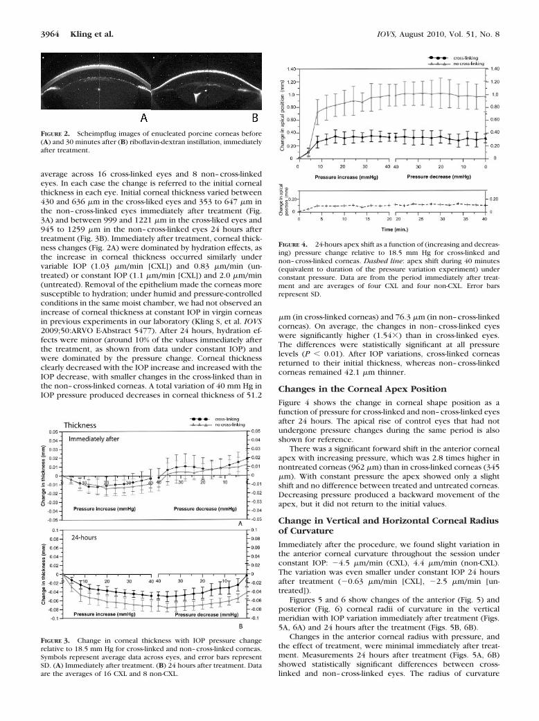
A dramatic decrease in corneal thickness occurred immediatelyafter the application of riboflavin-dextran solution (duration,30 minutes) to the deepithelialized corneas. Figure 2 showsexamples of Scheimpflug images before and after riboflavinapplication. On average, a decrease of 281 � 5 �m was foundafter the application of riboflavin. Another decrease was ob-served 30 minutes later, after UV illumination, which washigher in control eyes (88 �m) than in cross-linked eyes (31�m). After riboflavin instillation, the radius of curvaturechanged by 165 � 655 �m (anterior) and �774 � 1689 �m(posterior).
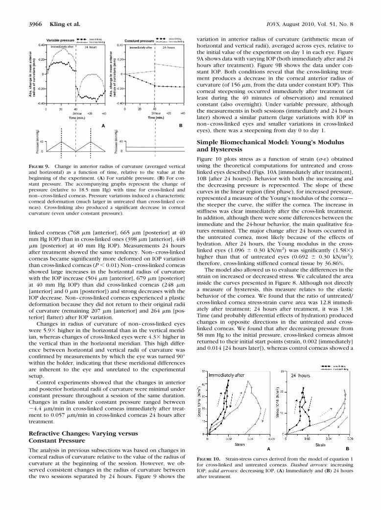
Figure 3 shows the change in corneal thickness as a func-tion of IOP change (first increased from 18.5 mm Hg to 58.5mm Hg, then decreased) immediately after treatment (Fig. 3A)and then 24 hours after treatment (Fig. 3B). Results are the
IOVS, August 2010, Vol. 51, No. 8 Corneal Biomechanics after CXL in Inflation Model 3963
average across 16 cross-linked eyes and 8 non–cross-linkedeyes. In each case the change is referred to the initial cornealthickness in each eye. Initial corneal thickness varied between430 and 636 �m in the cross-liked eyes and 353 to 647 �m inthe non–cross-linked eyes immediately after treatment (Fig.3A) and between 999 and 1221 �m in the cross-liked eyes and945 to 1259 �m in the non–cross-linked eyes 24 hours aftertreatment (Fig. 3B). Immediately after treatment, corneal thick-ness changes (Fig. 2A) were dominated by hydration effects, asthe increase in corneal thickness occurred similarly undervariable IOP (1.03 �m/min [CXL]) and 0.83 �m/min (un-treated) or constant IOP (1.1 �m/min [CXL]) and 2.0 �m/min(untreated). Removal of the epithelium made the corneas moresusceptible to hydration; under humid and pressure-controlledconditions in the same moist chamber, we had not observed anincrease of corneal thickness at constant IOP in virgin corneasin previous experiments in our laboratory (Kling S, et al. IOVS2009;50:ARVO E-Abstract 5477). After 24 hours, hydration ef-fects were minor (around 10% of the values immediately afterthe treatment, as shown from data under constant IOP) andwere dominated by the pressure change. Corneal thicknessclearly decreased with the IOP increase and increased with theIOP decrease, with smaller changes in the cross-linked than inthe non–cross-linked corneas. A total variation of 40 mm Hg inIOP pressure produced decreases in corneal thickness of 51.2
�m (in cross-linked corneas) and 76.3 �m (in non–cross-linkedcorneas). On average, the changes in non–cross-linked eyeswere significantly higher (1.54�) than in cross-linked eyes.The differences were statistically significant at all pressurelevels (P � 0.01). After IOP variations, cross-linked corneasreturned to their initial thickness, whereas non–cross-linkedcorneas remained 42.1 �m thinner.
Changes in the Corneal Apex Position
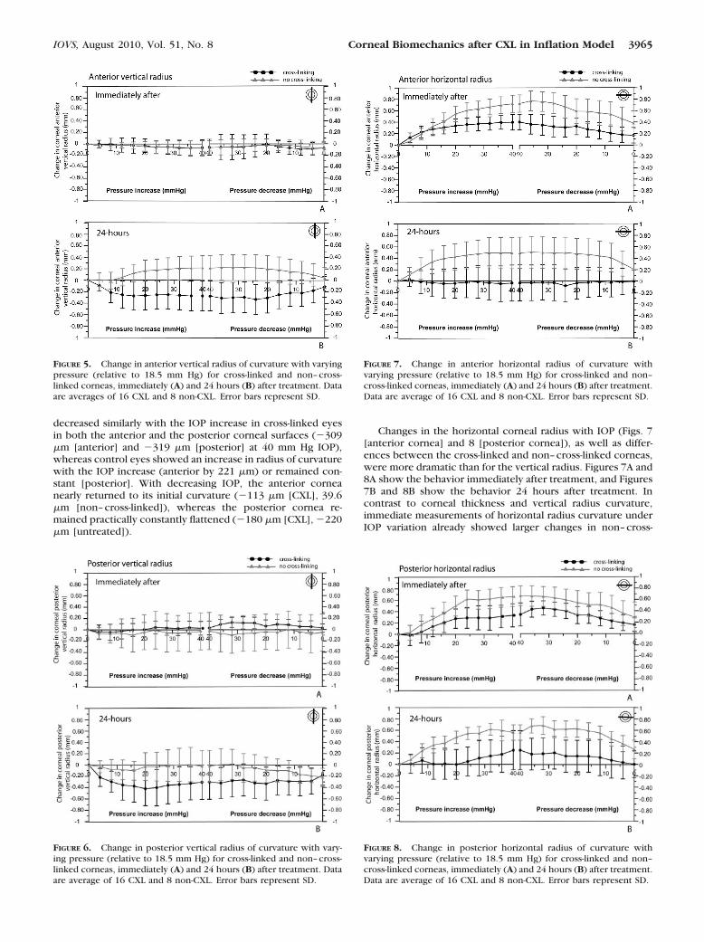
Figure 4 shows the change in corneal shape position as afunction of pressure for cross-linked and non–cross-linked eyesafter 24 hours. The apical rise of control eyes that had notundergone pressure changes during the same period is alsoshown for reference.
There was a significant forward shift in the anterior cornealapex with increasing pressure, which was 2.8 times higher innontreated corneas (962 �m) than in cross-linked corneas (345�m). With constant pressure the apex showed only a slightshift and no difference between treated and untreated corneas.Decreasing pressure produced a backward movement of theapex, but it did not return to the initial values.
Change in Vertical and Horizontal Corneal Radiusof Curvature
Immediately after the procedure, we found slight variation inthe anterior corneal curvature throughout the session underconstant IOP: �4.5 �m/min (CXL), 4.4 �m/min (non-CXL).The variation was even smaller under constant IOP 24 hoursafter treatment (�0.63 �m/min [CXL], �2.5 �m/min [un-treated]).
Figures 5 and 6 show changes of the anterior (Fig. 5) andposterior (Fig. 6) corneal radii of curvature in the verticalmeridian with IOP variation immediately after treatment (Figs.5A, 6A) and 24 hours after the treatment (Figs. 5B, 6B).
Changes in the anterior corneal radius with pressure, andthe effect of treatment, were minimal immediately after treat-ment. Measurements 24 hours after treatment (Figs. 5A, 6B)showed statistically significant differences between cross-linked and non–cross-linked eyes. The radius of curvature
FIGURE 2. Scheimpflug images of enucleated porcine corneas before(A) and 30 minutes after (B) riboflavin-dextran instillation, immediatelyafter treatment.
FIGURE 3. Change in corneal thickness with IOP pressure changerelative to 18.5 mm Hg for cross-linked and non–cross-linked corneas.Symbols represent average data across eyes, and error bars representSD. (A) Immediately after treatment. (B) 24 hours after treatment. Dataare the averages of 16 CXL and 8 non-CXL.
FIGURE 4. 24-hours apex shift as a function of (increasing and decreas-ing) pressure change relative to 18.5 mm Hg for cross-linked andnon–cross-linked corneas. Dashed line: apex shift during 40 minutes(equivalent to duration of the pressure variation experiment) underconstant pressure. Data are from the period immediately after treat-ment and are averages of four CXL and four non-CXL. Error barsrepresent SD.
3964 Kling et al. IOVS, August 2010, Vol. 51, No. 8
decreased similarly with the IOP increase in cross-linked eyesin both the anterior and the posterior corneal surfaces (�309�m [anterior] and �319 �m [posterior] at 40 mm Hg IOP),whereas control eyes showed an increase in radius of curvaturewith the IOP increase (anterior by 221 �m) or remained con-stant [posterior]. With decreasing IOP, the anterior corneanearly returned to its initial curvature (�113 �m [CXL], 39.6�m [non–cross-linked]), whereas the posterior cornea re-mained practically constantly flattened (�180 �m [CXL], �220�m [untreated]).
Changes in the horizontal corneal radius with IOP (Figs. 7[anterior cornea] and 8 [posterior cornea]), as well as differ-ences between the cross-linked and non–cross-linked corneas,were more dramatic than for the vertical radius. Figures 7A and8A show the behavior immediately after treatment, and Figures7B and 8B show the behavior 24 hours after treatment. Incontrast to corneal thickness and vertical radius curvature,immediate measurements of horizontal radius curvature underIOP variation already showed larger changes in non–cross-
FIGURE 5. Change in anterior vertical radius of curvature with varyingpressure (relative to 18.5 mm Hg) for cross-linked and non–cross-linked corneas, immediately (A) and 24 hours (B) after treatment. Dataare averages of 16 CXL and 8 non-CXL. Error bars represent SD.
FIGURE 6. Change in posterior vertical radius of curvature with vary-ing pressure (relative to 18.5 mm Hg) for cross-linked and non–cross-linked corneas, immediately (A) and 24 hours (B) after treatment. Dataare average of 16 CXL and 8 non-CXL. Error bars represent SD.
FIGURE 7. Change in anterior horizontal radius of curvature withvarying pressure (relative to 18.5 mm Hg) for cross-linked and non–cross-linked corneas, immediately (A) and 24 hours (B) after treatment.Data are average of 16 CXL and 8 non-CXL. Error bars represent SD.
FIGURE 8. Change in posterior horizontal radius of curvature withvarying pressure (relative to 18.5 mm Hg) for cross-linked and non–cross-linked corneas, immediately (A) and 24 hours (B) after treatment.Data are average of 16 CXL and 8 non-CXL. Error bars represent SD.
IOVS, August 2010, Vol. 51, No. 8 Corneal Biomechanics after CXL in Inflation Model 3965
linked corneas (768 �m [anterior], 665 �m [posterior] at 40mm Hg IOP) than in cross-linked ones (398 �m [anterior], 448�m [posterior] at 40 mm Hg IOP). Measurements 24 hoursafter treatment showed the same tendency. Non–cross-linkedcorneas became significantly more deformed on IOP variationthan cross-linked corneas (P � 0.01) Non–cross-linked corneasshowed large increases in the horizontal radius of curvaturewith the IOP increase (504 �m [anterior], 679 �m [posterior]at 40 mm Hg IOP) than did cross-linked corneas (248 �m[anterior] and 0 �m [posterior]) and strong decreases with theIOP decrease. Non–cross-linked corneas experienced a plasticdeformation because they did not return to their original radiiof curvature (remaining 207 �m [anterior] and 264 �m [pos-terior] flatter) after IOP variation.
Changes in radius of curvature of non–cross-linked eyeswere 5.9� higher in the horizontal than in the vertical merid-ian, whereas changes of cross-linked eyes were 4.3� higher inthe vertical than in the horizontal meridian. This high differ-ence between horizontal and vertical radii of curvature wasconfirmed by measurements by which the eye was turned 90°within the holder, indicating that these meridional differencesare inherent to the eye and unrelated to the experimentalsetup.
Control experiments showed that the changes in anteriorand posterior horizontal radii of curvature were minimal underconstant pressure throughout a session of the same duration.Changes in radius under constant pressure ranged between�4.4 �m/min in cross-linked corneas immediately after treat-ment to 0.057 �m/min in cross-linked corneas 24 hours aftertreatment.
Refractive Changes: Varying versusConstant Pressure
The analysis in previous subsections was based on changes incorneal radius of curvature relative to the value of the radius ofcurvature at the beginning of the session. However, we ob-served consistent changes in the radius of curvature betweenthe two sessions separated by 24 hours. Figure 9 shows the
variation in anterior radius of curvature (arithmetic mean ofhorizontal and vertical radii), averaged across eyes, relative tothe initial value of the experiment on day 1 in each eye. Figure9A shows data with varying IOP (both immediately after and 24hours after treatment). Figure 9B shows the data under con-stant IOP. Both conditions reveal that the cross-linking treat-ment produces a decrease in the corneal anterior radius ofcurvature (of 156 �m, from the data under constant IOP). Thiscorneal steepening occurred immediately after treatment (atleast during the 40 minutes of observation) and remainedconstant (also overnight). Under variable pressure, althoughthe measurements in both sessions (immediately and 24 hourslater) showed a similar pattern (large variations with IOP innon–cross-linked eyes and smaller variations in cross-linkedeyes), there was a steepening from day 0 to day 1.
Simple Biomechanical Model: Young’s Modulusand Hysteresis
Figure 10 plots stress as a function of strain (�-�) obtainedusing the theoretical computations for untreated and cross-linked eyes described (Figs. 10A [immediately after treatment],10B [after 24 hours]). Behavior with both the increasing andthe decreasing pressure is represented. The slope of thesecurves in the linear region (first phase), for increased pressure,represented a measure of the Young’s modulus of the cornea—the steeper the curve, the stiffer the cornea. The increase instiffness was clear immediately after the cross-link treatment.In addition, although there were some differences between theimmediate and the 24-hour behavior, the main qualitative fea-tures remained. The major change after 24 hours occurred inthe untreated cornea, most likely because of the effects ofhydration. After 24 hours, the Young modulus in the cross-linked eyes (1.096 � 0.30 kN/m2) was significantly (1.58�)higher than that of untreated eyes (0.692 � 0.30 kN/m2);therefore, cross-linking stiffened corneal tissue by 36.86%.
The model also allowed us to evaluate the differences in thestrain on increased or decreased stress. We calculated the areainside the curves presented in Figure 8. Although not directlya measure of hysteresis, this measure relates to the elasticbehavior of the cornea. We found that the ratio of untreated/cross-linked cornea stress-strain curve area was 12.8 immedi-ately after treatment; 24 hours after treatment, it was 1.38.Time (and probably differential effects of hydration) producedchanges in opposite directions in the untreated and cross-linked corneas. We found that after decreasing pressure from58 mm Hg to the initial pressure, cross-linked corneas almostreturned to their initial start points (strain, 0.002 [immediately]and 0.014 [24 hours later]), whereas control corneas showed a
FIGURE 10. Strain-stress curves derived from the model of equation 1for cross-linked and untreated corneas. Dashed arrows: increasingIOP; solid arrows: decreasing IOP. (A) Immediately and (B) 24 hoursafter treatment.
FIGURE 9. Change in anterior radius of curvature (averaged verticaland horizontal) as a function of time, relative to the value at thebeginning of the experiment. (A) For variable pressure. (B) For con-stant pressure. The accompanying graphs represent the change ofpressure (relative to 18.5 mm Hg) with time for cross-linked andnon–cross-linked corneas. Pressure variations induced a characteristiccorneal deformation (much larger in untreated than cross-linked cor-neas). Cross-linking also produced a significant decrease in cornealcurvature (even under constant pressure).
3966 Kling et al. IOVS, August 2010, Vol. 51, No. 8
large hysteresis and ended at higher strain (0.036 [immedi-ately] and 0.032 (24 hours later). We conclude that cross-linked corneas were more elastic (although stiffness is higher)and did not show any remaining plastic deformations, in con-trast to control eyes.
DISCUSSION
Comparison with Previous Clinical andExperimental Data
The first major effect found during treatment is the decrease ofcorneal thickness after riboflavin instillation, probably causedby the hyperosmolarity of the riboflavin-dextran solution. Wehave observed that this has a strong dehydrating effect on theporcine cornea, which is reduced in thickness by 54%. De-creased corneal thickness in patients has been reported re-cently in a clinical study31 in which corneal pachymetry wasmonitored with Scheimpflug imaging immediately after ribo-flavin instillation. On average, the corneal thickness decreasedby 18.7%, which may pose some safety concerns in thin cor-neas. Alternative hyposmolar riboflavin solution has been pro-posed to produce corneal swelling rather than dehydration toincrease the effective corneal thickness.32 We observed anincrease in corneal thickness caused by swelling 24 hours aftertreatment, as reported in previous studies on porcine corneas.7
However, we did not find that corneal hydration interferedwith the corneal response to pressure variation 24 hours aftertreatment or that swelling effects were different between un-treated and cross-linked corneas.
Even at constant pressure, we found a decrease in thecorneal radius of curvature 24 hours after cross-linking (166�m). This is consistent with clinical reports of corneal steep-ening during the first month after treatment.33 After the initialsteepening and 6 months after treatment, an average decreasein corneal curvature (flattening) of 2 D and a decrease inrefractive error of 1.14 D34 have been reported clinically. Ourresults show effects of cross-linking immediately and 24 hoursafter treatment. The fact that refractive changes are observedin patients during the 6 months after treatment suggests wouldhealing or long-term action balancing forces.
Previous inflation studies on excised porcine corneasshowed a rise in the apex as a function of increased IOP verysimilar to our observations in untreated corneas (Fig. 4). As didElsheiihk and Anderson,15 we found an initial phase (0–24 mmHg) during which the corneal apex rises with pressure at a lowstiffness (relatively high slope in Fig. 4), attributed to a relativelooseness of the collagen fibers, followed by a much increasedstiffness in the second phase, during which a significant in-crease in pressure results in a very small apical rise, attributedto the collagen fibers becoming taut. Previous studies usedtrephinate specimens (cornea and a narrow ring of scleraltissue) attached to a wet chamber rather than the whole globeused in the present study. The higher relative apical rise foundin our study may result from height changes in the limbusrather than from the mechanical rigid attachment in the trephi-nate experiments. The limited lateral range of the Scheimpflugcamera prevented us from accounting for the peripheral for-ward shift. Cross-linking reduced the apical rise by a factor of2.8 in untreated corneas. Most notably, in cross-linked corneas,the second phase was reached at significantly lower loads (15mm Hg), so that the regimen at physiological IOP was domi-nated by collagen with a much higher stiffness.
Although we cannot rule out a contribution of the entireeye motion to the apical radius displacement, the behavior wefound in corneal apical shift was consistent with the changes incorneal thickness. Non–cross-linked corneas did not fully re-turn to the initial values after increased and decreased IOP with
regard to apical position or corneal thickness. Cross-linkedcorneas tended to return to the original values both for apicalposition (within the measurement variability) and for cornealthickness. These results are consistent with the cross-linkedcornea being stiffer (but with a more elastic behavior) than thenon–cross-linked cornea.
A previous study35 reported the decrease in central cornealthickness on globe inflation in two virgin human corneas invitro using OCT (120 �m for a 206-mm Hg pressure increase).In that study, the posterior cornea was visible only in twocorneas, and no data were provided for lower pressures. Wefound an average decrease of 73.6 �m for an increase of 40 mmHg in porcine corneas (average of 16 eyes). Cross-linking pro-duced a reduction in the corneal thickness change by a factorof 2.28, consistent with an increase in corneal stiffness. Theincrease in the modulus of porcine corneal elasticity after CXLestimated from a simple model compares well with reports (afactor of 1.8�) from corneal strip extensometry.14 The abso-lute values obtained from inflation tests are lower than thoseobtained from corneal strip extensometry.
We measured changes in anterior and posterior cornealradii of curvature as a function of IOP variation (Figs. 5–8).Changes in the posterior cornea tend to parallel the changes inthe anterior cornea. We found highly significant differencesbetween untreated and cross-linked corneas, consistent withan increase in corneal stiffness, particularly in the horizontaldirection. We found high meridional differences both in theeffect of IOP on corneal curvature and in the effect of thecross-linking treatment. In untreated corneas, an increase of 40mm Hg produced 8.26 times more increase in the horizontalthan in the vertical anterior radius of curvature. The verticalposterior radius was nearly not altered by IOP variations,whereas the horizontal posterior radius changed by 500 �m inthe 0- to 40-mm Hg range. This result is in contrast to previouscorneal strip extensometry experiments on specimens cutalong the vertical or horizontal meridians of fresh porcinecorneas, which showed the same stress-strain responses alongthe vertical and horizontal directions and only marginally stifferresponses (2%–8%) diagonally.36 Microstructural studies of thefibrillar collagen structure in the porcine cornea show a pri-marily circumferential orientation. Other species, such ashorses, cows, and marmosets, have a primarily vertical orien-tation in the collagen fibrils; in humans, the stromal fibrils havea preferential orientation in the superior-inferior and temporal-nasal direction.37,38 The orientation-dependent corneal defor-mation found in our whole globe inflation experiments sug-gests that factors beyond the orientation of collagen fibrils playa role in the anisotropy of the biomechanical response.
Cross-linking was more effective in preventing an increasein anterior corneal radius (both anterior and posterior, imme-diately and 24 hours afterward) in the horizontal than in thevertical meridian. In fact, we found some vertical steepeningwith IOP in cross-linked corneas. The inherent astigmatism ofthe porcine eye (on average, the vertical meridian was 2.19%flatter than the vertical meridian) may play a role in the effec-tive UV-irradiation treatment area (and, therefore, the relativeexposure). However, differences of 710 �m in the treatmentarea are not expected to result in these meridional differencesin stiffness. Structural differences in the vertical and horizontalfibrils and a differential response to the formation of cross-linking bonds may be responsible for the observed meridionaldifferences in the response to cross-linking.
Implications of the Results
We have provided experimental data of the change in apexshift, corneal thickness, and anterior and posterior cornealradii of curvature in a porcine eye inflation model after corneal
IOVS, August 2010, Vol. 51, No. 8 Corneal Biomechanics after CXL in Inflation Model 3967
CXL (and control eyes after epithelium removal and riboflavininstillation but not after UV-A irradiation). Although differencesare expected on human corneas (particularly because of therelatively larger corneal cross-section treated) and in vivo, thecomplete experimental data set, along with estimations of theYoung’s modulus, are valuable input parameters in finite ele-ment models that will allow a better understanding and in-creased predictability of the cross-linking technique. To build amore sophisticated model with fewer limitations, we are plan-ning further experiments with a high-resolution OCT and alarger lateral range that will allow viewing of limbal and scleralregions.
We have related some experimental findings on porcinecorneas in vitro to clinical data found in patients. The use of asimilar methodology on human corneal specimens, along withparameters obtained in vivo (particularly in eyes with cornealdisorders susceptible to treatment) will provide further insightinto the mechanism of cross-linking.
Open questions such as the apparent anisotropy in thebiomechanical response of the porcine intact cornea and inresponse to treatment, yet to be confirmed in humans and ofparticular interest in pathologic corneas, require combinedstudies of microstructural analysis of the collagen fibers andbiomechanical approaches.
Acknowledgments
The authors thank Daniel Pascual and Carlos Meneses for technicalsupport and Michael Mrochen and Michael Bueler for initial discus-sions.
References
1. Wollensak G, Spoerl E, Seiler T. Riboflavin/ultraviolet-a-inducedcollagen crosslinking for the treatment of keratoconus. Am J Oph-thalmol. 2003;135(5):620–627.
2. Wollensak G, Wilsch M, Spoerl E, Seiler T. Collagen fiber diameterin the rabbit cornea after collagen crosslinking by riboflavin/UV-A.Cornea. 2004;23(5):503–507.
3. Spoerl E, Huhle M, Seiler T. Induction of cross-links in cornealtissue Exp Eye Res. 1998;66(1):97–103.
4. Wollensak G, Spoerl E, Wilsch M, Seiler T. Endothelial cell damageafter riboflavin-ultraviolet-A treatment in the rabbit. J CataractRefract Surg. 2003;29(9):1786–1790.
5. Wollensak G, Spoerl E, Wilsch M, Seiler T. Keratocyte apoptosisafter corneal collagen cross-linking using riboflavin/UV-A treat-ment. Cornea. 2004;23(1):43–49.
6. Spoerl E, Mrochen M, Sliney D, Trokel S, Seiler T. Safety of UV-A-riboflavin cross-linking of the cornea. Cornea. 2007;26(4):385–389.
7. Wollensak G, Aurich H, Pham DT, Wirbelbauer C. Hydration be-havior of porcine cornea crosslinked with riboflavin and ultravioletA. J Cataract Refract Surg. 2007;33(3):516–521.
8. Wollensak G. Crosslinkining treatment of progressive keratoconus:new hope. Curr Opin Ophthalmol. 2006;17(4):356–360.
9. Hafezi F, Kanellopoulos J, Wiltfang R, Seiler T. Corneal collagencrosslinking with riboflavin and ultraviolet A to treat inducedkeratectasia after laser in situ keratomileusis. J Cataract RefractSurg. 2007;33(12):2035–2040.
10. Ertan A, Colin J. Intracorneal rings for keratoconus and keratecta-sia. J Cataract Refract Surg. 2007;33(7):1303–1314.
11. Kohlhaas M, Spoerl E, Schilde T, Unger G, Wittig C, Pillunat LE.Biomechanical evidence of the distribution of cross-links in corne-astreated with riboflavin and ultraviolet A light. J Cataract RefractSurg. 2006;32(2):279–283.
12. Wollensak G, Iomdina E. Long-term biomechanical properties ofrabbit cornea after photodynamic collagen crosslinking. Acta Oph-thalmol. 2009;87(1):48–51.
13. Sporl E, Schreiber J, Hellmund K, et al. Studies on the stabilizationof the cornea in rabbits [in German]. Ophthalmologe. 2000;97(3):203–206.
14. Wollensak G, Spoerl E, Seiler T. Stress-strain measurements ofhuman and porcine corneas after riboflavin-ultraviolet-A-inducedcross-linking. J Cataract Refract Surg. 2003;29(9):1780–1785.
15. Elsheikh A, Anderson K. Comparative study of corneal strip and exten-sometry and inflation tests. J R Soc Interface. 2005;2(3):177–185.
16. Pallikaris IG, Kymionis GD, Ginis HS, et al. Ocular rigidity in livinghuman eyes. Invest Ophthalmol Vis Sci. 2005;46(2):409–414.
17. Asejczyk-Widlicka M, Pierscionek BK. The elasticity and rigidity of theouter coats of the eye. Br J Ophthalmol. 2008;92(10):1415–1418.
18. Jue B, Maurice DM. The mechanical properties of the rabbit andhuman cornea. J Biomech. 1986;19(10):847–853.
19. Hjortdal J. Regional elastic performance of the human cornea.J Biomech. 1996;29(7):931–942.
20. Anderson K, Elsheikh A, Newson T. Application of structuralanalysis to the mechanical behaviour of the cornea. J R Soc Inter-face. 2004;1:3–15.
21. Boyce BL, Grazier JM, Jones RE, Nguyen TD. Full-field deformationof bovine cornea under constrained inflation conditions. Bioma-terials. 2008;29:3896–3904.
22. Hennighausen H, Feldman ST, Bille JF, McCulloch AD. Anterior-posterior strain variation in normally hydrated and swollen rabbitcornea. Invest Ophthalmol Vis Sci. 1998;39(2):253–262.
23. Woo SL-Y, Kobayashi AS, Schlegel WA, Lawrence C. Nonlinearmaterial properties of intact cornea and sclera. Exp Eye Res.1972;14(1):29–39.
24. Shin TJ, Vito RP, Johnson LW, McCarey BE. The distribution ofstrain in the human cornea. J Biomech. 1997;30(5):497–503.
25. Srodka W, Iskander DR. Optically inspired biomechanical model ofthe human eyeball. J Biomed Opt. 2008;13(4):044034.
26. Elsheikh A, Alhasso D, Rama P. Biomechanical properties of hu-man and porcine corneas. Exp Eye Res. 2008;86(5):783–790.
27. Zeng Y, Yang J, Huang K, Lee Z, Lee X. A comparison of biome-chanical properties between human and porcine cornea. J Bio-mech. 2001;34(4):533–537.
28. Rosales P, Marcos S. Quantitative Scheimpflug imaging of the crystal-line and intraocular lens. J Refract Surg. 2009;25(5):421–428.
29. Perez-Escudero A, Dorronsoro C, Sawides L, Remon L, Merayo-Lloves J, Marcos S. Minor influence of myopic laser in situ kerato-mileusis on the posterior corneal surface. Invest Ophthalmol VisSci. 2009;50(9):4146–4154.
30. Cano D, Barbero S, Marcos S. Comparison of real and computer-simu-lated outcomes of LASIK refractive surgery. JOSA A. 2004;21:926–936.
31. Kymionis GD, Kounis GA, Portaliou DM, et al. Intraoperativepachymetry measurements during corneal collagen cross-linkingwith riboflavin and ultraviolet A Irradiation. Ophthalmology. 2009;116(12):2336–2339.
32. Hafezi F, Mrochen M, Iseli HP, Seiler T. Collagen crosslinking withultraviolet-A and hypoosmolar riboflavin solution in thin corneas. JCataract Refract Surg. 2009;35(4):621–624.
33. Doors M, Tahzib NG, Eggink FA, Berendschot TTJM, Webers CAB,Nuijts RMMA. Use of anterior segment optical coherence tomog-raphy to study corneal changes after collagen cross-linking. Am JOphthalmol. 2009;148(6):844–851
34. Vinciguerra P, Albe E, Trazza S, et al. Refractive, topographic, tomo-graphic and aberrometric analysis of keratoconic eyes undergoingcorneal cross-linking. Ophthalmology. 2009;116(3):369–378.
35. Johnson CS, Mian SI, Moroi S, Epstein D, Izatt J, Afshari NA. Roleof corneal elasticity in damping of intraocular pressure. InvestOphthalmol Vis Sci. 2007;48(6):2540–2544.
36. Elsheikh A, Alhasso D. Mechanical anisotropy of porcine corneaand correlation with stromal microstructure. Exp Eye Res. 2009;88(6):1084–1091.
37. Hayes S, Boote C, Lewis J, et al. Comparative study of fibrillarcollagen arrangement in the corneas of primates and other mam-mals. Anat Rec. 2007;290(12):1542–1550.
38. Meek KM, Newton R. Organization of collagen fibrils in the cor-neal stroma in relation to mechanical properties and surgicalpractice. J Refract Surg 1999;15(6):695–699.
3968 Kling et al. IOVS, August 2010, Vol. 51, No. 8
Related Documents