Coral Pathogens Identified for White Syndrome (WS) Epizootics in the Indo-Pacific Meir Sussman 1,2 *, Bette L. Willis 1 , Steven Victor 3 , David G. Bourne 2 1 ARC Centre of Excellence for Coral Reef Studies, School of Marine and Tropical Biology, James Cook University, Townsville, Australia, 2 Australian Institute of Marine Science (AIMS), PMB3, Townsville MC, Townsville, Australia, 3 Palau International Coral Reef Center (PICRC), Koror, Republic of Palau Abstract Background: White Syndrome (WS), a general term for scleractinian coral diseases with acute signs of advancing tissue lesions often resulting in total colony mortality, has been reported from numerous locations throughout the Indo-Pacific, constituting a growing threat to coral reef ecosystems. Methodology/Principal Findings: Bacterial isolates were obtained from corals displaying disease signs at three WS outbreak sites: Nikko Bay in the Republic of Palau, Nelly Bay in the central Great Barrier Reef (GBR) and Majuro Atoll in the Republic of the Marshall Islands, and used in laboratory-based infection trials to satisfy Henle-Koch’s postulates, Evan’s rules and Hill’s criteria for establishing causality. Infected colonies produced similar signs to those observed in the field following exposure to bacterial concentrations of 1 6 10 6 cells ml 21 . Phylogenetic 16S rRNA gene analysis demonstrated that all six pathogens identified in this study were members of the c-Proteobacteria family Vibrionacae, each with greater than 98% sequence identity with the previously characterized coral bleaching pathogen Vibrio coralliilyticus. Screening for proteolytic activity of more than 150 coral derived bacterial isolates by a biochemical assay and specific primers for a Vibrio family zinc- metalloprotease demonstrated a significant association between the presence of isolates capable of proteolytic activity and observed disease signs. Conclusion/Significance: This is the first study to provide evidence for the involvement of a unique taxonomic group of bacterial pathogens in the aetiology of Indo-Pacific coral diseases affecting multiple coral species at multiple locations. Results from this study strongly suggest the need for further investigation of bacterial proteolytic enzymes as possible virulence factors involved in Vibrio associated acute coral infections. Citation: Sussman M, Willis BL, Victor S, Bourne DG (2008) Coral Pathogens Identified for White Syndrome (WS) Epizootics in the Indo-Pacific. PLoS ONE 3(6): e2393. doi:10.1371/journal.pone.0002393 Editor: Niyaz Ahmed, Centre for DNA Fingerprinting and Diagnostics, India Received March 4, 2008; Accepted April 2, 2008; Published June 18, 2008 Copyright: ß 2008 Sussman et al. This is an open-access article distributed under the terms of the Creative Commons Attribution License, which permits unrestricted use, distribution, and reproduction in any medium, provided the original author and source are credited. Funding: This study was funded by an ARC Discovery grant and a James Cook University CRIG grant to BL Willis. The authors wish to thank CD Harvell, Chair of the World Bank Coral Disease Working Group, L Raymundo, University of Guam, and A Hooten from the IOC-GEF/World Bank Coral Reef Targeted Research and Capacity Building Program for contributing funding and support for travel and field work in Palau. Work on the research vessel Lady Basten and in the PC2 laboratory at Cape Ferguson was funded by the Australian Institute of Marine Science. Competing Interests: The authors have declared that no competing interests exist. * E-mail: [email protected] Introduction Reports on coral disease continue to rise [ 1] with currently 29 reported syndromes in the Caribbean [ 2] and 7 syndromes reported from the Indo-Pacific [ 3]. However, the causes for coral disease and the methods by which to investigate them are still heavily debated [ 4–6]. Most efforts are directed towards traditional surveillance [ 7], with comparatively less research directed towards developing strategies for active engagement in coral reef health management, disease prevention and cure [ 8– 10]. Unfortunately, a lack of knowledge of coral disease causative agents propels this debate to a stand still. To date, only 5 bacterial species and one fungal agent have been determined as causative agents for coral infectious diseases [ 11–17], and currently no diagnostic tools or management efforts are able to validate these findings at a level required for active intervention. [ 18–19]. The study of disease in complex environmental settings is often difficult. Modern studies have cast a shadow on traditional culturing methods that are required to satisfy Henle-Koch’s postulates [ 20], namely that a putative pathogen is first isolated on growth medium and then used in pure culture to duplicate disease signs in laboratory controlled infections. In many cases, more than 200 years after Henle-Koch’s own revolution, these experiments often fail, requiring the introduction of modern rules and criteria in order to establish disease causation [ 21–22]. These are often based on statistical associations rather than on ‘‘cause and effect’’. Most microorganisms cannot be easily cultured [ 23] and other disease components, namely host susceptibility and environmental factors may jointly contribute to successful infections in what is known as the ‘‘disease triad’’ [ 24]. To this end, modern diagnostic tools have been developed that can be applied to enhance our knowledge of coral disease without targeting either a single or a cultivable agent. These tools include cloning and denaturing gradient gel electrophoresis [25], fluorescent in situ hybridization [26], microarrays [27] and metagenomics [28–30], just to name a few, and are used to either detect new pathogens or validate their PLoS ONE | www.plosone.org 1 June 2008 | Volume 3 | Issue 6 | e2393

Welcome message from author
This document is posted to help you gain knowledge. Please leave a comment to let me know what you think about it! Share it to your friends and learn new things together.
Transcript

Coral Pathogens Identified for White Syndrome (WS)Epizootics in the Indo-PacificMeir Sussman1,2*, Bette L. Willis1, Steven Victor3, David G. Bourne2
1 ARC Centre of Excellence for Coral Reef Studies, School of Marine and Tropical Biology, James Cook University, Townsville, Australia, 2 Australian Institute of Marine
Science (AIMS), PMB3, Townsville MC, Townsville, Australia, 3 Palau International Coral Reef Center (PICRC), Koror, Republic of Palau
Abstract
Background: White Syndrome (WS), a general term for scleractinian coral diseases with acute signs of advancing tissuelesions often resulting in total colony mortality, has been reported from numerous locations throughout the Indo-Pacific,constituting a growing threat to coral reef ecosystems.
Methodology/Principal Findings: Bacterial isolates were obtained from corals displaying disease signs at three WSoutbreak sites: Nikko Bay in the Republic of Palau, Nelly Bay in the central Great Barrier Reef (GBR) and Majuro Atoll in theRepublic of the Marshall Islands, and used in laboratory-based infection trials to satisfy Henle-Koch’s postulates, Evan’s rulesand Hill’s criteria for establishing causality. Infected colonies produced similar signs to those observed in the field followingexposure to bacterial concentrations of 16106 cells ml21. Phylogenetic 16S rRNA gene analysis demonstrated that all sixpathogens identified in this study were members of the c-Proteobacteria family Vibrionacae, each with greater than 98%sequence identity with the previously characterized coral bleaching pathogen Vibrio coralliilyticus. Screening for proteolyticactivity of more than 150 coral derived bacterial isolates by a biochemical assay and specific primers for a Vibrio family zinc-metalloprotease demonstrated a significant association between the presence of isolates capable of proteolytic activity andobserved disease signs.
Conclusion/Significance: This is the first study to provide evidence for the involvement of a unique taxonomic group ofbacterial pathogens in the aetiology of Indo-Pacific coral diseases affecting multiple coral species at multiple locations.Results from this study strongly suggest the need for further investigation of bacterial proteolytic enzymes as possiblevirulence factors involved in Vibrio associated acute coral infections.
Citation: Sussman M, Willis BL, Victor S, Bourne DG (2008) Coral Pathogens Identified for White Syndrome (WS) Epizootics in the Indo-Pacific. PLoS ONE 3(6):e2393. doi:10.1371/journal.pone.0002393
Editor: Niyaz Ahmed, Centre for DNA Fingerprinting and Diagnostics, India
Received March 4, 2008; Accepted April 2, 2008; Published June 18, 2008
Copyright: � 2008 Sussman et al. This is an open-access article distributed under the terms of the Creative Commons Attribution License, which permitsunrestricted use, distribution, and reproduction in any medium, provided the original author and source are credited.
Funding: This study was funded by an ARC Discovery grant and a James Cook University CRIG grant to BL Willis. The authors wish to thank CD Harvell, Chair ofthe World Bank Coral Disease Working Group, L Raymundo, University of Guam, and A Hooten from the IOC-GEF/World Bank Coral Reef Targeted Research andCapacity Building Program for contributing funding and support for travel and field work in Palau. Work on the research vessel Lady Basten and in the PC2laboratory at Cape Ferguson was funded by the Australian Institute of Marine Science.
Competing Interests: The authors have declared that no competing interests exist.
* E-mail: [email protected]
Introduction
Reports on coral disease continue to rise [1] with currently 29
reported syndromes in the Caribbean [2] and 7 syndromes
reported from the Indo-Pacific [3]. However, the causes for coral
disease and the methods by which to investigate them are still
heavily debated [4–6]. Most efforts are directed towards
traditional surveillance [7], with comparatively less research
directed towards developing strategies for active engagement in
coral reef health management, disease prevention and cure [8–
10]. Unfortunately, a lack of knowledge of coral disease causative
agents propels this debate to a stand still. To date, only 5 bacterial
species and one fungal agent have been determined as causative
agents for coral infectious diseases [11–17], and currently no
diagnostic tools or management efforts are able to validate these
findings at a level required for active intervention. [18–19].
The study of disease in complex environmental settings is often
difficult. Modern studies have cast a shadow on traditional
culturing methods that are required to satisfy Henle-Koch’s
postulates [20], namely that a putative pathogen is first isolated on
growth medium and then used in pure culture to duplicate disease
signs in laboratory controlled infections. In many cases, more than
200 years after Henle-Koch’s own revolution, these experiments
often fail, requiring the introduction of modern rules and criteria
in order to establish disease causation [21–22]. These are often
based on statistical associations rather than on ‘‘cause and effect’’.
Most microorganisms cannot be easily cultured [23] and other
disease components, namely host susceptibility and environmental
factors may jointly contribute to successful infections in what is
known as the ‘‘disease triad’’ [24]. To this end, modern diagnostic
tools have been developed that can be applied to enhance our
knowledge of coral disease without targeting either a single or a
cultivable agent. These tools include cloning and denaturing
gradient gel electrophoresis [25], fluorescent in situ hybridization
[26], microarrays [27] and metagenomics [28–30], just to name a
few, and are used to either detect new pathogens or validate their
PLoS ONE | www.plosone.org 1 June 2008 | Volume 3 | Issue 6 | e2393

presence once detected. Nevertheless, the benefits from isolating
and culturing pathogens are still many, especially when precise
disease identification for health control purposes is needed [31].
The study of epidemiology has revolutionized many concepts
associated with disease studies [32] including some of the
terminology used in infectious disease classifications. Traditional
distinctions between primary vs. secondary, exogenous vs.
endogenous and opportunistic agents [33–34] are being replaced
by schemes classifying the genes involved in infectivity (the ability
to physically infect a host [35]) and virulence (the severity of
disease outcome inflicted by infection [36]). Modern studies have
demonstrated that host, pathogen and environment form a
constantly evolving disease equilibrium [37] contributing to a
growing list of newly emerging infectious diseases [38]. The
hierarchy of causation has been translated into causal models and
complex outbreaks are now considered as multi-factorial,
comprised of an often-unknown range of component causes
[32], which need to be explored both independently and in
conjunction with other causes. Nevertheless it remains a paradox,
that despite the growing complexity in our understanding of
disease causation, it is often expected that emerging infectious
outbreaks be successfully curtailed before causation is fully
established [39], shifting the focus from cure of individuals to
disease-prevention in entire populations.
The aims of this study were therefore twofold: firstly, to identify
possible causative agents for white syndromes widespread
throughout the Indo-Pacific by combining both traditional
microbial tools such as culturing with biochemical and molecular
methods, and secondly, to investigate the aetiology of WS in order
to recommend the development of novel diagnostic tools that
could be implemented and validated in an active coral reef health
management plan targeted ‘‘to protect against disease in the
framework of the concept of ecosystem management’’ [40].
Since 2003, a variety of white syndromes have been reported
from numerous locations throughout the Indo-Pacific and under
various names [3,41–44]. Willis et al. [3] suggested the use of a
common term: white syndrome (WS), for Indo-Pacific scleracti-
nian coral diseases displaying acute tissue loss exposing white
skeleton in the absence of other disease signs or established
causation. Three independent WS outbreaks were chosen for this
3-year study (2003–2006) in order to determine whether WS is one
disease or possibly many, and whether a standard disease
investigation protocol could be developed that could be used in
future monitoring and management efforts (for a short video clip
of a WS outbreak in the Republic of the Marshall Islands see
Movie S1 in Supporting Information).
Results
Higher bacterial counts on WS coralsDensities of cultivable bacteria (measured as CFU’s ml21 g21
wet weight) associated with corals sampled from each of the three
Indo-Pacific outbreak sites examined in this study were signifi-
cantly higher on corals displaying disease signs than on those
lacking disease signs (Fig. 1A–C). Mean CFU’s from Pachyseris
speciosa samples collected from Nikko Bay Palau (Fig. 1A) plated on
Figure 1. Bacterial density on corals sampled from the field: A.Mean CFU’s ml21 g21 from crushed Pachyseris speciosa fragmentssampled in Nikko Bay Palau. B. Mean CFU’s ml21 g21 from crushedMontipora aequituberculata fragments sampled in Nelly Bay GBR C.Mean CFU’s ml21 g21 from crushed Acropora cytheria fragmentssampled in Majuro Atoll the Marshall Islands. &–Bacterial isolates
streaked on TBCS agar. %-Bacterial isolates streaked on MA. Control–samples from coral fragments lacking disease signs. Healthy–Coraltissue lacking disease signs sampled from fragments displaying signs ofdisease. Interface–Coral tissue sampled at the border betweenexposed skeleton and healthy tissue. Skeleton–Exposed skeleton inareas of tissue lesions. CFU’s ml21 g21 are presented in a logarithmicscale. Bars = Standard Errors.doi:10.1371/journal.pone.0002393.g001
Coral Pathogens Identified
PLoS ONE | www.plosone.org 2 June 2008 | Volume 3 | Issue 6 | e2393
a general heterotrophic Marine Agar (MA) were ,20 times higher
for diseased corals (Mean 1.5060.426106 CFU’s ml21 g21) than
for corresponding samples lacking disease signs (Mean
8.060.56104 CFU’s ml21 g21). A ,200 fold difference was
observed when the same samples were plated on TCBS agar
selective for members of the family Vibrionacae (Mean
4.4261.846105 and mean 2.060.16103 CFU’s ml21 g21,
respectively), suggesting higher Vibrio densities on diseased corals.
Cultivable bacterial densities were also found to be significantly
higher on Montipora aequituberculata fragments (Nelly Bay GBR)
displaying visual WS disease lesions, compared to coral fragments
lacking lesions. Diseased fragments sampled from the interface (I)
between lesions and healthy tissue (Fig 1B), gave rise to ,7 times
more Vibrio CFU’s counts (Mean 4.9261.536102 CFU’s
ml21 g21) than the corresponding healthy fragments (H) from
the same corals (Mean 6.861.36101 CFU’s ml21 g21). Fragments
sampled from exposed coral skeleton (S) gave rise to ,50 times
more CFU’s (Mean 3.4260.776103 CFU’s ml21 g21) than
healthy fragments (H) from the corresponding corals. Fragments
sampled from Acropora cytherea corals (Marshall Islands) similarly
had a significantly higher mean CFU’s counts on TCBS for
samples derived from the lesion interface (I) and skeleton (S)
compared directly against healthy looking fragments (H) of the
corresponding corals (Fig. 1C), suggesting an association between
Vibrio densities and disease lesions within a coral colony.
Laboratory exposure trials were subsequently designed to test for
isolate infectivity and to satisfy Hill’s criterion 4 [22], namely that
disease signs follow a ‘‘time sequence’’ with cause (bacterial
presence) preceding effect (disease lesions).
Inoculation Experiment I: Exposed colonies displaydisease signs
Bacterial strains isolated from corals displaying disease signs at
each of the three outbreak sites (10 isolates from TCBS medium
plates and 10 isolates from MA medium plates per site) were
screened in infection trials with results from all inoculations
presented in table 1. All five P. speciosa fragments (Nikko Bay Palau)
inoculated with isolate P3 (16106 bacteria ml21) developed disease
signs following exposure for 96 h, while treatments with isolates P4
and P5 demonstrated lower infectivity (Fig. S1). Coral fragments in
control treatments (n = 17) including treatments with 7 other
TCBS derived isolates and 10 isolates from MA plates remained
unaffected for the duration of the experiment. Healthy fragments
of M. aequituberculata (Nelly Bay GBR) were only infected by one
strain (P1) of the 20 strains tested, with 40% of fragments
displaying disease signs after a 96 h exposure to P1. 100% and
12% of healthy A. cytherea fragments (Majuro Atoll Marshall
Islands) exposed to strains P2 and P7, respectively, displayed
disease signs after 36 h. A repeat of the experiment with strain P7
resulted in no further positive results and therefore the strain was
eliminated as a possible putative pathogen. Results from
inoculation experiment I satisfied Hill’s criterion 4 [22] of ‘‘time
sequence’’ (cause precedes effect) by demonstrating successful
infectivity following putative pathogen inoculations.
Inoculation experiment II: Fulfilling Henle-Koch’spostulates
Results from three replicated experimental inoculation trials
conducted to fulfil Henle-Koch’s’ postulates and determine the
virulence of putative pathogens by causing mortality to infected
corals are presented in table 2. Healthy colony fragments exposed
to putative pathogens P1–P6 (16106 cells ml21) displayed signs of
disease similar to those observed in the field in all experiments
(Fig. 2A–F). Exposure of M. aequituberculata to putative pathogen P1
resulted in lesions covered by a sulphurous deposit, which matched
disease signs in the field (Fig 2A–B). Exposure of P. speciosa to
putative pathogens P4 and P6 began by producing linear lesions
resembling field observed lesions (Fig. 2C–D), while P. speciosa
fragments exposed to P3 and P5 resulted in the development of
larger lesions similar to a second, more common type of lesion
observed at the site (Fig. 2E–F). Coral fragments inoculated with
control strains (non-pathogenic) and un-inoculated control frag-
ments did not develop signs of WS lesions (Fig. 3IA–B, 3IIA–B) in
contrast to lesion signs and mortality observed in all treatments
with putative pathogens (Fig 3IC–D, 3IIC–J). Bacterial isolates
from infected fragments retrieved at the conclusion of the
experimental exposure, demonstrated 100% 16S rRNA gene
sequence identity to inoculated strains. Recovery of inoculated
strains from infected fragments fulfilled Henle-Koch’s postulates
for all 6 proposed agents examined in this study.
The proportion of exposed fragments per tank that became
infected (infectivity) varied among the experiments, with 88% of
fragments exposed to P3–P6, 55% of fragments exposed to P1 and
94% of fragments exposed to strain P2 becoming infected.
Pathogenicity (proportion of exposed fragments that died)
measured 58%, 48% and 62%, and mortality rate, or virulence
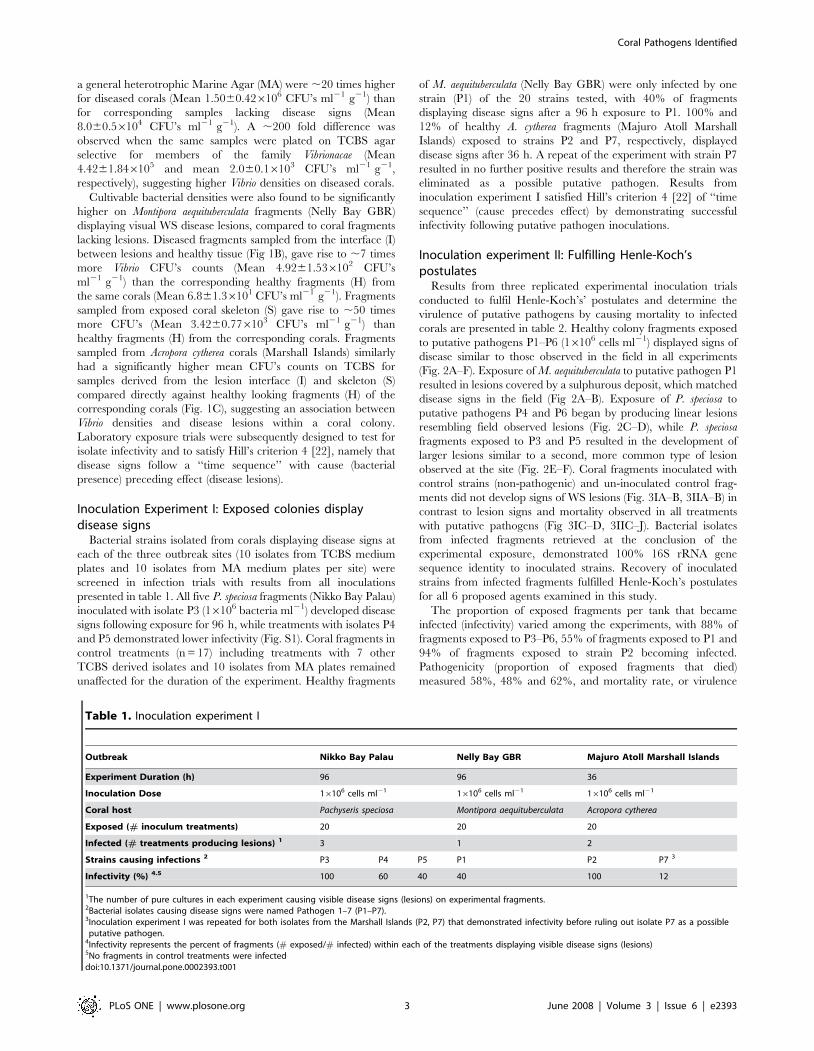
Table 1. Inoculation experiment I
Outbreak Nikko Bay Palau Nelly Bay GBR Majuro Atoll Marshall Islands
Experiment Duration (h) 96 96 36
Inoculation Dose 16106 cells ml21 16106 cells ml21 16106 cells ml21
Coral host Pachyseris speciosa Montipora aequituberculata Acropora cytherea
Exposed (# inoculum treatments) 20 20 20
Infected (# treatments producing lesions) 1 3 1 2
Strains causing infections 2 P3 P4 P5 P1 P2 P7 3
Infectivity (%) 4.5 100 60 40 40 100 12
1The number of pure cultures in each experiment causing visible disease signs (lesions) on experimental fragments.2Bacterial isolates causing disease signs were named Pathogen 1–7 (P1–P7).3Inoculation experiment I was repeated for both isolates from the Marshall Islands (P2, P7) that demonstrated infectivity before ruling out isolate P7 as a possibleputative pathogen.
4Infectivity represents the percent of fragments (# exposed/# infected) within each of the treatments displaying visible disease signs (lesions)5No fragments in control treatments were infecteddoi:10.1371/journal.pone.0002393.t001
Coral Pathogens Identified
PLoS ONE | www.plosone.org 3 June 2008 | Volume 3 | Issue 6 | e2393

(proportion of infected fragments that died) equalled 66%, 87%
and 66% for putative pathogens from Palau, Magnetic Island and
the Marshall Islands, respectively. Similarly, the times needed for
50% of the fragments to experience mortality (LT50) were 60h,
180h and 18h, respectively.
Aetiology of WS: Adhesion of pathogens to coral tissuePutative pathogen P1 (Nelly Bay GBR) demonstrated an 87%
reduction in mean seawater CFU’s (Fig 4A) within the first 12 h
following inoculation into aquaria with fragments of M. aequitu-
berculata (from mean 2.4860.376105 cells ml21 at t 0 to mean
3.1760.676104 cells ml21 at t 12). In comparison, only a 6%
reduction was observed when the same corals were inoculated with
control bacterial isolate MF1 (from mean 9.6061.816105 cells
ml21 at t 0 to mean 9.0761.016105 cells ml21 at t 12). CFU’s
from un-inoculated control aquaria averaged 3.060.696102 cells
ml21 after 12 h. After 36 h, mean CFU counts from aquaria
seawater treated with P1 dropped even further to 0.6% of the
original inoculation concentration (Men 1.4160.276103 cells
ml21), which was similar to the density of cells in control tanks
(Mean 2.2660.496103 cells ml21). In contrast, putative pathogens
that were inoculated into sterile seawater without corals main-
tained a constant density of viable counts in suspension throughout
the experiment (Fig. 4A), eliminating the possibility that bacteria
died from the seawater itself or may have settled on the sides or
bottom of aquaria. Vibrio density in aquaria containing M.
aequituberculata fragments, which were inoculated with non-
pathogen MF1 remained unchanged after 36 h, with mean
1.0460.156106 cell ml21 (100%) retrieved on TCBS agar plates.
CFU counts of crushed coral samples (CFU ml21 g21 wet
weight) from aquaria inoculated with P1 reached a mean of
1.6060.786105 ml21 g21 after 12 h (Fig 4B). In comparison,
fragments from aquaria inoculated with control bacteria (MF1), or
un-inoculated controls, resulted in CFU counts that were 94% and
97% lower after 12 h (Mean 9.0862.826103 ml21 g21 and mean
4.1262.456103 ml21 g21, respectively). Table S1 summarizes the
data from adhesion experiments conducted with putative patho-
gens and controls isolated from the three infection sites examined
in this study.
Loss of Symbiodinium followed by tissue lesionsDetailed photographs taken of A. hyacinthus fragments infected
experimentally with P2 (Fig 5A–C) revealed 2 distinct disease-
phases. An initial loss of Symbiodinium, visible as tissue paling was
observed after 9–12 h of exposure (Fig 5B–C) followed by
developing tissue lesions. Similar patterns of paling were also
observed when P. speciosa fragments were exposed to P3 (Fig 5D).
Paling and loss of Symbiodinium commenced in coenosarc tissue
(tissue between polyps) in distinct linear patterns starting 12 h post
inoculation and corresponding with the peak in viable CFU counts
retrieved from coral tissue. These early signs of disease then
developed into lesions that resembled those observed in the field
(Fig 5E–F), suggesting that disease progression was consistent
(Hill’s [22] criterion 2) and followed measurable steps (Evans’ Rule
F [21]). For a 24h time lapse video clip of A. hyacinthus inoculated
with pathogen P2, see Supporting Information Movie S2.
In all experimental treatments inoculated with putative
pathogens P1–P6, the proportion of fragments displaying acute
disease signs (lesions) increased with time to between 55% and
94% of fragments per tank (Fig 6A–C) conforming with Evans’
rules D and E [19], namely that disease occurs, temporally,
following specific incubation times and that the number of new
cases and the severity of outcome should correlate positively with
time. The proportion of P. speciosa fragments from Palau, M.
aequituberculata fragments from Nelly Bay and A. hyacinthus
fragments from the Marshall Islands displaying acute disease signs
increased consistently and significantly within the first 96 hours
(Fig. 6A–6B) and 12 hours (Fig. 6C) of the start of inoculation
experiment II, at each site, respectively, resembling standard
infection curves [32]. In contrast, 0–8% of fragments in inoculated
and un-inoculated control treatments developed disease signs
(Fig 6A–C).
Isolates associated with disease signs are proteolyticallyactive
Isolated bacteria (152 strains) recovered from both diseased and
healthy corals were screened for proteolytic activity using the
asocasein assay and specific PCR primers targeting the zinc-
binding site of a Vibrio family zinc-metalloprotease. A total of 48%
of strains (n = 33 strains) retrieved from diseased P. speciosa in the
field (Nikko Bay Palau) demonstrated high ($3U) or medium (1-
3U) proteolytic activity compared with 30% strains (n = 23 strains)
demonstrating high or medium activity that were retrieved from
Figure 2. WS signs observed in the laboratory and in the field:A. Montipora aequituberculata exposed to pathogen P1 in laboratoryinoculation experiment. B. M. aequituberculata with WS signs in thefield (Nelly Bay GBR). C. Pachyseris speciosa exposed to pathogen P6 inlaboratory inoculation experiment. D. P. speciosa with WS signs in thefield (Nikko Bay Palau). E. P. speciosa exposed to pathogen P3 inlaboratory inoculation experiment. F. P. speciosa with WS signs in thefield (Nikko Bay Palau).doi:10.1371/journal.pone.0002393.g002
Coral Pathogens Identified
PLoS ONE | www.plosone.org 4 June 2008 | Volume 3 | Issue 6 | e2393

non-diseased colony fragments sampled in the field (Table 3). This
difference, however, was not found to be statistically significant
(Pearson’s x2 = 1.825, DF = 1, p = 0.177). In contrast, 11 positive
PCR bands and derived partial sequences of the Vibrio zinc-
metalloprotease gene were obtained from DNA of isolates
retrieved from diseased P. speciosa sampled in the field compared
with only 1 partial sequence from a non-diseased colony fragment.
This difference was found to be significant by testing for Pearson’s
chi-square (x2 = 6.763, DF = 1, p = 0.0093).
Similar results were obtained by screening field isolates from
Nelly Bay GBR (Table S2). Bacteria demonstrating high and
medium proteolytic activity by the asocasein assay made up 70%
of all isolates retrieved from coral skeletons (S) exposed by WS
disease at Nelly Bay GBR and 57% of all isolates from the lesion
interfaces (I), compared with only 24% of all isolates obtained from
healthy (H) tissue fragments on diseased colonies, demonstrating a
significant difference in proteolytic activity between isolates
associated with disease signs (I+S) and healthy (H) tissue (Pearson’s
x2 = 6.446, DF = 1, p = 0.011). A significant difference was also
obtained for the same 38 isolates when screened by the molecular
method using PCR primers (Pearson’s x2 = 12.518, DF = 1,
p,0.0001). Finally, screenings by the molecular method per-
formed on DNA extracted from 56 isolates retrieved from both
infected and non-infected fragments at the conclusion of
inoculation experiment II in Palau (Table S3), demonstrated that
results obtained by screening field isolates were consistent with
screening laboratory derived isolates (Pearson’s x2 = 6.725,
DF = 1, p = 0.010). Thus, in both field and laboratory infections,
the presence of a Vibrio family zinc-metalloprotease was associated
with disease signs conforming to Evans’ rules B, C and G [21],
suggesting that bacterial proteolytic activity may cause or
contribute to observed WS lesions.
Pathogens identified by this study form a taxonomiccluster
Based on near complete 16S rRNA gene sequence comparisons,
the six pathogens clustered in a tight taxonomic group and were
found to share between 98–99% sequence identities with the
previously characterized coral-bleaching pathogen Vibrio coralliily-
ticus [14]. All isolates which tested positive for the zinc-
metalloprotease zinc-binding site and exhibited high proteolytic
activity (when screened by the asocasein assay) were used to
construct a maximum likelihood phylogenetic tree based on their
16S rRNA gene (Fig. 7). Our findings demonstrate that more
isolates possess the genetic capacity to become proteolytically
active than the six coral pathogens identified in this study,
suggesting that successful infections require the expression of
additional virulence genes, but also that other non-pathogens
might be indirectly involved in enhancing infections.
Discussion
This study reports the successful isolation and identification of
bacterial infectious agents implicated in a group of widespread
Indo-Pacific coral diseases that affect numerous species at various
geographical locations. Six coral pathogens were identified with
close 16S rRNA gene phylogenetic affiliation with the previously
identified coral pathogen V. coralliilyticus [14]. Vibrio pathogens
have been previously demonstrated to cause fish, eel, shrimp and
Figure 3. Inoculation experiment II: I A–B. Montipora aequituber-culata coral fragments in un-inoculated control treatment (t = 0h andt = 150h). I C–D. M. aequituberculata coral fragments exposed to 16106
cells ml 21 of culture P1 (t = 0h and t = 150h). II A–B. Pachyseris speciosacoral fragments in un-inoculated control treatment (t = 0h and t = 150h).II C–D. P. speciosa coral fragments exposed to 16106 cells ml21 ofculture P3 (t = 0h and t = 150h). II E–F. P. speciosa coral fragmentsexposed to 16106 cells ml21 of culture P4 (t = 0h and t = 150h). II G–H.P. speciosa coral fragments exposed to 16106 cells ml21 of culture P5
(t = 0h and t = 150h). II I–J. P. speciosa coral fragments exposed to16106 cells ml21 of culture P6 (t = 0h and t = 150h).doi:10.1371/journal.pone.0002393.g003
Coral Pathogens Identified
PLoS ONE | www.plosone.org 5 June 2008 | Volume 3 | Issue 6 | e2393
human mortalities [45–49]. Seasonal bleaching of the coral Oculina
patagonica in the Mediterranean Sea has been shown to be caused
by V. shiloi [5,11,50–51] and V. coralliilyticus has been identified as
the aetiological agent of Pocillopora damicornis bleaching in the
Indian Ocean [14,52–53]. Other coral diseases in the Caribbean,
such as White Band Disease type II, Yellow Blotch/Band and
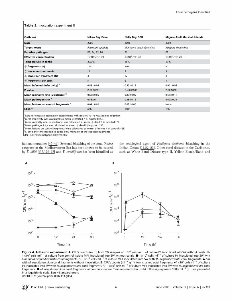
Table 2. Inoculation experiment II
Outbreak Nikko Bay Palau Nelly Bay GBR Majuro Atoll Marshall Islands
Date 2005 2003 2004
Target host/s Pachyseris speciosa Montipora aequituberculata Acropora hyacinthus
Putative pathogen P3, P4, P5, P6 1 P1 P2
Effective concentration 16106 cells ml21 16106 cells ml21 16106 cells ml21
Temperature in tanks 29.9uC 28uC 30uC
# fragments (n) 195 360 80
# inoculum treatments 11 3 3
# tanks per treatment (N) 3 12 4
# fragments per tank 5 6 4
Mean infected (Infectivity) 2 0.8860.08 0.5560.13 0.9460.05
P value P,0.00001 P = 0.00005 P,0.00001
Mean mortality rate (Virulence) 3 0.6660.05 0.8760.09 0.6660.11
Mean pathogenicity 4 0.5860.11 0.4860.15 0.6260.24
Mean lesions on control fragments 5 0.0460.02 0.0860.06 None
LT50 6 60h 180h 18h
1Data for separate inoculation experiments with isolates P3–P6 was pooled together.2Mean infectivity was calculated as mean #infected / # exposed6SE.3Mean mortality rate, or virulence, was calculated as mean # dead / # infected6SE.4Mean pathogenicity was calculated as mean # dead/ #exposed6SE.5Mean lesions on control fragments were calculated as mean # lesions / # controls6SE.6LT50 is the time needed to cause 50% mortality of the exposed fragments.doi:10.1371/journal.pone.0002393.t002
Figure 4. Adhesion experiment: A. CFU’s counts (ml21) from SW samples. N-16106 cells ml21 of culture P1 inoculated into SW without corals. e-16106 cells ml21 of culture from control isolate MF1 inoculated into SW without corals. &-16106 cells ml21 of culture P1 inoculated into SW withMontipora aequituberculata coral fragments. ,-16106 cells ml21 of culture MF1 inoculated into SW with M. aequituberculata coral fragments. m-SWwith M. aequituberculata coral fragments without inoculation. B. CFU’s counts (ml21 g21) from crushed coral fragments. N-16106 cells ml21 of cultureP1 inoculated into SW with M. aequituberculata coral fragments. ,- 16106 cells ml21 of culture MF1 inoculated into SW with M. aequituberculata coralfragments. &-M. aequituberculata coral fragments without inoculation. Time represents hours (h) following exposure.CFU’s ml21 g21 are presentedin a logarithmic scale. Bars = Standard errors.doi:10.1371/journal.pone.0002393.g004
Coral Pathogens Identified
PLoS ONE | www.plosone.org 6 June 2008 | Volume 3 | Issue 6 | e2393

Dark Spots Disease, were found to be associated with elevated
Vibrio spp. prevalence [4,54–56], suggesting the involvement of
Vibrio strains in numerous coral diseases including coral bleaching
[5,57–59].
This study has found high prevalence of Vibrio spp. to be
associated with WS signs in all diseased colonies sampled from
three WS outbreaks. An association between bacterial presence
and disease signs does not provide proof that bacteria actually
cause the disease. However, such an association already fulfils
requirements put forth by Evans’ rule A [21], namely that the
‘‘prevalence of the disease should be significantly higher in those
exposed to the putative cause than in cases controls not so
exposed’’. It also complies with the Read [60] definition of
virulence, which highlights an agent’s contribution to reduction in
host fitness caused by exploitation.
When bacterial strains were inoculated into tanks with healthy
coral fragments, only putative pathogens adhered to coral tissue
and a consistent peak in cultivable Vibrio abundance, 500–1000
fold greater than was found for control strains, was observed on
fragments exposed to putative pathogens 12 h post inoculation.
This experiment confirmed Hill’s criterion of ‘‘time sequence’’
[22], suggesting that following exposure and prior to the
development of visual disease signs (lesions), putative pathogens
were able to migrate towards the coral fragments, adhere to coral
tissue and survive initial contact in a viable state. In contrast,
control strains were unable to perform this transmission,
suggesting that motility towards corals and adhesion may be
regarded as traits involved in pathogenicity. Initial visual signs of
tissue paling and lesions were observed following a peak in
cultivable bacterial abundance for all six putative pathogens,
demonstrating a common aetiology of adhesion followed by
disease progression. Despite this peak in cultivable Vibrio
abundance 12 h post exposure, less than 1% of the original
inoculation was retrieved by plating coral fragments, potentially
indicating that Vibrio cells entered a viable but non-culturable
(VBNC) state [61], or alternatively, died.
The colonization of target hosts by Vibrio pathogens has been
studied in detail, particularly the ability of Vibrios to adhere to
mucus found either inside the gastro- internal track or externally
on fish or corals [62–63]. Denkin and Nelson [64] have
demonstrated that the transcription of zinc-metalloprotease by
the fish pathogen V. anguillarum is regulated by mucus and can only
occur after adhesion is completed. This duality in Vibrio function is
often referred to as the ‘‘transmission-virulence trade-off’’ [65] and
highlights the fact that the ultimate goal of Vibrio pathogenicity is
not to kill a host, or to complete a necessary biological life-cycle
within it, but to re-enter the environment in larger numbers and
initiate a new cycle of infections [66]. It explains why Vibrio
pathogens are commonly found in environmental reservoirs [26],
or transmitting through the water column, like pathogen P6
isolated in this study from seawater above infected corals at Nikko
Bay Palau.
This study has demonstrated that 55%–94% of coral fragments
exposed to pathogens cultured from diseased corals at their
respective field sites become infected and that 66%–87% of those
infected die, compared with significantly lower infection and
mortality for fragments exposed to control bacterial strains (0–8%).
These results conform to Evans’ rule G [21], requiring that
experimental reproduction of the disease should occur in higher
incidence in those exposed to the putative cause than in those not
so exposed. However, a proportion of exposed fragments did not
develop disease signs, demonstrating that the probability of
becoming infected may not be equal among healthy colony
fragments collected from the field, and that other host related
factors potentially contribute to successful infections. Such
unknown factors can be explored in future inoculation trials.
The presence of Vibrio spp. on both healthy and diseased corals
has led to the conclusion by some authors [67–68] that Vibrio
Figure 5. Disease progression: A. Acropora hyacinthus fragmennt inoculated with 16106 cells ml21 of culture P2 (t = 0h). B. Loss of Symbiodiniumfrom A. hyacinthus inoculated with 16106 cells ml21 of culture P2 (t = 12h). C. Polyp and surrounding tissue-loss of Symbiodinium from A. hyacinthusinoculated with 16106 cells ml21 of culture P2 (t = 12h). D. Loss of Symbiodinium cells from Pachyseris speciosa inoculated with 16106 cells ml21 ofculture P3 (t = 12h). E. Tissue lesions on P. speciosa inoculated with 16106 cells ml21 of culture P3 (t = 24h). F. Exposed skeleton on P. speciosainoculated with 16106 cells ml21 of culture P3 (t = 60h).doi:10.1371/journal.pone.0002393.g005
Coral Pathogens Identified
PLoS ONE | www.plosone.org 7 June 2008 | Volume 3 | Issue 6 | e2393

infections of corals may be opportunistic in nature. This
assumption fits well into models of disease occurring in
environmental settings, where multiple factors, such as host
density [69] and temperature [70] have been shown to influence
the probability of successful infections. Combinations of virulent
and a-virulent Vibrio strains are found readily in environmental
samples [71] with non-clinical V. cholera strains found to be capable
of causing infections despite lacking the cholera toxin gene [72].
Many Vibrios specialize in multiple host attachment and detach-
ment [73–75], suggesting a broad scope for potential coral
infections by Vibrios including possible host shifts due to fish
depletion from coral reefs [76]. Amaro and Biosca [46] have
demonstrated that Vibrio vulnificus biotype 2 is both a primary
pathogen for eels and an opportunistic pathogen for humans,
indicating that the identification of opportunistic pathogens
requires rigorous testing. Nevertheless, none of the claims to
define Vibrio coral infections as opportunistic have so far provided
conclusive evidence to show that suspects (identified by molecular
screening methods) found on healthy corals are in fact pathogenic
(whether opportunistic or not), or that only compromised hosts
become infected. In addition, not all coral mortalities are caused
by infectious agents, but rather by exposure to extreme conditions,
such as pesticides or high nutrient levels [77–78], which may result
in indirect shifts in microbial abundance. Infectious outbreaks can
be distinguished from non-infectious ones by plotting infection
Time (h)
0 6 9 12 18 24
Prop
ortio
n of
infe
cted
col
onie
s
0.00.10.20.30.40.50.60.70.80.91.0
0 12 72 96 144
Prop
ortio
n of
infe
cted
col
onie
s
0.00.10.20.30.40.50.60.70.80.91.0
0 48 96 192 288 336
Prop
ortio
n of
infe
cted
col
onie
s
0.00.10.20.30.40.50.60.70.80.91.0
B
C
A
Figure 6. Disease transmission: A. Mean proportion of infectedPachyseris speciosa coral fragments displaying WS signs followingexposure to cultures of P3–P6 in comparison to proportions in
inoculated and un-inoculated control treatments. B. Mean proportionof infected Montipora aequituberculata coral fragments displaying WSsigns following exposure to culture of P1 in comparison to proportionsin inoculated and un-inoculated control treatments. C. Mean propor-tion of infected Acropora cytherea coral fragments displaying WS signsfollowing exposure to culture P2 in comparison to proportions ininoculated and un-inoculated control treatments. &-Coral fragmentsinoculated with 16106 cells ml21 of putative pathogen cultures. &-Coral fragments inoculated with 16106 cells ml21 culture of non-pathogen isolates. %-Coral fragments without inoculation. Timerepresents hours (h) following exposure. Bars = Standard errors.doi:10.1371/journal.pone.0002393.g006
Table 3. Proteolytic activity of bacterial isolates (Nikko BayPalau)
Bacterial isolates retrievedfrom field Pachyseris speciosa 1
Total
Diseasedcolonies
Non-diseasedcolonies
+ve PCR product 2 11 1 12
2ve PCR product 2 22 22 44
Total 33 23 56
High proteolytic activity 3 6 4 10
Medium proteolytic activity 4 10 3 13
No proteolytic activity 5 17 16 33
Total 33 23 56
1Isolates retrieved from diseased and non-diseased Pachyseris speciosa coloniessampled in Nikko Bay Palau .
2Specific amplification of Vibrio zinc-metalloprotease active zinc binding site.3High proteolytic activity .3U measured by the asocasein assay.4Medium proteolytic activity 1-3U measured by the ascasein assay.5No proteolytic activity ,1U measured by the asocasein assay.doi:10.1371/journal.pone.0002393.t003
Coral Pathogens Identified
PLoS ONE | www.plosone.org 8 June 2008 | Volume 3 | Issue 6 | e2393

0.1
P6 LMG23694/EU372932
P5 LMG23692/EU372933
P2 LMG23691/EU372935
V. coralliilyticus AJ316167
EU372916
P1 LMG23696/EU372917
V. coralliilyticus AJ440004
V. coralliilyticus AJ440005
EU372929
Vibrio sp. PH1 AF513461
V. neptunius AJ316171
EU372920
EU372926
EU372937
EU372925
V. brasiliensis AJ316172
EU372930
P3 LMG23695/EU372934
EU372924
EU372927
EU372923
V. cholerae AY494843
V. vulnificus AY676131
V. splendidus AB038030
V. shiloi AF007115
EU372922
V. alginolyticus AY373027
V. parahaemolyticus AP005083
EU372938
EU372936
EU372921
Vibrio sp. BB4 AF319768
EU372918
EU372928
EU372939
P. ruthenica AF316891
EU372919
A. tumefaciens D14504
P4 LMG23693/EU372931
Figure 7. Phylogenetic tree of proteolitically-active isolates: Evolutionary distance maximum likelihood analysis based on 16S rRNA genesequences of isolates obtained by this study. Coral pathogens are marked in red. Reference strains are marked in black. Isolates that demonstratedhigh proteolytic activity (asocasein assay) and tested positive for a zinc-metalloprotease gene are presented in blue (Palau isolates) and in green(Nelly Bay GBR isolates). Nodes represent bootstrap values $50% based on 1000 re-samplings. Scale bar corresponds to 10% estimated sequencedivergence.doi:10.1371/journal.pone.0002393.g007
Coral Pathogens Identified
PLoS ONE | www.plosone.org 9 June 2008 | Volume 3 | Issue 6 | e2393

curves [32] to demonstrate a bell-shape increase and decrease in
incidence rate with time.
This study did not find evidence for the presence of coral
pathogens on healthy corals in the field, nor evidence that exposed
fragments might be successfully infected due to stress other than
the direct exposure to the pathogens themselves. Control
treatments in all inoculations remained healthy, including a
proportion of those exposed to pathogens. Further studies are
recommended to determine the prevalence of pathogens in field
samples by developing diagnostic tools to target specific virulence
genes in large scale screening efforts. These studies could then
determine the proportion of exposed corals in the field that
develop acute disease signs and should become an integral part of
establishing acute vs. chronic disease prevalence in environmental
studies.
This is the first study to diagnose proteolytic activity as a
possible component of the aetiology of WS through the screening
of more than 150 isolates from both diseased and non-diseased
corals. Zinc-metalloproteases have been characterized as virulence
factors in many Vibrio family pathogens, such as V. cholera [79], V.
vulnificus [80], V. harveyi [81] and V. anguillarum [82]. Vibrio zinc-
metalloproteases are involved in cleavage of connective tissue [83],
para-cellular perturbation [84], swarming and adhesion to mucus
[85] and detachment [86]. The coral bleaching pathogens V. shiloi
and V. coralliilyticus have been previously shown to harbour a zinc-
metalloprotease [53,87] along with other toxins that cause
photosynthetic 5 inhibition of coral Symbiodinium [88]. Serratia
marcescens, the aetiological agent of acroporid serratiosis (coral
White Pox disease [13]), resulting in acute tissue lesions, also
possesses a virulent zinc-metalloprotease capable of connective
tissue degradation [89]. However, it has been shown that both
clinical and non-clinical strains possess zinc-metalloprotease genes
[90], suggesting that it may not be the only virulence factor to
cause successful infections. This study provided similar results,
underlining the need to search for additional virulence factors in
future studies.
Recent studies by Ainsworth et al. [91] did not detect bacteria
associated with WS lesions of diseased corals sampled at Heron Island
on the GBR, using direct microscopic techniques. In contrast,
samples of WS corals obtained from Heron Island in this study for
screening purposes demonstrated an abundance of Vibrio spp. isolates
on WS lesions, including proposed putative pathogens that are
proteolytically active and possess a zinc-metalloprotease gene. These
contradicting findings underline the importance of ‘comparative
validation’ [92] in disease research and the need for standardized
protocols for disease detection using better diagnostic tools.
Further histopathological studies by Ainsworth et al. [68, 91]
utilizing commercial labelling kits have found that coral fragments
displaying WS signs test positive for DNA fragmentation. These
observations led to the hypothesis that WS is potentially the result
of coral programmed cell death. However, further proof is needed
in order establish whether DNA fragmentation (or apoptosis) in
corals is cause or effect. The induction of apoptosis by bacterial
pathogens (Salmonella Sp., E. coli, Shigella sp., C. difficile, L.
monocytogenes, C. parvum and others) has been previously demon-
strated by many studies [93-97], suggesting a possible link between
bacterial infections and apoptosis. This link can be tested in future
pathogen-exposure trials and used to design novel diagnostic
protocols for WS, which would target bacterial enzymes causing
DNA fragmentation.
In summary, this study demonstrated consistent results in
applying cost effective culturing techniques combined with
biochemical and molecular tools towards successful pathogen
isolation, coral disease investigation and sample screening. Future
research should be conducted to explore the virulence components
of all six pathogens identified in this study and to test the
contribution of multiple factors (pathogen, environment and host
related) to the aetiology of WS. Enhanced monitoring and
management of WS outbreaks will not only benefit coral health,
but would also further validate results obtained in this study.
Materials and Methods
Isolation and growth of bacteria from coral samplesFor inoculation experiment I, ten fragments (2–10 g wet weight)
from corals displaying WS disease signs and ten fragments (2–10 g
wet weight) from corals lacking WS disease signs were collected
from depths between 3–15 m at each of the following locations: 1)
Nelly Bay fringing reef (S19 109 E 146 529) at Magnetic Island in the
central section of the Great Barrier Reef (GBR) in September 2003;
2) Majuro Atoll the Republic of the Marshall Islands (N 9 009 E 168
009) in August 2004; and 3) Nikko Bay, an enclosed bay among rock
islands in the Republic of Palau (N 7 309 E 134 309) in February
2005. WS mainly affected plate colonies of Pachyseris speciosa in
Palau, tabular species of Acropora (A. cytherea, A. hyacinthus and A.
clathrata) in the Marshall Islands and plate colonies of Montipora
aequituberculata at Nelly Bay GBR. At each site, samples were
transported from the reef to the laboratory in sterile containers.
For calculating the abundance of bacteria associated with
diseased and non-diseased fragments, the following sub-samples
were obtained at each site: healthy tissue from coral fragments
with no disease (CON, n = 3); tissue adjacent to lesions on coral
fragments with WS disease signs (INF, n = 3); healthy tissue on
coral fragments displaying disease signs (H, n = 3); lesion interface
on coral fragments displaying disease signs (I, n = 3); and exposed
coral skeleton on coral fragments displaying disease signs (S, n = 3).
Samples were crushed and diluted with 10 ml of 0.22 mm
filtered seawater (Millipore, USA), and then vortexed for 3 min at
maximum speed before being left to settle for 3 min [26].
Supernatant (100 mL) was streaked on agar plates containing a
general heterotrophic bacterial medium (Marine Agar: 1.8%
Marine Broth, Difco-2216, USA 0.9% NaCl, 1.8% Agar Bacto,
Difco-214010, USA) and thiosulfate citrate bile salts sucrose
(TCBS) agar, a Vibrionacea selective growth medium (Difco, USA).
Plating was conducted in triplicates of serial dilutions (161021–
161026) followed by incubation overnight at 30uC. Cultivable
strains were quantified by counting colony forming units (CFU’s)
and the density of bacteria associated with corals was determined
as mean CFU’s per 1 ml of crush derived from 1 g (wet weight) of
coral tissue (CFU’s ml21 g21). Single CFU’s were picked from
both Marine Agar (MA) and TCBS plates and transferred to fresh
MA plates for further analyses.
Single isolates were grown in 250ml sterile flasks containing
sterile marine broth (MB) media incubated at 30 uC for 18h (i.e. to
end of the logarithmic phase) with constant shaking (150rpm). Cell
density in pure cultures was determined by plating triplicates of
serial dilutions on MA and by measuring absorbance (595nm) in
sterile microtitre well plates (n = 6).
Additional bacterial isolates from fragments displaying signs of
ongoing tissue loss in association with WS and from healthy
fragments (controls) were retrieved for screening purposes from
corals at Heron Island, GBR (March 2004) and at Dip Reef GBR
(November 2004).
DNA extraction, PCR amplification and gene sequencing.Genomic DNA extraction from pure cultures of bacterial
isolates retrieved by this study was performed using the Wizard
genomic DNA purification kit (Promega, USA) as per the
Coral Pathogens Identified
PLoS ONE | www.plosone.org 10 June 2008 | Volume 3 | Issue 6 | e2393
manufacturer’s instructions. The 16S rRNA gene was amplified by
using universal primers 27F and 1492R. [98]. In addition, primers
HA-F (59 –CATGAGGTCAGCCACGGTTTTACTGAGCAG)
and HA-R (59–CGCGCGGTTAAACACGCCACTC-
GAATGGTGAAC (Invitrogen, NZ) targeting a ,225 bp region
including the zinc binding site of Vibrio-family zinc-metallopro-
teasses [99] were used to screen all bacterial genomic DNA. PCR
reactions (50 mL) were run on an Eppendorf Mastercycler with the
reaction mix consisting of 10 pmol of each primer, 5 mL of
10xPCR buffer with 15 mM MgCl2, 50 nmol dNTP, 10 ng
template DNA and 1U Taq (iTaq, Intron Biotechnology, Korea).
DDW (Milli-Q, millipore) was added to the volume of 50 mL.
Cycling conditions consisted of: 1) 27F/1492R-a 5 min denatur-
ation step at 94uC followed by 30 cycles of 1 min at 94uC, 1 min
at 52uC and 1 min at 72uC and concluded by a 7 min extension
step at 72uC; and 2) HA-F/HA-R-a 5 min denaturation step at
94uC followed by 30 cycles of 20 sec at 94uC, 20 sec at 55uC and
1 min at 72uC, concluded by a 5 min extension step at 72uC.
Amplified bands of the correct size were confirmed on a 1%
ethidium bromide stained TAE agarose gel and amplified gene
products were sequenced at MACROGEN Inc. (Seoul, Korea) on
an ABI PRISM 3730XL analyzer (96 capillary-Applied Biosys-
tems, CA, USA) using the ABI PRISM BigDyeTM Terminator
Cycle Sequencing Kit. Retrieved gene sequences were aligned for
closest matches using BLAST [100]. In total, 152 partial sequences
were retrieved from coral fragments displaying WS signs and from
controls sampled at the three-infection sites.
Phylogenetic analysesSequences were checked for chimera formation with the
CHECK_CHIMERA software of the Ribosomal Database
Project [101]. Sequence data were aligned to the most similar
sequence using the BLAST database algorithm [100], and then
further analysed with the ARB software package [102]. Tree
topologies were evaluated by reconstructing phylogenies using
maximum likelihood evolutionary distance analysis (Phylip
Distance Method with Jukes and Cantor model) of aligned near
full-length sequences (.1200 bp). Regions of ambiguous sequence
(N) were removed from the analysis. Bootstrap values were
obtained for branching patterns using the Phylip software package
(version 3.65 [103]) and values $50% were included for main
nodes of the tree.
Infection experimentsInfection experiments were run as incurred matrices in 2
consecutive stages, described below as inoculation experiments I
and II.
Inoculation experiment I: Testing for infectivity ofbacterial isolates
To screen bacteria for infectivity (the ability to initiate visual
disease signs (lesions) regardless of their severity [35]), 20 isolates
retrieved from coral samples at each of the three sites (10 most
abundant isolates on both MA and TCBS plates from both healthy
and diseased colonies at each site) were grown to end logarithmic
phase in MB (as described above) and inoculated individually into
7L sterile aerated tanks (final inoculum concentration = 16106
cells ml21) containing 4–6 healthy fragments of corals collected
from sites without disease signs (i.e. healthy fragments of Pachyseris
speciosa from a healthy Palau site, healthy Acropora cytherea fragments
from a healthy Marshall Islands site, and healthy Montipora
aequituberculata fragments from a healthy Nelly Bay site). Prior to
bacterial inoculation, coral fragments were acclimatized for 5 days
to allow recovery from handling and fragmentation following a
protocol by Kushmaro et al. [50]. Each of the 20 culture
inoculations was tested in two tanks [n = 168–252 fragments per
site, N = 21 inoculation treatments including 1 negative control
treatment]. The negative control tanks contained coral fragments
with no bacteria added. Seawater in the tanks was replaced every
48 h and tanks were observed and photographed for 140 h in
order to detect developing disease signs. At the end of each
experiment, infectivity was calculated as the proportion of exposed
fragments per tank that became infected. Both infected and non-
infected fragments were crushed and individual CFU’s were
picked and transferred to fresh MA plates for further analyses and
DNA extraction, as previously described. Bacterial strains causing
disease signs in this experiment were given the simplified names:
P1–P7 (P1–from M. aequituberculata in Nelly Bay GBR; P2 and P7
from A. cytherea in Majuro Atoll the Republic of the Marshall
Islands; and P3–P6 from P. speciosa in Nikko Bay Palau) and were
inoculated as pure cultures in the following experiments under
these names. Bacterial strain P6 was isolated from seawater above
diseased P. speciosa colonies at Nikko Bay Palau. It caused
infections in a separate experiment and was therefore added to
the list of putative pathogens. Strain P7 from the Marshall Island
caused partial disease signs on only one A. cytherea fragment (out of
four fragments) when inoculated into two tanks (n = 4 fragments
per tank). Inoculation experiment I was repeated using this isolate
(n = 12 fragments in each of 3 tanks) and it was removed from the
putative pathogen list after failing to cause infections.
Inoculation experiment II: Replicated exposure trial tofulfil Henle-Koch’s postulates and test for virulence
To fulfil Henle-Koch’s postulates, a large multi-replicated
exposure trial using successful putative pathogens that initiated
disease signs in inoculation experiment I were grown as pure cultures
and inoculated (final inoculum concentration = 16106 cells ml21)
into multiple tanks with colony fragments (of P. speciosa, M.
aequituberculata A. hyacinthus) collected from non-disease sites and
acclimatized for 5 days. The number of fragments allocated to
inoculation tanks at each site (n) was between 80–360, distributed as
4–6 fragments per tank, and the number of tanks per inoculation/
control treatments (N) was between 4 and 12. At each site, 4 negative
controls were run including: 1 treatment comprising tanks to which
no bacteria were added, 2 treatments comprising tanks to which
control bacterial strains were added at identical concentrations, and
1 treatment comprising tanks to which sterile bacterial media was
added (1 ml MB per 1 L seawater) as potential ‘‘growth enhancer’’
for putative pathogens that might be already present on experimen-
tal coral fragments. Tanks were maintained temperatures identical
to those measured at infection sites. Fragments in each tank were
observed and photographed for the entire length of the experiment
and developing disease signs were recorded. The experiments were
terminated following mortality in infection tanks. Case mortality
rate, or virulence (the proportion of infected fragments in each tank
that died [36]) and the mean proportion of infected fragments per
tank were calculated. Pathogenicity (the proportion of exposed
fragments that died) was calculated following the formula of Thomas
and Elkinton [35]: Pathogenicity = infectivity6virulence (where
pathogenicity = # dying / # exposed, infectivity = # infected / #exposed and virulence = # dying / # infected). Finally, LT50 (the
estimated time it takes to kill 50% of the infected fragments) was
calculated as a temporal measure of virulence.
Both infected and non-infected fragments were crushed and
streaked on agar plates to determine mean CFU’s, as previously
described. Total DNA was extracted from retrieved isolates for
elucidation of taxonomic identity (16S rRNA gene sequence).
Coral Pathogens Identified
PLoS ONE | www.plosone.org 11 June 2008 | Volume 3 | Issue 6 | e2393

Complete alignment (100%) of the 16SrRNA gene sequences
retrieved from bacteria re-isolated from infected fragments and the
16S rRNA gene sequences of inoculated bacteria (P1–P6) allowed
the fulfilment of Henle-Koch’s postulates [20], namely, that:
1. An organism found only on infected corals could be isolated,
taxonomically identified, and grown in pure culture.
2. The isolated organism reproduced disease signs when inocu-
lated onto healthy corals.
3. An isolate retrieved from coral fragments that developed
disease signs in inoculation experiments is demonstrated to be
identical (by analysis of 16SrRNA partial gene sequences) with
the organism used for inoculations.
Other rules and criteria for supporting causality used bythis study
Results of experiments and screenings conducted in this study
were used to evaluate compliance with Evans’ rules [21] and Hill’s
criteria [22], defined as alternative requirements for establishing
disease causation. Both Evans’ rules and Hill’s criteria are listed in
the Supporting Information section (see Materials and Methods S1).
Adhesion of bacterial isolates to coralsTo further test the physical ability of putative pathogens to
migrate towards coral hosts, to adhere and to survive the initial
contact with the coral host, before initial signs of infection are
observed, mean bacterial CFU’s were quantified from random sub
samples of tank seawater (N = 4 seawater sub-samples per
treatment, each taken from a different tank) following inoculation
with the six coral pathogens (P1–P6) identified in Inoculation
Experiment I (final inoculum concentration = 16106 cells ml21).
One ml of tank seawater was collected at inoculation time (t = 0 h),
1 h post inoculation (t = 1 h) and then at 12 h intervals (t = 12 h,
t = 24 h, t = 36 h) from four infection tanks. 100 mL aliquots from
each sample were spread in triplicates on agar plates containing
Marine Agar and TCBS, as described previously. Mean CFU’s
ml21 g21 wet weight were determined from three crushed coral
samples per treatment at corresponding times [63]. In addition,
control bacterial strains were also tested to determine if they
adhered to coral fragments following inoculation into tanks.
Finally, both putative pathogens and control bacteria were
inoculated into four seawater tanks (per bacterial treatment)
lacking coral fragments to test their ability to survive and remain
suspended in the water column for the experiment’s duration.
Seawater samples were collected from these tanks and plated in
triplicate as described previously to determine mean bacterial
density in seawater (CFU’s ml21).
The asocasein proteolytic AssayThe proteolytic activities of supernatants derived from 152
isolated bacterial strains retrieved by this study were tested by the
asocasein assay based on a protocol by Windle and Kelleher [104].
Briefly, 1 ml of bacterial cultures grown to end logarithmic phase
were centrifuged at 10,000g (Eppendorf 5415D centrifuge) for
5 min. Supernatant was removed and filtered through a 0.22 mm
filter (Millipore, USA). 100 mL supernatant was incubated for
30min at 30uC with 5g ml21 of asocasein as substrate (Sigma,
USA) dissolved in Tris-Hcl (50 mM pH 8) containing 0.04%
NaN3 (wt vol21). The reaction was terminated by adding 10% (wt
vol21) of trichloroacetic acid (TCA) to a final concentration of
6.7% (wt vol21) and incubating samples for 1 min. Samples were
then centrifuged for 3 min at 10,000g and transferred to 700 mL of
525 mM NaOH. Absorbance of six replicates from triplicate
culture samples was measured in 96 microtitre well plates at
450 nm using a Wallac spectrophotometer (Perkin Elmer, USA).
Blank controls were prepared from supernatant derived from E.coli
cultures boiled at 100uC for 10 min, treated with 5mg ml21
asocasein and directly thereafter by TCA. Protease activity was
calculated as proteolytic units, when 1U = 10006(OD450
CFU21)6109 [64]. Isolates were divided into 3 groups based on
their proteolytic activity: High activity (.3U), medium activity (1-
3U) and no activity (,1U).
Statistical analysisMeans and Standard Errors (SE) for bacterial colony forming
unit (CFU) counts and for the proportion of infected colonies were
compared among treatments using One-Way ANOVA (Statistica,
StatSoft, Inc. USA). Colony forming unit (CFU) counts are
presented in this study using logarithmic scales.
The association between categorical values related to bacterial
isolates retrieved independently from diseased and non-diseased
corals and demonstrating positive or negative proteolytic activity
was estimated using 262 contingency tables (Pearson Chi-square).
Significant results were determined when a#0.05.
Coral pathogensSix coral pathogen strains that were identified by this study were
submitted to the public collection of BCCM/LMG at the Gehnt
University, Belgium and are available for acquisition under the
following accession numbers: LMG23691-isolate P2 from a WS
infected Acropora cytherea in Majuro Atoll the Republic of the
Martshall Islands, LMG23692-isolate P5 from a WS infected
Pachyseris speciosa in Nikko Bay Palau, LMG 23693-isolate P4 from
a WS infected P. speciosa in Nikko Bay Palau, LMG 23694-isolate
P6 from seawater above a WS infected P. speciosa in Nikko Bay
Palau, LMG 23695-isolate P3 from a WS infected P. speciosa in
Nikko Bay Palau and LMG 23696-isolate P1 from a WS infected
Montipora. aequituberculata in Nelly Bay the GBR. 16S rRNA gene
sequences of all coral pathogens identified by this study were
submitted to Genbank (www.ncbi.nih.nlm.gov/Genbank/) under
the following accession numbers: P1 (LMG23696)-EU372917, P2
(LMG23691)-EU372935, P3 (LMG23695)-EU372934, P4
(LMG23693)-EU372931, P5 (LMG23692)-EU372933, P6
(LMG23692)-EU372932. 16S rRNA gene sequences retrieved
from isolates demonstrating positive results for proteolytic activity
in both screening tests conducted by this study (asocasein assay
and PCR amplification) were submitted to Genbank under the
following accession numbers: EU372918-EU372930, EU372936-
EU372939, and are presented in Fig. 7.
Movie S2Acropora hyacinthus fragments were inoculated with 16106 cells
ml21 of pathogen P2 (aquarium situated on the left hand side of
the screen). Time lapse photography (every 20 seconds) was
carried out for 36 h. Control treatment with no inoculation
appears on the right hand side of the screen.
Supporting Information
Materials and Methods S1 Evans’ Rules and Hill’s Criteria
Found at: doi:10.1371/journal.pone.0002393.s001 (0.03 MB
DOC)
Figure S1 Inoculation experiment I, Palau: A–B. Pachyseris
speciosa coral fragments without inoculation (t = 0h and t = 96h).
C–D. P.speciosa coral fragments inoculated with 16106 cells ml21
of culture P3 (t = 0h and t = 96h). E–F. P.speciosa coral fragments
Coral Pathogens Identified
PLoS ONE | www.plosone.org 12 June 2008 | Volume 3 | Issue 6 | e2393

inoculated with 16106 cells ml21 of culture P4 (t = 0h and t = 96h).
G–H. P.speciosa coral fragments inoculated with 16106 cells ml21
of culture P5 (t = 0h and t = 96h).
Found at: doi:10.1371/journal.pone.0002393.s002 (1.19 MB TIF)
Table S1 Adhesion Experiment
Found at: doi:10.1371/journal.pone.0002393.s003 (0.03 MB
DOC)
Table S2 Proteolytic activity of bacterial isolates (Nelly Bay
GBR)
Found at: doi:10.1371/journal.pone.0002393.s004 (0.04 MB
DOC)
Table S3 Proteolytic activity of bacterial isolates (Palau)
Found at: doi:10.1371/journal.pone.0002393.s005 (0.03 MB
DOC)
Movie S1 WS outbreak in Majuro Atoll the Republic of the
Marshall Islands (August 2004)
Found at: doi:10.1371/journal.pone.0002393.s006 (10.34 MB
AVI)
Movie S2 Time lapse inoculation experiment of Acropora
hyacinthus fragments infected with 16106 cells ml21 of pathogen
P2 for 36 hours (aquarium situated on the left hand side of the
screen). Control treatment with no inoculation appears on the
right hand side of the screen.
Found at: doi:10.1371/journal.pone.0002393.s007 (6.74 MB AVI)
Acknowledgments
The authors would like to thank S Anthony, Dr. W Dunlap, Dr. D
Jacobson, E Matson, C Page, L Peplow, Prof. L Raymundo, members of
the Palau International Coral Reef Center, the College of the Marshall
Islands and the crew of the AIMS RSV Lady Basten for their assistance in
laboratory work and sample collection.
Author Contributions
Conceived and designed the experiments: BW DB MS. Performed the
experiments: MS SV. Analyzed the data: DB MS. Contributed reagents/
materials/analysis tools: BW DB. Wrote the paper: BW DB MS. Other:
Performed sample collection: SV MS.
References
1. Harvell CD, Mitchell CE, Ward JR, Altizer S, Dobson AP, et al. (2002)
Climate warming and disease risks for terrestrial and marine biota. Science296: 2158–2162.
2. Weil E (2004) Coral reef diseases in the wider Caribbean. In: Rosenberg E,
Loya Y, eds. Coral Health and Disease. Berlin: Springer-Verlag. pp 35–68.
3. Willis B, Page C, Dinsdale E (2004) Coral disease on the Great Barrier Reef. In:
Rosenberg E, Loya Y, eds. Coral Health and Disease. Berlin: Springer Verlag.pp 69–104.
4. Cervino JM, Hayes RL, Polson SW, Polson SC, Goreau TJ, et al. (2004)
Relationship of Vibrio species infection and elevated temperatures to yellow
blotch/band disease in Caribbean corals. Appl Environ Microbiol 70:6855–6864.
5. Rosenberg E, Falkovitz L (2004) The Vibrio shiloi/Oculina patagonica model
system of coral bleaching. Annu Rev Microbiol 58: 143–59 Review.
6. Lesser MP, Bythell JC, Gates RD, Johnstone RW, Hoegh-Guldberg O (2007)
Are infectious diseases really killing corals? Alternative interpretation of theexperimental and ecological data. J Exp Mar Biol Ecol 346: 36–44.
7. Morens DM, Gregory K, Folkers K, Fauci AS (2004) The challenge ofemerging and re-emerging infectious diseases. Nature 430: 242–249.
8. Efrony R, Loya Y, Bacharach E, Rosenberg E (2007) Phage therapy of coral
disease. Coral Reefs 26: 7–13.
9. Palumbi SR (2005) Germ theory for ailing corals. Nature 434: 713–714.
10. Pandolfi JM, Jackson JBC, Baron N, Bradbury RH, Guzman HM, et al. (2005)
Are U.S. Coral Reefs on the Slippery Slope to Slime? Science 307: 1725–1726.
11. Kushmaro A, Loya Y, Fine M, Rosenberg E (1996) Bacterial infection and
coral bleaching. Nature 380: 396.
12. Geiser DM, Taylor JW, Ritchie KB, Smith GW (1998) Cause of sea fan deathin the West Indies. Nature 394: 137–138.
13. Patterson KL, Porter GW, Ritchie KB, Polson SW, Mueller E, et al. (2002)
The etiology of white pox, a lethal disease of the Caribbean elkhorn coral,
Acropora Palmate. Proc Natl Acad Sci USA. 99: 8725–8730.
14. Ben-Haim Y, Thompson FL, Thompson CC, Cnockaert MC, Hoste B, et al.(2003a) Vibrio coralliilyticus sp. nov., a temperature-dependent pathogen of the
coral Pocillopora damicornis. Int J Syst Evol Microbiol. 53: 309–15.
15. Denner EBM, Smith GW, Busse HJ, Schumann P, Narzt T, et al. (2003)
Aurantimonas coralicida gen. nov., sp. nov., the causative agent of white plaguetype II on Caribbean scleractinian corals. Int J Syst Evol Microbiol 53:
1115–1122.
16. Barash Y, Sulam R, Loya Y, Rosenberg E (2005) Bacterial Strain BA-3 and a
filterable factor cause a white plague-like disease in corals from the Eilat coralreef. Aquat Microb Ecol 40: 183–189.
17. Thompson FL, Barash Y, Swabe T, Sharon G, Swings J, et al. (2006)
Thalassomonas loyana sp. Nov., a causative agent of the white plague-like disease
of corals on the Eilat coral reef. Int J Sys Evol Microbiol 56: 365–368.
18. Hiney MP (1997) How to test a test: Methods of field validation for non-culture- based detection techniques. Bull Eur Assoc Fish Pathol 17: 245–250.
19. Hiney MP, Smith PR (1998) Validation of Polymerase Chain Reaction–basedtechniques for proxy detection of bacterial fish pathogens: Framework,
problems and possible solutions for environmental applications. Aquaculture162: 41–68.
20. Koch R (1891) Uber bakteriologische Forschung Verhandlungen des X
internationalen medicinischen Congresses, Berlin 1890, 1, 35, august
Hirschwald Berlin.
21. Evans AS (1976) Causation and disease: The Henle-Koch postulates revisited.Yale J Biol Med 49: 175–195.
22. Hill AB (1965) The environment and disease: association or causation?
Proc R Soc Med 58: 295–300.
23. Amann RI, Ludwig W, Schleifer KH (1995) Phylogenetic identification and in
situ detection of individual microbial cells without cultivation. Microbiol Rev59: 143–69.
24. Snieszko SF (1974) The effects of environmental stress on outbreaks ofinfectious diseases of fishes. Journal of Fisheries Biology 6: 197–208.
25. Bourne DG (2005a) Microbial assessment of a disease outbreak on coral fromMagnetic Island (Great Barrier Reef, Australia). Coral Reefs 24: 304–312.
26. Sussman M, Loya Y, Fine M, Rosenberg E (2003) The marine firewormHermodice carunculata is a winter reservoir and spring-summer vector for the
coral-bleaching pathogen Vibrio shiloi. Environ Microbiol 5: 250–255.
27. Edge SE, Morgan MB, Gleason DF, Snell TW (2005) Development of a coral
cDNA array to examine gene expression profiles in Monastraea faveolata exposedto environmental stress. Mar Pollut Bull 51: 507–523.
28. DeLong EF (2005) Microbial community geonomics in the ocean. Nature RevMicrobiol 3: 459–469.
29. Edwards RA, Rohwer F (2005) Viral metagenomics Nature Rev Microbiol 3:504–510.
30. Yokuchi H, Fukuoka Y, Mukoyama D, Calugay R, Takeyama H, et al. (2006)Whole-metagenome amplification of a microbial community associated with
scleractinian coral by multiple displacement amplification using phi 29polymerase. Environ Microbiol 8: 1155–1163.
31. World Organization for Animal Health (OIE–2006) Manual of DiagnosticTests for Aquatic Animals. http://www.oie.int/eng/normes/fmanual/
A_00004.htm
32. Thrusfield M (2005) Veterinary Epidemiology 3rd Edition, Blackwell Science,
Oxford, UK. .
33. Dubos R (1965) Man Adapting.Yale University Press, New Haven. .
34. Stainer RY, Doudoroff M, Adelberg EA (1971) General microbiology.Macmillan, London, UK. .
35. Thomas SR, Elkinton JS (2004) Pathogenicity and virulence. J Invert Pathol 85:146–151.
36. Day T (2002) On the evolution of virulence and the relationship between
various measures of mortality. Proc R Soc Lond B 269: 1317–1323.
37. Osawa RO, Iyoda S, Nakayama SI, Wada A, Yamai S, et al. (2000) Genotypic
variations of Shiga toxin-converting phages from enterohaemorrhagic Esche-
richia coli O157: H7 isolates. Molecular Epidemiology 49: 565–574.
38. Cohen ML (2000) Changing patterns of infectious diseases. Nature 406:
762–767.
39. Wilesmith JW (1993) BSE: Epidemiological approaches, trials and tribulations.
Preventive Veterinary Medicine 18: 33–42.
40. FAO (1995) The challenge of sustainable forest management: what future forworld’s forests. Rome: FAO. pp 128.
41. Jones RJ, Bowyer J, Hoegh-Guldberg O, Blackall LL (2004) Dynamics of atemperature-related coral disease outbreak. Mar Ecol Prog Ser 281: 63–77.
42. Aeby GS (2006) Outbreak of coral disease in the Northwestern HawaiianIslands. Coral Reefs 24: 481.
43. Roff G, Hoegh-Guldberg O, Fine M (2006) Intra-colonial response toAcroporid ‘‘white syndrome’’ lesions in tabular Acropora spp. (Scleractinia)
Coral Reefs 25: 255–264.
44. Haapkyla J, Seymour AS, Trebilco J, Smith D (2007) Coral disease prevalence
and coral health in the Wakatobi Marine Park, South-East Sulawesi, Indonesia.
J Mar Bio Assoc.UK. 87: 403–414.
Coral Pathogens Identified
PLoS ONE | www.plosone.org 13 June 2008 | Volume 3 | Issue 6 | e2393
45. Milton DL, Norqvist A, Wolf-Watz H (1992) Cloning a metalloprotease gene
involved in the virulence mechanism of Vibrio anguillarum. J Bacterio 174:7235–7244.
46. Amaro C, Biosca EG (1996) Vibrio vulnificus biotype 2, pathogenic for eels, is
also an opportunistic pathogen for humans. Appl Environ Microbiol 62:1454–1457.
47. Faruque SM, Albert MJ, Mekalanos JJ (1998) Epidemiology, genetics andecology of Vibrio cholera. Microbiol Mol Biol Rev 62: 1301–1314.
48. Lightner DV (1988) Vibrio disease of penaeid shrimp. In: Sindermann CJ,
Lightner DV, eds. Disease diagnostics and control in North American MarineAquaculture. Amsterdam, The Netherlands: Elsevier. pp 42–47.
49. Linkous DA, Oliver JD (1999) Pathogenesis of Vibrio vulnificus FEMS MicrobiolLett 174: 207–214.
50. Kushmaro A, Rosenberg E, Fine M, Loya Y (1997) Bleaching of the coralOculina patagonica by Vibrio AK -1. Mar Ecol Prog Ser 147: 159–165.
51. Kushmaru A, Banin E, Loya Y, Stackebrandt E, Rosenberg E (2001) Vibrio
shiloi sp. nov., the causative agent of bleaching of the coral Oculina patagonica.Int J Sys Evol Microbiol 51: 1383–1388.
52. Ben-Hain Y, Rosenberg E (2002) A novel Vibrio sp. Pathogen of the coralPocillopora damicornis. Mar Biol 141: 47–55.
53. Ben-Haim Y, Zicherman-Keren M, Rosenberg E (2003b) Temperature-
regulated bleaching and lysis of the coral Pocillopora damicornis by the novelpathogen Vibrio coralliilyticus. Appl Environ Microbiol. 69: 4236–4242.
54. Richie KB, Smith WG (1998) Description of type II white band disease inacroporid corals. Rev Biol Trop 46 Suppl 5: 199–203.
55. Gil-Agudelo DL, Smith GW, Weil E (2006) The white band disease type IIpathogen in Puerto Rico. Int J Trop Biol 54 (Suppl 3): 59–67.
56. Gil-Agudelo DL, Fonseca DP, Weil E, Garzon-Ferreira J, Smith GW (2007)
Bacterial communities associated with the mucopolysaccharide layers of threecoral species affected and unaffected with dark spots disease. Can J Microbiol
53: 465–71.57. Ritchie KB, Dennis JH, McGrath T, Smith GW (1994) Bacteria associated
with bleached and nonbleached areas of Montastrea annularis. Proc Sym Nat Hist
Bahamas 5: 75–80.58. Barneah O, Ben-Dov E, Kramarsky-Winter E, Kushmaro A (2007)
Characterization of black band disease in Red Sea stony corals. EnvironMicrobiol 9: 1995–2006.
59. Bourne DG, Lida Y, Uthicke S, Smith-Keune C (2007) Changes in coralassociated microbial communities during a bleaching event. IZME online.
60. Read AF (1994) The evolution of virulence. Trends Microbiol 2: 73–76.
61. Israeli T, Banin E, Rosenberg E (2001) Growth, differentiation and death ofVibrio shiloi in coral tissue as a function of seawater temperature. Aqua Microb
Ecol 24: 1–8.62. Bordas MA, Balebona MC, Zorrilla I, Bprrego JJ, Morinigo MA (1996)
Kinetics of adhesion of selected fish pathogenic Vibrio strains to skin mucus of
Gilt-Head Sea Bream. Appl Environ Microbiol 62: 3650–3654.63. Banin E, Israely T, Fine M, Loya Y, Rosenberg E (2001a) Role of
endosymbiotic zooxanthellae and coral mucus in the adhesion of the coral-bleaching pathogen Vibrio shiloi to its host. FEMS Microbiol Lett 199: 33–37.
64. Denkin SM, Nelson DR (1999) Induction of protease activity in Vibrio
anguillarum by gastrointestinal mucus. Appl Environ Microbiol 65: 3555–3560.
65. Anderson RM, May RM (1982) Coevolution of hosts and parasites.
Parasitology 85: 411–426.66. Higgins DA, Pomianek ME, Kraml CM, Taylor RK, Semmelhack MF, et al.
(2007) The major Vibrio cholera autoinducer and its role in virulence factorproduction. Nature 450: 883–886.
67. Bourne DG, Munn CB (2005b) Diversity of bacteria associated with the coral
Pocillopora damicornis from the Great Barrier Reef. Environ Microbiol 7:1162–74.
68. Ainsworth TD, Kramaky-Winter E, Loya Y, Hoegh-Guldberg O, Fine M(2007b) Coral disease diagnostics: what’s between a plague and a band? Appl
Environ Microbiol 73: 981–992.
69. Bruno JF, Selig ER, Casey KS, Page CA, Willis BL, et al. (2007) Thermal stressand coral cover as drivers of coral disease outbreaks. PloS Biol 5: e124.
70. Colwell RR (1996) Global Climate and Infectious Disease: the CholeraParadigm. Science 274: 2025–2031.
71. Stelma GN Jr, Reyes AL, Peeler JT, Johnson CH, Spaulding PL (1992)Virulence characteristics of clinical and environmental isolates of Vibrio
vulnificus. Appl Environ Microbiol 58: 2776–2782.
72. Honda T, Lertpocasombat K, Hata A, Miwatani T, Finkelstein RA (1989)Purification and characterization of a protease produced by Vibrio cholera non-
01 and comparison with a protease of V. cholera 01. Infec Immun 57:2799–2803.
73. Nelson ET, Clements JD, Finkelstein RA (1976) Vibrio cholerae adherence and
colonization in experimental cholera: electron microscopic studies. InfectImmun 14: 527–547.
74. Tamplin ML, Gauzens AL, Huq A, Sack DA, Colwell RR (1990) Attachmentof Vibrio cholerae serogroup O1 to zooplankton and phytoplankton of Bangladesh
waters. Appl Environ Microbiol. 56: 1977–1980.75. Kirn TJ, Jude BA, Taylor RK (2005) A colonization factor links Vibrio cholerae
environmental survival and human infection. Nature 438: 863–866.
76. Connolly SR, Hughes TP, Bellwood DR, Karlson RH (2005) Community
structure of corals and reef fishes at multiple scales. Science 309: 1363–1365.77. Negri A, Vollhardt C, Humphrey C, Heyward A, Jones R, et al. (2005) Effects
of the herbicide diuron on the early life history stages of coral. Mar Pollut Bull
51: 370–383.78. Kline D, Kunz NM, Breitart M, Knowlton N, Rowher F (2006) Role of
elevated organic carbon levels and microbial activity in coral mortality.MarEcol Prog Ser 314: 119–125.
79. Finkelstein RA, Hanne LF (1982) Purification and characterization of the
soluble hemagglutinin (Cholera lectin) produced by Vibrio cholera. Infec Immu36: 1199–1208.
80. Miyoshi S, Shinoda S (1988) Role of the protease in the permeabilityenhancement by Vibrio vulnificus. Microbiol Immunol 32: 1025–1032.
81. Teo JW, Zhang LH, Poh CL (2003) Cloning and characterization of ametalloprotease from Vibrio harveyi strain AP6. Gene 303: 147–56.
82. Denkin SM, Nelson DR (2004) Regulation of Vibrio anguillarum empA
metalloprotease expression and its role in virulence. Appl Environ Microbiol70: 4193–4204.
83. Finkelstein RA, Boesman-Finkelstein M, Holt P (1983) Vibrio cholera
hemagglutinin/protease hydrolyzes fibronectin and ovomucin: F. M. Burnet
revisited. Proc Natl Acad Sci USA 80: 1092–1095.
84. Wu Z, Milton D, Nybom P, Sjo A, Magnusson KE (1996) Vibrio cholera
hemagglutinin/protease (HA/protease) causes morphological changes in
cultured epithelial cells and perturbs their paracellular barrier function.Microb Pathogenesis 21: 111–123.
85. Kim CM, Park RY, Chun HJ, Kim SY, Rhee JH, et al. (2007) Vibrio vulnificusmetalloprotease VvpE is essentially required for swarming. FEMS Microbiol
Lett 269: 170–179.
86. Finkelstein RA, Boesman-Finkelstein M, Chang Y, Hasse CC (1992) Vibrio
cholera hemagglutinin/protease, colonial variation, virulence, and detachment.
Infect Immu 60: 472–478.87. Ben-HaimY, Banin E, Kushmaro A, Loya Y, Rosenberg E (1999) Inhibition of
photosynthesis and bleaching of zooxanthellae by the coral pathogen Vibrio
shiloi. Environ Microbiol. 1: 223–229.88. Banin E, Khare SK, Naider F, Rosenberg E (2001b) Proline-rich peptide from
the coral pathogen Vibrio shiloi that inhibits photosynthesis of Zooxanthellae.Appl Environ Microbiol 67: 1536–1541.
89. Kamata R, Yamamoto T, Matsumoto K, Maeda HA (1985) Serratial proteasecauses vascular permeability reaction by activation of the Hageman factor–
dependent pathway in guinea pigs. Infect Immun 48: 747–753.
90. Booth BA, Finkelstein RA (1986) Presence of hemagglutinin/protease andother potential virulence factors in 01 and non 01 Vibrio cholera. J infect Dis 154:
183–186.91. Ainsworth TD, Kvennefors EC, Blackall LL, Fine M, Hoegh-Guldberg O
(2007a) Disease and cell death in white syndrome of Acroporid corals on the
Great Barrier Reef. Mar Biol 151: 19–29.92. Dane FC (1990) Measurement. In: Research methods. Pacific GroveCA:
Brooks Cole Publ. pp 258–259.93. Fiorentini C, Fabbri A, Falzano L, Fattorossi A, Matarrese P, et al. (1998)
Clostridium difficile toxin B induces apoptosis in intestinal cultured cells. InfectImmun 66: 2600–2665.
94. Keenan KP, Sharpnack DD, Collins H, Formal SB, O’Brien AD (1986)
Morphologic evaluation of the effects of Shiga toxin and E. Coli Shiga like-toxinon the rabbit intestine. Am J Pathol 125: 69–80.
95. Kim JM, Eckmann L, Savidge TC, Lowe DC, Witthoft T, et al. (1998)Apoptosis of human intestinal epithelial cells after bacterial invasion. J Clin.
Investig 102: 1815–1823.
96. McCole DF, Eckmann L, Laurent F, Kangnoff MF (2000) Intestinal epithelialcell apoptosis following Cryptosporidium parvum infection. Infec Immun 68:
1710–1713.97. Valenti P, Greco R, Pitari G, Rossi P, Ajello M, et al. (1999) Apoptosis of Caco-
2 intestinal cells invaded by Listeria monocytogenes: protective effect of lactoferrin.
Exp Cell Res 250: 197–202.98. Lane DJ (1991) 16S/23S rRNA sequencing. In: Stackebrandt E, Goodfellow M,
eds. Nucleic Acid Techniques in Bacterial Systematics. New York: Wiley. pp115–175.
99. Hase CC, Finkelstein RA (1991) Cloning and nucleotide sequence of the Vibrio
cholerae hemagglutinin/protease (HA/protease) gene and construction of an
HA/protease-negative strain. J Bacteriol 173: 3311–3317.
100. Altschul SF, Madden TL, Schaeffer AA, Zhang J, Zhang Z, et al. (1997)Gapped BLAST and PSI-BLAST: a new generation of protein database search
programs. Nucleic Acids Res 25: 3389–3402.101. Maidak BL, Olsen GJ, Larsen N, Overbeek R, McCaughey MJ, et al. (1996)
The Ribosomal Database Project (RDP). Nucleic Acids Research 24: 82–85.
102. Ludwig W, Strunk O, Westram R, Richter L, Meier H, et al. (2004) ARB: Asoftware package environment for sequence data. Nucleic Acids Research 32:
1363–1371.103. Retief JD (2000) Phylogenetic analysis using PHYLIP. Methods in Molecular
Biology 132: 243.104. Windle HJP, Kelleher D (1997) Identification and characterization of a
metalloprotease activity from Helicobacter pylori. Infect Immun 65: 3132–3137.
Coral Pathogens Identified
PLoS ONE | www.plosone.org 14 June 2008 | Volume 3 | Issue 6 | e2393
Related Documents