The Power is in Your Hands http://www.handsonlineeducation.com/Classes/APath3/path3entry.htm[3/13/18, 12:46:41 PM] Main Menu 1 Introduction click here 2 Muscular Disorders click here 3 Bone Disorders click here 4 Joint Disorders click here 5 Genetic Musculoskeletal Disorders click here 6 Other Connective Tissue Disorders click here 7 Neuromuscular Disorders click here Copyright HandsOn Therapy Schools 2009 PATH.3

Welcome message from author
This document is posted to help you gain knowledge. Please leave a comment to let me know what you think about it! Share it to your friends and learn new things together.
Transcript

The Power is in Your Hands
http://www.handsonlineeducation.com/Classes/APath3/path3entry.htm[3/13/18, 12:46:41 PM]
Main Menu
1 Introduction
click here
2 MuscularDisorders
click here
3 BoneDisorders
click here
4 JointDisorders
click here
5 GeneticMusculoskeletal
Disorders click here
6Other
ConnectiveTissue
Disorders click here
7 NeuromuscularDisorders
click here
Copyright HandsOn Therapy Schools 2009 PATH.3

The Power is in Your Hands
http://www.handsonlineeducation.com/Classes/APath3/path3pt1pg1.htm[3/13/18, 12:47:02 PM]
Musculoskeletal System Conditions
Injuries to muscles, bones, joints, ligaments, tendons, tendinoussheaths, bursae are hard to see on radiographs and MRI
Massage therapists are wellequipped to assess these
pt 1 Back Next
Copyright HandsOn Therapy Schools 2009 PATH3

The Power is in Your Hands
http://www.handsonlineeducation.com/Classes/APath3/path3pt1pg2.htm[3/13/18, 12:47:18 PM]
Bones
Terrific resilience, support and weight bearing capacity combined with a lightweight construction that provides a boney framework that protects vulnerableorgans and provides leverage for movement
Wolff’s law____________
Bone is living tissue thatremodels according tothe stresses that are
placed upon it
StructureCalcium, phosphorus on collagen matrix: concentric circles withholes for blood vessels
______________Long bones are spiraled
______________Shaft is hollow
______________Resilience, efficiency, lightweight construction
______________Osteoblasts (bone builders) and osteoclasts (bone clearers)under hormonal control pt 1
Back NextCopyright HandsOn Therapy Schools 2009 PATH3

The Power is in Your Hands
http://www.handsonlineeducation.com/Classes/APath3/path3pt1pg3.htm[3/13/18, 12:47:34 PM]
MusclesSpecialized thread like cells that with electrical and chemicalstimulation have the power contract while bearing weight
Massage moves fresh, highlyoxygenated blood, while flushing
old, toxic and stagnantinterstitial fluid out
Function: pull bony attachments together______________
Aerobic combustion: work with adequate supply ofoxygen; clean burning energy
______________Anaerobic combustion: without adequate supply ofoxygen; produces lactic acid, a nerve irritant
______________Delayed Muscle Soreness (DOMS) caused by increase oflactic acid; and/or calcium leakage from sarcomeres
pt 1 Back Next
Copyright HandsOn Therapy Schools 2009 PATH3

The Power is in Your Hands
http://www.handsonlineeducation.com/Classes/APath3/path3pt1pg4.htm[3/13/18, 12:47:56 PM]
JointsAllows movement between bones, providing the fulcrum that bones canuse ; constructed so that no rough surfaces ever touch
Other ConnectiveTissue:
Tendons, tendinoussheaths, ligaments, bursae
________________
General ConnectiveTissue Problems:
overuse, stress, cortisol,poor sleep: everything is
interrelated
Organized into three classes: Synarthroses (immovable, i.e. cranial)
______________Amphiarthroses (slightly movable, i.e. between vertabrae)
______________Diarthroses (freely movable, i.e. knee); most vulnerable toinjury
Copyright 2009 Walters Kluwers Health l Lippincott Williams & Wilkins
pt 1 Back Next
Copyright HandsOn Therapy Schools 2009 PATH3

The Power is in Your Hands
http://www.handsonlineeducation.com/Classes/APath3/path3pt2pg5.htm[3/13/18, 12:48:16 PM]
Muscular Disorders
Fibromyalgia
Myofascial Pain Syndrome
Myositis Ossificans
Shin Splints
Spasms, cramps
Strains
pt 2 Back Next
Copyright HandsOn Therapy Schools 2009 PATH3

The Power is in Your Hands
http://www.handsonlineeducation.com/Classes/APath3/path3pt2pg6.htm[3/13/18, 12:48:30 PM]
FibromyalgiaSyndrome involving chronic pain in muscles, tendons,ligaments, and other soft tissues, along with othersymptoms; frequently seen with chronic fatiguesyndrome, irritable bowel syndrome, S migraineheadaches, sleep disorders, and several other chronicconditions
Demographics2–3% of the U.S. population
85–90% of diagnoses are in women
Copyright 2009 Walters Kluwers Health l Lippincott Williams & Wilkins
Etiology
Not well understood. Consistent factors include…
Sleep disorder: little or no stage IVsleep
__________________
Fatigue: may be related to sleep;could also be mitochondrial inefficiency
__________________
Pain: may be related toneurotransmitters, esp. high substanceP and nerve growth factor levels
__________________
Tender points: Develop in all fourquadrants of the body
__________________
Other issues: oxidative stress, freeradicals, inefficient hypothalamic-pituitary-adrenal (HPA) axis,aspartame use, others
pt 2 Back Next

The Power is in Your Hands
http://www.handsonlineeducation.com/Classes/APath3/path3pt2pg7.htm[3/13/18, 12:48:55 PM]
more Fibromyalgia
Signs andSymptoms Diagnosis Complications Treatments Massage
Widespread painin shiftinglocations; canrange from adeep ache toburning andtingling Tender points:nine predictablepairs of these aredistributedamong allquadrants of thebody Stiffness afterrest Poor stamina Sensitivityamplification andlow paintolerance
Rule out similardiseases(challenging!) Diagnosticcriteria: Chronic pain for aminimum of 3months 11/18 tenderpoints are active(elicit diffuse painwith digitalpressure of about4 kg) Tender pointsmust bedistributed allover body Persistent fatigue Sleep notrefreshing;awaken withmorning stiffness
Depression,difficulty withrelationships andjobs, poor qualityof life
Education
Patient controls nutrition, sleep, exercise, stress
Medications
Guaifenesin
Tricyclic antidepressants
Drugs for restless leg syndrome (?)
Can be safeandappropriatewithintolerance ofclient Avoid ice Avoidovertreatment Don’t treattender pointslike triggerpoints
pt 2 Back Next
Copyright HandsOn Therapy Schools 2009 PATH3

The Power is in Your Hands
http://www.handsonlineeducation.com/Classes/APath3/path3pt2pg8.htm[3/13/18, 12:49:17 PM]
Myofascial Pain SyndromeThe development of trigger points Demographics
Affects men and women aboutequally
May be more prevalent with agePrecise incidence is not known
Copyright 2009 Walters Kluwers Health l Lippincott Williams &Wilkins
Etiology
Trigger points:
Microscopic injury leading to pain spasm cycle
Energy crisis: sustained involuntary contraction ofisolated group of sarcomeres
At neuromuscular junction (NMJ), central triggerpoint
At tenoperiosteal junction, attachment triggerpoint
May also involve folded, dehydrated collagen
Contraction causes a knot or taut band
Myofibers need more fuel
Ischemia prevents blood from flowing into area
This is adenosine triphosphate (ATP) energycrisis
Pain-sensitizing chemicals are released; muscletightens; more acetylcholine is released at NMJ;neutralizing enzymes can’t get near; this causes small,involuntary, painful contraction
Neurons become demyelinated, may contribute toreferred pain pattern (Fig. 3.4)
Satellite points form
Points may be active or latent
pt 2 Back Next
Copyright HandsOn Therapy Schools 2009 PATH3

The Power is in Your Hands
http://www.handsonlineeducation.com/Classes/APath3/path3pt2pg9.htm[3/13/18, 12:49:37 PM]
more Myofascial Pain Syndrome
Signs and Symptoms Diagnosis Treatments Massage
Taut bands or nodules Predictable trigger pointmap Referred pain pattern Regional pain
No consistent criteria;most people have sometrigger points
Eradicate trigger points:
Vapo-coolantspray Injections ofanesthetic Dry needling Botox tointerfere withacetylcholinerelease Acupuncture
Indicates massage
Sustainedischemicpressure istraditional Short,pulsingpressuremay bemoreeffective
pt 2

The Power is in Your Hands
http://www.handsonlineeducation.com/Classes/APath3/path3pt2pg10.htm[3/13/18, 12:50:03 PM]
Myositis OssificansMuscle inflammation with bone formation; Heterotopic ossification is more accurate: formation of osseoustissue outside of normal areas
EtiologyMost common is myositis ossificanstraumatica: blunt injury with bleedingbetween muscle sheaths
May be connected by astalk to nearby bonetissue or periosteumHardens at periphery,stays soft insideMay involve osteoblastsreleased from damagedperiosteum
Other forms associated withimmobility or bone abnormalities:
Spinal cord injury, Pagetdisease, hip replacementsurgery
Copyright 2009 Walters Kluwers Health l Lippincott Williams & Wilkins
pt 2 Back Next
Copyright HandsOn Therapy Schools 2009 PATH3

The Power is in Your Hands
http://www.handsonlineeducation.com/Classes/APath3/path3pt2pg11.htm[3/13/18, 12:50:19 PM]
more Myositis Ossificans
Signs and Symptoms Treatments Massage
Bruised sensation, then areafeels hard and tender Range of motion is limited Pain subsides, leaving ahardened mass (body eventuallyreabsorbs it)
Rest and isolate injury to preventexcessive bleeding Stretch to improve range ofmotion (ROM) post acute stage Surgical removal if necessary;can recur
Local contraindication Work within tolerance aroundedges
pt 2 Back Next
Copyright HandsOn Therapy Schools 2009 PATH3

The Power is in Your Hands
http://www.handsonlineeducation.com/Classes/APath3/path3pt2pg12.htm[3/13/18, 12:51:36 PM]
Shin SplintsUmbrella term for variety of lower leg problems
EtiologyAnatomy review
Lower leg muscles attach whole lengthof the bones Muscles are contained in four tightcompartments If feet don’t absorb, shock is translatedinto the lower leg Chronic overuse or misalignment Exercise without cooling down period Lower leg trauma All lead to edema inside compartments
Copyright 2009 Walters Kluwers Health l Lippincott Williams & Wilkins

The Power is in Your Hands
http://www.handsonlineeducation.com/Classes/APath3/path3pt2pg12.htm[3/13/18, 12:51:36 PM]
Copyright 2009 Walters Kluwers Health l Lippincott Williams & Wilkins
pt 2 Back Next
Copyright HandsOn Therapy Schools 2009 PATH3

The Power is in Your Hands
http://www.handsonlineeducation.com/Classes/APath3/path3pt2pg13.htm[3/13/18, 12:51:51 PM]
more Shin Splints
Signs and Symptoms Treatments Massage
Mild or severe pain Worse with muscle activity
Lower leg injuries
Tibialis anterior, tibialis posteriorinjury Medial tibial stress syndrome Periostitis Stress fractures Chronic compartment syndrome Acute compartment syndrome
Reduce activity Improve equipment (shoes,running surfaces, etc.) andtraining practices Hydrotherapy Steroid injection For acute compartmentsyndrome: surgery to split fascialsheaths
May indicate massage if noacute inflammation is present Can stretch lower leg musclesbetter than other interventions:good preventative Stress fractures, compartmentsyndrome need medical attention
pt 2 Back Next
Copyright HandsOn Therapy Schools 2009 PATH3

The Power is in Your Hands
http://www.handsonlineeducation.com/Classes/APath3/path3pt2pg14.htm[3/13/18, 12:52:17 PM]
Spasms, CrampsInvoluntary contraction of voluntary muscle; Cramps arestrong, painful, acute (charleyhorse); Spasms may bechronic
MassageIndicated, with caution
Watch for contraindicating conditionsRespect splinting mechanism
EtiologyFour main contributing factors:
NutritionIschemiaExercise-associated muscle crampingSplinting
pt 2 Back Next
Copyright HandsOn Therapy Schools 2009 PATH3

The Power is in Your Hands
http://www.handsonlineeducation.com/Classes/APath3/path3pt2pg15.htm[3/13/18, 12:52:38 PM]
StrainsInjury to muscle-tendon unit, with emphasis on muscledamage
MassageIndicated, with caution
Watch for contraindicating conditionsRespect splinting mechanism
EtiologyCan be specific trauma Chronic cumulative overuse Myofibers are torn, fibroblasts lay down scar tissue Graded by severity:
First degree: mildly painful, no functionallimitSecond degree: moderate injuryThird degree: rupture, possibly avulsionfracture
pt 2 Back Next
Copyright HandsOn Therapy Schools 2009 PATH3

The Power is in Your Hands
http://www.handsonlineeducation.com/Classes/APath3/path3pt2pg16.htm[3/13/18, 12:53:04 PM]
more Strains
Signs and Symptoms Treatments Massage
Mild to intense local pain Pain exacerbated by resistedmovement or passive stretching Usually no palpable heat orswelling Scar tissue may accumulate,leading to Impaired contractility Adhesions
Get an accurate diagnosis Control inflammation: RICE,PRICES Rehabilitate damaged tissues Prevent further injury
Can be extremely useful toshorten recovery time, improvequality of healing tissue
pt 2 Back Next
Copyright HandsOn Therapy Schools 2009 PATH3

The Power is in Your Hands
http://www.handsonlineeducation.com/Classes/APath3/path3pt3pg17.htm[3/13/18, 12:53:28 PM]
Bone Disorders
Avascular Osteonecrosis
Fractures
Osteoporosis
Paget Disease
Postural Deviations
pt 3 Back Next
Copyright HandsOn Therapy Schools 2009 PATH3

The Power is in Your Hands
http://www.handsonlineeducation.com/Classes/APath3/path3pt3pg18.htm[3/13/18, 12:53:48 PM]
Avascular OsteonecrosisBlood supply to bone is impeded; bone and blood vesselsdisintegrate, not replaced; high risk of fractures, arthritis,joint collapse
Demographics30–50 years old
10,000–20,000 diagnoses/year inUnited States
Leads to 50,000 hip replacementsurgeries/year
Legg-Calve-Perthes disease is inboys 3–12 years old
Copyright 2009 Walters Kluwers Health l Lippincott Williams & Wilkins
Etiology
Head of femur is most vulnerable
Emboli of blood clots, fat cells, nitrogenbubbles block arterioles Venous congestion also causes damage
Often a complication of other disorders
Decompression sickness Lupus or other autoimmune disease(steroids) Pancreatitis Hemophilia Sickle cell disease Alcoholism
pt 3 Back Next
Copyright HandsOn Therapy Schools 2009 PATH3

The Power is in Your Hands
http://www.handsonlineeducation.com/Classes/APath3/path3pt3pg19.htm[3/13/18, 12:54:05 PM]
more Avascular Osteonecrosis
Signs and Symptoms Diagnosis Treatments Massage
Joint pain duringmovement Becomes present atrest Looks like osteoarthritis
Joint collapse
Radiography, bonescans, computedtomography not usefulearly Magnetic resonanceimaging (MRI), biopsy,bone stress test forearly detection
Depends of age, causeNonsurgical: braces,crutches; electricalstimulation of bone Surgery: decompressmedullary canal;remove dead tissue;reshape or rebuild joint
Locally contraindicatesmassage May be helpful forpostural, movementcompensations
pt 3 Back Next
Copyright HandsOn Therapy Schools 2009 PATH3

The Power is in Your Hands
http://www.handsonlineeducation.com/Classes/APath3/path3pt3pg20.htm[3/13/18, 12:54:21 PM]
FracturesAny variety of broken bone: Simple, Incomplete or Compound; Alsostress, compression, march, greenstick, comminuted, impacted,compression, malunion, etc.
DemographicsChildren > adults (high-risk
behaviors)Elderly: brittle bones, easy
falls
pt 3 Back Next
Copyright HandsOn Therapy Schools 2009 PATH3

The Power is in Your Hands
http://www.handsonlineeducation.com/Classes/APath3/path3pt3pg21.htm[3/13/18, 12:54:36 PM]
more Fractures
Signs and Symptoms Treatments Massage
Usually obvious, may have to befound with radiography or bonescan
Usually heal well withimmobilization, relief fromweight-bearing or percussivestress Casts, pins or plates, reparativesurgery if necessary Grafting with various substances
Common sense: locally avoidwhile acute; work withcirculation, compensationpatterns
pt 3 Back Next
Copyright HandsOn Therapy Schools 2009 PATH3

The Power is in Your Hands
http://www.handsonlineeducation.com/Classes/APath3/path3pt3pg22.htm[3/13/18, 12:54:54 PM]
OsteoporosisPorous bones: calcium is removed faster than replaced Demographics
8 million women, 2 million men in theUnited States
34 million have precursor, osteopenia
(may be silent)
Women more at risk
Lower density to start with Childbearing
Hormone fluctuations at menopause
Most common in white and Asianwomen; other races can have it too
Copyright 2009 Walters Kluwers Health l Lippincott Williams & Wilkins
EtiologyBone density increases until about age 30Then bone density remains stable or decreases Calcium consumption may have influence onbone density, but so do other factors:
Other vitamins, minerals
Exercise habits
Blood pH
Other diseases
Medications
Mood
Calcium absorption
Requires acidic environment instomach Requires vitamins D, K (Too much vitamin A can impedecalcium uptake)
Calcium loss
Sweat, urine Meat-based proteins cause morecalcium to be excreted with urine Caffeine (coffee, soda) Medications

The Power is in Your Hands
http://www.handsonlineeducation.com/Classes/APath3/path3pt3pg22.htm[3/13/18, 12:54:54 PM]
Hyperthyroidism Heavy alcohol use Smoking Inflammatory bowel disease Hormonal imbalances Eating disorders
Maintaining bone density
Osteoblasts and osteoclasts, underhormonal control Most activity in trabecular bone(epiphyses and vertebral bodies) Loss of key struts increases risk ofcollapse Calcium is used outside of bones too Blood clotting Nerve transmission Buffer for pH balance in blood Osteoporosis develops when calciumabsorption/loss/maintenance balanceis lost Vertebrae and femur especiallyvulnerable
Copyright 2009 Walters Kluwers Health l Lippincott Williams & Wilkins
pt 3

The Power is in Your Hands
http://www.handsonlineeducation.com/Classes/APath3/path3pt3pg23.htm[3/13/18, 12:55:23 PM]
more Osteoporosis
Signs and Symptoms Diagnosis Treatment Massage
Silent while early Later: thinned,collapsed vertebrae,loss of height, widow’shump, back pain
Complications
Spontaneous fractures Hip fracture refers tohead of femur Slow healing: < 1/3return to previousactivity levels
DEXA: dual X-rayabsorptiometry Maybe ultrasound, CTPresence of fractures
Hormone replacementtherapy can slowprogression; these carryother possible risks Bisphosphates SERMS (selectiveestrogen receptormodulators) Exercise Diet, calciumsupplements
Prevention
Four main steps:
Get dietary calciumfrom absorbablesources Exercise Get vitamin D Avoid substances andbehaviors that pullcalcium off bone
Depends on resiliencyof client Adjust for fragility, etc. Can offer important painrelief
pt 3 Back Next

The Power is in Your Hands
http://www.handsonlineeducation.com/Classes/APath3/path3pt3pg24.htm[3/13/18, 12:55:44 PM]
Paget DiseaseBone is reabsorbed 50x faster than normal; replaced withdisorganized fibrous connective tissue; also called osteitisdeformans
DemographicsAbout 1 million in the United States
Men > women
Especially common in whites from
northwestern Europe
Family predisposition
EtiologyOsteoclasts become huge (5x larger than normal)and hyperactive Osteoclasts are also busy but can’t keep up Bone tissue is broken down/replaced ataccelerated pace Usually in one bone only Skull, vertebrae, pelvis, legs most often Doesn’t appear to progress from one bone toanother Cause is unknown; may involve slow-acting virusalong with genetic predisposition

The Power is in Your Hands
http://www.handsonlineeducation.com/Classes/APath3/path3pt3pg24.htm[3/13/18, 12:55:44 PM]
pt 3 Back Next
Copyright HandsOn Therapy Schools 2009 PATH3

The Power is in Your Hands
http://www.handsonlineeducation.com/Classes/APath3/path3pt3pg25.htm[3/13/18, 12:56:07 PM]
more Paget Disease
Signs and Symptoms Diagnosis Treatment Massage
No early symptoms Later: deep bone pain,palpable heat, problemsrelated to bone changes Loss of hearing Chronic headache Pinched nerves Change in leg shape
Complications
Fractures Arthritis Central nervous system(CNS) problems if skullbones are affected Loose teeth withmandible Heart failure 1% develop rare butaggressive form of bonecancer
Radiography or bonescan Blood test for alkalinephosphatase indicatesoveractive osteoblasts
Similar to osteoporosis Exercise, physicaltherapy Aspirin, pain relievers Calcitonin,bisphosphates Surgery if necessary
Requires caution butprobably safe for activeclients Work with health careteam

The Power is in Your Hands
http://www.handsonlineeducation.com/Classes/APath3/path3pt3pg26.htm[3/13/18, 12:56:27 PM]
Postural DeviationsOverdeveloped spinal curves: Hyperkyphosis (humpback),Hyperlordosis (“wayback), Scoliosis (S, C or reverse-C curve)
EtiologyDistortions happen in multipleplains (rotoscoliosis) Functional problem: soft tissuetension Structural problem: bonydistortion; Most cases areidiopathic; Some related tocongenital problems Cerebral palsy, polio, musculardystrophy, osteogenesisimperfecta, spina bifida
Copyright 2009 Walters Kluwers Health l Lippincott Williams & Wilkins
pt 3
Back NextCopyright HandsOn Therapy Schools 2009 PATH3

The Power is in Your Hands
http://www.handsonlineeducation.com/Classes/APath3/path3pt3pg27.htm[3/13/18, 12:56:40 PM]
more Postural Deviations
Signs and Symptoms Treatment Massage
Can be subtle or extreme Can lead to breathing problems,lung infections, heart problems
Scoliosis
1–2% of teenagers Girls > boys, 7:1, usually bend toright Mild is 30°–40°, treated withexercise, chiropractic, brace, etc. Severe is 40°+, will probablyprogress about 1° per year;candidate for surgery
Hyperkyphosis Overdeveloped thoracic curve May be congenital in young men:Scheuermann disease In older people may be related toosteoporosis, ankylosingspondylitis Surgery for 75°+ curvature
Hyperlordosis
Overpronounced lumbar curve:
Depends on type, age, severity
Can be especially effective forfunctional problems Even for others, can offer painrelief

The Power is in Your Hands
http://www.handsonlineeducation.com/Classes/APath3/path3pt3pg27.htm[3/13/18, 12:56:40 PM]
swayback Usually muscular imbalance Can cause significant low backpain
pt 3 Back Next
Copyright HandsOn Therapy Schools 2009 PATH3

The Power is in Your Hands
http://www.handsonlineeducation.com/Classes/APath3/path3pt4pg28.htm[3/13/18, 12:56:55 PM]
Joint Disorders
Ankylosing Spondylitis
Dislocations
Gout
Lyme Disease
Osteoarthritis
Patellofemoral Syndrome
Rheumatoid Arthritis
Spondylosis
Sprains
Temporomandibular Joint Disorders
pt 4 Back Next
Copyright HandsOn Therapy Schools 2009 PATH3

The Power is in Your Hands
http://www.handsonlineeducation.com/Classes/APath3/path3pt4pg29.htm[3/13/18, 12:57:06 PM]
Ankylosing SpondylitisProgressive inflammatory arthritis of the spine; alsocalled rheumatoid spondylitis
DemographicsInherited disorder; Usually appears in
men 16–35 years old
1% of U.S. population
Men > women 3:1
Copyright 2009 Walters Kluwers Health l Lippincott Williams & Wilkins
EtiologyProbably autoimmune, maybe triggered bybacterial infection No antinuclear antibodies: seronegativespondyloarthropathy Goes with Crohn disease, ulcerative colitis,psoriasis Usually begins with chronic inflammation atsacroiliac (SI) joint on one or both sides
Progresses up spine Joints become inflamed, cartilagedegenerates, discs ossify, vertebralbodies square off Vertebrae fuse in flexion Fusions are called syndesmophytes Can fuse at vertebral costal joints too

The Power is in Your Hands
http://www.handsonlineeducation.com/Classes/APath3/path3pt4pg29.htm[3/13/18, 12:57:06 PM]
Copyright 2009 Walters Kluwers Health l Lippincott Williams & Wilkins
pt 4
Back NextCopyright HandsOn Therapy Schools 2009 PATH3
The Power is in Your Hands
http://www.handsonlineeducation.com/Classes/APath3/path3pt4pg30.htm[3/13/18, 12:57:23 PM]
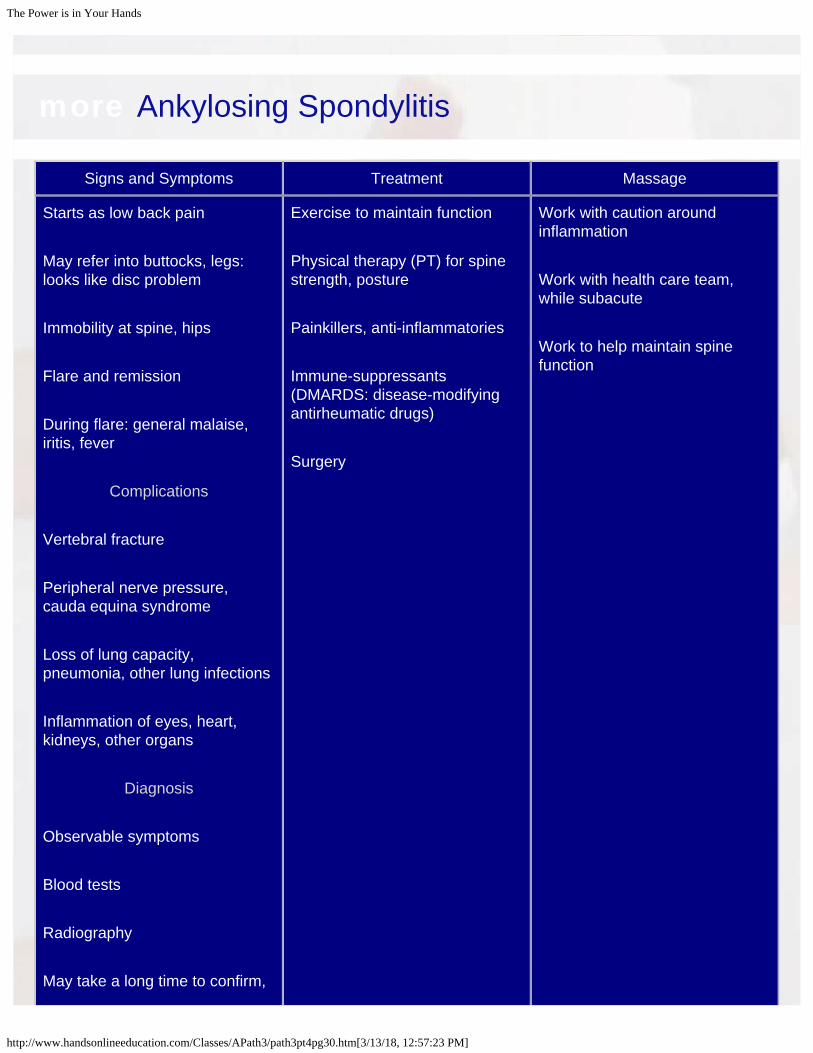
more Ankylosing Spondylitis
Signs and Symptoms Treatment Massage
Starts as low back pain May refer into buttocks, legs:looks like disc problem Immobility at spine, hips Flare and remission During flare: general malaise,iritis, fever
Complications
Vertebral fracture Peripheral nerve pressure,cauda equina syndrome Loss of lung capacity,pneumonia, other lung infections Inflammation of eyes, heart,kidneys, other organs
Diagnosis
Observable symptoms Blood tests Radiography May take a long time to confirm,
Exercise to maintain function Physical therapy (PT) for spinestrength, posture Painkillers, anti-inflammatories Immune-suppressants(DMARDS: disease-modifyingantirheumatic drugs) Surgery
Work with caution aroundinflammation Work with health care team,while subacute Work to help maintain spinefunction

The Power is in Your Hands
http://www.handsonlineeducation.com/Classes/APath3/path3pt4pg30.htm[3/13/18, 12:57:23 PM]
esp. in women
pt 4 Back Next
Copyright HandsOn Therapy Schools 2009 PATH3

The Power is in Your Hands
http://www.handsonlineeducation.com/Classes/APath3/path3pt4pg31.htm[3/13/18, 12:57:40 PM]
DislocationsBones in a joint are separated to that they no longerarticulate; Other soft tissue damage too
Copyright 2009 Walters Kluwers Health l Lippincott Williams & Wilkins
EtiologyUsually significant force Shoulder most often Fingers Congenital weakness in connective tissues(Marfan, Ehlers-Danlos) Hip dysplasia may be present at childbirth, canlead to osteoarthritis in adulthood

The Power is in Your Hands
http://www.handsonlineeducation.com/Classes/APath3/path3pt4pg31.htm[3/13/18, 12:57:40 PM]
Copyright 2009 Walters Kluwers Health l Lippincott Williams & Wilkins
pt 4
Back NextCopyright HandsOn Therapy Schools 2009 PATH3

The Power is in Your Hands
http://www.handsonlineeducation.com/Classes/APath3/path3pt4pg32.htm[3/13/18, 12:57:53 PM]
more Dislocations
Signs and Symptoms Treatment Massage
Swelling, discoloration, loss offunction, pain
Complications
Fibrosis, scar tissue Damage to blood vessels, otherstructures Ligament laxity Subluxation, spontaneousdislocation, osteoarthritis
For large joints: immediatereduction Radiography to rule out fracture Splinting, exercise, PT Other interventions: ligament-shortening surgery, thermalcapsulorrhaphy, proliferantinjections
Avoid while acute; in subacutestage work for scar tissueresolution, improved ROM Be careful about positioning oflax joints
pt 4 Back Next
Copyright HandsOn Therapy Schools 2009 PATH3

The Power is in Your Hands
http://www.handsonlineeducation.com/Classes/APath3/path3pt4pg33.htm[3/13/18, 12:58:07 PM]
GoutChemistry-based inflammatory arthritis Demographics
Men > women 10:1Women tend to be postmenopausal
1 million + in the United States
Copyright 2009 Walters Kluwers Health l Lippincott Williams & Wilkins
EtiologyUric acid is not extracted Metabolic gout: kidney function is normal; uric acidlevels are high Renal gout: uric acid is normal; kidneys are impaired Both: Kidneys are compromised and uric acid levelsare high May be triggered by:
Binge eating, drinking, surgery, suddenweight loss, infection
Uric acid accumulates, crystallizes Usually around great toe Usually sudden onset Tophi may develop later (deposits of sodium urate)
Risk Factors
High-purine diet (red meat, organ meats, shellfish,alcohol, lentils, mushrooms, peas, asparagus, spinach) Obesity Sudden weight changes Alcohol consumption

The Power is in Your Hands
http://www.handsonlineeducation.com/Classes/APath3/path3pt4pg33.htm[3/13/18, 12:58:07 PM]
Hypertension Some blood disorders One attack may be followed by others with increasingfrequency
pt 4
Back NextCopyright HandsOn Therapy Schools 2009 PATH3

The Power is in Your Hands
http://www.handsonlineeducation.com/Classes/APath3/path3pt4pg34.htm[3/13/18, 12:58:21 PM]
more Gout
Signs and Symptoms Treatment Massage
Sudden onset, usually at feet Extremely painful inflammation May cause fever May cause punched-outformation in bone Kidney stones, renal failure,high blood pressure,cardiovascular disease: allinterrelated
DiagnosisPain profile Distinguish from pseudogout forchemical accuracy Aspirated fluid shows uric acidcrystals
Drugs:
Pain relief (not aspirin)Anti-inflammatories
Metabolism/uric acid management Hydration Losing weight Changing diet
At least local contraindication;no ice! Get information oncardiovascular/kidney health
pt 4 Back Next
Copyright HandsOn Therapy Schools 2009 PATH3

The Power is in Your Hands
http://www.handsonlineeducation.com/Classes/APath3/path3pt4pg35.htm[3/13/18, 12:58:34 PM]
Lyme DiseaseInfection with spirochete Borrelia burgdorferi; Twospecies of deer ticks: Ixodes scapularis, Ixodespacificus
DemographicsMontana is only state with no Lyme
disease reported
90% cases in Northeast and mid-Atlantic, Wisconsin, Minnesota
At risk: work and play in grassy or
wooded areas
20,000 diagnoses/year in the UnitedStates; also in Europe and Asia
Copyright 2009 Walters Kluwers Health l Lippincott Williams & Wilkins
Etiology
Ticks live about 2 years In spring/summer of first year they crawl ontobushes and stems to find a warm-blooded host Pick up B. burgdorferi from deer or othermammals; pass on to humans Slow-growing bacterium that invades severaltypes of tissues
pt 4 Back Next
Copyright HandsOn Therapy Schools 2009 PATH3

The Power is in Your Hands
http://www.handsonlineeducation.com/Classes/APath3/path3pt4pg36.htm[3/13/18, 12:58:46 PM]
more Lyme Disease
Signs and Symptoms Treatment Massage
StagesEarly local disease
Symptoms appear 7–30 days aftertick bite. Bull’s-eye rash , highfever, fatigue, night sweats, stiffneck, headache. (Often no rash ispresent; looks like flu,mononucleosis)
Early disseminated disease
Systemic symptoms develop:
Cardiovascular:irregular heart beat,dizziness Neurological:headaches, Bell palsy,numbness, tingling,forgetfulness General: debilitatingfatigue
Late diseaseInfection of one or more joints:knee, elbow, shoulder. Usuallythree joints or fewer. Can causepermanent damage. Looks likerheumatoid arthritis. Symptoms usually last weeks tomonths, then subside Some get progressively worse
Antibiotics, long course for slow-growing bacteria (up to 12months)
Prevention
Long sleeves, pants Light-colored clothing Insect repellants Examine skin Remove ticks with tweezers,take to doctor (if removed within24 hours, risk of infection is verylow)
Contraindicated when joints areacutely inflamed Be careful aboutneurological/circulatorycomplications Know what ticks look like ifworking in endemic area

The Power is in Your Hands
http://www.handsonlineeducation.com/Classes/APath3/path3pt4pg36.htm[3/13/18, 12:58:46 PM]
Diagnosis
Difficult to be accurate Blood tests identify exposure, notwhether symptoms are related tocurrent infection False negatives Other tick-borne diseases
pt 4 Back Next
Copyright HandsOn Therapy Schools 2009 PATH3

The Power is in Your Hands
http://www.handsonlineeducation.com/Classes/APath3/path3pt4pg37.htm[3/13/18, 12:58:59 PM]
OsteoarthritisSynovial joints (especially weight bearing); Usually due toage, wear and tear; Also called degenerative joint disease
DemographicsMost common type of arthritis
20 million to 40 million in the United
States
Men about equal to women; womenhave it more severely
Leading risk factors:
AgeOverweight
Massage therapists: take care of
saddle joint!
Copyright 2009 Walters Kluwers Health l Lippincott Williams & Wilkins
Etiology
Precarious environment inside joints; once damageoccurs, it is difficult to reverse Cartilage
Articular cartilage: small number ofchondrocytes with proteoglycans thatattract water
Arrangement varies byregionsSuperficial (in joint space)IntermediateDeep (attaches to bone)
Resistance to shearing andcompressive forces
Chondrocytes are active all through life, replacingand rebuilding surface
Don’t migrate to areas of damageWhen cartilage is damaged,chondrocytes make less fluid andcollagen
Cartilage degradesOsteocytes in epiphysesbecome active: bone spurs,may be cystlike cavitiesunder cartilage
Causes
Age: dry, prone to injury Overweight: stress on knees, hips

The Power is in Your Hands
http://www.handsonlineeducation.com/Classes/APath3/path3pt4pg37.htm[3/13/18, 12:58:59 PM]
Lax ligaments: unstable joints History of trauma, arthroscopic surgery Repetitive pounding stress Others: Hormonal imbalance, nutritionaldeficiency, trigger foods, etc.
pt 4
Back NextCopyright HandsOn Therapy Schools 2009 PATH3

The Power is in Your Hands
http://www.handsonlineeducation.com/Classes/APath3/path3pt4pg38.htm[3/13/18, 12:59:14 PM]
more Osteoarthritis
Signs and Symptoms Treatment Massage
Deep pain, stiffness; especiallywithout warmup or with overuse At fingers: phalangeal epiphyseswiden At distal interphalangeal joints(DIPs): Heberden nodes At proximal interphalangealjoints (PIPs): Bouchard nodes
Diagnosis
Physical examination, patienthistory Rule out other causes of jointinflammation; radiography notconclusive
Goals: reduce inflammation, limitor reverse damage Nonsteroidal anti-inflammatorydrugs (carry some risks) Topical applications: camphor,menthol, capsaicin Exercise: within pain tolerance forthree goals:
Improve and maintainhealthy range ofmotionIncrease stamina andlose weightImprove the strengthof musclessurrounding affectedjoints
Nutritional supplements:Glucosamine and chondroitinsulfate
Popular and showresults for mild tomoderate arthritisGlucosamine mayaffect insulin levels indiabetic patientsMade from the shellsof shellfish (watch forallergies)Chondroitin may affectblood clotting
Arthroscopic procedures:
Can be useful to reduce pain,ease muscle tension; Doesn’trebuild damaged cartilage

The Power is in Your Hands
http://www.handsonlineeducation.com/Classes/APath3/path3pt4pg38.htm[3/13/18, 12:59:14 PM]
Proliferant injectionsCorticosteroidinjectionsSynovial fluidwithdrawalJoint lavage anddebridement
Joint replacement surgery:256,000 knee replacements,117,000 hip replacements peryear Procedures in development:numerous strategies are indevelopment:
Cartilage pasteDrill into epiphyses tostimulate cartilagegrowthTransplantosteochondral plugsOthers
pt 4 Back Next
Copyright HandsOn Therapy Schools 2009 PATH3

The Power is in Your Hands
http://www.handsonlineeducation.com/Classes/APath3/path3pt4pg39.htm[3/13/18, 1:00:16 PM]
Patellofemoral SyndromePatellar cartilage is damaged: precursor of osteoarthritis atthe knee; also called jumper’s knee; anterior knee painsyndrome; overuse syndrome
Etiology
Two main contributors
Overuse/overloading;Percussive activity withtwisting, jumping Poor alignment; Especially withoverweight, poorfootwear, unevensurfaces, muscularimbalance
pt 4 Back Next
Copyright HandsOn Therapy Schools 2009 PATH3

The Power is in Your Hands
http://www.handsonlineeducation.com/Classes/APath3/path3pt4pg40.htm[3/13/18, 1:00:28 PM]
more Patellofemoral Syndrome
Signs and Symptoms Treatment Massage
Pain at anterior aspect of knee Stiffness after immobility Difficulty with walking, especiallydown stairs Crepitus
Diagnosis Can be difficult; looks likepatellar tendinitis (whichresponds to massage)
Change activity Physical therapy: Quads, hams,tensor fascia latae (TFL), deeplateral rotators Ice Nonsteroidal anti-inflammatories(NSAIDs) Orthotics Knee brace, taping
Irritation is inside joint capsule;not in reach for massage; canaddress pain, stiffness, tension,alignment
pt 4 Back Next
Copyright HandsOn Therapy Schools 2009 PATH3

The Power is in Your Hands
http://www.handsonlineeducation.com/Classes/APath3/path3pt4pg41.htm[3/13/18, 1:00:40 PM]
Rheumatoid ArthritisAutoimmune attack on synovial membranes; can involveinflammation elsewhere too
Demographics
3.1 million in the United States
Women > men, 3:1
Mostly 20–50 years old, can be inchildren
Copyright 2009 Walters Kluwers Health l Lippincott Williams & Wilkins
Etiology
Immune system attacks synovial membranes
Can affect other areas: blood vessels,serous membranes, skin, eyes, lungs, liver,heart)
B cells, T cells, antibodies, inflammatory chemicals arepresent in joint during flare
Synovial membrane thickens, swellsFluid accumulatesInflamed tissue releases enzymes thaterode cartilageDeformation of joints
pt 4 Back Next
Copyright HandsOn Therapy Schools 2009 PATH3

The Power is in Your Hands
http://www.handsonlineeducation.com/Classes/APath3/path3pt4pg42.htm[3/13/18, 1:00:54 PM]
more Rheumatoid Arthritis
Signs and Symptoms Treatment Massage
Flare and remission Prodrome: malaise precedes sharp,specific joint pain Rheumatic nodules Joints are hot, painful, stiff
May improve with gentlemovementKnuckles in hands, toes,ankles, wrists
Bilateral, may not be symmetrical
Complications
During flares
Rheumatic nodules on thescleraSjögren syndromePleuritisCarditis or pericarditisHepatitisVasculitisRaynaud syndrome, skinulcers, bleeding intestinalulcers, and internalhemorrhaging.Bursitis and anemia, esp.with childhood onset
Between flares:
DislocationsRuptured tendons
Goals
Reduce painLimit inflammationStop damageImprove function
First-line drugs: NSAIDs,steroids, cyclo-oxygenase-2inhibitors (with exercise,hydrotherapy, PT,occupational therapy [OT]) Second-line drugs: biologicalresponse modifiers,immunosuppressant drugs Other: diet, exercise, stress-reduction Surgery if necessary
Avoid circulatory massagewhile acute Between flares work for painreduction, improved ROM,lower muscle tension

The Power is in Your Hands
http://www.handsonlineeducation.com/Classes/APath3/path3pt4pg42.htm[3/13/18, 1:00:54 PM]
Collapse at C1-C2
Diagnosis History, radiography, blood test forrheumatoid factor At least four of these:
Morning stiffness that lastsat least 1 hourArthritis in three or morejointsInvolvement of PIPs,metacarpophalangeal joints(MCPs), DIPsBilateralPositive serum rheumatoidfactorRheumatoid nodules
Radiographic evidence
pt 4 Back Next
Copyright HandsOn Therapy Schools 2009 PATH3

The Power is in Your Hands
http://www.handsonlineeducation.com/Classes/APath3/path3pt4pg43.htm[3/13/18, 1:01:12 PM]
SpondylosisOsteoarthritis at spine; Age-related changes of thevertebrae, discs, joints, and ligaments of the spine
Copyright 2009 Walters Kluwers Health l Lippincott Williams & Wilkins
Copyright 2009 Walters Kluwers Health l Lippincott Williams & Wilkins
Etiology
Osteophytes grow on vertebrae
Can be on vertebral bodies or facetsCan put pressure on nerve roots orspinal cord
Intervertebral joints analogy with synovial joints:
Vertebral bodies = articulating bonesAnnulus fibrosis = capsular ligamentNucleus pulposus = synovial fluidShearing and compressive forceswear on cartilage, disc thins, bonespurs develop
Not all osteophytes cause pain (radiography notdefinitive for cause of pain) Age contributes to ossification of anteriorlongitudinal ligament, posterior longitudinalligament, ligamentum flavum
DISH (diffuse idiopathic skeletalhyperostosis) may cause gradualpainless loss of ROM
More typical development of arthritis at facets, SIjoint, costovertebral joints
pt 4
Back NextCopyright HandsOn Therapy Schools 2009 PATH3

The Power is in Your Hands
http://www.handsonlineeducation.com/Classes/APath3/path3pt4pg44.htm[3/13/18, 1:01:25 PM]
more Spondylosis
Signs and Symptoms Treatment Massage
May be silent Painless progressive loss ofROM Pain if nerve roots arecompressed Spinal cord compression: pain,loss of bowel/bladder control
Complications
Spreading problems in the spine Nerve pain Secondary spasm Blood vessel pressure Spinal cord pressure
Diagnosis
Radiography, MRI
Anti-inflammatories, exercise,massage, acupuncture,hydrotherapy
Locally injectedsteroids, surgery
Caution for nerve irritation,positioning, muscle splinting
pt 4 Back Next
Copyright HandsOn Therapy Schools 2009 PATH3

The Power is in Your Hands
http://www.handsonlineeducation.com/Classes/APath3/path3pt4pg45.htm[3/13/18, 1:01:40 PM]
SprainsTorn ligaments Distinguishing Features
Sprains are injured ligaments, notmuscles or tendons
Sprains are more serious than
strains and tendinosis
Sprains tend to swell
Etiology
Linearly arranged collagen fibers link bone to bone
Injured when some fibers are rippedFirst, second, third degree (rupture)
Repair: laying down new collagen fibers
Begins disorganized and weakAligns according to weight-bearing forceWithout stress during healing, scar tissueremains weak and disorganized
pt 4
Back NextCopyright HandsOn Therapy Schools 2009 PATH3

The Power is in Your Hands
http://www.handsonlineeducation.com/Classes/APath3/path3pt4pg46.htm[3/13/18, 1:01:56 PM]
more Sprains
Signs and Symptoms Treatment Massage
Acute Stage
Pain, heat, redness, swelling,loss of function
Significant swelling,esp. if connected tojoint capsuleAnterior talofibularligament is mostcommonly sprained
Subacute Stage
Inflammation subsides
24–48 hours later,depending onseveritySome injuries goback and forth,depending on usage
Complications
Masking symptoms especially ofminor fractures Repeated injury, with poor-quality healing Ligament laxity collagen haspoor rebound; can lead toosteoarthritis
RICE (rest, ice, compression,elevation) PRICEMMM (protection, rest,ice, compression, elevation,medicine, mobility, modalities)
Indicated when subacute forimproved circulation, scar tissueformation, stiffness
pt 4

The Power is in Your Hands
http://www.handsonlineeducation.com/Classes/APath3/path3pt4pg47.htm[3/13/18, 1:02:17 PM]
Temporomandibular Joint DisordersCollection of signs and symptoms associated with jawproblems; also called TMD: temporomandibular jointdisorders
Demographics
An estimated 10 million in the UnitedStates (not all seek help)
Women > men
Copyright 2009 Walters Kluwers Health l Lippincott Williams & Wilkins
Etiology
TMJ has huge mobility:
Elevation, depression, retraction,protraction, side flexionJoint capsule stretches
Fibrocartilage disc can get injured (video clip 1) Muscles develop trigger points
Causes May be initiated by fall or motor vehicle accident(MVA): jawlash Can be spontaneous, connected to stress, bruxism Symptoms and causes can be circ Other factors
Misalignment at jaw, biteHormonal sensitivity?High overlap between ligament laxityand heart valve problems: connectivetissue quality issues?Frequently seen with fibromyalgia,chronic myofascial pain syndrome,irritable bowel syndrome
pt 4

The Power is in Your Hands
http://www.handsonlineeducation.com/Classes/APath3/path3pt4pg48.htm[3/13/18, 1:03:39 PM]
more Temporomandibular Joint Disorders
Signs and Symptoms Treatment Massage
Jaw, neck, and shoulder pain Limited range of motion Popping in the jaw Locking of the joint Grinding teeth (bruxism) Ear pain Headaches Chronic misalignment of cervicalvertebrae
Diagnosis
Differentiate from myofascial painsyndrome, other tension patternsthat cause pain in face and head
Sprain of ligament thatattachesstylomandibular joint tobase of the skull: alsocalled Ernest syndromeTrigeminal neuralgiaOccipital neuralgiaOsteomyelitis
MRI, radiography,electromyography, clinicalexamination can yield information
Nonsurgical: Hot/cold; PT,ultrasound, massage, anti-inflammatories, localanesthetics, splints, proliferantinjections Surgical: dissolve adhesionsand scar with injections;arthroscopic surgery; jointreplacement
Can be useful to interrupt theprocess before permanentdamage occurs
Reduce muscletension, improveawareness, addressreferred painpatterns

The Power is in Your Hands
http://www.handsonlineeducation.com/Classes/APath3/path3pt4pg48.htm[3/13/18, 1:03:39 PM]
on cartilage damage, musclefunction, subluxation
pt 4 Back Next
Copyright HandsOn Therapy Schools 2009 PATH3

The Power is in Your Hands
http://www.handsonlineeducation.com/Classes/APath3/path3pt5pg49.htm[3/13/18, 1:06:02 PM]
Genetic Musculoskeletal Disorders
Ehlers-Danlos Syndrome
Margan Syndrome
Muscular Dystrophy
Osteogenesis Imperfecta
pt 5 Back Next
Copyright HandsOn Therapy Schools 2009 PATH3

The Power is in Your Hands
http://www.handsonlineeducation.com/Classes/APath3/path3pt5pg50.htm[3/13/18, 1:06:24 PM]
Ehlers-Danlos SyndromeGroup of genetic disorders leading to connective tissueweakness
Demographics
Rare: about 50,000 in the UnitedStates, but many with mild form
Men = women
No racial predisposition
Etiology
Genetic mutation affects collagen, elastin,other extracellular matrix of connectivetissues
Hypermobility of jointsChronic joint painDelicate skinPoor wound healing
Most common form passed throughautosomal dominant genes: if one parent is acarrier, each child has a 50% chance ofdeveloping EDS Other types are recessive: both parents mustcarry the gene
pt 5
Back NextCopyright HandsOn Therapy Schools 2009 PATH3

The Power is in Your Hands
http://www.handsonlineeducation.com/Classes/APath3/path3pt5pg51.htm[3/13/18, 1:06:36 PM]
more Ehlers-Danlos Syndrome
Signs and Symptoms Treatment Massage
Depends on genetic anomaly
Easy bruising; poorwound healing; frequentjoint dislocations; eyeproblems (detachedretina, myopia); mitralvalve prolapseRarely: extremepostural deviations,baggy skin
Several types:
Classic EDSHypermobility EDSVascular EDSKyphoscoliosis EDSArthrochalasia EDSDermatosparaxis EDS
Diagnosis
Genetic testing not alwaysconclusive
Family history with signsand symptoms
Mild EDS may not be identified, butchildren can have it in moreextreme form: genetic counseling isimportant
Treated by symptom
Education topreserve jointfunctionSkin careSpecial care withdental workHigh-risk pregnancy
High doses of vitamin C mayimprove some connectivetissue strength
Appropriate if heart is healthyand joints not stretched too far
Delicate skin, easybruising
pt 5 Back Next
Copyright HandsOn Therapy Schools 2009 PATH3

The Power is in Your Hands
http://www.handsonlineeducation.com/Classes/APath3/path3pt5pg52.htm[3/13/18, 1:06:49 PM]
Marfan SyndromeGenetic mutation causes production of dysfunctionalfibrillin
Demographics
200,000 in the United States haveMarfan or a related disorder
Usually passed from parent to child
25% = spontaneous mutation
Copyright 2009 Walters Kluwers Health l Lippincott Williams & Wilkins
Etiology
Faulty protein fibers → connective tissues areweak Musculoskeletal system, meninges, heart, aorta,eyes most at risk
pt 5 Back Next
Copyright HandsOn Therapy Schools 2009 PATH3

The Power is in Your Hands
http://www.handsonlineeducation.com/Classes/APath3/path3pt5pg53.htm[3/13/18, 1:07:01 PM]
more Marfan Syndrome
Signs and Symptoms Treatment Massage
Ranges from mild to severe
Musculoskeletal systemanomalies: long fingersand toes, arms and legs;protruding or sunkensternum; posturaldeviationsCardiovascular systemanomalies: aortic andmitral valves maycollapse → heartproblems; risk ofaneurysm, aorticdissectionEye disorders: myopia,dislocated lens,detached retinaNervous systemanomalies: stretched,weakened dura mater:dural ectasiaOther symptoms: stretchmarks, hernias, flat feet,spondylolisthesis, andhammertoes
DiagnosisNo simple genetic test
Clinical examination,family history,observation
By symptom
Beta blockers toreduce force onaortaBlood pressuremedicationProphylacticantibiotics to protectheart valvesSurgery to correctspine, thorax, heartvalves if necessary
Can be appropriate with carefor delicate tissues, high risk ofheart/aorta problems Work with health care team
pt 5 Back Next
Copyright HandsOn Therapy Schools 2009 PATH3

The Power is in Your Hands
http://www.handsonlineeducation.com/Classes/APath3/path3pt5pg54.htm[3/13/18, 1:07:12 PM]
Muscular DystrophyGroup of related diseases with genetic anomalies; Degeneration, wasting of muscle tissue
Demographics
Duchenne and Becker are X-linked
Carried by mother, passed tosons
400–600 born each year
Other types not gender specific:
males = females
Etiology
Normal muscles use a protein, dystrophin, to helpconvert fat or glycogen into fuel
The most common forms of MD involveinadequate production dystrophinMuscle cells atrophy and die, replaced by fatand connective tissueContractures develop
Duchenne muscular dystrophy: most common: 1:3500male babies. No dystrophin is produced Becker muscular dystrophy: less common, less severe:1:30,000 boys, some dystrophin is produced Myotonic muscular dystrophy: most common adult-onset MD; myotonia, cataracts, GI dysfunction, heartproblems Other varieties
Congenital muscular dystrophyFacioscapulohumeral dystrophyLimb-girdle dystrophyEmery-Dreifuss muscular dystrophyOculopharyngeal muscular dystrophy
pt 5
Back NextCopyright HandsOn Therapy Schools 2009 PATH3

The Power is in Your Hands
http://www.handsonlineeducation.com/Classes/APath3/path3pt5pg55.htm[3/13/18, 1:07:26 PM]
more Muscular Dystrophy
Signs and Symptoms Treatment Massage
Vary by type
Duchenne and Beckerare similar
A toddler hasdifficultywalkingLeg pain,waddling gait,lumbar curve,walks on toesCan also affectspine, joints,heart, lungs
Most Becker MD patients die youngwith cardiac or respiratory failure
Diagnosis
Much easier to find now
Blood test for creatinekinaseLook for neurologicalproblemsBiopsy
Interventions to prolongactivity, life expectancy Massage, PT to minimizecontractures Surgery to release tighttendons, correct spine Steroids Assistive devices asnecessary
Sensation is intact: massage issafe
Check forcirculatory health,other complicationsof lost movement
Work with health care team
pt 5 Back Next
Copyright HandsOn Therapy Schools 2009 PATH3

The Power is in Your Hands
http://www.handsonlineeducation.com/Classes/APath3/path3pt5pg56.htm[3/13/18, 1:07:37 PM]
Osteogenesis ImperfectaGroup of genetic disorders that changes the quality of type Icollagen fibers; Four main subtypes; (other, much rarer types)
Demographics
Type I most common: 1 in 30,000births
Type II: 1 in 60,000 births
Type III: 1 in 70,000 births
Type IV and others: very rare
20,000–50,000 in United States
have OI
Males = females
Autosomal dominant: if one parenthas the gene, each child has a 50%
chance of having OI
About 25% of cases spontaneouswith no family history
Etiology
Type I collagen is a triple helix of intertwiningprocollagen fibers OI is shortage or faulty production of type I collagen
pt 5 Back Next
Copyright HandsOn Therapy Schools 2009 PATH3

The Power is in Your Hands
http://www.handsonlineeducation.com/Classes/APath3/path3pt6pg58.htm[3/13/18, 1:07:51 PM]
Other Connective Tissue Disorders
Baker Cyst
Bunions
Bursitis
Dupuytren Contracture
Ganglion Cysts
Hernia
Osgood-Schlatter Disease
Pes Planus, Pes Cavus
Plantar Fascitis
Scleroderma
Tendinopathies
Tenosynovitis
Whiplash
pt 6 Back Next
Copyright HandsOn Therapy Schools 2009 PATH3

The Power is in Your Hands
http://www.handsonlineeducation.com/Classes/APath3/path3pt6pg59.htm[3/13/18, 1:08:02 PM]
Baker CystSynovial cysts at the popliteal fossa, usually on medialside; also called popliteal cysts
Copyright 2009 Walters Kluwers Health l Lippincott Williams & Wilkins
Etiology
Joint capsule at knee develops a pouch
Common in children
In adults, may be related to other joint problems:
Osteoarthritis, rheumatoid arthritis,cruciate ligament tears, meniscus tears
Complications
Could impair blood flow
Risk of thrombophlebitis, deep veinthrombosis (DVT)
Risk of rupture, bleeding in joint, infection,posterior compartment syndrome

The Power is in Your Hands
http://www.handsonlineeducation.com/Classes/APath3/path3pt6pg59.htm[3/13/18, 1:08:02 PM]
Copyright 2009 Walters Kluwers Health l Lippincott Williams & Wilkins
pt 6
Back NextCopyright HandsOn Therapy Schools 2009 PATH3

The Power is in Your Hands
http://www.handsonlineeducation.com/Classes/APath3/path3pt6pg60.htm[3/13/18, 1:08:24 PM]
more Baker Cyst
Signs and Symptoms Treatment Massage
Usually silent; knee may bepainful from underlying problem May feel full or tight on medialaspect of calf
Ice, NSAIDs Aspiration, cortisone shots
May recur
Local contraindication; calfsymptoms may be a red flag forDVT
pt 6 Back Next
Copyright HandsOn Therapy Schools 2009 PATH3

The Power is in Your Hands
http://www.handsonlineeducation.com/Classes/APath3/path3pt6pg61.htm[3/13/18, 1:08:37 PM]
BunionsAlso called hallux valgus: laterally deviated big toe;at little toe: bunionette
Demographics
Women > men, 10:1
High-heeled, narrow-toed shoes
Genetic predisposition
Copyright 2009 Walters Kluwers Health l Lippincott Williams & Wilkins
Etiology
Factors that lead to misalignment betweenfirst metatarsal and proximal phalanx of greattoe:
Pes cavus, pes planusShape of the bonesMuscle imbalanceFootwear
Joint is distorted, bunion on top is irritated May develop bone spurs, osteoarthritis
pt 6 Back Next
Copyright HandsOn Therapy Schools 2009 PATH3

The Power is in Your Hands
http://www.handsonlineeducation.com/Classes/APath3/path3pt6pg62.htm[3/13/18, 1:08:48 PM]
more Bunions
Signs and Symptoms Treatment Massage
Lump on medial side ofmetatarsophalangeal (MTP) jointof great toe
May be hot andpainful
Remove irritants, improvefootwear Massage and exercise for foothealth ROM, traction, gentle friction Cortisone injection
Surgical correction
Locally contraindicated wheninflamed, otherwise appropriate
Work with othercompensationpatterns, intrinsic footmuscles
pt 6 Back Next
Copyright HandsOn Therapy Schools 2009 PATH3

The Power is in Your Hands
http://www.handsonlineeducation.com/Classes/APath3/path3pt6pg63.htm[3/13/18, 1:08:59 PM]
BursitisSynovial sacs outside joint capsules becomeinflamed
Copyright 2009 Walters Kluwers Health l Lippincott Williams & Wilkins
Copyright 2009 Walters Kluwers Health l Lippincott Williams & Wilkins
Etiology
Bursae act as shock absorbers and reducefriction where tendons cross over bones Repetitive stress irritates bursae
Pain, limited ROM, muscletightness
Accompanies general inflammation, gout,rheumatoid arthritis, etc. Can be from infection, especially at knee orolecranon
pt 6 Back Next
Copyright HandsOn Therapy Schools 2009 PATH3

The Power is in Your Hands
http://www.handsonlineeducation.com/Classes/APath3/path3pt6pg64.htm[3/13/18, 1:09:10 PM]
more Bursitis
Signs and Symptoms Treatment Massage
Pain on passive and activemovement Limited ROM (muscle splinting) Often no heat is palpable
Diagnosis
Patient history: consider otherlocal injuries
NSAIDs, warm packs Aspiration, cortisone injection Bursectomy (may grow back) New movement patterns!
Local contraindication whileacute Otherwise appropriate: work todecompress surroundingmuscles
Avoid infection
pt 6 Back Next
Copyright HandsOn Therapy Schools 2009 PATH3

The Power is in Your Hands
http://www.handsonlineeducation.com/Classes/APath3/path3pt6pg65.htm[3/13/18, 1:09:22 PM]
Dupuytren ContractureIdiopathic shrinking and thickening of palmar fascia; alsocalled palmar fasciitis
DemographicsMen > women
Middle-aged, Northern European
descent
Some genetic predisposition
Other risk factors:Smoking, alcohol use, seizure
disorders, type 1 and 2 diabetes
Copyright 2009 Walters Kluwers Health l Lippincott Williams & Wilkins
Etiology
Idiopathic
Looks like excessive posttrauma scartissue: type III collagen in palmar fascia andfingers
Collagen thickens and gets denser; living cells recede
Flexion may be normal; extension is limited
Similar connective tissue phenomena:
Plantar fibromatosis (Ledderhose disease)on sole of footPeyronie disease under skin on shaft ofpenisKnuckle pads (Garrod nodes) at DIPs ofhands
pt 6 Back Next
Copyright HandsOn Therapy Schools 2009 PATH3

The Power is in Your Hands
http://www.handsonlineeducation.com/Classes/APath3/path3pt6pg66.htm[3/13/18, 1:09:34 PM]
more Dupuytren Contracture
Signs and Symptoms Treatment Massage
Ring and little fingers affectedmost Begins as mildly tender bump;cord extends into palm, towardfinger Bilateral about 50% of time Can be slow or fast, mild orsevere Constricted nerve, blood supplymay lead to amputation
Without treatment, can lead toloss of function in affectedfingers Injections with cortisone,collagenase, needleaponeurotomy Surgery if necessary Recurs about one-third of time
As long as sensation is present,massage is safe; may not makesignificant changes May be useful post surgery tohelp recover function
pt 6 Back Next
Copyright HandsOn Therapy Schools 2009 PATH3

The Power is in Your Hands
http://www.handsonlineeducation.com/Classes/APath3/path3pt6pg67.htm[3/13/18, 1:09:46 PM]
Ganglion CystsPouches on joint capsules or tendinous sheaths
Copyright 2009 Walters Kluwers Health l Lippincott Williams & Wilkins
Etiology
May grow with trauma or overuse; many arespontaneous Filled with viscous fluid, may have multiple lobes May grow in a place to interfere with movement or limitfunction
Mucous cysts grow on DIPs, may distortgrowth of fingernail
pt 6 Back Next
Copyright HandsOn Therapy Schools 2009 PATH3

The Power is in Your Hands
http://www.handsonlineeducation.com/Classes/APath3/path3pt6pg68.htm[3/13/18, 1:09:57 PM]
more Ganglion Cysts
Signs and Symptoms Treatment Massage
Range from tiny to large Not usually painful unlessirritated
Usually resolve spontaneously Cortisone injection, aspiration,surgical removal (often growback)
Don’t smash with aBible!
Local contraindication May be irritated with friction Untreated bumps need diagnosis
pt 6 Back Next
Copyright HandsOn Therapy Schools 2009 PATH3

The Power is in Your Hands
http://www.handsonlineeducation.com/Classes/APath3/path3pt6pg69.htm[3/13/18, 1:10:06 PM]
HerniaHole in abdominal wall, diaphragm Demographics
5 million diagnosed per year
700,000 surgeries
Men with abdominal hernias >
women: 7:1
Copyright 2009 Walters Kluwers Health l Lippincott Williams & Wilkins
Etiology
Several factors
Weakness of abdominal wall; straining;childbirthSmall intestines can protrude, get caughtand damagedWeak spot at inguinal canal for men
pt 6 Back Next
Copyright HandsOn Therapy Schools 2009 PATH3

The Power is in Your Hands
http://www.handsonlineeducation.com/Classes/APath3/path3pt6pg70.htm[3/13/18, 1:10:16 PM]
more Hernia
Signs and Symptoms Treatment Massage
Inguinal hernia: most commonvariety; occur at inguinal ring Epigastric hernia: aboveumbilicus; linea alba splits Paraumbilical hernia: linea albasplits at umbilicus Umbilical hernia: most commonin newborn babies; usuallycloses by age 2 Femoral hernia: Most common inwomen; bulge at femoral ringbelow inguinal ligament. Risk ofstrangulation is high Hiatal hernia: Diaphragmatichiatus is stretched; stomachbulges into thorax Other hernias: at incisions,obturator, lateral aspect of rectusabdominus
Complications Bigger = safer for short term(less risk of strangulation) Strangulation can lead toinfection
Surgical repair
Truss is temporarysolution
Local contraindication at herniaand for recent surgery For past surgery, no cautions

The Power is in Your Hands
http://www.handsonlineeducation.com/Classes/APath3/path3pt6pg71.htm[3/13/18, 1:10:34 PM]
Osgood-Schlatter DiseaseIrritation and inflammation at quadriceps attachment ontibia; also called tibial tuberosity apophysitis
Demographics
Usually adolescent athletes Running, jumping sports
Boys > girls
Copyright 2009 Walters Kluwers Health l Lippincott Williams & Wilkins
Etiology
Rapid bone growth, especially at tibia and femurduring adolescence
Soft tissues may not keep up Quads are taxed with athletics
Stress at attachment leads to pain andinflammation Tibial tuberosity enlarges; microscopic fractures,possible avulsion
Usually unilateral
pt 6 Back Next
Copyright HandsOn Therapy Schools 2009 PATH3

The Power is in Your Hands
http://www.handsonlineeducation.com/Classes/APath3/path3pt6pg72.htm[3/13/18, 1:10:45 PM]
more Osgood-Schlatter Disease
Signs and Symptoms Treatment Massage
Acute: tibial tuberosity is hot,swollen, painful
Subacute: permanentremodeling of tibial tuberosity
Goals: reduce pain, limit damageto quad attachment Careful heating, warming upbefore activity Cooling down and stretching Rest if necessary Brace or cast followed byrehabilitative exercises Surgery if necessary
Locally contraindicated forcirculatory massage while acute Later, work to reduce pain atknee, stretch soft tissues,promote good quality healing
pt 6 Back Next
Copyright HandsOn Therapy Schools 2009 PATH3

The Power is in Your Hands
http://www.handsonlineeducation.com/Classes/APath3/path3pt6pg73.htm[3/13/18, 1:10:55 PM]
Pes Planus, Pes CavusPes planus = flat feet; Pes cavus = caved feet(jammed arches); Feet lack medial and lateralarches or arches don’t flatten and rebound
Etiology
Imbalance in forces at feet has repercussionsthrough the rest of the body Pes planus, cavus can be from congenitalproblems in bone shape; strength of footligaments; muscle imbalance; poor footwear
Underlying diseases that affectfeet
Charcot-Marie-Tooth syndrome; musculardystrophy; polio, cerebral palsy; neurologicaldamage
pt 6 Back Next
Copyright HandsOn Therapy Schools 2009 PATH3

The Power is in Your Hands
http://www.handsonlineeducation.com/Classes/APath3/path3pt6pg74.htm[3/13/18, 1:11:05 PM]
more Pes Planus, Pes Cavus
Signs and Symptoms Treatment Massage
Complications
Loss of shock absorption →
Change in footalignment Heel spurs Plantar fasciitis Neuromas Osteoarthritis at foot,knee, hip, SI, spine,TMJ, headaches, etc.
Especially an issue with poorperipheral circulation: diabetes,etc.
Improved footwear, orthotics PT to work with peroneuslongus, tibialis posterior If very extreme: surgical repair
Indicated
Can improve nutritionto ligaments, relievepain, work withcompensation
pt 6 Back Next
Copyright HandsOn Therapy Schools 2009 PATH3

The Power is in Your Hands
http://www.handsonlineeducation.com/Classes/APath3/path3pt6pg75.htm[3/13/18, 1:11:15 PM]
Plantar FascitisPain at plantar fascia; could be inflammatory ordegenerative
Demographics
2 million/year seek treatment
Men = women
Two groups more than others:Runners (up to 10%)
Older adults who are overweight
Etiology
Plantar fascia is vulnerable to damage
OverweightWorn-down shoesUnequal leg lengthFlat or pronated feet, jammed archesTight calf muscles
Secondary to
Gout, diabetes, rheumatoid arthritis
Fibers fray, become disorganized
Probably not usually inflamedDegeneration of collagen matrix(changes treatment options)
Radiography shows bone spurs (secondary,probably not causative of pain)
pt 6 Back Next

The Power is in Your Hands
http://www.handsonlineeducation.com/Classes/APath3/path3pt6pg76.htm[3/13/18, 1:11:30 PM]
more Plantar Fascitis
Signs and Symptoms Treatment Massage
Acutely painful after periods ofrest, immobility
Sharp, bruisedfeeling at anteriorcalcaneus or deep inarch Pain subsides withwarming up, returnswith fatigue
Remove tensions that reinjureplantar fascia
Warm, massagefoot/leg beforestandingOrthoticsNight splint to hold footin dorsiflexionNSAIDs, topical anti-inflammatories,massage, iceCortisone injections: Conservative;otherwise plantarfascia may ruptureShockwave lithotripsySurgery to divide,release damagedfasciaLong-lasting condition:6–18 months forresolution
Indicated to decrease tension incalf muscles, organize collagenwithin
pt 6 Back Next
Copyright HandsOn Therapy Schools 2009 PATH3

The Power is in Your Hands
http://www.handsonlineeducation.com/Classes/APath3/path3pt6pg77.htm[3/13/18, 1:11:43 PM]
SclerodermaAutoimmune disease leading to production of abnormalamounts of collagen, often in skin: hard skin; Othertissues may be affected
Demographics
About 300,000 in the United States
Women > men, 3–4:1
Copyright 2009 Walters Kluwers Health l Lippincott Williams & Wilkins
Etiology
Immune system attacks lining of small bloodvessels
Local edema, fibroblast stimulation Lots of type III collagen (basis for scartissue)
Local scleroderma: only skin is involved; mayaccumulate over years, then stabilize or reverse
Morphea scleroderma: oval patcheson trunk, face, extremities
Linear scleroderma: discolored line or band on aleg, arm, or over the forehead Systemic scleroderma: blood vessel damage inskin and other organs: digestive tract, heart,circulatory system, kidneys, lungs, synovialmembranes, tenosynovial sheaths
Limited systemic scleroderma: slowonset, may infiltrate other organs Diffuse scleroderma: sudden onset,earlier involvement of internal organs Sine scleroderma: internal organs only
Causes
Unknown; some factors:
Abnormal immune responses andchronic inflammation → excess

The Power is in Your Hands
http://www.handsonlineeducation.com/Classes/APath3/path3pt6pg77.htm[3/13/18, 1:11:43 PM]
collagen production Chimeric cells (genes of anotherperson) Chemical exposures Viral infections
pt 6
Back NextCopyright HandsOn Therapy Schools 2009 PATH3

The Power is in Your Hands
http://www.handsonlineeducation.com/Classes/APath3/path3pt6pg78.htm[3/13/18, 1:11:58 PM]
more Scleroderma
Signs and Symptoms Treatment Massage
CREST syndrome
C: Calcinosis:accumulation ofcalcium deposits in theskin, especially in thefingers R: Raynaudphenomenon E: Esophagealdysmotility S: Sclerodactyly:hardening of thefingers T: Telangiectasia
Other symptoms/complications:
Skin ulcers, changesin pigment, hair loss,weak muscles, swollenconnective tissues,lung damage, heartpain, arrhythmia, heartfailure, renal failure,trigeminal neuralgia,carpal tunnelsyndrome, Sjögrensyndrome
Manage symptoms,complications:
Drugs to manageRaynaud syndrome,kidney function,GERD, muscle andjoint pain, immunesystem overactivity PT, OT for flexibility,especially in hands Avoid smoking, coldtemperature, spicyfood
Depends on resiliency of client
Be careful ofcirculatory, kidneyhealth Bodywork thatdoesn’t challengefluid flow may bebeneficial
pt 6 Back Next
Copyright HandsOn Therapy Schools 2009 PATH3

The Power is in Your Hands
http://www.handsonlineeducation.com/Classes/APath3/path3pt6pg79.htm[3/13/18, 1:12:10 PM]
TendinopathiesInjury, damage to tendons
Etiology
Tendons are made of type I collagen in liquid groundsubstance
Some elastin fibers are woven in for stretchand rebound (limited) Looks hard, shiny, white
With injury:
Collagen degenerates Tendon becomes weak: tendinosis
CausesIntrinsic factors
Direct, shearing forces through tendonOveruse without recovery timePoor flexibilityUnderlying diseaseCortisone injection
Extrinsic factors:
Training errorsPoor equipmentFall or trauma
Damaged tendon looks dull gray or brown, soft More liquid ground substance Fibers are disrupted and not continuous Fibroblasts and extra blood vessels are active

The Power is in Your Hands
http://www.handsonlineeducation.com/Classes/APath3/path3pt6pg79.htm[3/13/18, 1:12:10 PM]
Fibroblasts produce type III fibers: thinner,weaker
Pro-inflammatory white blood cells not present: notusually inflammatory Tenoperiosteal junction, musculotendinous junctionmost at risk
pt 6
Back NextCopyright HandsOn Therapy Schools 2009 PATH3

The Power is in Your Hands
http://www.handsonlineeducation.com/Classes/APath3/path3pt6pg80.htm[3/13/18, 1:12:21 PM]
more Tendinopathies
Signs and Symptoms Treatment Massage
Looks like muscle strain: pain onresisted contraction, passivestretching Usually not palpably hot
Use of anti-inflammatories underquestion Steroids may give short-termrelief, but with long-term risks Rest, ice, stretching,rehabilitative exercise, patience
Respect acute injury (lymphaticwork may be beneficial) In postacute or chronic condition,can speed healing, help organizescar tissue, improve localnutrition
pt 6 Back Next
Copyright HandsOn Therapy Schools 2009 PATH3

The Power is in Your Hands
http://www.handsonlineeducation.com/Classes/APath3/path3pt6pg81.htm[3/13/18, 1:12:33 PM]
TenosynovitisTendons that pass through a synovialsheath become irritated and inflamed
Etiology
Tenosynovial sheath (also calledepitenon) becomes inflamed,shrinks around inner tendons
Usually related tooveruse At the thumb: DeQuervain tenosynovitis
Can occur as a complication ofother diseases, especiallyrheumatoid arthritis, gout, diabetes
pt 6 Back Next
Copyright HandsOn Therapy Schools 2009 PATH3

The Power is in Your Hands
http://www.handsonlineeducation.com/Classes/APath3/path3pt6pg82.htm[3/13/18, 1:12:44 PM]
more Tenosynovitis
Signs and Symptoms Treatment Massage
Local pain, sometimes with heatand a palpable nodule, at baseof fingers
Flexion is difficult;extension even moreso Crepitus, pop whenjoint extends
Anti-inflammatories, steroidinjection, surgery to splitsynovium
Avoid while acute Otherwise can help improveproduction of synovial fluid,freedom of movement
pt 6 Back Next
Copyright HandsOn Therapy Schools 2009 PATH3

The Power is in Your Hands
http://www.handsonlineeducation.com/Classes/APath3/path3pt6pg83.htm[3/13/18, 1:12:57 PM]
WhiplashAlso called cervical acceleration-deceleration(CAD); Mixture of injuries with MVAs or othertrauma
Demographics85% of neck pain from injury (?)
1 million cases of CAD/year from MVA
15.5 million people in the United States have
had whiplash
Copyright 2009 Walters Kluwers Health l Lippincott Williams & Wilkins
Etiology
Damage depends on variables: direction onimpact, speed, weight of vehicles, seatbelt,etc.
With 20 mph rear impact, force ismagnified at neck; Head ispropelled into flexion at 12g
Cervical muscles and ligaments can bestrained
Anterior and posterior longitudinalligaments also at risk:unreachable
Other structures:
Joint capsules at facetsSoft tissues of neck and throatIntervertebral discsSubluxation at vertebraeTMJSpinal cord, brain, nerves
pt 6
Back NextCopyright HandsOn Therapy Schools 2009 PATH3

The Power is in Your Hands
http://www.handsonlineeducation.com/Classes/APath3/path3pt6pg84.htm[3/13/18, 1:13:10 PM]
more Whiplash
Signs and Symptoms Treatment Massage
Symptoms and complicationsinterrelated
Often a delay in onsetof symptoms
Ligament sprains Damaged facet joint capsules Misaligned cervical vertebrae Damaged discs Spasm Trigger points Neurological symptoms TMJ disorders Headaches
Diagnosis
MRI, CT, nerve conduction tests(hard to evaluate soft tissuedamage with these)
Radicular painindicates nerve rootirritation General painsuggests referral fromsoft tissue injury
Neck collar (as short a time aspossible) Pain relievers, anti-inflammatories, muscle relaxants PT, massage to strengtheninjured muscles, reduce spasm,resolve trigger points, improvequality of healing tissue, etc.
Avoid mechanical massage whileacute Reflexive, energetic work maysupport autonomic recovery Rule out contraindicating injuries Then, look for progressiverelease of muscle spasm,improved connective tissuehealth
pt 6

The Power is in Your Hands
http://www.handsonlineeducation.com/Classes/APath3/path3pt7pg85.htm[3/13/18, 1:13:29 PM]
Neuromuscular Disorders
Carpal Tunnel Syndrome
Disc Disease
Myasthenia Gravis
Thoracic Outlet Syndrome
pt 7 Back Next
Copyright HandsOn Therapy Schools 2009 PATH3

The Power is in Your Hands
http://www.handsonlineeducation.com/Classes/APath3/path3pt7pg86.htm[3/13/18, 1:13:40 PM]
Carpal Tunnel SyndromeEntrapment of median nerve at carpal tunnelleading to symptoms in the hand
Demographics
Affects up to 10% adults at some time
Women > men, 3:1
Etiology
Pain may be from
Pressure directly on nerve Pressure impeding blood flowto nerve
Aggravating factors
EdemaSubluxation of carpal bonesFibrotic buildup
Underlying conditions
Diabetes, hypothyroidism,lymphedema, acromegaly,rheumatoid

The Power is in Your Hands
http://www.handsonlineeducation.com/Classes/APath3/path3pt7pg86.htm[3/13/18, 1:13:40 PM]
pt 7 Back Next
Copyright HandsOn Therapy Schools 2009 PATH3

The Power is in Your Hands
http://www.handsonlineeducation.com/Classes/APath3/path3pt7pg87.htm[3/13/18, 1:13:55 PM]
more Carpal Tunnel Syndrome
Signs and Symptoms Treatment Massage
Nerve signs
Tingling, pins and needles,burning, shooting pain,intermittentnumbness/weakness Thenar pad may atrophy May be worse at night(sleeping position)
Diagnosis
Description of symptoms; Tinel test,Phalen maneuver Nerve conduction test,electromyogram
Wrist splint Anti-inflammatories Cortisone injection Exercises Proliferants to tighten looseligaments Surgery: open or endoscopic
Depends on cause Work conservatively, monitorresults If work exacerbatessymptoms, stop!!
pt 7 Back Next
Copyright HandsOn Therapy Schools 2009 PATH3

The Power is in Your Hands
http://www.handsonlineeducation.com/Classes/APath3/path3pt7pg88.htm[3/13/18, 1:14:07 PM]
Disc DiseaseCollection of problems with nucleus pulposus orannulus fibrosis
Copyright 2009 Walters Kluwers Health l Lippincott Williams & Wilkins
Copyright 2009 Walters Kluwers Health l Lippincott Williams & Wilkins
EtiologyOuter layer of discs = 3 layers of annulusfibrosis Inner center = nucleus pulposus (spherical) Annulus fibers are strongest when tight,weakest when slack
Nucleus needs annulus to bestrong
Annulus begins to degenerate around age20–30; nucleus begins to shrink
Annulus can develop cracks,fissures; connecting vertebraedevelop osteophytes, →spondylosis
Types of Disc Problems
Herniated nucleus pulposusBulgeProtrusionExtrusionRuptureDegenerative disc diseaseInternal disc disruption
Progression
Person goes into flexion Person jerks upright, forcing nucleus intoposterior space

The Power is in Your Hands
http://www.handsonlineeducation.com/Classes/APath3/path3pt7pg88.htm[3/13/18, 1:14:07 PM]
Nucleus breaks through annulus or annuluscracks Damaged discs leak highly inflammatorypain-sensitizing chemicals Discs usually protrude posterolaterally; someother forms are possible Bulging directly posteriorly: cauda equinasyndrome (medical emergency)
pt 7
Back NextCopyright HandsOn Therapy Schools 2009 PATH3

The Power is in Your Hands
http://www.handsonlineeducation.com/Classes/APath3/path3pt7pg89.htm[3/13/18, 1:14:18 PM]
more Disc Disease
Signs and Symptoms Treatment Massage
From pressure on nerve tissue,inflammatory response May be intermittent Local and radicular pain Specific muscle weakness Parasthesia Reduced sensation Numbness
Complications
Spinal cord compression
Cauda equinasyndrome
Diagnosis
Damaged discs can look likeligament injury, bone spurs,tumors, infection Radiography, CT, myelogram,MRI
Goal: to allow bulgingnucleus/cracked annulus to recede Chiropractic, osteopathy:manipulation to create space Bed rest, traction PT: posture, good body mechanics Medication: muscle relaxants,painkillers Other interventions:
Chemonucleolysis Various types ofdiskectomy
Avoid while pain is acute(comes and goes)
Work to createspace in spine Adjust positioning,bolsters, supportcushions Work with otherhealth careproviders for bestoutcome
pt 7 Back Next
Copyright HandsOn Therapy Schools 2009 PATH3

The Power is in Your Hands
http://www.handsonlineeducation.com/Classes/APath3/path3pt7pg90.htm[3/13/18, 1:14:30 PM]
Myasthenia GravisGrave muscle weakness—W. Erb, 1890; Autoimmunedisease → degeneration/destruction of receptor sites atneuromuscular junctions
Demographics
Usually women in 20s, men in 50s
14 in 100,000 in the United States
Affects 36,000 people in the UnitedStates
EtiologyMotor neurons contact muscles at NMJ
Acetylcholine crosses synapse, beginsmuscle contraction
In MG the acetylcholine (ACh) receptor sites don’tfunction
ACh is released; muscle doesn’trespond Autoantibodies attack receptor sites
Thymus is involved
pt 7
Back NextCopyright HandsOn Therapy Schools 2009 PATH3

The Power is in Your Hands
http://www.handsonlineeducation.com/Classes/APath3/path3pt7pg91.htm[3/13/18, 1:14:43 PM]
more Myasthenia Gravis
Signs and Symptoms Treatment Massage
Weakness, fatigue in affectedmuscles
Often around eyesand lower face:ptosis, problemswith eating,drinking Symptoms worsein morning,evening Slowly progressive,can affect arms,legs, respiratorymuscles (this isnow rare)
Goals: boost nervetransmission, suppressimmune system activity atNMJ Meds keep ACh active, steroidsuppress immune system Surgery may remove thymus Plasmapheresis in crisis(removes antibodies)
MG involves motor loss but notsensory deficit: massage is safe
Excessive heat mayaggravate symptoms;avoid Immunosuppressant drugshave risks
pt 7 Back Next
Copyright HandsOn Therapy Schools 2009 PATH3

The Power is in Your Hands
http://www.handsonlineeducation.com/Classes/APath3/path3pt7pg92.htm[3/13/18, 1:14:54 PM]
Thoracic Outlet SyndromeNeurovascular entrapment; Between anteriorand medial scalene; Between clavicle and firstrib; Under coracoid process
Copyright 2009 Walters Kluwers Health l Lippincott Williams & Wilkins
EtiologyBrachial plexus is spinal nerves C5–T1
Any impingement between neckand destination makessymptoms C8 and T1 contribute to ulnarand median nerves; these aremost vulnerable
Axillary and subclavian veins/arteries alsoget pinched
Neurological TOS (nerveimpingement) Vascular TOS (vascularimpingement) Disputed TOS: symptoms arepresent, no impingement
Contributing Factors
Cervical ribs Muscle imbalance Connective tissue bands
Differential Diagnosis
Cervical misalignment

The Power is in Your Hands
http://www.handsonlineeducation.com/Classes/APath3/path3pt7pg92.htm[3/13/18, 1:14:54 PM]
Spondylosis Rib misalignment Other injuries
Rotator cuff, elbow, wrist, carpaltunnel syndrome, double crush,disc disease, cervical sprain
Other factors
Lung cancer, thrombosis
pt 7
Back NextCopyright HandsOn Therapy Schools 2009 PATH3

The Power is in Your Hands
http://www.handsonlineeducation.com/Classes/APath3/path3pt7pg93.htm[3/13/18, 1:16:05 PM]
more Thoracic Outlet Syndrome
Signs and Symptoms Treatment Massage
Nerve pain: shooting, electricalpain, numbness, reducedsensation, parasthesia
Vascular symptoms: feeling offullness, cold, weakness,asymmetrical color
Often worse at night,depending on sleep position
Diagnosis
Not all tests are accurate for allpeople
EAST (elevated arm stresstest)Wright hyperabduction testAdson testNerve velocity conduction,electromyogram, radiography,MRI, etc.
Depends on cause (need foraccurate diagnosis)
Muscleatrophy/tightness:exercise, stretching(massage)
Surgery for cervical rib,bone spurs
Indicated for muscle imbalance
Focus on balancearound the rib cageand shoulder
pt 7 Back to Lesson Home
Copyright HandsOn Therapy Schools 2009 PATH3

Access Code: 4EYWWMPlease write down code. You will be asked for it
Once you have successfully passed the test (70% correct), please
email Kim Jackson at [email protected]. We will email
you your CE certificate within 7 business days.
To Test
Related Documents