Copyright by Taneidra Walker Buie 2020

Welcome message from author
This document is posted to help you gain knowledge. Please leave a comment to let me know what you think about it! Share it to your friends and learn new things together.
Transcript

Copyright
by
Taneidra Walker Buie
2020

The Dissertation Committee for Taneidra Walker Buie Certifies that this is the
approved version of the following Dissertation:
DEVELOPMENT OF MULTIFUNCTIONAL ELECTROSPUN
WRAPS FOR BONE HEALING
Committee:
Elizabeth Cosgriff-Hernandez, Supervisor
Laura Suggs
Janeta Zoldan
David Laverty
DEVELOPMENT OF MULTIFUNCTIONAL ELECTROSPUN
WRAPS FOR BONE HEALING
by
Taneidra Walker Buie
Dissertation Presented to the Faculty of the Graduate School of
The University of Texas at Austin
in Partial Fulfillment
of the Requirements
for the Degree of
Doctor of Philosophy
The University of Texas at Austin
December 2020

Dedication
To my dear ancestors, who fought to make life better for me.
I am your wildest dreams.
v
Acknowledgements
This journey, by far, has been the most challenging, yet rewarding, journey that I
have ever endured. The growth that I have experienced both personally and professionally
would not have been possible without several key mentors, collaborators, peers, family
members, and friends.
First, I would like to give thanks to God. I know that it is not conventional for
science, religion, and spirituality to mix. Even frowned upon by some. However, nothing
in this world could stop me from giving You the praise and worship that You so deserve.
You made it my destiny to become something bigger than myself. Graduate school was the
vision that You implanted in me to bring this destiny to fruition. My faith in You and Your
vision of my life is what granted me the strength to see it through. So, thank you, thank
you, and thank you for showing me what is in store for me if I trust in Your leadership. It
was and will always be worth it.
Next, I would like to thank my advisor, Dr. Elizabeth Cosgriff-Hernandez. Without
you believing in me and consistently pushing me, I would have never realized the potential
that I harness as an independent researcher, a mentor, and a leader. You have instilled in
me that I am capable of achieving anything as long as I apply myself. It is your commitment
to my training that has allowed me to become the best professional version of myself that
I am today.
vi
My dissertation would not have had as great of an impact in the field if it were not
for my committee, Dr. Laura Suggs, Dr. Janet Zoldan, and Dr. David Laverty. Thank you
for your guidance and mentorship in bringing forward the pivotal change in direction that
was needed to advance this work. In addition to my committee, I would like to thank my
collaborators, Dr. Joseph Wenke and Dr. Michael Whitely for your assistance in advancing
the direction of the antimicrobial work and for providing valuable feedback whenever I
needed it. To Dr. Noah Cohen and Dr. Canaan Whitfield-Cargile, your help with designing
and evaluating the anastomosis bilayer wraps is greatly appreciated. Also, working with
you both provided me with one of many exciting moments in graduate school- developing
and observing dopamine-adhesive meshes stick to various tissues. So, thank you for that.
Another cherished moment was working with Jacob Blacutt (Dr. Vernita Gordon’s lab).
Your guidance and expertise, along with Dr. Gordon’s generosity, allowed me to develop
a microbial culture set-up and relevant protocols for our lab that will be used for various
future projects. Similarly, I would like to thank Austin Veith (Dr. Aaron Baker’s lab) for
training me, assisting me, and providing me with the supplies to perform my first animal
study- by far the highlight of my graduate journey.
Although I am completing my graduate journey at the University of Texas at
Austin, I did not start here. I would not have had a successful dissertation and an overall
amazing graduate experience had I not started with a strong foundation at Texas A&M
University. I was on the brink of canceling my plans to advance my degree and moving
back home to comfort, but a group of mentors and friends swooped in and supported me
during those tough times. A special thank you to Shawaneé Patrick, Dr. Samuel

vii
Merriweather, and Dr. Shannon Walton for always giving me guidance that often felt like
it was coming directly from a wise family member. Thank you to my Black Graduate
Student Association crew for giving me a space that felt familiar with like-minded people.
The mentorships and friendships that I established there definitely shaped my overall
outlook on my graduate experience. For the first time ever, I saw Black excellence in
STEM in the form of several influential leaders. I aspired to be like you all, and the only
way I saw that I could make that happen was to stay the course. So, thank you for
unknowingly influencing me to stay.
My dissertation would not have been possible if not for the funding sources that I
have received. I would like to acknowledge the National Science Foundation (Texas A&M
University Bridge-to-the-doctorate Program and Graduate Research Fellowship Program)
and the National Institutes of Health (R03 AI136060). I would also like to acknowledge
The University of Texas at Austin for their financial support through generous fellowships
and scholarships (2020 Agnes T. and Charles F. Wiebusch Fellowship and 2019
Engineering Foundation Endowed Graduate Presidential Scholarship) as well as for
funding me after completion of my external fellowships.
One of the most rewarding experiences during my time in graduate school was the
ability to work with several talented lab members. Although you may not read this, I have
to start by saying thank you, Dr. Alysha Kishan. Your foundational work paved the way
for me to achieve all that I have in this lab. I can only hope that I leave a legacy as great as
yours for the next generation in our lab. To my current lab members, you all have made
this journey much easier. I have grown to have a special bond with you all and will miss
viii
you all dearly. To my dearest Prachi Dhavalikar, I will miss you the most. We grew to be
more than lab members and more than friends. You and I have developed a bond over the
last five years that is indescribable and that extends beyond the confines of our academic
journey. I could not have imagined this journey without you, and I will forever cherish
your support, kindness, and friendship. You played a pivotal role in me finding one of my
greatest purposes- helping others to realize their potential. As you travel through life, I
hope that you hold on to the advice and words of encouragement that I have given you. I
would also like to give a special thanks to all of the chemistry gurus, Gabriel Rodriguez-
Rivera, Megan Wancura, and Dr. Malgorzata Chwatko. You all were always so willing to
help me feel a little less incompetent in this area. To the cell and microbial culture gurus,
Prachi Dhavalikar, Dana Jenkins and Ziyang Lan, thank you for helping me brainstorm
through different ideas for these studies throughout the years. To the electrospinning gurus,
Andrew Robinson and Sarah Jones, I know that our time together has been the shortest, but
I hope that you were able to learn from me as I have already learned so much from you
both. Aside from the technical aspects, I truly appreciated and will miss our camaraderie,
our unconditional personal and professional support of one another, our lunch dates at
Madam Mams (but not Taco Joint), and our much-needed happy hours. I could not have
asked for a more supportive group of people to work during graduate school.
I also could not have done most of this work without the amazing army of
undergraduate students with whom I had the pleasure of mentoring and training. To Joshua
McCune, Anupriya Jose, Sophia Ty, and Annika Balakrishnan, thank you for your hard
work, your persistence, and your desire to learn. You all were the heart of this research.
ix
You all treated this work as if it were your own dissertation, and that dedication is what
allowed us to make so much progress in our short time together. Working with you all
taught me more about myself than you could imagine. I grew to love mentoring because
you all made it so fun and worthwhile. I wish you all well on your next journey and can
only hope that our time together impacted your lives in the most meaningful way. Be great
in all that you do my little minions!
Last, but certainly not least, I am indebted to my loving and supportive family and
friends. Graduate school is full of many ups and downs, but you all were there to cheer me
on through all of them. To my husband, James (Jamie) Buie, thank you for weathering the
storm with me. Jamie, I know that this journey was not easy for you and that it took a toll
on our marriage. I recognize that you sacrificed a lot ‒ your hobbies, your friends, your
career‒ just to be by my side and stand in support of my goal. For that, I am eternally
grateful. Although I did not frequently say this, I could not have managed this journey
without you. I truly believe that we are coming out of this experience together much
stronger than we started. We can finally exhale. I love you. To my parents, Letitia and
Laferrell Walker, and my sister, Feaundra (Fee) Walker, I thank you for your unconditional
love, support, and encouragement. Daddy, without your tough love, life advice, and superb
negotiation skills, I would have not initially taken the risk to embark on this journey.
Mama, without your comforting words, daily conversations, and random care packages, I
would not have had the courage to stay away from everything that I have ever known and
loved just to achieve my goal. Fee, you often tell me that I am your role model. Those
words are what kept me on course when I wanted to give up many times on this journey.

x
Without you all backing my vision from God, graduate school would not have been a reality
for me. To my fur-baby, Coal, thank you for providing unspoken comfort during my most
stressful seasons during graduate school. You will never know the impact you had on me
and my sanity. You gave me unconditional love and licks that were always sure to brighten
any dark day. To my grandparents, Minnie and Frank Wilson and Julius Walker, as well
as a host of aunts, uncles, cousins, in-laws, and best friends, thank you for reminding me
of my perseverance, willfulness, and fortitude that has always guided me through
challenging times. These reminders grounded me when things started to seem impossible.
I initially started this journey to become a role model for you all; to become something
greater than myself. I wanted to be an example of the greatness that comes from our roots.
I hope that I have inspired you all to challenge yourselves and step out of your comfort
zones. Greatness awaits you too!
xi
Abstract
Development of Multifunctional Electrospun Wraps for Bone Healing
Taneidra Walker Buie, Ph.D.
The University of Texas at Austin, 2020
Supervisor: Elizabeth Cosgriff-Hernandez
The Masquelet technique is a two-staged procedure that uses an induced biological
membrane and bone graft to reconstruct critical-sized bone defects. However,
unpredictable clinical outcomes result due to the variable durability and the transient
vascular network of the induced membrane, as well as high incidences of osteomyelitis. To
this end, we have engineered a resorbable multifunctional electrospun wrap that guides
formation of the induced membrane with improved durability and enhanced angiogenesis
while simultaneously preventing infection. We achieve this by developing and combining
an antimicrobial poly(lactic-co-glycolic) acid (PLGA) mesh and an angiogenic crosslinked
gelatin mesh.
We first confirmed the ability of electrospun PLGA to provide sustained release of
gentamicin sulfate or gallium maltolate above its minimum inhibitory concentration
(MIC). Studies that evaluated antimicrobial activity indicated that osteomyelitis-derived
bacteria was not susceptible to released gallium maltolate at the hypothesized MIC and
xii
further established the accurate gallium maltolate MIC. The inhibitory concentration of
each antimicrobial on osteoblasts was compared to the respective MIC to determine if they
were safe and effective at released concentrations. Results concluded that the gentamicin
sulfate-loaded PLGA mesh is safer and more effective mesh. Next, the bioactivity retention
of vascular endothelial growth factor (VEGF) released from electrospun photo-crosslinked
gelatin-methacrylate was confirmed. Subcutaneous implantation of the VEGF-loaded
mesh in a rat corroborated resorption and the capacity for sustained release. A
multifunctional electrospun wrap was then engineered to prevent osteomyelitis and guide
formation of the induced membrane by combining the antimicrobial and angiogenic
platforms with co-electrospinning. The combination of the two fiber populations was
confirmed microscopically and offered independently tuned bimodal release of gentamicin
sulfate and VEGF.
Overall, this work provides the fundamentals to advance the development of a
multifunctional electrospun wrap that can guide formation of the induced membrane and
prevent osteomyelitis for improved clinical outcomes with the Masquelet technique. This
work offers a substrate that can recruit and support cellular adhesion, provide a template
for matrix deposition and tissue remodeling, and enable bimodal release of bioactive
agents. These studies also enhance the capacity of electrospun platforms to serve as stand-
alone therapies or combinatorial therapies in various bone regeneration applications.

xiii
Table of Contents
Dedication .......................................................................................................................... iv
Acknowledgements ..............................................................................................................v
Abstract .............................................................................................................................. xi
List of Tables ................................................................................................................... xvi
List of Figures ................................................................................................................. xvii
Chapter I: A Critical Review of Biomaterial Approaches for Improved Bone
Regeneration with the Masquelet Technique .......................................................................1
1.1 Bone Loss Management........................................................................................1
1.2 Biological Role of the Induced Membrane ...........................................................3
1.2.1 Characterization of the Induced Membrane ..................................3
1.2.2 Limitations of the Induced Membrane ..........................................4
1.2.3 Recent Approaches to Guide Membrane Formation ....................6
1.3 Improve Mechanical Durability ............................................................................7
1.3.1 Freeze-drying ................................................................................8
1.3.2 Microfluidic Spinning ...................................................................9
1.3.3 Electrospinning ...........................................................................10
1.3.4 Fibrous Scaffolds to Improve Durability ....................................11
1.4 Enhance Vascularization.....................................................................................12
1.4.1 Delivery of Angiogenic Factors ..................................................13
1.4.2 Gene Delivery .............................................................................16
1.4.3 Integrin Targeting .......................................................................17
1.4.4 Cell Delivery ...............................................................................19
1.4.5 Mechanisms to Enhance Vascularization ...................................20

xiv
1.5 Prevent Osteomyelitis .........................................................................................21
1.5.1 Antibiotics ...................................................................................22
1.5.2 Metals ..........................................................................................24
1.5.3 Antimicrobial Peptides................................................................26
1.5.4 Resorbable Matrices for Antimicrobial Delivery .......................28
1.5.5 Local Delivery of Antimicrobials ...............................................30
1.6 Summary and Approach .....................................................................................30
Chapter II: Comparative Efficacy of Resorbable Fiber Wraps Loaded with
Gentamicin Sulfate or Gallium Maltolate in the Treatment of Osteomyelitis ...................33
2.1 Introduction .........................................................................................................33
2.2 Materials and Methods........................................................................................37
2.3 Results .................................................................................................................46
2.4 Discussion ...........................................................................................................54
2.5 Conclusions .........................................................................................................60
Chapter III: Gelatin Matrices for Growth Factor Sequestration .......................................61
3.1 Polymeric Matrices for Growth Factor Delivery ................................................61
3.2 Affinity Sequestration to Control Growth Factor Release..................................63
3.3 Gelatin Matrices in Tissue Engineering..............................................................71
3.3.1 Gelatin Microparticles ................................................................72
3.3.2 Gelatin Scaffolds .........................................................................75
3.4 Future Perspectives in the Masquelet Technique ...............................................78
Chapter IV: A Multifaceted Matrix to Enhance Angiogenesis and Provide Infection
Control during Bone Regeneration ....................................................................................82
4.1 Introduction .........................................................................................................82

xv
4.2. Materials and Methods.......................................................................................85
4.3 Results .................................................................................................................95
4.4 Discussion .........................................................................................................106
4.5 Conclusion ........................................................................................................113
Chapter V: Conclusion .....................................................................................................115
5.1 Summary ...........................................................................................................115
5.2 Significance of Work ........................................................................................117
5.3 Challenges and Future Perspective ...................................................................121
Appendix A: In Vivo Performance of a Bilayer Wrap to Prevent Abdominal
Adhesions ........................................................................................................................126
A.1 Introduction ......................................................................................................126
A2. Materials and Methods .....................................................................................129
A.3 Results ..............................................................................................................138
A.4 Discussion ........................................................................................................149
A.5 Conclusions ......................................................................................................155
References ........................................................................................................................157
xvi
List of Tables
Table 1.1: Summary of angiogenic factors indicated to regulate angiogenesis. ...............14
Table 1.2: Summary of ligands commonly used for integrin targeting of vascular
endothelial cells. ..............................................................................................18
Table 1.3: Summary of the common classes of antibiotics indicated to treat bone
infection. ..........................................................................................................22
Table 1.4: Summary of metallic antimicrobials indicated to prevent bone infection. ......24
Table 1.5: Proposed structure-activity relationship of various antimicrobial peptides. ....26
Table A.1: Tensile properties of composite bilayer wrap formulations. * indicates
statistical differences as compared to the hydrogel foam-fiber composite,
(P<0.05). .......................................................................................................141
Table A.2: Adhesion scoring description. .......................................................................146
xvii
List of Figures
Figure 2.1: Fabrication of antimicrobial wraps. A) Schematic of electrospinning
apparatus and scanning electron micrographs of electrospun PLGA fibers
loaded with B) gentamicin sulfate and C) gallium maltolate. Main
micrograph scare bar = 100 µm. Inset micrograph scare bar= 30 µm. ..........47
Figure 2.2: Evaluation of in vitro release kinetics from electrospun antimicrobial
PLGA wraps in DI water. Cumulative release and daily release of A)
gentamicin sulfate and B) gallium maltolate was evaluated over 2 weeks.
Red-dashed line indicates hypothesized MIC for each antimicrobial. ...........49
Figure 2.3: Kirby Bauer assay was used to evaluate bioactivity of the antimicrobial
wraps. Representative images of the inhibition zones in response to A)
gentamicin sulfate and B) gallium maltolate released from the PLGA
wrap (bottom half images), as compared to negative control (blank PLGA
wrap, top right image) and the positive control (solubilized gentamicin
sulfate or gallium maltolate, top left image) after 24 h. Graphs display the
corresponding measurements of zone diameters. * indicates statistical
differences with respect to the gentamicin sulfate positive control
(P<0.05). .........................................................................................................51

xviii
Figure 2.4: The evaluation of the MIC and MBC of A) solubilized gentamicin sulfate
and B) solubilized gallium maltolate on MSSA bacterial colony growth
after 24 h. The mean initial bacteria density is denoted by the red dashed-
line. The MIC was deemed the lowest concentration that inhibited
bacterial growth such that the treated bacteria density (CFU/mL) is not
statistically different than the initial bacteria density. * indicates
statistical differences with respect to the initial bacteria density, (P<0.05).
The blue dashed-line represents a 99.9% reduction in the initial bacteria
density. The MBC was deemed the lowest concentration that reduced
bacterial growth ≥99.9% of the initial bacteria density. .................................53
Figure 2.5: Viability of differentiated MC3T3-E1 cells relative to TCPS control after
24 h exposure to various concentrations of A) solubilized gentamicin
sulfate and B) solubilized gallium maltolate. Red-dashed line indicates
relative IC50 calculated using GraphPad Prism 8. A relative IC50 was
identified as 566.5 ± 142.4 µM for gentamicin sulfate and 778.6 ± 326.1
µM for gallium maltolate. ..............................................................................54
Figure 3.1: The degree of crosslinking affects the hydrogel mesh size that governs
growth factor release from gelatin matrices. A) Low crosslinking results
in rapid swelling and diffusion. B) High crosslinking results in reduced
swelling and sustained diffusion. ...................................................................65
Figure 3.2: Effect of conjugation on growth factor sequestration in gelatin matrices.
Growth factor-conjugated gelatin matrix displays burst release due to
initial swelling that releases non-conjugated growth factors followed by
sustained growth factor release after proteolytic chain scission. ...................66

xix
Figure 3.3: Overview of the physiochemical properties governing growth factor
diffusion from gelatin matrices. The properties included growth factor
affinity to A) ligands, B) adaptor proteins, and C) nanomaterial additives
incorporated into gelatin matrices. .................................................................67
Figure 3.4: Effect of construct surface area-to-volume ratio on growth factor
diffusion from gelatin microparticles. A) Smaller microparticles have
shorter diffusion path lengths leading to rapid release of growth factors;
B) larger microparticles have longer diffusion path lengths and slower
release profiles. Scanning electrospun micrographs reprinted with
permission. .....................................................................................................74
Figure 3.5: Effect of construct surface area-to-volume ratio on growth factor
diffusion from gelatin fibers. The diffusion path length in electrospun
constructs are controlled by fiber diameter with A) thin fibers having
shorter diffusion path lengths and rapid release; B) thick fibers have
longer diffusion path lengths and slower release profiles. Representative
scanning electron micrographs reprinted with permission from [251]. .........77
Figure 3.6: Bimodal release of model proteins (FITC-bovine serum albumin and
TRITC-bovine serum albumin) from a single electrospun gelatin-based
mesh in collagenase. Figure reprinted with permission from. ......................80
Figure 4.1: NMR spectra of A) gelatin and B) gel-MA used to quantify
functionalization. ............................................................................................96

xx
Figure 4.2: Capillary-like network formation in response to unprocessed VEGF. A)
Representative images of network formation induced by increasing
concentrations of unprocessed VEGF. Cells stained with calcein-AM.
Scale bar is 200 µm. B) Quantified network formation/field of view
corresponding to the representative images. ..................................................98
Figure 4.3: Evaluation of the bioactivity of released VEGF from electrospun gel-MA
meshes. Representative images show capillary-like network formation
corresponding to blank releasate and releasate from VEGF-loaded
meshes. Cells stained with calcein-AM. Scale bar is 200 µm. Graph
displayed quantifies the network formation over 5 days of VEGF release.
* indicates statistical differences with respect to the network formation
induced by blank releasate at each time point. ...............................................98
Figure 4.4: In vivo evaluation of VEGF release kinetics from electrospun gel-MA in
a rat subcutaneous model. A) Mass loss of blank and VEGF-loaded
gelatin-methacrylate meshes over 6 weeks. B) Corresponding release of
VEGF from gelatin-methacrylate meshes. .....................................................99
Figure 4.5: Schematic of co-electrospinning apparatus. Blow out image depicts dual-
fiber population. Fluorescein (green) fibers are gel-MA and DAPI (blue)
fibers are PLGA. ...........................................................................................100
Figure 4.6: Stress-strain response for each electrospun wrap under tensile loading.
Blue line indicates electrospun gel-MA, red line indicates electrospun
PLGA, and green line indicates co-electrospun gel-MA and PLGA.
Arrows denotes tensile failure at the associated strain. ................................101
xxi
Figure 4.7: In vitro release of VEGF in PBS over 14 days. A) Cumulative release of
VEGF from electrospun gelatin-MA as compared to release from the co-
electrospun wrap. B) Corresponding daily release of VEGF from gel-MA
as compared to the co-electrospun wrap. Red-dashed line indicates the
lowest-targeted VEGF concentration. ..........................................................103
Figure 4.8: In vitro release of gentamicin sulfate in water over 14 days. A)
Cumulative release of gentamicin sulfate from electrospun PLGA as
compared to release from the co-electrospun wrap. B) Corresponding
daily release of gentamicin sulfate from PLGA as compared to the co-
electrospun wrap. Red-dashed line indicates the h ypothesized MIC. .........103
Figure 4.9: Fabrication of dopamine-modified PLGA wrap for tissue adhesion. A)
Schematic of the dopamine dip-coating process. B) ATR-FTIR of the un-
modified PLGA mesh (light gray line), pure dopamine (dark gray line),
and dopamine-modified PLGA mesh (black line). ......................................105
Figure 4.10: Evaluation of a dopamine-modified wrap on tissue adhesion. A)
Schematic of proposed reaction with the periosteum. B) Representative
image of the set-up for lap shear testing the dopamine-modified PLGA
mesh with bone. C) Average maximum shear strength of the dopamine-
modified PLGA mesh coated with increasing concentrations of
dopamine. Results of the un-modified PLGA mesh were not included in
the graph due to the shear forces being below the detection limit of the
instrument. * indicated statistical differences with respect to the 2 mg/mL
coating concentration. ..................................................................................106
xxii
Figure A.1: Iterative design of a composite bilayer wrap with requisite mechanical
properties. A) Schematic of composite bilayer wrap fabrication for bulk
hydrogel-fiber composite, electrosprayed hydrogel-fiber composite, and
hydrogel foam-fiber composite. B) Set up for mechanical testing of the
three composite formulations and C) resulting stress-strain response for
each composite bilayer wrap (arrows indicate tensile failure at the
associated strain). *Figure created with BioRender.com. ............................140
Figure A.2: Characterization of the hydrogel foam + TMPETA- fiber composite. A)
ATR-FTIR of the gelatin layer (gray line) and the hydrogel layer (black
line). B) Cross-sectional SEM depicting the intra-microarchitecture of
each layer (scale bar =30 µm). SEM blowouts display the plan view of
each layer (scale bar =100 µm). C) Representative images of hDF
attachment over 14 days (scale bar =200 µm) and quantified cell adhesion
on each layer. Cells stained with rhodamine phalloidin (F-
actin/cytoplasm) and SYBR green (DNA/nucleus). * indicates statistical
differences with respect to the hydrogel foam at each time point, (two-
way ANOVA with Sidak’s analysis, P<0.05). .............................................142
Figure A.3: In vivo intra-abdominal adhesion study results. A) Schematic of rat in vivo
abrasion model utilized to assess adhesion formation. B) Composite
adhesion scoring for all treatments, red data points indicate observed
displacement of the treatment specimen. * indicates statistical differences
as compared to Interceed®, (P<0.05). Representative images taken from
site where treatments were applied: C) PBS treated, D) Tisseel® treated,
E) gelatin mesh treated, F) hydrogel foam treated, G) composite bilayer
treated, H) Interceed® treated. *Figure created with BioRender.com ..........146

xxiii
Figure A.4: Effect of dopamine coating on tissue adhesion to the bilayer wrap. A)
Schematic of dopamine coating process and hypothesized reaction of
dopamine coating with the wound site. B) Schematic depicting the lap
shear test set-up and C) average maximum shear strength of the
dopamine coated bilayer wrap on porcine substrates as compared to
Interceed®. Un-modified gelatin control was not included in graph due to
shear forces below the detection limit of the instrument. * indicates
statistical differences as compared to Interceed®, (student’s t-test,
P<0.05). *Figure created with BioRender.com. ..........................................148
Figure A.5: Characterization of the dopamine-modified gelatin mesh. A) ATR-FTIR
of the un-modified gelatin (black line), pure dopamine (dark gray line),
and dopamine-modified gelatin mesh (light gray line). ...............................149
1
Chapter I: A Critical Review of Biomaterial Approaches for Improved
Bone Regeneration with the Masquelet Technique
1.1 BONE LOSS MANAGEMENT
Traumatic injuries to the long bones account for approximately 6% of delayed-
union and non-union fractures, indicated by a period of bone bridging exceeding 6 months
and incomplete bone bridging, respectively [1, 2]. Non-union fractures not only impose a
medical burden on the patient but also an economic burden with an estimated
hospitalization cost over $30,000 per patient [1]. Bone salvaging procedures have been
implemented to reduce the rate of delayed unions and non-unions. One common bone
salvaging procedure is the Ilizarov bone transport technique [3]. This technique was first
introduced in the 1950s by Gavriil Ilizarov and uses distraction osteogenesis to fill defect
voids [4]. It entails sectioning the bone and using an external fixation device to gradually
separate the cut ends to allow new bone to bridge the gap [5, 6]. The Ilizarov technique has
a bridging rate of 83% to 100%; nonetheless, this technique has lengthy recovery times,
high rate of pin-site infection (≤ 80%), poor alignment, and poor bone consolidation [6].
Another common procedure is the vascularized fibular autograft technique, first
implemented in 1975 by Gian Taylor [7]. It involves microsurgical attachment of a free
vascularized fibula to the vasculature surrounding the defect [7]. This technique offers
immediate blood supply to the damaged tissue, over 30 cm of viable cortical bone,
immediate soft-tissue coverage, and reduced donor site morbidity [8]. Despite a bridging

2
rate of 88% to 100%, this technique is limited by infection, stress fracture, and requisite
microsurgical expertise [6, 9]. An alternative approach to the aforementioned procedures
is the induced membrane technique. This technique was first described by Alain Masquelet
in 1986 and is now coined as the Masquelet technique. This two-stage procedure prompts
reconstruction of segmental bone defects utilizing a biological induced membrane,
morselized bone autograft, and bone marrow aspirate [3, 10]. In the first stage, a
polymethylmethacrylate (PMMA) cement spacer is implanted to prevent ingrowth of
fibrous tissue. Implantation of the PMMA spacer also stimulates a host response
characterized by cell infiltration and edema, which is then followed by acute inflammation,
chronic inflammation, and granulation tissue development that generates a fibrous capsule
(induced membrane) [11]. In the second stage, which takes place 6 to 10 weeks after the
first stage, the cement spacer is removed and is replaced with a mixture of morselized
cancellous autograft and bone aspirate harvested using the reamer-irrigator-aspirator
technique [2, 12]. The induced membrane that is formed during the second stage is used to
encapsulate the autograft during healing. The bridging rate for this procedure is comparable
to the Ilizarov technique and the vascularized free fibular autograft techniques (82 to
100%); however, the Masquelet technique does not impinge on daily activities, delay
weight bearing, or require technical expertise to the extinct of the other procedures [4, 9].
Despite the advantages over the other bone salvaging procedure, this technique is limited
by unpredictable clinical outcomes. Since its development, there have not been any
significant technical modifications to the Masquelet technique to address this limitation.
3
The failure to address this limitation creates an opportunity for further advancement to
improve and standardize healing.
1.2 BIOLOGICAL ROLE OF THE INDUCED MEMBRANE
It has been suggested that the induced membrane has a significant role in healing
during the Masquelet technique [11, 12]. Therefore, researchers have focused on
elucidating the biological role of the induced membrane to provide the fundamentals for
further innovation and improve outcomes. Histological analysis indicated that the 1-2 mm
thick membrane consist of three distinct layers, primarily composed of fibrous extracellular
matrix, a vascular network, cells, and paracrine factors [9, 13-16]. These features provide
a favorable environment for bone regeneration similar to the native periosteum However,
the induced membrane is formed by a variable host response that results in inconsistent
membrane compositions, which have been found to contribute to the unpredictable clinical
outcomes [17]. This membrane variability has lead to researchers investigating alternative
methods to guide formation of the induced membrane.
1.2.1 Characterization of the Induced Membrane
The matrix of the induced membrane is primarily composed of collagen type 1 and
elastin fibers which are responsible for the high tensile strength, toughness, and elasticity.
Accumulation of these fibers over time allows for surgical handling, provides mechanical
stimuli to cells for mechanotransduction, and serves as a barrier to protect the autograft
from resorption [9]. The vasculature of the membrane provides transport of signaling
molecules, nutrients, and waste to support cells during bone remodeling. It also facilitates
4
the transport of gases to help sustain the cell viability in the induced membrane [15, 18].
Blood vessels begin to form in the outermost layer, with vascularity seen as early as 2
weeks and peak density around 4 to 6 weeks after implantation [9]. The membrane is also
a source of bone marrow-derived stem cells measured by the presence of STRO-1-positive
cells [9, 15]. They are most prevalent in the outermost membrane and can be detected as
early as 2 weeks post-implantation [15, 19]. A subset of the cells in the membrane have
been shown to express markers of both embryonic and adult stems cells [20]. These cells
are capable of differentiating down osteogenic and chondrogenic lineages to further aid the
repair of injured tissue [15, 19]. Paracrine factors are also prevalent in the membrane. These
are soluble proteins (e.g. growth factors, cytokines) that are secreted by cells or transported
through the vasculature to induced cellular responses in nearby cells [15, 21, 22].
Angiogenic and osteogenic paracrine factors are detectable as early as 2 weeks, with levels
peaking between 4 and 6 weeks [15, 16, 21].
1.2.2 Limitations of the Induced Membrane
Unpredictable clinical outcomes are often due to the transient bioactivity and the
variable durability of the induced membrane. One of the most notable features that affects
the bioactivity of the induced membrane is the vasculature density. Approximately 40% of
the vasculature density decreases after 6 weeks [15, 16, 22, 23]. Vascular degeneration
causes a reduction nutrient, and waste transport which limits the healing capacity of the
membrane. A reduction in blood transport during the second stage of the procedure also
deprives blood-circulating paracrine factors to the transplanted MSCs and renders
5
autologous bone graft at risk for necrosis [24]. Surgical approaches that have been
suggested to improve the vasculature require tradeoffs with soft tissue healing and
durability. For example, researchers have advocated for the second stage of the procedure
to occur between 4 to 6 weeks, as opposed to 6 to 10 weeks, to capitalize on the peak
bioactive potential of the induced membrane. However, this tradeoff would compromise
the time required for soft tissue healing. Clinicians have postulated that epithelization and
revascularization of the soft tissue surrounding the defect occurs during the 6 to 10-week
period [25-28]. This time is also necessary to establish the mechanical properties of the
membrane, which are primarily determined by the composition of the extracellular matrix
secreted by adherent inflammatory cells (i.e. macrophages and fibroblasts) during the host
response [11, 15, 25]. As the degree of the host response can vary over this period,
inconsistency in durability occurs across cases [29]. The anatomical location of the defect
also has a role in the variability of the vasculature and the durability of the induced
membrane [25, 29, 30]. These variances can adversely impact surgical handling, barrier
properties, and mechanotransduction. Furthermore, the harmful effects of microbial
infection present another limitation that impacts the bioactive potential of the induced
membrane. Inadequate debridement during the first stage often leads to persistent infection.
This can cause chronic inflammation and tissue necrosis which impedes reconstruction and
requires a revision surgery. Overall, the degree of these limitations can vary across patient
populations which makes achieving predictable clinical outcomes challenging [6, 9, 26].
6
1.2.3 Recent Approaches to Guide Membrane Formation
Currently, no commercial products exist to standardize formation of the induced
membrane for improved bioactivity and durability. Researchers have attempted to modify
the properties of the induced membrane or inhibit infection for improved clinical outcomes
via alterations to the PMMA spacer [31-36]. Modification of PMMA spacer topography
was evaluated in an aim to enhance vascularization via an increase in membrane surface
area. This approach successfully increased the surface area of the induced membrane but
no assessment was performed to validate the effect on vascularization. There was also no
significant difference in bone formation as compared to treatment with a PMMA spacer
[34]. In another study, a calcium sulfate spacer was investigated as an alternative solution
to improve expression of growth factors that regulate angiogenesis and osteogenesis. This
approach was unsuccessful at enhancing expression and did not improve bone regeneration
as compared to a PMMA spacer [31]. Others have similarly evaluated the effect of spacer
material on membrane formation [32, 33, 35]. A titanium spacer generated an induced
membrane with biochemical expression comparable to a PMMA-induced membrane.
However, this membrane did not promote autograft integration as well as the PMMA-
induced membrane [33]. The titanium spacer was later evaluated with roughened
topography as a means to improve durability and biochemical expression of the membrane
[32, 35]. The roughened titanium space produced a more durable membrane than a PMMA
spacer with a 40% increase in tensile strain by 40% and a 58% reduction in the elastic
modulus without changing tensile strength or toughness. These results were attributed to
the isotropic mechanical properties of the membrane under tensile stress and indicated that
7
roughened topography improved the durability of the induced membrane, such that the
membrane can deform during surgical handling while retaining integrity. Careful
consideration should be given to the use of roughened spacers due to the significant
difference between the mechanical properties of the resulting membrane and the native
periosteum [32, 37]. The anisotropic mechanical properties of the periosteum exerts
mechanical stimuli to progenitor cells involved in osteogenesis that may not be present
with the more durable induced membrane [37]. A follow-up study confirmed that the
durable membrane did not improve biochemical expression and was inferior to the
performance of a thinner membrane induced by a PMMA spacer in bone regeneration [35].
Furthermore, antibiotic-loaded PMMA spacers were investigated and exhibited infection
clearance sufficient to restore biochemical expression comparable to non-infected induced
membranes. Nevertheless, the antibiotics did not enhance expression as to improve
treatment over the standard technique [36]. These failed attempts to enhance angiogenesis
and biochemical expression in the induced membrane highlight the need for a method to
better guide formation of the induced membrane during the Masquelet technique. An ideal
approach would guide membrane formation with a focus on the following key design
criteria: improve durability, enhance vascularization, and provide infection control.
1.3 IMPROVE MECHANICAL DURABILITY
Variations in the durability of the induced membrane significantly contribute to the
limitations of the Masquelet technique. It is necessary to standardize the durability to
improve surgical handling, protect the autograft from resorption, and provide proper
8
mechanical stimuli to progenitor cells. As previously described, the mechanical durability
of the induced membrane is controlled by the matrix secreted by macrophages and
fibroblasts during the host response to the spacer [11, 15, 25]. An adjunct substrate that can
provide a framework for cellular attachment and a template for guiding matrix deposition
and tissue remodeling has the potential to improve durability. One of the most notable
tissue engineering approaches used to guide cellular interactions for remodeling is
biomaterial scaffolding. Biomaterial scaffolds are often designed to mimic the fibrous and
porous microarchitecture of the matrix. The high surface-area-to-volume ratio of the
fibrous constituents selectively enhance adsorption of additional serum proteins that
promote cell attachment [38-40]. In addition to the ability of the microarchitecture to
enhance cellular attachment, the material properties of fibrous scaffolds can provide
biochemical cues to further guide cellular behavior during remodeling [39]. There are three
main fabrication techniques used to generate fibrous scaffolds which consist of freeze-
drying, microfluidic spinning, and electrospinning. Each of these techniques offer tunable
scaffold properties through material selection and processing conditions that can be used
to direct formation of the induced membrane with improved durability.
1.3.1 Freeze-drying
Freeze-drying is a form of thermally-induced phase separation. It involves freezing
a polymer solution at temperature below the freezing point of the solvent. This causes the
polymer to coalesce leading to a polymer-rich and polymer-free phase. The frozen solution
is then subjected to sublimation in which the solvent transitions directly from a solid state

9
to a gas state under a low pressure leaving a porous, fibrous polymer-rich network. Fiber
diameters of freeze-dried scaffolds range from nanometers to microns [38, 41, 42]. The
fiber diameter can be tuned by modification to the polymer concentration and the freezing
temperature. Lower polymer concentrations result in smaller fiber diameters due to a lower
polymer-rich phase. Similarly, freezing at lower temperatures results in smaller fibrous
structures [42]. Freeze-dried scaffolds are typically fabricated using water-soluble natural
and synthetic polymers. This is an advantage for applications that require cellular
interactions, as natural polymers impart bioactive sites that promote cell adhesion and
guide cellular behavior. Additionally, these polymers are resorbable which enable
remodeling [43]. The mechanical properties of the scaffolds can be strengthen through
crosslinking before or after freeze-drying [44]. Crosslinking can also be used to control the
resorption rate during remodeling. Despite their tunable fibrous properties, freeze-drying
can result in a laminar sheet-like microarchitecture instead of fibers if the polymer
concentration and freezing are not well controlled [42].
1.3.2 Microfluidic Spinning
Microfluidic spinning is a common manufacturing process involving an aqueous
polymer solution that flows through an oil-based sheath or in a silicone microchannel [45-
48]. Differences in flow rates, surface tension, and energy dissipation keeps the two
streams separated. This technique allows for precise control over the architecture and
uniform size of the resultant fibers. Microfluidic spinning produces fiber diameters that
range from nanometers to hundreds of microns, comparable in diameter to fibrils of the
10
native matrix [45]. Similar to freeze-drying, material selection for microfluidic spinning is
often limited to water-soluble synthetic and natural polymers. The mechanical properties
of these scaffolds are governed by crosslinking performed after precipitation in a
coagulation bath or in situ during flow [45]. Further tuning of mechanical properties can
be achieved by using a rotating spool in the coagulation bath. A rotating spool collects
fibers along a unidirectional axis leading to aligned fibrous matrices. Fiber alignment along
the axial direction of the loading force often results in higher tensile properties due to
greater resistance of fiber reorientation. Fiber alignment can modulate cell attachment, and
thus, can control directionality of the matrix to further control mechanical properties [49].
A rotating spool can also be used to control mechanical properties through modulation of
the fiber diameters. Higher rotational rates often lead to smaller fibers with greater
mechanical strength due to less architectural defects, as compared to larger fibers generated
at lower rates [50]. Smaller fibers can also permit greater cell infiltration due to larger pores
created by reduced fiber packing density [51]. Although microfluidic spinning offers many
advantages for cell adhesion and guiding cellular behavior, the hydrogel-like properties
limits its use in applications requiring structural support [45].
1.3.3 Electrospinning
The most common fabrication technique for producing fibrous scaffolds is
electrospinning, during which, an electric potential is applied to a polymer solution that is
constantly flowing from a syringe. Charge repulsion within the solution droplet at the end
of the capillary overcomes the solution surface tension leading to a polymer jet erupting
11
from the droplet towards a ground or oppositely charged collector [52]. The solution
parameters and electrospinning set-up can be modulated to generate fiber diameters that
range from nanometer to micron-sized fibers [45, 53].
The mechanical properties of electrospun fibers can be managed by the selection of
polymer, fiber orientation, collection time, and post-processing conditions. As previously
mentioned, natural polymers are chosen over synthetic polymers for constructs that require
greater bioactive sites to guide cell behavior [45, 54]. Furthermore, the fiber orientation
can be configured based on the conditions of the collector to control cellular alignment
[54]. Rotating mandrels at relatively high speeds often result in aligned electrospun meshes
with greater mechanical properties than electrospun meshes collected on a static plate [54].
An inherent limitation of electrospinning is the high fiber packing density that limits cell
infiltration due to small pores [54]. However, this can be overcome by using various
electrospinning set-ups such as co-electrospinning and co-axial electrospinning, which
enable the combination of materials to harness multiple material properties in a single
construct [54]. Materials with varying resorption rates can be combine such that a faster
resorption rate will reduce the fiber packing density and enable cell infiltration [51].
Although the electrospinning set up is highly versatile, sensitivity to ambient conditions
requires frequent modifications to the electrospinning set up [45].
1.3.4 Fibrous Scaffolds to Improve Durability
In summary, improving the durability of the induced membrane requires a
resorbable substrate that provides a template to guide cellular attachment and matrix

12
deposition as well as direct cellular behavior during tissue remodeling. Fibrous scaffolds
are ideal candidates due to their structural similarity to the native matrix which can drive
cellular interactions. The most commonly used fabrication techniques to generate fibrous
scaffolds include freeze-drying, microfluidic spinning, and electrospinning. These
techniques each offer unique and tunable properties such as biochemical signaling, fiber
diameter, and mechanical strength that can be used to improve durability of the induced
membrane.
1.4 ENHANCE VASCULARIZATION
In addition to increasing the durability of the induced membrane, it is also
imperative to enhance the vasculature to increase the bioactive potential of the membrane
during the second stage. Formation of the vasculature is primarily controlled by
angiogenesis which is a process where new vessels form from neighboring vessels [24].
This process is tightly regulated via the Notch-1 pathway which controls proliferation and
differentiation of endothelial cells. However, it is suggested that this pathway becomes
unregulated during the formation of the induced membrane leading to vessel degeneration
[22]. The transport of angiogenic factors decreases as a result of vasculature degeneration,
which impedes vascularization during the second stage of the procedure and delays bone
regeneration [15, 16, 21, 22]. Therefore, incorporation of bioactive cues into the adjunct
fibrous substrate to enhance angiogenesis will be important to increase the vasculature.
Recent approaches to enhance angiogenesis have focused on delivery of angiogenic

13
factors, gene delivery, integrin-targeting, and cell delivery, with many of these approaches
overlapping.
1.4.1 Delivery of Angiogenic Factors
Controlled release of angiogenic factors from polymeric matrices has been
investigated as a therapy to improve angiogenesis. This approach circumvents the adverse
effects of short growth factor half-lives, growth factor dilution in blood plasma, and
systemic toxicity associated with high levels of growth factor [55, 56]. The desired release
kinetics of the angiogenic factor from the polymer matrix helps to guide selection of the
polymer type and the technique for matrix fabrication [57].
Synthetic matrices offer several advantages for angiogenic factor delivery
including ease of fabrication, tunable degradation, and established use in controlled
delivery [58]. However, the harsh processing conditions required for fabrication of these
matrices, such as high temperatures or organic solvents, can denature factors leading to a
loss in bioactivity [59]. To circumvent this loss of bioactivity due to processing, angiogenic
factors can be incorporated after fabrication by adsorption onto the surface or absorption
into the polymer matrix, with subsequent delivery governed solely by diffusion.
Nevertheless, post-fabrication loading can restrict the encapsulation efficiency thereby
reducing the potential efficacy of the treatment [60]. Another concern with the use of
synthetic matrices for controlled delivery is that degradation of them can result in an
inflammatory response due to toxic byproducts or changes to the local pH [6].
14
As such, natural matrices and their derivatives are preferred over synthetic matrices
as they also offer ease of manufacture, tunable degradation, and established used in
controlled delivery. However, contrary to synthetic matrices, they are often processed in
aqueous solvents allowing for in-line loading of the angiogenic factors with a corollary
increase in encapsulation efficiency. Another advantage over synthetic matrices is that
degradation byproducts of biological materials are cytocompatible and are readily cleared
from the body [61, 62]. Nevertheless, controlled delivery from natural matrices is primarily
governed by an increase in the crosslink density or conjugation of the factor, resulting in
structural changes to the matrices or conformational changes to the factor, respectively
[63]. Affinity sequestration mechanisms have been explored as a means to sequester factors
for sustained release with minimal effect on the structural properties of natural matrices
and without loss of bioactivity. These mechanisms include extracellular matrix-derived
ligands (e.g. heparin, collagen-binding domains) [64, 65], aptamers [66], and nanomaterial
additives (e.g. nanodiamonds, nanoclays) [67, 68]. Despite the potential to sequester
angiogenic factors with minimal impact on the bioactivity, careful consideration must also
be given to the transient and reversible interactions that govern sequestration when
sustained preservation is desired [69]. Furthermore, it is difficult to mimic the endogenous
regulation of protein expression with delivery of angiogenic factors from biomaterials
carriers [70].
Table 1.1: Summary of angiogenic factors indicated to regulate angiogenesis.
Angiogenic factor Role in angiogenesis References

15
TNF-α
Cytokine family;
Activates inflammatory phase;
Upregulates expression of angiogenic factors in
inflammatory cells
[71, 72]
VEGF
Growth factor;
Stimulates proliferation and migration of
endothelial cells;
Stimulates formation of capillary like structures
[73, 74]
FGF-2
Growth factor;
Stimulates proliferation and migration of
endothelial cells;
Recruits pericytes;
Promotes matrix depositions for blood vessels;
Upregulates expression of VEGF;
[73, 74]
TGFβ-1
Cytokine;
Activates inflammatory phase;
Increases expression of angiogenic factors in
inflammatory cells
[71, 74]
PDGF Upregulates expression of VEGF [73, 74]
ANG 1/2
Promotes vessel maturation;
Mediates migration, adhesion and survival of
endothelial cells;
Disrupts the connections between the endothelium
and perivascular cells;
Promotes cell death and vascular regression;
Promotes neovascularization in the presence of
VEGF
[74, 75]

16
Common angiogenic factors used to enhance angiogenesis include cytokines and
angiogenic growth factors such as tumor necrosis factor-alpha (TNF-α), vascular
endothelial growth factor (VEGF), fibroblast growth factor (FGF), transforming growth
factor beta-1 (TGFβ-1), transforming platelet-derived growth factors (PDGF), and
angiopoietin (ANG). A subset of these factors have a direct role in initiating angiogenesis
(i.e. VEGF, FGF, TGFβ-1) whereas others regulate expression of angiogenic factors (i.e.
ANG, PDGF, TGFβ-1, TNF-α) (Table 1.1) [72, 74].
1.4.2 Gene Delivery
The limitations associated with delivery of angiogenic factors (i.e. timing of release
and dosing) introduce challenges for improving angiogenesis in tissue engineering
applications. Recent innovative strategies encompassing genetic engineering offer an
alternative solution to the delivery of angiogenic factors [70]. Specifically, gene delivery
enables foreign genetic material encoded for angiogenic factors to be integrated into the
host genome or replicated independently of it to induce overexpression of the gene. This
technique enables endogenously sustain levels of the selected angiogenic factor for
enhanced angiogenesis. There are two primary methods of delivery for genetic material: 1)
viral vectors and 2) non-viral vectors [76].
Viral vectors are regarded as those that integrate with the host genome.
Adenoviruses are the most clinically used viral vectors for gene delivery. They are non-
enveloped viruses containing dsDNA [76, 77]. The ability of adenoviruses to integrate with
the host genome enables high transfection rates and sustained expression of the angiogenic
17
factor. However, integration into the host genome increases the risk of provoking an
immune response. Non-viral vectors are those that do not integrate with the host genome.
The most commonly used non-viral vectors are plasmids which are bacterial dsDNA
molecules [77]. Plasmid vectors are regarded as the safest carriers as they do not integrate
with the host genome and have rapid clearance from the body. Although plasmid vectors
are safer than adenoviruses, their bioactive potential is limited by their transient presence
in the body [76, 77]. The risk of inducing an immune response and the limited transfection
efficiency have shifted the focus gene delivery to biomaterial matrices.
Biomaterial matrices offer protection and tunable delivery of genetic material to
host cells. Polymeric matrices are especially beneficial for entrapment and sustained
delivery of sensitive genetic material, as versatility in material selection and corollary
processing conditions broaden the mechanisms governing sequestration and release, as
previously described. As compared to conventional carriers, polymeric matrices have been
demonstrated to enhance expression of angiogenic factor and angiogenesis in tissue
ischemia [78]. Common polymeric matrices that have been investigated include hydrogels
[79-81], nanoparticles [82, 83], and porous constructs [84]. Despite the demonstrated
potential of polymeric matrices, degradation byproducts can induce an immune response,
as previously described [85].
1.4.3 Integrin Targeting
The adhesive interactions of vascular endothelial cells with the matrix aids in
regulation of angiogenesis [86, 87]. These interactions are governed by integrins, cell
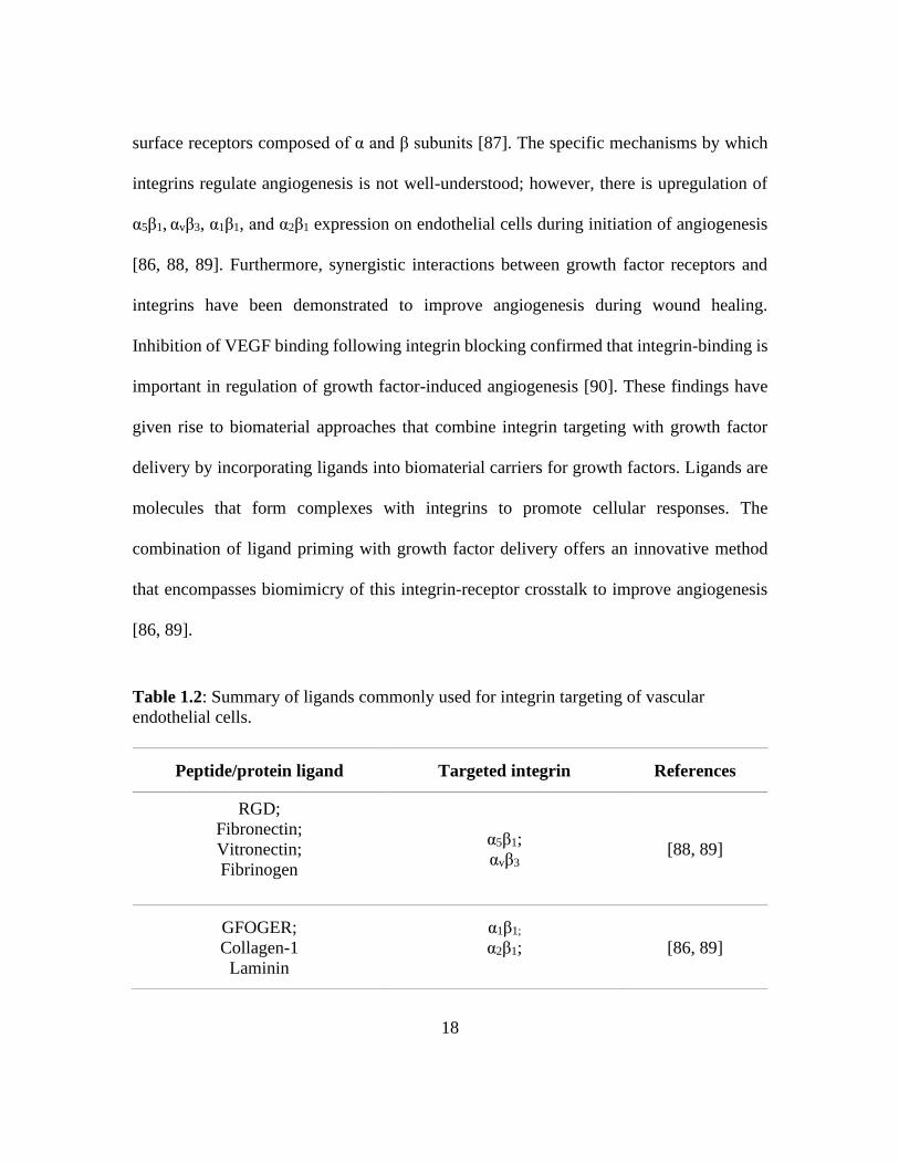
18
surface receptors composed of α and β subunits [87]. The specific mechanisms by which
integrins regulate angiogenesis is not well-understood; however, there is upregulation of
α5β1, αvβ3, α1β1, and α2β1 expression on endothelial cells during initiation of angiogenesis
[86, 88, 89]. Furthermore, synergistic interactions between growth factor receptors and
integrins have been demonstrated to improve angiogenesis during wound healing.
Inhibition of VEGF binding following integrin blocking confirmed that integrin-binding is
important in regulation of growth factor-induced angiogenesis [90]. These findings have
given rise to biomaterial approaches that combine integrin targeting with growth factor
delivery by incorporating ligands into biomaterial carriers for growth factors. Ligands are
molecules that form complexes with integrins to promote cellular responses. The
combination of ligand priming with growth factor delivery offers an innovative method
that encompasses biomimicry of this integrin-receptor crosstalk to improve angiogenesis
[86, 89].
Table 1.2: Summary of ligands commonly used for integrin targeting of vascular
endothelial cells.
Peptide/protein ligand Targeted integrin References
RGD;
Fibronectin;
Vitronectin;
Fibrinogen
α5β1;
αvβ3 [88, 89]
GFOGER;
Collagen-1
Laminin
α1β1;
α2β1;
[86, 89]

19
Selection of ligands with high specificity towards the aforementioned integrins is
critical for fabrication of effective angiogenic biomaterials [49]. Ligand priming for
production of angiogenic biomaterials is typically achieved by incorporation of peptides or
proteins summarized in Table 1.2 [87, 89]. These ligands have been incorporated into
biomaterial matrices using chemistries such as carbodiimides [91], periodate oxidation
[92], and Diels-Alder chemistries [93] and have been demonstrated to improve
vascularization [89]. Although ligand priming has the potential to improve angiogenesis
by imparting bioactivity into biomaterial matrices to mimic the interactions between cells,
matrix, and growth factors, there is concern with negative regulation of angiogenesis due
to peptide specificity [94].
1.4.4 Cell Delivery
Vascular endothelial cells and endothelial progenitor cells have vital roles in
angiogenesis. Their ability to organize into new vascular networks or integrate into existing
networks places them at the forefront of tissue engineering strategies for angiogenesis.
Researchers have turned to biomaterial scaffolds that enable sequestration of cells due to
the transient retention that results from direct transplantation [70]. Hydrogel-based
scaffolds are excellent candidates for angiogenic cell delivery as their mild processing
conditions enable cell encapsulation. They can be fabricated into architectural templates
that encourage cell organization into new vasculature networks [95, 96]. Furthermore,
modulation of the chemical and physical properties of hydrogels enables tunable release
kinetics for applications requiring cell release and integration into existing vasculature

20
networks [97]. Non-hydrogel-based scaffolds also provide platforms for which seeded cells
can proliferate, migrate, and organize into new vasculature networks. Similarly, material
properties, such as the mechanical strength, can be altered to influence cell behavior and
organization [98]. Although cell encapsulation and structural support are critical design
requirements for transplantation, they are only effective if cell viability is maintained [70].
A key factor responsible for failed cell delivery approaches is hypoxia, which is a
condition described by low-oxygenated environments. This is particularly true for cell-
encapsulated scaffolds that have low blood perfusion [70, 99]. Several groups have
investigated methods to enable oxygen production within the constructs. These methods
include embedment of inorganic peroxide species that interact with water to form hydrogen
peroxide and oxygen as an intermediary products [100-102]. Incorporation of inorganic
peroxide species has been shown to increase cell viability, with sustained oxygen level for
up to 10 days [101]. However, high concentrations of hydrogen peroxide pose a safety risk
for these approaches [99].
1.4.5 Mechanisms to Enhance Vascularization
Overall, increasing the vasculature is required to improve the bioactive potential of
the induced membrane at later stages of the Masquelet technique. Researchers have
developed several mechanisms to impart bioactive cues to increase the vasculature through
angiogenesis. These mechanisms include delivering angiogenic factors from polymeric
carriers, inducing expression of angiogenic factors via gene delivery from viral and non-
viral vectors, guiding endothelial cell behavior via integrin targeting biomaterials, and

21
delivering angiogenic cell lines using polymeric carriers. In some cases, these techniques
can be combined to generate a synergistic effect in angiogenesis. Incorporating these
mechanisms into a fibrous scaffold can generate a substrate that not only improves the
durability but also enhances angiogenesis in the induced membrane for sustained
bioactivity during treatment.
1.5 PREVENT OSTEOMYELITIS
Although guiding formation of the induced membrane to enhance the durability and
the vasculature will improve the Masquelet technique, preventing osteomyelitis remains a
challenge. Infection recurrence is one of the leading complications of the Masquelet
technique [6, 103, 104]. Despite radical debridement, up to 30% of reconstruction failures
are attributed to infection recurrence. Inadequate debridement is often followed up with a
second pass of debridement accompanied by systemic delivery of antimicrobials [6, 104,
105]. However, moderate levels of infection clearance result due to dilution of the
antimicrobials in the blood, off-target tissue absorption, and poor blood circulation around
the bone defect [106, 107]. High and potentially cytotoxic doses of antibiotics are
administered to overcome the potential loss due to systemic delivery. The reduced efficacy
and risk of toxicity has led clinicians to explore local antimicrobial delivery as an
alternative solution. Local delivery of antimicrobials can more precisely target the infected
tissue at greater concentrations that would normally be reduced via systemic delivery,
while avoiding systemic toxicity [107]. Furthermore, local delivery can expand the
selection of antimicrobials available for treatment of osteomyelitis.

22
1.5.1 Antibiotics
Since its discovery, antibiotics have become the standard choice among
antimicrobial agents used for infection control. They have been broadly used since
penicillin was introduced during the 20th century, and have been proven to effectively kill
bacteria primarily by preventing bacterial cell wall formation and inhibiting protein
biosynthesis and DNA replication [108-110]. Selection of antibiotics largely depends on
its bacterial spectrum activity, antibiotic sensitivity, and the route of administration [111,
112]. Among its diverse classes, the most commonly administered antibiotics are beta-
lactam antibiotics, glycopeptides, aminoglycosides, tetracyclines, fluoroquinolones, and
rifampicin [112]. Their corresponding mechanisms of action are summarized in Table 1.3.
Table 1.3: Summary of the common classes of antibiotics indicated to treat bone
infection.
Antibiotic
Class
Common
Forms
Mechanism
of Action
Spectrum
Activity References
Aminoglycosides
Gentamicin,
amikacin,
streptomycin
Inhibits protein
synthesis by binding
irreversibly to
bacteria’s 30S-
ribosomal subunit
Creates fissures in
bacterial cell
membrane causing
cell leakage and
increased antibiotic
uptake
Gram-
negative
bacteria
[112-114]

23
Beta-lactams
Penicillin,
oxacillin,
cephalosporin
Prevents bacterial
cell wall formation
by binding to the
active site of the
transpeptidase
Gram-positive
bacteria
[112, 115,
116]
Fluoroquinolones
Levofloxacin,
ciprofloxacin,
ofloxacin
Inhibits DNA
synthesis and
replication by
inhibiting DNA
topoisomerases
Gram-positive
bacteria [112, 117]
Glycopeptides Vancomycin
Prevents bacterial
cell wall formation
by inhibiting
transpeptidase and
peptidoglycan
synthesis
Gram-positive
bacteria [112, 118]
Rifampicin Rifampin
Suppresses
initiation of RNA
synthesis by
inhibiting DNA-
dependent RNA
polymerase activity
Mycobacteria [112, 119]
Tetracyclines Tetracycline,
Minocycline
Inhibits protein
synthesis by
preventing
aminoacyl-tRNA
from binding to the
ribosomal receptor
Gram-positive
and gram-
negative
bacteria
[112, 120]
Clinicians have routinely administered antibiotics to treat bone infection [108, 121].
The efficacy of gentamicin, cephalosporin, and levofloxacin have been demonstrated
24
against Staphylococcus aureus, including methicillin-resistant strains commonly known to
cause bone infection [122-125]. Similarly, experimental models of bone infection have
been successfully treated using vancomycin alone or in combination with rifampin [126,
127]. The established utility and proven effectiveness of antibiotics inarguably place them
at the forefront of antimicrobial agent selection [59, 123, 127, 128]. The long history of
antibiotics also provides extensive evidence that offers highly specific and supported
mechanisms of action against bacterial pathogens [109, 110, 121]. However, its widespread
administration throughout the decades raises concerns for bacterial resistance [108, 129] .
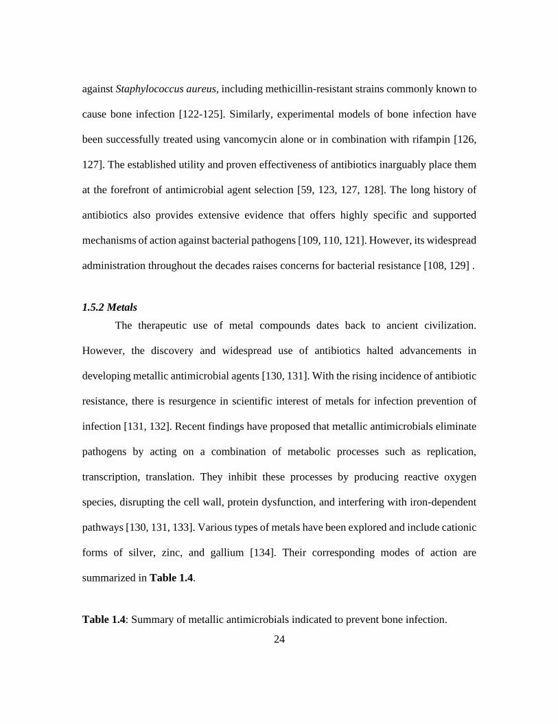
1.5.2 Metals
The therapeutic use of metal compounds dates back to ancient civilization.
However, the discovery and widespread use of antibiotics halted advancements in
developing metallic antimicrobial agents [130, 131]. With the rising incidence of antibiotic
resistance, there is resurgence in scientific interest of metals for infection prevention of
infection [131, 132]. Recent findings have proposed that metallic antimicrobials eliminate
pathogens by acting on a combination of metabolic processes such as replication,
transcription, translation. They inhibit these processes by producing reactive oxygen
species, disrupting the cell wall, protein dysfunction, and interfering with iron-dependent
pathways [130, 131, 133]. Various types of metals have been explored and include cationic
forms of silver, zinc, and gallium [134]. Their corresponding modes of action are
summarized in Table 1.4.
Table 1.4: Summary of metallic antimicrobials indicated to prevent bone infection.

25
Metal Common Applications Mechanism of Action References
Silver
Topical burn treatment,
wound dressings,
antimicrobial coating for
implants and orthopedic
fixtures
Produces reactive oxygen
species that induces bacterial
cell death;
Reacts with peptidoglycans
that puncture and destroy
bacterial cell wall;
[130, 133,
135]
Zinc
Topical treatment for
dermatologic conditions
(e.g. acne vulgaris,
dermal infection,
dandruff), dental
applications, oral rinses,
and nanoparticles
Generates reactive oxygen
species that leads to bacterial
cell death;
Reduces ATP synthesis and
inhibits enzymes critical to
cellular activity;
[136, 137]
Gallium Topical ointment for
burns, wound dressings
Inhibits bacterial and fungal
growth by interfering with
iron-dependent processes;
Competitively inhibits
binding of Fe (III) and
deprives the target pathogen
of this essential nutrient
[138-141]
The clinical use of metallic antimicrobials is limited; however, current research
presents its potency against a wide range of bacterial and fungal pathogens that cause bone
infection [136, 139]. In experimental models of osteomyelitis and periprosthetic infection,
silver and zinc have demonstrated efficacy against a broad spectrum of microorganisms
including both gram-negative and gram-positive bacteria [142, 143]. Similarly, gallium has
been reported to reduce bacterial activity of Staphylococcus aureus when metallurgically

26
added to titanium alloys, one of the most commonly used material for bone fixation devices
[144, 145]. This finding confirms that metallic antimicrobials are promising therapeutic
candidates for controlling orthopedic-related infections. Their broad-spectrum activity and
ability to exploit mechanisms independent of microbial metabolic pathways can potentially
address the ongoing challenge of antibiotic resistance. However, their exact mechanisms
of action and cytotoxic effects are not yet fully understood and effective dose
concentrations remain unclear [138, 142, 143].
1.5.3 Antimicrobial Peptides
In recent years, antimicrobial peptides (AMPs) have become viable alternatives to
conventional antibiotic treatment used in infection control [146]. AMPs are relatively small
molecules made of short sequences of amino acids that fold into an amphiphilic
configuration and behave as cationic species [146-148]. They contain approximately less
than 50 amino acids with almost 50% of the chain being hydrophobic species [146, 148].
Their overall net positive charge can be attributed to excess lysine and arginine residues,
and allows for preferential targeting of anionic bacterial cell membranes that causes pore
formation and membrane disintegration [147-149]. Despite similarities in general physical
properties, AMPs greatly vary in sequence and can be classified according to four main
categories of secondary structures whose structural characteristics and suggested modes of
action are summarized in Table 1.5 [150].
Table 1.5: Proposed structure-activity relationship of various antimicrobial peptides.

27
Class Structural
Characteristics
Mechanisms of
Antimicrobial
Activity
Examples References
α-helical
Peptides
Unstructured in
aqueous
environment but
can form barrel-
like bundles in
bacterial membrane
Creates clusters of
AMPs or toroidal
pores on the bacterial
cell membrane that
disrupts its processes
Magainin,
Cecropin,
Pexiganan
[151-153]
β-sheet
Peptides
Rigid structures
stabilized by
disulfide bonds
Perpendicularly
penetrates and
destroys bacterial cell
membrane by
inducing pores
β -defensins,
Protegrins,
Tachyplesin
[150-152]
Extended
Peptides
Possess irregular
secondary
structures but
contains a high
percentage of
proline, tryptophan,
arginine, and
histidine amino
acid residues
Non-membrane
active; Penetrates
bacterial cell
membrane and
interacts with
intracellular proteins;
induces cell
membrane leakage
Indolicin [151, 152,
154]
Loop
Peptides
Loop formation
using a single
disulfide bridge
Suggested to use
stereospecific targets
but remains unclear
Thanatin [151, 155]
Studies that have explored the antimicrobial activity of AMPs against bone
infections have demonstrated efficacy against a broad spectrum of pathogens, including
antibiotic resistant strains like Staphylococcus aureus, Pseudomonas aeruginosa, and
Staphylococcus epidermidis [156-159]. Considering its broad-spectrum activity and

28
selectivity in targeting pathogens, AMPs could potentially be adapted clinically as
antimicrobial agents [160]. However, AMPs currently remain comparably inferior to
conventional antibiotic treatment since their exact mechanisms of action are not firmly
established and concerns about acquired AMP resistance has been raised [148, 151, 155].
1.5.4 Resorbable Matrices for Antimicrobial Delivery
Local delivery systems for treatment of infection in orthopedic applications were
first introduced in bone cement stabilizers as a means to prevent infection during joint
arthroplasty procedures [161]. Bone cement has since become a long-standing approach to
implement local delivery of antibiotics, making its way into the Masquelet technique as an
antibiotic-embedded PMMA spacer [9, 28, 104, 162]. However, spacers fabricated from
PMMA bone cement are not resorbable, leading to poor release kinetics and the need for a
secondary removal procedure [36, 163, 164]. Another limitation with its use is that few
antimicrobials are able to withstand the exothermic conditions generated during
polymerization [36]. This limitation has peaked interested in the use of resorbable matrices.
Resorbable matrices used for local antimicrobial delivery are generally composed of
bioceramics or polymers. These materials have variable resorption rates, which prevent a
secondary removal procedure, and offer a method to tune release [107, 165]. Furthermore,
modifications to the physical properties such as surface-area-to-volume ratio, diffusion
path length, and swelling serve as additional methods to control release from resorbable
matrices [165].
29
Bioceramics are a class of bone substitutes that are commonly used as bone-void
fillers. One common bioceramic that is used as an off-label carrier for antimicrobials in the
treatment of bone infection is calcium sulfate. As a bone substitute, osteoclasts readily
adhere to and infiltrate calcium sulfate resulting in bulk erosion. Bulk erosion allows
greater penetration of water which facilitates rapid release via diffusion within 24 h [165,
166]. An alternative bioceramic that is used off-label for antimicrobial delivery is calcium
phosphate. Resorption of calcium phosphate is relatively slow, as compared to calcium
sulfate, resulting in diffusion independent of the resorption rate. Release from calcium
phosphate is primarily influenced by the diffusion path length through the porous matrix
and spans over days to weeks [163, 165]. In general, diffusion from bioceramic matrices is
relatively quick and should only be considered for treatment of acute infection.
In contrast to bioceramic matrices, polymeric matrices offer sustained release of
antimicrobials for treatment of chronic infection via selection of polymer type (e.g.
synthetic and natural), as previously described. This is particularly evident in synthetic
matrices that primarily release antimicrobials via degradation-mediated diffusion.
Modulation of the polymer formulation can significantly alter the degradation rate leading
to diffusion on the order of weeks to months [107, 163]. If greater sustained release is
desired, then surface-eroding synthetic matrices are advantageous due to the zero-order
release kinetics that result [163]. Contrarily, release from natural matrices is less controlled
due to release being primarily governed by swelling-based diffusion [107, 163, 165]. This
mechanism of diffusion limits sequestration to antimicrobials that are smaller than the
swollen network mesh size; however, modifications to the crosslink density can reduce the
30
mesh size and corollary swelling to provide better control over sequestration and release,
as previously described [167]. Nevertheless, one concern with the use of bioceramic and
polymeric matrices for antimicrobial delivery is that cytotoxic byproducts may exacerbate
the inflammatory response resulting in reduced efficacy of the antimicrobial [163, 165].
1.5.5 Local Delivery of Antimicrobials
In all, local delivery of antimicrobial can overcome limitations of osteomyelitis
associated with the Masquelet technique. Several different antimicrobials have been
demonstrated to be effective towards osteomyelitis. These include antibiotics, metal
compounds, and antimicrobial peptides. Controlled release of these antimicrobials is a key
design criteria for local treatment of infection. Researchers have investigated various
resorbable biomaterials such as bioceramics and polymers as carriers of antimicrobials and
have shown that these materials can be tuned to control release. As such, implementing
controlled delivery of an antimicrobial into the resorbable fibrous substrate for guiding
membrane formation can ultimately advance the Masquelet technique.
1.6 SUMMARY AND APPROACH
Despite high union rates with implementation of the Masquelet technique, there are
still challenges that need to be addressed for further advancement. Most notably, the non-
guided formation of the induced membrane yields variable membrane compositions with
unpredictable clinical outcomes. Of note is the mechanical durability of the induced
membrane. Although the membrane serves as a barrier to protect the autograft from
resorption, its mechanical properties vary across patient population which raises concerns

31
with surgical handling and mechanotransduction. The combination of biomaterial matrix
design and biomimicry presents an opportunity to reduce structural variability of the
induced membrane and improve tissue remodeling. Additionally, enhancement of
angiogenesis during the second stage of the procedure continues to be the focus of research
for increased vascularization and improved bioactivity. New approaches developed to
address this limitation include delivery of angiogenic factors, gene delivery, integrin-
targeting ligands, and cell delivery. Furthermore, local delivery of antimicrobials with
tunable release kinetics will be necessary to prevent infection recurrence associated with
inadequate debridement. Several attempts have been made to guide formation of the
induced membrane with most consisting of modification of the spacer material and
topography. Despite these attempts, researchers have yet to discover a substrate that
concurrently addresses these limitations with successful improvement of clinical outcomes.
To this end, we propose to develop a multifunctional electrospun wrap composed
of poly(lactide-co-glycolide) (PLGA) and in situ-crosslinked gelatin that can concurrently
address the limitations of durability, vascularity, and infection associated with the
Masquelet technique. This adjunct wrap will provide a resorbable fibrous framework that
permits cell attachment and serves as template to direct matrix deposition and remodeling.
It also offers independent control over the release of multiple factors to simultaneously
prevent infection and promote angiogenesis. The following chapters describe our work on
developing this novel adjunct wrap. First, the limitations of conventional PMMA bone
cement used for local delivery prompted a comparative analysis of gentamicin sulfate and
gallium maltolate released from electrospun PLGA adjunct wraps in the treatment of

32
osteomyelitis. Next, a critical review of gelatin matrices for growth factor delivery is
performed to elucidate potential mechanisms to sequester angiogenic growth factors for
enhanced angiogenesis. Co-electrospinning is then used to combine the candidate
antimicrobial wrap with a VEGF-loaded photo-crosslinked gelatin-methacrylate delivery
system, previously designed by our lab, to generate a substrate that controls cellular
responses and that provides bimodal release of an antimicrobial agent and VEGF. In all,
these platforms provide fundamental knowledge and tools to individually address the
complex biological processes necessary for improving the Masquelet technique in bone
loss management.

33
Chapter II: Comparative Efficacy of Resorbable Fiber Wraps Loaded
with Gentamicin Sulfate or Gallium Maltolate in the Treatment of
Osteomyelitis
2.1 INTRODUCTION
Treatment of critical-sized bone defects with the Masquelet technique remains a
clinical challenge due to the high incidence of osteomyelitis associated with these type of
defects [6, 17, 163, 168, 169]. The current standard of care is superficial debridement
followed by a systemic antibiotic regimen; however, a high rate of infection recurrence
results due to inadequate debridement and limited reach of systemic antimicrobials to the
targeted tissue [106, 107, 163, 170]. Another concern with the continued use of multiple
rounds of antibiotics is the potential development of bacterial tolerance and resistance as
well as side effects [164, 171, 172]. Implant-associated infection raises additional treatment
challenges due to bacterial colonization of the biomaterial surface and formation of biofilm
that confers antibiotic resistance [173]. To increase the efficacy of the antibiotic and limit
systemic effects during the Masquelet technique, clinicians and researchers have loaded
antibiotics in the poly(methyl methacrylate) (PMMA) bone cement spacer for local
delivery [162, 174, 175]. However, there is often suboptimal release, and the well-
documented exothermic effect caused by PMMA polymerization limits which
antimicrobials can be used [36, 163, 164]. Antimicrobial coatings used as adjunct therapies
offer an alternative solution to antimicrobial-loaded PMMA spacers [173, 176]. These
coatings can be applied to the PMMA spacer by coating the surface with polymer or
34
ceramic solutions containing antimicrobials [173]. Nevertheless, these coatings are often
not stable and typically require chemical modification of implants to enable stable
attachment [173]. The potential of antimicrobial coatings to migrate imposes risks that
could adversely affect treatment. Although, chemical modification of the PMMA spacer
can prevent migration, chemical modification may alter the properties of the PMMA spacer
and affect formation of the induced membrane. These limitations underscore the need for
a method to improve local antimicrobial treatment during the Masquelet technique.
Resorbable wraps are an emerging alternative that can be used broadly and provide
independently-controlled antimicrobial release kinetics [177-179]. These wraps can also
be used as stand-alone treatments to prevent infection in compound fractures that do not
require grafting. Hydrogel-based wraps, such as collagen fleece, offer promise as adjunct
therapies due to their simple fabrication and ease of application by clinicians.
Antimicrobials are loaded into the water-swollen matrix and lyophilized to encapsulate
them. Researchers have demonstrated sustained release with kinetic control provided by
modulation of the porosity or chemical crosslinking [172, 180-182]. Similarly,
polyelectrolyte multilayer films have been investigated as adjunct antimicrobial wraps for
bone defects due to their ease of fabrication, antimicrobial incorporation, and clinical
application. They are generally fabricated by layer-by-layer (LbL) assembly which enables
sequential adsorption of complementary electrostatic species to a pre-functionalized
substrate. Polyelectrolyte multilayer wraps have been reported to control release of
antimicrobials by diffusion through the various layers [123, 183]. Both hydrogel-based
wraps and polyelectrolyte multilayer wraps are able to modulate release and expand the

35
collection of antimicrobials that can be incorporated, as compared to PMMA spacers.
Although the collection of antimicrobials that can be used is expanded, the aqueous
processing conditions required for these fabrication techniques limit the collection to
water-soluble antibiotics [172, 183]. Careful consideration should be given to the potential
risk of tolerance development associated with overuse of antibiotics, and the variable tissue
penetration of antibiotics in bone [134, 171, 184]. Another concern with the use of these
fabrication techniques is that the antimicrobial encapsulation efficiency is limited by
swelling-based absorption into the bulk matrix or affinity-based adsorption onto the surface
[172, 183]. Restriction of the antimicrobial load can potentially reduce the efficacy of the
treatment. These constraints warrant the investigation of alternative fabrication techniques
that enable embedment of novel antimicrobials into adjunct wraps.
Cast antimicrobial films offer an alternative solution to hydrogel and
polyelectrolyte multilayer wraps due to the broad selection of materials and ease of
fabrication. This was demonstrated by polyester-based solutions that were blended with
various concentrations of gentamicin sulfate and cast into antimicrobial films that were
designed to augment orthopedic implants. Results confirmed a high loading capacity of
gentamicin sulfate; however, processing conditions of the films resulted in high burst
release due to significant levels of surface-bound gentamicin sulfate [185, 186]. In contrast
to films, electrospinning offers notable advantages for antimicrobial wraps due to its ease
of fabrication, versatility in materials, and tunable release kinetics [179, 187].
Electrospinning generates fibrous wraps by application of an electric field to a polymer
solution loaded with antimicrobial agents. Electrospun wraps enable greater control over

36
release kinetics by altering the chemical properties and fiber microarchitectures (e.g. fiber
diameter) [187]. Recent studies have established the potential of electrospun wraps to
control release of water-soluble antibiotics for treatment of osteomyelitis [188, 189]. Gao
et al. demonstrated that electrospun poly(lactic-co-glycolic) acid (PLGA) provided
controlled released of vancomycin over 4 weeks with corresponding bactericidal activity
resulting in significantly reduce bacterial density in an infected rabbit femoral defect model
after 8 weeks as compared to controls containing no vancomycin or systemic delivery of
vancomycin. However, the antimicrobial activity of the electrospun wrap was not
established with an osteomyelitis-derived strain [188]. This is important as antimicrobial
susceptibility varies among different bacterial strains [190]. This study was also limited by
the lack of evaluation of the potential cytotoxic effect of vancomycin as it relates to
establishing the safety and efficacy of the agents [188]. Similarly, Wei et al. demonstrated
sustained release of vancomycin from electrospun polycaprolactone (PCL) with released
concentrations up to 2 weeks. The wrap was confirmed to be cytocompatible with
osteoblasts and significantly decreased bacterial density in an infected rabbit femoral defect
model as compared to blank scaffolds over 12 weeks. However, the lack of relevant
osteomyelitis-derived strains used in the evaluation of treatment efficacy again limited the
potential impact of this study. The study also failed to perform a direct comparison of the
cytotoxic effects to the efficacy of the released vancomycin [169]. Furthermore, very few
studies have highlighted the capacity of electrospinning to enable loading and release of
novel antimicrobials [179].
37
The purpose of this study is to perform a comparative analysis of the efficacy of
gentamicin sulfate and gallium maltolate released from resorbable electrospun wraps in the
treatment of osteomyelitis. Electrospinning parameters were identified to generate
comparable release profiles of gentamicin sulfate and gallium maltolate from electrospun
poly(lactide-co-glycolide) (PLGA) wraps. Gallium maltolate is a novel antimicrobial that
mimics the ferric ion which allows it to exploit the iron-dependence of microorganisms
and inhibit growth [191]. To date, the antimicrobial potential of gallium maltolate has been
demonstrated in chronic wound applications but not in osteomyelitis applications [141,
192]. This study provides a direct comparison of the bactericidal activity of gentamicin
sulfate and gallium maltolate by identifying the minimum inhibitory concentration and
minimum bactericidal concentration on bacterial isolates from osteomyelitis. The
antimicrobial activity was then compared to the cytotoxicity in order to establish the safety
and efficacy of each agent. Finally, in vivo bactericidal activity and bone formation was
evaluated to determine the efficacy of an antimicrobial electrospun wrap for prevention of
bone infection in critical-sized bone defects.
2.2 MATERIALS AND METHODS
Materials
All chemicals and reagents were purchased from Sigma Aldrich (Milwaukee, WI)
and used as received unless otherwise noted.
PLGA Wrap Fabrication

38
50:50 PLGA (acid-terminated; inherent viscosity range 0.55–0.75 dL/g; DURECT
Corp., Cupertino, CA) was dissolved in 1,1,1,3,3,3-hexafluoro-2-propanol (HFIP)
(Halocarbon, Peachtree Corners, GA) to produce a 40% (w/v) polymer solution. Polymer
solutions were formulated with gentamicin sulfate at 1.25% of the polymer mass, gallium
maltolate (Gallixa LLC, Menlo Park, CA) at 25% of the polymer mass, or without the
incorporation of an antimicrobial agent (blank). The concentration of gentamicin sulfate
and gallium maltolate blended with the PLGA was determined by considering the release
kinetics obtained from preliminary scouting studies (data not included) and by considering
reported minimum inhibitory concentrations (MIC; lowest concentration that visibly
inhibited bacteria growth) of gentamicin sulfate and gallium maltolate [123, 141]. The
PLGA solutions were electrospun at ambient conditions (22°C, 40-50% relative humidity).
Briefly, solutions were horizontally ejected at a volumetric flow rate of 0.7 mL/h (KDS100;
KD Scientific, Holliston, MA) through an 18-gauge metal spinneret charged to 10 kV
(ES30P-5W/DDPM; Gamma High Voltage, Ormond Beach, FL). Fibers were collected on
a grounded static plate located 15 cm from the needle under ambient conditions. The
gentamicin sulfate/PLGA solution was electrospun for 4 h and the gallium maltolate/PLGA
solution was electrospun for 2 h. Meshes were then placed under vacuum for at least 24 h
prior to further characterization.
Fiber Characterization
An electrospun specimen (n= 3, 5 mm x 5 mm) was cut from the center of each
wrap (N=3/ group) to preclude edge effects. After sputter coating with 4 nm of gold

39
(Sputter Coater 108, Cressington Scientific Instruments, Oxhey, Watford, UK), fiber
microarchitecture was examined with scanning electron microscopy (SEM) (Phenom Pro;
NanoScience Instruments, Phoenix, AZ) at an accelerating voltage of 10 kV. Images
captured at 500X and 2000X via raster patterning on the front and back of each specimen
were analyzed to determine representative microarchitectures. Fiber diameter was
measured from the first 10 fibers to cross the midline of each 2000X micrograph (n=900
fibers/group).
Gentamicin Sulfate Encapsulation Efficiency
The encapsulation efficiency of gentamicin sulfate in the electrospun PLGA was
determined by first dissolving PLGA mesh specimens (1 cm x 6 cm; n=3) in 2 mL of
dichloromethane (DCM). Deionized water (DI water, 2 mL) was then added to each PLGA
solution to extract the gentamicin sulfate. The aqueous extracts were frozen and lyophilized
to concentrate the gentamicin sulfate. The dose of gentamicin sulfate in each sample was
evaluated by adding 0.5 mL of 2% ninhydrin to the lyophilized product and incubating at
120C for 15 min. The ninhydrin solutions were diluted with 1 mL of DI water to enable
spectrophotometric analysis (Infinite 200 Pro, Tecan, Morrisville, NC) at 570 nm. The
encapsulation efficiency was determined based on a standard curve and the theoretical
mass of gentamicin sulfate incorporated into the samples. Blank electrospun PLGA meshes
were used as controls.
In Vitro Release of Gentamicin Sulfate
40
Gentamicin sulfate release kinetics were determined by placing gentamicin sulfate-
loaded PLGA wraps (1 cm x 6 cm; n = 3) into 2 mL of DI water. DI water was selected, as
opposed to phosphate buffer, due to the low sensitivity of gentamicin sulfate in buffered
solution at the low concentrations [193]. Samples were incubated with rotation at 37C for
14 days. At daily time points, release medium was collected and replaced with fresh DI
water. The collected release medium was frozen and lyophilized to concentrate the
gentamicin sulfate. The dose released at each time point was determined using the
previously detailed ninhydrin assay. The percentage of gentamicin sulfate released daily
was calculated based on a standard curve and the initial mass of gentamicin sulfate
incorporated into each specimen. Blank electrospun PLGA meshes were used as controls.
The studies were repeated in triplicate and the results are represented in terms of the
average dose released (µg) and average cumulative release over time.
Gallium Maltolate Encapsulation Efficiency
The encapsulation efficiency of gallium maltolate in electrospun PLGA was
determined by first dissolving gallium maltolate-loaded PLGA samples (1 cm x 6 cm; n =
3) in 2 mL of DCM. Two-fold serial dilutions of the PLGA solutions were evaluated
spectrophotometrically at 322 nm to determine the dose of gallium maltolate loaded in each
sample. The encapsulation efficiency was calculated based on a standard curve and the
theoretical mass of gallium maltolate incorporated into the samples. Blank electrospun
PLGA meshes were used as controls.
41
In Vitro Release of Gallium Maltolate
Gallium maltolate release kinetics were determined by placing gallium maltolate-
loaded PLGA wraps (1 cm x 6 cm; n=3) into 2 mL of DI water. Samples were incubated
with rotation at 37C for 14 days. At daily time points, release medium was collected and
replaced with fresh DI water. Gallium maltolate concentration was determined
spectrophotometrically by measuring the absorbance at 307 nm. The percentage of gallium
maltolate released daily was calculated based on a standard curve and the initial mass of
gallium maltolate incorporated into each specimen. Blank electrospun PLGA samples were
used as controls. The studies were repeated in triplicate and results are represented in terms
of dose released (µg) and cumulative release over time.
Bacteria Culture Conditions
A single methicillin-susceptible Staphylococcus aureus (MSSA) isolate harvested
from a patient with osteomyelitis (49230; ATCC®, Manassas, VA) was cultured in brain
heart infusion broth (BHIB) (Sigma-Aldrich) and incubated at 37C for 16 to 18 h. The
bacterial cells were centrifuged at 3000 x g to pellet the suspension. The inoculum was
subsequently removed and replaced with Roswell Park Memorial Institute 1640 Medium
(RPMI) (Thermo Fisher Scientific, Waltham, MA) supplemented with 1% v/v sodium
pyruvate (100 mM, Thermo Fisher Scientific) and 1% v/v GlutaMAX™ (200 mM, Thermo
Fisher Scientific). The optical density of the bacterial inoculum was analyzed
spectrophotometrically at 625 nm. Bacterial density [colony-forming-units (CFU)/mL]
was determined based on a standard growth curve. Colony counts from 10-fold serial
42
dilutions of the inoculums grown on brain heart infusion agar (BHIA) (Sigma-Aldrich)
were used to confirm the bacteria density.
Antimicrobial Activity of Electrospun Wraps
The Kirby-Bauer assay was used to evaluate the antimicrobial activity of the
electrospun wraps. MSSA inoculum was suspended in supplemented RPMI at a turbidity
that matched a 0.5 MacFarland standard (1.5 x 10⁸ CFU/mL) and then spread onto BHIA.
Electrospun PLGA wraps containing gentamicin sulfate or gallium maltolate (n = 2) were
cut into 6 mm diameter disks and placed on the inoculated BHIA. Blank electrospun PLGA
disks were used as negative controls and Whatman™ #1 paper filter disks (6 mm) loaded
with 10 µg of gentamicin sulfate or 10 mg of gallium maltolate were used as positive
controls. The positive control for gentamicin sulfate was selected in accordance with the
Clinical Laboratory Standard Institute (CLSI) standard [194]. As gallium maltolate is not
a conventional antibiotic, the positive control was selected such that the dose released
would concentrate a fixed volume of agar (0.5 mL, ~ 15 mm diameter zone) greater than a
10-fold increase of the MIC after 24 h. This control was derived from a modification of the
pharmacodynamic ratio (maximum serum concentration/ MIC ≥ 10) that is used to
determine optimal patient outcomes in clinical settings [195-197]. After incubating the
BHIA at 37 °C for 24 h, the zones of inhibition were measured for each disk. Studies were
repeated in triplicate and results are represented in terms of the average zone diameter
(mm).

43
Bactericidal Activity of Gallium Maltolate
The susceptibility of MSSA to gallium maltolate was determined using a
microdilution assay according the guidelines of the CLSI [198]. Briefly, gallium maltolate
was dissolved in supplemented RPMI at a concentration of 16,000 M. Subsequent
dilutions were prepared from the sterile filtered gallium maltolate stock in supplemented
RPMI. In a 96-well plate, a 2-fold dilution of each concentration (n=3) was obtained using
the bacteria inoculum to yield final concentrations of gallium maltolate ranging from 2000
M to 8,000 M, each with an initial bacteria density of 5 x 10⁵ CFU/mL. Positive and
negative controls consisted of sterile supplemented RPMI and untreated bacteria,
respectively. The initial bacteria density was measured spectrophotometrically at 625 nm
and was confirmed by colony counting using 10-fold serial dilutions of the negative control
bacterial inoculum. The well plate was incubated at 37 °C for 16-18 h. A quantitative
method to determine the MIC was developed for this study, as visual determination can
vary across observers. This method defines the MIC as the lowest concentration that
inhibits bacterial growth such that the treated bacteria density (CFU/mL) is not statistically
different from the initial bacteria density. Colony counting using 10-fold serial dilutions of
the treated bacterial inoculums confirmed the MIC. The minimum bactericidal
concentration (MBC) of gallium maltolate was also evaluated quantitatively and was
determined to be the concentration at which the treated bacteria density was decreased by
99.9% as compared to the initial bacteria density [199]. The MIC and MBC for gentamicin
sulfate were validated using the same methods detailed above. Briefly, a stock solution of
gentamicin sulfate was dissolved at a concentration of 6 M in supplemented RPMI. Final

44
concentrations of gentamicin sulfate tested ranged from 0.5 M to 2.5 M. Studies were
repeated in triplicate and results are represented in terms of the average CFU/mL for each
concentration.
Differentiation of MC3T3-E1 Cells
The pre-osteoblastic cell line MC3T3-E1 (CRL-2593™; ATCC®, Manassas, VA)
was selected to assess cytotoxicity of gentamicin sulfate and gallium maltolate due to its
capacity to differentiate into osteoblasts and osteocytes. Briefly, MC3T3-E1 cells were first
expanded in alpha minimum essential media (α-MEM) (Caisson Lab, Smithfield, UT)
growth media, supplemented with 10% fetal bovine serum (Atlanta Biologicals, Flowery
Branch, GA) and 1% of penicillin-streptomycin (10,000 U/mL, Thermo Fisher Scientific)
at 37C. Media was replaced every 2-3 days until cells were near 80-90 % confluence. The
growth media was replaced with osteogenic media [growth media, supplemented with 10
mM β-glycerolphosphate (Sigma Aldrich), 50 μg/mL L-Ascorbic Acid (Sigma Aldrich),
and 10 nM dexamethasone (Sigma Aldrich)] to allow for differentiation. Osteogenic media
was replaced with fresh media every 2-3 days over 14 days.
IC50 and Selectivity Index for MC3T3-E1 Cells
Differentiated MC3T3-E1 cells (passage 2-6) were cultured in a 48-well plate at a
density of 25,000 cells/cm2 and incubated at 37C for 24 h. A cell viability assay was
performed on the cells to determine the inhibitory effect (IC50) and calculate the selectivity
index (IC50/MIC) of gentamicin sulfate and gallium maltolate. Briefly, gentamicin sulfate

45
and gallium maltolate were dissolved in osteogenic media to produce stock concentrations
at 50,000 M and 15,000 µM, respectively. A more concentrated gallium maltolate stock
solution was not used due to solubility in the media. Sterile-filtered gentamicin sulfate and
gallium maltolate stocks were further diluted in osteogenic media to yield final
concentrations ranging from 0.5 M to 50,000 M and 25 M to 15,000 M, respectively.
Osteogenic media on differentiated MC3T3-E1 cells was replaced with the gentamicin
sulfate or gallium maltolate treatments (n =3/concentration) and cultured for 24 h. Positive
and negative controls consisted of cells grown on tissue culture polystyrene (TCPS) with
osteogenic media replaced with fresh media or 70% ethanol, respectively. To determine
cell viability, resazurin (Sigma-Aldrich) was diluted in osteogenic media according to the
manufacturer’s instructions and incubated with the cells at 37C for 4 h. Fluorescence was
measured spectrophotometrically with excitation set to 544 nm and emission set to 590 nm.
Cell viability was then calculated by normalizing the antimicrobial treatments to the
positive control. Using an embedded algorithm for “dose-response inhibition” in GraphPad
Prism 8, the relative IC50 value was generated. Studies were repeated in triplicate with data
presented in terms of the average viability for each concentration. The IC50 was then used
to calculate the selectivity index (IC50/MIC).
Statistical Analysis
Data averages are accompanied with ± standard deviation. Statistical analysis was
performed utilizing a standard one-way ANOVA with Tukey’s post-hoc analysis unless
otherwise indicated in figure captions. Statistical significance was accepted at P<0.05.
46
2.3 RESULTS
Antimicrobial Loading and Release
Previous studies have demonstrated the ability of electrospinning to incorporate and
tune release of a variety of drugs. In this current study, we utilized electrospinning to
fabricate antimicrobial wraps loaded with gentamicin sulfate or gallium maltolate (Figure
2.1A). Electrospun PLGA was loaded with gentamicin sulfate or gallium maltolate, with a
blank wrap used as the control. The extent of antimicrobial encapsulation in each PLGA
wrap was determined by evaluating the loading efficiency. A loading efficiency of 96.9 ±
2.8% was calculated for gentamicin sulfate and 99.4 ± 12.5% for gallium maltolate. These
high efficiencies indicated that electrospinning enabled significant incorporation of two
distinct antimicrobials. The average fiber diameter of the gentamicin sulfate-loaded wrap
was 2.02 ± 0.44 µm and was 1.89 ± 0.34 µm for the gallium maltolate-loaded wrap (Figure
2.1B & C). The wraps were fabricated with similar fiber diameters in order to isolate the
effect of release kinetics for each antimicrobial, as differences in fiber diameter can
influence the diffusion path length and resorption period.
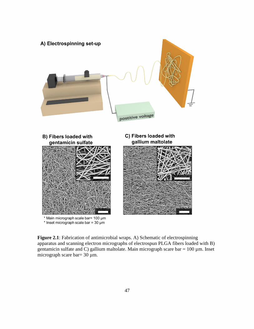
47
Figure 2.1: Fabrication of antimicrobial wraps. A) Schematic of electrospinning
apparatus and scanning electron micrographs of electrospun PLGA fibers loaded with B)
gentamicin sulfate and C) gallium maltolate. Main micrograph scare bar = 100 µm. Inset
micrograph scare bar= 30 µm.
48
Release kinetics were subsequently investigated after confirming high loading
efficiencies. Studies conducted in DI water demonstrated release profiles with similar burst
releases of 33.2 ± 2.6% from gentamicin sulfate-loaded wraps and 27.3 ± 1.7% from
gallium maltolate-loaded wraps. However, cumulative release after 14 days was lower for
gentamicin sulfate-loaded wraps than for gallium maltolate-loaded wraps (Figure 2.2).
This was possibly due to the higher loading fraction of gallium maltolate resulting in faster
release kinetics as compared to the gentamicin sulfate. Despite these differences, the daily
dose of gentamicin sulfate and gallium maltolate released were greater than the respective
MICs reported for S. aureus over 72 h (Figure 2.2) [123, 141]. This data demonstrated that
electrospun PLGA wraps enable the incorporation and controlled release of a diverse
portfolio of antimicrobials at therapeutic concentrations.

49
Figure 2.2: Evaluation of in vitro release kinetics from electrospun antimicrobial PLGA
wraps in DI water. Cumulative release and daily release of A) gentamicin sulfate and B)
gallium maltolate was evaluated over 2 weeks. Red-dashed line indicates hypothesized
MIC for each antimicrobial.
In Vitro Bacterial Inhibition and Cellular Response
To confirm the antimicrobial activity of the electrospun PLGA wraps, the Kirby
Bauer diffusion disk assay was used. The positive gentamicin sulfate control resulted in a
zone of inhibition of 17.9 ± 0.6 mm while the negative control did not inhibit growth, as
expected. Gentamicin sulfate released from PLGA wraps formed a zone of inhibition

50
comparable (17.8 ± 0.3 mm) to that of the positive control (Figure 2.3A). These results
indicated that the MSSA was susceptible to gentamicin sulfate and that electrospinning did
not impede gentamicin sulfate antimicrobial activity. Furthermore, the positive gallium
maltolate control did not form a zone of inhibition. This was unexpected as the control was
loaded based on the pharmacodynamic ratio (maximum serum concentration/MIC ≥10)
commonly used to predict clinical efficacy. Accordingly, released gallium maltolate did
not form a zone of inhibition (Figure 2.3B). The lack of zone formation suggested that
MSSA was not susceptible to gallium maltolate at the hypothesized MIC concentration.

51
Figure 2.3: Kirby Bauer assay was used to evaluate bioactivity of the antimicrobial
wraps. Representative images of the inhibition zones in response to A) gentamicin sulfate
and B) gallium maltolate released from the PLGA wrap (bottom half images), as
compared to negative control (blank PLGA wrap, top right image) and the positive
control (solubilized gentamicin sulfate or gallium maltolate, top left image) after 24 h.
Graphs display the corresponding measurements of zone diameters. * indicates statistical
differences with respect to the gentamicin sulfate positive control (P<0.05).
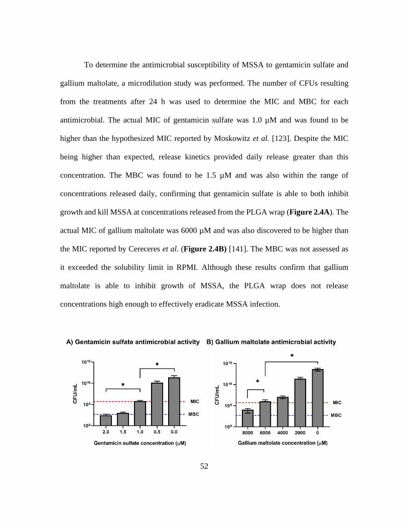
52
To determine the antimicrobial susceptibility of MSSA to gentamicin sulfate and
gallium maltolate, a microdilution study was performed. The number of CFUs resulting
from the treatments after 24 h was used to determine the MIC and MBC for each
antimicrobial. The actual MIC of gentamicin sulfate was 1.0 µM and was found to be
higher than the hypothesized MIC reported by Moskowitz et al. [123]. Despite the MIC
being higher than expected, release kinetics provided daily release greater than this
concentration. The MBC was found to be 1.5 µM and was also within the range of
concentrations released daily, confirming that gentamicin sulfate is able to both inhibit
growth and kill MSSA at concentrations released from the PLGA wrap (Figure 2.4A). The
actual MIC of gallium maltolate was 6000 µM and was also discovered to be higher than
the MIC reported by Cereceres et al. (Figure 2.4B) [141]. The MBC was not assessed as
it exceeded the solubility limit in RPMI. Although these results confirm that gallium
maltolate is able to inhibit growth of MSSA, the PLGA wrap does not release
concentrations high enough to effectively eradicate MSSA infection.
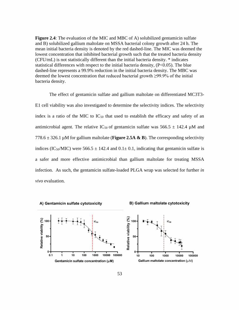
53
Figure 2.4: The evaluation of the MIC and MBC of A) solubilized gentamicin sulfate
and B) solubilized gallium maltolate on MSSA bacterial colony growth after 24 h. The
mean initial bacteria density is denoted by the red dashed-line. The MIC was deemed the
lowest concentration that inhibited bacterial growth such that the treated bacteria density
(CFU/mL) is not statistically different than the initial bacteria density. * indicates
statistical differences with respect to the initial bacteria density, (P<0.05). The blue
dashed-line represents a 99.9% reduction in the initial bacteria density. The MBC was
deemed the lowest concentration that reduced bacterial growth ≥99.9% of the initial
bacteria density.
The effect of gentamicin sulfate and gallium maltolate on differentiated MC3T3-
E1 cell viability was also investigated to determine the selectivity indices. The selectivity
index is a ratio of the MIC to IC50 that used to establish the efficacy and safety of an
antimicrobial agent. The relative IC50 of gentamicin sulfate was 566.5 ± 142.4 µM and
778.6 ± 326.1 µM for gallium maltolate (Figure 2.5A & B). The corresponding selectivity
indices (IC50/MIC) were 566.5 ± 142.4 and 0.1± 0.1, indicating that gentamicin sulfate is
a safer and more effective antimicrobial than gallium maltolate for treating MSSA
infection. As such, the gentamicin sulfate-loaded PLGA wrap was selected for further in
vivo evaluation.
54
Figure 2.5: Viability of differentiated MC3T3-E1 cells relative to TCPS control after 24
h exposure to various concentrations of A) solubilized gentamicin sulfate and B)
solubilized gallium maltolate. Red-dashed line indicates relative IC50 calculated using
GraphPad Prism 8. A relative IC50 was identified as 566.5 ± 142.4 µM for gentamicin
sulfate and 778.6 ± 326.1 µM for gallium maltolate.
2.4 DISCUSSION
The observed recurrence of osteomyelitis during treatment of the Masquelet
technique and the risk of implant-associated infection in critical-sized bone defects created
a clear need for a resorbable device that offered local antimicrobial delivery. We sought to
develop an electrospun antimicrobial wrap that could be used broadly as a stand-alone
treatment or as an adjunct treatment to prevent infection. Electrospinning was selected as
it allows higher antimicrobial encapsulation by blending antimicrobials with the polymer,
as compared to other wrap fabrication techniques that rely on swelling-based absorption or
surface adsorption. Furthermore, electrospinning enables tunable release of antimicrobials
by modulating chemical properties and fiber microarchitectures [200, 201]. Antibiotics are
traditionally selected as the antimicrobial agent incorporated into electrospun wraps as they
are effective against a wide spectrum of bacterial species making them useful for various
applications. They attack bacteria by disrupting a specific biological process such as cell
wall synthesis, nucleic acid synthesis, or protein synthesis [134, 202]. Although antibiotics
are effective at preventing infection, overuse of them can lead to bacterial modifications
that increase efflux, decrease binding, or cause inactivation of the antibiotics [202]. This
risk has prompted the investigation of novel antimicrobials like metals. Metal compounds
are of interest due to the ability to target multiple biological processes as opposed to the

55
single-target approach of antibiotics [134]. There are few studies that have investigated the
efficacy of metal compounds as an alternative solution to antibiotics in the treatment of
osteomyelitis.
To this end, we compared the efficacy of gentamicin sulfate and gallium maltolate
released from electrospun PLGA wraps in the treatment of osteomyelitis. PLGA was
selected as the polymer due to its established use as a controlled delivery system for low
molecular weight antimicrobials [185, 203, 204]. Modification of the co-polymer ratio,
molecular weight, or chemical end-groups can be used to tune release kinetics [205].
Gentamicin sulfate was chosen as the antibiotic owing to its broad antimicrobial spectrum
and frequent clinical use in bone infection [123]. The mode of action of gentamicin sulfate
involves inhibition of protein synthesis by targeting the 30S or 50S subunit of bacterial
ribosomes [202]. Furthermore, the potential to overcome antibiotic resistance reinforced
the selection of gallium maltolate as a metal-based alternative to gentamicin sulfate. The
atomic similarity to ferric iron improves bacterial uptake of gallium maltolate. Once taken
up by bacteria, the inability to reduce gallium ions disrupts the iron-redox process that is
responsible for iron transport, respiration, nucleic acid synthesis, and reactive oxygen
species protection leading to death [141, 206]. The concentration of gentamicin sulfate and
gallium maltolate blended with the PLGA was selected based on predicted release kinetics
of the PLGA wrap. It was postulated that the release kinetics would result in daily release
greater than the hypothesized MIC concentration for each antimicrobial over at least 72 h,
which is the critical period when vulnerable planktonic bacteria can progress into a resistant
biofilm [123, 141, 207, 208]. HFIP was selected as the electrospinning solvent, as organic
56
solvents enable solubilization of gallium maltolate at high concentrations [141]. Although
gentamicin sulfate is moderately soluble in HFIP, achieving release of the hypothesized
MIC does not require a high loading concentration as compared to gallium maltolate. It
was consequently determined that utilizing HFIP to solubilize both antimicrobials was
acceptable. HFIP evaporation during electrospinning ensured direct encapsulation of each
antimicrobial resulting in high encapsulation efficiencies. A high burst release was
expected to occur in response to the incompatible interactions between the hydrophilic
drugs and hydrophobic PLGA [203, 204]. This is of particular concern for analysis of
gentamicin sulfate release kinetics. Given the low loading concentration, a high burst
release reduces the amount of drug available at later time points during the sustained release
phase. The sustained release doses may fall below the detection limit of the ninhydrin assay
and prevent analysis. Therefore, the gentamicin sulfate-loaded wrap was electrospun for a
longer period to increase the thickness of the wrap and the corollary amount of drug
available during the sustained release phase. Although the wraps were fabricated with
different thicknesses, the fiber diameters were comparable and enabled normalization of
the release kinetics. The release profiles were expected to be comparable due to similar
drug-polymer interactions. However, the gallium maltolate-loaded wrap exhibited higher
cumulative release beyond the burst phase. It was hypothesized that the differences in
release kinetics were due to the loading concentration of each drug. Drugs loaded above a
polymer solubility threshold have the potential to aggregate into clusters in the amorphous
region of semi-crystalline polymers. As the aqueous medium penetrates the matrix, these
clusters are released via dissolution resulting in faster cumulative release than an evenly
57
dispersed drug load. This phenomenon has been demonstrated with other electrospun
polyester (i.e. polycaprolactone) meshes loaded with various concentrations of drugs [209,
210]. It is possible that the high loading fraction of gallium maltolate resulted in cluster
formation in the amorphous region of PLGA leading to faster release kinetics as compared
to the gentamicin sulfate-loaded wrap. Despite these differences, both wraps were able to
provide release of the respective antimicrobial above its hypothesized MIC in the first 72
h [207, 208]. These findings confirmed that electrospinning enables encapsulation and
release of different types of antimicrobials over a clinically relevant duration.
A potential concern with direct encapsulation of bioactive agents during
electrospinning is a loss of biological activity. The antimicrobial activity of each wrap was
evaluated using the Kirby Bauer assay which is a standardized assay for evaluation of
antimicrobial susceptibility [194]. MSSA was designated as the testing strain for its
prevalence in osteomyelitis [211]. The gentamicin sulfate-loaded wrap demonstrated
bioactivity comparable to the positive control indicating that the MSSA is susceptible to
gentamicin sulfate and that electrospinning does not impede the antimicrobial activity. As
gallium maltolate is not a conventional antibiotic, a standard has not been determined for
evaluating susceptibility. We selected the gallium maltolate standard based on a
modification of a pharmacodynamic ratio (maximum serum concentration/MIC ≥10) that
is commonly used to predict optimal clinical outcomes and suppression of resistance [195,
196]. Rather than considering a maximum serum concentration, a maximum agar
concentration in a constant volume of agar was considered. It was assumed that complete
release of the gallium maltolate from the paper filter disk would occur over 24 h and

58
concentrate the agar above the hypothesized dose based on uncontrolled diffusion from the
paper-filter disk [194]. However, the gallium maltolate control did not form a zone of
inhibition suggesting that MSSA was not susceptible to gallium maltolate at the
hypothesized MIC. This MIC was targeted based on the reported activity of gallium
maltolate against MRSA [141]. It is well known that antimicrobial activity is strain-
dependent so this data was not unforeseen [190]. We evaluated the susceptibility of MSSA
to soluble gallium maltolate to identify the MIC using a microdilution assay. Results
indicated that MSSA was susceptible to gallium maltolate at higher concentrations than
those reported for MRSA and that the current iteration of the PLGA wrap does not release
efficacious concentrations of gallium maltolate. Similarly, the MIC of gentamicin sulfate
was found to be higher than what was reported for MSSA. This was expected as Moskowitz
et al. evaluated a bacteria density below (5-fold) the CLSI standard density that was used
in present study [123]. It is probable that a low bacteria density will be susceptible to an
antimicrobial at a relatively low concentration as compared to a high bacteria density.
Despite this finding, the PLGA wrap was able to release daily concentrations above the
actual MIC of gentamicin sulfate.
To determine the ability of gallium maltolate to selectively target MSSA while
retaining cell viability of MC3T3-E1 cells at this relatively high MIC, we evaluated the
selective bioactivity as compared to gentamicin sulfate. A comparative analysis of the two
agents revealed a significantly lower selectivity index for gallium maltolate compared to
gentamicin sulfate. This indicated that the efficacy of gallium maltolate is undermined by
its cytotoxic effect on osteogenic cells and therefore should not be used to treat
59
osteomyelitis. Despite these findings, gallium maltolate-loaded PLGA wraps should be
further explored for controlled delivery in other biomedical applications such as chronic
wounds [141, 212]. Further evaluation of the efficacy of the gentamicin sulfate-loaded
PLGA wrap in the treatment of osteomyelitis is currently in progress in an infected rat
segmental defect model.
Overall, the gentamicin sulfate-loaded PLGA wrap utilizes material properties and
fibrous microarchitecture to maintain release in a controlled manner. Coupled with the
superior in vitro and in vivo antimicrobial performance, this wrap demonstrates a strong
potential as an adjunct therapy in the treatment of osteomyelitis associated with open
fractures and implant-mediated colonization. There were limitations in the present study
that necessitate future research. Current studies are in progress to evaluate the in vivo
antimicrobial activity of the gentamicin sulfate-loaded PLGA wrap. However, further
analysis is needed that provides a rigorous in vivo assessment of efficacy in bone
regeneration of infected defects. These studies will need to investigate bone volume
fraction, defect bridging, and total bone growth within the defect following treatment.
Results would be compared to antibiotic-loaded cement in order to assess improvement
upon clinical standards. In all, the gentamicin sulfate-loaded PLGA wrap has the potential
to have a direct impact on orthopedic applications through rational design of adjunct
infection control combined with the capacity to enhance fracture healing.

60
2.5 CONCLUSIONS
In the present study, we successfully performed a comparative analysis of efficacy
of two antimicrobial-loaded, resorbable meshes for the treatment of osteomyelitis during
the Masquelet technique. Our analysis resulted in the selection of an antimicrobial wrap
that sustains release of an antibiotic with established bactericidal activity for the
osteomyelitis bacterial strain. Specifically, the completed studies establish an initial proof-
of-concept wrap with the ability to reduce infection for potential use as an adjunct therapy
for contaminated fractures and implants. This wrap has been optimized for sustained
release to ensure bactericidal concentrations are retained throughout the treatment. The
ability of this treatment to selectively target bacteria associated with osteomyelitis while
avoiding cytotoxicity of osteoblasts was also evaluated to establish the potential safety and
efficacy of this treatment. Future work will build upon the present research by evaluating
fracture healing with the electrospun gentamicin sulfate-loaded PLGA wrap in a
contaminated femoral defect model.
61
Chapter III: Gelatin Matrices for Growth Factor Sequestration 1
3.1 POLYMERIC MATRICES FOR GROWTH FACTOR DELIVERY
The bioactive potential of the induced membrane is heavily regulated by the
vasculature; therefore, the temporal decrease in the vasculature observed during treatment
imposes significant limitations on the success of the Masquelet technique [15, 16, 21, 22].
Overcoming these limitations will require an increase the vasculature at later stages. This
can be achieved by implementing a strategy to enhance angiogenesis in the induced
membrane. One of the most common strategies investigated for enhanced angiogenesis in
tissue engineering applications is growth factor delivery. Angiogenic growth factors
instruct cellular responses such as cell migration, proliferation, and differentiation during
tissue repair and remodeling [72, 74, 213]. The ability of growth factors to direct cellular
behavior is dependent on the concentration as well as the spatial dispersion. Bolus delivery
of growth factors display limited efficacy and adverse side effects such as ectopic growth
and carcinogenic effects. Researchers attempt to address these limitations by developing
materials to provide localized delivery and controlled release [60].
Advances in polymeric material design over the last 25 years have enabled the
development of tunable platforms for growth factor delivery [214-216]. Synthetic
polymers (e.g. poly(lactide-co-glycolide) (PLGA), polylactide (PLA), polyglycolide
1 This work was reprinted with permission from “Gelatin matrices for growth factor sequestration”, by
Taneidra Walker Buie, Joshua McCune, and Elizabeth Cosgriff-Hernandez. Trends in Biotechnology,
2020, 38(5), 546-557. © 2019 Elsevier Ltd. All rights reserved.

62
(PGA), polycaprolactone (PCL)) offer several advantages including ease of manufacture,
tunable degradation, and established use in small molecule delivery [58]. However, the
harsh processing conditions required for fabrication of synthetic polymers, such as high
temperatures or organic solvents, can denature growth factors leading to a loss in
bioactivity [59]. To circumvent this loss of bioactivity due to processing, growth factors
can be loaded into the matrix after fabrication. Post-fabrication loading can restrict the
loading capacity to adsorption to the surface or absorption in the water-swollen polymer
matrix [60]. In addition, degradation of synthetic polymers can result in an inflammatory
response due to toxic by-products or changes to the local pH [58]. As an alternative, natural
polymers and their derivatives, such as collagen, gelatin, chitosan, and alginate, are often
processed in aqueous solvents. These mild processing conditions allow for in-line loading
of the growth factors with a corollary increase in loading capacity over synthetic matrices.
As biological materials, degradation byproducts are cytocompatible and readily cleared
from the body [61, 217]. One of the more common natural polymers used for growth factor
delivery is gelatin due to its versatile fabrication processing, ease of modification, and its
electrostatic properties that confer growth factor affinity [218, 219]. There has been an
increase in the development of gelatin delivery systems that provide tunable delivery of
growth factors to support bioactivity retention [220-222]. However, sequestration and
release is primarily governed by an increase in the crosslink density resulting in structural
changes to the gelatin matrix [63]. The focus of this review is to provide a summary of
alternative mechanisms to enhance growth factor sequestration in gelatin matrices, current

63
gelatin growth factor matrices in practice, and future perspectives of gelatin matrices in
tissue engineering.
3.2 AFFINITY SEQUESTRATION TO CONTROL GROWTH FACTOR RELEASE
The efficacy of growth factor therapy for tissue engineering applications is highly
dependent on retaining the bioactivity during fabrication and application. Gelatin matrices
offer advantages over synthetic polymeric carriers due to its mild fabrication conditions
(e.g. aqueous solution processing) and high growth factor loading during fabrication [223,
224]. Standard processing of collagen to generate gelatin also increases its solubility and
provides ease of fabrication as compared to collagen delivery vehicles. The selected
hydrolytic treatment (acidic or basic) determines the isoelectric point (IEP) of gelatin
matrices, the pH at which the charge on the gelatin is zero [225-227]. Acidic pre-treatment
results in positively-charged gelatin (type-A gelatin) with an IEP between pH 8-9. Alkaline
pre-treatment hydrolyzes amide residues to carboxyl residues leading to negatively-
charged gelatin (type-B gelatin) with an IEP between pH 4.8-5.4 [228]. The net charge of
gelatin enables electrostatic interactions with oppositely charged growth factors which
inherently sequesters growth factors. However, rapid dissolution of gelatin during
implantation requires gelatin matrices to be crosslinked into hydrogels [229]. The
crosslinking modality and degree of crosslinking can strongly impact the resulting physical
properties of the gelatin matrix [230, 231]. Reagent-based crosslinking modalities enable
homogenous crosslinking and are categorized as either non-zero length or zero-length
64
based on assisted bonding or direct bonding, respectively [232, 233]. Non-zero length
crosslinking reagents (e.g. aldehydes, isocyanates, and polyepoxides) react with free amine
residues and/or carboxylic acid residues to form intramolecular and intermolecular
crosslinks within a gelatin solution. Zero-length crosslinking agents (e.g. acyl azides and
carbodiimides) facilitate the direct reactions between carboxylic acid residues and amine
residues on the same gelatin molecule or adjacent gelatin molecules without intermediate
molecules in the network [234]. However, these chemical crosslinking modalities can have
residual unreacted reagents that could compromise the biocompatibility of the gelatin
matrices. Less toxic, covalent crosslinking modalities include natural enzymes such as
genipin, which is a natural reagent derived from the gardenia fruit. It facilitates crosslinking
in a two-step process that first reacts with amine residues of gelatin followed by reaction
with esters of genipin with amine residues of gelatin [229]. Photo-polymerization is another
common method of covalently crosslinking gelatin. This method requires functionalization
of gelatin with a primer (e.g. acrylamide, methacrylamide) in order to undergo photo-
polymerization in the presence of free radicals [235]. Use of these modalities offer versatile
methods for controlling the mechanical and physical properties of gelatin matrices.
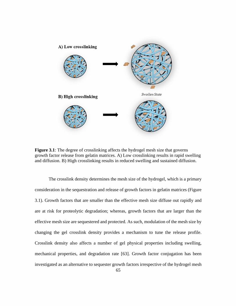
65
Figure 3.1: The degree of crosslinking affects the hydrogel mesh size that governs
growth factor release from gelatin matrices. A) Low crosslinking results in rapid swelling
and diffusion. B) High crosslinking results in reduced swelling and sustained diffusion.
The crosslink density determines the mesh size of the hydrogel, which is a primary
consideration in the sequestration and release of growth factors in gelatin matrices (Figure
3.1). Growth factors that are smaller than the effective mesh size diffuse out rapidly and
are at risk for proteolytic degradation; whereas, growth factors that are larger than the
effective mesh size are sequestered and protected. As such, modulation of the mesh size by
changing the gel crosslink density provides a mechanism to tune the release profile.
Crosslink density also affects a number of gel physical properties including swelling,
mechanical properties, and degradation rate [63]. Growth factor conjugation has been
investigated as an alternative to sequester growth factors irrespective of the hydrogel mesh

66
size. As mentioned previously, gelatin contains several chemical groups that enable
covalent crosslinking within gelatin or to adjacent gelatin molecules. These chemical
reactions can covalently bind growth factors to gelatin to enhance sequestration [236-238].
Release of conjugated growth factors will be delayed until cleavage of gelatin matrices
and/or linkers permit diffusion (Figure 3.2) [239].
Figure 3.2: Effect of conjugation on growth factor sequestration in gelatin matrices.
Growth factor-conjugated gelatin matrix displays burst release due to initial swelling that
releases non-conjugated growth factors followed by sustained growth factor release after
proteolytic chain scission.
Among these conjugation modalities are bi-functional crosslinkers such as
diisocyanates or susuccinimidyl valerate which facilitate covalent bonding with the
available free amines on gelatin and growth factors [239, 240]. However, it is possible for
side reactions to occur such as a single bi-functional crosslinker binding two growth factors
or multiple bi-functional crosslinkers binding a single growth factor due to the ratio of
amines present of growth factors [240]. The latter could affect the hydrogel mesh size by
behaving as an additional crosslink point with the gelatin. Alternatively, a two-step process

67
of functionalization of the gelatin and growth factor independently following by a
conjugation step provides additional control over the reaction. One of the most common
examples of this process is the use of methacrylated gelatin and acrylated or methacrylated
growth factors that undergo free radical polymerization in the presence of a photo-initiator
and UV irradiation [241]. Although conjugation modalities have been successful at
immbolizing growth factor for sustained sequestration, the poor control of conjugation sites
on growth factors, typically non-specific amino groups, puts these techniques at a high
potential for bioactivity loss [242]. To address this limitation, affinity sequestration has
been explored as a means to sequester the growth factor for sustained release without loss
of bioactivity and minimal effect of the gelatin physical properties. Common affinity
sequestration approaches will be described in detail with a focus on the relationships with
physiochemical properties and gelatin matrix design, Figure 3.3.
Figure 3.3: Overview of the physiochemical properties governing growth factor diffusion
from gelatin matrices. The properties included growth factor affinity to A) ligands, B)
adaptor proteins, and C) nanomaterial additives incorporated into gelatin matrices.
68
The established interactions of growth factors and the extracellular matrix (ECM)
is a rich field to draw design inspiration for sequestering growth factors. As such, ECM-
derived ligands are one of the most common moieties used to sequester growth factors in
gelatin matrices. These non-covalent bonds do not impair the stability or bioactivity of
growth factors [64, 65, 242]. Among these target ECM ligands is heparin, a negatively-
charged glycosaminoglycan that has binding domains for several growth factors. Heparin
binds several growth factors via electrostatic interactions between amino acid residues and
carboxyl groups. It has commonly been incorporated into gelatin through functionalization
by 1-ethyl-3-(3-dimethylaminopropyl) carbodiimide hydrochloride/N-
hydroxysulfosuccinimide (EDC/NHS). EDC/NHS reactions facilitate crosslinking
between carboxyl groups on heparin and amino acid residues on gelatin, but intramolecular
or intermolecular crosslinking of gelatin is also possible with this technique [65, 243]. This
can be avoided by first activating the carboxyl groups on heparin with EDC prior to its
incorporation into gelatin [244]. The heparin-modified gelatin matrix can then be used to
sequester growth factor with high affinity and without chemically modifying the growth
factor. An adapter protein is another type of ligand-based moiety that is composed of a
coiled peptide and a collagen-binding domain (CBD) derived from fibronectin. CBDs
derived from fibronectin have high affinity towards collagen and gelatin and facilitates
ready modification of gelatin matrices [64]. The coiled peptide tethered to the CBD enables
electrostatic binding with a complementary coiled peptide tethered to a growth factor of
interest. This technology has proven to be highly adaptable and can be modified to increase
the binding strength to gelatin by altering the source of CBD [64]. Furthermore, high-

69
throughput screening of DNA/RNA libraries using systematic evolution of ligands by
exponential enrichment (SELEX) technique enables selection of peptide and
oligonucleotide aptamers with high binding affinity by electrostatic interactions [66, 245].
Aptamers can be incorporated into gelatin matrices by standard bioconjugation methods
[66]. As an alternative to chemical modification of the gelatin matrix, nanomaterial
additives with growth factor affinity have been explored to generate gelatin nanocomposite
delivery systems. These additives can be readily mixed into gelatin precursor solutions and
provide a high surface area to facilitate growth factor adsorption. Common nanomaterials
used to sequester growth factors in gelatin matrices are nanodiamonds, carbon-based
nanoparticles with truncated octahedral structures, and nanoclays. Functional groups on
the surface of nanodiamonds determine interfacial interactions with growth factors. These
interaction include electrostatic, hydrogen bonds, dipole-dipole, and hydrophobic
adsorption and vary depending on the processing technique used during the synthesis of
the nanodiamonds [246]. Surface modification of the nanodiamonds through carboxylation
or hydroxylation can also be used to provide covalent conjugation to gelatin prior to
crosslinking of gelatin for greater stability [67, 246]. As compared to other widely used
carbon-based nanomaterials such as graphene oxide and carbon nanotubes, nanodiamonds
have greater biocompatibility [246]. Two-dimensional nanoclays are another type of
nanomaterial with superior biocompatibility. Nanoclays are discs composed of an
octahedral sheet of magnesium oxide inserted between two parallel tetrahedral sheets of
silica which results in negatively-charged surfaces and a positively-charged edge. Sodium
ions adsorbed to the surface of nanoclays during manufacturing foster ionic interactions

70
with neighboring nanoclays in dry environments. However, nanoclays dissociate in ionic
aqueous solutions due to favorable interactions between the sodium ions and hydroxide
molecules or other ions. Dissociation allows for rearrangement and greater access to the
charged surfaces by proteins [68]. Similar to nanodiamonds, nanoclays are generally
incorporated into gelatin solutions prior to crosslinking [68, 247].
There are several applications in tissue engineering that have used these affinity
sequestration approaches to achieve growth factor delivery ranging from 5 days to 25 days
including cardiovascular repair [247], angiogenesis [48, 66, 69], bone healing [65], and
wound healing [64, 248]. For example, adaptor proteins with coiled-CBDs specific for
gelatin were incorporated into EDC/NHS-crosslinked gelatin. Epidermal growth factor
(EGF) tethered with a complementary coil was added to the gelatin to allow for non-
covalent binding. This non-covalent binding resulted in sequestration of EGF for over four
days [64]. Given this relatively moderate time frame, this matrix could be employed in
wound healing to initiate cell proliferation for tissue regeneration. Alternatively, the strong
binding affinity of heparin to vascular endothelial growth factor (VEGF) has been used in
a gelatin composite wrap that was crosslinked with EDC/NHS. This matrix was able to
sustained release of VEGF over three weeks rendering it useful to direct capillary
formation, homogenization, and maturation of blood vessels that typically occurs during
the first three weeks of angiogenesis [48]. In addition to using these modalities for
moderate sequestration, they can be selected for high specificity to increase the time that
growth factors are preserved in gelatin matrices. This is especially true for aptamers as
demonstrated in a study that selected the acrydite oligonucleotide using SELEX due to its

71
high bind specificity for VEGF [66]. Another advantage of these affinity sequestration is
that they can be used for concentrated localization of growth factors to a particular area in
the matrix. For example, a single nanodiamond can bind multiple growth factors based on
its high surface area that allows for increased adsorption [246]. Furthermore, spatial
concentration of nanodiamonds in gelatin matrices provides another mean to sequester
growth factors by regulating the diffusion path length as later discussed. Despite the
potential to sequester growth factors with minimal impact on the bioactivity, careful
consideration must be given to the transient and reversible interactions that govern
sequestration when sustained preservation is desired. Another consideration is that freely
encapsulated nanomaterials can potentially bind the gelatin matrix due to their high surface
area resulting in changes to the physical properties [69].
3.3 GELATIN MATRICES IN TISSUE ENGINEERING
An accompanying aspect that affects growth factor release kinetics is the
fabrication technique, which determines the diffusion path length through the gelatin
matrix [249]. Advancements in fabrication of gelatin-based matrices have provided several
opportunities to create systems for the controlled delivery of growth factors. The geometry
and size of gelatin matrices can be altered through various fabrication techniques to control
growth factor sequestration. These strategies are primarily focused on changing the
surface-area-to-volume ratio and the diffusion path length of the embedded growth factors
[250, 251]. As a general consideration, an increase in surface area will decrease the
diffusion path length with a corollary increase in the release kinetics. These considerations

72
can be applied to the design of the gelatin delivery system regardless of the resulting
geometry (e.g. microparticles, gels, fibrous meshes). The following section will describe
fabrication techniques that control growth factor sequestration for growth factor delivery
in a variety of tissue engineering applications.
3.3.1 Gelatin Microparticles
Gelatin microparticles offer several advantages as a growth factor delivery vehicle
such as a high surface area-to-volume ratio. Typically, smaller microparticles display faster
growth factor release rates due to an increase in surface area and a shorter diffusional path
length of embedded growth factors (Figure 3.4) [250, 252-254]. This was demonstrated in
a study that showed microparticles that were 0.20 ± 0.04 µm in average diameter resulted
in release of 70% of bone morphogenetic protein 2 (BMP-2) as compared to 12% from
microparticles with an average diameter of 26 ± 6.0 µm after four weeks [250].
Microsphere size and shape can be tuned through various manufacturing techniques with
the most common technique being water-in-oil emulsions induced by mechanical agitation
such as high-speed stirring of a gelatin solution and an organic phase. These emulsions are
then cooled to allow for gelation of the microparticles followed by precipitation [250, 255,
256]. Processing parameters such as mixing speed and solvent selection are used to control
microparticle size; however, this technique typically results in a large particle size
distribution [252, 257]. As an alternative to high-speed stirring, microfluidic devices have
been used to achieve monodispersed particle size distributions. This technique consists of
coaxial flow between an aqueous gelatin solution and an oil-based sheath with each phase
73
set to different flow rates to control the droplet size [258, 259]. Particles are collected in a
coagulation bath prior to chemical crosslinking. If smaller microparticles are desired, then
electrospraying can be used to form microparticles. This technique applies an electric field
to a low viscosity gelatin solution as it is being extruded from a syringe. Charge repulsion
within the solution droplet at the end of the capillary overcomes the solution surface tension
leading to a solution droplets erupting from the droplet towards a ground or oppositely
charged collector. As the solvent evaporates from the droplet during flight to the collector,
a repulsive threshold is reached within the droplets leading to solution fission into smaller
dried particles [252, 260]. Gelatin microparticles are most commonly crosslinked
following fabrication by chemical reagents (e.g. glutaraldehyde) [261-263]. It can be
difficult to control the crosslinking density and these chemical reagents are typically
cytotoxic [237, 264]. Genipin and carbodiimides are alternative reagents used to provide
greater control over crosslinking of gelatin microparticles with less cytotoxicity [236, 265,
266].
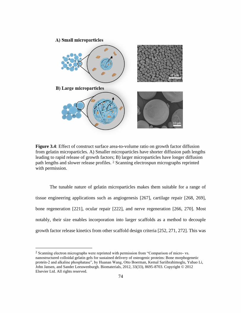
74
Figure 3.4: Effect of construct surface area-to-volume ratio on growth factor diffusion
from gelatin microparticles. A) Smaller microparticles have shorter diffusion path lengths
leading to rapid release of growth factors; B) larger microparticles have longer diffusion
path lengths and slower release profiles. 2 Scanning electrospun micrographs reprinted
with permission.
The tunable nature of gelatin microparticles makes them suitable for a range of
tissue engineering applications such as angiogenesis [267], cartilage repair [268, 269],
bone regeneration [221], ocular repair [222], and nerve regeneration [266, 270]. Most
notably, their size enables incorporation into larger scaffolds as a method to decouple
growth factor release kinetics from other scaffold design criteria [252, 271, 272]. This was
2 Scanning electron micrographs were reprinted with permission from “Comparison of micro- vs.
nanostructured colloidal gelatin gels for sustained delivery of osteogenic proteins: Bone morphogenetic
protein-2 and alkaline phosphatase”, by Huanan Wang, Otto Boerman, Kemal Sariibrahimoglu, Yubao Li,
John Jansen, and Sander Leeuwenburgh. Biomaterials, 2012, 33(33), 8695-8703. Copyright © 2012
Elsevier Ltd. All rights reserved.

75
demonstrated in a study that incorporated gelatin microparticles loaded with VEGF into a
porous lithium calcium polyphosphate scaffold for bone repair associated with
glucocorticoids-induced osteonecrosis of the femoral head. Microparticles were fabricated
through emulsion templating and crosslinked in glutaraldehyde prior to diffusional loading
of VEGF [263]. In addition to the use of gelatin microparticles in composite scaffolds,
microparticles can also be directly injected as a slurry of particles for more rapid growth
factor delivery. Hirose et al. reported the delivery of basic fibroblast growth factor (bFGF)
and interferon-beta (IFNβ) from gelatin microparticles as a method to establish a
proliferative vitreoretinopathy disease model. The microparticles were fabricated using
emulsion templating and crosslinked in glutaraldehyde prior to diffusional loading of bFGF
or IFNβ [222]. As a general consideration, direct application of gelatin microparticles may
result in a higher initial burst release that results from immediate exposure to aqueous
solutions as compared to microparticles embedded within a composite.
3.3.2 Gelatin Scaffolds
Several researchers have explored the use of gelatin constructs to act as both a
controlled growth factor delivery vehicle and as a scaffolding material. Gelatin scaffolds
have been fabricated using a variety of methods including electrospinning, microfluidics,
freeze-drying, and porogen leaching. The resulting porous, three-dimensional architectures
template new tissue formation by supporting cell attachment, proliferation, and migration
[45, 53]. These same pores can alter the diffusion path length that affects growth factor
release profiles as previously described in delivery vehicles. Electrospinning has become
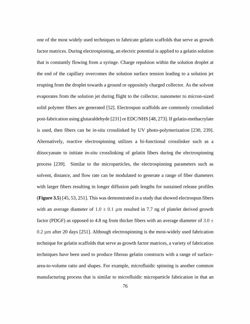
76
one of the most widely used techniques to fabricate gelatin scaffolds that serve as growth
factor matrices. During electrospinning, an electric potential is applied to a gelatin solution
that is constantly flowing from a syringe. Charge repulsion within the solution droplet at
the end of the capillary overcomes the solution surface tension leading to a solution jet
erupting from the droplet towards a ground or oppositely charged collector. As the solvent
evaporates from the solution jet during flight to the collector, nanometer to micron-sized
solid polymer fibers are generated [52]. Electrospun scaffolds are commonly crosslinked
post-fabrication using glutaraldehyde [231] or EDC/NHS [48, 273]. If gelatin-methacrylate
is used, then fibers can be in-situ crosslinked by UV photo-polymerization [230, 239].
Alternatively, reactive electrospinning utilizes a bi-functional crosslinker such as a
diisocyanate to initiate in-situ crosslinking of gelatin fibers during the electrospinning
process [239]. Similar to the microparticles, the electrospinning parameters such as
solvent, distance, and flow rate can be modulated to generate a range of fiber diameters
with larger fibers resulting in longer diffusion path lengths for sustained release profiles
(Figure 3.5) [45, 53, 251]. This was demonstrated in a study that showed electrospun fibers
with an average diameter of 1.0 ± 0.1 µm resulted in 7.7 ng of platelet derived growth
factor (PDGF) as opposed to 4.8 ng from thicker fibers with an average diameter of 3.0 ±
0.2 µm after 20 days [251]. Although electrospinning is the most-widely used fabrication
technique for gelatin scaffolds that serve as growth factor matrices, a variety of fabrication
techniques have been used to produce fibrous gelatin constructs with a range of surface-
area-to-volume ratio and shapes. For example, microfluidic spinning is another common
manufacturing process that is similar to microfluidic microparticle fabrication in that an

77
aqueous gelatin solution is flowed through an oil-based sheath or in a silicone microchannel
[45, 46]. Differences in flow rates, surface tension, and energy dissipation keeps the two
streams separated. This technique allows for precise control over the architecture and
uniform size of the resultant fibers. Precipitation of the gelatin fibers can also be achieved
through a coagulation bath. Fibers produced by microfluidic spinning range from
nanometers to hundreds of microns [45] and are generally crosslinked by glutaraldehyde
[46], UV photo-initiation [274, 275] following precipitation.
Figure 3.5: Effect of construct surface area-to-volume ratio on growth factor diffusion
from gelatin fibers. The diffusion path length in electrospun constructs are controlled by
fiber diameter with A) thin fibers having shorter diffusion path lengths and rapid release;
B) thick fibers have longer diffusion path lengths and slower release profiles. 3
Representative scanning electron micrographs reprinted with permission from [251].
3 Scanning electrospun micrographs were reprinted with permission from “The effected of controlled
release of PDGF-BB from heparin-conjugated electrospun PCL/gelatin scaffolds on cellular bioactivity and
infiltration”, by Jongman Lee, James Yoo, Anthony Atala, and Sang Jin Lee. Biomaterials, 2012, 33(28),
6709-6720. Copyright © 2012 Elsevier Ltd. All rights reserved.
78
The breadth of architectures available enables the use of gelatin scaffolds in a range
of applications such as wound healing [231], bone regeneration [230, 276], and
angiogenesis [48, 273]. A primary advantage of fibrous gelatin matrices is that they have
the potential to be applied as a stand-alone treatment for tissue engineering grafts [277].
For example, electrospun gelatin fiber meshes loaded with FGF-2 were fabricated for
potential use as a tissue engineering construct. Gelatin fibers were crosslinked by both EDC
and glutaraldehyde and FGF-2 was bound to gelatin fibers by electrostatic avidin-biotin-
complexes. These composite meshes displayed enhanced cell attachment and proliferation,
key targets for enhanced tissue regeneration [231]. In another application, electrospun
gelatin wraps containing transforming growth factor beta 2 (TGFβ2) were fabricated and
crosslinked by genipin. The scaffolds displayed enhanced proliferation and migration with
potential application as a medial layer of vascular grafts for modulation of the hemostatic
environment [278]. Although fibrous gelatin grafts permit controlled release of growth
factors and support cell proliferation and migration, densely packed fibers can limit cell
infiltration [279, 280].
3.4 FUTURE PERSPECTIVES IN THE MASQUELET TECHNIQUE
Gelatin has evolved as one of the most widely studied growth factor delivery
vehicles due to its native physiochemical properties that enable high loading efficiencies
and tunable crosslinking and fabrication processes that provide a broad range of
mechanisms to sequester growth factors for temporal release during tissue engineering.

79
This technology meets many design requirements set forth for enhancement of
angiogenesis during management of critical-sized bone defects using the Masquelet
induced membrane technique. However, tissue regeneration is typically regulated by
expression of multiple biochemical cues over various periods. This creates a clear need for
a multifactor release system with independent control over temporal release of each factor.
To this end, we have developed a gelatin-based bimodal release system that can address
this need. This release system consists of two in situ crosslinked gelatin carriers combined
by co-electrospinning.
One of the carriers entail photo-crosslinked gelatin that enables burst release of
growth factors. Functionalization of gelatin with methacrylate groups (gel-MA) enables
gelatin to undertake free radical polymerization in the presence of a photo-initiator and UV
irradiation [28]. In-line loading of the gelatin-methacrylate with a growth factor embeds
the growth factor in the gelatin matrix during polymerization resulting in release that is
governed by swelling-based diffusion. Modulation of the initial functionalization
stoichiometry can tune the crosslink density, and therefore the swelling ratio, to control
release kinetics. This gelatin carrier is useful for delivering angiogenic growth factors that
are potent during the initiation of angiogenesis (e.g. VEGF and ANG-2) [281]. The second
carrier consists of diisocyanate-crosslinked gelatin (gel-NCO) that imparts sustained
growth factor release. A reaction between the amines present on the lysine residues of
gelatin and the diisocyanate crosslinker facilitates polymerization of the gelatin molecules.
The incorporation of a growth factor into the gelatin solution results in conjugation of the
growth factor via a reaction between the diisocyanate crosslinker and its amine groups.
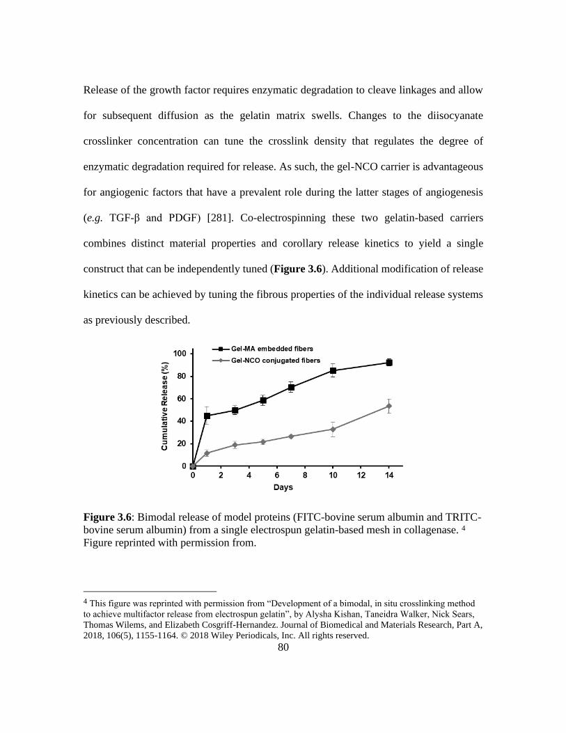
80
Release of the growth factor requires enzymatic degradation to cleave linkages and allow
for subsequent diffusion as the gelatin matrix swells. Changes to the diisocyanate
crosslinker concentration can tune the crosslink density that regulates the degree of
enzymatic degradation required for release. As such, the gel-NCO carrier is advantageous
for angiogenic factors that have a prevalent role during the latter stages of angiogenesis
(e.g. TGF-β and PDGF) [281]. Co-electrospinning these two gelatin-based carriers
combines distinct material properties and corollary release kinetics to yield a single
construct that can be independently tuned (Figure 3.6). Additional modification of release
kinetics can be achieved by tuning the fibrous properties of the individual release systems
as previously described.
Figure 3.6: Bimodal release of model proteins (FITC-bovine serum albumin and TRITC-
bovine serum albumin) from a single electrospun gelatin-based mesh in collagenase. 4
Figure reprinted with permission from.
4 This figure was reprinted with permission from “Development of a bimodal, in situ crosslinking method
to achieve multifactor release from electrospun gelatin”, by Alysha Kishan, Taneidra Walker, Nick Sears,
Thomas Wilems, and Elizabeth Cosgriff-Hernandez. Journal of Biomedical and Materials Research, Part A,
2018, 106(5), 1155-1164. © 2018 Wiley Periodicals, Inc. All rights reserved.

81
The bimodal release system shows much promise and could overcome the
limitation of a transient vasculature density in the induced membrane to improve clinical
outcomes. However, the release system requires further investigation to establish clinical
significance. Our previous work has demonstrated tunable release with use of model-
proteins but this potential has yet to be investigated for actual angiogenic factors of interest.
This study is required to elucidate the impact of their isoelectric potentials and molecular
weights on release kinetics. Although crosslinking is presumed to preserve bioactivity of
the growth factor, additional studies are needed to assess the effect of physical and
chemical processing applied during fabrication on bioactivity retention. Finally, in vivo
evaluation of the individual release systems and the bimodal release system is required to
confirm the predicted release kinetics and establish their efficacy in angiogenesis. The
following chapter further investigates the potential of a co-electrospun gelatin-based
bimodal release system as an adjunct therapy for enhancing angiogenesis in the induced
membrane during the Masquelet technique.

82
Chapter IV: A Multifaceted Matrix to Enhance Angiogenesis and
Provide Infection Control during Bone Regeneration
4.1 INTRODUCTION
Successfully improving the Masquelet technique requires a substrate that provides
antimicrobial prophylaxis and guides formation of the induced membrane for improved
durability and vascularity. Resorbable fibrous meshes offer a platform that can be
engineered to achieve these goals, independent of the PMMA spacer [188, 282-286].
Fibrous meshes produced by electrospinning are of particular interest due to the versatility
in materials and fibrous microarchitecture (e.g. fiber diameter and fiber alignment) that
permits biomimicry of the extracellular matrix for cell scaffolding [54, 287]. Electrospun
meshes are fabricated by applying electrostatic forces to a polymer solution. Modulation
of the electrospinning parameters enable tunable structural properties that can be used as a
template to direct cell behavior and matrix deposition for a more robust membrane with
improved durability [287]. Beyond cell scaffolding, electrospun meshes serve as matrices
for bioactive agents, with tunable release that can be used to further guide cell behavior for
angiogenesis as well as inhibit bacterial growth [288, 289]. To our knowledge, only a few
studies have demonstrated this potential for the Masquelet technique [282, 283].
Yu et al. performed a proof-of-concept study that evaluated the ability of an
electrospsun poly(lactic-co-glycolic) acid (PLGA) wrap to induced formation of a
membrane absent of a PMMA spacer. The electrospun wrap was completely resorbed after
6 weeks. Histological analysis of the membrane revealed a cell-rich layer with expression

83
of angiogenic and osteogenic growth factors. These results confirmed the ability of the
electrospun PLGA wrap to induce membrane formation. However, no instruction beyond
material cues and scaffolding were provided by the electrospun PLGA to improve
formation of the membrane. The potential of the membrane to promote bone regeneration
with and without autologous bone graft was also assessed. Despite the demonstrated
membrane bioactivity, incomplete bone bridging and poor bone consolidation resulted
from the absence of bone graft and indicated that the PLGA-induced membrane was not
sufficient to promote bone regeneration alone. The lack of comparison between the PLGA-
induced membrane and a PMMA-induced membrane composition further challenged the
proposed potential of the electrospun PLGA-induced membrane. This control is important
to include as to establish the efficacy of wrap as an improved or synergistic solution to the
standard technique [283].
The findings from the initial study provided the fundamentals for the development
of a composite substrate encompassing the electrospun PLGA wrap. In this study, Yu et
al. modified the electrospun PLGA system to contain vancomycin/ceftazidime-loaded
layer and a BMP-2-loaded layer. The potential of the composite substrate was evaluated
on membrane formation absent of a PMMA spacer and subsequent bone regeneration with
bone substitute after 8 weeks. Results demonstrated that the PLGA fibers released the
antibiotics and BMP-2 for up to 4 weeks. The composite substrate also induced formation
of a membrane that was rich in cells, biochemical cues, and blood vessels. Satisfactory
bone bridging and mechanical strength were observed with use of the composite substrate
as compared to treatment without the composite substrate. However, this study similarly
84
failed to compare these findings to that of a PMMA-induced membrane to confirm
improvement or synergy. Another limitation of this study is that the composite substrate
was not evaluated in an infected femoral defect model to validate efficacy of the
antimicrobial potential [282].
The purpose of this study was to engineer a multifunctional wrap as an adjunct
therapy that can guide membrane formation while inhibiting infection. In preceding
chapters, we have highlighted the ability of a resorbable electrospun PLGA mesh to control
release of antimicrobial agents and demonstrated the antimicrobial potential in treatment
of osteomyelitis. We have also elucidated the ability of a gelatin mesh to control release of
growth factors. Therefore, in this chapter, the capacity of a previously developed photo-
crosslinked gelatin-methacrylate (gel-MA) electrospinning mechanism to enable in-line
loading of vascular endothelial growth factor (VEGF) for enhanced angiogenesis was first
confirmed [290]. Characterization of the in vitro VEGF bioactivity retention was evaluated
to validate the safety of electrospinning and crosslinking on VEGF bioactivity. In vivo
analysis of gel-MA resorption and corollary release of VEGF was assessed in a rat
subcutaneous model to establish proof-of-concept for controlled release. We then utilized
co-electrospinning to combine the VEGF-loaded gel-MA release system with the
gentamicin sulfate-loaded PLGA release system to create the multifunctional wrap. The
co-electrospun wrap was investigated to corroborate dual-fiber populations and
maintenance of wrap integrity after swelling. The retention of the release profiles of VEGF
and gentamicin sulfate from the co-electrospun wrap was then evaluated in comparison to
individually established release profiles. To improve clinical application and prevent

85
displacement during treatment, the co-electrospun wrap was further modified with tissue
adhesive moieties. In all, these studies provide initial evaluation of an adjunct
multifunctional wrap that can guide membrane formation during the Masquelet technique
with potential to simultaneously inhibit osteomyelitis for improve clinical outcomes.
4.2. MATERIALS AND METHODS
Materials
All chemicals and reagents were purchased from Sigma Aldrich (Milwaukee, WI)
and used as received unless otherwise noted.
Gelatin-methacrylate Synthesis
Gelatin-methacrylate (Gel-MA) was synthesized by dropping 2-isocyanatoethyl
methacrylate (IEMA) into a 5 % (wt/wt) solution of gelatin (porcine, type B) in dimethyl
sulfoxide (DMSO). IEMA was added such that the molar ratio of isocyanate to amine was
4X, with the assumption of 11 lysine residues per gelatin molecule. The reaction was
performed under nitrogen at 37°C with stirring for 3 h. The solution was then dialyzed
against reverse osmosis water for 72 h to remove non-functionalized IEMA and DMSO.
The solution was then frozen and lyophilized to isolate the gel-MA product. The degree
of functionalization with IEMA was quantified using NMR based on the method adapted
from Ovsianikov et al.[291]. Briefly, 10 mg of gel-MA sample was dissolved in 1 mL of
deuterium oxide and analyzed using a 400 MHZ Varian MR-400. The methacrylation ratio
was defined as the percentage of amino groups that were modified in gel-MA. The NMR

86
spectrum was normalized to the phenylalanine sign (6.9 -7.5 ppm) to account for the
concentration of gelatin. The known ratio of 0.011 mol phenylalanine/100 g gelatin was
considered in the integration of this peak (5 protons) to calculate a ratio corresponding to
approximately 0.05297 mol/100 g [292]. The total amount of primary amine groups in
gelatin is known to be approximately 0.0385 mol/100 g [291]. The two new peaks at 5.4-
5.7 ppm on the gel-MA spectrum corresponding to the methacrylate group were integrated
and used in the following calculation to determine the degree of methacrylation:
𝐷𝑒𝑔𝑟𝑒𝑒 𝑜𝑓 𝑚𝑒𝑡ℎ𝑎𝑐𝑟𝑦𝑙𝑎𝑡𝑖𝑜𝑛 = 𝐼5.7𝑝𝑝𝑚
𝐼7.2𝑝𝑝𝑚 ×
0.05297 𝑚𝑜𝑙
100 ×
100
0.0385 𝑚𝑜𝑙
Electrospinning Gelatin-methacrylate
Gel-MA was dissolved at 7 % (wt/wt) in 2,2,2-trifluoroethanol. Ethylene glycol
dimethacrylate was added as a crosslinker at 20 % (wt/wt) of the polymer and a photo-
initiator, lithium phenyl-2,4,6-trimethylbenzoylphosphinates, was added at 5 wt% of the
polymer. The solution was stirred at 37°C overnight prior to electrospinning.
Pentaerythritol tetrakis(3-mercaptoproprionate) was added at 10 wt% of the polymer
immediately prior to electrospinning to reduce oxygen inhibition and facilitate
crosslinking. The gel-MA solution (1 mL) was then added to a syringe protected from light
and dispensed at 1.0 mL/h (KDS100; KD Scientific; Holliston, MA) through a blunted 18
G needle. The syringe pump was placed 12 cm away from a grounded copper plate and a
voltage source charged to 10 kV (ES30P-5W/DDPM; Gamma Scientific; San Diego, CA)
was used to apply to the needle. A UV lamp was placed above the collector to allow for in
situ photo-crosslinking. Each mesh had a total collection time of 1 h followed by one

87
additional hour of UV exposure. For VEGF-loaded meshes, 0.2 µg of recombinant human
VEGF (R&D Systems; Minneapolis, MN) reconstituted in a 0.1 % (wt/v) BSA was added
to the electrospinning solution. The loading concentration was selected based on
consideration of the model release kinetics and literature-targeted release concentrations
required to induce formation of a mature vasculature [290, 293].
Water Uptatke
The crosslinking density of the electrospun gel-MA wrap was determined by water
uptake analysis. Electrospun wraps (N=3) were soaked in deionized water (DI) overnight
at 37°C to remove uncrosslinked gelatin and swell the specimens. The wraps were blotted
dry and weighed after immersion to obtain the swollen weight. Specimens were then
vacuum dried overnight and weighed again to obtain the dried weight. Water uptake was
calculated using the following calculation:
𝑊𝑎𝑡𝑒𝑟 𝑢𝑝𝑡𝑎𝑘𝑒 (%) = 𝑤𝑠−𝑤𝑑
𝑤𝑑𝑋 100,
where ws indicates the weight of the swollen specimen and wd is the weight of the dried
specimen.
Endothelial Cell Tubulogenesis
The human umbilical vein endothelial cell line (HUVECs; Promocell; Heidelberg,
Germany) was selected to assess bioactivity retention due to its potential to organize into
tube-like vascular networks [294]. Briefly, HUVECs were first expanded in EGM-2
endothelial cell growth media (Promocell), supplemented with 1% of penicillin-

88
streptomycin (10,000 U/mL; Thermo Fisher Scientific; Waltham, MA) at 37C. Media was
replaced every 48 h until cells were near 80-90 % confluence.
A specimen (10 x 15 mm2) was cut from an electrospun VEGF-loaded gel-MA
mesh and immersed in EGM-2 endothelial cell growth media, supplemented with 0.2 U/mL
of collagenase up to 7 days, with releasate being collected and replaced with fresh media
at 1, 3, and 5 days. Releasate collected from a blank gel-MA mesh at each time point were
used as the negative control. Releasates and controls were stored at -80°C until further use.
Growth factor-reduced Matrigel (Corning; Corning, NY) was thawed at 4°C overnight. On
the day of use, the Matrigel was transferred onto ice to preserve its liquid state. Matrigel-
coated well plates were prepared by plating 50 μL of Matrigel into a 96-well plate at a
horizontal level that allows the Matrigel to distribute evenly without bubbles. Plates were
incubated for 30 min at 37°C to solidify the Matrigel. Releasates and controls were thawed
at 37°C for 10 min prior to use. HUVECs were reconstituted in releasates or controls at a
density of 40,000 cells/ 100 μL. Cell suspensions were carefully plated on top of the
Matrigel and incubated at 37°C for 4 h. To assess network formation, cells were stained
with Calcein, AM (Invitrogen; Carlsbad, CA) and imaged using a fluorescence microscope
(Nikon Eclipse TE2000-S; Melville, NY). Network formation was captured through raster
patterning of 3 wells/group at each time point. Automated counts of network formation
were achieved using the Angiogenesis Analyzer plugin for ImageJ, which detects
characteristic points and elements of endothelial cell networks [295]. VEGF bioactivity
retention was measured by the ability of released VEGF to encourage morphogenesis of
HUVECs into capillary-like networks as a function of VEGF concentration compared to

89
the negative controls at each time point. A standard curve of capillary-like networks was
formed based on various concentrations of unprocessed VEGF in cell culture media. This
curve was then used to extrapolate the number of networks that would form based on the
expected released concentrations at each time point that was determined from model
release kinetics. Direct comparison of the network formation in response to unprocessed
VEGF and released VEGF was used to determine the degree of bioactivity retention at each
time point. Bioactivity retention was deemed acceptable at >80 %. Studies were repeated
in triplicate and results are represented in terms of the average networks formed at each
time point for each group.
Animal Care
All procedures were approved by the University of Texas Institutional Animal Care
and Use Committee (IACUC, Animal Use Protocol# 2019-00183).
In Vivo Assessment of Resorption
In vivo resorption rates and corollary release kinetics of VEGF from electrospun
gel-MA meshes were assessed in a pilot study. Eight-week old Sprague-Dawley rats (n =
2/time point) were randomly assigned to blank electrospun gel-MA meshes or electrospun
gel-MA meshes. Animals were anesthetized and placed on a nose cone flowing with 2%
isoflurane (Animal Health International; Greeley, CO) in oxygen at a rate of 2 L/min. The
dorsal surface was clipped and prepped for surgery with povidone-iodine and 70% ethanol.
The dorsum was covered with a sterile drape and a 4-cm incision was made in the central

90
third of the dorsum. Blunt dissections were made in the left and right dorsolateral areas to
prepare four implant pockets. UV sterilized blank and VEGF-loaded electrospun gel-MA
specimens (1 x 6 cm2, n=2/ rat) were placed in the pockets in a sterile manner. The incision
was then closed with stainless steel surgical wound clips (Braintree Scientific; Braintree,
MA) and all animals were percutaneously administered carprofen (5 mg/kg; Animal Health
International). One week after the initial procedure, all rats were anesthetized and the
wound clips were removed. At 2, 4, and 6 weeks, the rats were euthanized via carbon
dioxide inhalation followed by bilateral thoracotomy to ensure death. The incisions were
opened and any remaining specimens were harvested. To evaluate resorption, harvested
specimens were lyophilized and dried masses at each time point were compared to the
initial mass of the sample. Release of VEGF was assessed using an ELISA kit (R&D
Systems). Briefly, lyophilized specimens were degraded in 200 U/mL of collagenase to
release remaining VEGF loaded in the specimens at each time point. Solutions were
analyzed following the manufactures instructions. In vivo release was confirmed by
comparing the remaining VEGF content at each time to the initial content loaded in
specimens that were not implanted.
Co-electrospinning
Co-electrospun meshes were fabricated by electrospinning the gel-MA with PLGA.
Briefly, a gel-MA solution was prepared as previously described. 50:50 PLGA (acid-
terminated; inherent viscosity range 0.55–0.75 dL/g; DURECT Corp.; Cupertino, CA) was
dissolved in 1,1,1,3,3,3-hexafluoro-2-propanol (HFIP; Halocarbon; Peachtree Corners,

91
GA) to produce a 40% (w/v) polymer solution. Gel-MA (1 mL) and PLGA (4 mL)
solutions were placed into their respective syringes and were placed on either side of the
mandrel to minimize interaction of the electric fields. Gel-MA electrospinning parameters
were set according to the specifications previously described above. PLGA was
electrospun at a flow rate of 0.7 mL/h through a 20 G blunted needle charged to 8 kV at a
distance of 15 cm from the collector. Fibers were collected at ambient conditions on a
rotating grounded mandrel. The total collection time for gel-MA was 1 h and 4 h for PLGA.
The fabricated meshes were vacuum dried for a minimum of 12 h prior to characterization.
For VEGF-loaded gel-MA fibers, 0.2 µg of rhVEGF reconstituted in a 0.1 % (wt/v) BSA
was added to the electrospinning solution. For gentamicin sulfate-loaded PLGA fibers, 20
mg of gentamicin sulfate powder was dissolved in the electrospinning solution. This dose
was determined by considering the release kinetics obtained from preliminary scouting
studies (data not included) and by considering reported minimum inhibitory concentrations
(MIC; lowest concentration that visibly inhibited bacteria growth) of gentamicin sulfate
for the osteomyelitis-derived strain UAMS-1 [123].
Characterization of Co-electrospun Meshes
Fluorescence microscopy of co-electrospun meshes was performed using a
confocal fluorescence imaging system (MaiTai HP; Spectra Physics; Mountain View, CA).
The gel-MA solution was mixed with 2 wt% fluorescein, and the PLGA solution was mixed
with 0.1 wt% DAPI. These fluorescently-doped solutions were co-electrospun onto a glass
slide attached to the rotating mandrel using the parameters described above.
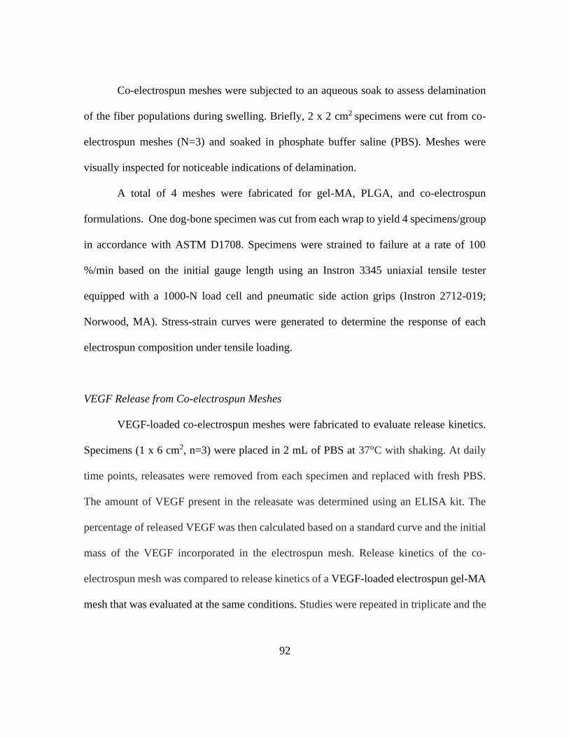
92
Co-electrospun meshes were subjected to an aqueous soak to assess delamination
of the fiber populations during swelling. Briefly, 2 x 2 cm2 specimens were cut from co-
electrospun meshes (N=3) and soaked in phosphate buffer saline (PBS). Meshes were
visually inspected for noticeable indications of delamination.
A total of 4 meshes were fabricated for gel-MA, PLGA, and co-electrospun
formulations. One dog-bone specimen was cut from each wrap to yield 4 specimens/group
in accordance with ASTM D1708. Specimens were strained to failure at a rate of 100
%/min based on the initial gauge length using an Instron 3345 uniaxial tensile tester
equipped with a 1000-N load cell and pneumatic side action grips (Instron 2712-019;
Norwood, MA). Stress-strain curves were generated to determine the response of each
electrospun composition under tensile loading.
VEGF Release from Co-electrospun Meshes
VEGF-loaded co-electrospun meshes were fabricated to evaluate release kinetics.
Specimens (1 x 6 cm2, n=3) were placed in 2 mL of PBS at 37°C with shaking. At daily
time points, releasates were removed from each specimen and replaced with fresh PBS.
The amount of VEGF present in the releasate was determined using an ELISA kit. The
percentage of released VEGF was then calculated based on a standard curve and the initial
mass of the VEGF incorporated in the electrospun mesh. Release kinetics of the co-
electrospun mesh was compared to release kinetics of a VEGF-loaded electrospun gel-MA
mesh that was evaluated at the same conditions. Studies were repeated in triplicate and the

93
data is presented in terms of average cumulative percent release over time and average dose
released (ng).
Gentamicin Sulfate Release from Co-electrospun Meshes
Gentamicin sulfate-loaded co-electrospun meshes were fabricated to evaluate
release kinetics. Specimens (1 x 6 cm2, n=3) were placed in 2 mL of distilled water at 37°C
with shaking. Deionized water was selected, as opposed to phosphate buffer, due to the
low sensitivity of gentamicin sulfate in buffered solution at the low concentrations. At daily
time points, release medium was collected and replaced with fresh deionized water. The
collected release medium was frozen and lyophilized to concentrate the gentamicin sulfate.
The dose released at each time point was determined using a ninhydrin assay. Briefly, 0.5
mL of 2% ninhydrin was added to the lyophilized product and incubated at 120C for 15
min. The ninhydrin solutions were diluted with 1 mL of distilled water to enable
spectrophotometric analysis (Infinite 200 Pro; Tecan; Morrisville, NC) at 570 nm. The
percentage of released gentamicin sulfate was then calculated based on a standard curve
and the initial mass of the gentamicin sulfate incorporated in the electrospun mesh. Release
kinetics of the co-electrospun mesh was compared to release kinetics of a gentamicin
sulfate-loaded electrospun PLGA mesh that was evaluated at the same conditions. Studies
were repeated in triplicate and the data is presented in terms of average cumulative percent
release over time and average dose released (µM).
Dopamine Tissue Adhesive Coating

94
Dopamine hydrochloride was dissolved at 2 or 20 mg/mL in 10 mM tris. 1 mL of
each solution was then carefully pipetted onto a PLGA electrospun mesh. After 24 h, the
solutions were aspirated and 1 mL of 3 wt% sodium periodate in distilled water was applied
to the meshes. After 1 minute, the sodium periodate solution was aspirated and meshes
were washed in 50 mL of distilled water for 5 h with water changes every hour. Samples
were subsequently frozen and lyophilized prior to characterization. The dopamine-
modified PLGA layer was affirmed using ATR-FTIR spectroscopy (Nicolet iS10;
ThermoFisher Scientific; Waltham, MA) at a resolution of 2 cm-1 for 32 scans.
Adhesion Strength Measurement
Adhesive properties of the dopamine-coated PLGA meshes were determine using
lap-shear tensile stress measurements in accordance with the ASTM standard F2255-05,
with some modification. To assess adhesion to bone, fresh porcine cortical bone with in-
tact periosteum was cut into 2.5 x 5 cm2 segments with uniform thickness and hydrated in
PBS for immediate use. Un-modified and adhesive PLGA meshes (N=4) were fabricated
and stored in a dry location at room temperature. A 2.5 × 1 cm2 specimen was removed
from each mesh to account for batch variability. The porcine test substrates were removed
from PBS and blotted with sterile gauze to remove excess PBS prior to coming into contact
with the un-modified PLGA specimens or adhesive PLGA specimens. The specimens were
positioned between two tissue substrates with a 1 cm overlap. They were then compressed
with a force of approximately 1 N to allow for the bond to set. Specimens were further
conditioned in a humidity chamber at 37°C for 30 min prior to being strained to failure at
95
a rate of 5 mm/min using an Instron 3345 uniaxial tensile tester equipped with a 100-N
load cell and pneumatic side action grips (Instron 2712-019). The maximum strength and
failure strain were recorded. The adhesive strength was calculated by maximum strength
divided by the initial bond area.
Statistical Analysis
Data averages are accompanied with ± standard deviation. Statistical analysis was
performed utilizing a standard one-way ANOVA with Tukey’s post-hoc analysis unless
otherwise indicated in figure captions. Statistical significance was accepted at P<0.05.
4.3 RESULTS
Fabrication and Characterization of Gelatin-methacrylate Wrap
Controlled delivery of angiogenic factors has been demonstrated to improve
angiogenesis in tissue engineering applications. Implementation of this technology can
address limitations of the Masquelet technique for treatment of critical-sized bone defects.
In this present study, we used a previously developed in situ crosslinking mechanism to
enable in-line loading and encapsulation of VEGF during gel-MA electrospinning [290].
NMR analysis confirmed that approximately 85% of gelatin lysines were functionalized
with methacrylate groups (Figure 4.1). Assessment of the crosslinking density confirmed
a water uptake of 282 ± 80 % indicating successful photo-polymerization of the gel-MA
wrap.

96
Figure 4.1: NMR spectra of A) gelatin and B) gel-MA used to quantify functionalization.
To confirm VEGF encapsulation and evaluate the effect of fabrication on
bioactivity retention, HUVECs were suspended in VEGF releasate collected at each time
point and seeded onto Matrigel®. Success was determined by the ability of HUVECs to
organize into significantly greater numbers of capillary-like networks as a function of
VEGF concentration as compared to the blank gel-MA releasate (negative control). A
standard curve of network formation was created to confirm cellular response to
unprocessed VEGF at increasing concentrations (Figure 4.2). VEGF-loaded gel-MA
releasate resulted in 27 ± 6 networks after 1 day of release, 14 ± 6 networks after 3 days of
release, and 11 ± 5 networks after 5 days of release. The quantity of networks formed at
each time point was significantly greater than the quantity formed by blank releasate at the

97
corresponding time point. These results indicated that VEGF bioactivity was not severely
impeded by fabrication (Figure 4.3). The degree of bioactivity retention was determined
at each time point by comparing the number of releasate-induced networks to the number
of networks that formed in response to unprocessed VEGF at theoretical released
concentrations. The theoretical concentration at each time point was derived from
previously determined release kinetics using model growth factors [290]. Results specified
that the estimated bioactivity retention was greater than 80% by the fifth day of release,
signifying that the in situ crosslinking mechanism successfully encapsulated VEGF and
that the bioactivity was retained following fabrication.

98
Figure 4.2: Capillary-like network formation in response to unprocessed VEGF. A)
Representative images of network formation induced by increasing concentrations of
unprocessed VEGF. Cells stained with calcein-AM. Scale bar is 200 µm. B) Quantified
network formation/field of view corresponding to the representative images.
Figure 4.3: Evaluation of the bioactivity of released VEGF from electrospun gel-MA
meshes. Representative images show capillary-like network formation corresponding to
blank releasate and releasate from VEGF-loaded meshes. Cells stained with calcein-AM.
Scale bar is 200 µm. Graph displayed quantifies the network formation over 5 days of
VEGF release. * indicates statistical differences with respect to the network formation
induced by blank releasate at each time point.
After evaluating the effect of fabrication on loading and bioactivity retention, initial
in vivo studies were conducted to evaluate gel-MA resorption and corollary release. VEGF-
loaded and blank gel-MA specimens were implanted subcutaneously into the dorsum of
Sprague-Dawley rats and resorption and release were assessed at 2, 4, and 6 weeks. Studies
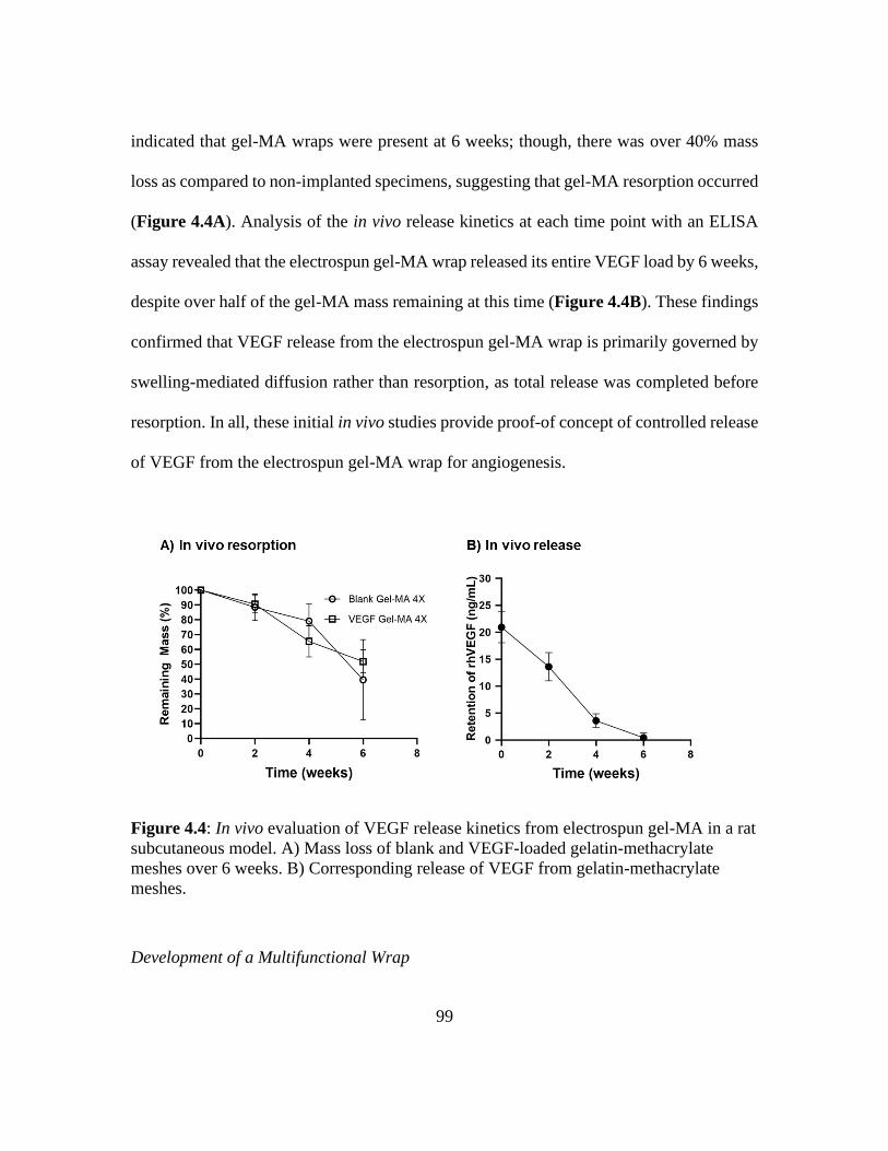
99
indicated that gel-MA wraps were present at 6 weeks; though, there was over 40% mass
loss as compared to non-implanted specimens, suggesting that gel-MA resorption occurred
(Figure 4.4A). Analysis of the in vivo release kinetics at each time point with an ELISA
assay revealed that the electrospun gel-MA wrap released its entire VEGF load by 6 weeks,
despite over half of the gel-MA mass remaining at this time (Figure 4.4B). These findings
confirmed that VEGF release from the electrospun gel-MA wrap is primarily governed by
swelling-mediated diffusion rather than resorption, as total release was completed before
resorption. In all, these initial in vivo studies provide proof-of concept of controlled release
of VEGF from the electrospun gel-MA wrap for angiogenesis.
Figure 4.4: In vivo evaluation of VEGF release kinetics from electrospun gel-MA in a rat
subcutaneous model. A) Mass loss of blank and VEGF-loaded gelatin-methacrylate
meshes over 6 weeks. B) Corresponding release of VEGF from gelatin-methacrylate
meshes.
Development of a Multifunctional Wrap

100
Treatment of critical-sized bone defects with the Masquelet technique is limited by
induced membrane bioactivity and durability. Bacteria colonization of implants further
complicates treatment with high incidences of osteomyelitis. We used co-electrospinning
to combine the VEGF-loaded gel-MA release system and a gentamicin-loaded PLGA
release system into a multifunctional wrap with the potential to enhance the induced
membrane and reduce infection as an adjunct therapy. It was hypothesized that combining
the two release systems would allow independent control over multifactor release. Dual-
fiber population was confirmed via fluorescent labeling of each solution with fluorescein
(gel-MA) and DAPI (PLGA) (Figure 4.5).
Figure 4.5: Schematic of co-electrospinning apparatus. Blow out image depicts dual-
fiber population. Fluorescein (green) fibers are gel-MA and DAPI (blue) fibers are
PLGA.
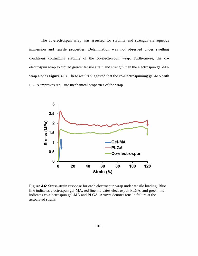
101
The co-electrospun wrap was assessed for stability and strength via aqueous
immersion and tensile properties. Delamination was not observed under swelling
conditions confirming stability of the co-electrospun wrap. Furthermore, the co-
electrospun wrap exhibited greater tensile strain and strength than the electrospun gel-MA
wrap alone (Figure 4.6). These results suggested that the co-electrospinning gel-MA with
PLGA improves requisite mechanical properties of the wrap.
Figure 4.6: Stress-strain response for each electrospun wrap under tensile loading. Blue
line indicates electrospun gel-MA, red line indicates electrospun PLGA, and green line
indicates co-electrospun gel-MA and PLGA. Arrows denotes tensile failure at the
associated strain.

102
VEGF and gentamicin sulfate release from the co-electrospun wrap were evaluated
daily over 14 days to validate retention of the respective release kinetics when combined.
VEGF release kinetics from the co-electrospun wrap was assessed with an ELISA assay
and was shown to have a burst release comparable to the gel-MA wrap. Similarly, there
was no significant difference between the cumulative release after 14 days (97.8 ± 2.5 %
for gel-MA wrap and 86.1 ± 3.1 % for co-electrospun wrap) indicating that co-
electrospinning did not impact release kinetics of the electrospun gel-MA component
(Figure 4.7A). Analysis of the dose of VEGF that was released daily from the co-
electrospun wrap revealed an overall decrease in the released amount at each time point as
compared to the gel-MA wrap (Figure 4.7B). This decrease was attributed to off-target
fiber collection in the electrospinning set-up due to the build-up of fibers on the
surrounding electrospinning apparatuses. The loss of fiber collection resulted in daily dose
of VEGF below the targeted concentration range for angiogenesis (0.5 -11 ng/ml) over 10
days [293, 296]. A similar trend was observed for the comparison of release kinetics of
gentamicin sulfate from the co-electrospun wrap and the PLGA wrap over 14 days (Figure
4.8). However, the lower dose that was released from the co-electrospun wrap at each time
point was still greater than the hypothesized MIC concentration for treating osteomyelitis
(1µM) [123]. Overall, this data demonstrated that co-electrospinning enables independent
control over multifactor release from a single adjunct therapy.
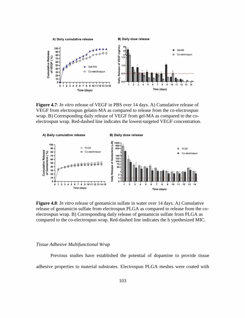
103
Figure 4.7: In vitro release of VEGF in PBS over 14 days. A) Cumulative release of
VEGF from electrospun gelatin-MA as compared to release from the co-electrospun
wrap. B) Corresponding daily release of VEGF from gel-MA as compared to the co-
electrospun wrap. Red-dashed line indicates the lowest-targeted VEGF concentration.
Figure 4.8: In vitro release of gentamicin sulfate in water over 14 days. A) Cumulative
release of gentamicin sulfate from electrospun PLGA as compared to release from the co-
electrospun wrap. B) Corresponding daily release of gentamicin sulfate from PLGA as
compared to the co-electrospun wrap. Red-dashed line indicates the h ypothesized MIC.
Tissue Adhesive Multifunctional Wrap
Previous studies have established the potential of dopamine to provide tissue
adhesive properties to material substrates. Electrospun PLGA meshes were coated with

104
dopamine using the reported dip coating method to improve clinical application and
prevent displacement during healing (Figure 4.9A). Infrared spectral analysis confirmed
the presence of the dopamine coating indicated by dopamine specific peaks on the PLGA
wrap (Figure 4.9B). Lap shear analysis was used to determine the tissue adhesive potential
of the dopamine-coated PLGA mesh as compared to an un-modified PLGA mesh.
Specimens were applied to fresh porcine cortical bone with an in-tact periosteum and
evaluated under tensile loading. The average shear strength of the dopamine-modified
PLGA mesh increased with higher dopamine concentrations (Figure 4.10C). The average
shear strength of the un-modified PLGA mesh was not included due to shear forces being
below the detection limit of the load cell. Overall, these result demonstrated the potential
of dopamine to confer tissue adhesive properties to biomaterials.

105
Figure 4.9: Fabrication of dopamine-modified PLGA wrap for tissue adhesion. A)
Schematic of the dopamine dip-coating process. B) ATR-FTIR of the un-modified PLGA
mesh (light gray line), pure dopamine (dark gray line), and dopamine-modified PLGA
mesh (black line).

106
Figure 4.10: Evaluation of a dopamine-modified wrap on tissue adhesion. A) Schematic
of proposed reaction with the periosteum. B) Representative image of the set-up for lap
shear testing the dopamine-modified PLGA mesh with bone. C) Average maximum shear
strength of the dopamine-modified PLGA mesh coated with increasing concentrations of
dopamine. Results of the un-modified PLGA mesh were not included in the graph due to
the shear forces being below the detection limit of the instrument. * indicated statistical
differences with respect to the 2 mg/mL coating concentration.
4.4 DISCUSSION
The Masquelet induced membrane technique is commonly used for management of
critical-sized bone defects. Despite the potential of the induced membrane to aid bone
regeneration, the durability of it varies depending on the topography of the spacer and the
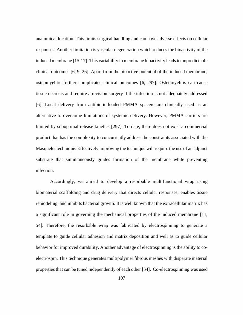
107
anatomical location. This limits surgical handling and can have adverse effects on cellular
responses. Another limitation is vascular degeneration which reduces the bioactivity of the
induced membrane [15-17]. This variability in membrane bioactivity leads to unpredictable
clinical outcomes [6, 9, 26]. Apart from the bioactive potential of the induced membrane,
osteomyelitis further complicates clinical outcomes [6, 297]. Osteomyelitis can cause
tissue necrosis and require a revision surgery if the infection is not adequately addressed
[6]. Local delivery from antibiotic-loaded PMMA spacers are clinically used as an
alternative to overcome limitations of systemic delivery. However, PMMA carriers are
limited by suboptimal release kinetics [297]. To date, there does not exist a commercial
product that has the complexity to concurrently address the constraints associated with the
Masquelet technique. Effectively improving the technique will require the use of an adjunct
substrate that simultaneously guides formation of the membrane while preventing
infection.
Accordingly, we aimed to develop a resorbable multifunctional wrap using
biomaterial scaffolding and drug delivery that directs cellular responses, enables tissue
remodeling, and inhibits bacterial growth. It is well known that the extracellular matrix has
a significant role in governing the mechanical properties of the induced membrane [11,
54]. Therefore, the resorbable wrap was fabricated by electrospinning to generate a
template to guide cellular adhesion and matrix deposition and well as to guide cellular
behavior for improved durability. Another advantage of electrospinning is the ability to co-
electrospin. This technique generates multipolymer fibrous meshes with disparate material
properties that can be tuned independently of each other [54]. Co-electrospinning was used
108
to combine gelatin and PLGA into a multifaceted wrap. Gelatin-based fibers are of
particular interest due to both the matrix-like fibrous structure and the RGD (arginine-
glycine-aspartate) ligands that inherently promote cellular adhesion [298]. Gelatin also has
potential as a carrier for VEGF, a potent regulator of angiogenesis, due to its mild
processing conditions [299]. Angiogenesis is a process by which new blood vessels form
from pre-existing blood vessels. Biochemical cues, such as VEGF, stimulate endothelial
cells and smooth muscle cells that are organized into a pre-existing blood vessel to
destabilize, migrate, proliferate, and organize into vascular tubes. These newly formed
tubes are able to form a stable anastomosis with the pre-existing vascular network leading
to a greater vascular network [300]. Controlled delivery of VEGF between 0.5 to 11 ng/mL
daily over 10 days has been demonstrated to increase mature and stable (i.e. persisting
independently of VEGF delivery) capillary formation by approximately 50% [293, 296].
The release of VEGF from a gelatin wrap within this therapeutic range could enhance
angiogenesis to increase the vasculature and corollary bioactivity of the induced
membrane. In the present study, we have used photo-crosslinked gel-MA to enable
controlled resorption and release of VEGF. Modulation of the gel-MA crosslink density
can be used to tune swelling-based diffusion of embedded VEGF [290]. Furthermore,
PLGA was selected as the additional electrospun polymer due to the demonstrated capacity
to control release of gentamicin sulfate. Gentamicin sulfate is a broad-spectrum antibiotic
that inhibits protein synthesis by targeting bacterial ribosomes. Controlled delivery of
gentamicin sulfate from PLGA above the MIC (1 µM) can potentially inhibit viability of
osteomyelitis-derived isolates like methicillin-susceptible Staphylococcus aureus (MSSA)

109
[123]. Modification of the chemical and fibrous properties of PLGA offer many
mechanisms to tune diffusion of gentamicin sulfate [46, 47]. In addition, these properties
can be tuned to control the mechanical properties and impart additional strength to the
multifunctional wrap.
To enhance angiogenesis, the electrospun gel-MA was loaded with VEGF based on
consideration of target release concentrations and model release kinetics identified in
previous studies [290, 293]. It is well known that growth factors are sensitive to
environmental conditions; therefore, we first evaluated the effects of fabrication and post-
processing on VEGF bioactivity using an endothelial cell tubulogenesis assay, whereby
endothelial cells organize into capillary-like networks in response to VEGF concentration.
This study confirmed that electrospinning did not severely reduce the bioactivity of VEGF
loaded into electrospun gel-MA wrap. These results are in-line with past studies that
demonstrated significant bioactivity retention of VEGF following electrospinning [301,
302]. In vivo evaluation of the VEGF-loaded gel-MA wrap in a rat subcutaneous model
showed that VEGF released over a month and was exhausted before complete resorption
of the wrap. This was expected as previous studies have revealed that the release kinetics
of proteins from gel-MA are primarily governed by swelling-based diffusion rather than
degradation-based diffusion [237, 290]. However, the overall rate of release and resorption
is slower than what has been modeled in previous in vitro studies in our laboratory [290].
This was not unforeseen, as modeling the in vivo environment has proven to be challenging
and can only be used to surmise resorption and release [55]. Slower release and resorption
rates may be of advantage due to the potential to benefit the overall goal of sustaining

110
release of VEGF. Ultimately, the findings from these studies serve as initial proof-of-
concept for a VEGF-loaded electrospun gel-MA wrap capable of enhancing angiogenesis.
Dual-collection of the gel-MA and PLGA fibers were confirmed via fluorescent
imaging. The requisite tensile properties of the co-electrospun wrap were attributed to the
PLGA due to the higher tensile strain and strength of the PLGA wrap as compared to the
gel-MA wrap. The strength was also greater than what has been reported for the induced
membrane when under tensile stress [32]. This suggests that integration of the co-
electrospun wrap into the induced membrane can impart additional structural support
during formation of the membrane. Furthermore, the release kinetics of the co-electrospun
wrap for VEGF and gentamicin sulfate were evaluated. Both fiber populations in the co-
electrospun wrap retained relatively similar release profiles as compared to the individual
release systems. These results confirmed the ability of co-electrospinning to enable
bimodal release with independently tuned release kinetics. However, the concentration
released from each fiber population in the co-electrospun wrap was lower than what was
exhibited for the individual wraps. This was attributed to off-target collection of the fibers
due to the charge repulsion of the two electrospinning jets [303]. Reduced collection of the
fibers lowers the amount of drug available for release. Although this is could be
problematic for angiogenesis, released doses of gentamicin sulfate were still above the
targeted concentration for antimicrobial activity. These results indicated that VEGF-loaded
gel-MA will need to be electrospun for a longer period to compensate for the reduction of
VEGF available due to off-target collection.

111
To improve application of the co-electrospun wrap, a dopamine hydrochloride
(dopamine) coating was applied via dip-coating [304]. Dopamine has been identified as a
derivative of the foot protein that allows aquatic mussels to adhere to inorganic and organic
wet surfaces under high shear stress due to water flow using both covalent and non-
covalent interactions. This discovery has led to the incorporation of dopamine in
biomaterials to promote tissue adhesion. Oxidation of the dopamine catechol enables
covalent bonding with primary amines by nucleophilic attack [304, 305]. We hypothesized
that coating the co-electrospun wrap with dopamine would facilitate attachment to the
periosteum of the host bone and prevent the need of suturing to secure the wrap, as suturing
imparts further tissue damage. Another advantage of incorporating dopamine into the co-
electrospun wrap is the strong affinity of cells to dopamine coatings [306, 307]. Ku et al.
demonstrated significantly enhanced HUVEC adhesion, viability, and stress fiber
formation on dopamine-coated polycaprolactone nanofibers as compared to un-modified
nanofibers and gelatin-coated nanofibers [306]. Similarly, Lee et al. reported on the
potential for polydopamine-coated substrates to support fibroblast adhesion [304].
Polydopamine coatings have been shown to have higher adsorption of serum proteins like
fibronectin as compared to unmodified surfaces. It has been hypothesized that cell adhesion
to dopamine coatings is governed by increased cellular affinity to the adsorbed serum
proteins [258]. Therefore, incorporation of dopamine can exert additional cell binding
properties to improve the composition and durability of the induced membrane. In vitro
analysis of the dopamine-modified PLGA demonstrated the potential for tissue adhesion
of the co-electropsun wrap to the periosteum as compared to un-modified PLGA. These
112
results suggested that a dopamine coating can be implemented to effectively secure the
wrap around the defect without suturing. In vitro studies to evaluate the effect of dopamine
on cell adhesion are currently under investigation.
In all, the co-electrospun wrap offers bimodal release of multiple factors to enhance
angiogenesis and prevent infection. The combination of this technology with biomimicry
of the native matrix structure could be used to guide formation of the induced membrane
to standardize clinical outcomes following the Masquelet technique. The current studies
presented here establish initial evaluation of the multifunctional electrospun wrap with
sustained bioactivity, optimize handling, controlled release, and tissue adhesiveness.
However, there were limitations of these studies that require further investigation. In vivo
angiogenesis in response to the electrospun VEGF-loaded gel-MA mesh has yet to be
investigated to confirm efficacy of the treatment. These studies would need to evaluate the
change in vasculature density and CD31+ blood vessels, which are specific to mature blood
vessel, following treatment as compared to no treatment and blank meshes. Preliminary
studies are in progress to confirm the potential of released gentamicin sulfate to reduce
infection in an osteomyelitis model and improve bone regeneration as compared to no
treatment and a PMMA bone cement carrier. Fibrous properties can be altered to control
release kinetics if bacterial density is above the critical threshold known to cause infection
[308]. Selection of a different PLGA co-polymer ratio can also modulate release. Another
proposed advantage of the multifunctional electrospun wrap is the ability to improve the
durability of the induced membrane for better surgical handling by providing templating
and biochemical signaling. Cellular behavior in response to the co-electrospun wrap was

113
not experimentally investigated in the present study but cellular adhesion and proliferation
in the presence of gelatin and dopamine is established in the literature [298, 306].
Following completion of the aforementioned studies, in vivo assessment of the
multifunctional electrospun wrap with the Masquelet induced membrane technique will be
required to determine its efficacy in guiding formation of the membrane for improved
healing outcomes.
4.5 CONCLUSION
The aim of this study was to engineer a device that can be implemented to reduce
variable clinical outcomes achieved by the Masquelet induced membrane technique. The
studies performed have established initial evaluation of an adjunct multifunctional
electrospun wrap that can influence formation of the induce membrane rather than relying
on the non-guided biological process. This adjunct wrap has been designed to improve
durability for improved surgical handling, enhance angiogenesis to increase the vasculature
and corollary bioactivity, and provide infection control. The bioactivity retention of VEGF
was confirmed following in-line loading into electrospun gel-MA. In situ crosslinking of
gel-MA exhibited sustained in vivo release of VEGF. Co-electrospinning enabled the
combination of the angiogenic release system with the antibiotic release system. The co-
electrospun wrap possessed enhanced mechanical properties with the capacity for bimodal
release of VEGF and gentamicin sulfate. Bimodal released profiles from the co-electrospun
wrap validated the potential to independently control angiogenesis and provide infection
control from a single substrate. Additionally, the ability exert tissue adhesive properties
114
was evaluated and confirmed the potential improve surgical application of the adjunct.
Overall, this work defines the fundamentals for an engineered substrate that can guide
formation of the induced membrane to improve treatment of critical-sized bone defects.
Future studies will build on these findings by further characterizing the multifunctional
electrospun wrap in in vitro cell culture and in relevant animal models.

115
Chapter V: Conclusion
5.1 SUMMARY
The induced membrane has a critical role in facilitating bone regeneration during
the Masquelet technique [9]. However, unpredictable clinical outcomes highlight the need
for a method to better guide formation of the induced membrane. Several research attempts
have been made to modify the spacer for improved formation of the induced membrane,
with the majority of these attempts focused on altering expression of biochemical cues,
enhancing angiogenesis, and increasing durability of the membrane [31-36]. However, no
single approach has definitively guided formation better than the standard technique. An
ideal approach would augment the Masquelet technique with a platform that offers cellular
recruitment, guides cell behavior, and serves as a template to direct matrix deposition and
remodeling for improved durability and enhanced angiogenesis. Due to the frequent
incidence of osteomyelitis associated with critical-sized defects, the approach should also
encompass infection control [6, 103, 104]. The platforms discussed in this work provide
the framework for the development of a multifaceted adjunct wrap with potential to address
these needs.
We first compared the efficacy of two distinct antimicrobials released from a
resorbable fibrous mesh in the treatment of osteomyelitis. The resorbable mesh was
electrospun to allow direct encapsulation of gentamicin sulfate or gallium maltolate into
the PLGA fibers. Release kinetics were then evaluated to confirm sustained release above
116
the hypothesized MIC for each antimicrobial [123, 141]. Not only was sustained release
above the hypothesized MICs achieved, but it lasted longer than the critical period when
bacteria is susceptible to antimicrobials (72 h) [207, 208]. Next, antimicrobial meshes were
cultured with an osteomyelitis-derived isolate to determine the potential of released drug
to inhibit growth. The sub-optimal antimicrobial activity of gallium maltolate led to further
evaluation of the MIC and MBC for each antimicrobial. The poor antimicrobial activity of
the gallium-loaded PLGA mesh was attributed to a higher calculated MIC as compared to
the hypothesized MIC. The antimicrobial activity of each drug was then evaluated against
its cytotoxicity on osteoblasts to establish the safety and efficacy. From these studies, the
gentamicin sulfate-loaded PLGA mesh was selected as a candidate adjunct wrap to treat
osteomyelitis during the Masquelet technique.
We then developed a resorbable wrap to enhance formation of the induced
membrane by electrospinning a photo-crosslinked gelatin-methacrylate (gel-MA) mesh
loaded with VEGF. The resorbable fibers serve as scaffolding to support cell attachment
and guide cell behavior and matrix deposition during tissue remodeling for improved
durability. Delivery of VEGF from the gel-MA mesh can provide further instruction on
cellular behavior for enhanced angiogenesis during formation. VEGF bioactivity retention
following fabrication and in vivo release were evaluated as an initial proof-of-concept study
for controlled growth factor delivery. Electrospinning did not impede VEGF bioactivity
and enabled controlled release up to 4 weeks in vivo. To engineer a substrate that provided
infection control and enhanced formation of the induced membrane, we adapted an
electrospun gelatin bimodal release system developed by our lab [290]. This modified

117
bimodal release system was fabricated by co-electrospinning the gentamicin sulfate-loaded
PLGA platform selected in the previous section with the VEGF-loaded photo-crosslinked
gel-MA platform. The combination of the two platforms not only resulted in bimodal
release of VEGF and gentamicin sulfate with the potential to be independently tuned, but
also resulted in a more structurally stable wrap that can improve surgical application and
further guide cellular behavior.
In summary, successfully improving the Masquelet technique requires infection
control and enhancement of the induced membrane. This work underscores the potential
of this multifaceted substrate to serve as an adjunct wrap to enhance formation of the
induced membrane during the Masquelet technique for improve bone regeneration through
1) antimicrobial activity, 2) support for cellular attachment and guided matrix deposition,
and 3) instruction on cellular behavior to promote angiogenesis.
5.2 SIGNIFICANCE OF WORK
By combining biomaterial scaffolding with drug delivery and biochemical
signaling, an adjunct substrate has been engineered to addresses several limitations of the
Masquelet technique. Herein, we describe novel mechanisms imparted into this substrate
to achieve these goals and the impact that the discoveries from this work will have in the
broader field.
In the first section, two antimicrobial wraps were investigated to establish their
potential as treatments in osteomyelitis. The findings from these studies contributed to the
growing area of research containing few reports on the potential of electrospun
118
antimicrobial wraps as adjunct therapies in bone regeneration. Studies that have evaluated
electrospun antimicrobial wraps as adjunct therapies in infected bone defects did not
evaluate them using relevant osteomyelitis-derived isolates [169, 188]. Our work has
provided analysis of electrospun antimicrobial wraps against an osteomyelitis-derived
isolate to establish better clinical relevance. Furthermore, existing electrospun
antimicrobial wraps used in treatment of osteomyelitis often contain antibiotics [169, 188].
This work highlighted the potential of electrospinning to enable incorporation and
controlled release of a novel antimicrobial, gallium maltolate. Gallium maltolate has been
investigated in treatment for chronic wounds [141, 212]; however, this is the first study to
evaluate its potential in treatment for osteomyelitis. We provided the first known report of
the MIC and MBC for gallium maltolate against an osteomyelitis-derived isolate. These
results build on the fundamentals of gallium maltolate as a broad-spectrum antimicrobial
agent. The MIC of gentamicin sulfate has been discussed in the treatment of osteomyelitis
but not the MBC [123]. Our work has identified the MBC of gentamicin sulfate which can
be used to guide future development of carriers for this drug. Moreover, very few studies
have investigated the cytotoxicity of antimicrobials released from electrospun wraps on
osteogenic cell lines. Those that have seldomly compare the cytotoxicity of the drug to its
antimicrobial activity as to determine if the antimicrobial will be released at concentrations
that are both safe and effective (selectivity index) [169, 188]. Our studies identified the
selectivity indices for each antimicrobial and determined that gentamicin sulfate was a
safer and more effective drug than gallium maltolate in the treatment of osteomyelitis.
119
These findings further expand the knowledge of antimicrobials available to effectively treat
osteomyelitis.
The candidate antimicrobial wrap was then combined with an electrospun bioactive
wrap to generate a multifaceted therapy. To our knowledge, this work established the first
resorbable adjunct wrap to guide membrane formation with cellular recruitment,
templating, and biochemical signaling. An electrospun photo-crosslinked gel-MA mesh
that was previously developed in our lab was utilized to provide scaffolding and
instructions for cells through biomimicry of the native matrix fibrous structure and
bioactivity of the RGD ligands on gelatin [290]. These features offer a framework to
actively guide cellular behavior during membrane formation as compared to other
researched substrates that only rely on scaffolding. In addition, the gel-MA mesh was
loaded with VEGF to promote angiogenesis. Previous work has established the potential
of this gel-MA mesh to provide controlled release of model-proteins for bone regeneration
[290]. We demonstrated significant in vitro bioactivity retention as well as in vivo release
of VEGF from the gel-MA mesh up to 4 weeks. These results further contribute to the
development of this gelatin release system for controlled delivery of growth factors. These
findings also build upon the sparse reports that describe bioactivity retention of growth
factors following in-line loading into electrospun fibers. By co-electrospinning the VEGF-
loaded gel-MA mesh with the gentamicin sulfate-loaded PLGA, we achieved bimodal
release from a single construct with the potential to be independently tuned. Co-
electrospinning also enables multiple material properties to be combined. This technique
could provide additional mechanical cues that further guide cell behavior during membrane
120
formation. The increased strength of the wrap imparted by the PLGA fibers can also be
used to reinforce the mechanical properties of induced membrane during formation until
complete remodeling is achieved. Ultimately, these methods enable the wrap to address
limitations of osteomyelitis and the induced membrane simultaneously.
The individual electrospun platforms that were combined to overcome the
limitations of the Masquelet technique can also be used to address other indications in bone
healing. For example, compound fractures are often exposed to contaminants that can lead
to osteomyelitis and delay or prevent healing. The gentamicin sulfate-loaded PLGA wrap
can be used as a stand-alone local prophylaxis to overcome this drawback. Although the
risk of infection is lower in non-compound fractures, there can still be significant damage
to the bone resulting in critical-sized defects that require bone grafting. Allografts are
prevalently used in critical-sized defects that require a substantial amount of graft and
structural support due to their availability as compared to autografts. However, sterilization
and processing of allografts removes the periosteum, which contains cellular and vascular
constituents that confer bioactivity. This results in poor bone union and mechanical
strength [309, 310]. Allograft revitalization has since become a major objective in bone
regeneration. The electrospun gel-MA wrap can serve as a bioactive carrier for growth
factors to emulate the biochemical signaling that is present during periosteum-mediated
healing and guide cell behavior to enhance regeneration. This technology can also augment
synthetic bone grafts that have superior biocompatibility, mechanical properties, and
tunable degradation rates but that lack bioactivity to actively promote healing. When
contamination and significant bone loss are of concern in severe compound fractures, the

121
multifunctional electrospun wrap can augment bone grafts to address both indications
simultaneously. In all, the ability to address multiple limitations places this work at the
forefront of innovation in bone regeneration.
We have developed a multifunctional adjunct wrap capable of simultaneously
inhibiting bacterial growth and guiding formation of the induced membrane to address
these needs. The overall significance of this work includes expanding knowledge of
antimicrobials available to effectively treat osteomyelitis, imparting structural and
biochemical cues for active guidance of cellular behavior and tissue remodeling, and
combining multiple release systems to achieve bimodal release from a single construct.
Although the mechanisms described here were aimed at improving the Masquelet
technique, the broader impact lies within the development of independent electrospun
platforms that be used in a variety of bone regeneration applications.
5.3 CHALLENGES AND FUTURE PERSPECTIVE
The work presented here describes several advances in improving the Masquelet
technique. However, further investigation must be performed for clinical translation.
A primary focus of this work has been on evaluating and identifying an adjunct
antimicrobial therapy in the treatment of osteomyelitis. Although we have demonstrated
the potential of the gentamicin sulfate-loaded PLGA wrap to reduce bacterial
contamination at released concentrations that are safe on osteoblasts, bone healing is
orchestrated by more than just osteoblasts. Additional work is needed to evaluate
cytotoxicity of gentamicin sulfate on other cell lines such as chondrocytes and endothelial
122
cells to better determine the potential efficacy of the wrap. It is also important to evaluate
the high burst release from the wrap on nephrotoxicity due high incidences of gentamicin
sulfate-induced nephrotoxicity [311]. The antimicrobial wrap is electrospun for 4 h to
achieve a relatively high drug load that enables in vitro detection of release kinetics during
the sustained release phase. This results in concentrations released that are significantly
greater than the calculated MIC and MBC of gentamicin sulfate. However, no adverse
effects are expected due to the total load of gentamicin sulfate in the PLGA wrap being
below (20-fold decrease) the clinically-administered dose known to induce necrosis and
renal dysfunction in animals [311]. The most robust method to evaluate nephrotoxicity will
be in vivo analysis due to the challenges associated with modeling cellular responses in
vitro. If released doses are found to be nephrotoxic, then a reduction of the wrap’s thickness
will be required to reduce the available drug load and subsequent released concentrations.
The evaluation of in vivo antimicrobial activity is also warranted in an infected bone defect
model. It has been reported that there is a bacterial threshold (>103 CFU/g of tissue) for
reliable infection that impedes bone healing [308]. Current studies are in progress to
evaluate the remaining bacterial load and bone healing following treatment with the
gentamicin sulfate-loaded PLGA wrap. These studies are important to validate in vitro
modeling of antimicrobial activity and cytotoxicity. They will also establish the potential
efficacy of local delivery at relatively low doses over a shorter duration as compared to
systemic treatment and other local delivery devices. If the released concentrations are
found not to be effective, then additional modification of the wrap will be necessary. One
of the advantages of this electrospun wrap is the ability to be adapted as needed to achieve

123
clinical efficacy in not only bone regeneration but many other drug delivery applications;
however, the presented work did not demonstrate this ability. Tunable release kinetics are
currently being investigated through modification of various parameters during
electrospinning. These modifications include changes to the solution properties such as the
polymer viscosity and the polymer type or changes to the electrospinning parameters such
as the voltage and flow rate. Modulation of these parameters can be used to alter the fiber
diameter or the degradation rate of the gentamicin sulfate-loaded wrap if tunable release
kinetics are needed following in vivo evaluation [45, 53]. Furthermore, commercialization
of the antimicrobial wrap will require that the fabrication time is reduced. This must be
completed without compromising the effective dose loaded into the wrap. This can be
achieved by using a different solvent such as chloroform to solubilize and load gentamicin
sulfate at higher concentrations [185, 186]. A higher loading concentration will maintain
the requisite total drug load with less polymer being collection over a shorter period of
time. Careful consideration should be given when modifying the loading concentration as
to not increase the gentamicin sulfate concentration above a critical threshold that would
cause drug aggregation [209, 210]. This could result in changes to the release kinetics and
alter the clinical efficacy.
In this work, we have also demonstrated the potential of an engineered substrate to
serve as an adjunct wrap to guide formation of the induced membrane. We have shown that
this wrap provides bimodal release of gentamicin sulfate and VEGF with sustained
bioactivity to both prevent infection and enhance angiogenesis of the induced membrane.
Another proposed key feature of this wrap is the ability to recruit cells and guide cellular

124
behavior to influence membrane formation with improved durability. However, in vitro
cell studies that evaluate attachment, matrix deposition, proliferation, and migration are
needed to validate this claim. Additional in vivo studies are required to determine the
duration that the wrap retains its structural integrity. Degradation must occur at a rate that
allows for tissue remodeling. If degradation proceeds tissue remodeling, then the fiber
populations can be tuned independently to slow this rate by mechanisms such as altering
the crosslink density of the gel-MA and using a higher molecular weight PLGA or PLGA
with a different co-polymer ratio that is more hydrophobic [290, 312]. In terms of
enhancing angiogenesis, it was observed in in vitro studies that co-electrospinning reduced
the total load of VEGF due to a reduction in fiber collection as compared to the fiber
collection of gel-MA during independent electrospinning. A longer collection time will be
required to overcome the loss of fibers during co-electrospinning and increase the available
load. The overall potential of the multifunctional wrap to guide formation of the induced
membrane will need to be evaluated in vivo to obtain clinically relevant information. It will
be important to observe the tensile properties of the induced membrane as to determine if
the multifunctional wrap improved durability that improves handling. If mechanical
properties are not improved, then the fiber alignment can be modified to guide matrix
deposition and orientation and corollary mechanical properties [54]. However, doing so
may alter mechanotransduction during the second stage of the procedure and impact bone
regeneration. Another important aspect to observe is the vascularity of the induced
membrane. In vivo analysis of the VEGF-loaded gel-MA mesh should be performed
subcutaneously first to confirm the angiogenic potential of the released concentrations. If

125
vascularization is not enhanced, then the wrap can be optimized by increasing its thickness
to increase the available dose of VEGF. The effect of improved durability and enhanced
vascularization should then be evaluated on bone healing to establish the efficacy of the
multifunctional wrap. This can be achieved with histological analysis, radiographic
analysis, and mechanical analysis. These same observations should made for the wrap in
infected bone defects as to determine the potential to simultaneously prevent infection and
promote membrane formation for improved bone healing in contaminated bone defects.
Despite the need for additional investigation to further develop and confirm the
potential the multifunctional wrap, the work presented here has advanced the wrap towards
the goal of being an adjunct substrate to improve bone regeneration during the Masquelet
technique. First, an electrospun gentamicin sulfate-loaded PLGA wrap was recognized as
an ideal adjunct therapy to treat osteomyelitis. Then the electrospun antimicrobial mesh
was combined with an electrospun VEGF-loaded gel-MA mesh to establish a resorbable
substrate that offers bacterial inhibition, templating for cellular recruitment and matrix
deposition, and biochemical signaling to direct cellular behavior during tissue remodeling.
The findings from these studies provide the fundamentals for further development of the
multifunctional electrospun wrap as well as contribute to the knowledge of electrospun
platforms available for use as stand-alone therapies or combined constructs to meet the
complex requisites for improving tissue regeneration for a variety of applications.

126
Appendix A: In Vivo Performance of a Bilayer Wrap to Prevent
Abdominal Adhesions 5
A.1 INTRODUCTION
Intestinal anastomoses following bowel resection are among the most frequently
performed procedures in general surgery [313]. Despite advancements in suturing and
stapling techniques, there is a high incidence of post-operative complications with up to
30% of patients presenting with anastomotic leakage and up to 90% of patients developing
intra-abdominal adhesions [314-317]. Anastomotic leakage is of particular concern due to
its associated high morbidity and mortality rates as a result of infection, peritonitis, and
sepsis [316, 318, 319]. Treatment of anastomotic leakage often requires prolonged
hospitalization and a secondary operation to minimize infection. Intra-abdominal
adhesions form vascularized fibrotic bridges between adjacent tissues that can restrict
normal organ movement and blood supply leading to chronic pain and bowel obstruction
[320, 321]. The severity and widespread occurrence of these two complications underscore
the need for a method to improve patient outcomes after these surgical procedures.
The two most prevalent approaches to reduce anastomotic leakage are
reinforcement of the anastomotic closure with surgical sealants (i.e., fibrin and
cyanoacrylate glues) and an adjunct wrap placed over the anastomosis. Despite in vivo
experimental efforts to develop a method using surgical sealants to strengthen the suture
5 This work was reprinted with permission from “In vivo performance of a bilayer wrap to prevent
abdominal adhesions”, by Alysha Kishan, Taneidra Buie, Canaan Whitfield-Cargile, Anupriya Jose, Laura
Bryan, Noah Cohen, and Elizabeth Cosgriff-Hernandez. Acta Biomaterialia, 2020, 115, 116-126. © 2020
Acta Materialia Inc. Published by Elsevier Ltd. All rights reserved.
127
line, results have displayed high variability of outcomes that were attributed to dosage
variations and inconsistencies in application [322-324]. As an alternative, several groups
have evaluated tissue-derived wraps to externally reinforce the anastomosis and enhance
anastomotic healing including patient-specific omentum, bovine pericardium, human
amniotic membrane, and porcine collagen foams [325-330]. Bovine pericardium, in
particular, has been shown to improve wound healing with significant increases in burst
pressures of the treated anastomoses [328]. Although these materials effectively enhance
healing and reduce anastomotic leakage, none have gained commercial success possibly
due to the increased incidence and severity of intra-abdominal adhesions associated with
their use [331, 332].
To address the complications of intra-abdominal adhesions after intestinal
surgeries, several adhesion barriers have been developed and commercialized. Adhesion
barriers act to physically separate the injured tissue from the adjacent viscera during the
critical period of post-operative healing (5-7 days), thereby reducing the formation of
fibrotic adhesions [333]. Seprafilm, composed of sodium hyaluronate and
carboxymethylcellulose, and Interceed®, composed of an oxidized regenerated cellulose,
are adhesion barriers that effectively reduce the incidence and severity of adhesions both
experimentally and clinically [320, 334, 335]. Despite established efficacy, these barriers
have limitations. The efficacy of Interceed® is significantly reduced in the presence of
blood due to fibrin deposition and fibroblast penetration which reduces the capacity to
prevent adhesions [336-338]. Application of Seprafilm® immediately after anastomoses
delays inflammatory-mediated processes that promote healing of the suture line which

128
leads to an increased rate of anastomotic leakage [337-339]. As a result, several clinical
studies have recommended that surgeons avoid applying Seprafilm directly onto fresh
suture lines [334, 339, 340].
To date, current treatments to enhance anastomotic healing to prevent leakage often
result in increased adhesions; whereas, barrier materials to reduce adhesions can delay
anastomotic healing and increase anastomotic leakage. These findings suggest that
therapeutic strategies that only address one of these complications independent of the other
will ultimately fail. Our laboratory developed a new approach to concurrently address these
complications using a composite bilayer wrap with selective bioactivity to both reduce
intra-abdominal adhesions and enhance anastomotic healing. The inner layer consists of an
electrospun gelatin mesh to promote cell adhesion and remodeling [341]. Reactive
electrospinning developed in our laboratory was utilized to modulate the degree of gelatin
crosslinking and the corollary resorption rate. In order to resist adhesion formation, a
resorbable poly(ethylene glycol) (PEG) hydrogel was selected as an outer layer due to its
inherent non-fouling properties [342]. Iterative design of hydrogel and electrospun mesh
formulations was utilized to identify a bilayer structure with requisite surgical handling
characteristics and retention of wrap integrity after swelling. Candidate bilayer wraps were
then evaluated to confirm selective bioactivity over two weeks. As an in vivo proof-of-
concept study, adhesion prevention was assessed using a rat colonic abrasion model in
comparison to a clinical control using Interceed. Based on initial in vivo results, the
bilayer wrap was further modified to include an additional tissue adhesive component to
prevent displacement after implantation. Collectively, these studies provide an initial
129
evaluation of a new bilayer wrap design to address the multimodal complications
associated with intestinal anastomoses.
A2. MATERIALS AND METHODS
Materials
All chemicals were purchased from Sigma Aldrich (Milwaukee, WI) and used as
received unless otherwise noted.
Polymer Synthesis
Poly(ethylene glycol) diacrylate (PEGDA) was synthesized according to a method
adapted from Hahn, et al. [343]. Briefly, acryloyl chloride was added dropwise to a solution
of PEG 2 kDa or 6 kDa diol and triethylamine in dichloromethane (DCM) under nitrogen.
The molar ratio of PEG, acryloyl chloride, and triethylamine was 1:2:4, respectively. After
the addition of acryloyl chloride, the reaction was stirred for an additional 24 hours at room
temperature. The resulting solution was then washed with 8 molar equivalents of 2 M
potassium bicarbonate to remove acidic byproducts. The product was then precipitated in
cold diethyl ether, filtered, and dried under vacuum.
PEGDA with thio-β esters (PEGDTT) was synthesized as previously described
[342]. d,l-dithiolthreitol (DTT) and triethylamine (TEA) were added dropwise to a solution
of PEGDA (2 kDa) in DCM. The molar ratio of DTT, PEG and triethylamine was 3:2:0.9,
respectively. After the addition of the DTT and triethylamine, the reaction was stirred for
24 hours at room temperature. The resulting solution was then precipitated in cold
130
diethethyl ether, washed, filtered, dried under ambient conditions for 24 hours then placed
under vacuum to remove any excess solvent.
Characterization of PEGDA and PEGDTT was confirmed using proton nuclear
magnetic resonance (1H-NMR) spectroscopy. Proton NMR spectra of control and
functionalized polymers were recorded on a Mercury 300 MHz spectrometer using a
TMS/solvent signal as an internal reference. Percent conversions of PEG diol to acrylate
endgroups was greater than 85%. 1H-NMR (CDCl3): δ 3.6 ppm (m, -OCH2CH2), 5.8 ppm
(dd, -CH=CH2), 6.1 (dd, -CH=CH2) and 6.4 ppm (dd, -CH=CH2).
Reactive Electrospinning of Gelatin
In situ crosslinking of electrospun gelatin was performed as previously reported
[341]. Briefly, a double-barrel syringe with an attached mixing head and a diisocyanate
crosslinker were utilized to generate electrospun scaffolds that crosslink during the
electrospinning process via reaction of the isocyanate with the pendant amines of lysine
residues in gelatin. Bovine-derived gelatin, 1,4-diazabicyclo[2,2,2]octane (DABCO) and
hexamethylene diisocyanate (HDI) were each dissolved in 2,2-trifluoroethanol (TFE). The
concentration of HDI was determined such that the crosslinker density would equal a 5:1
ratio of isocyanate:amine. Double-barrel syringes were loaded with 12 wt% gelatin in TFE
and 5 wt% (of solids) DABCO solution in one barrel and HDI in TFE solution in the other.
The double barrel syringe solutions were pumped through a mixing head into an 18-gauge
blunted needle at a rate of 3.0 mL/hr. The needles were placed 12 cm away from the
collector to allow for adequate solvent evaporation and fiber drying. A high voltage of 10
131
kV was then applied to the needle. Meshes were electrospun for 3 hours and stored under
vacuum for at least 24 hours prior to further use.
Bilayer Wrap Fabrication
A PEG-based hydrogel coating method was selected through iterative design using
3 distinct methods. Namely, a bulk hydrogel coating, an electrosprayed hydrogel coating,
and a hydrogel foam coating were tested. Bulk hydrogel coatings were fabricated from a
10 wt% PEGDTT:PEGDA 75:25 solution in distilled water. A photo-initiator solution (1
mg Irgacure 2959 per 10 µl 70% ethanol) was added at 1 vol% of precursor solution. The
PEG hydrogel precursor solution was pipetted between 1-mm spacer plates and exposed to
UV light to initiate crosslinking. After 45 seconds of UV illumination, a 0.2-mm thick
gelatin mesh was placed on top of the surface of the hydrogel. Samples were then exposed
to UV light for 5 additional minutes to complete hydrogel formation. Bilayer wraps were
swollen in reverse osmosis water overnight followed by lyophilization before further use.
Electrospraying precursor solutions were prepared from 30 wt% 75:25
PEGDTT:PEGDA in 70:30 DCM:EtOH solvent. A photo-initiator solution (1 mg Irgacure
2959 per 0.01 ml 70% ethanol) was added at 3 vol% of precursor solution. The hydrogel
solution was then added to a syringe and electrospun gelatin meshes were attached to a
copper plate as the collector. A voltage of 7.5 kV was applied to the needle tip and -2 kV
was applied to the collector, which was set 13 cm away from the needle tip to allow for
solvent evaporation and particle drying. The polymer solution was dispensed at a rate of
1.0 mL/hr using a syringe pump. A UV lamp was placed above the electrospraying set up
132
to enable in situ curing. The hydrogel solution was electrosprayed for 3 hours, after which
the resulting bilayer wrap was vacuumed for at least 24 hours prior to characterization.
Finally, PEG-based hydrogel foam coatings were prepared using an emulsion
templating method developed previously in our laboratory. Hydrogel precursor solutions
were prepared by mixing 20 wt% PEGDTT:PEGDA 75:25 in a 10 wt% solution of
Pluronics F-68 in water, and a photo-initiator solution added at 1% of the total polymer of
lithium phenyl-2,4,6-trimethylbenzoylphosphinates (LAP). Precursor solutions were made
with and without 10 wt% trimethyloylpropane ethyodylate triacrylate (TMPE-TA) as an
additional crosslinker. Once thoroughly mixed, light mineral oil was added at volume
fractions of 50% of the continuous polymer precursor phase. The water and oil composition
was mixed at 2500 rpm for 5 minutes using a FlackTek Speedmixer DAC 150 FVZ-K.
Electrospun gelatin meshes were affixed to 1.0- mm spacer plates and hydrogel emulsions
were then transferred into the mold. Samples were exposed to UV light for 6 minutes to
complete foam formation. Wraps were then subjected to a series of 3 1-hour soaks to
remove the oil content: 1) 100% DCM; 2) 50/50 DCM/ethanol; 3) 100% ethanol. After the
last ethanol soak, wraps were soaked in distilled water overnight prior to lyophilization
before further use.
Bilayer Wrap Characterization
Scanning electron microscopy (SEM) (JOEL 6500) was utilized to image
specimens. Lyophilized samples were stored under vacuum prior to imaging. A total of 4
composite bilayer wraps were fabricated for each composite formulation. One specimen
133
was taken from each wrap to yield 4 specimens/group. Specimens were coated with 4 nm
of gold using a sputter coater (Sputter Coater 108, Cressingtion Scientific Instruments).
Confirmation of two distinct layers was performed by evaluating the chemical structure of
each using attenuated total reflectance-Fourier transform infrared (ATR-FTIR)
spectroscopy (Nicolet iS10 (Thermo Scientific) at a resolution of 2 cm-1 for 32 scans.
A previously described, 4 composite bilayer wraps were fabricated for each
composite formulation. One dog-bone specimen was cut from each wrap to yield 4
specimens/group in accordance with ASTM D1708. Specimens were strained to failure at
a rate of 100 %/min based on the initial gauge length using an Instron 3345 uniaxial tensile
tester equipped with a 100-N load cell and pneumatic side action grips (Instron 2712-019).
The elastic modulus, tensile strength, and ultimate elongation were calculated from the
resultant engineering stress-strain curves. A secant modulus at 2% strain was calculated
for the elastic modulus and subsequently referred to as “modulus”. Toughness was
calculated as the area under the stress-strain curve.
Selective bioactivity over time was assessed via cell adhesion. Adult human dermal
fibroblasts (hDFs, isolated from adult skin, Invitrogen) were cultured in growth media
containing 16.5% fetal bovine serum (FBS, Atlanta Biologicals), 1% L-glutamine (Life
Technologies), and Minimum Essential Media α (MEM α, Life Technologies) to 80%
confluence and utilized at Passage 5. A total of 4 hydrogel foam fiber composite bilayer
wraps were fabricated and 8 mm-disks were removed from each wrap using a biopsy punch
(Integra Miltex). Specimens were placed in a tissue cultured 48-well plate (Corning) and
were sterilized for 30 minutes under UV. They were then immersed in PBS and incubated
134
at 37 °C for 2 weeks. PBS was selected as the hydrogel component is hydrolytically
degradable and incubation in PBS is a common analogue for real-time hydrolytic
degradation under physiological conditions (e.g. pH and ion concentration) [344-346].
Specimens (n=4/group) were then taken at 1, 7, and 14 days to test selective bioactivity.
Human dermal fibroblasts were seeded upon either the hydrogel foam layer or the gelatin
layer at 10,000 cells/cm2 in growth media supplemented with 1 vol% penicillin-
streptomycin. Following a 3-hour incubation, cells were fixed and stained with rhodamine
phalloidin (F-actin/cytoplasm, Biotium) and SYBR green (DNA/nucleus, Thermo Fisher
Scientific). A 3-hour culture period was selected as it is considered a typical length of time
to allow cell attachment [347-349]. Cell adhesion was calculated from manual cell counts
of images obtained through raster patterning of 4 specimens/group/end point using a
fluorescence microscope (Nikon Eclipse TE2000-S).
Animal Care
All procedures were approved by the Texas A&M University Institutional Animal
Care and Use Committee (IACUC, Animal Use Protocol# 2017-0316).
In Vivo Assessment
First, in vivo resorption rates for each component of the bilayer mesh was assessed.
Sprague-Dawley rats (n = 3/group) were randomly assigned to 1 of 2 groups: 1) hydrogel
foam or 2) electrospun gelatin mesh to assess degradation. Animals were anesthetized with
isoflurane in oxygen and maintained on isoflurane in oxygen via a nose cone. The ventral
135
abdomen was clipped and prepped for surgery with chlorohexidine and 70% ethanol. The
abdomen was draped with a sterile paper drape and a 2-cm ventral mid-line incision made
in the central third of the abdomen. The appropriate dried mesh or gel was paced in the
abdomen in a sterile manner. The body wall was closed with 5-0 PDS in an intradermal
pattern and the skin was closed with 5-0 PDS in a simple continuous pattern. A
circumferential abdominal bandage was placed and all animals were administered
buprenorphine (0.01 mg/kg, subcutaneously) immediately post-operatively. At each time
point, the rats were euthanized via carbon dioxide asphyxiation. The abdomen was opened
and entire abdomen thoroughly explored macroscopically to identify remaining specimens
or confirm full resorption.
The effect of treatment on the formation of intra-abdominal adhesions was assessed
using a rat cecum abrasion model as previously described [350]. Sprague-Dawley rats (n =
5/group) were randomly assigned to 1 of 6 groups: 1) cecum abrasion with no treatment;
2) cecum abrasion with fibrin glue applied; 3) cecum abrasion with Interceed ® applied;
4) cecum abrasion with gelatin mesh only applied; 5) cecum abrasion with hydrogel foam
only applied; 6) cecum abrasion with bilayer wrap applied. Specimens were sterilized using
ethylene oxide and allowed to vent for 3 days prior to use. Twelve-week-old rat were
anesthetized and prepared for surgery as described above. A 2-cm ventral mid-line incision
made in the central third of the abdomen. The cecum was exteriorized and a template used
to abrade the cecal serosa in a standard location, size (1 x 2 cm2), and severity of abrasion
(40 strokes of a dry gauze with mild pressure). Similarly, a 1 x 2 cm2 area of body wall
was resected from the right side of the abdomen centered 1 cm dorsal to the incision. A

136
single simple interrupted suture (5-0 monocryl) was placed between the cecum and the
body wall at a site approximately 1 cm distant (cranial) to the abraded area of both the
cecum and body wall. The treatments were then applied. Briefly, Tisseel® tissue sealant
was used as fibrin glue (Baxter healthcare) and was thawed in a water bath 1 hour prior to
use and used within 4 hours of thawing. Approximately 200 µL of fibrin glue was applied
to the abraded surface of the cecum for every group except PBS and Interceed®. The
appropriate mesh was then immediately applied to the area making every effort to cover
the abraded area with the mesh. For Interceed®-treated rats, fibrin glue was not applied
and instead Interceed® was applied according to manufacturer’s directions (applied dry to
a non-bleeding surface). Similarly, approximately 500 µL PBS was pipetted onto the
damaged portion of the cecum 3 times for the PBS group. The cecum was then returned to
the abdomen. The body wall was closed with 5-0 PDS in a simple continuous pattern and
the skin was closed with 5-0 PDS in a simple continuous pattern. A circumferential
abdominal bandage was placed and all animals were administered buprenorphine (0.01
mg/kg subcutaneously) immediately post-operatively.
At each time point, animals were euthanized via carbon dioxide asphyxiation. The
abdominal wall was opened at the left flank to avoid disturbing site of adhesion and
retracted to expose the right body wall. Adhesions were photographed and scored discretely
for size, strength, and maturity as described in Table A.2. One observer blinded to
treatment group scored all of the animals. As the small scale used to determine adhesion
scores (0-3, 0-5) does not capture the differences between large, mature adhesions, and
137
smaller, less cohesive adhesions, a composite score was assigned to each rat based on the
following calculation:
𝐶𝑜𝑚𝑝𝑜𝑠𝑖𝑡𝑒 𝑠𝑐𝑜𝑟𝑒 = 𝑠𝑖𝑧𝑒 × (𝑠𝑡𝑟𝑒𝑛𝑔𝑡h + 𝑚𝑎𝑡𝑢𝑟𝑖𝑡𝑦)
Dopamine Tissue Adhesive Coating
Dopamine hydrochloride was dissolved at 50 mg/mL in 10 mM tris. 1 mL of
solution was then carefully pipetted onto the gelatin layer of a swollen bilayer wrap. After
24 hours, the solution was aspirated and 1 mL of 3 wt% sodium periodate in distilled water
was applied to the bilayer wrap. After 1 minute, the sodium periodate solution was
aspirated and wraps were washed in 50 mL of distilled water for 5 hours with water changes
every hour. Samples were subsequently frozen and lyophilized prior to characterization.
The dopamine-modified gelatin layer was affirmed using ATR-FTIR spectroscopy at a
resolution of 2 cm-1 for 32 scans.
Adhesion Strength Measurement
Adhesive properties of the dopamine-coated gelatin layer were determine using lap-
shear tensile stress measurements in accordance with the ASTM standard F2255-05. Fresh
split thickness porcine skin was cut into 2.5 x 5 cm2 segments with uniform thickness and
hydrated in PBS for immediate use. A total of 4 un-modified and adhesive gelatin meshes
were fabricated and stored in a dry location at room temperature. A 2.5 × 1 cm2 specimen
was removed from each mesh to account for batch variability. Interceed® (Ethicon) sheets
were cut into comparable dimensions for comparison to the un-modified and adhesive

138
gelatin meshes. Interceed® was selected as a clinical control due to its ability to securely
adhere to the anastomosis without sutures when meticulous hemostasis has been achieved
[336]. The porcine test substrates were removed from PBS and blotted with sterile gauze
to remove excess PBS prior to coming into contact with the un-modified gelatin specimens,
adhesive gelatin specimens, or Interceed®. The specimens were positioned between two
tissue substrates with a 1 cm overlap. They were then compressed with a force of
approximately 1 N to allow for the bond to set. Specimens were further conditioned in a
humidity chamber at 37°C for 30 minutes prior to being strained to failure at a rate of 5
mm/min using an Instron 3345 uniaxial tensile tester equipped with a 100-N load cell and
pneumatic side action grips (Instron 2712-019). The maximum strength and failure strain
were recorded. The adhesive strength was calculated by maximum strength divided by the
initial bond area.
Statistical Analysis
Data are displayed as mean ± standard deviation for each composition. Statistical
analysis was performed utilizing a standard one-way ANOVA unless otherwise noted in
the figure captions. Statistical significance was accepted at P< 0.05.
A.3 RESULTS
Bilayer Wrap Optimization
139
Multiple bilayer formulations were investigated for their suitability to produce an
optimized adhesion barrier with enhanced handleability and stability in vitro. The proposed
PEG-based hydrogel layer for adhesion prevention was applied to the inner electrospun
gelatin layer in 3 forms: a hydrogel layer, an electrosprayed layer, and a foam layer (Figure
A.1A-C). Each of the 3 bilayer wrap fabrication methods were assessed for handling in
terms of tensile properties and stability via delamination assessment upon swelling. The
hydrogel bilayer wrap displayed the most brittle behavior, as evidenced by the stress-strain
curves in Figure A.1C. The electrosprayed bilayer wrap had the lowest toughness (0.4 ±
0.2 MJ/m3) and reached tensile failure at 3.5 ± 1.2% elongation (Table A.1). The hydrogel
foam composite bilayer wrap displayed the highest toughness of 23.6 ± 1.7 MJ/m3 and
reached an elongation of 41.0 ± 4.0%. As such, this composite formulation was selected
for future testing. However, swelling analysis indicated delamination upon immersion in
water. In order to decrease the degree of swelling and thereby resist delamination, the
crosslinker, trimethylolpropane ethoxylate triacrylate (TMPE-TA), was added to the foam
precursor solution. The TMPE-TA containing bilayer was again evaluated to ensure
maintenance of handleability and stability in vitro. The formulation displayed a further
significant increase in toughness, from 23.6 ± 1.7 to 28.8 ± 1.5 MJ/m3 (Table A.1).
Furthermore, the formulation displayed no evidence of delamination upon swelling. This
optimized bilayer wrap, containing an electrospun gelatin layer and PEG-TMPETA foam
was subjected to further characterization prior to assessment in vivo.
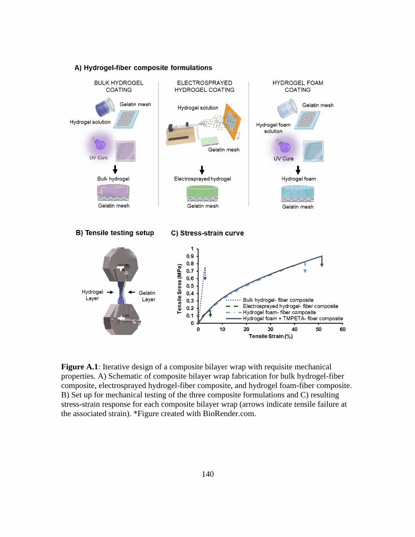
140
Figure A.1: Iterative design of a composite bilayer wrap with requisite mechanical
properties. A) Schematic of composite bilayer wrap fabrication for bulk hydrogel-fiber
composite, electrosprayed hydrogel-fiber composite, and hydrogel foam-fiber composite.
B) Set up for mechanical testing of the three composite formulations and C) resulting
stress-strain response for each composite bilayer wrap (arrows indicate tensile failure at
the associated strain). *Figure created with BioRender.com.

141
Table A.1: Tensile properties of composite bilayer wrap formulations. * indicates
statistical differences as compared to the hydrogel foam-fiber composite, (P<0.05).
Composite
formulations
2% Secant
modulus
(MPa)
Ultimate tensile
strength (MPa)
Elongation
(%)
Toughness
(MJ/m3)
Bulk hydrogel -
fiber composite 22.8 ± 4.8 * 0.6 ± 0.2* 2.5 ± 0.5 * 1.0 ± 0.4 *
Electrosprayed
hydrogel - fiber
composite
4.8 ± 0.7 0.1 ± 0.1 * 3.5 ± 1.2 * 0.4 ± 0.2 *
Hydrogel foam
-
fiber composite
4.7 ± 0.7 0.7 ± 0.1* 41.0 ± 4.0 * 23.6 ± 1.7 *
Hydrogel foam
+ TMPETA -
fiber composite
4.1 ± 0.2 0.9 ± 0.1* 48.3 ± 3.4 * 28.8 ± 1.5 *
Bilayer Wrap Characterization
ATR-FTIR spectroscopy was utilized to evaluate the chemical composition of each
layer in the selected bilayer wrap. Spectral analysis indicated that although the hydrogel
was visible on both layers, as demonstrated by the ether peak at 1110 cm-1, the amine
stretch at 3350 cm-1 of gelatin was only present on the inner layer (Figure A.2A). Scanning
electron microscopy of the selected bilayer wrap, displayed in Figure A.2B, demonstrated
that the inner gelatin layer consisted of homogenous, smooth fibers while the outer
hydrogel foam layer displayed a porous intra-architecture encased between the fibrous
layer and a smooth, outer film.

142
Figure A.2: Characterization of the hydrogel foam + TMPETA- fiber composite. A)
ATR-FTIR of the gelatin layer (gray line) and the hydrogel layer (black line). B) Cross-
sectional SEM depicting the intra-microarchitecture of each layer (scale bar =30 µm).
SEM blowouts display the plan view of each layer (scale bar =100 µm). C)
Representative images of hDF attachment over 14 days (scale bar =200 µm) and
quantified cell adhesion on each layer. Cells stained with rhodamine phalloidin (F-
actin/cytoplasm) and SYBR green (DNA/nucleus). * indicates statistical differences with
respect to the hydrogel foam at each time point, (two-way ANOVA with Sidak’s
analysis, P<0.05).
Next, the potential to maintain selective bioactivity over the desired application
period (target 2 weeks) was evaluated by assessing hDF adhesion on each layer at selected

143
time intervals. Bilayer wrap samples were incubated in PBS for 2 weeks and removed at
1, 7, and 14 days to prior to culturing with cells to determine the effect of degradation on
bioactivity. After each time point, hDFs were seeded onto each side of the bilayer wrap for
3 hours and cell adhesion was evaluated. There was significant cell adhesion on the
electrospun gelatin layer throughout the 14-day period, (Figure A.2C). In contrast, the anti-
fouling properties of the hydrogel foam layer prevented cellular adhesion resulting in cells
being washed away prior to analysis. Overall, these results suggested that selective
bioactivity was maintained.
Assessment of In Vivo Degradation
Prior to evaluating the efficacy of the bilayer wrap, an in vivo degradation scouting
study was conducted. Samples of each composition were implanted within the abdominal
cavity of rats and degradation was assessed visually after 7, 14, and 21 days. Initial studies
indicated that gelatin meshes were present at 7 days and had lost mechanical integrity by
14 days. Hydrogel foam specimens remained at 21 days, suggesting that a longer study
must be conducted to fully evaluate the in vivo degradation rate. These initial results
indicated that the degradation profiles of each component were within range of the targeted
rate, permitting initial evaluation for adhesion prevention.
In Vivo Evaluation of Intra-abdominal Adhesion Formation
Intestinal adhesion formation between the cecum and peritoneum of rats was
evaluated by observation of abdominal wound sites within a rat abrasion model after 14
144
days (Figure A.3). Initial scouting studies indicated that applying the bilayer wrap to the
wound site with sutures resulted in strong adhesion formation initiated by leakage, and
ultimately infection. As a result, fibrin glue was used to affix the bilayer wrap to the
abraded area. The adhesions were evaluated macroscopically to discretely evaluate their
area, strength, and maturity (data not shown). A summary of the adhesion scoring method
is described in Table A.2. In addition to the adhesion scoring system, a composite score
was also calculated in an attempt to better capture the differences in adhesions by
considering the correlation between size, strength, and maturity. Figure A.3B displays a
summary of the composite scores as well as representative images from each group (Figure
A.3C-H). Control rats, and rats with only fibrin glue application, showed the development
of strong, thick adhesions between the cecum and peritoneum in all instances, with
composite scores of 18 ± 9 and 20 ± 4, respectively. Interceed® was utilized as a clinical
control and showed complete prevention of adhesions in all specimens (composite score =
0 ± 0). Gelatin meshes applied alone saw a range of adhesion formation in the rats, with a
composite score of 12 ± 10 that was statistically greater than Interceed®. This was expected
as gelatin is known to promote cell adhesion, which the bilayer aims to utilize to promote
healing. Hydrogel foam control and bilayer wrap treated rats had composite scores of 8 ±
7 and 8 ± 5 each. Though these scores were higher than that for Interceed® treated rats,
they were not statistically different. The higher scores were attributed to a subset of rats in
both groups presenting with immature adhesions at the wound site. It was noted that the
foam and bilayer wraps had been displaced from the wound site in each of the rats where
adhesions were reported. Few adhesions were found in rats where the foams or wraps

145
remained in place. As such, it was concluded that the fibrin application was insufficient to
maintain placement and that wraps needed to be formulated such that they adhered to the
wound bed for further studies.
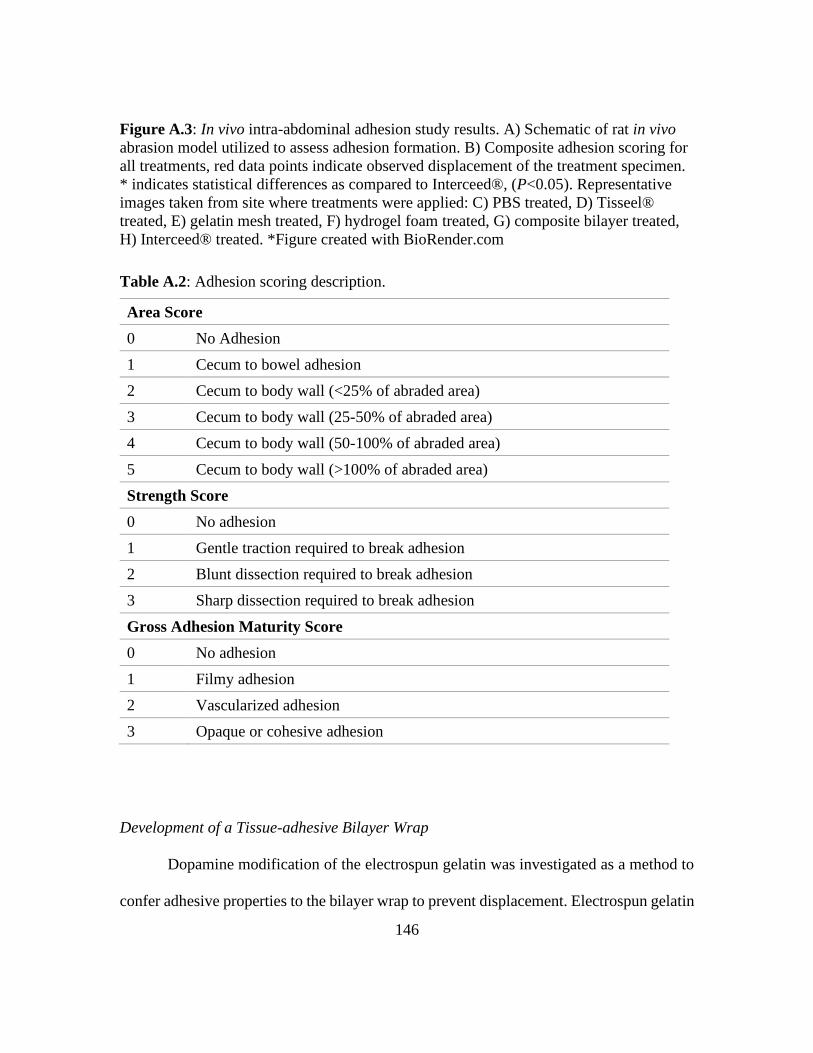
146
Figure A.3: In vivo intra-abdominal adhesion study results. A) Schematic of rat in vivo
abrasion model utilized to assess adhesion formation. B) Composite adhesion scoring for
all treatments, red data points indicate observed displacement of the treatment specimen.
* indicates statistical differences as compared to Interceed®, (P<0.05). Representative
images taken from site where treatments were applied: C) PBS treated, D) Tisseel®
treated, E) gelatin mesh treated, F) hydrogel foam treated, G) composite bilayer treated,
H) Interceed® treated. *Figure created with BioRender.com
Table A.2: Adhesion scoring description.
Area Score
0 No Adhesion
1 Cecum to bowel adhesion
2 Cecum to body wall (<25% of abraded area)
3 Cecum to body wall (25-50% of abraded area)
4 Cecum to body wall (50-100% of abraded area)
5 Cecum to body wall (>100% of abraded area)
Strength Score
0 No adhesion
1 Gentle traction required to break adhesion
2 Blunt dissection required to break adhesion
3 Sharp dissection required to break adhesion
Gross Adhesion Maturity Score
0 No adhesion
1 Filmy adhesion
2 Vascularized adhesion
3 Opaque or cohesive adhesion
Development of a Tissue-adhesive Bilayer Wrap
Dopamine modification of the electrospun gelatin was investigated as a method to
confer adhesive properties to the bilayer wrap to prevent displacement. Electrospun gelatin

147
meshes were coated with dopamine through the reported dip-coating method (Figure
A.4A). Dopamine-modified gelatin meshes were then oxidized and lyophilized to preserve
their oxidized state. Infrared spectral analysis indicated the presence of the dopamine-
specific peaks in the modified gelatin meshes (Figure A.5). Lap shear testing was utilized
to evaluate the adhesive properties of the dopamine-modified gelatin mesh as compared to
un-modified gelatin, Interceed®, and fibrin glue. Specimens were applied to split-thickness
porcine skin and subjected to tensile testing as depicted in Figure A.4B. The mean shear
strength of the dopamine-modified electrospun gelatin layer was 11.6 ± 2.9 kPa. This value
is significantly higher than values reported for fibrin glue (4.6-6.9 kPa) and comparable to
that obtained from commercial Interceed® (Figure A.4C) indicating that dopamine can
potentially prevent displacement [304, 351]. The shear strength of the un-modified gelatin
mesh was not included as, compared to other groups, the hydrated gelatin wrap had shear
strength associated with forces that were below the detection limit of the load cell.

148
Figure A.4: Effect of dopamine coating on tissue adhesion to the bilayer wrap. A)
Schematic of dopamine coating process and hypothesized reaction of dopamine coating
with the wound site. B) Schematic depicting the lap shear test set-up and C) average
maximum shear strength of the dopamine coated bilayer wrap on porcine substrates as
compared to Interceed®. Un-modified gelatin control was not included in graph due to
shear forces below the detection limit of the instrument. * indicates statistical differences
as compared to Interceed®, (student’s t-test, P<0.05). *Figure created with
BioRender.com.
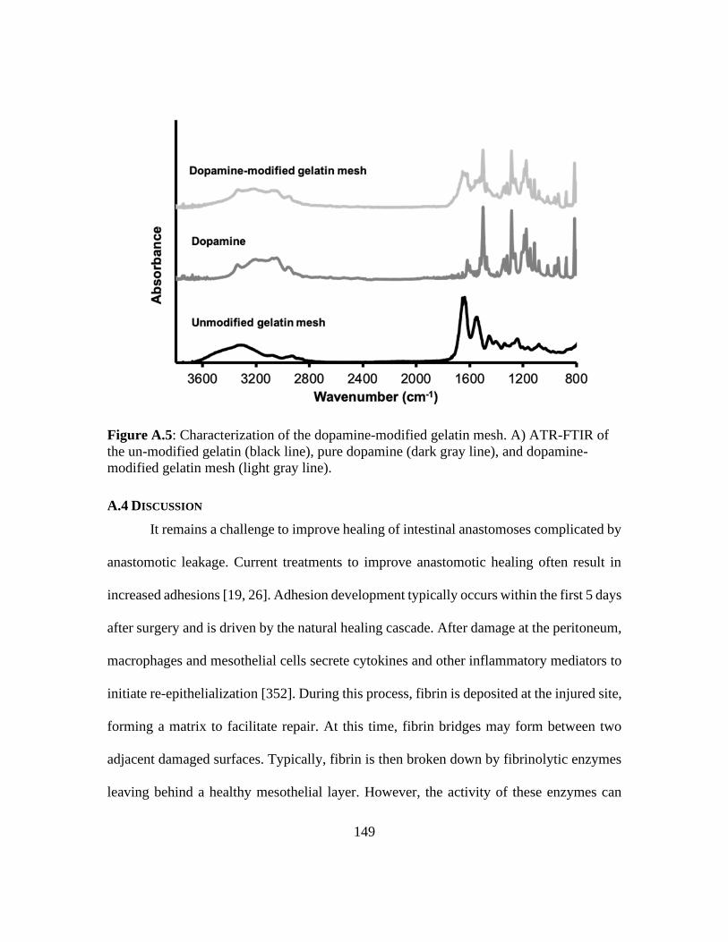
149
Figure A.5: Characterization of the dopamine-modified gelatin mesh. A) ATR-FTIR of
the un-modified gelatin (black line), pure dopamine (dark gray line), and dopamine-
modified gelatin mesh (light gray line).
A.4 DISCUSSION
It remains a challenge to improve healing of intestinal anastomoses complicated by
anastomotic leakage. Current treatments to improve anastomotic healing often result in
increased adhesions [19, 26]. Adhesion development typically occurs within the first 5 days
after surgery and is driven by the natural healing cascade. After damage at the peritoneum,
macrophages and mesothelial cells secrete cytokines and other inflammatory mediators to
initiate re-epithelialization [352]. During this process, fibrin is deposited at the injured site,
forming a matrix to facilitate repair. At this time, fibrin bridges may form between two
adjacent damaged surfaces. Typically, fibrin is then broken down by fibrinolytic enzymes
leaving behind a healthy mesothelial layer. However, the activity of these enzymes can

150
become compromised due to surgical damage limiting fibrin breakdown. This results in an
influx of fibroblasts, capillaries, and nerves that lead to the development of permanent
fibrous connective tissue, also known as adhesions [353]. Adhesion barriers such as
Seprafilm® and Interceed® typically prevent adhesion formation by physical separation
between tissues during the first 5 days. Contrarily, there is concern with the use of adhesion
barriers as adhesion formation has an integral role in the 1 to 2 week critical re-
epithelialization phase of the anastomotic healing [338, 354]. Prevention of adhesion
formation has been shown to delay anastomotic healing leading to increased incidences of
leakage [331, 338, 354]. Thus, barrier films that solely address adhesion prevention fail to
holistically address the clinical needs and can lead to more severe complications associated
with anastomotic leakage. Key requirements for an improved adhesion barrier include that
it promotes anastomotic healing, is simple to apply, is stable in aqueous environments,
degrades within the targeted time-frame, and maintains its position during treatment [355].
To this end, we have developed a resorbable bilayer wrap that resists adhesion
formation while maintaining the potential to promote healing of the anastomosis site. The
inner layer consists of a crosslinked gelatin electrospun mesh. Gelatin, a natural polymer,
inherently promotes cellular adhesion through its RGD (arginine-glycine-aspartate) ligand
[298]. The fibrous structure of the electrospun mesh also promotes cellular integration by
mimicking the native structure of the extracellular matrix [356]. As the gelatin layer of the
bilayer wrap is present to promote this re-epithelialization, the targeted degradation time
for the wrap is 2 weeks to provide a buffer for delayed healing. Previous work demonstrates
a methodology to crosslink gelatin during the electrospinning process with a diisocyanate
151
for improved fiber morphology retention and controlled enzymatic degradation. Enzymatic
degradation facilitates cleavage of peptide bonds within the gelatin structures resulting in
resorption byproducts that are cytocompatible and cleared from the body [341, 357]. In
vitro studies demonstrated that the gelatin layer reached full degradation in enzymatic
solution by 14 days [341]. Furthermore, the outer layer of the wrap is a degradable PEG-
based hydrogel foam. PEG-based hydrogels have an intrinsic resistance to cell adhesion
and thereby have the potential to prevent adhesion to surrounding tissue. This was
demonstrated by West et al. who utilized an in situ crosslinked polyethylene glycol-co-
lactic acid diacrylate hydrogel to effectively reduce the adhesion formation by 60% in a
similar rat model [358]. Although adhesion formation can be prevented during the first 5
days by the PEG-based foam, it was hypothesized that the PEG layer degradation rate
should be slower than that of the gelatin layer (targeted 3 weeks). PEGDA degradation is
primarily mediated by hydrolysis of the esters within the acrylate endgroups. As there are
low numbers of esters present in PEGDA, the in vivo degradation rate is slower than desired
[344, 359]. A modified PEGDA-based foam with thio-β ester linkages was utilized to
control hydrolytic degradation to achieve a targeted degradation rate. The incorporation of
thio-β ester linkages increases the positive atomic charge on the carbonyl carbon of
proximal acrylate esters bonds which heightens ester hydrolysis via nucleophilic reactivity.
Higher stoichiometric monomer ratios of thio-β ester linkages to PEGDA results in more
hydrolytically labile sites leading to faster degradation [360]. Previous in vitro work
demonstrated that the modified PEG hydrogel degraded via hydrolysis within 3 weeks and
that degradation byproducts were cytocompatible [342, 360].

152
In contrast to other PEG-based formulations, we hypothesized that the PEG foam
would provide the requisite toughness and elongation for improved handleability and ease
of application of the bilayer wrap. The favorable mechanical properties of the foam-based
bilayer wrap were attributed to the foam’s microarchitecture (i.e., porosity and pore size)
which were consistent with mechanical properties of hydrogel foams possessing the similar
microarchitecture [361, 362]. It has been noted that hydrogel-based foams with similar
porosity and pore sizes have relatively lower tensile strength and modulus with corollary
enhanced elongation [362]. Equally important to the ease of application is the stability of
the bilayer wrap in terms of delamination. The bilayer wrap displayed delamination which
was attributed to appreciable hydrogel layer swelling. TMPE-TA was added to the
hydrogel foam precursor solution as it is well established in literature that the addition of a
crosslinker reduces scaffold swelling [363]. The addition of TMPE-TA further increased
the strength of the bilayer wrap improving handleability and making it more conducive for
clinical application.
A common drawback of commercial adhesion barriers is that they compromise
anastomotic healing. An advantage of the bilayer wrap is that is has distinct layers to impart
selective bioactivity. Although ATR-FTIR analysis indicated that hydrogel was present on
both layers, the presence of hydrogel on the inner layer does not preclude the cell adhesive
characteristics of gelatin [364]; therefore, these results were considered to be acceptable.
Fibroblasts were selected for evaluation of selective bioactivity as they are the most
prevalent cell type during the healing process and are commonly utilized as a screening
method for adhesion evaluation [365]. Cell adhesion primarily on the gelatin layer
153
indicated that cell adhesion was unaffected by the presence of a minuscule amount of PEG
foam. This was expected as gelatin is widely incorporated into PEG-based scaffolds to
enhance cellular adhesion [366, 367]. The presence of hydrogel on the gelatin layer is
preferred over the presence of gelatin on the hydrogel layer as the gelatin would reduce the
adhesion resistant properties of the hydrogel layer [364].
In vivo evaluation of the bilayer wrap in a rat abrasion model demonstrated that the
selected compositions of gelatin and PEG-based hydrogel foam were within the targeted
degradation time-frame. The bilayer wrap resulted in reduced adhesion formation. Further
reduction, closer to that of Interceed®, is expected provided that there is maintenance of
the position of the bilayer wrap relative to the wound site. Wraps noted as being displaced
resulted in minor adhesions and indicated a need to more securely affix the wraps to the
wound. Recent studies have reported that dopamine layers can be added to substrates in
order to impart tissue adhesive properties through simple dip-coating [304]. This
technology is derived from mussels, which are known to adhere to all types of organic and
inorganic surfaces under wet conditions. Dopamine hydrochloride (dopamine) has been
identified as the compound which facilitates mussel adhesion to substrates through both
covalent and noncovalent interactions [368]. As a result, dopamine has become a popular
candidate to promote tissue adhesion in devices [369]. We hypothesized that a dopamine
coating could facilitate crosslinking with the wound surface as the catechol transitions into
a quinone upon oxidation, which can then form a covalent bond with primary amines
through nucleophilic attack [351, 370]. This reaction would permit adhesion between the
dopamine-coated gelatin layer and the wound surface. In vitro evaluation of the adhesive
154
gelatin layer demonstrated marked tissue adhesion as compared to that of un-modified
gelatin, comparable to that of Interceed®, and higher than adhesion strengths reported for
fibrin glue [337-339]. These findings indicate that the adhesive dopamine coating can be
implemented to effectively prevent displacement of the bilayer wrap during healing to
prevent adhesion formation.
Overall, these unique wraps utilize material properties to maintain selective
bioactivity throughout degradation in a controlled manner. By combining the native
properties of PEG hydrogels and gelatin, this bilayer approach could be used to
simultaneously enhance anastomotic healing while preventing abdominal adhesions,
unlike any current clinical standard. As a first step towards this goal, the presented studies
provide an initial evaluation of the bilayer wrap with optimized handling, controlled
degradation, and tissue adhesiveness. Despite promising initial results, there were
limitations in the current study that necessitate future research. Regarding the in vitro
characterization, only hydrolytic degradation was considered during evaluation of the
selective bioactivity despite enzymatic degradation being the primary mechanism of
gelatin degradation. In vivo evaluation of gelatin control meshes did confirm that
enzymatic degradation would not impede the cell instructive behavior of gelatin as
adhesions readily formed over 2 weeks. In addition, future studies are needed that provide
in vitro characterization of the dopamine-modified bilayer wrap to ensure that the
mechanical properties and selective bioactivity is maintained. The adhesive strength of
dopamine-modified gelatin meshes was also not experimentally evaluated against fibrin
glue but the shear strength of fibrin glue is well documented in the literature using the

155
ASTM F2255 – 05 standard. Following validation of the dopamine-modified bilayer wrap,
prevention of intra-abdominal adhesion with the dopamine adhesive-modified bilayer wrap
will need to be evaluated in vivo. Most notably, the current studies did not evaluate the
efficacy of the bilayer wrap to promote anastomotic healing. The demonstration of reduced
adhesions from this study using an abrasion model provides the requisite proof of principle
for future study in the more rigorous anastomoses animal model. These future animal
studies will need to assess anastomotic healing by evaluating the tensile strength and burst
strength of the treated anastomoses as well as collagen deposition and organization at the
site. Moreover, results will need to be compared to Interceed® in order to assess
improvement upon clinical standards. The full in vivo degradation rate and
biocompatibility assessment of each layer of the bilayer wrap will also need to be
evaluated. If the degradation profile significantly differs from our initial scouting, the rates
may be adjusted according to crosslink density (gelatin layer) and DTT content (hydrogel
layer). Collectively, this tunable system has the potential to have a direct impact upon
patients through rational design of a more effective adhesion barrier with the capacity to
enhance anastomotic healing.
A.5 CONCLUSIONS
In this study, we successfully fabricated a bilayer wrap that retains the advantages
of each respective material to form a composite device for the simultaneous treatment of
adhesion formation and anastomotic healing. Specifically, the completed studies establish
initial efficacy to prevent surgical adhesions with the potential to enhance anastomotic
156
healing. This wrap has been optimized for easy surgical handling, tailored degradation to
match the physiological cascade, selective bioactivity to control cellular behavior, and
tissue adhesiveness for facile application. This work resulted in an initial proof-of-concept
device that was shown to effectively prevent adhesion formation in vivo. Future studies
will evaluate the effect of improved retention at the injury site on reducing adhesions and
the benefit of the gelatin matrix in improving anastomotic healing. Given the prevalence
of these procedures and the high morbidity and healthcare costs associated with
complications, the proposed bilayer mesh could have a strong clinical impact.
157
References
[1] F. Ghaffarpasand, M. Shahrezaei, M. Dehghankhalili, Effects of Platelet Rich Plasma
on Healing Rate of Long Bone Non-union Fractures: A Randomized Double-Blind
Placebo Controlled Clinical Trial, Bull Emerg Trauma 4(3) (2016) 134-140.
[2] P.R. Stafford, B.L. Norris, Reamer-irrigator-aspirator bone graft and bi Masquelet
technique for segmental bone defect nonunions: a review of 25 cases, Injury 41
(2010) S72-S77.
[3] A.C. Masquelet, T. Begue, The Concept of Induced Membrane for Reconstruction of
Long Bone Defects, Orthopedic Clinics 41(1) (2010) 27-37.
[4] P.V. Giannoudis, Treatment of bone defects: Bone transport or the induced membrane
technique?, Injury 47(2) (2016) 291-292.
[5] P. Yin, Q. Ji, T. Li, J. Li, Z. Li, J. Liu, G. Wang, S. Wang, L. Zhang, Z. Mao, P. Tang,
A Systematic Review and Meta-Analysis of Ilizarov Methods in the Treatment of
Infected Nonunion of Tibia and Femur, PLOS ONE 10(11) (2015) e0141973.
[6] R. Morris, M. Hossain, A. Evans, I. Pallister, Induced membrane technique for treating
tibial defects gives mixed results, The Bone & Joint Journal 99-B(5) (2017) 680-
685.
[7] G.I. TAYLOR, G.D.H. MILLER, F.J. HAM, The Free Vascularized Bone Graft: A
Clinical Extension of Microvascular Techniques, Plastic and Reconstructive
Surgery 55(5) (1975) 533-544.
[8] W.C. Pederson, D.W. Person, Long bone reconstruction with vascularized bone grafts,
Orthopedic Clinics 38(1) (2007) 23-35.
[9] M.A. Yee, M.P. Mead, A.I. Alford, D.J. Hak, C. Mauffrey, M.E. Hake, Scientific
understanding of the induced membrane technique: current status and future
directions, Journal of orthopaedic trauma 31 (2017) S3-S8.
[10] C. Mauffrey, M.E. Hake, V. Chadayammuri, A.-C. Masquelet, Reconstruction of
Long Bone Infections Using the Induced Membrane Technique: Tips and Tricks,
Journal of Orthopaedic Trauma 30(6) (2016) e188-e193.
[11] J.M. Anderson, A. Rodriguez, D.T. Chang, Foreign body reaction to biomaterials,
Semin Immunol 20(2) (2008) 86-100.
[12] G. Cox, E. Jones, D. McGonagle, P.V. Giannoudis, Reamer-irrigator-aspirator
indications and clinical results: a systematic review, Int Orthop 35(7) (2011) 951-
956.
[13] R. Gouron, L. Petit, C. Boudot, I. Six, M. Brazier, S. Kamel, R. Mentaverri,
Osteoclasts and their precursors are present in the induced-membrane during bone

158
reconstruction using the Masquelet technique, Journal of Tissue Engineering and
Regenerative Medicine 11(2) (2017) 382-389.
[14] C. Christou, R.A. Oliver, Y. Yu, W.R. Walsh, The Masquelet technique for membrane
induction and the healing of ovine critical sized segmental defects, PloS one 9(12)
(2014) e114122-e114122.
[15] D. Henrich, C. Seebach, C. Nau, S. Basan, B. Relja, K. Wilhelm, A. Schaible, J. Frank,
J. Barker, I. Marzi, Establishment and characterization of the Masquelet induced
membrane technique in a rat femur critical-sized defect model, Journal of Tissue
Engineering and Regenerative Medicine 10(10) (2016) E382-E396.
[16] X. Wang, F. Wei, F. Luo, K. Huang, Z. Xie, Induction of granulation tissue for the
secretion of growth factors and the promotion of bone defect repair, Journal of
Orthopaedic Surgery and Research 10(1) (2015) 147.
[17] E. Roddy, M.R. DeBaun, A. Daoud-Gray, Y.P. Yang, M.J. Gardner, Treatment of
critical-sized bone defects: clinical and tissue engineering perspectives, European
Journal of Orthopaedic Surgery & Traumatology 28(3) (2018) 351-362.
[18] B.J. Allsopp, D.J. Hunter-Smith, W.M. Rozen, Vascularized versus Nonvascularized
Bone Grafts: What Is the Evidence?, Clin Orthop Relat Res 474(5) (2016) 1319-
1327.
[19] R.J. Cuthbert, S.M. Churchman, H.B. Tan, D. McGonagle, E. Jones, P.V. Giannoudis,
Induced periosteum a complex cellular scaffold for the treatment of large bone
defects, Bone 57(2) (2013) 484-492.
[20] J. Patel, K.P. Gudehithlu, G. Dunea, J.A. Arruda, A.K. Singh, Foreign body-induced
granulation tissue is a source of adult stem cells, Translational Research 155(4)
(2010) 191-199.
[21] P.H. Pelissier, A.C. Masquelet, R. Bareille, S.M. Pelissier, J. Amedee, Induced
membranes secrete growth factors including vascular and osteoinductive factors
and could stimulate bone regeneration, Journal of Orthopaedic Research 22(1)
(2004) 73-79.
[22] Q. Tang, H. Jin, M. Tong, G. Zheng, Z. Xie, S. Tang, J. Jin, P. Shang, H. Xu, L. Shen,
Inhibition of Dll4/Notch1 pathway promotes angiogenesis of Masquelet’s induced
membrane in rats, Experimental & molecular medicine 50(4) (2018) 1-15.
[23] O.-M. Aho, P. Lehenkari, J. Ristiniemi, S. Lehtonen, J. Risteli, H.-V. Leskelä, The
Mechanism of Action of Induced Membranes in Bone Repair, JBJS 95(7) (2013)
597-604.
[24] J. Filipowska, K.A. Tomaszewski, Ł. Niedźwiedzki, J.A. Walocha, T. Niedźwiedzki,
The role of vasculature in bone development, regeneration and proper systemic
functioning, Angiogenesis 20(3) (2017) 291-302.
159
[25] M. Tarchala, E.J. Harvey, J. Barralet, Biomaterial‐stabilized soft tissue healing for
healing of critical‐sized bone defects: the Masquelet technique, Advanced
healthcare materials 5(6) (2016) 630-640.
[26] A.C. Masquelet, Induced Membrane Technique: Pearls and Pitfalls, Journal of
Orthopaedic Trauma 31 (2017) S36-S38.
[27] P.V. Giannoudis, O. Faour, T. Goff, N. Kanakaris, R. Dimitriou, Masquelet technique
for the treatment of bone defects: tips-tricks and future directions, Injury 42(6)
(2011) 591-598.
[28] V. Chadayammuri, M. Hake, C. Mauffrey, Innovative strategies for the management
of long bone infection: a review of the Masquelet technique, Patient Safety in
Surgery 9(1) (2015) 32.
[29] A. Masquelet, N.K. Kanakaris, L. Obert, P. Stafford, P.V. Giannoudis, Bone Repair
Using the Masquelet Technique, JBJS 101(11) (2019) 1024-1036.
[30] J.-C. Aurégan, T. Bégué, Induced membrane for treatment of critical sized bone
defect: a review of experimental and clinical experiences, Int Orthop 38(9) (2014)
1971-1978.
[31] Y.-f. Ma, N. Jiang, X. Zhang, C.-h. Qin, L. Wang, Y.-j. Hu, Q.-r. Lin, B. Yu, B.-w.
Wang, Calcium sulfate induced versus PMMA-induced membrane in a critical-
sized femoral defect in a rat model, Scientific Reports 8(1) (2018) 637.
[32] N. Gaio, A. Martino, Z. Toth, J.T. Watson, D. Nicolaou, S. McBride-Gagyi, Masquelet
technique: The effect of altering implant material and topography on membrane
matrix composition, mechanical and barrier properties in a rat defect model, J
Biomech 72 (2018) 53-62.
[33] S. McBride‐Gagyi, Z. Toth, D. Kim, V. Ip, E. Evans, J.T. Watson, D. Nicolaou,
Altering spacer material affects bone regeneration in the Masquelet technique in a
rat femoral defect, Journal of Orthopaedic Research® 36(8) (2018) 2228-2238.
[34] V. Luangphakdy, G. Elizabeth Pluhar, N.S. Piuzzi, J.-C. D'Alleyrand, C.S. Carlson,
J.E. Bechtold, J. Forsberg, G.F. Muschler, The Effect of Surgical Technique and
Spacer Texture on Bone Regeneration: A Caprine Study Using the Masquelet
Technique, Clin Orthop Relat Res 475(10) (2017) 2575-2585.
[35] Z. Toth, M. Roi, E. Evans, J.T. Watson, D. Nicolaou, S. McBride-Gagyi, Masquelet
Technique: Effects of Spacer Material and Micro-topography on Factor Expression
and Bone Regeneration, Annals of Biomedical Engineering 47(1) (2019) 174-189.
[36] S.R. Shah, B.T. Smith, A.M. Tatara, E.R. Molina, E.J. Lee, T.C. Piepergerdes, B.A.
Uhrig, R.E. Guldberg, G.N. Bennett, J.C. Wenke, A.G. Mikos, Effects of Local
Antibiotic Delivery from Porous Space Maintainers on Infection Clearance and
Induction of an Osteogenic Membrane in an Infected Bone Defect, Tissue
Engineering Part A 23(3-4) (2017) 91-100.
160
[37] S.H. McBride, S.F. Evans, M.L. Knothe Tate, Anisotropic mechanical properties of
ovine femoral periosteum and the effects of cryopreservation, J Biomech 44(10)
(2011) 1954-1959.
[38] L.A. Smith, X. Liu, P.X. Ma, Tissue Engineering with Nano-Fibrous Scaffolds, Soft
Matter 4(11) (2008) 2144-2149.
[39] K.M. Woo, V.J. Chen, P.X. Ma, Nano-fibrous scaffolding architecture selectively
enhances protein adsorption contributing to cell attachment, Journal of Biomedical
Materials Research Part A 67A(2) (2003) 531-537.
[40] B.P. Chan, K.W. Leong, Scaffolding in tissue engineering: general approaches and
tissue-specific considerations, European Spine Journal 17(4) (2008) 467-479.
[41] L.A. Smith, P.X. Ma, Nano-fibrous scaffolds for tissue engineering, Colloids and
Surfaces B: Biointerfaces 39(3) (2004) 125-131.
[42] D. Verma, K.S. Katti, D.R. Katti, Polyelectrolyte-complex nanostructured fibrous
scaffolds for tissue engineering, Materials Science and Engineering: C 29(7) (2009)
2079-2084.
[43] H.-F. Ko, C. Sfeir, P.N. Kumta, Novel synthesis strategies for natural polymer and
composite biomaterials as potential scaffolds for tissue engineering, Philosophical
Transactions of the Royal Society A: Mathematical, Physical and Engineering
Sciences 368(1917) (2010) 1981-1997.
[44] J. Koivurinta, E. Hämäläinen, M. Kellomäki, The effect of cross-linking time on a
porous freeze-dried collagen scaffold using 1-ethyl-3-(3-dimethylaminopropyl)
carbodiimide as a cross-linker, Journal of Applied Biomaterials and Biomechanics
6(2) (2008) 89-94.
[45] J. Cheng, Y. Jun, J. Qin, S.-H. Lee, Electrospinning versus microfluidic spinning of
functional fibers for biomedical applications, Biomaterials 114 (2017) 121-143.
[46] H. Hiramatsu, A. Hori, Y. Yajima, M. Yamada, M. Seki, Microfluidics-based wet
spinning of protein microfibers as solid scaffolds for 3D cell cultivation, 2016
International Symposium on Micro-NanoMechatronics and Human Science
(MHS), IEEE, 2016, pp. 1-4.
[47] X. Shi, S. Ostrovidov, Y. Zhao, X. Liang, M. Kasuya, K. Kurihara, K. Nakajima, H.
Bae, H. Wu, A. Khademhosseini, Microfluidic spinning of cell‐responsive grooved
microfibers, Advanced Functional Materials 25(15) (2015) 2250-2259.
[48] K. Wang, X. Chen, Y. Pan, Y. Cui, X. Zhou, D. Kong, Q. Zhao, Enhanced
vascularization in hybrid PCL/gelatin fibrous scaffolds with sustained release of
VEGF, Biomed Res Int 2015 (2015) 865076-865076.
[49] P. Lee, R. Lin, J. Moon, L.P. Lee, Microfluidic alignment of collagen fibers for in
vitro cell culture, Biomedical microdevices 8(1) (2006) 35-41.
161
[50] C. Haynl, E. Hofmann, K. Pawar, S. Förster, T. Scheibel, Microfluidics-Produced
Collagen Fibers Show Extraordinary Mechanical Properties, Nano Letters 16(9)
(2016) 5917-5922.
[51] S. Soliman, S. Sant, J.W. Nichol, M. Khabiry, E. Traversa, A. Khademhosseini,
Controlling the porosity of fibrous scaffolds by modulating the fiber diameter and
packing density, Journal of Biomedical Materials Research Part A 96A(3) (2011)
566-574.
[52] M. Erencia, F. Cano, J.A. Tornero, M.M. Fernandes, T. Tzanov, J. Macanás, F.
Carrillo, Electrospinning of gelatin fibers using solutions with low acetic acid
concentration: Effect of solvent composition on both diameter of electrospun fibers
and cytotoxicity, Journal of Applied Polymer Science 132(25) (2015).
[53] Z. Ghassemi, G. Slaughter, Cross-linked electrospun gelatin nanofibers for cell-based
assays, Conf Proc IEEE Eng Med Biol Soc 2018 (2018) 6088-6091.
[54] A.P. Kishan, E.M. Cosgriff‐Hernandez, Recent advancements in electrospinning
design for tissue engineering applications: a review, Journal of Biomedical
Materials Research Part A 105(10) (2017) 2892-2905.
[55] Z.S. Patel, S. Young, Y. Tabata, J.A. Jansen, M.E.K. Wong, A.G. Mikos, Dual
delivery of an angiogenic and an osteogenic growth factor for bone regeneration in
a critical size defect model, Bone 43(5) (2008) 931-940.
[56] J. Delgado, E. Sánchez, M. Baro, R. Reyes, C. Évora, A. Delgado, A platelet derived
growth factor delivery system for bone regeneration, Journal of Materials Science:
Materials in Medicine 23(8) (2012) 1903-1912.
[57] S. Bhatia, Natural Polymers vs Synthetic Polymer, Natural Polymer Drug Delivery
Systems: Nanoparticles, Plants, and Algae, Springer International Publishing,
Cham, 2016, pp. 95-118.
[58] L.M. Caballero Aguilar, S.M. Silva, S.E. Moulton, Growth factor delivery: Defining
the next generation platforms for tissue engineering, Journal of Controlled Release
306 (2019) 40-58.
[59] S. Zhang, H. Uludağ, Nanoparticulate systems for growth factor delivery,
Pharmaceutical Research 26(7) (2009) 1561.
[60] Z. Wang, Z. Wang, W.W. Lu, W. Zhen, D. Yang, S. Peng, Novel biomaterial strategies
for controlled growth factor delivery for biomedical applications, NPG Asia
Materials 9(10) (2017) e435-e435.
[61] A.K. Bajpai, S.K. Shukla, S. Bhanu, S. Kankane, Responsive polymers in controlled
drug delivery, Progress in Polymer Science 33(11) (2008) 1088-1118.
[62] M.E. Hoque, T. Nuge, K.Y. Tshai, N. Nordin, V. Prasad, Gelatin Based Scaffolds For
Tissue Engineering – A review, Polymers Research Journal 9 (2015) 15-32.
162
[63] M.S. Rehmann, K.M. Skeens, P.M. Kharkar, E.M. Ford, E. Maverakis, K.H. Lee,
A.M. Kloxin, Tuning and Predicting Mesh Size and Protein Release from Step
Growth Hydrogels, Biomacromolecules 18(10) (2017) 3131-3142.
[64] C. Addi, F. Murschel, B. Liberelle, N. Riahi, G. De Crescenzo, A highly versatile
adaptor protein for the tethering of growth factors to gelatin-based biomaterials,
Acta Biomaterialia 50 (2017) 198-206.
[65] J.-h. Lee, Y.J. Lee, H.-j. Cho, D.W. Kim, H. Shin, The incorporation of bFGF
mediated by heparin into PCL/gelatin composite fiber meshes for guided bone
regeneration, Drug delivery and translational research 5(2) (2015) 146-159.
[66] X. Zhang, M.R. Battig, N. Chen, E.R. Gaddes, K.L. Duncan, Y. Wang, Chimeric
aptamer–gelatin hydrogels as an extracellular matrix mimic for loading cells and
growth factors, Biomacromolecules 17(3) (2016) 778-787.
[67] S. Basu, S. Pacelli, J. Wang, A. Paul, Adoption of nanodiamonds as biomedical
materials for bone repair, Nanomedicine 12(24) (2017) 2709-2713.
[68] A.K. Gaharwar, L.M. Cross, C.W. Peak, K. Gold, J.K. Carrow, A. Brokesh, K.A.
Singh, 2D Nanoclay for Biomedical Applications: Regenerative Medicine,
Therapeutic Delivery, and Additive Manufacturing, Advanced Materials 31(23)
(2019) 1900332.
[69] G. Cidonio, C.R. Alcala-Orozco, K.S. Lim, M. Glinka, I. Mutreja, Y.-H. Kim, J.I.
Dawson, T.B.F. Woodfield, R.O.C. Oreffo, Osteogenic and angiogenic tissue
formation in high fidelity nanocomposite Laponite-gelatin bioinks, Biofabrication
11(3) (2019) 035027.
[70] J.R. García, A.J. García, Biomaterial-mediated strategies targeting vascularization for
bone repair, Drug delivery and translational research 6(2) (2016) 77-95.
[71] R.J. Bodnar, Chemokine Regulation of Angiogenesis During Wound Healing, Adv
Wound Care (New Rochelle) 4(11) (2015) 641-650.
[72] K.D. Hankenson, M. Dishowitz, C. Gray, M. Schenker, Angiogenesis in bone
regeneration, Injury 42(6) (2011) 556-561.
[73] Y. Bai, G. Yin, Z. Huang, X. Liao, X. Chen, Y. Yao, X. Pu, Localized delivery of
growth factors for angiogenesis and bone formation in tissue engineering,
International Immunopharmacology 16(2) (2013) 214-223.
[74] S. Stegen, N. van Gastel, G. Carmeliet, Bringing new life to damaged bone: The
importance of angiogenesis in bone repair and regeneration, Bone 70 (2015) 19-27.
[75] E. Fagiani, G. Christofori, Angiopoietins in angiogenesis, Cancer Letters 328(1)
(2013) 18-26.
[76] M. Malecki, P. Kolsut, R. Proczka, Angiogenic and antiangiogenic gene therapy, Gene
Therapy 12(1) (2005) S159-S169.
163
[77] R. Gupta, J. Tongers, D.W. Losordo, Human studies of angiogenic gene therapy, Circ
Res 105(8) (2009) 724-736.
[78] H.-J. Park, F. Yang, S.-W. Cho, Nonviral delivery of genetic medicine for therapeutic
angiogenesis, Advanced Drug Delivery Reviews 64(1) (2012) 40-52.
[79] H. Kasahara, E. Tanaka, N. Fukuyama, E. Sato, H. Sakamoto, Y. Tabata, K. Ando, H.
Iseki, Y. Shinozaki, K. Kimura, E. Kuwabara, S. Koide, H. Nakazawa, H. Mori,
Biodegradable gelatin hydrogel potentiates the angiogenic effect of fibroblast
growth factor 4 plasmid in rabbit hindlimb ischemia, Journal of the American
College of Cardiology 41(6) (2003) 1056-1062.
[80] D. Trentin, H. Hall, S. Wechsler, J.A. Hubbell, Peptide-matrix-mediated gene transfer
of an oxygen-insensitive hypoxia-inducible factor-1α variant for local induction of
angiogenesis, Proceedings of the National Academy of Sciences 103(8) (2006)
2506-2511.
[81] A. Paul, A. Hasan, H.A. Kindi, A.K. Gaharwar, V.T. Rao, M. Nikkhah, S.R. Shin, D.
Krafft, M.R. Dokmeci, D. Shum-Tim, Injectable graphene oxide/hydrogel-based
angiogenic gene delivery system for vasculogenesis and cardiac repair, ACS nano
8(8) (2014) 8050-8062.
[82] J. Kim, A.C. Mirando, A.S. Popel, J.J. Green, Gene delivery nanoparticles to modulate
angiogenesis, Advanced Drug Delivery Reviews 119 (2017) 20-43.
[83] S.-W. Kang, H.-W. Lim, S.-W. Seo, O. Jeon, M. Lee, B.-S. Kim, Nanosphere-
mediated delivery of vascular endothelial growth factor gene for therapeutic
angiogenesis in mouse ischemic limbs, Biomaterials 29(8) (2008) 1109-1117.
[84] J.-H. Jang, C.B. Rives, L.D. Shea, Plasmid Delivery in Vivo from Porous Tissue-
Engineering Scaffolds: Transgene Expression and Cellular Transfection, Molecular
Therapy 12(3) (2005) 475-483.
[85] S.-o. Han, R.I. Mahato, Y.K. Sung, S.W. Kim, Development of Biomaterials for Gene
Therapy, Molecular Therapy 2(4) (2000) 302-317.
[86] K.R. Turner, C. Adams, S. Staelens, H. Deckmyn, J. San Antonio, Crucial Role for
Endothelial Cell α2β1 Integrin Receptor Clustering in Collagen-Induced
Angiogenesis, The Anatomical Record 303(6) (2020) 1604-1618.
[87] A. Duro-Castano, E. Gallon, C. Decker, M.J. Vicent, Modulating angiogenesis with
integrin-targeted nanomedicines, Advanced Drug Delivery Reviews 119 (2017)
101-119.
[88] L.A. Rocha, D.A. Learmonth, R.A. Sousa, A.J. Salgado, αvβ3 and α5β1 integrin-
specific ligands: From tumor angiogenesis inhibitors to vascularization promoters
in regenerative medicine?, Biotechnology Advances 36(1) (2018) 208-227.
164
[89] J.R. García, A.Y. Clark, A.J. García, Integrin-specific hydrogels functionalized with
VEGF for vascularization and bone regeneration of critical-size bone defects,
Journal of Biomedical Materials Research Part A 104(4) (2016) 889-900.
[90] D.R. Senger, C.A. Perruzzi, M. Streit, V.E. Koteliansky, A.R. de Fougerolles, M.
Detmar, The α1β1 and α2β1 integrins provide critical support for vascular
endothelial growth factor signaling, endothelial cell migration, and tumor
angiogenesis, The American journal of pathology 160(1) (2002) 195-204.
[91] C. Ferris, L. Stevens, K.J. Gilmore, E. Mume, I. Greguric, D. Kirchmajer, G.G.
Wallace, Peptide modification of purified gellan gum, Journal of Materials
Chemistry B 3(6) (2015) 1106-1115.
[92] M.Ø. Dalheim, J. Vanacker, M.A. Najmi, F.L. Aachmann, B.L. Strand, B.E.
Christensen, Efficient functionalization of alginate biomaterials, Biomaterials 80
(2016) 146-156.
[93] E.D. Gomes, S.S. Mendes, H. Leite-Almeida, J.M. Gimble, R.Y. Tam, M.S. Shoichet,
N. Sousa, N.A. Silva, A.J. Salgado, Combination of a peptide-modified gellan gum
hydrogel with cell therapy in a lumbar spinal cord injury animal model,
Biomaterials 105 (2016) 38-51.
[94] Y. Maeshima, U.L. Yerramalla, M. Dhanabal, K.A. Holthaus, S. Barbashov, S.
Kharbanda, C. Reimer, M. Manfredi, W.M. Dickerson, R. Kalluri, Extracellular
matrix-derived peptide binds to αvβ3 integrin and inhibits angiogenesis, Journal of
Biological Chemistry 276(34) (2001) 31959-31968.
[95] M. Nikkhah, N. Eshak, P. Zorlutuna, N. Annabi, M. Castello, K. Kim, A. Dolatshahi-
Pirouz, F. Edalat, H. Bae, Y. Yang, A. Khademhosseini, Directed endothelial cell
morphogenesis in micropatterned gelatin methacrylate hydrogels, Biomaterials
33(35) (2012) 9009-9018.
[96] S.K. Seidlits, C.T. Drinnan, R.R. Petersen, J.B. Shear, L.J. Suggs, C.E. Schmidt,
Fibronectin–hyaluronic acid composite hydrogels for three-dimensional
endothelial cell culture, Acta Biomaterialia 7(6) (2011) 2401-2409.
[97] A.C. Gaffey, M.H. Chen, C.M. Venkataraman, A. Trubelja, C.B. Rodell, P.V. Dinh,
G. Hung, J.W. MacArthur, R.V. Soopan, J.A. Burdick, P. Atluri, Injectable shear-
thinning hydrogels used to deliver endothelial progenitor cells, enhance cell
engraftment, and improve ischemic myocardium, The Journal of Thoracic and
Cardiovascular Surgery 150(5) (2015) 1268-1277.
[98] X. Wu, E. Rabkin-Aikawa, K.J. Guleserian, T.E. Perry, Y. Masuda, F.W.H.
Sutherland, F.J. Schoen, J. John E. Mayer, J. Bischoff, Tissue-engineered
microvessels on three-dimensional biodegradable scaffolds using human
endothelial progenitor cells, American Journal of Physiology-Heart and Circulatory
Physiology 287(2) (2004) H480-H487.
165
[99] G. Camci-Unal, N. Alemdar, N. Annabi, A. Khademhosseini, Oxygen-releasing
biomaterials for tissue engineering, Polymer International 62(6) (2013) 843-848.
[100] B.S. Harrison, D. Eberli, S.J. Lee, A. Atala, J.J. Yoo, Oxygen producing biomaterials
for tissue regeneration, Biomaterials 28(31) (2007) 4628-4634.
[101] S.H. Oh, C.L. Ward, A. Atala, J.J. Yoo, B.S. Harrison, Oxygen generating scaffolds
for enhancing engineered tissue survival, Biomaterials 30(5) (2009) 757-762.
[102] S.-M. Ng, J.-Y. Choi, H.-S. Han, J.-S. Huh, J.O. Lim, Novel microencapsulation of
potential drugs with low molecular weight and high hydrophilicity: Hydrogen
peroxide as a candidate compound, International Journal of Pharmaceutics 384(1)
(2010) 120-127.
[103] B. Taylor, B. French, T. Fowler, J. Russell, A. Poka, Induced Membrane Technique
for Reconstruction To Manage Bone Loss, The Journal of the American Academy
of Orthopaedic Surgeons 20 (2012) 142-50.
[104] M.L. Azi, A.A.d.A. Teixeira, R.B. Cotias, A. Joeris, M.J. Kfuri, Membrane Induced
Osteogenesis in the Management of Posttraumatic Bone Defects, Journal of
Orthopaedic Trauma 30(10) (2016) 545-550.
[105] T. Apard, N. Bigorre, P. Cronier, F. Duteille, P. Bizot, P. Massin, Two-stage
reconstruction of post-traumatic segmental tibia bone loss with nailing,
Orthopaedics & Traumatology: Surgery & Research 96(5) (2010) 549-553.
[106] M. Angel, C.P.-F. Juan, R.-R. Carmen, S.-G. Carmen, H.A. Mohammad, R. Rosa,
R.-G. Arantza, B. Ana, L.-C. Enrique, Antibiotic Release from Calcium Phosphate
Materials in Oral and Maxillofacial Surgery. Molecular, Cellular and
Pharmaceutical Aspects, Current Pharmaceutical Biotechnology 18(1) (2017) 52-
63.
[107] A.D. Hanssen, Local Antibiotic Delivery Vehicles in the Treatment of
Musculoskeletal Infection, Clinical Orthopaedics and Related Research® 437
(2005) 91-96.
[108] N.W. Cortés-Penfield, P.A. Kulkarni, The history of antibiotic treatment of
osteomyelitis, Open forum infectious diseases, Oxford University Press US, 2019,
p. ofz181.
[109] G. Kapoor, S. Saigal, A. Elongavan, Action and resistance mechanisms of
antibiotics: A guide for clinicians, Journal of anaesthesiology, clinical
pharmacology 33(3) (2017) 300-305.
[110] G. Kaufman, Antibiotics: mode of action and mechanisms of resistance, Nursing
standard 25(42) (2011).
[111] C.J. Mathews, Bone and joint infections, Medicine 42(5) (2014) 266-270.
166
[112] H.S. Fraimow, Systemic antimicrobial therapy in osteomyelitis, Semin Plast Surg,
Thieme Medical Publishers, 2009, p. 90.
[113] L.S. Gonzalez III, J.P. Spencer, Aminoglycosides: a practical review, American
family physician 58(8) (1998) 1811.
[114] F. Dowd, B. Johnson, A. Mariotti, P. Kumar, Pharmacology of specific drug groups:
antibiotic therapy, Pharmacol. Ther. Dent (2017) 457-487.
[115] K.F. KONG, L. Schneper, K. Mathee, Beta‐lactam antibiotics: from antibiosis to
resistance and bacteriology, Apmis 118(1) (2010) 1-36.
[116] J. Popovich, M. Stephens, H. Celaya, S. Suwarno, S. Barclay, E. Yee, D.A. Dean,
M. Farris, S.E. Haydel, Building and Breaking the Cell Wall in Four Acts: A
Kinesthetic and Tactile Role-Playing Exercise for Teaching Beta-Lactam
Antibiotic Mechanism of Action and Resistance, Journal of microbiology &
biology education 19(1) (2018).
[117] J.M. Blondeau, Fluoroquinolones: mechanism of action, classification, and
development of resistance, Survey of ophthalmology 49(2) (2004) S73-S78.
[118] D. Zeng, D. Debabov, T.L. Hartsell, R.J. Cano, S. Adams, J.A. Schuyler, R.
McMillan, J.L. Pace, Approved glycopeptide antibacterial drugs: mechanism of
action and resistance, Cold Spring Harbor perspectives in medicine 6(12) (2016)
a026989.
[119] G.R. Hartmann, P. Heinrich, M.C. Kollenda, B. Skrobranek, M. Tropschug, W.
Weiß, Molecular mechanism of action of the antibiotic rifampicin, Angewandte
Chemie International Edition in English 24(12) (1985) 1009-1014.
[120] I. Chopra, M. Roberts, Tetracycline antibiotics: mode of action, applications,
molecular biology, and epidemiology of bacterial resistance, Microbiology and
molecular biology reviews 65(2) (2001) 232-260.
[121] B. Spellberg, B.A. Lipsky, Systemic antibiotic therapy for chronic osteomyelitis in
adults, Clinical infectious diseases 54(3) (2012) 393-407.
[122] P. Zhang, J. Qin, B. Zhang, Y. Zheng, L. Yang, Y. Shen, B. Zuo, F. Zhang,
Gentamicin-loaded silk/nanosilver composite scaffolds for MRSA-induced chronic
osteomyelitis, Royal Society open science 6(5) (2019) 182102.
[123] J.S. Moskowitz, M.R. Blaisse, R.E. Samuel, H.-P. Hsu, M.B. Harris, S.D. Martin,
J.C. Lee, M. Spector, P.T. Hammond, The effectiveness of the controlled release of
gentamicin from polyelectrolyte multilayers in the treatment of Staphylococcus
aureus infection in a rabbit bone model, Biomaterials 31(23) (2010) 6019-6030.
[124] C. Jacqueline, G. Amador, J. Caillon, V. Le Mabecque, E. Batard, A.-F. Miègeville,
D. Biek, Y. Ge, G. Potel, A. Hamel, Efficacy of the new cephalosporin ceftaroline
in the treatment of experimental methicillin-resistant Staphylococcus aureus acute
osteomyelitis, Journal of antimicrobial chemotherapy 65(8) (2010) 1749-1752.

167
[125] A.C. Matos, C.F. Marques, R.V. Pinto, I.A. Ribeiro, L.M. Gonçalves, M.A. Vaz, J.
Ferreira, A.J. Almeida, A.F. Bettencourt, Novel doped calcium phosphate-PMMA
bone cement composites as levofloxacin delivery systems, International journal of
pharmaceutics 490(1-2) (2015) 200-208.
[126] J. Zhou, X. Zhou, J. Wang, H. Zhou, J. Dong, Treatment of osteomyelitis defects by
a vancomycin-loaded gelatin/β-tricalcium phosphate composite scaffold, Bone &
Joint Research 7(1) (2018) 46-57.
[127] P. Vergidis, M.S. Rouse, G. Euba, M.J. Karau, S.M. Schmidt, J.N. Mandrekar, J.M.
Steckelberg, R. Patel, Treatment with linezolid or vancomycin in combination with
rifampin is effective in an animal model of methicillin-resistant Staphylococcus
aureus foreign body osteomyelitis, Antimicrobial agents and chemotherapy 55(3)
(2011) 1182-1186.
[128] D. Stengel, K. Bauwens, J. Sehouli, A. Ekkernkamp, F. Porzsolt, Systematic review
and meta-analysis of antibiotic therapy for bone and joint infections, The Lancet
infectious diseases 1(3) (2001) 175-188.
[129] S.B. Zaman, M.A. Hussain, R. Nye, V. Mehta, K.T. Mamun, N. Hossain, A review
on antibiotic resistance: alarm bells are ringing, Cureus 9(6) (2017).
[130] W. Sim, R.T. Barnard, M. Blaskovich, Z.M. Ziora, Antimicrobial silver in medicinal
and consumer applications: A patent review of the past decade (2007–2017),
Antibiotics 7(4) (2018) 93.
[131] J.A. Lemire, J.J. Harrison, R.J. Turner, Antimicrobial activity of metals:
mechanisms, molecular targets and applications, Nature Reviews Microbiology
11(6) (2013) 371-384.
[132] G.V. Vimbela, S.M. Ngo, C. Fraze, L. Yang, D.A. Stout, Antibacterial properties
and toxicity from metallic nanomaterials, International journal of nanomedicine 12
(2017) 3941.
[133] J.-Y. Maillard, P. Hartemann, Silver as an antimicrobial: facts and gaps in
knowledge, Critical reviews in microbiology 39(4) (2013) 373-383.
[134] R.J. Turner, Metal-based antimicrobial strategies, Microb Biotechnol 10(5) (2017)
1062-1065.
[135] L.C. Yun’an Qing, R. Li, G. Liu, Y. Zhang, X. Tang, J. Wang, H. Liu, Y. Qin,
Potential antibacterial mechanism of silver nanoparticles and the optimization of
orthopedic implants by advanced modification technologies, International journal
of nanomedicine 13 (2018) 3311.
[136] J. Pasquet, Y. Chevalier, J. Pelletier, E. Couval, D. Bouvier, M.-A. Bolzinger, The
contribution of zinc ions to the antimicrobial activity of zinc oxide, Colloids and
Surfaces A: Physicochemical and Engineering Aspects 457 (2014) 263-274.

168
[137] M.M. Almoudi, A.S. Hussein, M.I.A. Hassan, N.M. Zain, A systematic review on
antibacterial activity of zinc against Streptococcus mutans, The Saudi dental
journal 30(4) (2018) 283-291.
[138] A.B. Kelson, M. Carnevali, V. Truong-Le, Gallium-based anti-infectives: targeting
microbial iron-uptake mechanisms, Current opinion in pharmacology 13(5) (2013)
707-716.
[139] L. Bernstein, Gallium, therapeutic effects, Encyclopedia of metalloproteins (2013)
823-835.
[140] S. Hijazi, D. Visaggio, M. Pirolo, E. Frangipani, L. Bernstein, P. Visca,
Antimicrobial activity of gallium compounds on ESKAPE pathogens, Frontiers in
cellular and infection microbiology 8 (2018) 316.
[141] S. Cereceres, Z. Lan, L. Bryan, M. Whitely, T. Wilems, H. Greer, E.R. Alexander,
R.J. Taylor, L. Bernstein, N. Cohen, C. Whitfield-Cargile, E. Cosgriff-Hernandez,
W.S. C., D.V. R., W.J. S., A.D. G., Bactericidal activity of 3D-printed hydrogel
dressing loaded with gallium maltolate, APL Bioengineering 3(2) (2019) 026102.
[142] S.K. Nandi, A. Shivaram, S. Bose, A. Bandyopadhyay, Silver nanoparticle deposited
implants to treat osteomyelitis, Journal of Biomedical Materials Research Part B:
Applied Biomaterials 106(3) (2018) 1073-1083.
[143] N. Garino, P. Sanvitale, B. Dumontel, M. Laurenti, M. Colilla, I. Izquierdo-Barba,
V. Cauda, M. Vallet-Regì, Zinc oxide nanocrystals as a nanoantibiotic and
osteoinductive agent, RSC advances 9(20) (2019) 11312-11321.
[144] A. Cochis, B. Azzimonti, R. Chiesa, L. Rimondini, M. Gasik, Metallurgical Gallium
Additions to Titanium Alloys Demonstrate a Strong Time-Increasing Antibacterial
Activity without any Cellular Toxicity, ACS Biomaterials Science & Engineering
5(6) (2019) 2815-2820.
[145] N.S. Moghaddam, M.T. Andani, A. Amerinatanzi, C. Haberland, S. Huff, M. Miller,
M. Elahinia, D. Dean, Metals for bone implants: Safety, design, and efficacy,
Biomanufacturing Reviews 1(1) (2016) 1.
[146] N.K. Brogden, K.A. Brogden, Will new generations of modified antimicrobial
peptides improve their potential as pharmaceuticals?, International journal of
antimicrobial agents 38(3) (2011) 217-225.
[147] B. Mishra, S. Reiling, D. Zarena, G. Wang, Host defense antimicrobial peptides as
antibiotics: design and application strategies, Current opinion in chemical biology
38 (2017) 87-96.
[148] S.A. Baltzer, M.H. Brown, Antimicrobial peptides–promising alternatives to
conventional antibiotics, Journal of molecular microbiology and biotechnology
20(4) (2011) 228-235.

169
[149] B. Bechinger, S.-U. Gorr, Antimicrobial peptides: mechanisms of action and
resistance, Journal of dental research 96(3) (2017) 254-260.
[150] H. Jenssen, P. Hamill, R.E. Hancock, Peptide antimicrobial agents, Clinical
microbiology reviews 19(3) (2006) 491-511.
[151] M.-D. Seo, H.-S. Won, J.-H. Kim, T. Mishig-Ochir, B.-J. Lee, Antimicrobial
peptides for therapeutic applications: a review, Molecules 17(10) (2012) 12276-
12286.
[152] M. Amerikova, I. Pencheva El-Tibi, V. Maslarska, S. Bozhanov, K. Tachkov,
Antimicrobial activity, mechanism of action, and methods for stabilisation of
defensins as new therapeutic agents, Biotechnology & Biotechnological Equipment
33(1) (2019) 671-682.
[153] I. Zelezetsky, A. Tossi, Alpha-helical antimicrobial peptides—Using a sequence
template to guide structure–activity relationship studies, Biochimica et Biophysica
Acta (BBA)-Biomembranes 1758(9) (2006) 1436-1449.
[154] L.T. Nguyen, E.F. Haney, H.J. Vogel, The expanding scope of antimicrobial peptide
structures and their modes of action, Trends in biotechnology 29(9) (2011) 464-
472.
[155] Y. Park, K. Hahm, Antimicrobial peptides (AMPs): peptide structure and mode of
action, Journal of biochemistry and molecular biology 38(5) (2005) 507.
[156] H.P. Stallmann, C. Faber, A.V.N. Amerongen, P.I. Wuisman, Antimicrobial
peptides: review of their application in musculoskeletal infections, Injury 37(2)
(2006) S34-S40.
[157] A.M. Eid, A.A. Eid, Multidrug resistance in bone infection: antimicrobial peptides
as a therapeutic tool, The Egyptian Orthopaedic Journal 48(2) (2013) 113.
[158] M. Kazemzadeh‐Narbat, S. Noordin, B.A. Masri, D.S. Garbuz, C.P. Duncan, R.E.
Hancock, R. Wang, Drug release and bone growth studies of antimicrobial peptide‐
loaded calcium phosphate coating on titanium, Journal of Biomedical Materials
Research Part B: Applied Biomaterials 100(5) (2012) 1344-1352.
[159] P. Melicherčík, O. Nešuta, V. Čeřovský, Antimicrobial peptides for topical treatment
of osteomyelitis and implant-related infections: study in the spongy bone,
Pharmaceuticals 11(1) (2018) 20.
[160] C. Faber, H.P. Stallmann, D. Lyaruu, U. Joosten, C. von Eiff, A. van Nieuw
Amerongen, P.I. Wuisman, Comparable efficacies of the antimicrobial peptide
human lactoferrin 1-11 and gentamicin in a chronic methicillin-resistant
Staphylococcus aureus osteomyelitis model, Antimicrobial agents and
chemotherapy 49(6) (2005) 2438-2444.
170
[161] J.G.E. Hendriks, J.R. van Horn, H.C. van der Mei, H.J. Busscher, Backgrounds of
antibiotic-loaded bone cement and prosthesis-related infection, Biomaterials 25(3)
(2004) 545-556.
[162] C. Nau, C. Seebach, A. Trumm, A. Schaible, K. Kontradowitz, S. Meier, H.
Buechner, I. Marzi, D. Henrich, Alteration of Masquelet's induced membrane
characteristics by different kinds of antibiotic enriched bone cement in a critical
size defect model in the rat's femur, Injury 47(2) (2016) 325-334.
[163] J.A. Inzana, E.M. Schwarz, S.L. Kates, H.A. Awad, Biomaterials approaches to
treating implant-associated osteomyelitis, Biomaterials 81 (2016) 58-71.
[164] B. Li, K.V. Brown, J.C. Wenke, S.A. Guelcher, Sustained release of vancomycin
from polyurethane scaffolds inhibits infection of bone wounds in a rat femoral
segmental defect model, Journal of Controlled Release 145(3) (2010) 221-230.
[165] O. Kluin, H. van der Mei, H. Busscher, D. Neut, Biodegradable vs non-biodegradable
antibiotic delivery devices in the treatment of osteomyelitis, Expert opinion on drug
delivery 10 (2013).
[166] J. Ferguson, M. Dudareva, N. Riley, D. Stubbs, B. Atkins, M. McNally, The use of
a biodegradable antibiotic-loaded calcium sulphate carrier containing tobramycin
for the treatment of chronic osteomyelitis: a series of 195 cases, The bone & joint
journal 96(6) (2014) 829-836.
[167] P. Gao, X. Nie, M. Zou, Y. Shi, G. Cheng, Recent advances in materials for extended-
release antibiotic delivery system, The Journal of Antibiotics 64(9) (2011) 625-634.
[168] M.A.T. Aroni, P.F.D. Costa Neto, G.J.P.L.D. Oliveira, R.A.C. Marcantonio, E.
Marcantonio Junior, Bone repair induced by different bone graft substitutes in
critical-sized defects in rat calvaria, Revista de Odontologia da UNESP 48 (2019).
[169] P.-F. Wei, Z.-Y. Yuan, W. Jing, B.-B. Guan, Z.-H. Liu, X. Zhang, J.-P. Mao, D.-F.
Chen, Q. Cai, X.-P. Yang, Regenerating infected bone defects with
osteocompatible microspheres possessing antibacterial activity, Biomaterials
science 7(1) (2019) 272-286.
[170] J. Hatzenbuehler, T.J. Pulling, Diagnosis and management of osteomyelitis,
American family physician 84(9) (2011) 1027-1033.
[171] H.M. Nguyen, C.J. Graber, Limitations of antibiotic options for invasive infections
caused by methicillin-resistant Staphylococcus aureus: is combination therapy the
answer?, Journal of Antimicrobial Chemotherapy 65(1) (2009) 24-36.
[172] J.D. Caplin, A.J. García, Implantable antimicrobial biomaterials for local drug
delivery in bone infection models, Acta Biomaterialia 93 (2019) 2-11.
[173] K. Vasilev, J. Cook, H.J. Griesser, Antibacterial surfaces for biomedical devices,
Expert Review of Medical Devices 6(5) (2009) 553-67.

171
[174] S. Kasha, S.S. Rathore, H. Kumar, Antibiotic Cement Spacer and Induced Membrane
Bone Grafting in Open Fractures with Bone Loss: A Case Series, Indian J Orthop
53(2) (2019) 237-245.
[175] T.M. Wong, T.W. Lau, X. Li, C. Fang, K. Yeung, F. Leung, Masquelet Technique
for Treatment of Posttraumatic Bone Defects, The Scientific World Journal 2014
(2014) 710302.
[176] J.A. Lyndon, B.J. Boyd, N. Birbilis, Metallic implant drug/device combinations for
controlled drug release in orthopaedic applications, Journal of Controlled Release
179 (2014) 63-75.
[177] J. Wang, L. Wang, Z. Zhou, H. Lai, P. Xu, L. Liao, J. Wei, Biodegradable Polymer
Membranes Applied in Guided Bone/Tissue Regeneration: A Review, Polymers
8(4) (2016) 115.
[178] S.P. Noel, H. Courtney, J.D. Bumgardner, W.O. Haggard, Chitosan Films: A
Potential Local Drug Delivery System for Antibiotics, Clin Orthop Relat Res
466(6) (2008) 1377-1382.
[179] S. Jin, J. Li, J. Wang, J. Jiang, Y. Zuo, Y. Li, F. Yang, Electrospun silver ion-loaded
calcium phosphate/chitosan antibacterial composite fibrous membranes for guided
bone regeneration, International journal of nanomedicine 13 (2018) 4591-4605.
[180] S. Radin, P. Ducheyne, Controlled release of vancomycin from thin sol–gel films on
titanium alloy fracture plate material, Biomaterials 28(9) (2007) 1721-1729.
[181] T.S. Sørensen, l.I. Sørensen, S. Merser, Rapid release of gentamicin from collagen
sponge: in vitro comparison with plastic beads, Acta orthopaedica Scandinavica
61(4) (1990) 353-356.
[182] O. Kilian, H. Hossain, I. Flesch, U. Sommer, H. Nolting, T. Chakraborty, R.
Schnettler, Elution kinetics, antimicrobial efficacy, and degradation and
microvasculature of a new gentamicin‐loaded collagen fleece, Journal of
Biomedical Materials Research Part B: Applied Biomaterials 90(1) (2009) 210-
222.
[183] H.F. Chuang, R.C. Smith, P.T. Hammond, Polyelectrolyte Multilayers for Tunable
Release of Antibiotics, Biomacromolecules 9(6) (2008) 1660-1668.
[184] E.A. Masters, R.P. Trombetta, K.L. de Mesy Bentley, B.F. Boyce, A.L. Gill, S.R.
Gill, K. Nishitani, M. Ishikawa, Y. Morita, H. Ito, S.N. Bello-Irizarry, M.
Ninomiya, J.D. Brodell, Jr., C.C. Lee, S.P. Hao, I. Oh, C. Xie, H.A. Awad, J.L.
Daiss, J.R. Owen, S.L. Kates, E.M. Schwarz, G. Muthukrishnan, Evolving concepts
in bone infection: redefining "biofilm", "acute vs. chronic osteomyelitis", "the
immune proteome" and "local antibiotic therapy", Bone Res 7 (2019) 20-20.

172
[185] M. Aviv, I. Berdicevsky, M. Zilberman, Gentamicin-loaded bioresorbable films for
prevention of bacterial infections associated with orthopedic implants, Journal of
Biomedical Materials Research Part A 83A(1) (2007) 10-19.
[186] X. Zhang, U.P. Wyss, D. Pichora, M.F. Goosen, Biodegradable controlled antibiotic
release devices for osteomyelitis: optimization of release properties, Journal of
pharmacy and pharmacology 46(9) (1994) 718-724.
[187] K. Ye, H. Kuang, Z. You, Y. Morsi, X. Mo, Electrospun Nanofibers for Tissue
Engineering with Drug Loading and Release, Pharmaceutics 11(4) (2019) 182.
[188] J. Gao, G. Huang, G. Liu, Y. Liu, Q. Chen, L. Ren, C. Chen, Z. Ding, A
biodegradable antibiotic-eluting PLGA nanofiber-loaded deproteinized bone for
treatment of infected rabbit bone defects, Journal of Biomaterials Applications
31(2) (2016) 241-249.
[189] S. Wei, C. Jian, F. Xu, T. Bao, S. Lan, G. Wu, B. Qi, Z. Bai, A. Yu, Vancomycin–
impregnated electrospun polycaprolactone (PCL) membrane for the treatment of
infected bone defects: An animal study, Journal of Biomaterials Applications 32(9)
(2018) 1187-1196.
[190] H. Saderi, P. OULIA, N.M. JALALI, Difference in epidemiology and antibiotic
susceptibility of methicillin resistant and methicillin susceptible Staphylococcus
aureus isolates, (2009).
[191] M. Coleman, K. Kuskie, M. Liu, K. Chaffin, M. Libal, S. Giguère, L. Bernstein, N.
Cohen, In vitro antimicrobial activity of gallium maltolate against virulent
Rhodococcus equi, Veterinary Microbiology 146(1) (2010) 175-178.
[192] L. Giacani, L.R. Bernstein, A.M. Haynes, B.C. Godornes, G. Ciccarese, F. Drago,
A. Parodi, S. Valdevit, L. Anselmi, C.F. Tomasini, A.M. Baca, Topical treatment
with gallium maltolate reduces Treponema pallidum subsp. pertenue burden in
primary experimental lesions in a rabbit model of yaws, PLOS Neglected Tropical
Diseases 13(1) (2019) e0007076.
[193] P. Frutos, S. Torrado, M.E. Perez-Lorenzo, G. Frutos, A validated quantitative
colorimetric assay for gentamicin, Journal of Pharmaceutical and Biomedical
Analysis 21(6) (2000) 1149-1159.
[194] J. Hudzicki, Kirby-Bauer disk diffusion susceptibility test protocol, (2009).
[195] V.H. Tam, S. Kabbara, G. Vo, A.N. Schilling, E.A. Coyle, Comparative
pharmacodynamics of gentamicin against Staphylococcus aureus and
Pseudomonas aeruginosa, Antimicrobial agents and chemotherapy 50(8) (2006)
2626-2631.
[196] C.C. Llanos-Paez, S. Hennig, C.E. Staatz, Population pharmacokinetic modelling,
Monte Carlo simulation and semi-mechanistic pharmacodynamic modelling as
173
tools to personalize gentamicin therapy, Journal of Antimicrobial Chemotherapy
72(3) (2017) 639-667.
[197] K. Kanellakopoulou, E.J. Giamarellos-Bourboulis, Carrier Systems for the Local
Delivery of Antibiotics in Bone Infections, Drugs 59(6) (2000) 1223-1232.
[198] C.L.S. Institute, Methods for Dilution Antimicrobial Susceptibility Tests for Bacteria
That Grow Aerobically; Approved Standards—Ninth Edition, 2012.
[199] D. Baldoni, A. Steinhuber, W. Zimmerli, A. Trampuz, In vitro activity of gallium
maltolate against Staphylococci in logarithmic, stationary, and biofilm growth
phases: comparison of conventional and calorimetric susceptibility testing
methods, Antimicrobial agents and chemotherapy 54(1) (2010) 157-163.
[200] K. Kim, Y.K. Luu, C. Chang, D. Fang, B.S. Hsiao, B. Chu, M. Hadjiargyrou,
Incorporation and controlled release of a hydrophilic antibiotic using poly (lactide-
co-glycolide)-based electrospun nanofibrous scaffolds, Journal of controlled
release 98(1) (2004) 47-56.
[201] M. Brewster, G. Verreck, I. Chun, J. Rosenblatt, J. Mensch, A. Van Dijck, M. Noppe,
A. Arien, M. Bruining, J. Peeters, The use of polymer-based electrospun nanofibers
containing amorphous drug dispersions for the delivery of poorly water-soluble
pharmaceuticals, Die Pharmazie-An International Journal of Pharmaceutical
Sciences 59(5) (2004) 387-391.
[202] G. Kapoor, S. Saigal, A. Elongavan, Action and resistance mechanisms of
antibiotics: A guide for clinicians, J Anaesthesiol Clin Pharmacol 33(3) (2017) 300-
305.
[203] W. Chaisri, A.H. Ghassemi, W.E. Hennink, S. Okonogi, Enhanced gentamicin
loading and release of PLGA and PLHMGA microspheres by varying the
formulation parameters, Colloids and Surfaces B: Biointerfaces 84(2) (2011) 508-
514.
[204] P. Coimbra, J.P. Freitas, T. Gonçalves, M.H. Gil, M. Figueiredo, Preparation of
gentamicin sulfate eluting fiber mats by emulsion and by suspension
electrospinning, Materials Science and Engineering: C 94 (2019) 86-93.
[205] H.K. Makadia, S.J. Siegel, Poly Lactic-co-Glycolic Acid (PLGA) as Biodegradable
Controlled Drug Delivery Carrier, Polymers 3(3) (2011) 1377-1397.
[206] A. Rangel-Vega, L. Bernstein, E.-A. Mandujano Tinoco, S.-J. García-Contreras, R.
García-Contreras, Drug repurposing as an alternative for the treatment of
recalcitrant bacterial infections, Frontiers in Microbiology 6(282) (2015).
[207] R. Chandki, P. Banthia, R. Banthia, Biofilms: A microbial home, J Indian Soc
Periodontol 15(2) (2011) 111-114.

174
[208] J. Penn-Barwell, C. Murray, J. Wenke, Early antibiotics and debridement
independently reduce infection in an open fracture model, The Journal of Bone and
Joint Surgery. British volume 94(1) (2012) 107-112.
[209] M.V. Natu, H.C. de Sousa, M.H. Gil, Effects of drug solubility, state and loading on
controlled release in bicomponent electrospun fibers, International Journal of
Pharmaceutics 397(1) (2010) 50-58.
[210] M. Zamani, M. Morshed, J. Varshosaz, M. Jannesari, Controlled release of
metronidazole benzoate from poly ε-caprolactone electrospun nanofibers for
periodontal diseases, European Journal of Pharmaceutics and Biopharmaceutics
75(2) (2010) 179-185.
[211] H.S. Fraimow, Systemic antimicrobial therapy in osteomyelitis, Semin Plast Surg
23(2) (2009) 90-99.
[212] S.P. Lawless, N.D. Cohen, S.D. Lawhon, A.M. Chamoun-Emanuelli, J. Wu, A.
Rivera-Vélez, B.R. Weeks, C.M. Whitfield-Cargile, Effect of gallium maltolate on
a model of chronic, infected equine distal limb wounds, PloS one 15(6) (2020)
e0235006.
[213] A. Khademhosseini, R. Langer, A decade of progress in tissue engineering, Nature
Protocols 11 (2016) 1775.
[214] J.A. Hubbell, Bioactive biomaterials, Current opinion in biotechnology 10(2) (1999)
123-129.
[215] T.P. Richardson, M.C. Peters, A.B. Ennett, D.J. Mooney, Polymeric system for dual
growth factor delivery, Nature Biotechnology 19 (2001) 1029.
[216] V. Luginbuehl, L. Meinel, H.P. Merkle, B. Gander, Localized delivery of growth
factors for bone repair, European journal of pharmaceutics and biopharmaceutics
58(2) (2004) 197-208.
[217] M.E. Hoque, T. Nuge, T.K. Yeow, N. Nordin, R. Prasad, Gelatin based scaffolds for
tissue engineering-a review, Polymers Research Journal 9(1) (2015) 15.
[218] N. Alemdar, Fabrication of a novel bone ash-reinforced gelatin/alginate/hyaluronic
acid composite film for controlled drug delivery, Carbohydrate Polymers 151
(2016) 1019-1026.
[219] T. Kobayashi, M. Mizuta, N. Hiwatashi, Y. Kishimoto, T. Nakamura, S.-i.
Kanemaru, S. Hirano, Drug delivery system of basic fibroblast growth factor using
gelatin hydrogel for restoration of acute vocal fold scar, Auris Nasus Larynx 44(1)
(2017) 86-92.
[220] N. Hiwatashi, S. Hirano, M. Mizuta, I. Tateya, S.-i. Kanemaru, T. Nakamura, J. Ito,
K. Kawai, S. Suzuki, Biocompatibility and Efficacy of Collagen/Gelatin Sponge
Scaffold With Sustained Release of Basic Fibroblast Growth Factor on Vocal Fold

175
Fibroblasts in 3-Dimensional Culture, Annals of Otology, Rhinology &
Laryngology 124(2) (2015) 116-125.
[221] P.N. Dang, N. Dwivedi, L.M. Phillips, X. Yu, S. Herberg, C. Bowerman, L.D.
Solorio, W.L. Murphy, E. Alsberg, Controlled dual growth factor delivery from
microparticles incorporated within human bone marrow‐derived mesenchymal
stem cell aggregates for enhanced bone tissue engineering via endochondral
ossification, Stem cells translational medicine 5(2) (2016) 206-217.
[222] F. Hirose, J. Kiryu, Y. Tabata, H. Tamura, K. Musashi, N. Takase, H. Usui, S.
Kuwayama, A. Kato, N. Yoshimura, Y. Ogura, T. Yasukawa, Experimental
proliferative vitreoretinopathy in rabbits by delivery of bioactive proteins with
gelatin microspheres, European Journal of Pharmaceutics and Biopharmaceutics
129 (2018) 267-272.
[223] A. Sionkowska, S. Skrzyński, K. Śmiechowski, A. Kołodziejczak, The review of
versatile application of collagen, Polymers for Advanced Technologies 28(1)
(2017) 4-9.
[224] M. Lalande, C. Comby-Zerbino, M. Bouakil, P. Dugourd, F. Chirot, J.-C. Poully,
Isolated Collagen Mimetic Peptide Assemblies Have Stable Triple-Helix
Structures, Chemistry – A European Journal 24(52) (2018) 13728-13733.
[225] P. Aramwit, N. Jaichawa, J. Ratanavaraporn, T. Srichana, A comparative study of
type A and type B gelatin nanoparticles as the controlled release carriers for
different model compounds, Materials Express 5(3) (2015) 241-248.
[226] M. Foox, M. Zilberman, Drug delivery from gelatin-based systems, Expert Opinion
on Drug Delivery 12(9) (2015) 1547-1563.
[227] M. Nikkhah, Akbari, M. , Paul, A. , Memic, A. , Dolatshahi‐Pirouz, A. and
Khademhosseini, A., Gelatin-Based Biomaterials For Tissue Engineering And
Stem Cell Bioengineering, Biomaterials from Nature for Advanced Devices and
Therapies2016, pp. 37-62.
[228] S. Afewerki, A. Sheikhi, S. Kannan, S. Ahadian, A. Khademhosseini, Gelatin-
polysaccharide composite scaffolds for 3D cell culture and tissue engineering:
Towards natural therapeutics, Bioengineering & Translational Medicine 4(1)
(2019) 96-115.
[229] M. Nikkhah, M. Akbari, A. Paul, A. Memic, A. Dolatshahi‐Pirouz, A.
Khademhosseini, Gelatin‐Based Biomaterials For Tissue Engineering And Stem
Cell Bioengineering, Biomaterials from Nature for Advanced Devices and
Therapies (2016) 37-62.
[230] W.-H. Lin, J. Yu, G. Chen, W.-B. Tsai, Fabrication of multi-biofunctional gelatin-
based electrospun fibrous scaffolds for enhancement of osteogenesis of
mesenchymal stem cells, Colloids and Surfaces B: Biointerfaces 138 (2016) 26-31.
176
[231] H. Lee, S. Lim, M.S. Birajdar, S.-H. Lee, H. Park, Fabrication of FGF-2 immobilized
electrospun gelatin nanofibers for tissue engineering, International Journal of
Biological Macromolecules 93 (2016) 1559-1566.
[232] J. Ratanavaraporn, R. Rangkupan, H. Jeeratawatchai, S. Kanokpanont, S.
Damrongsakkul, Influences of physical and chemical crosslinking techniques on
electrospun type A and B gelatin fiber mats, International Journal of Biological
Macromolecules 47(4) (2010) 431-438.
[233] C.N. Grover, J.H. Gwynne, N. Pugh, S. Hamaia, R.W. Farndale, S.M. Best, R.E.
Cameron, Crosslinking and composition influence the surface properties,
mechanical stiffness and cell reactivity of collagen-based films, Acta Biomaterialia
8(8) (2012) 3080-3090.
[234] C.E. Campiglio, N. Contessi Negrini, S. Farè, L. Draghi, Cross-Linking Strategies
for Electrospun Gelatin Scaffolds, Materials (Basel) 12(15) (2019) 2476.
[235] K. Yue, G. Trujillo-de Santiago, M.M. Alvarez, A. Tamayol, N. Annabi, A.
Khademhosseini, Synthesis, properties, and biomedical applications of gelatin
methacryloyl (GelMA) hydrogels, Biomaterials 73 (2015) 254-271.
[236] P.A. Turner, J.S. Thiele, J.P. Stegemann, Growth factor sequestration and enzyme-
mediated release from genipin-crosslinked gelatin microspheres, Journal of
biomaterials science. Polymer edition 28(16) (2017) 1826-1846.
[237] A.H. Nguyen, J. McKinney, T. Miller, T. Bongiorno, T.C. McDevitt, Gelatin
methacrylate microspheres for controlled growth factor release, Acta biomaterialia
13 (2015) 101-110.
[238] P. Jaipan, A. Nguyen, R.J. Narayan, Gelatin-based hydrogels for biomedical
applications, MRS Communications 7(3) (2017) 416-426.
[239] A.P. Kishan, T. Walker, N. Sears, T. Wilems, E.M. Cosgriff‐Hernandez,
Development of a bi‐modal, in situ crosslinking method to achieve multi‐factor
release from electrospun gelatin, Journal of Biomedical Materials Research Part A
(2018).
[240] J. Su, S.C. Satchell, J.A. Wertheim, R.N. Shah, Poly(ethylene glycol)-crosslinked
gelatin hydrogel substrates with conjugated bioactive peptides influence
endothelial cell behavior, Biomaterials 201 (2019) 99-112.
[241] S. Prakash Parthiban, D. Rana, E. Jabbari, N. Benkirane-Jessel, M. Ramalingam,
Covalently immobilized VEGF-mimicking peptide with gelatin methacrylate
enhances microvascularization of endothelial cells, Acta Biomaterialia 51 (2017)
330-340.
[242] A.C. Mitchell, P.S. Briquez, J.A. Hubbell, J.R. Cochran, Engineering growth factors
for regenerative medicine applications, Acta Biomaterialia 30 (2016) 1-12.

177
[243] Z. Li, T. Qu, C. Ding, C. Ma, H. Sun, S. Li, X. Liu, Injectable gelatin derivative
hydrogels with sustained vascular endothelial growth factor release for induced
angiogenesis, Acta Biomaterialia 13 (2015) 88-100.
[244] C. Claaßen, L. Sewald, G.E.M. Tovar, K. Borchers, Controlled Release of Vascular
Endothelial Growth Factor from Heparin-Functionalized Gelatin Type A and
Albumin Hydrogels, Gels 3(4) (2017) 35.
[245] D.G. Belair, N.N. Le, W.L. Murphy, Design of growth factor sequestering
biomaterials, Chem Commun (Camb) 50(99) (2014) 15651-15668.
[246] S. Pacelli, F. Acosta, A.R. Chakravarti, S.G. Samanta, J. Whitlow, S. Modaresi,
R.P.H. Ahmed, J. Rajasingh, A. Paul, Nanodiamond-based injectable hydrogel for
sustained growth factor release: Preparation, characterization and in vitro analysis,
Acta Biomaterialia 58 (2017) 479-491.
[247] R. Waters, S. Pacelli, R. Maloney, I. Medhi, R.P. Ahmed, A. Paul, Stem cell
secretome-rich nanoclay hydrogel: a dual action therapy for cardiovascular
regeneration, Nanoscale 8(14) (2016) 7371-7376.
[248] J.C. Bragg, H. Kweon, Y. Jo, K.G. Lee, C.-C. Lin, In situ formation of silk-gelatin
hybrid hydrogels for affinity-based growth factor sequestration and release, RSC
Advances 6(115) (2016) 114353-114360.
[249] G. Thakur, D. Rousseau, R.R. Rafanan, Gelatin based matrices for drug delivery
applications, Gelatin: Production, Applications and Health Implications (2013).
[250] H. Wang, O.C. Boerman, K. Sariibrahimoglu, Y. Li, J.A. Jansen, S.C.G.
Leeuwenburgh, Comparison of micro- vs. nanostructured colloidal gelatin gels for
sustained delivery of osteogenic proteins: Bone morphogenetic protein-2 and
alkaline phosphatase, Biomaterials 33(33) (2012) 8695-8703.
[251] J. Lee, J.J. Yoo, A. Atala, S.J. Lee, The effect of controlled release of PDGF-BB
from heparin-conjugated electrospun PCL/gelatin scaffolds on cellular bioactivity
and infiltration, Biomaterials 33(28) (2012) 6709-6720.
[252] M.B. Oliveira, J.F. Mano, Polymer‐based microparticles in tissue engineering and
regenerative medicine, Biotechnology progress 27(4) (2011) 897-912.
[253] K.M.Z. Hossain, U. Patel, I. Ahmed, Development of microspheres for biomedical
applications: a review, Progress in biomaterials 4(1) (2015) 1-19.
[254] M.C. Echave, P. Sánchez, J.L. Pedraz, G. Orive, Progress of gelatin-based 3D
approaches for bone regeneration, Journal of Drug Delivery Science and
Technology 42 (2017) 63-74.
[255] W. Habraken, O. Boerman, J. Wolke, A. Mikos, J. Jansen, In vitro growth factor
release from injectable calcium phosphate cements containing gelatin
microspheres, Journal of Biomedical Materials Research Part A 91(2) (2009) 614-
622.
178
[256] Y. Li, H. Meng, Y. Liu, A. Narkar, B.P. Lee, Gelatin microgel incorporated
poly(ethylene glycol)-based boadhesive with enhanced adhesive property and
bioactivity, ACS Applied Materials & Interfaces 8(19) (2016) 11980-11989.
[257] V. Gupta, Y. Khan, C.J. Berkland, C.T. Laurencin, M.S. Detamore, Microsphere-
based scaffolds in regenerative engineering, Annual review of biomedical
engineering 19 (2017) 135-161.
[258] X. Zhao, S. Liu, L. Yildirimer, H. Zhao, R. Ding, H. Wang, W. Cui, D. Weitz,
Injectable Stem Cell-Laden Photocrosslinkable Microspheres Fabricated Using
Microfluidics for Rapid Generation of Osteogenic Tissue Constructs, Advanced
Functional Materials 26(17) (2016) 2809-2819.
[259] K.-S. Park, C. Kim, J.-O. Nam, S.-M. Kang, C.-S. Lee, Synthesis and
characterization of thermosensitive gelatin hydrogel microspheres in a microfluidic
system, Macromolecular Research 24(6) (2016) 529-536.
[260] N. Bock, M.A. Woodruff, D.W. Hutmacher, T.R. Dargaville, Electrospraying, a
reproducible method for production of polymeric microspheres for biomedical
applications, Polymers 3(1) (2011) 131-149.
[261] T.A. Holland, Y. Tabata, A.G. Mikos, In vitro release of transforming growth factor-
β1 from gelatin microparticles encapsulated in biodegradable, injectable
oligo(poly(ethylene glycol) fumarate) hydrogels, Journal of Controlled Release
91(3) (2003) 299-313.
[262] Y. Kimura, M. Ozeki, T. Inamoto, Y. Tabata, Adipose tissue engineering based on
human preadipocytes combined with gelatin microspheres containing basic
fibroblast growth factor, Biomaterials 24(14) (2003) 2513-2521.
[263] Y. Luo, D. Li, X. Xie, P. Kang, Porous, lithium-doped calcium polyphosphate
composite scaffolds containing vascular endothelial growth factor (VEGF)-loaded
gelatin microspheres for treating glucocorticoid-induced osteonecrosis of the
femoral head, Biomedical Materials 14(3) (2019) 035013.
[264] K. De Clercq, C. Schelfhout, M. Bracke, O. De Wever, M. Van Bockstal, W. Ceelen,
J.P. Remon, C. Vervaet, Genipin-crosslinked gelatin microspheres as a strategy to
prevent postsurgical peritoneal adhesions: In vitro and in vivo characterization,
Biomaterials 96 (2016) 33-46.
[265] R.T. Annamalai, P.A. Turner, W.F. Carson, B. Levi, S. Kunkel, J.P. Stegemann,
Harnessing macrophage-mediated degradation of gelatin microspheres for
spatiotemporal control of BMP2 release, Biomaterials 161 (2018) 216-227.
[266] H. Zhuang, S. Bu, L. Hua, M.A. Darabi, X. Cao, M. Xing, Gelatin-methacrylamide
gel loaded with microspheres to deliver GDNF in bilayer collagen conduit
promoting sciatic nerve growth, International journal of nanomedicine 11 (2016)
1383.
179
[267] Y. Zhao, Z. Liu, C. Pan, Z. Li, J. Zhou, J. Wang, Z. Yin, X. Wang, Preparation of
gelatin microspheres encapsulated with bFGF for therapeutic angiogenesis in a
canine ischemic hind limb, Journal of Biomaterials Science, Polymer Edition 22(4-
6) (2011) 665-682.
[268] L.D. Solorio, E.L. Vieregge, C.D. Dhami, P.N. Dang, E. Alsberg, Engineered
cartilage via self-assembled hMSC sheets with incorporated biodegradable gelatin
microspheres releasing transforming growth factor-β1, Journal of Controlled
Release 158(2) (2012) 224-232.
[269] R. Tsaryk, A. Gloria, T. Russo, L. Anspach, R. De Santis, S. Ghanaati, R.E. Unger,
L. Ambrosio, C.J. Kirkpatrick, Collagen-low molecular weight hyaluronic acid
semi-interpenetrating network loaded with gelatin microspheres for cell and growth
factor delivery for nucleus pulposus regeneration, Acta Biomaterialia 20 (2015) 10-
21.
[270] H. Matsumine, R. Sasaki, Y. Tabata, M. Matsui, M. Yamato, T. Okano, H. Sakurai,
Facial nerve regeneration using basic fibroblast growth factor‐impregnated gelatin
microspheres in a rat model, Journal of tissue engineering and regenerative
medicine 10(10) (2016) E559-E567.
[271] T.N. Vo, F.K. Kasper, A.G. Mikos, Strategies for controlled delivery of growth
factors and cells for bone regeneration, Advanced Drug Delivery Reviews 64(12)
(2012) 1292-1309.
[272] I. Banerjee, D. Mishra, T.K. Maiti, PLGA Microspheres Incorporated Gelatin
Scaffold: Microspheres Modulate Scaffold Properties, International Journal of
Biomaterials 2009 (2009) 9.
[273] C.J. Tsao, L. Pandolfi, X. Wang, S. Minardi, C. Lupo, M. Evangelopoulos, T.
Hendrickson, A. Shi, G. Storci, F. Taraballi, Electrospun patch functionalized with
nanoparticles allows for spatiotemporal release of VEGF and PDGF-BB promoting
in vivo neovascularization, ACS applied materials & interfaces 10(51) (2018)
44344-44353.
[274] Y. Zuo, X. He, Y. Yang, D. Wei, J. Sun, M. Zhong, R. Xie, H. Fan, X. Zhang,
Microfluidic-based generation of functional microfibers for biomimetic complex
tissue construction, Acta biomaterialia 38 (2016) 153-162.
[275] J. Rickman, G. Tronci, H. Liang, S.J. Russell, Rotation-assisted wet-spinning of UV-
cured gelatin fibres and nonwovens, Journal of Materials Science 54(14) (2019)
10529-10547.
[276] G. An, W. Zhang, D. Ma, B. Lu, G. Wei, Y. Guang, C. Ru, Y. Wang, Influence of
VEGF/BMP-2 on the proliferation and osteogenetic differentiation of rat bone
mesenchymal stem cells on PLGA/gelatin composite scaffold, Eur Rev Med
Pharmacol Sci 21(10) (2017) 2316-2328.
180
[277] M. Norouzi, I. Shabani, H.H. Ahvaz, M. Soleimani, PLGA/gelatin hybrid
nanofibrous scaffolds encapsulating EGF for skin regeneration, Journal of
biomedical materials research Part A 103(7) (2015) 2225-2235.
[278] D.C. Ardila, E. Tamimi, T. Doetschman, W.R. Wagner, J.P. Vande Geest,
Modulating smooth muscle cell response by the release of TGFβ2 from tubular
scaffolds for vascular tissue engineering, Journal of Controlled Release 299 (2019)
44-52.
[279] J. Wu, Y. Hong, Enhancing cell infiltration of electrospun fibrous scaffolds in tissue
regeneration, Bioact Mater 1(1) (2016) 56-64.
[280] Z. Tan, X. Gao, T. Liu, Y. Yang, J. Zhong, C. Tong, Y. Tan, Electrospun vein grafts
with high cell infiltration for vascular tissue engineering, Materials Science and
Engineering: C 81 (2017) 407-415.
[281] A.A. Ucuzian, A.A. Gassman, A.T. East, H.P. Greisler, Molecular mediators of
angiogenesis, Journal of Burn Care & Research 31(1) (2010) 158-175.
[282] Y.-H. Yu, D. Lee, Y.-H. Hsu, Y.-C. Chou, S.W. Ueng, C.-K. Chen, S.-J. Liu, A
Three-Dimensional Printed Polycaprolactone Scaffold Combined with Co-Axially
Electrospun Vancomycin/Ceftazidime/Bone Morphological Protein-2 Sheath-Core
Nanofibers for the Repair of Segmental Bone Defects During the Masquelet
Procedure, International journal of nanomedicine 15 (2020) 913-925.
[283] Y.-H. Yu, R.-C. Wu, D. Lee, C.-K. Chen, S.-J. Liu, Artificial membrane induced by
novel biodegradable nanofibers in the Masquelet procedure for treatment of
segmental bone defects, Journal of Nanomaterials 2018 (2018).
[284] M. Shin, H. Yoshimoto, J.P. Vacanti, In vivo bone tissue engineering using
mesenchymal stem cells on a novel electrospun nanofibrous scaffold, Tissue
engineering 10(1-2) (2004) 33-41.
[285] A.K. Ekaputra, Y. Zhou, S.M. Cool, D.W. Hutmacher, Composite electrospun
scaffolds for engineering tubular bone grafts, Tissue Engineering Part A 15(12)
(2009) 3779-3788.
[286] Z. Guo, S. Wu, H. Li, Q. Li, G. Wu, C. Zhou, In vitro evaluation of electrospun
PLGA/PLLA/PDLLA blend fibers loaded with naringin for guided bone
regeneration, Dental materials journal (2018) 2016-220.
[287] S.H. Lim, H.-Q. Mao, Electrospun scaffolds for stem cell engineering, Advanced
Drug Delivery Reviews 61(12) (2009) 1084-1096.
[288] H. Zigdon-Giladi, A. Khutaba, R. Elimelech, E.E. Machtei, S. Srouji, VEGF release
from a polymeric nanofiber scaffold for improved angiogenesis, Journal of
biomedical materials research. Part A 105(10) (2017) 2712-2721.
181
[289] C. Del Gaudio, S. Baiguera, M. Boieri, B. Mazzanti, D. Ribatti, A. Bianco, P.
Macchiarini, Induction of angiogenesis using VEGF releasing genipin-crosslinked
electrospun gelatin mats, Biomaterials 34(31) (2013) 7754-65.
[290] A. Kishan, T. Walker, N. Sears, T. Wilems, E. Cosgriff‐Hernandez, Development of
a bimodal, in situ crosslinking method to achieve multifactor release from
electrospun gelatin, Journal of Biomedical Materials Research Part A 106(5) (2018)
1155-1164.
[291] A. Ovsianikov, A. Deiwick, S. Van Vlierberghe, P. Dubruel, L. Moller, G. Drager,
B. Chichkov, Laser fabrication of three-dimensional CAD scaffolds from
photosensitive gelatin for applications in tissue engineering, Biomacromolecules
12(4) (2011) 851-858.
[292] T. Billiet, B.V. Gasse, E. Gevaert, M. Cornelissen, J.C. Martins, P. Dubruel,
Quantitative Contrasts in the Photopolymerization of Acrylamide and
Methacrylamide‐Functionalized Gelatin Hydrogel Building Blocks,
Macromolecular bioscience 13(11) (2013) 1531-1545.
[293] E. Groppa, S. Brkic, E. Bovo, S. Reginato, V. Sacchi, N. Di Maggio, M.G. Muraro,
D. Calabrese, M. Heberer, R. Gianni‐Barrera, VEGF dose regulates vascular
stabilization through Semaphorin3A and the Neuropilin‐1+ monocyte/TGF‐β1
paracrine axis, EMBO molecular medicine 7(10) (2015) 1366-1384.
[294] K. Xu, O. Cleaver, Tubulogenesis during blood vessel formation, Semin Cell Dev
Biol 22(9) (2011) 993-1004.
[295] G. Carpentier, M. Martinelli, J. Courty, I. Cascone, Angiogenesis analyzer for
ImageJ, 4th ImageJ User and Developer Conference proceedings, 2012, pp. 198-
201.
[296] M.D. Hoffman, D.S. Benoit, Emulating native periosteum cell population and
subsequent paracrine factor production to promote tissue engineered periosteum-
mediated allograft healing, Biomaterials 52 (2015) 426-440.
[297] S.R. Shah, B.T. Smith, A.M. Tatara, E.R. Molina, E.J. Lee, T.C. Piepergerdes, B.A.
Uhrig, R.E. Guldberg, G.N. Bennett, J.C. Wenke, A.G. Mikos, Effects of Local
Antibiotic Delivery from Porous Space Maintainers on Infection Clearance and
Induction of an Osteogenic Membrane in an Infected Bone Defect, Tissue
Engineering Part A 23(3-4) (2016) 91-100.
[298] N. Davidenko, C.F. Schuster, D.V. Bax, R.W. Farndale, S. Hamaia, S.M. Best, R.E.
Cameron, Evaluation of cell binding to collagen and gelatin: a study of theeffect of
2D and 3D architecture and surface chemistry, Journal of Materials Science:
Materials in Medicine 27(10) (2016) 148.
[299] C. Del Gaudio, S. Baiguera, M. Boieri, B. Mazzanti, D. Ribatti, A. Bianco, P.
Macchiarini, Induction of angiogenesis using VEGF releasing genipin-crosslinked
electrospun gelatin mats, Biomaterials 34(31) (2013) 7754-7765.

182
[300] M. Rajabi, S.A. Mousa, The role of angiogenesis in cancer treatment, Biomedicines
5(2) (2017) 34.
[301] L. Wu, Y. Gu, L. Liu, J. Tang, J. Mao, K. Xi, Z. Jiang, Y. Zhou, Y. Xu, L. Deng, L.
Chen, W. Cui, Hierarchical micro/nanofibrous membranes of sustained releasing
VEGF for periosteal regeneration, Biomaterials 227 (2020) 119555.
[302] H. Zhang, X. Jia, F. Han, J. Zhao, Y. Zhao, Y. Fan, X. Yuan, Dual-delivery of VEGF
and PDGF by double-layered electrospun membranes for blood vessel
regeneration, Biomaterials 34(9) (2013) 2202-2212.
[303] S.A. Theron, A.L. Yarin, E. Zussman, E. Kroll, Multiple jets in electrospinning:
experiment and modeling, Polymer 46(9) (2005) 2889-2899.
[304] H. Lee, S.M. Dellatore, W.M. Miller, P.B. Messersmith, Mussel-inspired surface
chemistry for multifunctional coatings, science 318(5849) (2007) 426-430.
[305] Y.H. Ding, M. Floren, W. Tan, Mussel-inspired polydopamine for bio-surface
functionalization, Biosurface and Biotribology 2(4) (2016) 121-136.
[306] S.H. Ku, C.B. Park, Human endothelial cell growth on mussel-inspired nanofiber
scaffold for vascular tissue engineering, Biomaterials 31(36) (2010) 9431-9437.
[307] H. Lee, S.M. Dellatore, W.M. Miller, P.B. Messersmith, Mussel-Inspired Surface
Chemistry for Multifunctional Coatings, Science 318(5849) (2007) 426.
[308] S. Shiels, D. Tennent, A. Lofgren, J. Wenke, Topical rifampin powder for
orthopaedic trauma part II: Topical rifampin allows for spontaneous bone healing
in sterile and contaminated wounds: TOPICAL RIFAMPIN ALLOWS
FRACTURE HEALING, Journal of Orthopaedic Research 36 (2018).
[309] M.D. Hoffman, D.S.W. Benoit, Emerging ideas: Engineering the periosteum:
revitalizing allografts by mimicking autograft healing, Clin Orthop Relat Res
471(3) (2013) 721-726.
[310] M.D. Hoffman, D.S.W. Benoit, Emulating native periosteum cell population and
subsequent paracrine factor production to promote tissue engineered periosteum-
mediated allograft healing, Biomaterials 52 (2015) 426-440.
[311] M.P. Mingeot-Leclercq, P.M. Tulkens, Aminoglycosides: nephrotoxicity,
Antimicrobial agents and chemotherapy 43(5) (1999) 1003-1012.
[312] P. Gentile, V. Chiono, I. Carmagnola, P.V. Hatton, An overview of poly(lactic-co-
glycolic) acid (PLGA)-based biomaterials for bone tissue engineering, Int J Mol
Sci 15(3) (2014) 3640-3659.
[313] C.f.D. Control, Prevention, CDC/NCHS National Hospital Discharge Survey, 2010.
Number and rate of discharges from short-stay hospitals and days of care, with
average length of stay and standard error, by selected first-listed diagnostic
categories: United States, 2010. CDC Web site, 2013.
183
[314] B. Davis, D.E. Rivadeneira, Complications of colorectal anastomoses, Surgical
Clinics 93(1) (2013) 61-87.
[315] J. Hammond, S. Lim, Y. Wan, X. Gao, A. Patkar, The burden of gastrointestinal
anastomotic leaks: an evaluation of clinical and economic outcomes, Journal of
Gastrointestinal Surgery 18(6) (2014) 1176-1185.
[316] N. Hyman, T.L. Manchester, T. Osler, B. Burns, P.A. Cataldo, Anastomotic leaks
after intestinal anastomosis: it's later than you think, Annals of surgery 245(2)
(2007) 254.
[317] J.W. Ogilvie, D.W. Dietz, L. Stocchi, Anastomotic leak after restorative
proctosigmoidectomy for cancer: what are the chances of a permanent ostomy?,
International journal of colorectal disease 27(10) (2012) 1259-1266.
[318] B.C. Paun, S. Cassie, A.R. MacLean, E. Dixon, W.D. Buie, Postoperative
complications following surgery for rectal cancer, Annals of surgery 251(5) (2010)
807-818.
[319] J.M. Daly, J.J. DeCosse, Complications in surgery of the colon and rectum, Surgical
Clinics of North America 63(6) (1983) 1215-1231.
[320] N. Bölgen, I. Vargel, P. Korkusuz, Y.Z. Menceloğlu, E. Pişkin, In vivo performance
of antibiotic embedded electrospun PCL membranes for prevention of abdominal
adhesions, Journal of Biomedical Materials Research Part B: Applied Biomaterials
81(2) (2007) 530-543.
[321] A.H. DeCherney, Cllinical problem of intraperitoneal postsurgical adhesion
formation following generaly surgery and the use of adheson prevention barriers
Surgical Clinics of North America 77(3) (1997) 671-688.
[322] T.Z. Nursal, R. Anarat, S. Bircan, S. Yildirim, A. Tarım, M. Haberal, The effect of
tissue adhesive, octyl-cyanoacrylate, on the healing of experimental high-risk and
normal colonic anastomoses, The American journal of surgery 187(1) (2004) 28-
32.
[323] A.C. van der Ham, W.J. Kort, I.M. Weijma, H. Jeekel, Transient protection of
incomplete colonic anastomoses with fibrin sealant: an experimental study in the
rat, Journal of Surgical Research 55(3) (1993) 256-260.
[324] A. Van der Ham, W. Kort, I. Weijma, V. Ingh, H. Den, J. Jeekel, Effect of fibrin
sealant on the healing colonic anastomosis in the rat, British journal of surgery
78(1) (1991) 49-53.
[325] P. Senthil‐Kumar, T. Ni, M.A. Randolph, G.C. Velmahos, I.E. Kochevar, R.W.
Redmond, A light‐activated amnion wrap strengthens colonic anastomosis and
reduces peri‐anastomotic adhesions, Lasers in surgery and medicine 48(5) (2016)
530-537.
184
[326] A. Agnifili, M. Schietroma, A. Carloni, S. Mattucci, G. Caterino, N. Lygidakis, F.
Carlei, The value of omentoplasty in protecting colorectal anastomosis from
leakage. A prospective randomized study in 126 patients, Hepato-gastroenterology
51(60) (2004) 1694-1697.
[327] F. Merad, J.-M. Hay, A. Fingerhut, Y. Flamant, J.-M. Molkhou, Y. Laborde,
Omentoplasty in the prevention of anastomotic leakage after colonic or rectal
resection: a prospective randomized study in 712 patients. French Associations for
Surgical Research, Annals of surgery 227(2) (1998) 179.
[328] G.F. Hagerman, W.B. Gaertner, G.R. Ruth, M.L. Potter, R.E. Karulf, Bovine
pericardium buttress reinforces colorectal anastomoses in a canine model, Diseases
of the colon & rectum 50(7) (2007) 1053-1060.
[329] J. Hoeppner, K. Willa, S. Timme, D. Tittelbach-Helmrich, U. Hopt, T. Keck, G.
Marjanovic, Reinforcement of colonic anastomoses with a collagenous double-
layer matrix extracted from porcine dermis, European Surgical Research 45(2)
(2010) 68-76.
[330] M. Uludag, B. Citgez, O. Ozkaya, G. Yetkin, O. Ozcan, N. Polat, A. Isgor, Effects
of amniotic membrane on the healing of normal and high-risk colonic anastomoses
in rats, International journal of colorectal disease 24(7) (2009) 809.
[331] J. Hoeppner, V. Crnogorac, G. Marjanovic, E. Jüttner, T. Keck, H.-F. Weiser, U.T.
Hopt, Small intestinal submucosa for reinforcement of colonic anastomosis,
International journal of colorectal disease 24(5) (2009) 543-550.
[332] J. Hoeppner, B. Wassmuth, G. Marjanovic, S. Timme, U.T. Hopt, T. Keck,
Anastomotic sealing by extracellular matrices (ECM) improves healing of colonic
anastomoses in the critical early phase, Journal of Gastrointestinal Surgery 14(6)
(2010) 977-986.
[333] S. Matsuda, N. Se, H. Iwata, Y. Ikada, Evaluation of the antiadhesion potential of
UV cross-linked gelatin films in a rat abdominal model, Biomaterials 23(14) (2002)
2901-2908.
[334] J.M. Becker, M.T. Dayton, V.W. Fazio, D.E. Beck, S.J. Stryker, S.D. Wexner, B.G.
Wolff, P.L. Roberts, L.E. Smith, S.A. Sweeney, Prevention of postoperative
abdominal adhesions by a sodium hyaluronate-based bioresorbable membrane: a
prospective, randomized, double-blind multicenter study, Journal of the American
College of surgeons 183(4) (1996) 297-306.
[335] A.B.S. Group, Prevention of postsurgical adhesions by INTERCEED (TC7), an
absorbable adhesion barrier: a prospective randomized multicenter clinical study,
Fertil Steril 51(6) (1989) 933-8.
[336] A. Hirschelmann, G. Tchartchian, M. Wallwiener, A. Hackethal, R.L. De Wilde, A
review of the problematic adhesion prophylaxis in gynaecological surgery, Arch
Gynecol Obstet 285(4) (2012) 1089-1097.

185
[337] D. Wiseman, L. Gottlick-Iarkowski, L. Kamp, Effect of different barriers of oxidized
regenerated cellulose (ORC) on cecal and sidewall adhesions in the presence and
absence of bleeding, Journal of Investigative Surgery 12(3) (1999) 141-146.
[338] Q. Zeng, Z. Yu, J. You, Q. Zhang, Efficacy and safety of Seprafilm for preventing
postoperative abdominal adhesion: systematic review and meta-analysis, World
journal of surgery 31(11) (2007) 2125-2131.
[339] D.E. Beck, Z. Cohen, J.W. Fleshman, H.S. Kaufman, H. van Goor, B.G. Wolff, A
prospective, randomized, multicenter, controlled study of the safety of Seprafilm®
adhesion barrier in abdominopelvic surgery of the intestine, Diseases of the colon
& rectum 46(10) (2003) 1310-1319.
[340] T. Horii, H. Tsujimoto, H. Miyamoto, K. Yamanaka, S. Tanaka, H. Torii, Y.
Ozamoto, H. Takamori, E. Nakamachi, Y. Ikada, Physical and biological properties
of a novel anti‐adhesion material made of thermally cross‐linked gelatin film:
Investigation of the usefulness as anti‐adhesion material, Journal of Biomedical
Materials Research Part B: Applied Biomaterials (2017).
[341] A. Kishan, R. Nezarati, C. Radzicki, A. Renfro, J. Robinson, M. Whitely, E. Cosgriff-
Hernandez, In situ crosslinking of electrospun gelatin for improved fiber
morphology retention and tunable degradation, Journal of Materials Chemistry B
3(40) (2015) 7930-7938.
[342] S. Cereceres, T. Touchet, M.B. Browning, C. Smith, J. Rivera, M. Höök, C.
Whitfield-Cargile, B. Russell, E. Cosgriff-Hernandez, Chronic wound dressings
based on collagen-mimetic proteins, Adv Wound Care (New Rochelle) 4(8) (2015)
444-456.
[343] M.S. Hahn, M.K. McHale, E. Wang, R.H. Schmedlen, J.L. West, Physiologic
Pulsatile Flow Bioreactor Conditioning of Poly(ethylene glycol)-based Tissue
Engineered Vascular Grafts, Annals of Biomedical Engineering 35(2) (2007) 190-
200.
[344] M.B. Browning, E. Cosgriff-Hernandez, Development of a biostable replacement for
PEGDA hydrogels, Biomacromolecules 13(3) (2012) 779-786.
[345] S. Cereceres, Z. Lan, L. Bryan, M. Whitely, T. Wilems, N. Fabela, C. Whitfield, E.
Cosgriff-Hernandez, In Vivo Characterization of Poly(ethylene glycol) Hydrogels
with Thio-β Esters, Annals of Biomedical Engineering 48 (2019).
[346] M. Deshmukh, Y. Singh, S. Gunaseelan, D. Gao, S. Stein, P.J. Sinko, Biodegradable
poly(ethylene glycol) hydrogels based on a self-elimination degradation
mechanism, Biomaterials 31(26) (2010) 6675-6684.
[347] S.-C. Wu, W.-H. Chang, G.-C. Dong, K.-Y. Chen, Y.-S. Chen, C.-H. Yao, Cell
adhesion and proliferation enhancement by gelatin nanofiber scaffolds, Journal of
Bioactive and Compatible Polymers 26(6) (2011) 565-577.

186
[348] G.B. Schneider, A. English, M. Abraham, R. Zaharias, C. Stanford, J. Keller, The
effect of hydrogel charge density on cell attachment, Biomaterials 25(15) (2004)
3023-3028.
[349] M. Wancura, M. Talanker, S. Toubbeh, A. Bryan, E. Cosgriff-Hernandez, Bioactive
hydrogel coatings of complex substrates using diffusion-mediated redox initiation,
Journal of Materials Chemistry B 8(19) (2020) 4289-4298.
[350] D. Poehnert, M. Abbas, H.H. Kreipe, J. Klempnauer, M. Winny, High reproducibility
of adhesion formation in rat with meso-stitch approximation of injured cecum and
abdominal wall, International journal of medical sciences 12(1) (2015) 1-6.
[351] J. Jiang, W. Wan, L. Ge, S. Bu, W. Zhong, M. Xing, Mussel-inspired nanofibrous
sheet for suture-less stomach incision surgery, Chemical Communications 51(41)
(2015) 8695-8698.
[352] Y. Liu, M.B. Chan-Park, Hydrogel based on interpenetrating polymer networks of
dextran and gelatin for vascular tissue engineering, Biomaterials 30(2) (2009) 196-
207.
[353] A.K. Davey, P.J. Maher, Surgical adhesions: a timely update, a great challenge for
the future, Journal of minimally invasive gynecology 14(1) (2007) 15-22.
[354] D. Ntourakis, M. Katsimpoulas, A. Tanoglidi, C. Barbatis, P.E. Karayannacos, T.N.
Sergentanis, N. Kostomitsopoulos, A. Machairas, Adhesions and Healing of
Intestinal Anastomoses:The Effect of Anti-Adhesion Barriers, Surgical Innovation
23(3) (2016) 266-276.
[355] D.-J. Yang, F. Chen, Z.-C. Xiong, C.-D. Xiong, Y.-Z. Wang, Tissue anti-adhesion
potential of biodegradable PELA electrospun membranes, Acta biomaterialia 5(7)
(2009) 2467-2474.
[356] X.M. Mo, C.Y. Xu, M. Kotaki, S. Ramakrishna, Electrospun P(LLA-CL) nanofiber:
a biomimetic extracellular matrix for smooth muscle cell and endothelial cell
proliferation, Biomaterials 25(10) (2004) 1883-1890.
[357] Y.S. Choi, S.R. Hong, Y.M. Lee, K.W. Song, M.H. Park, Y.S. Nam, Studies on
gelatin-containing artificial skin: II. Preparation and characterization of cross-
linked gelatin-hyaluronate sponge, Journal of Biomedical Materials Research 48(5)
(1999) 631-639.
[358] J.L. West, J.A. Hubbell, Comparison of covalently and physically cross-linked
polyethylene glycol-based hydrogels for the prevention of postoperative adhesions
in a rat model, Biomaterials 16(15) (1995) 1153-1156.
[359] M.B. Browning, S.N. Cereceres, P.T. Luong, E.M. Cosgriff-Hernandez,
Determination of the in vivo degradation mechanism of PEGDA hydrogels, Journal
of biomedical materials research. Part A 102(12) (2014) 4244-4251.

187
[360] S. Cereceres, Z. Lan, L. Bryan, M. Whitely, T. Wilems, N. Fabela, C. Whitfield-
Cargile, E. Cosgriff-Hernandez, In Vivo Characterization of Poly(ethylene glycol)
Hydrogels with Thio-β Esters, Annals of Biomedical Engineering 48(3) (2019)
953-967.
[361] S. Kovačič, E. Žagar, C. Slugovc, Strength versus toughness of emulsion templated
Poly(Dicyclopentadiene) foams, Polymer 169 (2019) 58-65.
[362] Y. Wan, Q. Wu, S. Wang, S. Zhang, Z. Hu, Mechanical Properties of Porous
Polylactide/Chitosan Blend Membranes, Macromolecular Materials and
Engineering 292(5) (2007) 598-607.
[363] M. Browning, T. Wilems, M. Hahn, E. Cosgriff‐Hernandez, Compositional control
of poly (ethylene glycol) hydrogel modulus independent of mesh size, Journal of
Biomedical Materials Research Part A 98(2) (2011) 268-273.
[364] C.B. Hutson, J.W. Nichol, H. Aubin, H. Bae, S. Yamanlar, S. Al-Haque, S.T. Koshy,
A. Khademhosseini, Synthesis and characterization of tunable poly (ethylene
glycol): gelatin methacrylate composite hydrogels, Tissue Engineering Part A
17(13-14) (2011) 1713-1723.
[365] S.H. Bae, S.R. Son, S. Kumar Sakar, T.H. Nguyen, S.W. Kim, Y.K. Min, B.T. Lee,
Evaluation of the potential anti‐adhesion effect of the PVA/Gelatin membrane,
Journal of Biomedical Materials Research Part B: Applied Biomaterials 102(4)
(2014) 840-849.
[366] Y. Li, H. Meng, Y. Liu, A. Narkar, B.P. Lee, Gelatin Microgel Incorporated
Poly(ethylene glycol)-Based Bioadhesive with Enhanced Adhesive Property and
Bioactivity, ACS applied materials & interfaces 8(19) (2016) 11980-11989.
[367] J. Zhang, A. Mujeeb, J. Feng, Y. Li, Y. Du, J. Lin, Z. Ge, Physically entrapped gelatin
in polyethylene glycol scaffolds for three-dimensional chondrocyte culture, Journal
of Bioactive and Compatible Polymers 31(5) (2016) 513-530.
[368] M. Mehdizadeh, H. Weng, D. Gyawali, L. Tang, J. Yang, Injectable citrate-based
mussel-inspired tissue bioadhesives with high wet strength for sutureless wound
closure, Biomaterials 33(32) (2012) 7972-7983.
[369] H. Zhang, L.P. Bré, T. Zhao, Y. Zheng, B. Newland, W. Wang, Mussel-inspired
hyperbranched poly (amino ester) polymer as strong wet tissue adhesive,
Biomaterials 35(2) (2014) 711-719.
[370] M. Mehdizadeh, J. Yang, Design strategies and applications of tissue bioadhesives,
Macromolecular bioscience 13(3) (2013) 271-288.
Related Documents