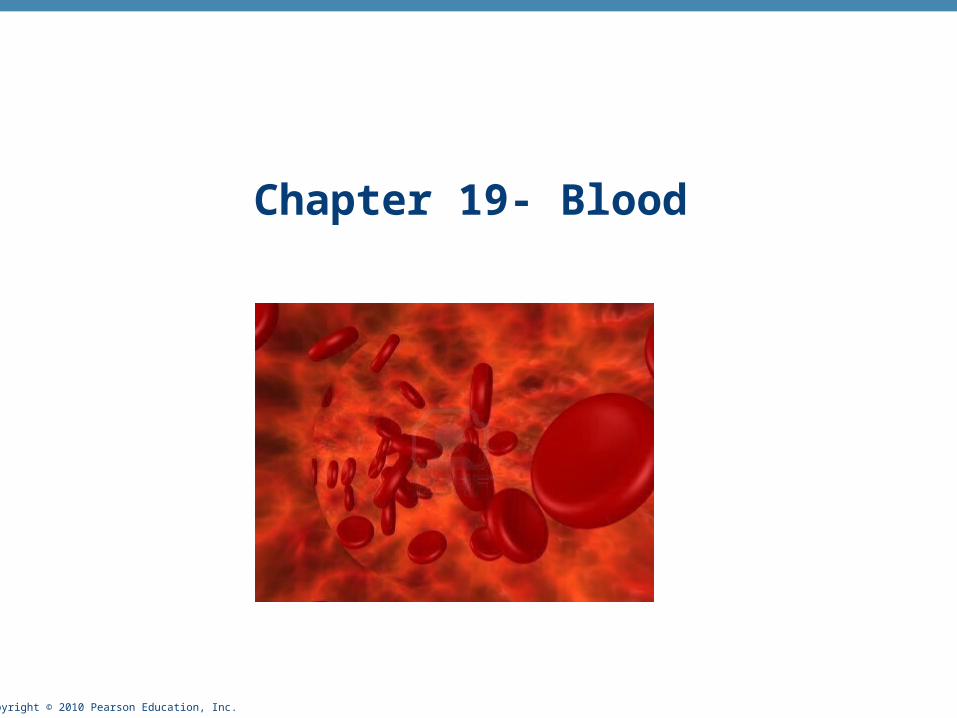
pyright © 2010 Pearson Education, Inc. Chapter 19- Blood

Copyright © 2010 Pearson Education, Inc. Chapter 19- Blood.
Dec 17, 2015
Welcome message from author
This document is posted to help you gain knowledge. Please leave a comment to let me know what you think about it! Share it to your friends and learn new things together.
Transcript
Copyright © 2010 Pearson Education, Inc.
Chapter 19- Blood
Copyright © 2010 Pearson Education, Inc.
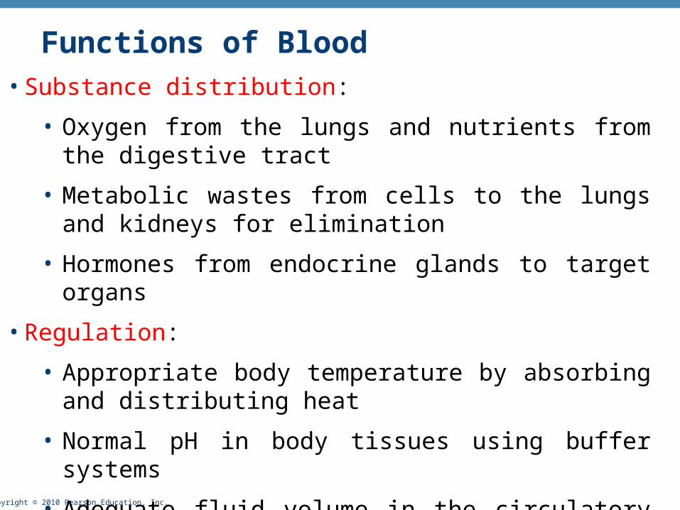
Functions of Blood• Substance distribution:
• Oxygen from the lungs and nutrients from the digestive tract
• Metabolic wastes from cells to the lungs and kidneys for elimination
• Hormones from endocrine glands to target organs
• Regulation:
• Appropriate body temperature by absorbing and distributing heat
• Normal pH in body tissues using buffer systems
• Adequate fluid volume in the circulatory system

Copyright © 2010 Pearson Education, Inc.
Functions of Blood
• Body protection
• prevents blood loss by:
• Activating plasma proteins and platelets
• Initiating clot formation when a vessel is broken
• Blood prevents infection by:
• Synthesizing and utilizing antibodies
• Activating complement proteins
• Activating WBCs to defend the body against foreign invaders
Copyright © 2010 Pearson Education, Inc.
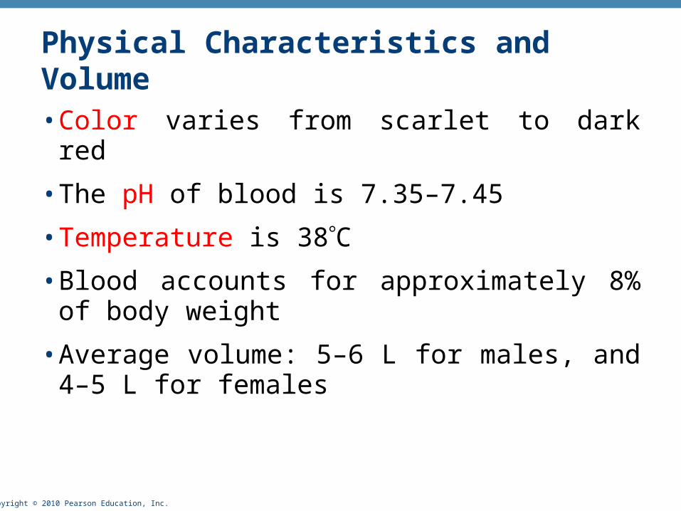
Physical Characteristics and Volume
• Color varies from scarlet to dark red
• The pH of blood is 7.35–7.45
• Temperature is 38C
• Blood accounts for approximately 8% of body weight
• Average volume: 5–6 L for males, and 4–5 L for females

Copyright © 2010 Pearson Education, Inc.
Composition of Blood• What type of tissue is blood?
• It is composed of liquid plasma (matrix) and formed elements
• Formed elements include:
• Erythrocytes, or red blood cells (RBCs)
• Leukocytes, or white blood cells (WBCs)
• Platelets

Copyright © 2010 Pearson Education, Inc.
Blood Plasma
• Blood plasma contains over 100 solutes, including:
• Proteins – albumin, globulins, clotting proteins, and others
• Lactic acid, urea, creatinine
• Organic nutrients – glucose, carbohydrates, amino acids
• Electrolytes – sodium, potassium, calcium, chloride, bicarbonate
• Respiratory gases – oxygen and carbon dioxide

Copyright © 2010 Pearson Education, Inc.
• Accounts for 46-63% of blood volume
• 92% of plasma is water
• In many respects, plasma composition resembles that of the interstitial fluid
• The differences between plasma and interstitial fluids are:
• Concentration of dissolved oxygen and carbon dioxide
• Concentration of dissolved proteins (plasma proteins can not cross capillary walls)
Plasma
Copyright © 2010 Pearson Education, Inc.
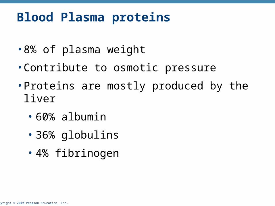
Blood Plasma proteins
• 8% of plasma weight
• Contribute to osmotic pressure
• Proteins are mostly produced by the liver
• 60% albumin
• 36% globulins
• 4% fibrinogen

Copyright © 2010 Pearson Education, Inc.
• Albumins
• 60% of plasma proteins
• Responsible for viscosity and osmotic pressure of blood
• Important in the transport of fatty acids, thyroid hormones and steroids hormones
• Globulins
• ~35% of plasma proteins
• Include immunoglobins (antibodies) which attack foreign proteins and pathogens
• Include transport globulins which bind ions, hormones and other compounds
Plasma proteins -
Copyright © 2010 Pearson Education, Inc.
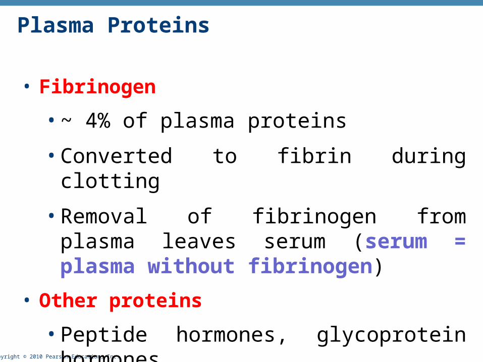
• Fibrinogen
• ~ 4% of plasma proteins
• Converted to fibrin during clotting
• Removal of fibrinogen from plasma leaves serum (serum = plasma without fibrinogen)
• Other proteins
• Peptide hormones, glycoprotein hormones
Plasma Proteins

Copyright © 2010 Pearson Education, Inc.
Formed Elements
• Erythrocytes, leukocytes, and platelets make up the formed elements
• Only WBCs are complete cells
• RBCs have no nuclei or organelles, and platelets are just cell fragments
• Most formed elements survive in the bloodstream for only a few days
• Most blood cells do not divide but are renewed by cells in bone marrow

Copyright © 2010 Pearson Education, Inc.
• What is the function of erythrocytes?
• What elements in their structure support their function?
• Function – carry respiratory gases – mainly oxygen
• They contain hemoglobin and have a biconcave structure that helps with moving into small blood
vessels

Copyright © 2010 Pearson Education, Inc.
Erythrocytes (RBCs)
• Biconcave discs, anucleate, essentially no organelles
• Filled with hemoglobin (Hb), a protein that functions in gas transport
• The biconcave disc shape
• provides a large surface to volume ratio – help with fast exchange between the cell and the plasma
• allows RBCs to stack – facilitate flow through narrow blood vessels
• Allows the RBCs to bend and flex when entering small capillaries
• Structural characteristics contribute to its gas transport function
• Erythrocytes are more than 97% hemoglobin
• ATP is generated anaerobically through glycolysis (they lack mitochondria), so the erythrocytes do not consume the oxygen they transport

Copyright © 2010 Pearson Education, Inc.
• RBC membranes have glycoprotein antigens on their external surfaces
• These antigens are:
• Unique to the individual
• Recognized as foreign if transfused into another individual
• Promoters of agglutination and are referred to as agglutinogens
• Presence or absence of these antigens is used to classify blood groups
Human Blood Groups
Copyright © 2010 Pearson Education, Inc.
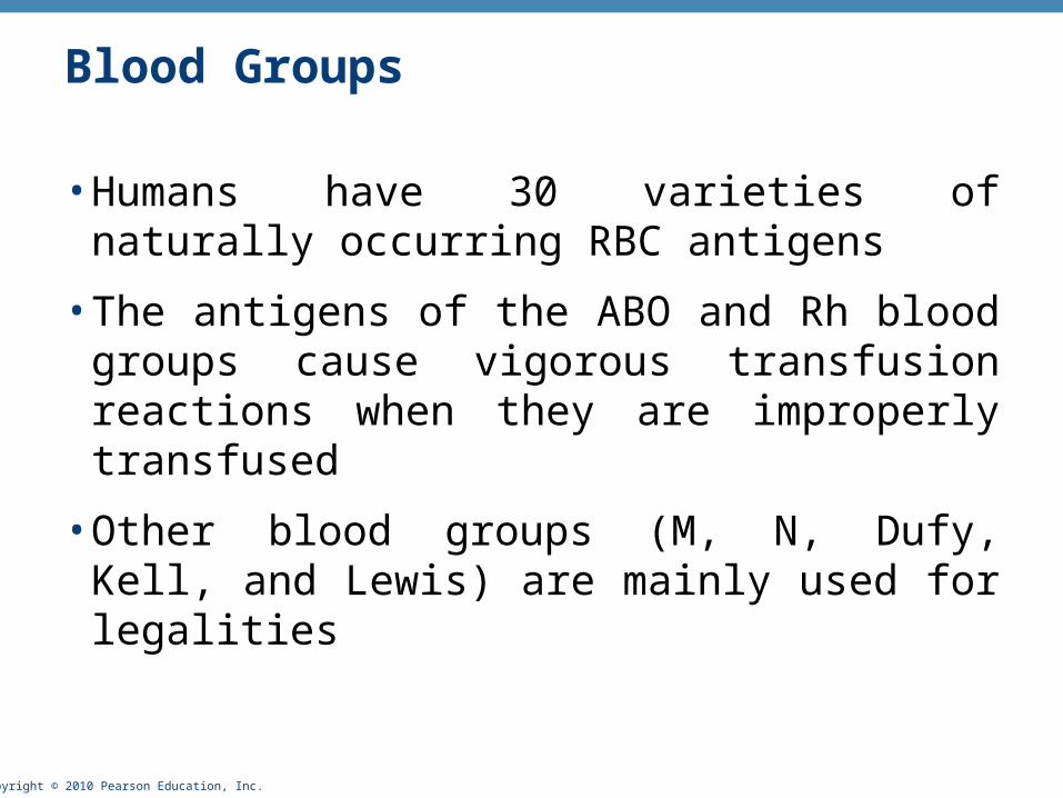
• Humans have 30 varieties of naturally occurring RBC antigens
• The antigens of the ABO and Rh blood groups cause vigorous transfusion reactions when they are improperly transfused
• Other blood groups (M, N, Dufy, Kell, and Lewis) are mainly used for legalities
Blood Groups

Copyright © 2010 Pearson Education, Inc.
• The ABO blood groups consists of:
• Two antigens (A and B) on the surface of the RBCs
• Two antibodies in the plasma (anti-A and anti-B)
• ABO blood groups may have various types of antigens and preformed antibodies
• Agglutinogens and their corresponding antibodies cannot be mixed without serious hemolytic reactions
ABO Blood Groups
Copyright © 2010 Pearson Education, Inc.
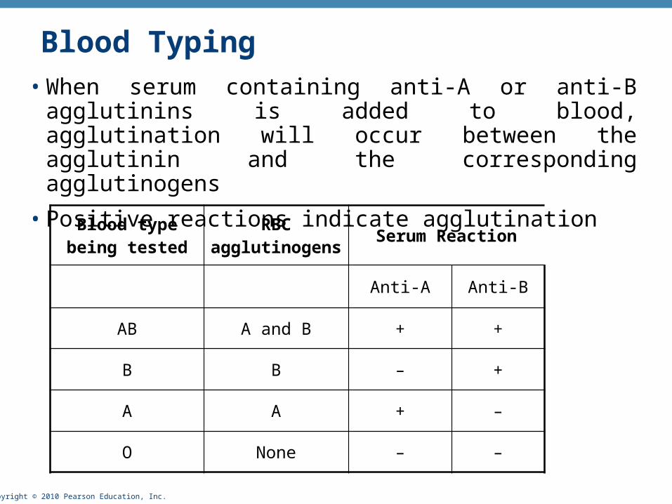
Blood Typing• When serum containing anti-A or anti-B agglutinins is added to
blood, agglutination will occur between the agglutinin and the corresponding agglutinogens
• Positive reactions indicate agglutination
Blood type being tested
RBC agglutinogens
Serum Reaction
Anti-A Anti-B
AB A and B + +
B B – +
A A + –
O None – –

Copyright © 2010 Pearson Education, Inc.
• Transfusion reactions occur when mismatched blood is infused
• Donor’s cells are attacked by the recipient’s plasma agglutinins causing:
• Diminished oxygen-carrying capacity
• Clumped cells that impede blood flow
• Ruptured RBCs that release free hemoglobin into the bloodstream
• Circulating hemoglobin precipitates in the kidneys and causes renal failure
Transfusion Reactions
Copyright © 2010 Pearson Education, Inc.
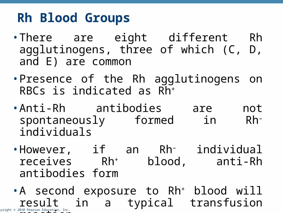
• There are eight different Rh agglutinogens, three of which (C, D, and E) are common
• Presence of the Rh agglutinogens on RBCs is indicated as Rh+
• Anti-Rh antibodies are not spontaneously formed in Rh– individuals
• However, if an Rh– individual receives Rh+ blood, anti-Rh antibodies form
• A second exposure to Rh+ blood will result in a typical transfusion reaction
Rh Blood Groups
Copyright © 2010 Pearson Education, Inc.
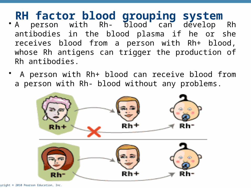
RH factor blood grouping system
• A person with Rh- blood can develop Rh antibodies in the blood plasma if he or she receives blood from a person with Rh+ blood, whose Rh antigens can trigger the production of Rh antibodies.
• A person with Rh+ blood can receive blood from a person with Rh- blood without any problems.
Copyright © 2010 Pearson Education, Inc.
Homeostatic Imbalance: Hemolytic Disease of the Newborn• Also called erythroblastosis fetalis
• Rh– mother becomes sensitized when exposure to Rh+ blood causes her body to synthesize anti-Rh antibodies
• Anti-Rh antibodies cross the placenta and destroy the RBCs of an Rh+ baby
• The baby can be treated with prebirth transfusions and exchange transfusions after birth
• RhoGAM serum containing anti-Rh can prevent the Rh– mother from becoming sensitized

Copyright © 2010 Pearson Education, Inc.
• The hematocrit (also called volume of packed red cells, VPRC, or packed cell volume, PCV) is a measure of the relative percentage of blood cells (mainly erythrocytes) in a given volume of whole blood.
• Normal hematocrit for Adult Females: 37-48% (ave. 42%)
• Normal hematocrit for Adult Males: 42-52% (ave. 47%)
Hematocrit Measurement
Copyright © 2010 Pearson Education, Inc.
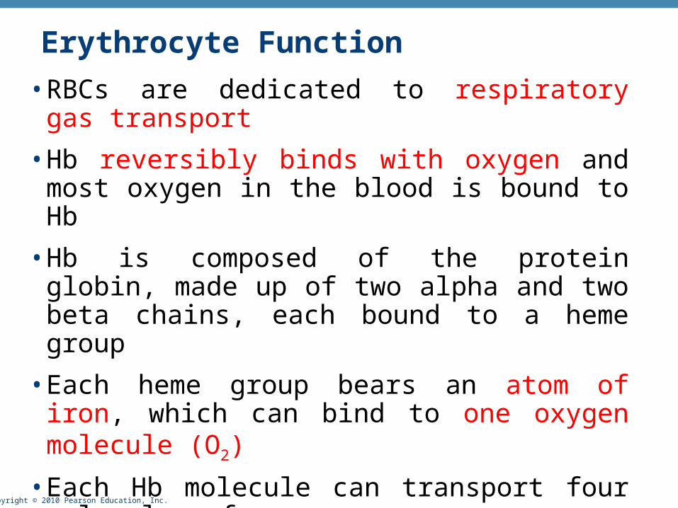
Erythrocyte Function
• RBCs are dedicated to respiratory gas transport
• Hb reversibly binds with oxygen and most oxygen in the blood is bound to Hb
• Hb is composed of the protein globin, made up of two alpha and two beta chains, each bound to a heme group
• Each heme group bears an atom of iron, which can bind to one oxygen molecule (O2)
• Each Hb molecule can transport four molecules of oxygen
• Each RBC contains ~280 million Hb molecules

Copyright © 2010 Pearson Education, Inc.
Hemoglobin (Hb)
• O2 loading in the lungs
• Produces oxyhemoglobin (ruby red)
• O2 unloading in the tissues
• Produces deoxyhemoglobin or reduced hemoglobin (dark red)
• CO2 loading in the tissues
• Produces carbaminohemoglobin (carries 20% of CO2 in the blood)
• We will talk about hemoglobin with the respiratory system

Copyright © 2010 Pearson Education, Inc.
Regulation and Requirements for Erythropoiesis
• Circulating erythrocytes – the number remains constant and reflects a balance between RBC production and destruction
• Normal body requirements for oxygen is 250 ml/min
• Almost all of that is for use in oxidative phosphorylation by the mitochondria
• Too few RBCs leads to tissue hypoxia (A deficiency of oxygen in the tissues
• Causes of hypoxia
• Hemorrhage or increased RBC destruction reduces RBC numbers
• Insufficient hemoglobin (e.g., iron deficiency)
• Reduced availability of O2 (e.g., high altitudes)
• Too many RBCs causes undesirable blood viscosity

Copyright © 2010 Pearson Education, Inc.
Hormonal Control of Erythropoiesis
• Erythropoiesis is hormonally controlled and depends on adequate supplies of iron, amino acids, and B vitamins
• Erythropoietin (EPO) release by the kidneys is triggered by:
• Hypoxia due to decreased RBCs
• Decreased oxygen availability (high altitude)
• Increased tissue demand for oxygen
• When certain kidney cells become hypoxic they release hypoxia inducing factors (HIF) which in turn accelarate the synthesis and release of erythropoietin

Copyright © 2010 Pearson Education, Inc.
The rate of erythropoiesis is controlled not
by RBC number but by their ability to
transport oxygen

Copyright © 2010 Pearson Education, Inc. Figure 17.6, step 5
Kidney (and liver toa smaller extent)releaseserythropoietin. Erythropoietin
stimulates redbone marrow.
Enhancederythropoiesisincreases RBCcount.
O2- carryingability of bloodincreases.
Homeostasis: Normal blood oxygen levels
Stimulus:Hypoxia (low bloodO2- carrying ability)
due to• Decreased
RBC count• Decreased amount
of hemoglobin• Decreased
availability of O2
1
2
3
4
5
IMBALANCE
IMBALANCE

Copyright © 2010 Pearson Education, Inc.
• Erythropoiesis requires:
• Proteins, lipids, and carbohydrates
• Iron
• vitamin B12, and folic acid (necessary for DNA production)
• Location of iron in the body
• The body stores iron in Hb (65%), the liver, spleen, and bone marrow
• Intracellular iron is stored in protein-iron complexes such as ferritin and hemosiderin
• Circulating iron is loosely bound to the transport protein transferrin
Dietary Requirements of Erythropoiesis
Copyright © 2010 Pearson Education, Inc.
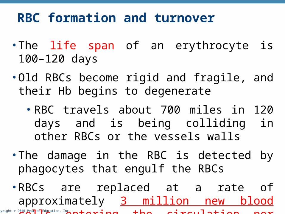
RBC formation and turnover
• The life span of an erythrocyte is 100–120 days
• Old RBCs become rigid and fragile, and their Hb begins to degenerate
• RBC travels about 700 miles in 120 days and is being colliding in other RBCs or the vessels walls
• The damage in the RBC is detected by phagocytes that engulf the RBCs
• RBCs are replaced at a rate of approximately 3 million new blood cells entering the circulation per second.
Copyright © 2010 Pearson Education, Inc.
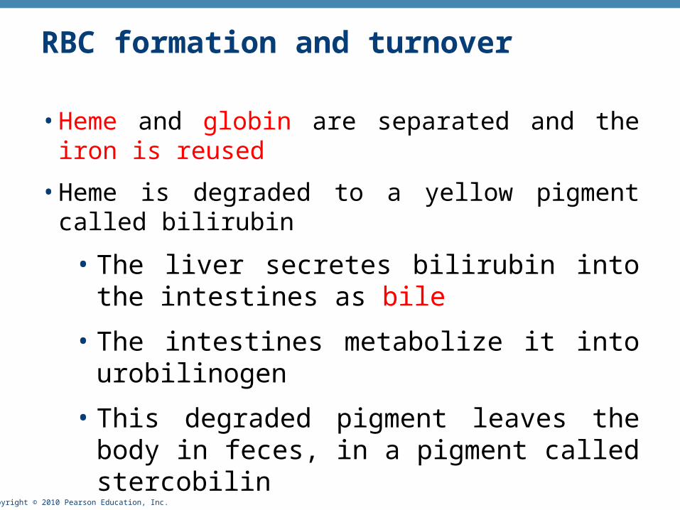
RBC formation and turnover
• Heme and globin are separated and the iron is reused
• Heme is degraded to a yellow pigment called bilirubin
• The liver secretes bilirubin into the intestines as bile
• The intestines metabolize it into urobilinogen
• This degraded pigment leaves the body in feces, in a pigment called stercobilin
• Globin is metabolized into amino acids and is released into the circulation

Copyright © 2010 Pearson Education, Inc. Figure 17.7, step 4
Low O2 levels in blood stimulatekidneys to produce erythropoietin.1
Erythropoietin levels risein blood.2
Erythropoietin and necessaryraw materials in blood promoteerythropoiesis in red bone marrow.
3
New erythrocytesenter bloodstream;function about 120 days.
4
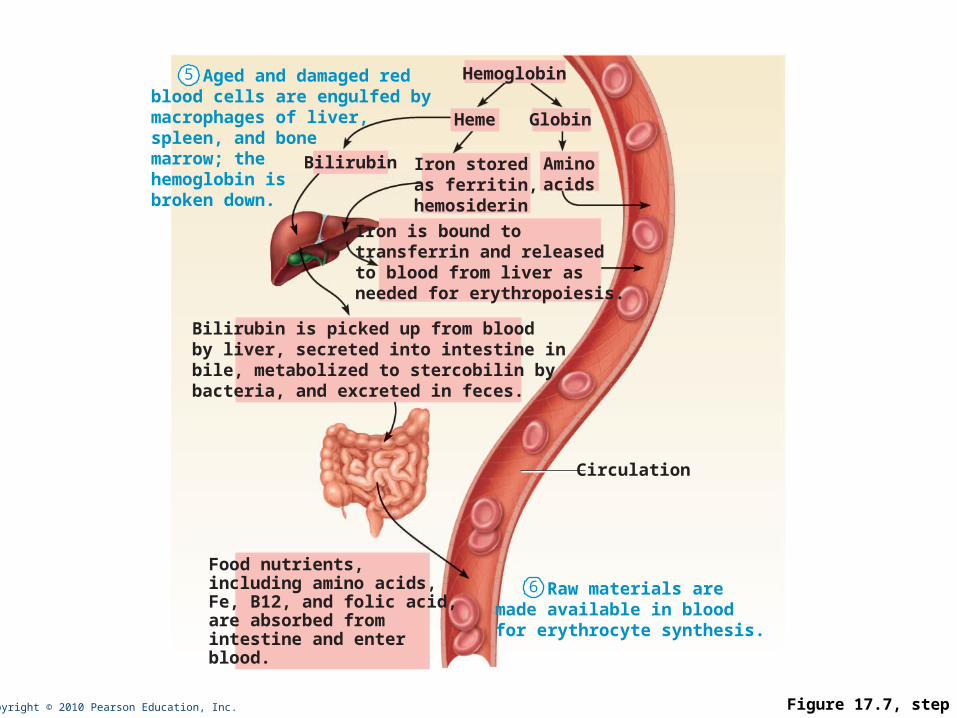
Copyright © 2010 Pearson Education, Inc. Figure 17.7, step 6
Aged and damaged redblood cells are engulfed bymacrophages of liver,spleen, and bonemarrow; thehemoglobin isbroken down.
5
Raw materials aremade available in bloodfor erythrocyte synthesis.
6
Hemoglobin
Aminoacids
Globin
Iron is bound totransferrin and releasedto blood from liver asneeded for erythropoiesis.
Food nutrients,including amino acids,Fe, B12, and folic acid,are absorbed fromintestine and enterblood.
Heme
Circulation
Iron storedas ferritin,hemosiderin
Bilirubin
Bilirubin is picked up from bloodby liver, secreted into intestine inbile, metabolized to stercobilin bybacteria, and excreted in feces.
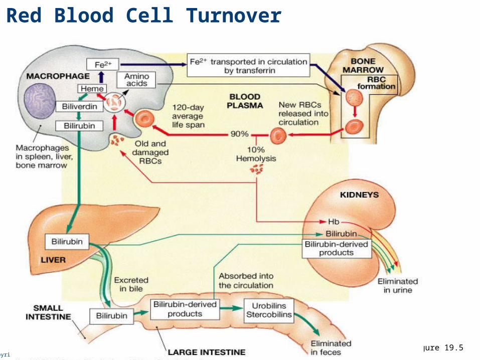
Copyright © 2010 Pearson Education, Inc.Figure 19.5
Red Blood Cell Turnover

Copyright © 2010 Pearson Education, Inc.
• Anemia – blood has abnormally low oxygen-carrying capacity
• It is a symptom rather than a disease itself
• Blood oxygen levels cannot support normal metabolism
• Signs/symptoms include fatigue, paleness, shortness of breath, and chills
• Hemorrhagic anemia – result of acute or chronic loss of blood
• Hemolytic anemia – prematurely ruptured RBCs
• Aplastic anemia – destruction or inhibition of red bone marrow
Erythrocyte Disorders
Copyright © 2010 Pearson Education, Inc.
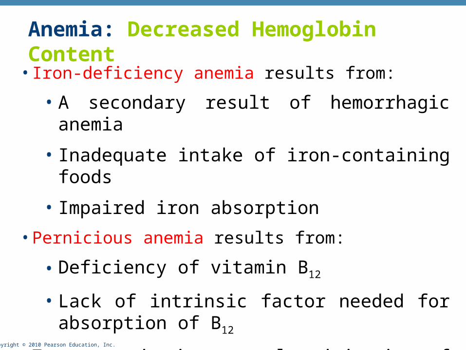
• Iron-deficiency anemia results from:
• A secondary result of hemorrhagic anemia
• Inadequate intake of iron-containing foods
• Impaired iron absorption
• Pernicious anemia results from:
• Deficiency of vitamin B12
• Lack of intrinsic factor needed for absorption of B12
• Treatment is intramuscular injection of B12
Anemia: Decreased Hemoglobin Content

Copyright © 2010 Pearson Education, Inc.
Anemia: Abnormal Hemoglobin• Sickle-cell anemia – results from a defective gene coding for an
abnormal Hb called hemoglobin S (HbS)
• HbS has a single amino acid substitution in the beta chain
• This defect causes RBCs to become sickle-shaped in low oxygen situations

Copyright © 2010 Pearson Education, Inc.
http://pathy.med.nagoya-u.ac.jp/atlas/img/t2/img0027.jpg
Thalassemias – absent or faulty globin chain in Hb RBCs are thin, delicate, and deficient in Hb
Anemia: Abnormal Hemoglobin
Copyright © 2010 Pearson Education, Inc.
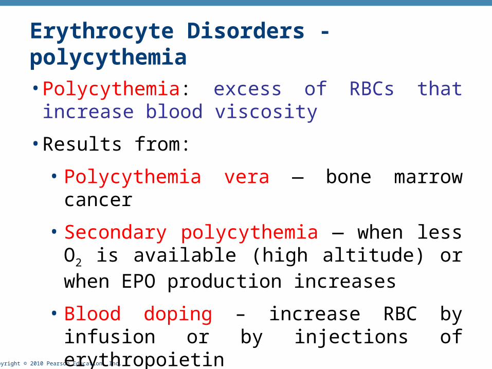
Erythrocyte Disorders - polycythemia
• Polycythemia: excess of RBCs that increase blood viscosity
• Results from:
• Polycythemia vera — bone marrow cancer
• Secondary polycythemia — when less O2 is available (high altitude) or when EPO production increases
• Blood doping – increase RBC by infusion or by injections of erythropoietin

Copyright © 2010 Pearson Education, Inc.
Leukocytes (WBCs)
• Leukocytes, the only blood components that are complete cells:
• Are less numerous than RBCs
• Make up 1% of the total blood volume
• Can leave capillaries via diapedesis
• Move through tissue spaces
• Leukocytosis – WBC count over 11,000 / mm3
• Normal response to bacterial or viral invasion

Copyright © 2010 Pearson Education, Inc.
Types of WBC• Five types of WBC: neutrophils, eosinophils, basophils,
monocytes, lymphocytes
• First 4 are part of the body non-specific defense and lymphocytes are part of the specific defense (will be discussed later in the course)
• WBC are divided into 2 groups on the basis of their appearance after staining:
• Granulocytes – visible stained granules
• Agranulocytes – non-visible granules
Copyright © 2010 Pearson Education, Inc.
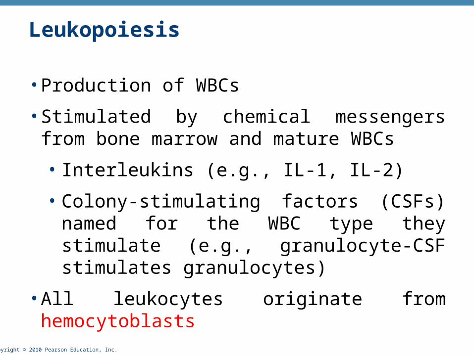
Leukopoiesis
• Production of WBCs
• Stimulated by chemical messengers from bone marrow and mature WBCs
• Interleukins (e.g., IL-1, IL-2)
• Colony-stimulating factors (CSFs) named for the WBC type they stimulate (e.g., granulocyte-CSF stimulates granulocytes)
• All leukocytes originate from hemocytoblasts
Copyright © 2010 Pearson Education, Inc.
Leukocyte Disorders
• Leukopenia
• Abnormally low WBC count—drug induced
• Leukemias
• Cancerous conditions involving WBCs
• Named according to the abnormal WBC clone involved
• Myelocytic leukemia involves myeloblasts
• Lymphocytic leukemia involves lymphocytes

Copyright © 2010 Pearson Education, Inc.
Leukemia
• Bone marrow totally occupied with cancerous leukocytes
• Immature nonfunctional WBCs in the bloodstream
• Death caused by internal hemorrhage and overwhelming infections
• Treatments include irradiation, antileukemic drugs, and stem cell transplants

Copyright © 2010 Pearson Education, Inc.
Platelets
• Small fragments of megakaryocytes
• Formation is regulated by thrombopoietin
• Blue-staining outer region, purple granules
• Granules contain serotonin, Ca2+, enzymes, ADP, and platelet-derived growth factor (PDGF)
Copyright © 2010 Pearson Education, Inc.
Hemostasis – “bleeding stoppage”
• A series of reactions for stoppage of bleeding
• Hemostatis is a balance of the physiological process which
• prevents excessive bleeding after vessel injury,
• maintains a viable circulation by keeping the blood in an uncoagulated state.
Copyright © 2010 Pearson Education, Inc.
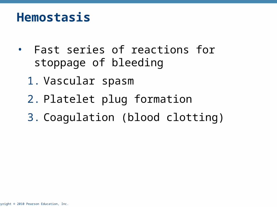
Hemostasis
• Fast series of reactions for stoppage of bleeding
1. Vascular spasm
2. Platelet plug formation
3. Coagulation (blood clotting)
Copyright © 2010 Pearson Education, Inc.
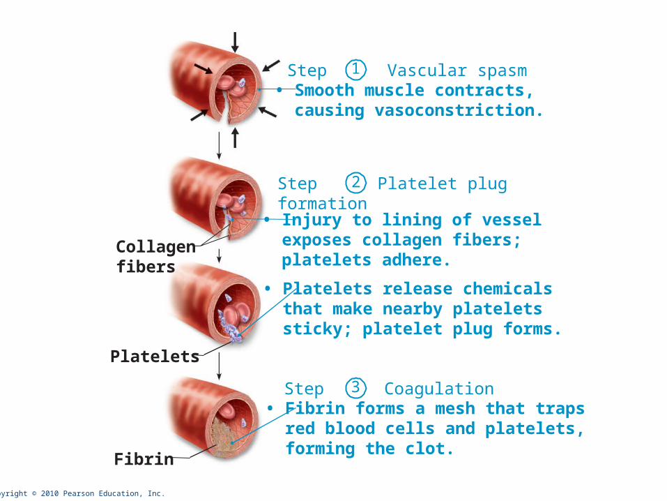
Collagenfibers
Platelets
Fibrin
Step Vascular spasm• Smooth muscle contracts, causing vasoconstriction.
Step Platelet plugformation
• Injury to lining of vessel exposes collagen fibers; platelets adhere.
• Platelets release chemicals that make nearby platelets sticky; platelet plug forms.
Step Coagulation• Fibrin forms a mesh that traps red blood cells and platelets, forming the clot.
1
2
3
Copyright © 2010 Pearson Education, Inc.
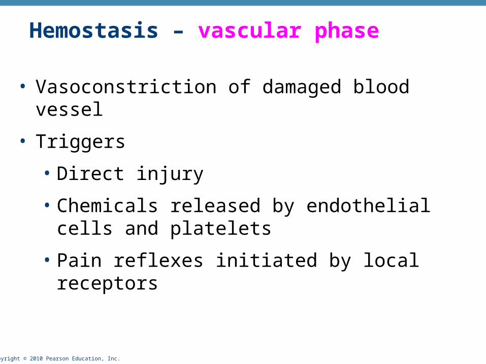
Hemostasis – vascular phase
• Vasoconstriction of damaged blood vessel
• Triggers
• Direct injury
• Chemicals released by endothelial cells and platelets
• Pain reflexes initiated by local receptors
Copyright © 2010 Pearson Education, Inc.
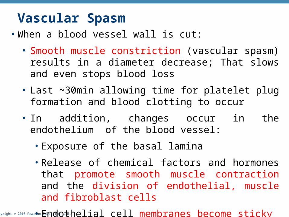
Vascular Spasm• When a blood vessel wall is cut:
• Smooth muscle constriction (vascular spasm) results in a diameter decrease; That slows and even stops blood loss
• Last ~30min allowing time for platelet plug formation and blood clotting to occur
• In addition, changes occur in the endothelium of the blood vessel:
• Exposure of the basal lamina
• Release of chemical factors and hormones that promote smooth muscle contraction and the division of endothelial, muscle and fibroblast cells
• Endothelial cell membranes become sticky
Copyright © 2010 Pearson Education, Inc.
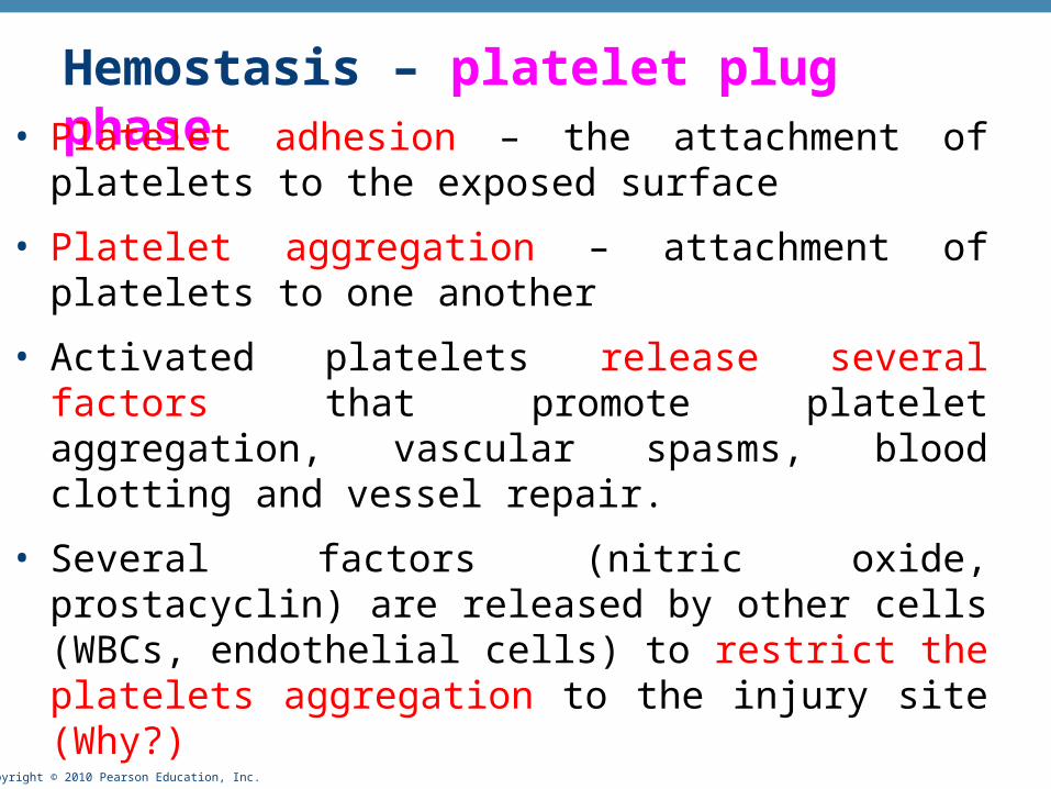
Hemostasis – platelet plug phase• Platelet adhesion – the attachment of platelets to the exposed
surface
• Platelet aggregation – attachment of platelets to one another
• Activated platelets release several factors that promote platelet aggregation, vascular spasms, blood clotting and vessel repair.
• Several factors (nitric oxide, prostacyclin) are released by other cells (WBCs, endothelial cells) to restrict the platelets aggregation to the injury site (Why?)
Copyright © 2010 Pearson Education, Inc.
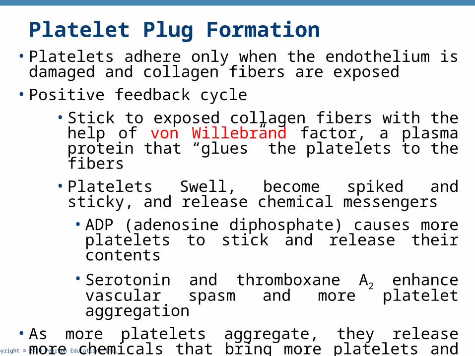
Platelet Plug Formation• Platelets adhere only when the endothelium is damaged and
collagen fibers are exposed
• Positive feedback cycle
• Stick to exposed collagen fibers with the help of von Willebrand factor, a plasma protein that “glues” the platelets to the fibers
• Platelets Swell, become spiked and sticky, and release chemical messengers
• ADP (adenosine diphosphate) causes more platelets to stick and release their contents
• Serotonin and thromboxane A2 enhance vascular spasm and more platelet aggregation
• As more platelets aggregate, they release more chemicals that bring more platelets and so on.
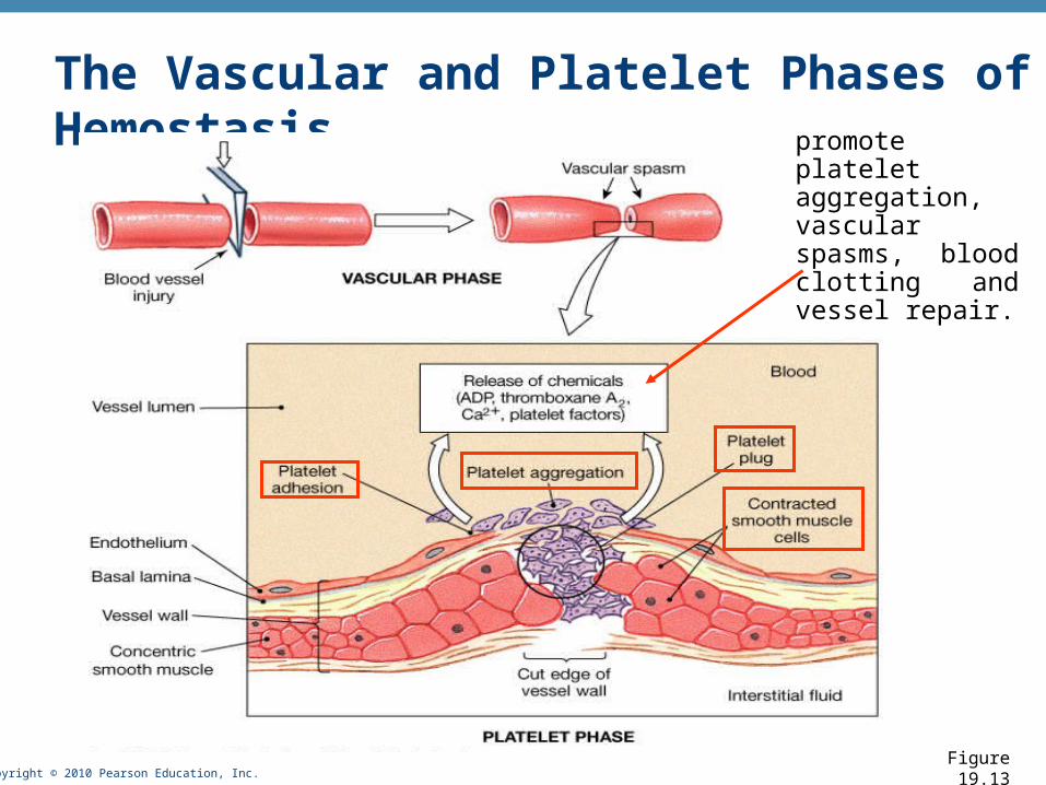
Copyright © 2010 Pearson Education, Inc.Figure 19.13
The Vascular and Platelet Phases of Hemostasispromote platelet aggregation, vascular spasms, blood clotting and vessel repair.
Copyright © 2010 Pearson Education, Inc.
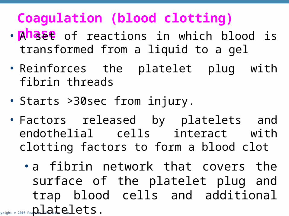
Coagulation (blood clotting) phase• A set of reactions in which blood is transformed from a
liquid to a gel
• Reinforces the platelet plug with fibrin threads
• Starts >30sec from injury.
• Factors released by platelets and endothelial cells interact with clotting factors to form a blood clot
• a fibrin network that covers the surface of the platelet plug and trap blood cells and additional platelets.
• Blood clot seals the open/cut portion of the vessel
Copyright © 2010 Pearson Education, Inc.
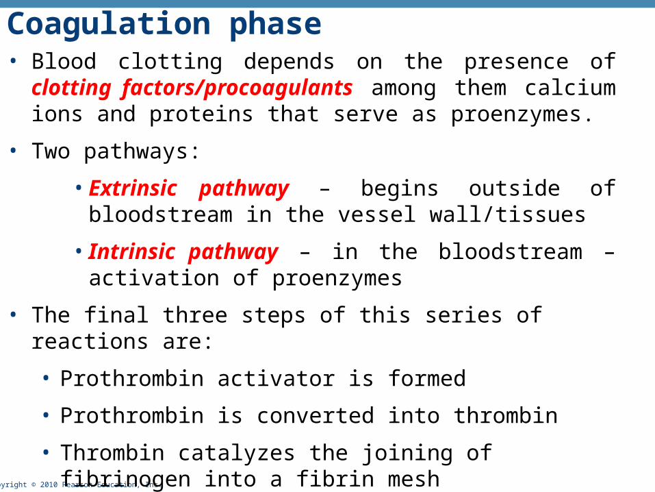
Coagulation phase• Blood clotting depends on the presence of clotting
factors/procoagulants among them calcium ions and proteins that serve as proenzymes.
• Two pathways:
• Extrinsic pathway – begins outside of bloodstream in the vessel wall/tissues
• Intrinsic pathway – in the bloodstream – activation of proenzymes
• The final three steps of this series of reactions are:
• Prothrombin activator is formed
• Prothrombin is converted into thrombin
• Thrombin catalyzes the joining of fibrinogen into a fibrin mesh

Copyright © 2010 Pearson Education, Inc.Figure 19.14a
The Coagulation Phase of Hemostasis
Extrinsic pathway
Intrinsic pathway
common pathway

Copyright © 2010 Pearson Education, Inc.
Clot retraction/syneresis
• Final phase of healing
• Platelets contract and pull the edges of the vessel together
• Actin and myosin in platelets contract within 30–60 minutes
• Reduction of the damaged area allow fibroblasts, muscle cells and endothelium to complete repairs
Copyright © 2010 Pearson Education, Inc.
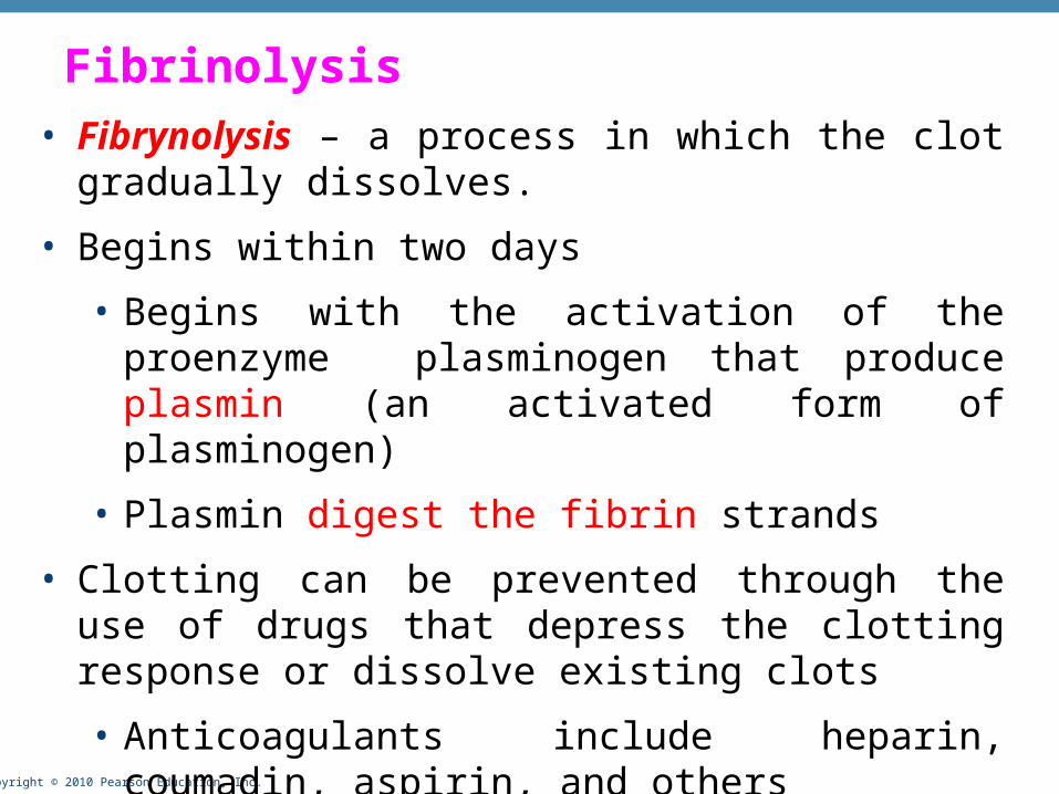
Fibrinolysis• Fibrynolysis – a process in which the clot gradually
dissolves.
• Begins within two days
• Begins with the activation of the proenzyme plasminogen that produce plasmin (an activated form of plasminogen)
• Plasmin digest the fibrin strands
• Clotting can be prevented through the use of drugs that depress the clotting response or dissolve existing clots
• Anticoagulants include heparin, coumadin, aspirin, and others

Copyright © 2010 Pearson Education, Inc.
Related Documents