Copper–zinc alloy nanoparticle based enzyme-free superoxide radical sensing on a screen-printed electrode Burak Derkus, Emel Emregul n , Kaan C. Emregul Ankara University, Science Faculty, Department of Chemistry, Tandoğan, Ankara 06100, Turkey article info Article history: Received 11 August 2014 Received in revised form 29 October 2014 Accepted 2 November 2014 Available online 18 November 2014 Keywords: Superoxide dismutase Enzyme-free biosensor Screen-printed electrode CuZn nanoparticles Nanobiosenosors abstract In this paper, amperometric enzyme-free sensors using superoxide dismutase (SOD) enzyme as a catalyst for the dismutation reaction of superoxides into oxygen and hydrogen peroxide, enabling superoxide radical detection have been described. For this purpose, the surfaces of screen-printed platinum electrodes have been modified with gelatin composites of CuO, ZnO and CuZn nanoparticles with the expectation of an increase in catalytic effect toward the dismutation reaction. SOD containing electrodes were also prepared for comparative studies in which glutaraldehyde was used as a cross- linker for the immobilization of SOD to the nanocomposite materials. Electrochemical measurements were carried out using a screen-printed electrochemical system that included potassiumferrocyanide (K 4 [Fe(CN) 6 ]) and potassiumferricyanide (K 3 [Fe(CN) 6 ]) as the redox probes. The results revealed that the enzyme-free detection method using CuZn nanoparticles can determine superoxide radicals with high performance compared to other detection methods prepared with different nanoparticles by mimicking the active region of superoxide dismutase enzyme. The anodic (ks a ) and cathodic (ks c ) electron transfer rate constants and the anodic (α a ) and cathodic (α c ) transfer coefficients were evaluated and found to be ks a ¼6.31 s 1 and α a ¼0.81, ks c ¼1.48 s 1 and α c ¼0.19 for the gelatin–CuZn–SOD electrode; ks a ¼6.15 s 1 and α a ¼0.79, ks c ¼1,63 s 1 and α c ¼0.21 for the enzyme-free gelatin–CuZn electrode. The enzyme-free electrode showed nearly 80% amperometric performance with respect to the enzyme containing electrode indicating the superior functionality of enzyme-free electrode for the detection of superoxide radicals. & 2014 Elsevier B.V. All rights reserved. 1. Introduction Due to its overwhelming reaction rate owing to a radical mechanism and high specificity [1], the superoxide dismutase (SOD) enzyme offers a great potential for sensitive quantification of superoxide radicals in various biological samples especially in cancerous tissues using biosensing technology. Superoxide radicals (O 2 d ) are known to damage some biological molecules [2–4] and signal pathways, and also play important roles in heart disease [5], cancer [6] and neuronal dejeneration [7]. Hence, the study of super- oxide radicals has attracted considerable attention in recent years. SODs are metalloenzymes which have iron (Fe), manganese (Mn), nickel (Ni), or copper–zinc (CuZn) ions in their active region that catalyze the dismutation of superoxide to oxygen and hydrogen peroxide [8]. Copper–zinc superoxide dismutase is present in the cytosol, nucleus, peroxisomes, and mitochondrial intermembrane space of human cells, acting as an antioxidant enzyme by lowering the steady-state concentration of superoxide. The human enzyme is a 32-kDa homodimer, with a copper- and zinc-binding site each per 153-amino acid subunits [9–11]. The copper site is the heart of the enzymatic active site where SOD1 protein catalyzes the dispro- portionation of superoxide to give dioxygen and hydrogen per- oxide. This catalysis is a two-step process: one molecule of superoxide first reduces the cupric ion to form dioxygen and then a second molecule of O 2 d reoxidizes the cuprous ion to form hydrogen peroxide. Detailed information about the catalyzer effect of CuZn can be found in literature [12]. The dismutation of superoxide proceeds via a two-step reaction if the enzyme includes CuZn [13]: O 2 þ Cu(II)ZnSOD-O 2 þ Cu(I)ZnSOD O 2 þ Cu(I)ZnSOD þ 2H þ -H 2 O 2 þ Cu(II)ZnSOD The overall reaction is O 2 þ 2H þ -H 2 O 2 þ O 2 This reaction is often used to determine the superoxide anion. In order to understand the role of O 2 d in pathology and physiology and the relationship between O 2 d and environmental stresses, Contents lists available at ScienceDirect journal homepage: www.elsevier.com/locate/talanta Talanta http://dx.doi.org/10.1016/j.talanta.2014.11.003 0039-9140/& 2014 Elsevier B.V. All rights reserved. n Corresponding author. Tel.: þ90 312 212 60 40. E-mail address: [email protected] (E. Emregul). Talanta 134 (2015) 206–214

Welcome message from author
This document is posted to help you gain knowledge. Please leave a comment to let me know what you think about it! Share it to your friends and learn new things together.
Transcript

Copper–zinc alloy nanoparticle based enzyme-free superoxide radicalsensing on a screen-printed electrode
Burak Derkus, Emel Emregul n, Kaan C. EmregulAnkara University, Science Faculty, Department of Chemistry, Tandoğan, Ankara 06100, Turkey
a r t i c l e i n f o
Article history:Received 11 August 2014Received in revised form29 October 2014Accepted 2 November 2014Available online 18 November 2014
Keywords:Superoxide dismutaseEnzyme-free biosensorScreen-printed electrodeCuZn nanoparticlesNanobiosenosors
a b s t r a c t
In this paper, amperometric enzyme-free sensors using superoxide dismutase (SOD) enzyme as acatalyst for the dismutation reaction of superoxides into oxygen and hydrogen peroxide, enablingsuperoxide radical detection have been described. For this purpose, the surfaces of screen-printedplatinum electrodes have been modified with gelatin composites of CuO, ZnO and CuZn nanoparticleswith the expectation of an increase in catalytic effect toward the dismutation reaction. SOD containingelectrodes were also prepared for comparative studies in which glutaraldehyde was used as a cross-linker for the immobilization of SOD to the nanocomposite materials. Electrochemical measurementswere carried out using a screen-printed electrochemical system that included potassiumferrocyanide(K4[Fe(CN)6]) and potassiumferricyanide (K3[Fe(CN)6]) as the redox probes. The results revealed that theenzyme-free detection method using CuZn nanoparticles can determine superoxide radicals with highperformance compared to other detection methods prepared with different nanoparticles by mimickingthe active region of superoxide dismutase enzyme. The anodic (ksa) and cathodic (ksc) electron transferrate constants and the anodic (αa) and cathodic (αc) transfer coefficients were evaluated and found to beksa¼6.31 s�1 and αa¼0.81, ksc¼1.48 s�1 and αc¼0.19 for the gelatin–CuZn–SOD electrode; ksa¼6.15 s�1
and αa¼0.79, ksc¼1,63 s�1 and αc¼0.21 for the enzyme-free gelatin–CuZn electrode. The enzyme-freeelectrode showed nearly 80% amperometric performance with respect to the enzyme containingelectrode indicating the superior functionality of enzyme-free electrode for the detection of superoxideradicals.
& 2014 Elsevier B.V. All rights reserved.
1. Introduction
Due to its overwhelming reaction rate owing to a radicalmechanism and high specificity [1], the superoxide dismutase(SOD) enzyme offers a great potential for sensitive quantificationof superoxide radicals in various biological samples especially incancerous tissues using biosensing technology. Superoxide radicals(O2
d� ) are known to damage some biological molecules [2–4] andsignal pathways, and also play important roles in heart disease [5],cancer [6] and neuronal dejeneration [7]. Hence, the study of super-oxide radicals has attracted considerable attention in recent years.SODs are metalloenzymes which have iron (Fe), manganese (Mn),nickel (Ni), or copper–zinc (CuZn) ions in their active region thatcatalyze the dismutation of superoxide to oxygen and hydrogenperoxide [8].
Copper–zinc superoxide dismutase is present in the cytosol,nucleus, peroxisomes, and mitochondrial intermembrane space ofhuman cells, acting as an antioxidant enzyme by lowering the
steady-state concentration of superoxide. The human enzyme is a32-kDa homodimer, with a copper- and zinc-binding site each per153-amino acid subunits [9–11]. The copper site is the heart of theenzymatic active site where SOD1 protein catalyzes the dispro-portionation of superoxide to give dioxygen and hydrogen per-oxide. This catalysis is a two-step process: one molecule ofsuperoxide first reduces the cupric ion to form dioxygen and thena second molecule of O2
d� reoxidizes the cuprous ion to formhydrogen peroxide. Detailed information about the catalyzer effectof CuZn can be found in literature [12].
The dismutation of superoxide proceeds via a two-step reactionif the enzyme includes CuZn [13]:
O2�þCu(II)ZnSOD-O2þCu(I)ZnSOD
O2�þCu(I)ZnSODþ2Hþ-H2O2þCu(II)ZnSOD
The overall reaction is
O2�þ2Hþ-H2O2þO2
This reaction is often used to determine the superoxide anion. Inorder to understand the role of O2
d� in pathology and physiologyand the relationship between O2
d� and environmental stresses,
Contents lists available at ScienceDirect
journal homepage: www.elsevier.com/locate/talanta
Talanta
http://dx.doi.org/10.1016/j.talanta.2014.11.0030039-9140/& 2014 Elsevier B.V. All rights reserved.
n Corresponding author. Tel.: þ90 312 212 60 40.E-mail address: [email protected] (E. Emregul).
Talanta 134 (2015) 206–214

it is essential to determine O2d� in a variety of in vitro and in vivo
models. Due to its low concentration, high reactivity, and shortlifetime, it is still an analytical challenge to detect the localconcentration of O2
d� , especially in biological systems. Determinationof free radicals is usually carried out with spectrometry, fluorometry,chemiluminensence, and electron spin resonance [14–16]. Recentattempts have concentrated on electrochemical methods due to theirdirect, real-time measurements and capability for in vivo detection[17–19].
Recently the combination of nanomaterials with biologicalagents has provided a novel way for the fabrication of diagnostictools. Oxide nanoparticles like zinc oxide (ZnO), tin oxide (SnO),copper oxide (CuO) and titanium dioxide (TiO2) are often used toimmobilize biomolecules due to their biocompatibility. CuO is asemiconductor nanoparticle with 1.2 eV band-gap which has manyapplications such as the fabrication of electrochemical biosensors [20],optical and photovoltaic devices [21], heterogeneous catalysis [22],anode materials for lithium-ion batteries [23] and enzyme-freeglucose biosensor [24]. Various shapes of CuO have been producedsuch as nanowires [25], nanorods [26], nano-flowers [27], andnanoellipsoids [28]. One of the interesting metal oxide nanoparticleswhich has amazing properties is ZnO nanoparticles. ZnO has anisoelectric point of 9.5, which is quite high compared with othernanoparticles [29]. At biological pH values, proteins with a lowisoelectric point can be immobilized on positively charged ZnOnanoparticle surfaces via electrostatic forces [30]. ZnO nanoparticlesare also nontoxic, have good biocompatibility and high stability. Allthese properties make ZnO nanoparticles ideal materials for biosen-sing applications.
Screen-printed electrodes (SPEs) are particularly useful due totheir disposability, minimum sample preparation, simplicity of theapparatus, obtaining of fast results, cost effectiveness, and lack ofrequirement of surface pre-treatment. Working with SPEs usingcyclic voltammetry (CV), electrochemical impedance spectroscopy(EIS), or amperometry prepares it for point-of-care applications.Personal glucose biosensor used by those suffering from diabetescan be shown as a widespread example of commercialized SPE.
This study is aimed at developing novel enzyme-free sensorsfor the detection of superoxide radicals in biological samples.For this purpose, ZnO, CuO and CuZn nanoparticles were usedin combination with gelatin hydro gel to design the sensors.
The requirement of biosensors to have an effective surface areaand the catalytic nature of CuZn in the active region of SODenzyme makes the CuZn multicomponent an excellent candidatefor superoxide radical biosensing application. Thus in addition toCuO and ZnO nanoparticles, CuZn nanostructures were alsostudied as a superoxide radical sensing platform. Designingenzyme-free diagnostic tools makes them economical and biolo-gically durable compared to those including enzymes.
2. Materials and methods
2.1. Materials
Superoxide dismutase (EC. 1.15.1.1, 75KU) from bovine erythro-cytes, xanthine oxidase (EC 1.1.3.22, 0.3 U mg�1, from milk),xanthine (2,6-dihydroxypurine) sodium salt, gelatin from porcinebone, glutaraldehyde cross-linking agents, CuZn alloy nanoparticles(150 nm), CuO nanoparticles (o50 nm), ZnO nanoparticles(o50 nm), sodium chloride (NaCl), potassium chloride (KCl), sodiumbicarbonate (NaHCO3), potassium ferrocyanide (K4[Fe(CN)6]), potas-siumferricyanide (K3[Fe(CN)6]), sodium dihydrogenorthophosphate(NaH2PO4), and disodium hydrogen orthophosphate (Na2HPO4)were purchased from Sigma (St Louis, MO, USA). De-ionized waterwas purified using a MilliPore Simplicity unit to a resistivityZ18.2 MΩ cm. Electrochemical measurements were carried out witha Gamry Instrument using Framework Version 5.50 software.
2.2. Preparation of biosensors
The polymer was prepared by dissolving in phosphate buffer(0.05 M, pH 7.4) so as to obtain the desired ratio (2%) in the finalsolution. To prepare the electrodes containing SOD, nanoparticles,glutaraldehyde cross-linking agent and SOD were added tothe gelatin containing situated in an eppendorf, respectively(Scheme 1). Homogeneity was provided by vortexing after eachaddition for a period of 30 s. Next, 2 μL of the mixed solution wereadded dropwise to the electrode surface. Enzyme-free electrodeswere prepared using the same protocol without the enzyme. Thesemodified electrodes were left at room temperature for 2 h toensure a stable dry surface. Electrochemical measurements werecarried out in a 5 mL electrochemical cell. In order to trigger the
Scheme 1. Schematic presentation and preparation steps of gelatin-nanoparticle-SOD screen-printed electrode. After modification of the platinum SPE electrode with theenzyme and cross-linker containing gelatin solution, amperometric results are obtained with injection of xanthin into xanthin oxidase containing electrochemical cell .
B. Derkus et al. / Talanta 134 (2015) 206–214 207
dismutation reaction, the desired concentration of xanthine in100 μL of buffer was injected into the cell containing 4.9 mL totalvolume of buffer and xanthine oxidase.
2.3. Electrochemical study
All-in-one platinum screen printed electrodes with a 2 mmdiameter working surface, counter and silver reference electrodeswere used for the biosensor design. Platinum SPE electrodes wereused due to their reusable feature. When the electrodes areelectrochemically cleaned and reactivated in sulfuric acid, theycan be used over and over again until their surface is destroyed.The other reason that makes SPE electrodes superior is their highstability, durability and conductivity. Printing of platinum onceramic templates also prevents the electrode system from theadverse effect of acids or bases. A 5 mL volume electrochemicalcell for screen-printed electrode was used in all the experiments.CV and EIS were performed in PB buffer containing 0.1 M KCl and0.5 mM FeðCNÞ63�=4� . Cyclic voltamograms were obtained bycycling the potential between �0.4 and 0.6 V with a scan rate of100 mVs�1. EIS measurements were recorded within the fre-quency range of 0.01 Hz to 100 kHz at open circuit potential. Allexperimental procedures for the development of the biosensorswere performed at least three times.
The electron-transfer coefficient and electron-transfer rateconstant could be determined based on the Laviron theory (Eqs. (1)and (2)) [31]
Epc¼ E″0þ RTαsnF
� RTαsnF
ln v ð1Þ
Epα¼ E00þ RTð1�αsÞnF–
RTð1�asÞnF ln v ð2Þ
where n is the electron transfer number, R is the gas constant(R¼8.314 J mol�1 K�1), T is the temperature in Kelvin (T¼298 K)and F is the Faraday constant (F¼96493 C mol�1).
When nΔEp4200 mV, the electron transfer rate ks could beestimated with Laviron's equation (Eq. (3)) [31]
ks¼ anFnRT
ð3Þ
The effective surface areas were determined by CV in 0.5 mMFeðCNÞ63�=4� /0.1 M KCl solution in the potential range of �400 toþ600 mV. Scan rates of 10, 50, 100, 200, 500, and 1000 mV s�1
were employed. The reduction peak current was determined andeffective surface areas of electrodes were calculated by theRandles–Sevcik equation (Eq. (4)) as described below
ip ¼ ð2:69� 105Þn3=2D1=2CAv1=2 ð4Þ
where n is the number of transferred electrons for the redoxreaction, D is the diffusion coefficient (6.70�10�6 cm2 s�1),C is the molar concentration of ferricyanide (0.5 mM), A is theeffective surface area (cm2), and v is the scan rate (V s�1). Thevalue of n is equal to one for cyclic voltammograms obtained inFeðCNÞ63�=4� , due to the following half reaction taking place atthe electrode:
Fe(CN)63�þe�-Fe(CN)64�
Graphs using ip and v1 2 were drawn and slopes (k) werecalculated. Using the modified Randles–Sevcik equation (Eq. (5)),effective surface areas were calculated
A¼ k=ðð2:69� 105Þn3=2D1=2CÞ ð5Þ
2.4. Principle of the method
The biosensor used to determine the Od�2 was obtained by
coupling an amperometric electrode for hydrogen peroxide withthe superoxide dismutase enzyme or nanoparticles immobilizedon a gelatin support system. The capacity of the O2
d� biosensorwas determined in the following way. The O2
d� is produced inaqueous solution by oxidation of xanthine to uric acid in thepresence of xanthine oxidase
XanthineþH2OþO2 XOD-
Uricacidþ2Hþ þO2��
SOD or CuZn nanoparticles immobilized on the electrodecatalyzes the dismutation reaction of the O2
d� releasing oxygenand hydrogenperoxide according to the reaction given below:
2Hþ þO2d� SOD
-H2O2þO2
The H2O2 can be detected at the electrode surface in accor-dance with the following reaction:
H2O2-2HþþO2þ2e�
The current generated by oxidation of hydrogen peroxide at theworking electrode held at 650 mV relative to the Ag/AgCl electrodeis proportional to the concentration of O2
d� in solution. Oxidationof H2O2 generates the electrons which create a current on theelectrode surface which is the principle reaction of superoxidedetection. Glucose detection is also dependant on this phenom-enon. Our previous experiences have shown that 650 mV is theoptimum potential for the oxidation of H2O2 in super oxidedetection systems [1,32,33].
2.5. Effects of scavengers
The effect of molecules regarded as scavengers for the superoxideradical were determined in vitro using gelatin–CuZn–SOD and gelatin–CuZn electrodes. The response of the electrodes to the superoxideradical both in the presence and absence of scavenger molecules suchas acetylsalicylic acid, aspirin, and aspirin containing vitamin C weredetermined. The selected scavenger molecule (12.5 gL�1) obtained bydissolving and homogenizing the weighed sample in phosphate buffer(0.05 M, pH 7.4) was added to the cell containing phosphate buffer(0.05 M, pH 7.4) and XOD (0.7 U) right before measurement.
2.6. Medical application
The biosensor response was investigated on healthy andcancerous brain tissue, meningioma (grade I, WHO 2000) obtainedfrom Ankara University, Medical School, Oncology Department.The healthy or cancerous brain tissue (0.5 g) was homogenized indistilled water (3 mL) using a Bandalin homogenizer. A solution ofthe homogenized healthy or cancerous brain tissue (100 μL) wasadded and the biosensor response recorded. The tissues werestored at �20 1C before use.
3. Results and discussion
3.1. Optimization and characterization of nanobiosensors
3.1.1. Optimization of nanobiosensorsDifferent parameters affecting the nanobiosensor's performance,
namely, the gelatin matrix, glutaraldehyde cross-linker, SOD enzyme,nanoparticles and xanthin oxidase concentrations were investigatedand the results have been presented in supplementary materials(Figs. S1–13). Maximum amperometric responses were obtained fora gelatin ratio of 2% with the gelatin–CuO–SOD biosensor. Low gelatin
B. Derkus et al. / Talanta 134 (2015) 206–214208
ratios increased the nanoparticle and/or enzyme leakage due toincreased pore size, and smooth film formation was not observed atlow polymer concentrations. At higher levels the response increaseddue to an increase in the amount of nanoparticle/enzyme bondage.Finally, further increase caused decrease of the biosensor response,probably due to excess nanoparticle/enzyme binding, leading to
inactivation. Diverse concentrations of cross-linker were used toimmobilize SOD onto the carrier systems. Increasing the glutaralde-hyde concentration reduced the response of immobilized SOD, due todeactivation of the enzyme molecules and formation of a tight gelstructure because of the excess cross-linker. Adversely, the lowcross-linker concentration decreased the amperometric signal due to
Fig. 1. D Surface topography image of gelatin (A) and roughness graph; and image of gelatin–CuZn–SOD (B) obtained by AFM. Although enzyme incorporation decreases theporosity and active surface area, nanoparticles increase it as presented in the roughness graphs.
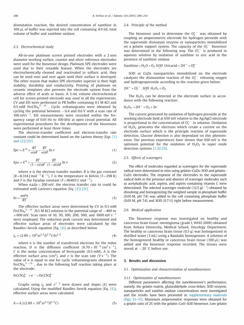
Fig. 2. SEM micrographs of the gelatine (A), gelatine–CuZn nanocomposite (B), gelatine–SOD (C), and gelatine–CuZn–SOD electrode (D).
B. Derkus et al. / Talanta 134 (2015) 206–214 209
insufficient immobilization of SOD to gelatin. Optimum concentrationof glutaraldehyde was found as 0.0032M inwhich the electrodes gavethe best results. Many electrodes were prepared using differentamounts of nanoparticles for optimization of the nanoparticles'content. The results showed that low nanoparticle levels resulted inlow amperometric signals due to insufficient electron transfer. Opti-mum nanoparticle content was found to be 0.0003 g. Enzymeoptimization was performed using different concentrations of SODwhere the optimum concentration was found to be 100 U. Theamperometric response increased with increasing SOD concentrationup to a limit followed by a decrease in the signal. This behavior can beattributed to an increase in enzyme–enzyme cross-linking. In addition,
with increased enzyme loading, oversaturation within the matrixpores may have occurred leading to restriction of product andsubstrate diffusion. Similar results were obtained for xanthine optimi-zation. The same optimum values were obtained for ZnO- and CuZn-based nanobiosensors. Phosphate buffer was chosen as the bestreaction environment (Figs. S14–16).
3.1.2. Surface topography study with AFM and SEMSurface topographies of gelatin and gelatin–CuZn–SOD electro-
des were investigated with atomic force microscopy (AFM) (Fig. 1).The AFM image of gelatin shows a porous and well settled gelatincarrier surface. Gelatin also covers almost the entire surfaceenabling an efficient immobilization surface. With enzyme immo-bilization, the surface roughness of gelatin is seen to decreasealthough the nanoparticles embedded in the gelatin matrix lead toenormous enzyme dimensions. Though the nanoparticles cannotbe seen distinctly in the image, the effect of nanoparticles on theroughness can be seen on the roughness graph. Such a nanoporousrough surface morphology results in an increased effective surfacearea for enzyme immobilization preventing the leaching ofenzymes from the electrode surface.
The gelatine surface is quite rough and not a wholly formedstructure as seen from Fig. 2(A). The gelatin CuZn nanocompositestructure on the other hand is relatively smooth and seems to form acomplete matrix compared to the gelatin structure (Fig. 2(B)), makingit suitable for biosensor studies. The SOD immobilized micrographgiven in Fig. 2(C) shows the immobilized enzyme structure. Due to thefact that a hydrogel is used as polymer matrix the embeddednanostructures cannot be seen clearly. But looking at Fig. 2(D) wecan see that with the incorporation of enzymes on the surface, due toincreasing surface tension, the nanostructure around the enzymes canbe seen embedded in the gelatin structure.
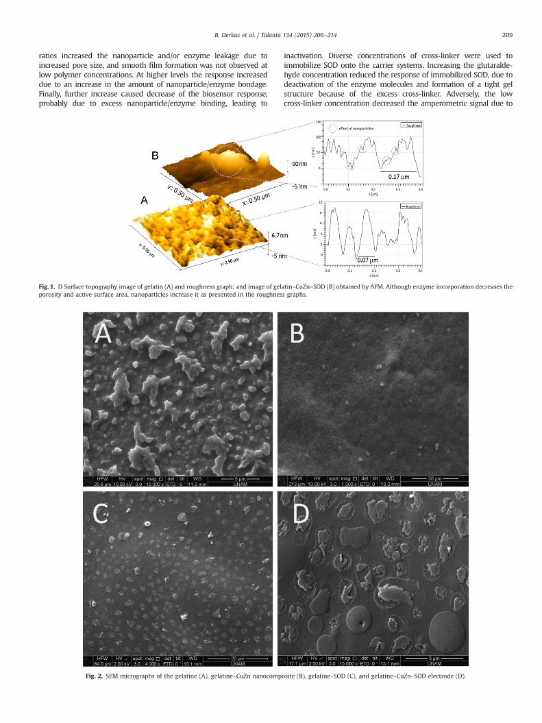
Fig. 3. (A) Nyquist diagram of CuZn-based electrode: (a) gelatin modified elec-trode; (b) CuZn embedded gelatin electrode; (c) SOD immobilized gelatin–CuZnelectrode; (d) 10 mM xanthin injected gelatin–CuZn–SOD electrode; and (e) 10 mMxanthin injected gelatin–SOD electrode. (B) Cyclic voltammograms of gelatinelectrodes containing different nanoparticles: (a) CuO embeded gelatin electrode;(b) ZnO embedded gelatin electrode; (c) CuZn embedded gelatin electrode; and(d) SOD immobilized gelatin–CuZn electrode. Solution composition: 0.1 M KCl,0.5 mM K3[Fe(CN)6]/K4[Fe(CN)6], pH 7.4. The frequency range 0.01 Hz to 100 kHz at220 mV open circuit potential. Scan rates 100 mVs�1.
Table 1Linear calibration equations of the electrodes.
Electrode type LRE (anodic) LRE (cathodic)
Gelatin–CuO–SOD Ipa/μA¼(36.78)þ(0.8270.04)ν/mV s�1 Ipc/μA¼�(37.84) (0.7870.03) ν/mV s�1
Gelatin–ZnO–SOD Ipa/μA¼(36.84)þ(0.9070.05)ν/mV s�1 Ipc/μA¼�(32.33)�(0.6870.04) ν/mV s�1
Gelatin–CuZn–SOD Ipa/μA¼(30.58)þ(0.9570.03)ν/mV s�1 Ipc/μA¼�(83.53)�(1.1470.05) ν/mV s�1
Gelatin–CuZn Ipa/μA¼(30.03)þ(0.6970.02)ν/mV s�1 IIpc/μA¼�(60.96)�(0.6570.03) ν/mV s�1
LRE is abbreviation of Linear Regression Equation.
Table 2Electrochemical performance of the electrodes.
Electrode type ksa (s�1) αa ksc (s�1) αc
Gelatin–CuO–SOD 5.84 0.75 1.94 0.25Gelatin–ZnO–SOD 5.92 0.76 1.86 0.24Gelatin–CuZn–SOD 6.31 0.81 1.48 0.19Gelatin–CuZn 6.15 0.79 1.63 0.21
Table 3Analytical data of the electrodes.
Electrode type Correlationcoefficient
Limit ofdetection(μM)
Calibration equation
Gelatin–CuO–SOD 0.9965/0.9817 0.89 I/μA¼33.68[Xanthin]þ0.64I/μA¼1.48[Xanthin]þ13.44
Gelatin–ZnO–SOD 0.9932/0.9930 1.64 I/μA¼18.19[Xanthin]þ2.90I/μA¼1.86[Xanthin]þ1.18
Gelatin–CuZn–SOD 0.9533/0.9836 0.31 I/μA¼95.62[Xanthin]þ1.13I/μA¼2.17[Xanthin]þ17.98
B. Derkus et al. / Talanta 134 (2015) 206–214210
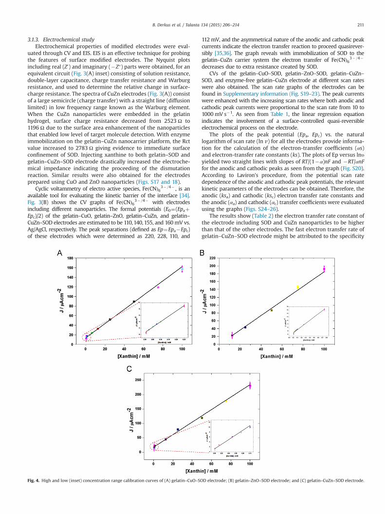
3.1.3. Electrochemical studyElectrochemical properties of modified electrodes were eval-
uated through CV and EIS. EIS is an effective technique for probingthe features of surface modified electrodes. The Nyquist plotsincluding real (Z0) and imaginary (�Z″) parts were obtained, for anequivalent circuit (Fig. 3(A) inset) consisting of solution resistance,double-layer capacitance, charge transfer resistance and Warburgresistance, and used to determine the relative change in surface-charge resistance. The spectra of CuZn electrodes (Fig. 3(A)) consistof a large semicircle (charge transfer) with a straight line (diffusionlimited) in low frequency range known as the Warburg element.When the CuZn nanoparticles were embedded in the gelatinhydrogel, surface charge resistance decreased from 2523 Ω to1196 Ω due to the surface area enhancement of the nanoparticlesthat enabled low level of target molecule detection. With enzymeimmobilization on the gelatin–CuZn nanocarrier platform, the Rctvalue increased to 2783 Ω giving evidence to immediate surfaceconfinement of SOD. Injecting xanthine to both gelatin–SOD andgelatin–CuZn–SOD electrode drastically increased the electroche-mical impedance indicating the proceeding of the dismutationreaction. Similar results were also obtained for the electrodesprepared using CuO and ZnO nanoparticles (Figs. S17 and 18).
Cyclic voltammetry of electro active species, FeðCNÞ63�=4� , is anavailable tool for evaluating the kinetic barrier of the interface [34].Fig. 3(B) shows the CV graphs of FeðCNÞ63�=4� with electrodesincluding different nanoparticles. The formal potentials (E0¼(EpaþEpc)/2) of the gelatin–CuO, gelatin–ZnO, gelatin–CuZn, and gelatin–CuZn–SOD electrodes are estimated to be 110,140,155, and 160mV vs.Ag/AgCl, respectively. The peak separations (defined as Ep¼Epa�Epc)of these electrodes which were determined as 220, 228, 110, and
112mV, and the asymmetrical nature of the anodic and cathodic peakcurrents indicate the electron transfer reaction to proceed quasirever-sibly [35,36]. The graph reveals with immobilization of SOD to thegelatin–CuZn carrier system the electron transfer of FeðCNÞ63�=4�
decreases due to extra resistance created by SOD.CVs of the gelatin–CuO–SOD, gelatin–ZnO–SOD, gelatin–CuZn–
SOD, and enzyme-free gelatin–CuZn electrode at different scan rateswere also obtained. The scan rate graphs of the electrodes can befound in Supplementary information (Fig. S19–23). The peak currentswere enhanced with the increasing scan rates where both anodic andcathodic peak currents were proportional to the scan rate from 10 to1000 mV s�1. As seen from Table 1, the linear regression equationindicates the involvement of a surface-controlled quasi-reversibleelectrochemical process on the electrode.
The plots of the peak potential (Epa, Epc) vs. the naturallogarithm of scan rate (ln v) for all the electrodes provide informa-tion for the calculation of the electron-transfer coefficients (αs)and electron-transfer rate constants (ks). The plots of Ep versus lnυyielded two straight lines with slopes of RT/(1�α)nF and �RT/αnFfor the anodic and cathodic peaks as seen from the graph (Fig. S20).According to Laviron's procedure, from the potential scan ratedependence of the anodic and cathodic peak potentials, the relevantkinetic parameters of the electrodes can be obtained. Therefore, theanodic (ksa) and cathodic (ksc) electron transfer rate constants andthe anodic (αa) and cathodic (αc) transfer coefficients were evaluatedusing the graphs (Figs. S24–26).
The results show (Table 2) the electron transfer rate constant ofthe electrode including SOD and CuZn nanoparticles to be higherthan that of the other electrodes. The fast electron transfer rate ofgelatin–CuZn–SOD electrode might be attributed to the specificity
Fig. 4. High and low (inset) concentration range calibration curves of (A) gelatin–CuO–SOD electrode; (B) gelatin–ZnO–SOD electrode; and (C) gelatin–CuZn–SOD electrode.
B. Derkus et al. / Talanta 134 (2015) 206–214 211
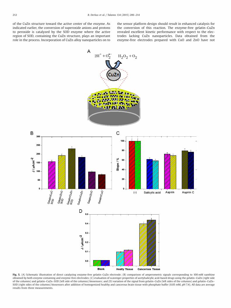
of the CuZn structure toward the active center of the enzyme. Asindicated earlier, the conversion of superoxide anions and protonsto peroxide is catalyzed by the SOD enzyme where the activeregion of SOD, containing the CuZn structure, plays an importantrole in the process. Incorporation of CuZn alloy nanoparticles on to
the sensor platform design should result in enhanced catalysis forthe conversion of this reaction. The enzyme-free gelatin–CuZnrevealed excellent kinetic performance with respect to the elec-trodes lacking CuZn nanoparticles. Data obtained from theenzyme-free electrodes prepared with CuO and ZnO have not
Fig. 5. (A) Schematic illustration of direct catalyzing enzyme-free gelatin–CuZn electrode; (B) comparison of amperometric signals corresponding to 100 mM xanthineobtained by both enzyme containing and enzyme-free electrodes; (C) evaluation of scavenger properties of acetylsalicylic acid-based drugs using the gelatin–CuZn (right sideof the columns) and gelatin–CuZn–SOD (left side of the columns) biosensors; and (D) variation of the signal from gelatin–CuZn (left sides of the columns) and gelatin–CuZn–SOD (right sides of the columns) biosensors after addition of homogenized healthy and cancerous brain tissue with phosphate buffer (0.05 mM, pH 7.4). All data are averageresults from three measurements.
B. Derkus et al. / Talanta 134 (2015) 206–214212
been presented here because of their poor electrochemical andanalytical performance
The effective surface areas of the electrodes were also deter-mined using CV, with the aid of the Randles–Sevcik equation [37]where the peak currents (ip) vs. ln v graphs (Figs. S27–29) wereutilized. The results revealed the ZnO embedded electrodes tohave a 14.4% higher surface area than CuZn embedded electrodes.This situation can be attributed to high particle size of CuZn alloynanoparticles (150 nm) compared to CuO and ZnO (o50 nm). TheZnO embedded electrode also has an effective surface area onaverage 11.1% higher than the CuO embedded electrode, which isthought to be the result of the high isoelectric point of ZnO(8.7–10.3).
3.1.4. Comparison of the developed nanobiosensors with and withoutenzyme
The amperometric response of the nanobiosensors towardsO2d� was investigated in the buffer solution. A simple and efficient
method was used for the generation of O2d� by oxidation of
xanthine to uric acid in the presence of xanthine oxidase at anapplied potential of 650 mV. When xanthine is injected into themedium, a superoxide “burst” occurs followed by a rapid decreaseto the baseline level due to dismutation. Table 3 shows the mainanalytical data of the nanobiosensors obtained from the calibra-tion curves (Fig. 4).
The detection limits of the biosensors were calculated as0.89 μM for gelatin–CuO–SOD, 1.64 μM for gelatin–ZnO–SOD, and0.31 μM for gelatin–CuZn–SOD electrodes (at a signal-to-noiseratio of 3). Peak intensities of both electrodes, SOD containingelectrodes and direct catalyzing enzyme-free electrodes (Fig. 5(A)),were compared using 100 mM of xanthine (Fig. 5(B)). The gelatin–ZnO and gelatin–CuZn electrodes containing SOD gave a moreintense peak with respect to the enzyme-free electrodes. On theother hand, the gelatin–CuZn electrode was seen to give a moreintense peak in comparison to the gelatin–CuO–SOD electrode.Enzyme-free gelatin–CuO and gelatin–ZnO electrodes were seento give weak peaks around 35% and 42% compared to the gelatin–CuZn–SOD, which is not functional in designing biosensors. How-ever, the enzyme-free gelatin–CuZn electrode gave a peak of 79%which is lucrative for superoxide radical sensing.
3.1.5. Effect of scavengersSome molecules and drugs are potential scavengers of super-
oxide radicals [38,39]. For this purpose, acetylsalicylic acid,Aspirin, and Aspirin C were used and amperometric measure-ments were carried out using both gelatin–CuZn–SOD andenzyme-free gelatin–CuZn electrodes in the absence and presenceof these scavengers (Fig. 5(C)). The antioxidant properties of thescavenger molecules tested were evaluated from the percentageratio of the slope values of the calibration plot of the gelatin–CuZn–SOD electrode (Fig. 4(B)). Addition of salycylic acid, aspirinand aspirin C samples decreased the signal strengths as theantioxidant species reacted with the superoxide radical, thusreducing its concentration in the solution. There was a consequentdecrease in the amount of H2O2 released and as a result in theamperometric signal. It can be deduced that pure salycylic acid hasa more intense scavenger effect. Hence the developed biosensorcan also be used for in vitro determination of antioxidant proper-ties of salicylic acid-based drugs.
3.1.6. Medical applicationThe biosensor response was also tested on healthy and cancer-
ous brain tissue (Fig. 5(D)) using both gelatin–CuZn–SOD andenzyme-free gelatin–CuZn electrode. Different signals wereobtained from the biosensor depending on whether the tissue
was healthy or cancerous. Cancerous brain tissue contains higherquantities of superoxide radicals compared to a healthy tissue. Thisis probably due to smaller quantities of scavenger molecules orendogenous SOD being present in the cancerous tissue. The valuesobtained by both electrodes were seen to be fairly close to eachother, indicating the viability of the enzyme-free electrode forclinical analysis of superoxide radicals.
4. Conclusion
Technology has always been an indispensable part in thedevelopment of biosensors. The performance of biosensors isbeing strongly improved using new materials such as nanoparti-cles or other smart materials. The use of nanoparticles in biosensortechnology allows innovation in designing more sensitive, eco-nomical, rapid, easy-to-use, and lab-on-chip type diagnostic sys-tems. In recent years, enzyme-free catalyzing of some redoxreactions have become trendy. Therefore the designability ofsensors for the determination of superoxide radicals was investi-gated in this study. The results obtained from the enzyme-freesensors were hopeful but not as advanced as those from thebiosensor containing enzyme. The gelatin–CuZn enzyme-freesensor showed an electrochemical performance of 80% comparedto the enzyme containing electrode. On the other hand, theenzyme-free sensors prepared with CuO and ZnO nanoparticlesdid not show the same advanced performance. It seems that thedetection of superoxide radicals will be feasible using enzyme-freesensors in biological samples with further advanced studies.
Acknowledgments
This work is financially supported by the Scientific and Tech-nological Research Council of Turkey (no. 113S255).
Appendix A. Supporting information
Supplementary data associated with this article can be found inthe online version at http://dx.doi.org/10.1016/j.talanta.2014.11.003.
References
[1] O. Kocabay, E. Emregul, S. Aras, K.C. Emregul, Bioprocess. Biosyst. Eng. 35 (6)(2012) 923–930.
[2] R.S. Sohal, B.H. Sohal, W.C. Orr, Free Radical Biol. Med. 19 (1995) 499–504.[3] R. Misiaszek, C. Crean, A. Joffe, N.E. Geacintov, V. Shafirovich, J. Biol.Chem. 279
(2004) 32106–32115.[4] J.R. Woods, Placenta 22 (2001) S38–S44.[5] S. Mak, G.E. Newton, Chest 120 (2001) 2035–2046.[6] P. Kovacic, J.D. Jacintho, Curr. Med. Chem. 8 (2001) 773–796.[7] B.E. Leonard, Int. J. Dev. Neurosci. 19 (2001) 305–312.[8] I. Fridovich, Acc. Chem. Res. 5 (1972) 321.[9] I. Fridovich, J. Biol. Chem. 272 (1997) 18515.[10] A. Okado-Matsumoto, I. Fridovich, J. Biol. Chem. 276 (2001) 38388.[11] L.A. Sturtz, K. Diekert, L.T. Jensen, R. Lill, V.C. Culotta, J. Biol. Chem. 276 (2001)
38084.[12] J.S. Valentine, P.A. Doucette, S.Z. Potter, Annu. Rev. Biochem. 74 (2005)
563–593.[13] B.K. Beissenhirtz, F.W. Scheller, M.S. Viezzoli, F. Lisdat, Anal. Chem. 78 (3)
(2006) 928–935.[14] K. Prasad, J. Kalra, B. Bhardwaj, J. Exp. Pathol. 70 (1989) 463.[15] J. Vasquez-Vivar, N. Hogg, K.A. Pritchard, B. Kalyanaraman, FEBS Lett. 403
(1997) 130.[16] T. Ohyashiki, M. Nunomura, T. Katoh, Biochim. Biophys. Acta 1421 (1992) 139.[17] T. Oshaka, Y. Shintane, F. Matsumoto, T. Okajima, K. Tokuda, Bioelectrochem.
Bioenerg. 37 (1995) 73.[18] K. Tammeveski, T.T. Tenno, A.A. Mashirin, E.W. Hillhouse, P. Manning, C.
J. McNeil, Free Radic. Res. Commun. 25 (1998) 973.[19] K. Tanaka, Y. Muto, Bioelectrochem. Bioenerg. 29 (1992) 143.[20] S. Luo, F. Su, C. Liu, J. Li, R. Liu, Y. Xiao, Y. Li, X. Liu, Q. Cai, Talanta 86 (2011)
157–163.
B. Derkus et al. / Talanta 134 (2015) 206–214 213

[21] S. Anandan, X.-G. Wen, S.-H. Yang, Mater. Chem. Phys. 93 (2005) 35.[22] Z.-L. Jin, X.-J. Zhang, Y.-X. Li, S.-B. Li, G.-X. Lu, Catal. Commun. 8 (2008) 1267.[23] S.F. Zheng, J.S. Hu, L.S. Zhong, W.G. Song, L.J. Wan, Chem. Mater. 20 (2008)
3617.[24] N.Q. Dung, D. Patil, H. Jung, D. Kim, Biosens. Bioelectron. 42 (2013) 280–286.[25] Z.-J. Zhuang, X.-D. Su, H.-Y. Yuan, Q. Sun, D. Xiao, M. Choi, Analyst 133 (2008)
126.[26] X. Wang, C.-G. Hu, H. Liu, G.-J. Du, X.-S. He, Y. Xi, Sens. Actuators B: Chem. 144
(2010) 220.[27] A. Umar, M.M. Rahman, A. Al-Hajry, Y.B. Hahn, Electrochem. Commun. 11
(2009) 278.[28] J. Liu, X. Huang, Y. Li, K.M. Sulieman, X. He, F. Sun, Cryst. Growth Des. 6 (2006)
16901.[29] M. Ahmad, C. Pan, L. Gan, Z. Nawaz, J. Zhu, J. Phys. Chem. C 17 (2010) 243–250.[30] P. Norouzi, V.K. Gupta, F. Faridbod, M. Pirali-Hamedani, B. Larijani, M.
R. Ganjali, Anal. Chem. 83 (2011) 1564–1570.
[31] E. Laviron, J. Electroanal. Chem. 101 (1979) 19–28.[32] E. Emregul, O. Kocabay, B. Derkus, T. Yumak, K.C. Emregul, A. Sinag, K. Polat,
Bioelectrochemistry 90 (2013) 8–17.[33] E. Emregul, Anal. Bioanal. Chem. 383 (6) (2005) 947–954.[34] B. Rezaei, N. Majidi, H. Rahmani, T. Khayamian, Biosens. Bioelectron. 26 (2011)
2130–2134.[35] Z. Deng, Q. Rui, X. Yin, H. Liu, Y. Tian, Anal. Chem. 80 (2008) 5839–5846.[36] H. Liu, Y. Tian, P. Xia, Langmuir 24 (2008) 6359–6366.[37] J. Shi, J.C. Claussen, E.S. McLamore, A. Haque, D. Jaroch, A.R. Diggs, P. Calvo-
Marzal, J.L. Rickus, D.M. Porterfield, Nanotechnology 22 (2011) 355502.[38] B.N. Ames, R. Cathcart, E. Schwiers,and, P. Hochstein, Proc. Natl. Acad. Sci. USA
78 (1981) 6858.[39] A. Nandi, I.B. Chatterjee, J. Biosci. 11 (1–4) (1987) 435–441.
B. Derkus et al. / Talanta 134 (2015) 206–214214
Related Documents