COPI Activity Coupled with Fatty Acid Biosynthesis Is Required for Viral Replication Sara Cherry 1* , Amit Kunte 2 , Hui Wang 3 , Carolyn Coyne 4 , Robert B. Rawson 2 , Norbert Perrimon 3 1 University of Pennsylvania School of Medicine, Philadelphia, Pennsylvania, United States of America, 2 University of Texas Southwestern Medical Center, Dallas, Texas, United States of America, 3 Harvard Medical School, Howard Hughes Medical Institute, Boston, Massachusetts, United States of America, 4 Children’s Hospital of Pennsylvania, Philadelphia, Pennsylvania, United States of America During infection by diverse viral families, RNA replication occurs on the surface of virally induced cytoplasmic membranes of cellular origin. How this process is regulated, and which cellular factors are required, has been unclear. Moreover, the host–pathogen interactions that facilitate the formation of this new compartment might represent critical determinants of viral pathogenesis, and their elucidation may lead to novel insights into the coordination of vesicular trafficking events during infection. Here we show that in Drosophila cells, Drosophila C virus remodels the Golgi apparatus and forms a novel vesicular compartment, on the surface of which viral RNA replication takes place. Using genome-wide RNA interference screening, we found that this step in the viral lifecycle requires at least two host encoded pathways: the coat protein complex I (COPI) coatamer and fatty acid biosynthesis. Our results integrate, clarify, and extend numerous observations concerning the cell biology of viral replication, allowing us to conclude that the coupling of new cellular membrane formation with the budding of these vesicles from the Golgi apparatus allows for the regulated generation of this new virogenic organelle, which is essential for viral replication. Additionally, because these pathways are also limiting in flies and in human cells infected with the related RNA virus poliovirus, they may represent novel targets for antiviral therapies. Citation: Cherry S, Kunte A, Wang H, Coyne C, Rawson RB, et al. (2006) COPI activity coupled with fatty acid biosynthesis is required for viral replication. PLoS Pathog 2(10): e102. DOI: 10.1371/journal.ppat.0020102 Introduction Viruses, because of their small genome size, are dependent on a multitude of cellular factors to replicate within their hosts. Not only do they have to co-opt cellular factors in order to complete their replication cycle, but also they must efficiently and simultaneously coordinate many steps of their replication cycle using host-encoded machinery. This can require complicated compartmentalization of various steps in their lifecycle. For example, single-strand RNA viruses must simultaneously coordinate transcription, RNA replica- tion, and RNA packaging activities using the same genomic RNA template. One example of subcellular separation of these activities is the observation that all positive-strand RNA viruses, a group that includes poliovirus, undergo RNA replication in associ- ation with membranes of infected cells [1]. Depending on the specific virus, these membranes can be derived from a variety of sources within the host cell, including the endoplasmic reticulum (ER), Golgi apparatus, mitochondria, chloroplasts, or from the endolysosomal compartment [2]. While the purpose of this compartmentalization has not been defini- tively established, several models have been discussed. One model suggests that this process provides a structural frame- work for replication, fixing the RNA replication machinery onto a confined two-dimensional space [3]. This compart- mentalization of RNA replication may be important due to the fact that the viral RNA must be used for competing enzymatic activities: replication and transcription must both access templates using different machines. The separation of RNA templates into defined compartments may prevent interference between these processes. Another model postu- lates that the compartment may be generated by a cellular autophagic process. This may allow for the nonlytic release of virions [4], which in vivo could allow the virus to circumvent presentation by the immune system. Another possibility is that autophagy may play an antiviral role in clearing the cytoplasm of infectious virus. Therefore, the purpose of this targeted localization has yet to be understood. Poliovirus, and picornaviridae in general (nonenveloped, positive-strand RNA viruses), do not use native organelle membranes for replication but instead actively induce the formation of a novel cytoplasmic vesicular compartment in infected cells [5,6]. During replication of these viruses, there is a massive rearrangement of intracellular membranes whereby the cytoplasm of the cell becomes densely packed with vesicles of nonuniform size [7]. Immunoelectron micro- scopy has revealed that the cytoplasmic surfaces of these vesicles are the sites of viral RNA replication and that a membrane compartment is essential for replication [8]. The mechanisms whereby these viruses induce this vesicular compartment and direct their RNA replication complex to Editor: Beth Levine, University of Texas Southwestern Medical Center, United States of America Received April 11, 2006; Accepted August 25, 2006; Published October 13, 2006 DOI: 10.1371/journal.ppat.0020102 Copyright: Ó 2006 Cherry et al. This is an open-access article distributed under the terms of the Creative Commons Attribution License, which permits unrestricted use, distribution, and reproduction in any medium, provided the original author and source are credited. Abbreviations: Arf, ADP-ribosylation factor; COPI, coat protein complex I; DCV, Drosophila C virus; dsRNA, double-strand RNA; ER, endoplasmic reticulum; GFP, green fluorescent protein; GO, Gene Ontology; RNAi, RNA interference; siRNA, small interfering RNA; SREBP, sterol regulatory element binding protein * To whom correspondence should be addressed. E-mail: [email protected]. edu PLoS Pathogens | www.plospathogens.org October 2006 | Volume 2 | Issue 10 | e102 0900

Welcome message from author
This document is posted to help you gain knowledge. Please leave a comment to let me know what you think about it! Share it to your friends and learn new things together.
Transcript

COPI Activity Coupled with Fatty AcidBiosynthesis Is Required for Viral ReplicationSara Cherry
1*, Amit Kunte
2, Hui Wang
3, Carolyn Coyne
4, Robert B. Rawson
2, Norbert Perrimon
3
1 University of Pennsylvania School of Medicine, Philadelphia, Pennsylvania, United States of America, 2 University of Texas Southwestern Medical Center, Dallas, Texas,
United States of America, 3 Harvard Medical School, Howard Hughes Medical Institute, Boston, Massachusetts, United States of America, 4 Children’s Hospital of
Pennsylvania, Philadelphia, Pennsylvania, United States of America
During infection by diverse viral families, RNA replication occurs on the surface of virally induced cytoplasmicmembranes of cellular origin. How this process is regulated, and which cellular factors are required, has been unclear.Moreover, the host–pathogen interactions that facilitate the formation of this new compartment might representcritical determinants of viral pathogenesis, and their elucidation may lead to novel insights into the coordination ofvesicular trafficking events during infection. Here we show that in Drosophila cells, Drosophila C virus remodels theGolgi apparatus and forms a novel vesicular compartment, on the surface of which viral RNA replication takes place.Using genome-wide RNA interference screening, we found that this step in the viral lifecycle requires at least two hostencoded pathways: the coat protein complex I (COPI) coatamer and fatty acid biosynthesis. Our results integrate,clarify, and extend numerous observations concerning the cell biology of viral replication, allowing us to conclude thatthe coupling of new cellular membrane formation with the budding of these vesicles from the Golgi apparatus allowsfor the regulated generation of this new virogenic organelle, which is essential for viral replication. Additionally,because these pathways are also limiting in flies and in human cells infected with the related RNA virus poliovirus, theymay represent novel targets for antiviral therapies.
Citation: Cherry S, Kunte A, Wang H, Coyne C, Rawson RB, et al. (2006) COPI activity coupled with fatty acid biosynthesis is required for viral replication. PLoS Pathog 2(10):e102. DOI: 10.1371/journal.ppat.0020102
Introduction
Viruses, because of their small genome size, are dependenton a multitude of cellular factors to replicate within theirhosts. Not only do they have to co-opt cellular factors inorder to complete their replication cycle, but also they mustefficiently and simultaneously coordinate many steps of theirreplication cycle using host-encoded machinery. This canrequire complicated compartmentalization of various stepsin their lifecycle. For example, single-strand RNA virusesmust simultaneously coordinate transcription, RNA replica-tion, and RNA packaging activities using the same genomicRNA template.
One example of subcellular separation of these activities isthe observation that all positive-strand RNA viruses, a groupthat includes poliovirus, undergo RNA replication in associ-ation with membranes of infected cells [1]. Depending on thespecific virus, these membranes can be derived from a varietyof sources within the host cell, including the endoplasmicreticulum (ER), Golgi apparatus, mitochondria, chloroplasts,or from the endolysosomal compartment [2]. While thepurpose of this compartmentalization has not been defini-tively established, several models have been discussed. Onemodel suggests that this process provides a structural frame-work for replication, fixing the RNA replication machineryonto a confined two-dimensional space [3]. This compart-mentalization of RNA replication may be important due tothe fact that the viral RNA must be used for competingenzymatic activities: replication and transcription must bothaccess templates using different machines. The separation ofRNA templates into defined compartments may preventinterference between these processes. Another model postu-lates that the compartment may be generated by a cellular
autophagic process. This may allow for the nonlytic release ofvirions [4], which in vivo could allow the virus to circumventpresentation by the immune system. Another possibility isthat autophagy may play an antiviral role in clearing thecytoplasm of infectious virus. Therefore, the purpose of thistargeted localization has yet to be understood.Poliovirus, and picornaviridae in general (nonenveloped,
positive-strand RNA viruses), do not use native organellemembranes for replication but instead actively induce theformation of a novel cytoplasmic vesicular compartment ininfected cells [5,6]. During replication of these viruses, thereis a massive rearrangement of intracellular membraneswhereby the cytoplasm of the cell becomes densely packedwith vesicles of nonuniform size [7]. Immunoelectron micro-scopy has revealed that the cytoplasmic surfaces of thesevesicles are the sites of viral RNA replication and that amembrane compartment is essential for replication [8]. Themechanisms whereby these viruses induce this vesicularcompartment and direct their RNA replication complex to
Editor: Beth Levine, University of Texas Southwestern Medical Center, UnitedStates of America
Received April 11, 2006; Accepted August 25, 2006; Published October 13, 2006
DOI: 10.1371/journal.ppat.0020102
Copyright: � 2006 Cherry et al. This is an open-access article distributed under theterms of the Creative Commons Attribution License, which permits unrestricteduse, distribution, and reproduction in any medium, provided the original authorand source are credited.
Abbreviations: Arf, ADP-ribosylation factor; COPI, coat protein complex I; DCV,Drosophila C virus; dsRNA, double-strand RNA; ER, endoplasmic reticulum; GFP,green fluorescent protein; GO, Gene Ontology; RNAi, RNA interference; siRNA, smallinterfering RNA; SREBP, sterol regulatory element binding protein
* To whom correspondence should be addressed. E-mail: [email protected]
PLoS Pathogens | www.plospathogens.org October 2006 | Volume 2 | Issue 10 | e1020900
these specific intracellular membranes, and the cellularcomponents involved in this process, are not established.Previous studies have implicated a number of differenttrafficking processes including coat protein complex I(COPI), COPII, and autophagy-mediated processes, with themembrane being derived from the ER via a COPII coatamer–mediated process or the Golgi via either COPI or autophagicvesicles [4,9–12]. Studies investigating how picorna andrelated viruses induce the formation of cellular membraneshave used small molecule inhibitors such as Brefeldin A,which have pleiotropic effects, or have used steady-statelocalization experiments that are complicated by the fact thatmarkers from many different intracellular organelles can befound residing on virus-induced membranes, making itdifficult to assign a specific role for specific cellularcomponents in the ontogeny of the vesicles.
To overcome some of these limitations, we used a genome-wide loss-of-function analysis to identify factors required forthe generation of this compartment and viral RNA repli-cation. To accomplish this, we studied Drosophila C virus(DCV), a natural pathogen of Drosophila that readily infectsthe cells and animals [13]. DCV is a dicistrovirus that is inmany ways similar to picornaviruses such as poliovirus. Bothare encoded by a single positive-strand RNA genome, with agenome-linked protein at the 59 end and a poly adenosine tailat the 39 end [14–16]. They also share many physical andmorphological properties of the viral structural proteinsincluding the requirement for virally encoded proteases forprocessing [17,18]. In addition, they are translated by aspecialized, cap-independent, internal ribosome entry site(IRES) mechanism [19].
Using this Drosophila system allowed us to apply forwardgenetics to both the whole organism and cell culture to screenfor host-encoded factors whose loss blocks viral replication.Recently, we conducted a genome-wide RNA interference(RNAi) screen and found over 100 genes required for efficientviral replication in tissue culture [20]. We have begun tovalidate the importance of these genes for virus replicationby several approaches and to determine the stage in the virallifecycle affected by the loss of each. Using this loss-of-function strategy, we found that DCV, like mammalian
picornaviruses, induces a cytoplasmic vesicular compartmentupon which viral RNA replication takes place. The formationof this structure was dependent on the cellular activity ofCOPI along with the generation of new membrane by thefatty acid biosynthetic pathway. Together, these host-encodedgenes drive the formation of these vesicles for viral RNAreplication. Moreover, animals mutant for fatty acid biosyn-thesis were attenuated for viral replication demonstratingthat this pathway is required and limiting for infection bothin vivo and in vitro. Importantly, we also found that COPI,but not COPII, was also required for poliovirus infection ofhuman cells, demonstrating the generality of our findings.
Results
Gene Ontology Analysis Reveals Overrepresented CellularFunctions Necessary for Viral ReplicationGenome-wide RNAi screening in Drosophila cells using high-
throughput imaging identified 66 ribosomal proteins and 45nonribosomal genes as required for DCV replication [20]. Inour first study, we showed that knock-down of theseribosomal proteins blocked translation of IRES-containingviruses, including poliovirus, due to a requirement for higherlevels of ribosomal function than for host messages, showing aunique sensitivity of this class of viruses to ribosomeattenuation [20]. To identify roles for the remaining genesin viral replication, we analyzed Gene Ontology (GO)associations of the 45 nonribosomal genes required forDCV replication in Drosophila cells. This analysis showed thatthese genes fall into a small number of functional categories(Figure 1A). In addition, Table 1 lists the 45 genes, theireffects on viral replication, GO category, and whether thedouble-strand RNA (dsRNA) amplicons identified havepotential off-target effects as predicted by 21–base pairoverlaps with other annotated genes. Based on the associa-tions with Gene Ontology (GO) categories, some biologicalprocesses were significantly overrepresented. Specifically,statistical analysis revealed that vesicular trafficking processeswere overrepresented while Drosophila-specific genes wereunderrepresented (30% of genome, 4% of set), suggestingthat DCV selectively targets conserved features of the hostcells, and not species-specific functions.One notable group of genes within the vesicle trafficking
category included five of the seven COPI coatamer proteins:alphaCOP, betaCOP, beta’COP, gammaCOP, and zetaCOP (Figure1B, p , 0.001 for enrichment relative to representation in thegenome, by Fisher exact test). To confirm that loss of COPIwas responsible for the phenotype and to determine whetherthe two COPI genes (deltaCOP and epsilonCOP [CG9543]) notidentified in the screen were erroneously missed (i.e., falsenegatives), we synthesized additional dsRNAs for the sevenCOPI coatamer genes and tested them for their role in DCVreplication. These analyses revealed that dsRNAs against allCOPI components except epsilonCOP blocked DCV replica-tion, although some differences in the extent of the effectwere observed, possibly reflecting variability in the efficiencyof the gene silencing mediated by the dsRNAs (Figure 1B).Thus, we can conclude that deltaCOP was a false negative inthe primary screen. However, neither of the dsRNAs directedagainst epsilonCOP had an effect on DCV replication. RT-PCRanalysis demonstrated that epsilonCOP mRNA was depleted,demonstrating that the dsRNA was functional (Figure S1).
PLoS Pathogens | www.plospathogens.org October 2006 | Volume 2 | Issue 10 | e1020901
COPI, Fatty Acids, and Viral Replication
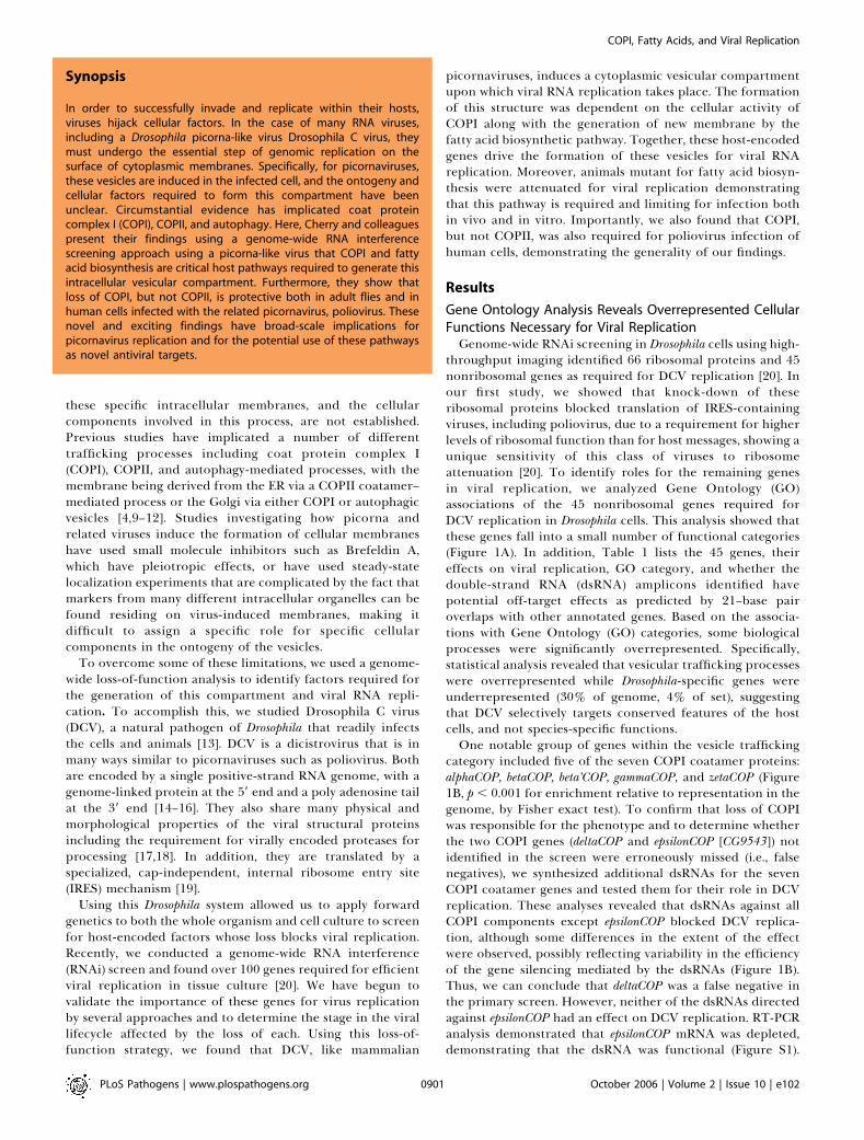
Synopsis
In order to successfully invade and replicate within their hosts,viruses hijack cellular factors. In the case of many RNA viruses,including a Drosophila picorna-like virus Drosophila C virus, theymust undergo the essential step of genomic replication on thesurface of cytoplasmic membranes. Specifically, for picornaviruses,these vesicles are induced in the infected cell, and the ontogeny andcellular factors required to form this compartment have beenunclear. Circumstantial evidence has implicated coat proteincomplex I (COPI), COPII, and autophagy. Here, Cherry and colleaguespresent their findings using a genome-wide RNA interferencescreening approach using a picorna-like virus that COPI and fattyacid biosynthesis are critical host pathways required to generate thisintracellular vesicular compartment. Furthermore, they show thatloss of COPI, but not COPII, is protective both in adult flies and inhuman cells infected with the related picornavirus, poliovirus. Thesenovel and exciting findings have broad-scale implications forpicornavirus replication and for the potential use of these pathwaysas novel antiviral targets.

However, the protein may be long-lived—something we couldnot assess due to the absence of antibodies directed againsteither this or the other Drosophila COPI proteins. Never-theless, these data suggest that it may have an unknownhomolog which can substitute for its function. It should benoted that in yeast only six of the seven coatamercomponents are essential—only epsilonCOP is dispensible[21]. This suggests that epsilonCOP may be dispensable forCOPI function in both yeast and Drosophila.
COPI, but Not Secretion, Is Required for DCV ReplicationTwo vesicular coat complexes, COPI and COPII, are
required for trafficking between the ER and Golgi [22].Formation of transport vesicles is dependent on the recruit-ment of these cytosolic coat proteins to the surface of the
donor compartment membrane from which they bud.Whereas the COPII coatamer is required for the anterogradetransport of proteins from the ER to the Golgi, the COPIcoatamer is required for retrograde transport of recycledproteins and membrane from the Golgi to the ER [23].Blocking either the COPI or the COPII coatamer pathwaysblocks protein secretion, as observed in yeast mutants for theorthologous genes and in studies using RNAi screening forgenes required for general secretion [24]. However, we onlyidentified COPI-associated genes and none of the COPIIcoatamer components (Sec13, Sec31, Sec23, and Sec24[CG1472]) in our screen, suggesting that either the COPIIcoatamer proteins are refractory to RNAi-mediated deple-tion in our cells or DCV replication specifically requiresCOPI function but not COPII.
Figure 1. COPI Coatamer Complex Is Required for Viral Replication
(A) Frequency of encoded functional groups as curated by GO (The FlyBase Consortium) and manually assigned to representative categories for allverified candidates. Categories that are overrepresented with p , 0.05 are indicated.(B) Decreased viral replication post dsRNA treatment with dsRNA against alphaCOP, betaCOP, beta’COP, gammaCOP, deltaCOP, and zetaCOP ascompared to dsRNA treatment with GFP or epsilonCOP. Images were quantified as the percentage of infected cells (FITC-anti DCV [green]) divided by(Hoescht 33342 [red]).DOI: 10.1371/journal.ppat.0020102.g001
PLoS Pathogens | www.plospathogens.org October 2006 | Volume 2 | Issue 10 | e1020902
COPI, Fatty Acids, and Viral Replication
To rule out a role for COPII in DCV replication, wemodified a microscopy-based assay to quantitatively monitorsecretion of a plasma membrane reporter protein, Delta, totest whether the dsRNAs directed against COPII componentswere effective [25]. Using Drosophila cells that were stably
transformed with an inducible membrane-bound form ofDelta (DeltaNdeMyc cells [26]), we were able to assess theeffects of different dsRNAs on the secretory pathway usingflow cytometry. We pretreated cells with dsRNAs for 3 d andthen induced the transgene with the addition of copper
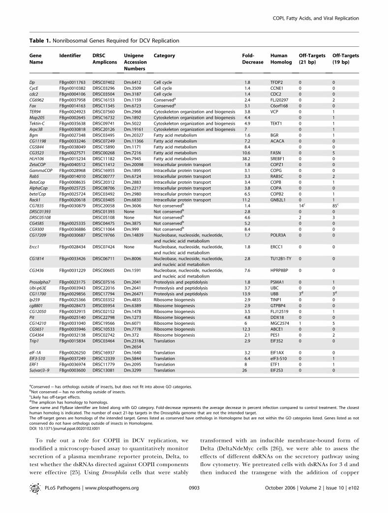
Table 1. Nonribosomal Genes Required for DCV Replication
Gene
Name
Identifier DRSC
Amplicons
Unigene
Accession
Numbers
Category Fold-
Decrease
Human
Homolog
Off-Targets
(21 bp)
Off-Targets
(19 bp)
Dp FBgn0011763 DRSC07402 Dm.6412 Cell cycle 1.8 TFDP2 0 0
CycE FBgn0010382 DRSC03296 Dm.3509 Cell cycle 1.4 CCNE1 0 0
cdc2 FBgn0004106 DRSC03504 Dm.3187 Cell cycle 1.4 CDC2 0 0
CG6962 FBgn0037958 DRSC16153 Dm.1159 Conserveda 2.4 FLJ20297 0 2
Fax FBgn0014163 DRSC11345 Dm.6723 Conserveda 3.1 C6orf168 0 0
TER94 FBgn0024923 DRSC07560 Dm.2968 Cytoskeleton organization and biogenesis 3.8 VCP 0 1
Map205 FBgn0002645 DRSC16732 Dm.1892 Cytoskeleton organization and biogenesis 4.4 0 1
Tektin-C FBgn0035638 DRSC09741 Dm.5022 Cytoskeleton organization and biogenesis 4.9 TEKT1 0 1
Arpc3B FBgn0030818 DRSC20126 Dm.19161 Cytoskeleton organization and biogenesis 7 0 1
Bgm FBgn0027348 DRSC03495 Dm.20327 Fatty acid metabolism 1.6 BGR 0 1
CG11198 FBgn0033246 DRSC07249 Dm.11366 Fatty acid metabolism 7.2 ACACA 0 0
CG5844 FBgn0038049 DRSC15890 Dm.1171 Fatty acid metabolism 8.4 0 0
CG3523 FBgn0027571 DRSC00268 Dm.7216 Fatty acid metabolism 10.6 FASN 0 5
HLH106 FBgn0015234 DRSC11182 Dm.7945 Fatty acid metabolism 38.2 SREBF1 0 0
ZetaCOP FBgn0040512 DRSC11412 Dm.20098 Intracellular protein transport 1.8 COPZ1 0 0
GammaCOP FBgn0028968 DRSC16955 Dm.1895 Intracellular protein transport 3.1 COPG 0 0
Rab5 FBgn0014010 DRSC00777 Dm.6724 Intracellular protein transport 3.3 RAB5C 0 2
BetaCop FBgn0008635 DRSC20312 Dm.2883 Intracellular protein transport 3.4 COPB 0 1
AlphaCop FBgn0025725 DRSC08706 Dm.2217 Intracellular protein transport 3.8 COPA 0 0
beta’Cop FBgn0025724 DRSC03492 Dm.2980 Intracellular protein transport 6.5 COPB2 0 1
Rack1 FBgn0020618 DRSC03405 Dm.6830 Intracellular protein transport 11.2 GNB2L1 0 1
CG7835 FBgn0030879 DRSC20058 Dm.3606 Not conservedb 1.4 14c 85c
DRSC01393 DRSC01393 None Not conservedb 2.8 0 0
DRSC05108 DRSC05108 None Not conservedb 4.6 2 3
CG4585 FBgn0025335 DRSC04475 Dm.3875 Not conservedb 5.2 0 0
CG9300 FBgn0036886 DRSC11064 Dm.999 Not conservedb 8.4 0 0
CG17209 FBgn0030687 DRSC19766 Dm.14839 Nucleobase, nucleoside, nucleotide,
and nucleic acid metabolism
1.7 POLR3A 0 0
Ercc1 FBgn0028434 DRSC07424 None Nucleobase, nucleoside, nucleotide,
and nucleic acid metabolism
1.8 ERCC1 0 0
CG1814 FBgn0033426 DRSC06711 Dm.8006 Nucleobase, nucleoside, nucleotide,
and nucleic acid metabolism
2.8 TU12B1-TY 0 0
CG3436 FBgn0031229 DRSC00605 Dm.1591 Nucleobase, nucleoside, nucleotide,
and nucleic acid metabolism
7.6 HPRP8BP 0 0
Prosalpha7 FBgn0023175 DRSC07516 Dm.2041 Proteolysis and peptidolysis 1.8 PSMA1 0 1
Ubi-p63E FBgn0003943 DRSC22016 Dm.2641 Proteolysis and peptidolysis 3.7 UBC 0 0
CG11700 FBgn0029856 DRSC17794 Dm.26471 Proteolysis and peptidolysis 13.9 UBB 3d 3d
Ip259 FBgn0025366 DRSC03352 Dm.4835 Ribosome biogenesis 2.9 TINP1 0 0
cg8801 FBgn0028473 DRSC05954 Dm.6389 Ribosome biogenesis 2.9 GTPBP4 0 0
CG12050 FBgn0032915 DRSC02152 Dm.1478 Ribosome biogenesis 3.5 FLJ12519 0 1
Pit FBgn0025140 DRSC22798 Dm.1273 Ribosome biogenesis 4.8 DDX18 0 0
CG14210 FBgn0031040 DRSC19566 Dm.6071 Ribosome biogenesis 6 MGC2574 1 5
CG5651 FBgn0035946 DRSC10533 Dm.7778 Ribosome biogenesis 12.3 ABCE1 0 0
CG4364 FBgn0032138 DRSC02742 Dm.372 Ribosome biogenesis 2.1 PES1 0 2
Trip1 FBgn0015834 DRSC03464 Dm.23184,
Dm.2654
Translation 2.9 EIF3S2 0 0
eIF-1A FBgn0026250 DRSC16937 Dm.1640 Translation 3.2 EIF1AX 0 0
EIF3-S10 FBgn0037249 DRSC12339 Dm.5844 Translation 6.4 eIF3-S10 0 1
ERF1 FBgn0036974 DRSC11779 Dm.2095 Translation 8 ETF1 0 1
Su(var)3–9 FBgn0003600 DRSC13081 Dm.3299 Translation 26 EIF2S3 0 0
aConserved¼ has orthologs outside of insects, but does not fit into above GO categories.bNot conserved¼ has no ortholog outside of insects.cLikely has off-target effects.dThe amplicon has homology to homologs.Gene name and FlyBase identifier are listed along with GO category. Fold-decrease represents the average decrease in percent infection compared to control treatment. The closesthuman homolog is indicated. The number of exact 21-bp targets in the Drosophila genome that are not the intended target.The off-target genes are homologs of the intended target. Genes listed as conserved have orthologs in Homologene but are not within the GO categories listed. Genes listed as notconserved do not have orthologs outside of insects in Homologene.DOI: 10.1371/journal.ppat.0020102.t001
PLoS Pathogens | www.plospathogens.org October 2006 | Volume 2 | Issue 10 | e1020903
COPI, Fatty Acids, and Viral Replication

sulfate for 2 h. Cell surface levels of Delta were measured bystaining unpermeabilized cells with an antibody that recog-nizes extracellular Delta. Using this assay on control cells, wedetected a large increase in surface Delta expression ascompared to uninduced cells (Figure 2A, compare green andpurple). Treatment of the cells with dsRNA against COPI(betaCOP) or COPII (sec23) components significantly decreasedthe extracellular levels of Delta as expected (Figure 2A). WhiledsRNA treatment against sec23 did not block secretion asmuch as knockdown of betaCOP, treatment with dsRNAagainst Syntaxin 5 (Syx5), a t-SNARE required at the Golgifor secretion [27], was able to block secretion to a similarextent as COPI. Despite this, treatment with dsRNA againstsec23 or Syx5 had no effect on viral replication (unpublisheddata). These data suggest that DCV replication does notrequire a functional secretory pathway per se but insteadspecifically requires COPI (but not COPII) coatamer function.
COPI Is Required Downstream of EntryWe next sought to determine which step in the viral
lifecycle requires COPI function, and so we first determinedwhether depletion of COPI by dsRNA treatment affectedDCV entry in tissue culture cells. We had previouslydetermined that viral entry requires clathrin-mediatedendocytosis and thus did not anticipate a requirement forCOPI during entry. Nevertheless, we tested whether pretreat-ment with dsRNAs against control (GFP [green fluorescentprotein]), COPI (betaCOP), or COPII (sec23) affected viralentry. To do this, the cells were pretreated with dsRNAs for 3d and, then incubated at 4 8C to block endocytosis. Next,virions were added at a multiplicity of infection of 10 andallowed to bind to the surface of these dsRNA-treated cells.After 1 h of binding, the cells were washed to removeunbound virions and then returned to 25 8C to allowendocytosis to resume. After 3 h, the cells were immunos-tained to visualize virions during entry through the endocyticcompartment. Using this assay, and quantitation withautomated image analysis, we found that depletion of COPI(or COPII) did not block viral entry (Figure 2B).
To verify that this assay is sensitive to genes required forDCV entry, we tested the effect of Rab5, another cellularfactor identified in our screen. Rab5 encodes a small GTPaserequired for endocytosis, and as such should be required forDCV uptake [28]. Indeed, we found that treatment with Rab5dsRNA resulted in a 4-fold decrease in viral entry (Figure 2B).We did not identify other genes involved in clathrin-mediated endocytosis in this screen, suggesting that genessuch as clathrin may be difficult to deplete in the Drosophilacell lines used. However, our RNAi screen was able to uncovera component of the endocytic apparatus required for viralentry (Rab5) and further underscores the role for COPI asrequired for a step in the viral lifecycle postentry.
DCV Induces a Cytoplasmic Vesicular Compartment that Isthe Site of RNA Replication
All positive-strand RNA viruses undergo RNA replicationon the surface of intracellular membranes [1]. Picornaviruses,which are similar in many respects to DCV, induce theformation of cytoplasmic vesicles of nonuniform size, and it ison the surface of these vesicles that viral RNA replicationoccurs [5–7]. We used ultrastructural analysis to test whetherDCV might induce a similar vesicular compartment and
found that, indeed, vesicles were induced in the cytoplasm ofinfected cells but not in the uninfected control cells (compareFigure 3A to Figure 3B). While the vesicles were nonuniformin size, they averaged 115 nm in diameter.We next determined whether DCV uses these vesicles as a
site for RNA replication. To this end, we generated anantibody against the DCV helicase, an integral component ofthe RNA replication machinery. We stained cells that wereeither uninfected or infected with DCV with the anti-helicaseantibody and monitored localization using immunoelectronmicroscopy. We found that the DCV helicase was localized tothe surface of these vesicles and did not stain the uninfectedcells (Figure 3F–3G and unpublished data). Greater than 90%of the gold labeling was vesicle associated, demonstrating thatthe RNA replication machinery is indeed compartmentbound. Importantly, the vesicles were detectable by 10 hpostinfection but not at an earlier time point (unpublisheddata), consistent with our previous findings that new viralprotein synthesis begins at approximately 7 h postinfection[13]. Because these viruses do not encapsidate the RNApolymerase, RNA replication cannot begin until the genomicRNA is translated to produce the viral factors required forRNA replication including the helicase. Altogether, our datademonstrate that the vesicles form postentry and posttrans-lation and are the site of RNA replication.
Vesicle Formation Is COPI DependentBecause DCV induces a cytoplasmic vesicular compartment
and requires the COPI coatamer for replication, we testedwhether this compartment might be generated by the activityof the COPI complex. If the COPI coatamer directly formsthese vesicles, then this might explain why DCV replication isdependent on COPI, but not COPII, function. Thus, we testedwhether depletion of COPI affected the formation of DCV-dependent vesicles by treating cells with dsRNAs againstCOPI (betaCOP), COPII (sec23), or a control (GFP) followed byDCV infection. At 10 h postinfection, both control cells(Figure 3B) and COPII-treated cells (sec23, Figure 3D) weredensely packed with the newly formed vesicles, despite thefact that dsRNA against sec23 reduced transport of Delta tothe cell surface. Randomly selected cells were quantitated interms of the presence or absence of the characteristiccytoplasmic vesicles; 87% of control cells and 83% of sec23-depleted cells were filled with the vesicles. In contrast, theCOPI-depleted cells had a 2.5-fold reduction in the percent-age of cells that contained vesicles (34%, Figure 3C),suggesting that the COPI coatamer is required to generatethis vesicular compartment during DCV replication.
DCV Replication Disrupts the Golgi ApparatusSince the COPI coatamer normally buds vesicles from the
Golgi apparatus [22,29], we reasoned that the virus-inducedvesicular compartment might be generated by COPI-medi-ated budding of vesicles from the Golgi during DCVreplication. Although the COPI machinery required fortrafficking is conserved between yeast, mammals, and insects,there are striking differences in the morphologies of theGolgi apparatus. In Drosophila, the Golgi cisternae are notinterconnected to form a single copy organelle that istypically juxtanuclear. Instead, the vesicles are stacked anddispersed throughout the cytoplasm, as is the case in plantsand yeast [30, 31]. We tested whether the morphology of the
PLoS Pathogens | www.plospathogens.org October 2006 | Volume 2 | Issue 10 | e1020904
COPI, Fatty Acids, and Viral Replication

Figure 2. COPI Is Specifically Required for DCV Replication Postentry
(A) FACS analysis demonstrates that COPI (betaCOP) and COPII (sec23) are required for Delta secretion. DeltaWTNdeMYC cells were treated with dsRNAand subsequently Delta expression was induced for 2 h. Extracellular Delta expression was monitored by FACS. The bar was set such that 98% of thetotal uninduced cells were negative for Delta staining. Induction leads to a shift in the population to express surface Delta, such that only 31% of thecells remain negative. Under these conditions, COPI, COPII, and SREBP block surface staining.(B) DCV entry requires endocytosis (Rab5) but not COPI (bCOP) or COPII (sec23) function. Cells were pretreated with dsRNA, infected at 4 8C to allowsurface binding, followed by 3 h at 25 8C to release the block to endocytosis and monitor viral trafficking. Viral uptake was measured by determining thepercentage of cells (red) that contained virus (green). Green, anti-DCV; red, Alexa-fluor-568-phalloidin.DOI: 10.1371/journal.ppat.0020102.g002
PLoS Pathogens | www.plospathogens.org October 2006 | Volume 2 | Issue 10 | e1020905
COPI, Fatty Acids, and Viral Replication

Golgi was altered during DCV replication by infectingDrosophila cells with DCV and monitoring the structure ofthe Golgi using an antibody to a membrane-bound Golgiresident protein. Using confocal microscopy, we found, as hasbeen observed by others, that the Golgi has a punctatemorphology in uninfected Drosophila cells [30,32] (Figure 4A,uninfected cells). In contrast, the morphology of the Golgiwas altered in the infected cells. The punctae seemed smallerand increased in number throughout the cytoplasm of thecell (Figure 4A, infected cells). This disruption resembles thelarge number of dispersed vesicles observed by electronmicroscopy. These observations were made using two differ-ent antibodies against the Golgi (anti-Golgi and DG13).Moreover, we co-stained infected cells with antibodies toDCV helicase and a Golgi marker (DG13) using immunoelec-tron microscopy. We found that the DCV-induced vesiclesthat are coated by the viral helicase (5-nm gold particles) arealso positive for the Golgi marker (12 nm) (Figure 3H). Thisdemonstrates that these vesicles are indeed derived from theGolgi apparatus.
Because Golgi morphology was disrupted by DCV duringreplication, and depletion of the COPI coatamer blockedDCV replication; we reasoned that the Golgi morphology ofCOPI-depleted cells might be altered. Indeed, treatment ofcells with dsRNA to COPI (betaCOP) led to a decrease in Golgi
staining (Figure 4B), which was distinct from the change inGolgi morphology observed upon infection (compare thesignal levels in uninfected cells in Figure 4A and 4B). Theabsolute levels of Golgi markers were decreased in COPI-treated cells, whereas the signal was dispersed in DCV-infected cells as observed by confocal microscopy. In contrast,treatment with dsRNA to a ribosomal protein, RpS6, whichblocks DCV translation, and thus replication at a differentstep in the lifecycle, had no effect on Golgi morphology(unpublished data). Moreover, depletion of Syx5 (t-SNARE) orCOPII (sec23) led to a loss in Golgi staining but had no effecton viral replication (Figure 4C and 4D). This demonstratesthat loss of Golgi per se is not sufficient to block viralreplication. Instead, the specific loss of COPI results in a lossof the Golgi, a defect in vesicle formation, and a block in viralreplication. Together, this suggests that the vesicular com-partment is formed by COPI coatamer-mediated disassemblyfrom the Golgi during infection and that the Golgi disruptionmediated by the loss of COPII does not block COPI access tothe appropriate target for vesicle formation.
Fatty Acid Biosynthesis Is Required to Generate ThisVesicular CompartmentThe apparent requirement for the generation of a COPI-
dependent vesicular compartment for DCV replication led us
Figure 3. Ultrastructural Analysis Reveals Virus-Dependent Vesicular Compartment
(A) Uninfected cells with intact Golgi.(B) Vesicles were generated at 10 h postinfection throughout the cytoplasm of cells pretreated with dsRNA against GFP and infected with DCV.(C–E) Cells were pretreated with dsRNA against COPI (bCOP) (C), COPII (sec23) (D), or SREBP (E), infected with DCV, and prepared for electron microscopy.(F) Immunoelectron microscopy of Drosophila cells infected with DCV and the RNA replication machinery was visualized using anti-DCV helicase and asecondary antibody coupled to 10-nm gold particles. The surfaces of cytoplasmic vesicles (arrows) are stained.(G) Higher-magnification view of DCV helicase–labeled vesicle.(H) Immunoelectron microscopy of Drosophila cells infected with DCV and the RNA replication machinery was visualized using anti-DCV helicase and asecondary antibody coupled to 5-nm gold particles. The Golgi was visualized using an anti-Golgi antibody (DG13) and a secondary antibody coupled to12-nm gold particles.DOI: 10.1371/journal.ppat.0020102.g003
PLoS Pathogens | www.plospathogens.org October 2006 | Volume 2 | Issue 10 | e1020906
COPI, Fatty Acids, and Viral Replication

to hypothesize that there might be additional genes identifiedin our genome-wide RNAi screen that might also be requiredto generate this vesicular compartment. Therefore, we testedwhether any of the other genes had a phenotypic effect on themorphology of the Golgi, and thus might be required for thegeneration of the vesicular compartment. We screened the 45genes and found that while the majority had no effect on theGolgi as monitored by immunofluorescence microscopy, twoadditional genes led to a loss in Golgi staining similar to thatseen on treatment with dsRNAs against the COPI coatamercomponents. Both of these (HLH106 and CG3523) encodefactors required for fatty acid biosynthesis (Figure 4F andunpublished data).
HLH106, also known as sterol regulatory element bindingprotein (SREBP), is the master transcriptional regulator offatty acid metabolism, and directly controls the transcriptionof CG3523 which encodes fatty acid synthase, the first rate-limiting enzymatic step in the pathway [33]. These resultsindicate that fatty acid biosynthesis is required both for DCVreplication and for maintenance of the Golgi compartment.Additionally, we found that depletion of these factors had noeffect on viral entry (unpublished data). Instead, depletion ofthese factors blocked the formation of the virus-inducedvesicular compartment as measured by electron microscopy(Figure 3, unpublished data).
Our finding that fatty acid biosynthesis was required forthe formation of this compartment is consistent with theobservation that infected cells have a net increase inmembrane due to the large number of cytoplasmic vesicles(2-fold increase in total membrane). Our results are alsosupported by the observation that cerulenin, a fatty acidsynthase inhibitor, blocks positive-strand RNA virus (includ-
ing poliovirus) replication in tissue culture [34]. While SREBPin mammals is cholesterol responsive and as such regulatesboth fatty acid metabolism and cholesterol biosynthesis, ininsects SREBP responds to palmitate levels (Drosophila is acholesterol auxotroph [35]). Nevertheless, Drosophila SREBPcontrols many of the same regulators of fatty acid biosyn-thesis, suggesting conserved functions in viral replication.
SREBP, COPI, and Fatty Acid Biosynthesis Are Limiting forViral Replication in AnimalsTo test the dependence of DCV infection on host fatty acid
biosynthesis in vivo, we infected Drosophila SREBP mutantswith DCV. SREBP-null adults were generated by rescuing thelarval lethality of the mutation with an RU486-inducible wild-type SREBP transgene that is expressed exclusively duringlarval development [36]. Withdrawal of RU486 results in adultsthat do not express the rescue construct and as such are nullfor SREBP as measured by Western blot analysis (Figure 5A).Therefore, SREBP is dispensable in adult flies. We challengedthese mutants or their heterozygous siblings with DCV andmonitored viral replication using two methods. First, we usedimmunoblot analysis to monitor viral capsid production as afunction of time postinfection. Consistent with the cell-basedresults, SREBPmutant flies had reduced levels of viral antigenproduction as compared to heterozygous matched siblingsnormalized to cellular tubulin levels (Figure 5B). Second, wemonitored viral RNA production by RT-PCR and found thatloss of SREBP severely attenuated viral replication (Figure 5C).This demonstrates that the transcriptional master regulator offatty acid biosynthesis, SREBP, is required and limiting forviral replication in animals.We also found that there was a synthetic interaction
between fatty acid biosynthesis and COPI activity in vivo.
Figure 4. COPI-Dependent Golgi Disassembly in DCV Infected Cells
Confocal analysis of cells pretreated with the indicated dsRNA and infected with DCV.(A) Golgi morphology of DCV-infected control cells (GFP) reveals that the normal punctate staining in uninfected cells is dispersed during viralreplication.(B–F) Loss of COPI (bCOP) (B), SREBP (E), or CG3523 (F) but not COPII (sec23) (C) or Syx5 (D) results in a decrease in viral infection. Note that the Golgi stainis reduced in uninfected COPI, COPII, SREBP, CG2523, and Syx5, but only the loss in COPI, SREBP, or CG3523 results in a decrease in DCV replication.Green, anti-Golgi (DG13); red, anti-DCV; blue, Hoescht 33342.DOI: 10.1371/journal.ppat.0020102.g004
PLoS Pathogens | www.plospathogens.org October 2006 | Volume 2 | Issue 10 | e1020907
COPI, Fatty Acids, and Viral Replication

While heterozygous mutants of gammaCOP or fatty acidsynthase (CG3523) were unaffected in their ability to supportDCV replication, flies carrying mutant alleles for bothgammaCOP and fatty acid synthase were attenuated in theirability to support DCV replication as measured by viralantigen production (Figure S2). Therefore, the pathways thatwere limiting in vitro for viral replication were also limitingin animals.
Poliovirus Infection Is Also Sensitive to Depletion of COPIbut Not COPII
Because attenuation of COPI, but not COPII, blocked DCVreplication, we hypothesized that this effect may be general-izable to mammalian picornaviruses such as poliovirus. Totest whether attenuation of these pathways protected humancells from poliovirus replication, we infected human cellspretreated with small interfering RNAs (siRNAs) againstcontrol, alphaCOP or sec23B with poliovirus. We assayedinfection by immunofluorescence staining of infected cellsusing an antibody against VP1, a capsid protein (Figure 6A).We quantitated these images and observed a significantreduction of viral infection in cells transfected with siRNAsagainst alphaCOP as compared to control siRNA or siRNAagainst sec23B (Figure 6B). To confirm that the siRNA againstsec23B was functional, we measured the levels of sec23B andalphaCOP by RT-PCR. We found that treatment with eithersec23B or alphaCOP siRNA led to a depletion of the cognatemRNA, demonstrating that sec23B is depleted yet is dispen-
sable for poliovirus replication in human cells (Figure 6C).Therefore, these data suggest that poliovirus replication inhuman cells, like DCV replication in Drosophila cells, requiresCOPI, while COPII activity is dispensable.
Discussion
In this work, we have shown the dependence of DCVreplication on an array of host-encoded factors. Using anunbiased loss-of-function screen, we identified a number offactors involved in different stages of the viral lifecycle.Moreover, because this RNAi methodology does not result innull alleles, but instead in hypomorphic phenotypes, we wereable to identify the limiting components for viral replication inthe cell. Interestingly, these host factors are significantly biasedtoward conserved genes as opposed to species-specific factors,demonstrating thatDCV specifically, andperhaps virusesmoregenerally, may selectively target conserved functions in thecell. This may help explain how some viruses can readily infectdisparate hosts (mosquitoes, humans, etc.) and parsimoniouslyexpand their range to new species. Moreover, because theserequired, but limiting, genes are essential for DCV replicationinDrosophila cells, onemay presume that related virusesmay bedependent on similar cellular factors for replication. Indeed,we confirmed that poliovirus replication in mammalian cells isalso dependent on COPI.We found that DCV, like picornaviruses, induces the
formation of a cytoplasmic vesicular compartment in
Figure 5. Attenuation of Fatty Acid Biosynthesis in Animals Is Protective
(A) SREBP-null flies were generated by rescuing the larval lethality using an inducible transgene and have no detectable SREBP protein as adults asmeasured by immunoblot probed with anti-SREBP.(B) These SREBP-null flies are resistant to viral infection as measured by viral antigen production post infection. Heterozygous or homozygous SREBPmutant flies were challenged with DCV, and viral antigen production was measured as a function of time postinfection. Protein lysates were generated(hours postinfection indicated [Hr p.i.], normalized, and probed with anti-DCV or anti-tubulin for normalization.(C) SREBP-null flies are resistant to viral infection as measured by viral RNA production postinfection. Heterozygous or homozygous SREBP mutant flieswere challenged with DCV, and viral RNA production was measured by RT-PCR at the indicated time points postinfection (Hr p.i.).DOI: 10.1371/journal.ppat.0020102.g005
PLoS Pathogens | www.plospathogens.org October 2006 | Volume 2 | Issue 10 | e1020908
COPI, Fatty Acids, and Viral Replication

infected cells and that this compartment is the site of viralRNA replication. Among the conserved genes whose dis-ruption significantly reduced DCV replication were a groupof vesicular trafficking genes including the COPI coatamer.Our data show that COPI was required downstream of viralentry and was essential for the production of this vesicularcompartment. However, we cannot rule out the possibilitythat COPI is required postentry, prevesicle formation for astep in the viral lifecycle independent of its role in secretion.In contrast, our data clearly show that both COPII andautophagy were dispensable for both viral replication andvesicle formation. For autophagy, we directly tested the rolesof Beclin-1, Atg5, Atg18, and Atg12 and found no defect inDCV replication (unpublished data). This is importantbecause there has been much debate on the ontogeny ofvesicles that arise during picornavirus replication. This is due,at least in part, to the experimental approaches used todetermine the factors involved. For example, it is difficult to
discern cause from effect in studies utilizing marker co-localization strategies, as many different cellular factors andcompartments may be associated with the vesicles. Byundertaking a loss-of-function study, we were able to identifyspecific factors required to form the vesicular compartment,rather than simply identifying factors found associated withthe membranous compartment under steady state conditions.While it is possible that some of the vesicles associated withviral replication are generated via a COPII or an autophagicprocess, our data clearly demonstrate that these latterprocesses are not essential for bulk vesicle formation or forviral replication.Because the COPI coatamer normally targets and buds from
the Golgi and DCV-induced vesicles required COPI activity,we analyzed the state of the Golgi during infection. We foundthat normal Golgi morphology was disrupted during viralreplication with many small punctae distributed throughoutthe cytoplasm of the cell, consistent with the disassembly of
Figure 6. Attenuation of COPI but Not COPII Protects Human Cells from Poliovirus Infection
(A) Poliovirus infection of Caco-2 cells pretransfected with siRNAs against alphaCOP results in inhibition of viral replication but not control siRNA orsiRNA against sec23B as measured by immunofluorescence analysis of infected cells (nuclei [blue] ¼ DAPI, infected cells [green] ¼ FITC-conjugatedmouse anti-enterovirus VP1).(B) Percent infection (FITC-positive cells/DAPI * 100) is shown for two independent experiments performed in triplicate where error bars represent onestandard deviation. *p , 0.05.(C) Plaque-forming units/mL (pfu/mL) are shown for the experiments performed in (B). *p , 0.05.(D) RT-PCR on Caco-2 cells treated with siRNA against alphaCOP or sec23B demonstrates that treatment with either siRNA leads to a significantdepletion of the cellular mRNA and amplification of GAPDH was used as loading control.DOI: 10.1371/journal.ppat.0020102.g006
PLoS Pathogens | www.plospathogens.org October 2006 | Volume 2 | Issue 10 | e1020909
COPI, Fatty Acids, and Viral Replication

the Golgi by the COPI coatamer and the formation of thevesicles used for RNA replication. We also demonstrated thata Golgi marker co-localizes with the RNA replicationmachinery at the DCV-induced vesicles. At present, we cannotdetermine if this compartment is formed directly by theconversion of the Golgi into a novel specific structure orwhether membrane is derived from other cellular structures.This is in part because an intact Golgi was not required forvesicle formation, suggesting that the COPI coatamer targets aspecific membrane component that remains accessible even ifthe Golgi is apparently disrupted by the loss of cellular factorsincluding COPII or Syx5. This raises the possibility that DCVmay derive membranes from other structures that are targetsfor COPI-dependent vesicle formation or that DCV may insome way redirect COPI to other sites. Possible explanationsfor the source of COPI-mediated membrane recruitmentinclude the ER-intermediate compartment, as COPI has alsobeen localized to this area, or the possible anterogradetransport activity of the COPI coatamer. DCV, which is clearlydependent on COPI for the generation of its virogenicorganelle and must in some way interdict normal COPItrafficking pathways, may prove to be a useful tool fordissecting COPI functions and the complex relationshipsbetween COPI, the Golgi, and the generation of vesicularstructures upon which viral replication occurs.
Under normal circumstances, in order to drive theformation of COPI vesicles, the COPI coatamer complex isbound by the small GTPase ADP-ribosylation factor 1 (Arf1).These coatamer-GDP bound Arf1 complexes are activated byguanine nucleotide exchange factors to induce vesicleformation (reviewed in [37]). Our screen and additionalexperiments did not identify Drosophila Arf1, nor any of theother Arfs present in Drosophila, as being required for viralreplication. This may be due to some functional redundancybetween Arfs. Recent work on poliovirus demonstrated that anumber of different Arfs were recruited to the poliovirusreplication complexes [12]. Moreover, in a screen for factorsinvolved in general secretion in Drosophila cells, two Arfs wereessential [38]. Together, this may point to functional redun-dancy of Arfs under certain conditions in a variety of systems.
Once we identified the COPI complex as necessary forvesicle formation and Golgi maintenance, we reexamined theadditional factors identified during the initial screen andfound that suppression of fatty acid metabolism (SREBP andCG3523) decreased viral replication in vitro and in vivo andalso prevented the formation of the vesicular compartment.Thus, we were able to assign a functional role for fatty acidmetabolism in viral replication. This is perhaps not surprisingsince morphometric analysis of membrane profiles in DCV-infected cells indicated that there was a 2-fold increase inmembrane surface area within 10 h of infection. Thus, virusinfection leads to membrane redistribution involving boththe Golgi and COPI as well as membrane expansion throughde novo lipid biogenesis. Because the size of the virallyinduced vesicles (mean¼115 nm) was significantly larger thanthat of normal COPI vesicles (mean ¼ 50 nm), they may beformed in part by active membrane biosynthesis at thebudding site. Nevertheless, this coupling of fatty acid biosyn-thesis with COPI coatamer budding from the Golgi results inGolgi disassembly and the formation of a novel cytoplasmicvesicular compartment upon which viral RNA replicationtakes place.
More generally, it is not clear why it would be beneficial fora virus to generate a new membranous structure rather thanuse preexisting membranes for replication. Indeed, someviruses (e.g., flock house virus) use preexisting membranes forRNA replication instead of generating new structures [39].Moreover, how viruses such as DCV coordinate and activatethese cellular processes has yet to be shown, making thisDrosophila-DCV system an ideal model both to identify cellularfactors required for viral replication and to study how virus-dependent cellular structures and organelles are generated denovo from preexisting components within a cell.Importantly, the work presented herein confirms the
relevance of the cell culture model used to perform the initialRNAi screen by demonstrating a requirement in vivo for themachinery which produces the virogenic organelle. Moreover,we found that both COPI and fatty acid biosyntheticmachinery were essential for viral replication in adult animals.This additional evidence supports the generality of thefindings and is the first demonstration of the importance ofthe virus-induced membranous organelle for infection of thenative host of a virus of this class. In addition, we extended ourfinding to the related picornavirus, poliovirus. Using a similarloss-of-function strategy in human cells, we found that, likeDCV infection of Drosophila cells, poliovirus infection ofhuman cells required COPI while COPII appeared dispen-sable. Therefore, our screen for host factors in this Drosophilamodel system provides insight into cellular genes required forviral replication in higher organisms.More work will be necessary to identify the mechanistic
aspects of vesicle formation and viral functions dependentthereon. Nevertheless, our results form a basis for inves-tigation of virus–host interactions, the limiting cellularcomponents required for the viral lifecycle, and potentialantiviral targets in a system that is amenable to both forwardand reverse genetic analysis and thus is uniquely suited torapid and comprehensive dissection.
Materials and Methods
Cells, antibodies, and reagents. SL2 cells, DL2 cells, and DeltaWT-deMYC cells were grown and maintained as described previously[13,26], as were the production and purification of DCV [13].Antibodies were obtained from the following sources: anti-DCV[13], anti-Golgi (Calbiochem, San Diego, California, United States),DG13 (anti-Golgi, gift of Vivek Malhotra, University of California SanDiego), anti-Delta (C594.9B, Iowa Hybridoma Bank, University ofIowa, Iowa City, Iowa, United States), anti-tubulin (Sigma, St. Louis,Missouri, United States), and anti-dSREBP (IgG 3B, prepared asdescribed [35]). Polyclonal anti-DCV helicase antibodies weregenerated in rabbits against a peptide corresponding to residues483–498 of the replicase polyprotein within the helicase domain.Fluorescently labeled secondary antibodies against chicken wereobtained from Jackson ImmunoResearch (West Grove, Pennsylvania,United States), and other secondary antibodies were from MolecularProbes (Eugene, Oregon, United States). Additional chemicals wereobtained from Sigma.
RNAi. dsRNAs for RNAi were generated and used for RNAi for 3 das described [20]. Amplicons used were generated by the DRSC andare described at http://flyrnai.org.
Secretion assay. DeltaWTdeMYC cells were treated with dsRNAand pulsed with 0.5 mM CuSO4 for 2 h. All subsequent steps wereperformed at 4 8C. The cells were washed in FACS buffer (2% fetalcalf serum, azide, PBS) and stained with anti-Delta (1:200) for 45 min.The cells were washed and stained with Alexa-488 anti-mouse for 45min. Cells were analyzed on a FACScan flow cytometer with CellQuest software (Becton-Dickinson, Palo Alto, California, UnitedStates), live cells were gated using forward scatter and side scatter,and propidium iodide was used to exclude dead cells.
PLoS Pathogens | www.plospathogens.org October 2006 | Volume 2 | Issue 10 | e1020910
COPI, Fatty Acids, and Viral Replication

Viral entry, infection, and immunofluorescence. Entry experimentswere performed as described [13]. Cells were pretreated with dsRNAand placed at 4 8C. Virions were added (multiplicity of infection¼10)and incubated for 2 h. The cells were washed and incubated at roomtemperature for 3 h. Cells were then stained with anti-DCV asdescribed and counterstained with Alexa-594 phalloidin (Invitrogen,Carlsbad, California, United States). For other experiments, cells wereinfected and stained as previously described [20]. Cells were imagedusing automated microscopy [20] or confocal microscopy (Leica TCSSP2 AOBS) as indicated, and percent infection was measured. Imagequantitation was performed using MetaMorph software (MolecularDevices, Sunnyvale, California, United States).
Electron microscopy. Cells were fixed on the dish in 2% PFA/2.5%glutaraldehyde in 0.1 M sodium cacodylate buffer (pH 7.4) for 1 h atRT, postfixed in 1% osmium tetroxide/1.5% potassium ferrocyanidein water for 30 min, and stained in 1% uranyl acetate in maleatebuffer (pH 5.2) for 30 min at RT. After dehydration in a gradedethanol series, cells were removed from the dish in propyleneoxideand pelleted at 3,000 rpm for 3 min. Pellets were embedded in Epon.Ultrathin sections (approximately 80 to 90 nm) were mounted oncopper grids, stained with 2% uranyl acetate in acetone followed by0.2% lead citrate, and examined in a JEOL 1200EX transmissionelectron microscope, and images were recorded at a primarymagnification of 39,700.
For preparation of cryosections, cells were fixed in 4% parafor-maldehyde (in 0.1 M sodium phosphate buffer [pH 7.4]) in amicrofuge tube and pelleted for 3 min at 3,000 rpm. The supernatantwas carefully removed, and fresh 4% paraformaldehyde was added.After 2-h fixation at room temperature, the cell pellets were washedwith PBS containing 0.2 M glycine. Cell pellets were infiltrated with2.3 M sucrose in PBS for 15 min and frozen in liquid nitrogen. Frozensamples were sectioned at �120 8C, transferred to formvar-carbon–coated copper grids, and floated on PBS. Gold labeling was carriedout at room temperature on a piece of parafilm. All antibodies andProtein A-gold were diluted 1% BSA. Grids were floated on drops of1% BSA for 10 min, transferred to 5-ll drops of primary antibody,and incubated for 30 min. The grids were then washed in PBS for atotal of 15 min and transferred to drops of Protein A-gold or gold-labeled secondary antibody. Embedding of the labeled grids wascarried out on ice in 0.3% uranyl acetate in 2% methyl cellulose. Thegrids were examined in a JEOL 1200EX transmission electronmicroscope, and images were recorded at a primary magnificationof 325,000 [40].
The total amount of membrane per cell was calculated bymeasuring the linear amount of membrane of similar sized sectionsof cells using MetaMorph software for uninfected (n¼6) and infected(n ¼ 6) cells.
Fly stocks and viral infections. All flies were obtained from theBloomington stock center unless stated otherwise and were main-tained on standard medium at 24 8C. RU486-inducible Gal4 (S1106)was used to rescue the larval lethality of the SREBP-null allele (189) byfeeding larvae 200 lM RU486 which induced the expression of theUAS-SREBP rescue construct as described [36]. Following eclosion,adults were maintained on standard medium. The 4- to 5-d-old adultsof the stated genotypes were inoculated with DCV as previouslydescribed [13].
Immunoblotting and RT-PCR. Flies were collected at the timepoints indicated. The flies were lysed in radioimmunoprecipitationbuffer supplemented with a protease inhibitor cocktail (Boehringer-Ingelheim, Ingelheim, Germany). Samples were separated by 10%SDS-PAGE and blotted as previously described [13]. For RT-PCR, flieswere lysed and total RNA was extracted using TRIzol (Invitrogen)according to the manufacturer. cDNA was prepared using AMVreverse transcriptase (Invitrogen) and the virus-specific primer DCV2and used for PCR with the DCV1 and DCV2 primers as described [41].
Poliovirus experiments. Poliovirus Sabin 2 was a kind gift from theCenters for Disease Control and Prevention (Atlanta, Georgia, UnitedStates). Virus was expanded by growth in HeLa cells and concen-trated by ultracentrifugation through a sucrose cushion, and titers
were determined by plaque assay on HeLa cells. For infections, Caco-2 cells were cultured as described [42] and transfected with 10 nMCOPI, Sec23B, or control siRNAs (Dharmacon smart pools) usingHiPerFect transfection reagent according to the manufacturer’sprotocol (Qiagen, Valencia, California, United States). Transfectedcells were incubated with PV at a multiplicity of 1 PFU/cell in virusbinding buffer (DMEM supplemented with 1 mM HEPES for 1 h at 48C). Following washing, virus infection was initiated by shifting cellsto 37 8C in tissue culture medium for 7 h. Cells were fixed andpermeabilized before staining with ice-cold methanol/acetone (3:1)for 5 min at room temperature. Cells were incubated with FITC-conjugated mouse anti-enterovirus VP1 (Ncl-Entero; NovocastraLaboratories, Newcastle upon Tyne, United Kingdom) for 1 h,washed, and mounted with Vectashield (Vector Laboratories,Burlingame, California, United States) containing DAPI. Images werecaptured with a confocal laser-scanning microscope (Leica, Exton,Pennsylvania, United States). Plaque-forming assays were performedin HeLa cells as described [42].
Statistical analysis. Statistical analyses for Figure 1 were performedusing the Fisher exact test for small samples; the v2 test was used insituations were all counts are above five. Student’s t-test wasperformed on the data from Figure 6.
Supporting Information
Figure S1. RNAi against epsilonCOP and sec23 Leads to Depletion ofmRNA
RT-PCR of cells treated with the indicated dsRNA. Total RNA waspurified 3 d post treatment, used as a template for cDNA, andamplified using primers specific for the indicated genes. The amountof imput cDNA was varied to assess the linearity of the PCRconditions.
Found at DOI: 10.1371/journal.ppat.0020102.sg001 (44 KB PDF).
Figure S2. DCV Replication Requires High Levels of COPI and FattyAcid Synthase in Adults
The flies carrying a mutant allele of gammaCOP (gammaCop[S057302a]) and fatty acid synthase AU: Make CG3523 italic? If so,also gammaCOP(CG3523 [Df(2L)JS17]) are resistant to viral infectionas measured by viral antigen production post infection. Fliesheterozygous for each mutant or the compound mutants werechallenged with DCV, and viral antigen production was measured byWestern blot 24 h post infection. Protein lysates were generated andprobed with anti-DCV or anti-tubulin for normalization.
Found at DOI: 10.1371/journal.ppat.0020102.sg002 (16 KB PDF).
Acknowledgments
We thank Maria Ericsson for expert assistance with electron micro-scopy and Frederic Bard and Vivek Malhotra for antibodies. We alsothank Matthew Tudor for statistical analysis and Matthew Tudor,Matthew Gibson, Max Nibert, and Robert Doms for helpfuldiscussions and critical reading of the manuscript. We thankChristians Villalta for microinjections and Susan Armknecht,Molecular Devices, and the Drosophila RNAi Screening Center forproviding reagents and expertise.
Author contributions. SC conceived, designed, and analyzed theexperiments and wrote the manuscript. SC and HW performed theexperiments. AK and RBR contributed reagents and data for Figure5. CC generated the data for Figure 6. NP contributed toexperimental design and the drafting of the manuscript.
Funding. This work was supported by NIAID grant RO1-AI051365-01A1.
Competing interests. The authors have declared that no competinginterests exist.
References1. Buck KW (1996) Comparison of the replication of positive-strand RNA
viruses of plants and animals. Adv Virus Res 47: 159–251.2. Salonen A, Ahola T, Kaariainen L (2005) Viral RNA replication in
association with cellular membranes. Curr Top Microbiol Immunol 285:139–173.
3. Lyle JM, Bullitt E, Bienz K, Kirkegaard K (2002) Visualization andfunctional analysis of RNA-dependent RNA polymerase lattices. Science296: 2218–2222.
4. Jackson WT, Giddings TH Jr, Taylor MP, Mulinyawe S, Rabinovitch M, et al.(2005) Subversion of cellular autophagosomal machinery by RNA viruses.PLoS Biol 3: e156. DOI: 10.1371/journal.pbio.0030156
5. Bienz K, Egger D, Pfister T, Troxler M (1992) Structural and functionalcharacterization of the poliovirus replication complex. J Virol 66: 2740–2747.
6. Bienz K, Egger D, Pfister T (1994) Characteristics of the poliovirusreplication complex. Arch Virol Suppl 9: 147–157.
7. Dales S, Eggers HJ, Tamm I, Palade GE (1965) Electron microscopic study ofthe formation of poliovirus. Virology 26: 379–389.
PLoS Pathogens | www.plospathogens.org October 2006 | Volume 2 | Issue 10 | e1020911
COPI, Fatty Acids, and Viral Replication
8. Schlegel A, Giddings TH Jr, Ladinsky MS, Kirkegaard K (1996) Cellularorigin and ultrastructure of membranes induced during poliovirusinfection. J Virol 70: 6576–6588.
9. Egger D, Bienz K (2005) Intracellular location and translocation of silentand active poliovirus replication complexes. J Gen Virol 86: 707–718.
10. Gazina EV, Mackenzie JM, Gorrell RJ, Anderson DA (2002) Differentialrequirements for COPI coats in formation of replication complexes amongthree genera of Picornaviridae. J Virol 76: 11113–11122.
11. Rust RC, Landmann L, Gosert R, Tang BL, Hong W, et al. (2001) CellularCOPII proteins are involved in production of the vesicles that form thepoliovirus replication complex. J Virol 75: 9808–9818.
12. Belov GA, Fogg MH, Ehrenfeld E (2005) Poliovirus proteins inducemembrane association of GTPase ADP-ribosylation factor. J Virol 79:7207–7216.
13. Cherry S, Perrimon N (2004) Entry is a rate-limiting step for viral infectionin a Drosophila melanogaster model of pathogenesis. Nat Immunol 5: 81–87.
14. King LA, Moore NF (1988) Evidence for the presence of a genome linkedprotein in two insect picornaviruses, cricket paralysis virus and DrosophilaC virus. FEMS Lett 50: 41.
15. Eaton BT, Steacie AD (1980) Cricket paralysis virus RNA has a terminalpoly(A). J Gen Virol 50: 167–171.
16. Johnson KN, Christian PD (1998) The novel genome organization of theinsect picorna-like virus Drosophila C virus suggests this virus belongs to apreviously undescribed virus family. J Gen Virol 79: 191–203.
17. Scotti PD, Longworth JF, Plus N, Croizier G, Reinganum C (1981) Thebiology and ecology of strains of an insect small RNA virus complex. AdvVirus Res 26: 117–143.
18. Tate J, Liljas L, Scotti P, Christian P, Lin T, et al. (1999) The crystalstructure of cricket paralysis virus: The first view of a new virus family. NatStruct Biol 6: 765–774.
19. Wilson JE, Powell MJ, Hoover SE, Sarnow P (2000) Naturally occurringdicistronic cricket paralysis virus RNA is regulated by two internalribosome entry sites. Mol Cell Biol 20: 4990–4999.
20. Cherry S, Doukas T, Armknecht S, Whelan S, Wang H, et al. (2005)Genome-wide RNAi screen reveals a specific sensitivity of IRES-containingRNA viruses to host translation inhibition. Genes Dev 19: 445–452.
21. Duden R, Kajikawa L, Wuestehube L, Schekman R (1998) epsilon-COP is astructural component of coatomer that functions to stabilize alpha-COP.EMBO J 17: 985–995.
22. Lee MC, Miller EA, Goldberg J, Orci L, Schekman R (2004) Bi-directionalprotein transport between the ER and Golgi. Annu Rev Cell Dev Biol 20:87–123.
23. Rabouille C, Klumperman J (2005) Opinion: The maturing role of COPIvesicles in intra-Golgi transport. Nat Rev Mol Cell Biol 6: 812–7.
24. Kaiser CA, Gimeno RE, Shaywitz DA (1997) Protein secretion, membranebiogenesis, and endocytosis. In Pringle JR, Broach JR, Jones EW, editors.The molecular and cellular biology of the yeast saccharomyces: Cell cycle
and cell biology. Cold Spring Harbor (New York): Cold Spring HarborLaboratory Press. pp. 91–228.
25. Kondylis V, Rabouille C (2003) A novel role for dp115 in the organizationof tER sites in Drosophila. J Cell Biol 162: 185–198.
26. Klueg KM, Parody TR, Muskavitch MA (1998) Complex proteolyticprocessing acts on Delta, a transmembrane ligand for Notch, duringDrosophila development. Mol Biol Cell 9: 1709–1723.
27. Parlati F, Varlamov O, Paz K, McNew JA, Hurtado D, et al. (2002) DistinctSNARE complexes mediating membrane fusion in Golgi transport based oncombinatorial specificity. Proc Natl Acad Sci U S A 99: 5424–5429.
28. Maxfield FR, McGraw TE (2004) Endocytic recycling. Nat Rev Mol Cell Biol5: 121–132.
29. Letourneur F, Gaynor EC, Hennecke S, Demolliere C, Duden R, et al. (1994)Coatomer is essential for retrieval of dilysine-tagged proteins to theendoplasmic reticulum. Cell 79: 1199–1207.
30. Ripoche J, Link B, Yucel JK, Tokuyasu K, Malhotra V (1994) Location ofGolgi membranes with reference to dividing nuclei in syncytial Drosophilaembryos. Proc Natl Acad Sci U S A 91: 1878–1882.
31. Satiat-Jeunemaitre B, Cole L, Bourett T, Howard R, Hawes C (1996)Brefeldin A effects in plant and fungal cells: Something new about vesicletrafficking? J Microsc 181 (Pt 2): 162–177.
32. Rabouille C, Kuntz DA, Lockyer A, Watson R, Signorelli T, et al. (1999) TheDrosophila GMII gene encodes a Golgi alpha-mannosidase II. J Cell Sci 112(Pt 19): 3319–3330.
33. Slabas AR, Brown A, Sinden BS, Swinhoe R, Simon JW, et al. (1994) Pivotalreactions in fatty acid synthesis. Prog Lipid Res 33: 39–46.
34. Ahlquist P, Noueiry AO, Lee WM, Kushner DB, Dye BT (2003) Host factorsin positive-strand RNA virus genome replication. J Virol 77: 8181–8186.
35. Seegmiller AC, Dobrosotskaya I, Goldstein JL, Ho YK, Brown MS, et al.(2002) The SREBP pathway in Drosophila: Regulation by palmitate, notsterols. Dev Cell 2: 229–238.
36. Kunte AS, Matthews KA, Rawson RB (2006) Fatty acid auxotrophy inDrosophila larvae lacking SREBP. Cell Metab 3: 439–448.
37. Nickel W, Brugger B, Wieland FT (2002) Vesicular transport: The coremachinery of COPI recruitment and budding. J Cell Sci 115: 3235–3240.
38. Bard F, Casano L, Mallabiabarrena A, Wallace E, Saito K, et al. (2006)Functional genomics reveals genes involved in protein secretion and Golgiorganization. Nature 439: 604–607.
39. Miller DJ, Schwartz MD, Ahlquist P (2001) Flock house virus RNA replicateson outer mitochondrial membranes in Drosophila cells. J Virol 75: 11664–11676.
40. Griffiths G (1993) Fine structure immunocytochemistry. Heidelberg(Germany): Spinger Verlag.
41. Johnson KN, Christian PD (1999) Molecular characterization of DrosophilaC virus isolates. J Invertebr Pathol 73: 248–254.
42. Coyne CB, Bergelson JM (2006) Virus-induced Abl and Fyn kinase signalspermit coxsackievirus entry through epithelial tight junctions. Cell 124:119–131.
PLoS Pathogens | www.plospathogens.org October 2006 | Volume 2 | Issue 10 | e1020912
COPI, Fatty Acids, and Viral Replication
Related Documents