1 Convolution-Free Medical Image Segmentation using Transformers Davood Karimi, Serge Vasylechko , and Ali Gholipour Department of Radiology, Boston Children’s Hospital, Harvard Medical School, Boston, MA, USA Abstract Like other applications in computer vision, medical image segmentation has been most successfully addressed using deep learning models that rely on the convolution operation as their main building block. Convolutions enjoy important properties such as sparse interactions, weight sharing, and translation equivariance. These properties give convolutional neural networks (CNNs) a strong and useful inductive bias for vision tasks. However, recent works have also highlighted the limitations of CNNs for segmenting fine and complex structures in medical images. In this work we show that a different deep neural network architecture, based entirely on self-attention between neighboring image patches and without any convolution operations, can achieve more accurate segmentations than CNNs. Given a 3D image block, our network divides it into n 3 3D patches, where n =3 or 5 and computes a 1D embedding for each patch. The network predicts the segmentation map for the center patch of the block based on the self-attention between these patch embeddings. We show that the proposed model can achieve segmentation accuracies that are better than the state of the art CNNs on three datasets. We also propose methods for pre-training this model on large corpora of unlabeled images. Our experiments show that with pre-training the advantage of our proposed network over CNNs can be significant when labeled training data is small. Index Terms: Medical image segmentation, deep learning, transformers, attention Fig. 1. Proposed convolution-free network for 3D medical image segmentation. Fig. 2. Example segmentations predicted by the proposed network and a state of the art CNN. arXiv:2102.13645v2 [eess.IV] 3 Apr 2022

Welcome message from author
This document is posted to help you gain knowledge. Please leave a comment to let me know what you think about it! Share it to your friends and learn new things together.
Transcript
1
Convolution-Free Medical Image Segmentationusing Transformers
Davood Karimi, Serge Vasylechko , and Ali GholipourDepartment of Radiology, Boston Children’s Hospital, Harvard Medical School, Boston, MA, USA
Abstract
Like other applications in computer vision, medical image segmentation has been most successfully addressed using deeplearning models that rely on the convolution operation as their main building block. Convolutions enjoy important propertiessuch as sparse interactions, weight sharing, and translation equivariance. These properties give convolutional neural networks(CNNs) a strong and useful inductive bias for vision tasks. However, recent works have also highlighted the limitations ofCNNs for segmenting fine and complex structures in medical images. In this work we show that a different deep neural networkarchitecture, based entirely on self-attention between neighboring image patches and without any convolution operations, canachieve more accurate segmentations than CNNs. Given a 3D image block, our network divides it into n3 3D patches, wheren = 3 or 5 and computes a 1D embedding for each patch. The network predicts the segmentation map for the center patch of theblock based on the self-attention between these patch embeddings. We show that the proposed model can achieve segmentationaccuracies that are better than the state of the art CNNs on three datasets. We also propose methods for pre-training this modelon large corpora of unlabeled images. Our experiments show that with pre-training the advantage of our proposed network overCNNs can be significant when labeled training data is small.
Index Terms: Medical image segmentation, deep learning, transformers, attention
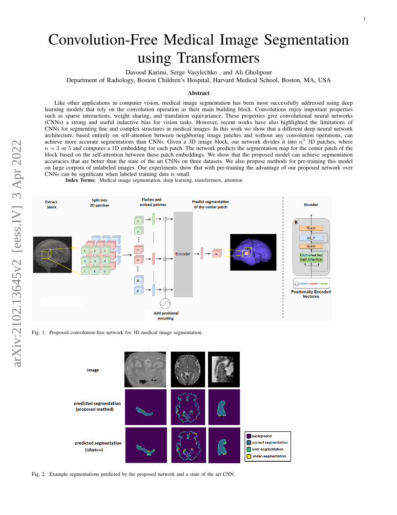
Fig. 1. Proposed convolution-free network for 3D medical image segmentation.
Fig. 2. Example segmentations predicted by the proposed network and a state of the art CNN.
arX
iv:2
102.
1364
5v2
[ee
ss.I
V]
3 A
pr 2
022
2
I. INTRODUCTION
Image segmentation is a central task in medical imageanalysis. It is routinely used for quantifying the size and shapeof the volume/organ of interest, population studies, diseasequantification, treatment planning, and computer-aided inter-vention. In most medical applications, manual segmentation byan expert is regarded as the gold standard. However, automaticmethods promise much faster, cheaper, and more reproduciblesegmentations.
Classical methods in medical image segmentation run thegamut from region growing [11] and deformable models [36]to atlas-based methods [32], Bayesian approaches [29], graphcuts [26], clustering methods [12], and more. In the past fewyears, deep learning (DL) methods have emerged as a highlycompetitive alternative and they have achieved remarkablelevels of accuracy [3] [5] [18] [20] [38] [37] [19]. It appearsthat DL methods have largely replaced the classical methodsfor medical image segmentation.
Due to their success, DL methods for medical image seg-mentation have attracted much attention from the researchcommunity. Recent surveys of the published research on thistopic can be found in [14] [31]. Some of the main directionsof these studies include improving network architectures, lossfunctions, and training strategies. Surprisingly, the one com-mon feature in all of these works is the use of the convolutionoperation as the main building block of the networks. Theproposed network architectures differ in terms of the waythe convolutional operations are arranged, but they all buildupon the same convolution operation. There have been severalattempts to use recurrent networks [10] [2] and attentionmechanisms [6] for medical image segmentation. However, allof those models still rely on the convolution operation. Somerecent studies have gone so far as to suggest that in practicemost of these architectures achieve largely similar results [17].
Similar to semantic segmentation, other computer visiontasks such as image classification and object detection havealso been most successfully addressed with convolutionalneural networks (CNNs) [30], [23]. These observations attestto the importance of the convolution operation to image under-standing and modeling. The effectiveness of the convolutionoperation can be attributed to a number of key properties,including: 1) local (sparse) connections, 2) parameter (weight)sharing, and 3) translation equivariance [25] [24]. These prop-erties are in large part inspired by the neuroscience of vision[28], and give CNNs a very strong inductive bias. In fact,a convolutional layer can be interpreted as a fully connectedlayer with an “infinitely strong prior” over its weights [13].As a result, modern deep CNNs have been able to tackle avariety of vision tasks with amazing success.
In other important applications, most prominently in naturallanguage processing, the dominant architectures were thosebased on recurrent neural networks (RNNs) [7], [16]. Thedominance of RNNs ended with the introduction of thetransformer model, which proposed to replace the recurrencewith an attention mechanism that could learn the relationshipbetween different parts of a sequence as a whole [34].
The attention mechanism has had a profound impact on the
field of natural language processing. In vision applications,on the other hand, its impact has been much more limited.A recent survey of applications of transformer networks incomputer vision can be found in [21]. The number of pixelsin typical images is much larger than the number of units ofdata (e.g., words) in natural language processing applications.This makes it impossible to apply standard attention models toimages. As a result, despite numerous efforts (e.g., [35] [15][9]) attention mechanism has not yet resulted in the type ofsea change that has occurred in natural language processing.The recently-proposed vision transformer (ViT) appears to bea major step towards adapting transformer/attention modelsfor computer vision applications [8]. The main insight inthat work is to consider image patches, rather than pixels,to be the units of information in images. ViT embeds imagepatches into a shared space and learns the relation betweenthese embeddings using self-attention modules. Given massiveamounts of training data and computational resources, ViTwas shown to surpass CNNs in image classification accu-racy. Subsequent work has shown that by using knowledgedistillation from a CNN teacher and using standard trainingdata and computational resources, transformer networks canachieve image classification accuracy levels on par with CNNs[33].
The goal of this work is to explore the potential of selfattention-based deep neural networks for 3D medical imagesegmentation. We propose a network architecture that is basedon self-attention between linear embeddings of 3D imagepatches, without any convolution operations. We train thenetwork on three medical image segmentation datasets andcompare its performance with a state of the art CNN. Thecontributions of this work are as follows:
1) We propose the first convolution-free deep neural networkfor medical image segmentation.
2) We show that our proposed network can achieve segmen-tation accuracies that are better than or at least on parwith a state of the art CNN on three different medicalimage segmentation datasets. We show that, unlike recentworks on image classification ([8] [33]), our network canbe effectively trained for 3D medical image segmentationwith datasets of ∼ 20− 200 labeled images.
3) We propose pre-training methods that can improve ournetwork’s segmentation accuracy when large corpora ofunlabeled training images are available. We show thatwhen labeled training images are fewer in number, ournetwork performs better than a state of the art CNN withpre-training.
II. MATERIALS AND METHODS
A. Proposed network
Figure 1 shows our proposed network for convolution-freemedical image segmentation. The input to the network is a 3Dblock B ∈ IRW×W×W×c, where W denotes the extent of theblock (in voxels) in each dimension and c is the number ofimage channels. The block B is partitioned into n3 contiguousnon-overlapping 3D patches {pi ∈ IRw×w×w×c}Ni=1, wherew = W/n is the side length of each patch and N = n3 denotes

3
the number of patches in the block. We choose n to be an oddnumber. In the experiments presented in this paper n is either3 or 5, corresponding to 27 or 125 patches, respectively. Thenetwork uses the image information in all N patches in Bto predict the segmentation for the center patch, as describedbelow.
Each of the N patches is flattened into a vector of sizeIRw3c and embedded into IRD using a trainable mappingE ∈ IRD×w3c. Unlike [8], we do not use any additional tokenssince that did not improve the segmentation performance ofour network in any way. The sequence of embedded patchesX0 = [Ep1; ...;EpN ] + Epos constitutes the input to ournetwork. The matrix Epos ∈ IRD×N is intended to learn apositional encoding. Without such positional information, thetransformer ignores the ordering of the input sequence becausethe attention mechanism is permutation-invariant. Therefore,in most NLP applications such embedding has been very im-portant. For image classification, on the other hand, authors of[8] found that positional encoding resulted in relatively smallimprovements in accuracy and that simple 1D raster encodingwas as good as more elaborate 2D positional encodings. Forthe experiments presented in this paper, we left the positionalencoding as a free parameter to be learned along with thenetwork parameters during training because we do not knowa priori what type of positional encoding would be useful. Wediscuss the results of experimental comparisons with differentpositional encodings below.
The encoder has K stages, each consisting of a multi-head self-attention (MSA) and a subsequent two-layer fullyconnected feed-forward network (FFN). Both the MSA andFFN modules include residual connections, ReLU activations,and layer normalization. More specifically, starting with thesequence of embedded and position-encoded patches, X0
described above, the kth stage of the encoder will performthe following operations to map Xk to Xk+1:
1) Xk goes through nh separate heads in MSA. The ith head:a) Computes the query, key, and value sequences from the
input sequence:
Qk,i = Ek,iQ Xk,Kk,i = Ek,i
K Xk, V k,i = Ek,iV Xk
where EQ, EK , Ev ∈ IRDh×D
b) Computes the self-attention matrix and then the trans-formed values:
Ak,i = Softmax(QTK)/√Dh
SAk,i = Ak,iV k,i
2) Outputs of the nh heads are stacked and reprojected backonto IRD
MSAk = Ekreproj[SAk,0; ...;SAk,nh ]T
where Ereproj ∈ IRD×Dhnh
3) The output of the MSA module is computed as:
XkMSA = MSAk +Xk
4) XkMSA goes through a two-layer FFN to obtain the output
of the kth encoder stage:
Xk+1 = XkMSA + Ek
2
(ReLU((Ek
1XkMSA + bk1)
)+ bk2
The output of the last encoder stage, XK , will pass througha final FFN layer that projects it onto the IRNnclass and thenreshaped into IRn×n×n×nclass , where nclass is the number ofclasses (for binary segmentation, nclass = 2):
Y = Softmax(EoutX
K + bout)).
Y is the predicted segmentation for the center patch of theblock.
B. Implementation and training
The network was implemented in TensorFlow 1.16 and runon an NVIDIA GeForce GTX 1080 GPU on a Linux machinewith 120 GB of memory and 16 CPU cores. We compare ourmodel with 3D UNet++ [39], which is a state of the art CNNfor medical image segmentation.
We trained the network parameters to maximize the Dicesimilarity coefficient, DSC [27], between Y and the ground-truth segmentation of the center patch using Adam [22]. Weused a batch size of 10 and a learning rate of 10−4, whichwas reduced by half if after a training epoch the validationloss did not decrease.
Pre-training: Manual segmentation of complex structuressuch as the brain cortical plate can take several hours of amedical expert’s time for a single image. Therefore, methodsthat can achieve high accuracy with fewer labeled trainingimages are highly advantageous. To improve the network’sperformance when labeled training images are insufficient, wepropose to first pre-train the network on unlabeled data fordenoising or inpainting tasks. For denoising pre-training, weadd Gaussian noise with SNR = 10 dB to the input blockB, whereas for inpainting pre-training we set the center patchof the block to constant 0. In both cases, the target is thenoise-free center patch of the block. For pre-training, we use adifferent output layer (without the softmax operation) and trainthe network to minimize the `2 norm between the predictionand the target. To fine-tune the pre-trained network for thesegmentation task, we introduce a new output layer withsoftmax and train the network on the labeled data as explainedin the above paragraph. We fine-tune the entire network, ratherthan only the output layer, on the labeled images becauseWe have found that fine-tuning the entire network for thesegmentation task leads to better results.
C. Data
Table I shows the datasets used in this work. We used ∼1/5 of the training images for validation. The final modelswere evaluated on the test images. The images were split intotrain/validation/test at random.
4
TABLE IDATASETS USED FOR EXPERIMENTS IN THIS WORK.
target organ image modality [ntrain, ntest]Brain cortical plate T2 MRI [18, 9]Pancreas CT [231, 50]Hippocampus MRI [220, 40]
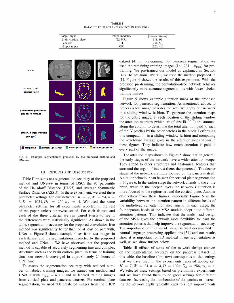
Fig. 3. Example segmentations predicted by the proposed method andUNet++.
III. RESULTS AND DISCUSSION
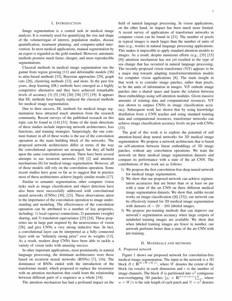
Table II presents test segmentation accuracy of the propsoedmethod and UNet++ in terms of DSC, the 95 percentileof the Hausdorff Distance (HD95) and Average SymmetricSurface Distance (ASSD). In these experiment, we used theseparameter settings for our network: K = 7,W = 24, n =3, D = 1024, Dh = 256, nh = 4. We used the sameparameter settings for all experiments reported in the restof the paper, unless otherwise stated. For each dataset andeach of the three criteria, we ran paired t-tests to see ifthe differences were statistically significant. As shown in thetable, segmentation accuracy for the proposed convolution-freemethod was significantly better than, or at least on part with,UNet++. Figure 3 shows example slices from test images ineach dataset and the segmentations predicted by the proposedmethod and UNet++. We have observed that the propsoedmethod is capable of accurately segmenting fine and complexstructures such as the brain cortical plate. In terms of trainingtime, our network converged in approximately 24 hours ofGPU time.
To assess the segmentation accuracy with reduced num-ber of labeled training images, we trained our method andUNet++ with ntrain = 5, 10, and 15 labeled training imagesfrom cortical plate and pancreas datasets. For cortical platesegmentation, we used 500 unlabeled images from the dHCP
dataset [4] for pre-training. For pancreas segmentation, weused the remaining training images (i.e., 231− ntrain) for pre-training. We pre-trained our model as explained in SectionII-B. To pre-train UNet++, we used the method propsoed in[1]. Figure 4 shows the results of this experiment. With thepropsoed pre-training, the convolution-free network achievessignificantly more accurate segmentations with fewer labeledtraining images.
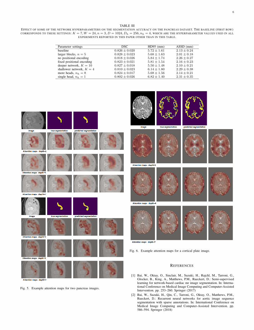
Figure 5 shows example attention maps of the proposednetwork for pancreas segmentation. As mentioned above, toprocess a test image of a desired size, we apply our networkin a sliding window fashion. To generate the attention mapsfor the entire image, at each location of the sliding windowthe attention matrices (which are of size IRN×N ) are summedalong the column to determine the total attention paid to eachof the N patches by the other patches in the block. Performingthis computation in a sliding window fashion and computingthe voxel-wise average gives us the attention maps shown inthese figures. They indicate how much attention is paid toevery part of the image.
The attention maps shown in Figure 5 show that, in general,the early stages of the network have a wider attention scope.They attend to other structures and anatomical features thatsurround the organ of interest (here, the pancreas). The deeperstages of the network are more focused on the pancreas itself.A similar behaviour can be seen for cortical plate segmentationin Figure 6. In the earlier stage the network attends to the entirebrain, while in the deeper layers the network’s attention ismore focused to the regions around the cortical plate. Anotherobservation from these figures, especially Figure 5 is thevariability between the attention pattern in different heads ofthe multi-head self-attention mechanism. In each stage, thefour separate heads of the MSA module adopt quite differentattention patterns. This indicates that the multi-head designof the MSA gives the network more flexibility to learn theattention patterns that help improve the segmentation accuracy.The importance of multi-head design is well documented innatural language processing applications [34] and our resultsshow it is important for 3D medical image segmentation aswell, as we show further below.
Table III effects of some of the network design choiceson the segmentation accuracy on the pancreas dataset. Inthis table, the baseline (first row) corresponds to the settingsthat we have used in the experiments reported above, i.e.,K = 7,W = 24, n = 3, D = 1024, Dh = 256, nh = 4.We selected these settings based on preliminary experimentsand we have found them to be good settings for differentdatasets. Increasing the number/size of the patches or increas-ing the network depth typically leads to slight improvements

5
TABLE IISEGMENTATION ACCURACY OF THE PROPOSED METHOD AND UNET++. BETTER RESULTS FOR EACH DATASET/CRITERION ARE IN BOLD TYPE.
ASTERISKS DENOTE STATISTICALLY SIGNIFICANT DIFFERENCE (p < 0.01 IN PAIRED T-TEST.)
Dataset Method DSC HD95 (mm) ASSD (mm)
Brain cortical plate Proposed 0.879± 0.026∗ 0.92± 0.04 0.24± 0.03UNet++ 0.860± 0.024 0.91± 0.04 0.25± 0.04
PancreasProposed 0.826± 0.020∗ 5.72± 1.61∗ 2.13± 0.24∗
UNet++ 0.808± 0.021 6.67± 1.80 2.45± 0.21
Hippocampus Proposed 0.881± 0.017∗ 1.10± 0.19∗ 0.40± 0.04UNet++ 0.852± 0.022 1.33± 0.26 0.41± 0.07
Fig. 4. Segmentation accuracy (in DSC) for the proposed network and UNet++ with reduced labeled training data on the cortical plate (left) and pancreas(right) datasets.
in accuracy. Furthermore, using a fixed positional encoding orno positional encoding slightly reduces segmentation accuracycompared with free-parameter/learnable positional encoding.Finally, using a single-head attention significantly reduces thesegmentation accuracy, which indicates the importance of themulti-head design to enable the network to learn a morecomplex relation between neighboring patches.
IV. CONCLUSIONS
The convolution operation has a strong basis in the structureof the mammalian primary visual cortex and it is well suitedfor developing powerful techniques for image modeling andimage understanding. In recent years, CNNs have been shownto be highly effective in tackling various computer visionproblems. However, there is no reason to believe that no othermodel can outperform CNNs on a specific vision task. Medicalimage analysis applications, in particular, pose specific chal-lenges such as 3D nature of the images and small number oflabeled images. In such applications, other models can be moreeffective than CNNs. In this work we presented a new modelfor 3D medical image segmentation. Unlike all recent modelsthat use convolutions as their main building block, our modelis based on self-attention between neighboring 3D patches.Our results show that the proposed network can outperforma state of the art CNN on three medical image segmentationdatasets. With pre-training for denoising and in-painting taskson unlabeled images, our network also performed better than a
CNN when only 5-15 labeled training images were available.We expect that the network proposed in this paper should beeffective for other tasks in medical image analysis such asanomaly detection and classification.
ACKNOWLEDGMENT
This project was supported in part by the National Institute ofBiomedical Imaging and Bioengineering and the National In-stitute of Neurological Disorders and Stroke of the National In-stitutes of Health (NIH) under award numbers R01EB031849,R01NS106030, and R01EB032366; in part by the Office ofthe Director of the NIH under award number S10OD0250111;in part by the National Science Foundation (NSF) underaward 2123061; and in part by a Technological Innovationsin Neuroscience Award from the McKnight Foundation. Thecontent of this paper is solely the responsibility of the authorsand does not necessarily represent the official views of theNIH, NSF, or the McKnight Foundation.
The DHCP dataset is provided by the developing Hu-man Connectome Project, KCL-Imperial-Oxford Consortiumfunded by the European Research Council under the EuropeanUnion Seventh Framework Programme (FP/2007-2013) / ERCGrant Agreement no. [319456]. We are grateful to the familieswho generously supported this trial.
6
TABLE IIIEFFECT OF SOME OF THE NETWORK HYPERPARAMETERS ON THE SEGMENTATION ACCURACY ON THE PANCREAS DATASET. THE BASELINE (FIRST ROW)CORRESPONDS TO THESE SETTINGS: K = 7,W = 24, n = 3, D = 1024, Dh = 256, nh = 4, WHICH ARE THE HYPERPARAMETER VALUES USED IN ALL
EXPERIMENTS REPORTED IN THIS PAPER OTHER THAN IN THIS TABLE.
Parameter settings DSC HD95 (mm) ASSD (mm)baseline 0.826± 0.020 5.72± 1.61 2.13± 0.24larger blocks, n = 5 0.828± 0.023 5.68± 1.63 2.01± 0.18no positional encoding 0.818± 0.026 5.84± 1.74 2.26± 0.27fixed positional encoding 0.823± 0.021 5.81± 1.54 2.16± 0.23deeper network, K = 10 0.827± 0.018 5.50± 1.48 2.10± 0.21shallower network, K = 4 0.810± 0.023 6.14± 1.80 2.29± 0.38more heads, nh = 8 0.824± 0.017 5.68± 1.56 2.14± 0.21single head, nh = 1 0.802± 0.026 6.82± 1.40 2.31± 0.35
Fig. 5. Example attention maps for two pancreas images.
Fig. 6. Example attention maps for a cortical plate image.
REFERENCES
[1] Bai, W., Oktay, O., Sinclair, M., Suzuki, H., Rajchl, M., Tarroni, G.,Glocker, B., King, A., Matthews, P.M., Rueckert, D.: Semi-supervisedlearning for network-based cardiac mr image segmentation. In: Interna-tional Conference on Medical Image Computing and Computer-AssistedIntervention. pp. 253–260. Springer (2017)
[2] Bai, W., Suzuki, H., Qin, C., Tarroni, G., Oktay, O., Matthews, P.M.,Rueckert, D.: Recurrent neural networks for aortic image sequencesegmentation with sparse annotations. In: International Conference onMedical Image Computing and Computer-Assisted Intervention. pp.586–594. Springer (2018)
7
[3] Bakas, S., Reyes, M., Jakab, A., Bauer, S., Rempfler, M., Crimi, A.,Shinohara, R.T., Berger, C., Ha, S.M., Rozycki, M., et al.: Identifyingthe best machine learning algorithms for brain tumor segmentation,progression assessment, and overall survival prediction in the bratschallenge. arXiv preprint arXiv:1811.02629 (2018)
[4] Bastiani, M., et al.: Automated processing pipeline for neonatal diffusionmri in the developing human connectome project. NeuroImage 185, 750–763 (2019)
[5] Bernard, O., Lalande, A., Zotti, C., Cervenansky, F., Yang, X., Heng,P.A., Cetin, I., Lekadir, K., Camara, O., Ballester, M.A.G., et al.:Deep learning techniques for automatic mri cardiac multi-structuressegmentation and diagnosis: is the problem solved? IEEE transactionson medical imaging 37(11), 2514–2525 (2018)
[6] Chen, J., Lu, Y., Yu, Q., Luo, X., Adeli, E., Wang, Y., Lu, L., Yuille,A.L., Zhou, Y.: Transunet: Transformers make strong encoders formedical image segmentation. arXiv preprint arXiv:2102.04306 (2021)
[7] Chung, J., Gulcehre, C., Cho, K., Bengio, Y.: Empirical evaluation ofgated recurrent neural networks on sequence modeling. arXiv preprintarXiv:1412.3555 (2014)
[8] Dosovitskiy, A., Beyer, L., Kolesnikov, A., Weissenborn, D., Zhai,X., Unterthiner, T., Dehghani, M., Minderer, M., Heigold, G., Gelly,S., et al.: An image is worth 16x16 words: Transformers for imagerecognition at scale. arXiv preprint arXiv:2010.11929 (2020)
[9] Dou, H., Karimi, D., Rollins, C.K., Ortinau, C.M., Vasung, L., Velasco-Annis, C., Ouaalam, A., Yang, X., Ni, D., Gholipour, A.: A deepattentive convolutional neural network for automatic cortical plate seg-mentation in fetal mri. arXiv preprint arXiv:2004.12847 (2020)
[10] Gao, Y., Phillips, J.M., Zheng, Y., Min, R., Fletcher, P.T., Gerig,G.: Fully convolutional structured lstm networks for joint 4d med-ical image segmentation. In: 2018 IEEE 15th International Sympo-sium on Biomedical Imaging (ISBI 2018). pp. 1104–1108 (2018).https://doi.org/10.1109/ISBI.2018.8363764
[11] Gibbs, P., Buckley, D.L., Blackband, S.J., Horsman, A.: Tumour volumedetermination from mr images by morphological segmentation. Physicsin Medicine & Biology 41(11), 2437 (1996)
[12] Goldszal, A.F., Davatzikos, C., Pham, D.L., Yan, M.X., Bryan, R.N.,Resnick, S.M.: An image-processing system for qualitative and quanti-tative volumetric analysis of brain images. Journal of computer assistedtomography 22(5), 827–837 (1998)
[13] Goodfellow, I., Bengio, Y., Courville, A., Bengio, Y.: Deep learning,vol. 1. MIT press Cambridge (2016)
[14] Hesamian, M.H., Jia, W., He, X., Kennedy, P.: Deep learning techniquesfor medical image segmentation: achievements and challenges. Journalof digital imaging 32(4), 582–596 (2019)
[15] Ho, J., Kalchbrenner, N., Weissenborn, D., Salimans, T.: Axial atten-tion in multidimensional transformers. arXiv preprint arXiv:1912.12180(2019)
[16] Hochreiter, S., Schmidhuber, J.: Long short-term memory. Neural com-putation 9(8), 1735–1780 (1997)
[17] Isensee, F., Kickingereder, P., Wick, W., Bendszus, M., Maier-Hein,K.H.: No new-net. In: International MICCAI Brainlesion Workshop. pp.234–244. Springer (2018)
[18] Kamnitsas, K., Ledig, C., Newcombe, V.F., Simpson, J.P., Kane, A.D.,Menon, D.K., Rueckert, D., Glocker, B.: Efficient multi-scale 3d cnnwith fully connected crf for accurate brain lesion segmentation. Medicalimage analysis 36, 61–78 (2017)
[19] Karimi, D., Samei, G., Kesch, C., Nir, G., Salcudean, S.E.: Prostatesegmentation in mri using a convolutional neural network architectureand training strategy based on statistical shape models. InternationalJournal of Computer Assisted Radiology and Surgery 13(8), 1211–1219(Aug 2018). https://doi.org/10.1007/s11548-018-1785-8, https://doi.org/10.1007/s11548-018-1785-8
[20] Karimi, D., et al.: Accurate and robust deep learning-basedsegmentation of the prostate clinical target volume in ultra-sound images. Medical Image Analysis 57, 186 – 196 (2019).https://doi.org/https://doi.org/10.1016/j.media.2019.07.005, http://www.sciencedirect.com/science/article/pii/S1361841519300623
[21] Khan, S., Naseer, M., Hayat, M., Zamir, S.W., Khan, F.S., Shah, M.:Transformers in vision: A survey. arXiv preprint arXiv:2101.01169(2021)
[22] Kingma, D.P., Ba, J.: Adam: A method for stochastic optimization. In:Proceedings of the 3rd International Conference on Learning Represen-tations (ICLR) (2014)
[23] Krizhevsky, A., Sutskever, I., Hinton, G.E.: Imagenet classification withdeep convolutional neural networks. In: Advances in neural informationprocessing systems. pp. 1097–1105 (2012)
[24] Le Cun, Y., Boser, B., Denker, J.S., Henderson, D., Howard, R.E.,Hubbard, W., Jackel, L.D.: Handwritten digit recognition with a back-propagation network. In: Proceedings of the 2nd International Confer-ence on Neural Information Processing Systems. pp. 396–404 (1989)
[25] LeCun, Y., Bengio, Y., Hinton, G.: Deep learning. nature 521(7553),436 (2015)
[26] Mahapatra, D., Buhmann, J.M.: Prostate mri segmentation us-ing learned semantic knowledge and graph cuts. IEEE Transac-tions on Biomedical Engineering 61(3), 756–764 (March 2014).https://doi.org/10.1109/TBME.2013.2289306
[27] Milletari, F., Navab, N., Ahmadi, S.A.: V-net: Fully convolutional neuralnetworks for volumetric medical image segmentation. In: 3D Vision(3DV), 2016 Fourth International Conference on. pp. 565–571. IEEE(2016)
[28] Olshausen, B.A., Field, D.J.: Emergence of simple-cell receptive fieldproperties by learning a sparse code for natural images. Nature381(6583), 607–609 (1996)
[29] Prince, J.L., Pham, D., Tan, Q.: Optimization of mr pulse sequencesfor bayesian image segmentation. Medical Physics 22(10), 1651–1656(1995)
[30] Ren, S., He, K., Girshick, R., Sun, J.: Faster r-cnn: Towards real-timeobject detection with region proposal networks. In: Advances in neuralinformation processing systems. pp. 91–99 (2015)
[31] Taghanaki, S.A., Abhishek, K., Cohen, J.P., Cohen-Adad, J., Hamarneh,G.: Deep semantic segmentation of natural and medical images: Areview. Artificial Intelligence Review pp. 1–42 (2020)
[32] Thompson, P.M., Toga, A.W.: Detection, visualization and animation ofabnormal anatomic structure with a deformable probabilistic brain atlasbased on random vector field transformations. Medical image analysis1(4), 271–294 (1997)
[33] Touvron, H., Cord, M., Douze, M., Massa, F., Sablayrolles, A., Jegou,H.: Training data-efficient image transformers & distillation throughattention. arXiv preprint arXiv:2012.12877 (2020)
[34] Vaswani, A., Shazeer, N., Parmar, N., Uszkoreit, J., Jones, L., Gomez,A.N., Kaiser, L., Polosukhin, I.: Attention is all you need. arXiv preprintarXiv:1706.03762 (2017)
[35] Wang, H., Zhu, Y., Green, B., Adam, H., Yuille, A., Chen, L.C.:Axial-deeplab: Stand-alone axial-attention for panoptic segmentation.In: European Conference on Computer Vision. pp. 108–126. Springer(2020)
[36] Wang, Y., Guo, Q., Zhu, Y.: Medical image segmentation based ondeformable models and its applications. In: Deformable Models, pp.209–260. Springer (2007)
[37] Zeng, Q., Karimi, D., Pang, E.H., Mohammed, S., Schneider, C.,Honarvar, M., Salcudean, S.E.: Liver segmentation in magnetic reso-nance imaging via mean shape fitting with fully convolutional neuralnetworks. In: International Conference on Medical Image Computingand Computer-Assisted Intervention. pp. 246–254. Springer (2019)
[38] Zeng, Q., Samei, G., Karimi, D., Kesch, C., Mahdavi, S.S., Abolmae-sumi, P., Salcudean, S.E.: Prostate segmentation in transrectal ultrasoundusing magnetic resonance imaging priors. International journal of com-puter assisted radiology and surgery 13(6), 749–757 (2018)
[39] Zhou, Z., Siddiquee, M.M.R., Tajbakhsh, N., Liang, J.: Unet++: A nestedu-net architecture for medical image segmentation. In: Deep learning inmedical image analysis and multimodal learning for clinical decisionsupport, pp. 3–11. Springer (2018)
Related Documents
![arXiv:1812.00020v2 [cs.CV] 28 Mar 2019 · (4-RoSy) field to define a domain for convolution on a sur-face. Though 4-RoSy fields have several properties favor-able for convolution](https://static.cupdf.com/doc/110x72/600c557e21fef825644cb65e/arxiv181200020v2-cscv-28-mar-2019-4-rosy-ield-to-deine-a-domain-for-convolution.jpg)


