RESEARCH Open Access Conus vexillum venom induces oxidative stress in Ehrlich's ascites carcinoma cells: an insight into the mechanism of induction Mohamed A Abdel-Rahman 1* , Ismail M Abdel-Nabi 1,2 , Mohamed S El-Naggar 1 , Osama A Abbas 3 and Peter N Strong 4 Abstract Background: It is estimated that venoms of marine cone snails (genus Conus) contain more than 100,000 different small peptides with a wide range of pharmacological and biological actions. Some of these peptides were developed into potential therapeutic agents and as molecular tools to understand biological functions of nervous and cardiovascular systems. In this study we examined the cytotoxic and anticancer properties of the marine vermivorous cone snail Conus vexillum (collected from Hurgada and Sharm El-Shaikh, Red Sea, Egypt) and suggest the possible mechanisms involved. The in vitro cytotoxic effects of Conus venom were assessed against Ehrlich’s ascites carcinoma (EAC) cells. Results: Conus venom treatment resulted in concentration-dependent cytotoxicity as indicated by a lactate dehydrogenase leakage assay. Apoptotic effects were measured in vivo by measuring levels of reactive oxygen species and oxidative defense agents in albino mice injected with EAC cells. Conus venom (1.25 mg/kg) induced a significant increase (p < 0.05) in several oxidative stress biomarkers (lipid peroxidation, protein carbonyl content and reactive nitrogen intermediates) of EAC cells after 3, 6, 9 and 12 hours of venom injection. Conus venom significantly reduced (p < 0.05) the activities of oxidative defense enzymes (catalase and superoxide dismutase) as well as the total antioxidant capacity of EAC cells, as evidenced by lowered levels of reduced glutathione. Conclusions: These results demonstrate the cytotoxic potential of C. vexillum venom by inducing oxidative stress mediated mechanisms in tumor cells and suggest that the venom contains novel molecules with potential anticancer activity. Keywords: Conus vexillum venom, Ehrlich’s cells, Oxidative stress, Cancer, Egypt Background It is acknowledged that natural products are one of the major sources for drug discovery and used to treat sev- eral life-threading diseases including cancer [1]. Venoms of animal species (such as snakes, scorpions, spiders and cone snails) are a combination of unique bioactive mole- cules that display a plethora of molecular targets and functions. Some of these molecules are currently being developed as candidate or to provide a critical template for the design of others [2]. Cancer is defined as a disturbance in mechanisms involved in the control of cell growth and division [3,4]. It is well known that resistance to apoptotic signals is one of the characteristic features of cancer cells [5]. Chemotherapy is the usage of conventional antitumor drugs to treat several types of cancerous cells. While chemotherapy can be quite effective in treating advanced or metastatic diseases, it often accompanied with many harmful side-effects through inducing severe damage to normal cells and tissues [6-8]. Moreover through particular cellular and molecular changes, cancer cells eventually become not susceptible to chemotherapeutic agents. These changes include up-regulation the en- zymes of drug detoxification and drug transporters and * Correspondence: [email protected] 1 Department of Zoology, Faculty of Science, Suez Canal University, Ismailia 41522, Egypt Full list of author information is available at the end of the article © 2013 Abdel-Rahman et al.; licensee BioMed Central Ltd. This is an Open Access article distributed under the terms of the Creative Commons Attribution License (http://creativecommons.org/licenses/by/2.0), which permits unrestricted use, distribution, and reproduction in any medium, provided the original work is properly cited. Abdel-Rahman et al. Journal of Venomous Animals and Toxins including Tropical Diseases 2013, 19:10 http://www.jvat.org/content/19/1/10

Welcome message from author
This document is posted to help you gain knowledge. Please leave a comment to let me know what you think about it! Share it to your friends and learn new things together.
Transcript
Abdel-Rahman et al. Journal of Venomous Animals and Toxins including Tropical Diseases 2013, 19:10http://www.jvat.org/content/19/1/10
RESEARCH Open Access
Conus vexillum venom induces oxidative stress inEhrlich's ascites carcinoma cells: an insight intothe mechanism of inductionMohamed A Abdel-Rahman1*, Ismail M Abdel-Nabi1,2, Mohamed S El-Naggar1, Osama A Abbas3
and Peter N Strong4
Abstract
Background: It is estimated that venoms of marine cone snails (genus Conus) contain more than 100,000 differentsmall peptides with a wide range of pharmacological and biological actions. Some of these peptides weredeveloped into potential therapeutic agents and as molecular tools to understand biological functions of nervousand cardiovascular systems. In this study we examined the cytotoxic and anticancer properties of the marinevermivorous cone snail Conus vexillum (collected from Hurgada and Sharm El-Shaikh, Red Sea, Egypt) and suggestthe possible mechanisms involved. The in vitro cytotoxic effects of Conus venom were assessed against Ehrlich’sascites carcinoma (EAC) cells.
Results: Conus venom treatment resulted in concentration-dependent cytotoxicity as indicated by a lactatedehydrogenase leakage assay. Apoptotic effects were measured in vivo by measuring levels of reactive oxygenspecies and oxidative defense agents in albino mice injected with EAC cells. Conus venom (1.25 mg/kg) induced asignificant increase (p < 0.05) in several oxidative stress biomarkers (lipid peroxidation, protein carbonyl content andreactive nitrogen intermediates) of EAC cells after 3, 6, 9 and 12 hours of venom injection. Conus venomsignificantly reduced (p < 0.05) the activities of oxidative defense enzymes (catalase and superoxide dismutase) aswell as the total antioxidant capacity of EAC cells, as evidenced by lowered levels of reduced glutathione.
Conclusions: These results demonstrate the cytotoxic potential of C. vexillum venom by inducing oxidative stressmediated mechanisms in tumor cells and suggest that the venom contains novel molecules with potentialanticancer activity.
Keywords: Conus vexillum venom, Ehrlich’s cells, Oxidative stress, Cancer, Egypt
BackgroundIt is acknowledged that natural products are one of themajor sources for drug discovery and used to treat sev-eral life-threading diseases including cancer [1]. Venomsof animal species (such as snakes, scorpions, spiders andcone snails) are a combination of unique bioactive mole-cules that display a plethora of molecular targets andfunctions. Some of these molecules are currently beingdeveloped as candidate or to provide a critical templatefor the design of others [2].
* Correspondence: [email protected] of Zoology, Faculty of Science, Suez Canal University, Ismailia41522, EgyptFull list of author information is available at the end of the article
© 2013 Abdel-Rahman et al.; licensee BioMedCreative Commons Attribution License (http:/distribution, and reproduction in any medium
Cancer is defined as a disturbance in mechanismsinvolved in the control of cell growth and division [3,4].It is well known that resistance to apoptotic signals isone of the characteristic features of cancer cells [5].Chemotherapy is the usage of conventional antitumordrugs to treat several types of cancerous cells. Whilechemotherapy can be quite effective in treating advancedor metastatic diseases, it often accompanied with manyharmful side-effects through inducing severe damageto normal cells and tissues [6-8]. Moreover throughparticular cellular and molecular changes, cancer cellseventually become not susceptible to chemotherapeuticagents. These changes include up-regulation the en-zymes of drug detoxification and drug transporters and
Central Ltd. This is an Open Access article distributed under the terms of the/creativecommons.org/licenses/by/2.0), which permits unrestricted use,, provided the original work is properly cited.
Abdel-Rahman et al. Journal of Venomous Animals and Toxins including Tropical Diseases 2013, 19:10 Page 2 of 10http://www.jvat.org/content/19/1/10
alteration in the molecular targets of drugs. Also, drugtolerance may be due to increase the ability of cancercells to evade apoptosis through suppression apoptoticpathways and improve the machinery of DNA repair[9-11]. Thus, development of alternative anticancerdrugs with minimal side-effects is urgently required.The scientific literature on the antitumor activities of
snake venoms extends back many decades [12,13]. Morerecently, peptides with specific antitumor activity havebeen isolated, such as cystatin [14]. However the hightoxicity of snake venoms often halts their therapeuticproperties and in most cases, the cytotoxic doses formalignant and normal cells are the same. In addition tosnake venom, anti-cancer efficacy of bee venom hasbeen reported using both in vitro and in vivo approaches[15,16]. Bee venom has been reported to be cytotoxicand induce apoptosis in various tumour cell lines[17,18]. The promotion of apoptosis through severalcancer cell death mechanisms is essential for beevenom-induced anticancer effects (and the bee venompeptide, melittin) [19]. Scorpion venom also possessthe property of inhibiting growth of various types ofcancers [20]. Several cytotoxic antitumor peptides havebeen characterized from scorpion venom, which exerttheir action by distinct mechanisms: as an ion channelblocker, as a matrix metalloproteinase (MMP-2) inhibi-tor and as an apoptosis inducer, activating intracellularpathways [21-23].The venoms of cone snails, a family of widely distrib-
uted marine mollusks, contain a large number of smallpeptides (conotoxins and conopeptides) which haveevolved separately in approximately 700 species of spe-cific fish, mollusk and worm-hunting animals [24]. It isestimated that more than 50,000 conopeptides exist, butless than 0.1% have been functionally characterized.Those that have been examined are specific for an amaz-ingly diverse set of important pharmacological targets,including voltage gated ion channels (Na, K, Ca), neuro-transmitter receptors (acetylcholine, glutamate) andtransporters (noradrenaline) [2]. Their small size, relativeease of synthesis and structural stability also make themimportant templates for the design of novel drugs andindeed several are presently in clinical trials [25].We have recently demonstrated that crude venom of the
vermivorous snail C. vexillum caused an array of cytotoxiceffects in mammalian systems which were attributed tothe venom’s ability to induce oxidative stress [26]. Venomscollected from different locations in the Red Sea (Hurgadaand Sharm El-Shaikh) also showed clear differences invenom potency. The present study was designed to extendour earlier work, by examining the cytotoxic activity andmechanism of action of C. vexillum venom on EAC cells,both in vitro and in vivo. To the best of our knowledge,this is the first study of its kind on this Conus species.
MethodsVenom preparationSpecimens of C. vexillum were collected from two differ-ent sites (Hurgada and Sharm El-Shaikh) in the Red Sea,Egypt [26]. Venom glands were dissected as describedpreviously by Cruz et al. [27] and crude venom fromeach location was extracted and lyophilized [26]. Pooledvenoms were stored at −80°C until further use.
Experimental animals and ascites tumorAll animal procedures and experimental protocols were ap-proved by the Research Ethics Committee of Suez CanalUniversity and were carried out in accordance with theGuide for the Care and Use of Laboratory Animals (http://www.nap.edu/catalog/12910.html). Adult male Swiss albinomice weighing 20–25 g were purchased from the breedingunit of Theodor Bilharz Research Institute (Giza, Egypt).The animals were maintained under controlled conditionsof temperature, humidity and on a 12 hour-light/dark cycle,with free access to standard pellet diet and water. The firstinoculum of EAC cell line was purchased from the Depart-ment of Tumor Biology, National Cancer Institute, CairoUniversity. The cell line was inoculated by serial intraperi-toneal (IP) passages of 106 cells per mouse. Cells weregrown in the peritoneal cavity of mice and transferred everyten days to new animals. Mice were monitored daily forsigns of tumor progression, including the amount ofabdominal distension and signs of illness and distress.The volume of ascites fluid was determined by needle(18–22 gauge) aspiration. Withdrawal of ascites fluidwas performed under aseptic conditions.
Isolation of EAC cellsBased on the method described by Mookerjee et. al. [28]peritoneal fluid containing the tumor cells was with-drawn, collected in sterile petri plates and incubated at37°C for two hours. The cells of macrophage lineageadhered to the bottom of the petri dishes. Since EACcells do not adhere to synthetic surfaces in vitro, thenon-adherent cell population was gently aspirated andwashed repeatedly with phosphate buffered saline [29].More than 93% of the non-adherent cells were mor-phologically characterized as EAC cells by Wrightstaining [30] and viability was assessed by Trypan Bluedye exclusion [31].
Lactate dehydrogenase (LDH) assayEAC cells (1 × 10⁶ cells/mL) were incubated in a 96-wellplate with varying concentrations (10, 20 and 30 μg/mL)of C. vexillum venom for one hour at 37°C. LDH re-leased into the medium was determined spectrophoto-metrically (340 nm) by measuring the rate of decrease ofNAPDH, using commercial kit (Spinreact, Sant Estevede Bas, Spain). Data was expressed as units/liter (U/L).
Abdel-Rahman et al. Journal of Venomous Animals and Toxins including Tropical Diseases 2013, 19:10 Page 3 of 10http://www.jvat.org/content/19/1/10
Antitumor activity of Conus venomThe antitumor activity of C. vexillum venom from eachgeographical location was evaluated. Fifty-four mice wereinoculated with the EAC cells as previously mentioned.After successful establishment of the tumor (ten days aftertumor inoculation), mice were randomly assorted intothree groups. A Hurgada-treated group (24 animals) and aSharm El-Shaikh-treated group (24 animals) were bothinjected IP with a sublethal dose of C. vexillum venom(1.25 mg/kg) [26]. A third control group (six animals) wasinjected with 100 μL of sterile saline.Ascites fluid containing EAC cells was drawn from the
peritoneal cavity of treated and control groups after 3, 6,9 and 12 hours of venom administration. The followingbiochemical parameters were measured in the EAC cellsof treated and control groups: the level of lipid peroxi-dation (LPx) and protein carbonyl contents (PCC),glutathione (reduced form) content (GSH), the activ-ities of Cu/Zn-superoxide dismutase (Cu/Zn-SOD),catalase (CAT) and total antioxidant capacity (TAC).The level of nitric oxide (NO) was measured in thesupernatant of ascites fluid.
Malondialehyde (MDA) assayThe extent of lipid peroxidation in EAC cells was deter-mined by measuring the production of malondialdehyde,an indicator of oxidative damage and oxidative stress[31]. Malondialdehyde was determined by incubatingsamples with thiobarbituric acid and measuring reactionproducts at 532 nm [32]. 1,1,3,3-tetramethoxypropane[malonaldehyde bis(dimethyl acetal), Sigma-Aldrich,USA] was used as an external standard and the resultswere expressed as μmoles of MDA/mg.
Protein carbonyl content (PCC)PCC was quantified using dinitrophenylhydrazide(DNPH) [33]. Protein was precipitated with an equalvolume of 1% trichloroacetic acid (TCA) and the pelletwas resuspended in 1 mL of 2,4- dinitrophenylhydrazide(Sigma-Aldrich, USA), 10 mM, dissolved in 2 N HCl. Basedon the method described by Fulle et al. [34], separateblanks were prepared by adding 1 mL of 2 N HCl withoutDNPH. Samples were left at room temperature for onehour in the dark and vortexed every 15 minutes. An equalvolume of 20% TCA was added and after centrifugation(12,000 × g, 15 minutes, 4°C), pellets were washed (threetimes) with 1 mL of ethanol: ethyl acetate (1:1) to removefree DNPH and lipid contaminants. The final pellet wasdissolved in 6 M guanidine (1 mL, 1 hour, 37°C shakingwater bath). The solution was centrifuged (12,000 × g, 15minutes) and the carbonyl content (nmol/mg), measured asprotein phenylhydrazone derivatives, was determined at370 nm using an absorption coefficient of 22,000 M−1 Cm1.
Estimation of nitric oxide (NO)Nitric oxide was measured in the supernatant of ascitesfluid by a spectrophotometric method based on the Griessreaction [35]. One hundred microliters of supernatant wasmixed with an equal volume of Griess reagent (one part0.1% naphthylethylendiamine dihydrochloride in distilledwater plus one part 1% sulfanilamide in 5% concentratedH3PO4), at room temperature for ten minutes. The ab-sorbance was measured at 540 nm. Sodium nitrite wasused as a standard.
Reduced glutathione (GSH)The content of glutathione in EAC cells of control andtreated groups was estimated according to Beutler et al.[36]. Aliquots of 0.2 mL of ascites fluid were added to1.8 mL distilled water and 3 mL of precipitating solution(1.67 gm glacial metaphosphoric acid, 0.2 gm EDTA and30 gm NaCl in 100 mL distilled water) and the mixturewas centrifuged (2200 × g, 15 minutes, 4°C). To 1 mLof supernatant it was added sequentially, sodiumdihydrogen phosphate (4 mL, 0.3 M) and DTNB re-agent [0.5 mL, 40 mg 5,5` dithiobis-2-nitrobenzoicacid (Sigma-Aldrich, USA) dissolved in 100 mL 1%sodium citrate] and the absorbance was measured at412 nm. Reduced glutathione (Sigma-Aldrich, USA)was used as standard.
Cu/Zn-superoxide dismutase (Cu/Zn-SOD) and catalase(CAT)Superoxide dismutase (Cu/Zn SOD; EC 1.15.1.1) activitywas estimated in the lysate of EAC cells according toMisra and Fridovich [37]. The rate of inhibition of autooxidation was monitored at 560 nm; the amount ofenzyme required to produce 50% inhibition is defined asone unit of enzyme activity. SOD activity was expressedas units/mL. Catalase (CAT; EC 1.11.1.6) activity wasdetermined using the method of Aebi [38]. Sampleswere prepared in phosphate buffer (50 mM, pH 7) andTriton-X 100 (1%, v/v) was added to increase the ob-servable CAT activity by releasing the enzyme fromperoxisomes [39]. CAT was measured by monitoringthe decomposition of H2O2 at 240 nm. The enzymeactivity was calculated using a molar extinction coeffi-cient of 43.6 mol.
Total antioxidant capacity (TAC)The total antioxidant capacity in EAC cell lysates wasdetermined according to Koracevic et al. [40]. As inour previous work [26], the ability of antioxidantsto inhibit the H2O2-induced oxidation of 2,2’-azinobis(3-ethylbenzothiazoline-6-sulphonate; ABTS) was mea-sured spectrophotometrically by the reduction in concen-tration of the cation radical ABTS+, absorbing at 600 nmand expressed as mmol/L.
Abdel-Rahman et al. Journal of Venomous Animals and Toxins including Tropical Diseases 2013, 19:10 Page 4 of 10http://www.jvat.org/content/19/1/10
Statistical analysisSPSSW statistical software (v. 17.01 SPSS Inc., USA)was used in all data analyses [41]. Descriptive analysesincluding mean and standard error were applied to allbiochemical measurements. Differences in the effectsof Conus venom between control and treated groupswere assessed using the Student's unpaired t-test [42].One-way analysis of variance (ANOVA) followed by aDunnett post hoc test was performed to evaluate even-tual significant differences in the biochemical measure-ments between control and treated groups.
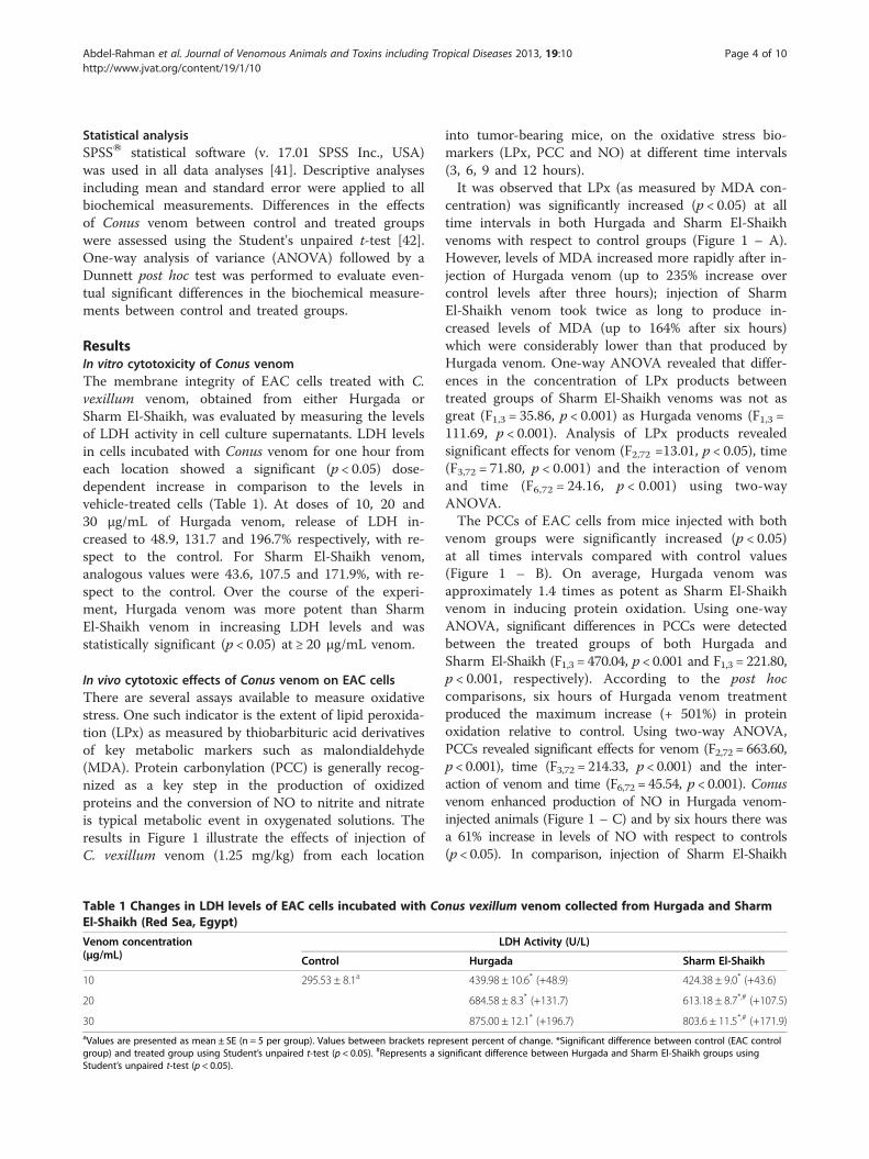
ResultsIn vitro cytotoxicity of Conus venomThe membrane integrity of EAC cells treated with C.vexillum venom, obtained from either Hurgada orSharm El-Shaikh, was evaluated by measuring the levelsof LDH activity in cell culture supernatants. LDH levelsin cells incubated with Conus venom for one hour fromeach location showed a significant (p < 0.05) dose-dependent increase in comparison to the levels invehicle-treated cells (Table 1). At doses of 10, 20 and30 μg/mL of Hurgada venom, release of LDH in-creased to 48.9, 131.7 and 196.7% respectively, with re-spect to the control. For Sharm El-Shaikh venom,analogous values were 43.6, 107.5 and 171.9%, with re-spect to the control. Over the course of the experi-ment, Hurgada venom was more potent than SharmEl-Shaikh venom in increasing LDH levels and wasstatistically significant (p < 0.05) at ≥ 20 μg/mL venom.
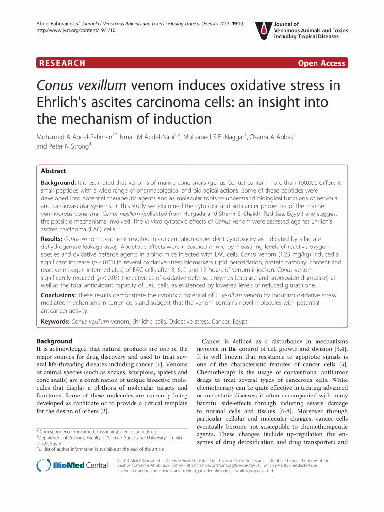
In vivo cytotoxic effects of Conus venom on EAC cellsThere are several assays available to measure oxidativestress. One such indicator is the extent of lipid peroxida-tion (LPx) as measured by thiobarbituric acid derivativesof key metabolic markers such as malondialdehyde(MDA). Protein carbonylation (PCC) is generally recog-nized as a key step in the production of oxidizedproteins and the conversion of NO to nitrite and nitrateis typical metabolic event in oxygenated solutions. Theresults in Figure 1 illustrate the effects of injection ofC. vexillum venom (1.25 mg/kg) from each location
Table 1 Changes in LDH levels of EAC cells incubated with CoEl-Shaikh (Red Sea, Egypt)
Venom concentration(μg/mL) Control
10 295.53 ± 8.1a
20
30aValues are presented as mean ± SE (n = 5 per group). Values between brackets repgroup) and treated group using Student’s unpaired t-test (p < 0.05). #Represents a sStudent’s unpaired t-test (p < 0.05).
into tumor-bearing mice, on the oxidative stress bio-markers (LPx, PCC and NO) at different time intervals(3, 6, 9 and 12 hours).It was observed that LPx (as measured by MDA con-
centration) was significantly increased (p < 0.05) at alltime intervals in both Hurgada and Sharm El-Shaikhvenoms with respect to control groups (Figure 1 – A).However, levels of MDA increased more rapidly after in-jection of Hurgada venom (up to 235% increase overcontrol levels after three hours); injection of SharmEl-Shaikh venom took twice as long to produce in-creased levels of MDA (up to 164% after six hours)which were considerably lower than that produced byHurgada venom. One-way ANOVA revealed that differ-ences in the concentration of LPx products betweentreated groups of Sharm El-Shaikh venoms was not asgreat (F1,3 = 35.86, p < 0.001) as Hurgada venoms (F1,3 =111.69, p < 0.001). Analysis of LPx products revealedsignificant effects for venom (F2,72 =13.01, p < 0.05), time(F3,72 = 71.80, p < 0.001) and the interaction of venomand time (F6,72 = 24.16, p < 0.001) using two-wayANOVA.The PCCs of EAC cells from mice injected with both
venom groups were significantly increased (p < 0.05)at all times intervals compared with control values(Figure 1 – B). On average, Hurgada venom wasapproximately 1.4 times as potent as Sharm El-Shaikhvenom in inducing protein oxidation. Using one-wayANOVA, significant differences in PCCs were detectedbetween the treated groups of both Hurgada andSharm El-Shaikh (F1,3 = 470.04, p < 0.001 and F1,3 = 221.80,p < 0.001, respectively). According to the post hoccomparisons, six hours of Hurgada venom treatmentproduced the maximum increase (+ 501%) in proteinoxidation relative to control. Using two-way ANOVA,PCCs revealed significant effects for venom (F2,72 = 663.60,p < 0.001), time (F3,72 = 214.33, p < 0.001) and the inter-action of venom and time (F6,72 = 45.54, p < 0.001). Conusvenom enhanced production of NO in Hurgada venom-injected animals (Figure 1 – C) and by six hours there wasa 61% increase in levels of NO with respect to controls(p < 0.05). In comparison, injection of Sharm El-Shaikh
nus vexillum venom collected from Hurgada and Sharm
LDH Activity (U/L)
Hurgada Sharm El-Shaikh
439.98 ± 10.6* (+48.9) 424.38 ± 9.0* (+43.6)
684.58 ± 8.3* (+131.7) 613.18 ± 8.7*,# (+107.5)
875.00 ± 12.1* (+196.7) 803.6 ± 11.5*,# (+171.9)
resent percent of change. *Significant difference between control (EAC controlignificant difference between Hurgada and Sharm El-Shaikh groups using
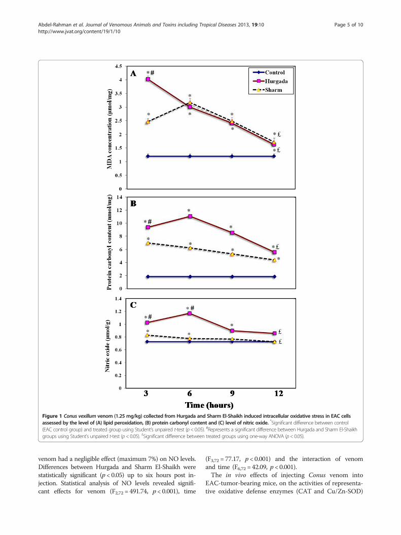
Figure 1 Conus vexillum venom (1.25 mg/kg) collected from Hurgada and Sharm El-Shaikh induced intracellular oxidative stress in EAC cellsassessed by the level of (A) lipid peroxidation, (B) protein carbonyl content and (C) level of nitric oxide. *Significant difference between control(EAC control group) and treated group using Student’s unpaired t-test (p< 0.05). #Represents a significant difference between Hurgada and Sharm El-Shaikhgroups using Student’s unpaired t-test (p< 0.05). £Significant difference between treated groups using one-way ANOVA (p< 0.05).
Abdel-Rahman et al. Journal of Venomous Animals and Toxins including Tropical Diseases 2013, 19:10 Page 5 of 10http://www.jvat.org/content/19/1/10
venom had a negligible effect (maximum 7%) on NO levels.Differences between Hurgada and Sharm El-Shaikh werestatistically significant (p < 0.05) up to six hours post in-jection. Statistical analysis of NO levels revealed signifi-cant effects for venom (F2,72 = 491.74, p < 0.001), time
(F3,72 = 77.17, p < 0.001) and the interaction of venomand time (F6,72 = 42.09, p < 0.001).The in vivo effects of injecting Conus venom into
EAC-tumor-bearing mice, on the activities of representa-tive oxidative defense enzymes (CAT and Cu/Zn-SOD)
Abdel-Rahman et al. Journal of Venomous Animals and Toxins including Tropical Diseases 2013, 19:10 Page 6 of 10http://www.jvat.org/content/19/1/10
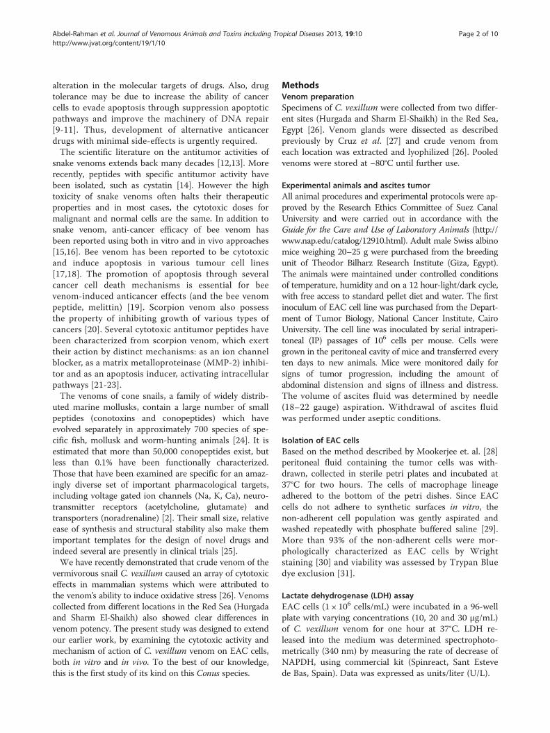
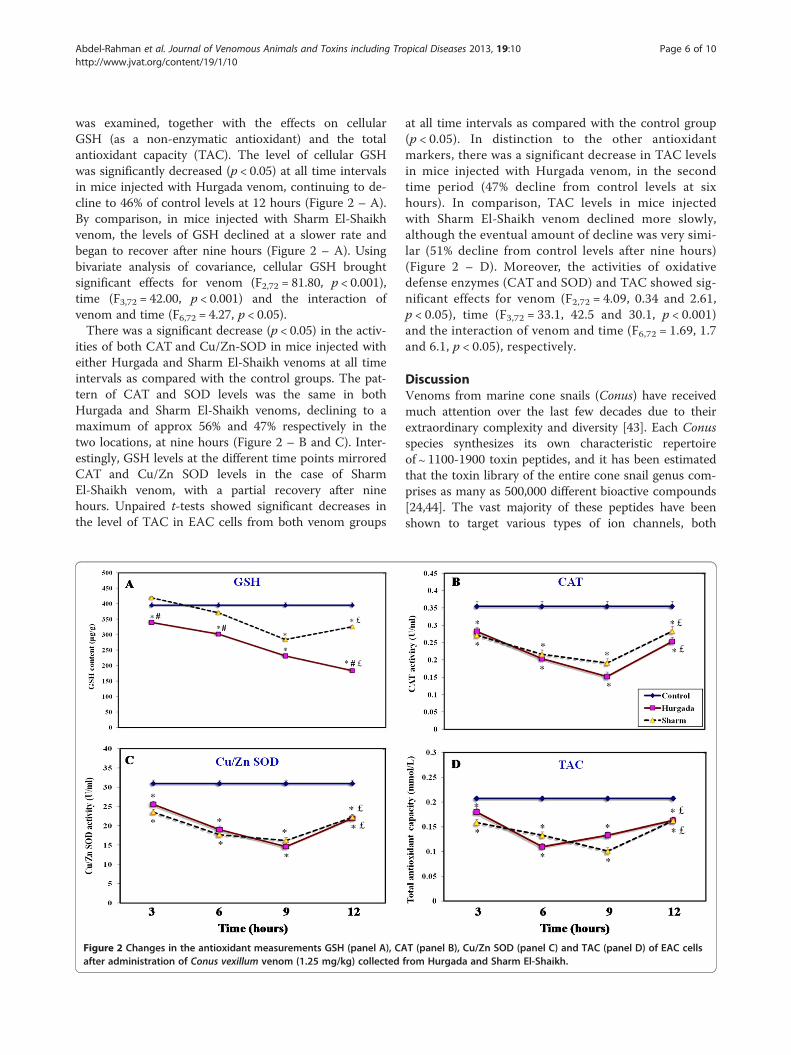
was examined, together with the effects on cellularGSH (as a non-enzymatic antioxidant) and the totalantioxidant capacity (TAC). The level of cellular GSHwas significantly decreased (p < 0.05) at all time intervalsin mice injected with Hurgada venom, continuing to de-cline to 46% of control levels at 12 hours (Figure 2 – A).By comparison, in mice injected with Sharm El-Shaikhvenom, the levels of GSH declined at a slower rate andbegan to recover after nine hours (Figure 2 – A). Usingbivariate analysis of covariance, cellular GSH broughtsignificant effects for venom (F2,72 = 81.80, p < 0.001),time (F3,72 = 42.00, p < 0.001) and the interaction ofvenom and time (F6,72 = 4.27, p < 0.05).There was a significant decrease (p < 0.05) in the activ-
ities of both CAT and Cu/Zn-SOD in mice injected witheither Hurgada and Sharm El-Shaikh venoms at all timeintervals as compared with the control groups. The pat-tern of CAT and SOD levels was the same in bothHurgada and Sharm El-Shaikh venoms, declining to amaximum of approx 56% and 47% respectively in thetwo locations, at nine hours (Figure 2 – B and C). Inter-estingly, GSH levels at the different time points mirroredCAT and Cu/Zn SOD levels in the case of SharmEl-Shaikh venom, with a partial recovery after ninehours. Unpaired t-tests showed significant decreases inthe level of TAC in EAC cells from both venom groups
Figure 2 Changes in the antioxidant measurements GSH (panel A), CAafter administration of Conus vexillum venom (1.25 mg/kg) collected
at all time intervals as compared with the control group(p < 0.05). In distinction to the other antioxidantmarkers, there was a significant decrease in TAC levelsin mice injected with Hurgada venom, in the secondtime period (47% decline from control levels at sixhours). In comparison, TAC levels in mice injectedwith Sharm El-Shaikh venom declined more slowly,although the eventual amount of decline was very simi-lar (51% decline from control levels after nine hours)(Figure 2 – D). Moreover, the activities of oxidativedefense enzymes (CAT and SOD) and TAC showed sig-nificant effects for venom (F2,72 = 4.09, 0.34 and 2.61,p < 0.05), time (F3,72 = 33.1, 42.5 and 30.1, p < 0.001)and the interaction of venom and time (F6,72 = 1.69, 1.7and 6.1, p < 0.05), respectively.
DiscussionVenoms from marine cone snails (Conus) have receivedmuch attention over the last few decades due to theirextraordinary complexity and diversity [43]. Each Conusspecies synthesizes its own characteristic repertoireof ∼ 1100-1900 toxin peptides, and it has been estimatedthat the toxin library of the entire cone snail genus com-prises as many as 500,000 different bioactive compounds[24,44]. The vast majority of these peptides have beenshown to target various types of ion channels, both
T (panel B), Cu/Zn SOD (panel C) and TAC (panel D) of EAC cellsfrom Hurgada and Sharm El-Shaikh.
Abdel-Rahman et al. Journal of Venomous Animals and Toxins including Tropical Diseases 2013, 19:10 Page 7 of 10http://www.jvat.org/content/19/1/10
ligand-gated and voltage-gated [45]. In comparison,there are only a few reports describing the cytotoxic ac-tions of Conus venoms on eukaryotic cells [26,46,47].The first cytolytic peptide (conolysin-Mt) was iso-
lated from the vermivorous cone snail venom Conusmustelinus [47]. The characterization of conolysinexpanded the known repertoire of conopeptide mecha-nisms to include membrane perturbation. Cytolyticpeptides, defined by their ability to partially or com-pletely destroy cell membranes, are among the largestgroup of toxins produced by living organisms, whichinclude bacteria, viruses, insects, scorpions, spiders,reptiles, and marine invertebrates [48]. By targetingthe lipid bilayer of the cell membrane, cytolytic pep-tides can affect a wide range of biological processes. Inour study, an attempt has been made to elucidate thecytotoxic potential of Conus venom (collected fromHurgada and Sharm El-Shaikh) and its underlyingmechanism of action, using EAC cells.The cytotoxic potential of Conus venom from these
two geographical sites has been evaluated in vitro againstEAC cells by measuring LDH activity. It is well docu-mented that the in vitro release of LDH provides anaccurate measure of cell membrane integrity and cellviability. This assay is based on the release of the cyto-solic enzyme LDH from cells, which catalyses theconversion of lactate to pyruvate and cannot be detectedextracellularly unless cell damage has occurred. TheLDH levels in EAC cells incubated with Conus venomfor one hour showed a significant (p < 0.01) dose-dependent increase in comparison to the levels invehicle-treated cells. The release of LDH indicates aloss of cell membrane integrity and is therefore an in-direct method to assess the venom-induced cytotox-icity [20,49]. Abdel-Rahman et al. [26] attributed thedirect cytotoxic effects of C. vexillum on mammaliancells to the presence of proteolytic enzymes in thecrude venom. Proteolytic enzymes in Conus venom areprimarily responsible for venom-induced necroticactivity and could be injected by cone snails to elicitproteolytic degradation of the extracellular matrix inthe prey [46]. Indeed, Cathepsin D and kallikrein-likeproteins have recently been identified in the venomglands of C. victoriae and C. novaehollandiae, respect-ively [43]. Cathepsin D (aspartyl protease) and kallikrein(trypsin and serine proteases) have also been found inother animal venoms [50-52].Interestingly, our toxicological data revealed significant
differences in the efficacy of C. vexillum venom collectedfrom the two Egyptian locations, Hurgada and SharmEl-Shaikh. The data showed that the venom obtainedfrom Hurgada was more potent than that obtained fromSharm El-Shaikh. We have attributed the difference invenom potency to variation in the expression of peptides
from C. vexillum collected from these two locations[26]. For example, the presence of the m/z peptide2924.7 (calcium channel blocker) in the venom of SharmEl-Shaikh may explain why venom-induced oxidativestress was significantly higher in the case of Hurgadavenom. The presence of this calcium channel blocker inSharm El-Shaikh venom delays rises in intracellularcalcium concentrations in venom-treated cells and henceslows the elevation of oxidative stress markers incomparison with Hurgada venom [26].It is well documented that oxidative stress is extremely
deleterious to cells and reactive oxygen species (ROS)are engaged in the etiology and progression of severaldiseases including cancer. Several environmental oxi-dants such as heat shock, UV irradiation, infections, andtoxins [26,53,54] can induce oxidative stress that shiftingthe cellular redox status to a more oxidized state. In nor-mal conditions, enzymatic and non-enzymatic antioxi-dants are capable of neutralizing harmful effects of ROSand protect cellular components from damage. However,under conditions of excessive oxidative stress, ROS candamage cellular components and interfere with criticalcellular activities [55]. Numerous studies have providedevidence that ROS are directly involved in oxidativedamage with cellular macromolecules such as lipids,proteins and nucleic acids, leading to cell death [56].Cancer cells have active protective mechanisms to pre-vent lipid peroxidation. It has been demonstrated thatthe presence of relatively low levels of the NADPH-cytochrome-P450 electron transport chain may explainthe remarkable decrease of lipid peroxidation in cancercells when compared to normal cells [57].In order to examine Conus venom induced oxida-
tive stress, the changes of intracellular ROS of EACcells were measured. Conus venom administration(1.25 mg/kg) to tumor-bearing albino mice inducedsignificant increases in the oxidative stress biomarkers(MDA, PCC) of the propagated EAC cells. We haverecently suggested possible mechanisms by which thevenom of C. vexillum induces cellular oxidative dam-age in murine cells [26]. Damage could be attributedto phospholipase A2 activity – for example an enzymeanalogous to conodipine-M – on cell membranes, causingthe release of arachidonic acid [58]. Arachidonic acid canpotentiate cell damage by converting apoptosis to necrosisthrough lipid peroxidation and the promotion of DNAfragmentation [59].NO may also play a prominent role in cytotoxicity in-
duced by Conus venom. Our results showed that Conusvenom significantly increased the production of NO inthe ascites fluid of treated animals, especially in Hurgadavenom. The elevation of NO levels may be due to theactivation of macrophages present in the ascites fluid. Ithas been found that macrophages play an important role
Abdel-Rahman et al. Journal of Venomous Animals and Toxins including Tropical Diseases 2013, 19:10 Page 8 of 10http://www.jvat.org/content/19/1/10
against neoplastic cells [60]. Activated macrophagesinitiate cell death programs through the release ofsoluble immune mediators [61]. One mechanism thatmacrophages use to exert their cytolytic effects on targettumors is by the release of NO through activation ofNADPH-dependent NO synthase [62,63]. NO can causegrowth arrest and apoptosis because of its high reactivitywith iron-and thiol-containing macromolecules, therebyinhibiting enzymes of the TCA cycle and those involvedin mitochondrial respiration, as well as DNA synthesisand repair [64,65]. It can be noted that our data revealedthat increases in NO levels paralleled a significantincrease in the level of PCC, which is an indication ofprotein damage of EAC cells.Several previous studies have clarified the influence
of ROS overproduction on intracellular Ca2+ levels.Chandra et al. [66] found that increases in the concen-trations of intracellular ROS were a sign of the onset ofapoptotic processes; they cited ROS production as acritical determinant of the toxicity associated withexposure to chemotherapeutic drugs. It has beenshown that oxidative stress increases intracellular Ca2+
concentrations, leading to activation of endonucleaseswhich degrade DNA and, ultimately, contribute to celldamage [67]. Orrenius et al. [68] found that free rad-ical overproduction may inhibit Ca2+ ATPases and thisleads to altered regulation of Ca2+ levels and cell death.Ermak et al. [53] clearly demonstrated the influence ofrising intracellular Ca2+ concentration induced by oxi-dative stress. They found that oxidative stress increasesthe process of Ca2+ diffusion from both the endoplas-mic reticulum and extracellular environment into thecytoplasm. Consequently, the elevation of cytoplasmicCa2+ concentrations causes Ca2+ influx into both mito-chondria and nuclei. High concentration of Ca2+ inmitochondria leads to cell damage through disruptionnormal cellular metabolism. In nuclei, high Ca2+ levelsmodulate transcription factors and nucleases thatregulate cell apoptosis. Moreover, the enzymes (e.g.nitric oxide synthase) that produce free radicals can beactivated by the elevation of Ca2+ level [69]. Therefore,by increasing cytoplasmic Ca2+ concentrations, oxidantscan also indirectly cause more oxidant production andfurther raise Ca2+ levels.In the present work, increased levels of cellular oxi-
dative stress in EAC cells treated with Conus venomwere also accompanied by a remarkable decrease inthe levels of antioxidants in treated cells. The resultsclearly demonstrated that Conus venom significantlyreduced the content of GSH of tumor cells. Modifica-tions of GSH metabolism have been postulated as be-ing useful in cancer therapy [70]. Indeed, theelevation of intracellular GSH content has been asso-ciated with mitogenic stimulation [71,72]. GSH has
been suggested as a potential regulator of protein syn-thesis, DNA synthesis and cell proliferation [72,73].Several previous in vivo and in vitro studies have re-vealed that the amino acid precursors (glutamine andmethionine) for GSH synthesis play a crucial role incancer metabolism. Tumour cells use the amino acidglutamine as a main respiratory fuel [74] and methio-nine in cell growth [75]. Moreover, our data showedthat the depletion of GSH content was concomitantwith a reduction in the activity of enzymatic antioxi-dants (CAT and SOD) as well as a reduction in thelevel of TAC. The observed reduction in the level ofantioxidants of tumor cells from venom-treated ani-mals may explain the role of oxidative stress in celldamage. It is well established that antioxidant defenseenzymes such as SOD and CAT play a crucial role inmaintaining cellular homeostasis by detoxifying thegenerated ROS such as superoxide radicals and H2O2
[76]. Our findings are corroborate those of Sun et al.[77], who reported that the inhibition of cell growthand apoptotic activity of the snake venom fractionOHAP-1 (Okinawa Habu apoxin protein-1) was re-versed by the addition of GSH and catalase to rat andhuman malignant glioma cell lines.
ConclusionsConus venoms have provided extensive libraries of toxinsactive against a diverse range of ion channel proteins,belonging to both voltage-gated and neurotransmitter-activated gene families. The recognition that cone snailsalso have cytolytic peptides in their venom arsenalsshould not be surprising, given that similarly actingpeptides in snake venom have been known for thirtyyears but it was only a few years ago that the firstConus venom cytolytic peptide, conolysin MT wasdiscovered [47]. Our present study aims at contribut-ing to the study of the mechanism of action of thesecytolytic toxins from Cone snails that interact withand disrupt cell membranes. Our data indicate that C.vexillum venom contains components which induceoxidative stress mechanisms in Erhlich’s ascitescarcninoma cells and suggest that the venom containsnovel molecules with potential anticancer activity.
Ethics committee approvalThe present study was approved by the Research EthicsCommittee of Suez Canal University and was carried outin accordance with the Guide for the Care and Use ofLaboratory Animals (by the Committee for the Updateof the Guide for the Care and Use of LaboratoryAnimals of the National Research Council, Washington,USA, 2011).
Abdel-Rahman et al. Journal of Venomous Animals and Toxins including Tropical Diseases 2013, 19:10 Page 9 of 10http://www.jvat.org/content/19/1/10
Competing interestsThe authors declare no conflicts of interest.
Authors’ contributionsMAA designed and supervised the entire project, participated in analyzingthe data and wrote the initial draft of the manuscript. IMA reviewed themanuscript. MSE carried out biochemical experiments and participated inanalyzing the data. OAA reviewed the manuscript. PNS reviewed themanuscript and participated in its coordination. All authors read andapproved the final manuscript.
AcknowledgmentsWe would like to thank The Red Sea Protectorate Management Unit (RSPMU)in Hurgada and Sharm El-Shaikh for giving us the logistical support andpermission to collect Conus species from these locations.
Financial sourceThis project has been financially supported by the Sector of Post-graduateStudies and Researches Affairs, Suez Canal University (Ismailia, Egypt).
Author details1Department of Zoology, Faculty of Science, Suez Canal University, Ismailia41522, Egypt. 2Department of Biological Sciences, Faculty of Science, TaibahUniversity, Madinah, KSA. 3Department of Zoology, Faculty of Sciences, PortSaid University, Port Said, Egypt. 4Biomedical Research Center, BiosciencesDivision, Sheffield Hallam University, Sheffield, UK.
Received: 29 June 2012 Accepted: 14 August 2012Published: 1 May 2013
References1. Mann J: Natural products in cancer chemotherapy: past, present and
future. Nat Rev Cancer 2002, 2(2):143–148.2. Lewis RJ, Garcia ML: Therapeutic potential of venom peptides. Nat Rev
Drug Discov 2003, 2(10):790–802.3. Renan MJ: How many mutations are required for tumorigenesis?
Implications from human cancer data. Mol Carcinog 1993, 7(3):139–146.4. Wang Z: Roles of K+ channels in regulating tumour cell proliferation and
apoptosis. Pflugers Arch 2004, 448(3):274–286.5. Hanahan D, Weinberg RA: The hallmarks of cancer. Cell 2000, 100(1):57–70.6. Espinosa E, Zamora P, Feliu J, Barón MG: Classification of anticancer drugs – a
new system based on therapeutic targets. Cancer Treat Rev 2003, 29(6):515–523.7. Cassidy J, Misset JL: Oxaliplatin-related side-effects: characteristics and
management. Semin Oncol 2002, 29(5 Suppl 15):11–20.8. Kalyanaraman B, Joseph J, Kalivendi S, Wang S, Konorev E, Kotamraju S:
Doxorubicin-induced apoptosis: implications in cardiotoxicity. Mol CellBiochem 2002, 234–235(1–4):119–124.
9. Naumov GN, Towson JL, MacDonald IC, Wilson SM, Bramwell VH, GroomAC, et al: Ineffectiveness of doxorubicin treatment on solitary dormantmammary carcinoma cells or late-developing metastasis. Breast CancerRes Treat 2003, 82(3):199–206.
10. Benjamin CW, Hiebsch RR, Jones DA: Caspase activation in MCF7 cellsresponding to etoposide treatment. Mol Pharmacol 1998, 53(3):446–450.
11. Gatti L, Zunino F: Overview of tumor cell chemoresistance mechanisms.Methods Mol Med 2005, 111:127–148.
12. Abu-Sinna G, Esmat AY, Al-Zahaby AS, Soliman NA, Ibrahim TM:Fractionation and characterization of Cerastes cerastes cerastes snakevenom and the antitumor action of its lethal and non-lethal fractions.Toxicon 2003, 42(2):207–215.
13. El-Refael MF, Sarkar NH: Snake venom inhibits the growth of mousemammary tumor cells in vitro and in vivo. Toxicon 2009, 54(1):33–41.
14. Xie Q, Tang N, Wan R, Qi Y, Lin X, Lin J: Recombinant snake venomcystatin inhibits the growth, invasion and metastasis of B16F10 cells andMHCC97H cells in vitro and in vivo. Toxicon 2011, 57(5):704–711.
15. Orsolić N, Sver L, Verstovsek S, Terzić S, Basić I: Inhibition of mammarycarcinoma cell proliferation in vitro and tumor growth in vivo by beevenom. Toxicon 2003, 41(7):861–870.
16. Huh JE, Baek YH, Lee MH, Choi DY, Park DS, Lee JD: Bee venom inhibitstumor angiogenesis and metastasis by inhibiting tyrosinephosphorylation of VEGFR-2 in LLC-tumor-bearing mice. Cancer Lett 2010,292(1):98–110.
17. Chu ST, Cheng HH, Huang CJ, Chang HC, Chi CC, Su HH, et al:Phospholipase A2-independent Ca2+ entry and subsequent apoptosisinduced by melittin in human MG63 osteosarcoma cells. Life Sci 2007,80(4):364–369.
18. Son DJ, Lee JW, Lee YH, Song HS, Lee CK, Hong JT: Therapeutic applicationof anti-arthritis, pain-releasing, and anti-cancer effects of bee venomand its constituent compounds. Pharmacol Ther 2007, 115(2):246–270.
19. Bhoopathi P, Chetty C, Kunigal S, Vanamala SK, Rao JS, Lakka SS:Blockade of tumor growth due to matrix metalloproteinase-9inhibition is mediated by sequential activation of beta1-integrin, ERK,and NF-kappaB. J Biol Chem 2008, 283(3):1545–1552.
20. Zargan J, Sajad M, Umar S, Naime M, Ali S, Khan H: Scorpion (Androctonuscrassicauda) venom limits growth of transformed cells (SH-SY5Y andMCF-7) by cytotoxicity and cell cycle arrest. Exp Mol Pathol 2011,91(1):447–454.
21. Jäger H, Dreker T, Buck A, Giehl K, Gress T, Grissmer S: Blockage ofintermediate-conductance Ca2+-activated K+ channels inhibit humanpancreatic cancer cell growth in vitro. Mol Pharmacol 2004, 65(3):630–638.
22. Deshane J, Garner CC, Sontheimer H: Chlorotoxin inhibits glioma invasionvia matrix metalloproteinase-2. J Biol Chem 2003, 278(6):4135–4144.
23. Gupta SD, Gomes A, Debnath A, Saha A, Gomes A: Apoptosis induction inhuman leukemic cells by a novel protein Bengalin, isolated from Indianblack scorpion: through mitochondrial pathway and inhibition of heatshock proteins. Chem Biol Interact 2010, 183(2):293–303.
24. Davis JM, Jones A, Lewis RJ: Remarkable inter- and intraspecies complexityof conotoxins revealed by LC/MS. Peptides 2009, 30(7):11222–11227.
25. Scott DA, Wright CE, Angus JA: Actions of intrathecal ω-conotoxins CVID,GVIA, MVIIA, and morphine in acute and neuropathic pain in the rat.Eur J Pharmacol 2002, 451(3):279–286.
26. Abdel-Rahman MA, Abbas OA, El-Naggar MS, Abdel-Nabi IM, Strong PN:Intraspecific variation in the venom of the vermivorous cone snail Conusvexillum. Comp Biochem Physiol C Toxicol Pharmacol 2011, 154(4):318–325.
27. Cruz LJ, Ramilo CA, Corpuz GP, Olivera BM: Conus peptides: phylogeneticrange of biological activity. Biol Bull 1992, 183(1):159–164.
28. Mookerjee A, Basu JM, Dutta P, Majumder J, Bhattacharya S, et al:Overcoming drug-resistant in cancer by a newly developed copperchelate through host protective cytokine mediated apoptosis. Clin CancerRes 2006, 12:4339–4349.
29. Viñuela JE, Rodriguez R, Gil J, Coll J, De La Concha EG, Subiza JL: Antigenshedding vs. development of natural suppressor cells as mechanism oftumor escape in mice bearing Ehrlich tumor. Int J Cancer 1991, 47(1):86–91.
30. Stuehr DJ, Marletta MA: Induction of nitrite/nitrate synthesis in murinemacrophages in BCG infection, lymphokines or interferon-γ. J Immunol1987, 139(2):518–525.
31. Tavazzi B, Di Pierro D, Amorini AM, Fazzina G, Tuttobene M, Giardina B, et al:Energy metabolism and lipid peroxidation of human erythrocytes as afunction of increased oxidative stress. Eur J Biochem 2000, 267(3):684–689.
32. Yagi K: Lipid peroxides and human diseases. Chem Phys Lipids 1978,45(2–4):337–351.
33. Reznick AZ, Packer L: Oxidative damage to proteins: spectrophotometricmethod for carbonyl assay. Methods Enzymol 1994, 233:357–363.
34. Fulle S, Mecocci P, Fano G, Vecchiet I, Vecchini A, et al: Specific oxidativealterations in vastus lateralis muscle of patients with the diagnosis ofchronic fatigue syndrome. Free Radic Biol Med 2000, 29:1252–1259.
35. Green LC, Wagner DA, Glogowski J, Skipper PL, Wishnok JS, TannenbaumSR: Analysis of nitrate, nitrite and [15N] nitrate in biological fluids.Anal Biochem 1982, 126(1):131–138.
36. Beutler E, Doron O, Kelly BM: Improved method for the determination ofblood glutathione. J Lab Clin Med 1963, 61:882–888.
37. Misra HP, Fridovich I: The role of superoxide anion in the autoxidation ofepinephrine and a simple assay for superoxide dismutase. J Biol Chem1972, 247(10):3170–3175.
38. Aebi H: Catalase in vitro. In Oxygen radicals in biological systems: MethsEnzymol. Edited by Packer L. Orlando: Academic; 1984:121–126.
39. Cohen G, Dembiec D, Marcus J: Measurement of catalase activity in tissueextracts. Anal Biochem 1970, 34:30–38.
40. Koracevic D, Koracevic G, Djordjevic V, Andrejevic S, Cosic V: Method forthe measurement of antioxidant activity in human fluids. J Clin Pathol2001, 54(5):356–361.
41. Dancey CP, Reidy J: Statistics without maths for psychology. Harlow: PearsonEducation; 2002.
Abdel-Rahman et al. Journal of Venomous Animals and Toxins including Tropical Diseases 2013, 19:10 Page 10 of 10http://www.jvat.org/content/19/1/10
42. Snedecor GW: Statistical Methods. 4th edition. Iowa: Iowa College Press;1956.
43. Safavi-Hemami H, Siero WA, Gorasia DG, Young ND, Macmillan D,Williamson NA, et al: Specialisation of the venom gland proteome inpredatory cone snails reveals functional diversification of the conotoxinbiosynthetic pathway. J Proteome Res 2011, 10(9):3904–3919.
44. Tayo LL, Lu BW, Cruz LJ, Yates JR 3rd: Proteomic analysis provides insightson venom processing in Conus textile. J Proteome Res 2010,9(5):2292–2301.
45. Ekberg J, Craik DJ, Adams DJ: Conotoxin modulation of voltage-gatedsodium channels. Int J Biochem Cell Biol 2008, 40(1):2363–2368.
46. Saminathan R, Babuji S, Sethupathy S, Viswanathan P, Balasubramanian T,Gopalakrishanakone P: Clinico-toxinological characterization of the acuteeffects of the venom of the marine snail, Conus loroisii. Acta Trop. 2006,97(1):75–87.
47. Biggs JS, Rosenfeld Y, Shai Y, Olivera BM: Conolysin-Mt: a conus peptidethat disrupts cellular membranes. Biochemistry 2007, 46(44):12586–12593.
48. Alouf JE: Pore-forming bacterial protein toxins: An overview. Curr TopMicrobiol Immunol 2001, 257:1–14.
49. Debnath A, Chatterjee U, Das M, Vedasiromoni JR, Gomes A: Venom ofIndian monocellate cobra and Russell’s viper show anticancer activity inexperimental models. J Ethnopharmacol 2007, 111(3):681–684.
50. da Silveira RB, dos Santos Filho JF, Mangili OC, Veiga SS, Gremski W, NaderHB, et al: Identification of proteases in the extract of venom glands frombrown spiders. Toxicon 2002, 40(6):815–822.
51. Asgari S, Zhang G, Zareie R, Schmidt O: A serine proteinase homologvenom protein from an endoparasitoid wasp inhibits melanization ofthe host hemolymph. Insect Biochem Mol Biol 2003, 33(10):1017–1024.
52. Matsui T, Fujimura Y, Titani K: Snake venom proteases affecting hemostasisand thrombosis. Biochim Biophys Acta 2000, 1477(1–2):146–156.
53. Ermak G, Davies KJ: Calcium and oxidative stress: from cell signaling tocell death. Mol Immunol 2001, 38(10):713–721.
54. Abdel-Rahman MA, Omran M, Abdel-Nabi IM, Nassier O, Schemerhorn B:Neurotoxic and cytotoxic effects of venom from different populations ofthe Egyptian Scorpio maurus palmatus. Toxicon 2010, 55(2–3):298–306.
55. Sardar N, Singh M, Chatterjee M: Vitamin D3 as a modulator of cellularantioxidant defence in murine lymphoma. Nutr Res 2000, 20(1):91–102.
56. Chan PH: Reactive oxygen radicals in signalling and damage in theischemic brain. J Cereb Blood Flow Metab 2001, 21(1):2–14.
57. Faber M, Coudray C, Hida H, Mousseau M, Favier A: Lipid peroxidationproducts, and vitamin and trace element status in patients with cancerbefore and after chemotherapy, including adriarnycin. Bio Trace Elem Res1995, 47(1–3):117.
58. McIntosh JM, Ghomaschi F, Gelb MH, Dooley DJ, Stoehr SJ, Giordani AB,et al: Conodipine-M, a novel phospholipase A2 isolated from the venomof the marine snail Conus magus. J Biol Chem 1995, 270(8):3518–3526.
59. Higuchi Y, Yoshimoto T: Archidonic acid converts the glutathionedepletion-induced apoptosis to necrosis by promoting lipid peroxidationin glioma cells. Arch Biochem Biophys 2002, 400(1):133–140.
60. Jackson PG, Evans SR: Intraperitoneal macrophages and tumor immunity:A review. J Surg Oncol 2000, 75(2):146–154.
61. Chattopadhyay S, Das T, Sa G, Ray PK: Protein A-activated macrophagesinduce apoptosis in Ehrlich’s ascites carcinoma through a nitricoxide-dependent pathway. Apoptosis 2002, 7(1):49–57.
62. Farias-Eisner R, Sherman MP, Aeberhard E, Chaudhuri G: Nitric oxide is animportant mediator for tumoricidal activity in vivo. Proc Natl Acad Sci U SA 1994, 91(20):9407–9411.
63. Bredt DS: Endogenous nitric oxide synthesis: Biological functions andpathophysiology. Free Radic Res 1990, 31(6):577–596.
64. Cui S, Reichner JS, Mateo RB, Albina JE: Activated murine macrophagesinduce apoptosis in tumor cells through nitric oxide-dependent or -independent mechanisms. Cancer Res 1994, 54(9):2462–2467.
65. Stamler JS: Redox signaling: Nitrosylation and related target interactionsof nitric oxide. Cell 1994, 78(6):931–936.
66. Chandra J, Samali A, Orrenius S: Triggering and modulation of apoptosisby 612 oxidative stress. Free Radic Biol Med 2000, 29(3–4):323–333.
67. Cantoni O, Sestili F, Cattabeni F, Bellomo G, Pou S, Cohen M, et al: CalciumChelator Quin-2 prevents hydrogenperoxide induced DNA breakage andcytotoxicity. Eur J Biochem 1982, 182(2):209–212.
68. Orrenius S, Burkitt MJ, Kass GE, Dybukt GM, Nicotera P: Calcium ions andoxidative cell injury. Ann Neurol 1992, 32:S33–S42.
69. Lynch DR, Dawson TM: Secondary mechanisms in neuronal trauma.Curr Opin Neurol 1994, 7(6):510–516.
70. Estrela JM, Ortega A, Obrador E: Glutathione in cancer biology andtherapy. Crit Rev Clin Lab Sci 2006, 43(2):143–181.
71. Shaw JP, Chou IN: Elevation of intracellular glutathione contentassociated with mitogenic stimulation of quiescent fibroblasts. J CellPhysiol 1986, 129(2):193–198.
72. Ortega A, Mena S, Estrela JM: Glutathione in cancer cell death. Cancers2011, 3(1):1285–1310.
73. Suthanthiran M, Anderson ME, Sharma VK, Meister A: Glutathione regulatesactivation-dependent DNA synthesis in highly purified normal human Tlymphocytes stimulated via the CD2 and CD3 antigens. Proc Natl Acad SciUSA 1990, 87(9):3343–3347.
74. Moreadith RW, Lehninger AL: The pathways of glutamate and glutamineoxidation by tumor cell mitochondria. Role of mitochondrial NAD(P)+-dependent malic enzyme. J Biol Chem 1984, 259(10):6215–6221.
75. Hoffman RM: Altered methionine metabolism and transmethylation incancer. Anticancer Res 1985, 5(1):1–30.
76. Halliwell B, Gutteridge JM: Free radicals in biology and medicine. 3rd edition.Oxford: University Press; 2000.
77. Sun LK, Yoshii Y, Hyodo A, Tsurushima H, Saito A, Harakuni T, et al:Apoptotic effect in the glioma cells induced by specific proteinextracted from Okinawa Habu (Trimeresurus flavoviridis) venom inrelation to oxidative stress. Toxicology in Vitro 2003, 17(2):169–177.
doi:10.1186/1678-9199-19-10Cite this article as: Abdel-Rahman et al.: Conus vexillum venom inducesoxidative stress in Ehrlich's ascites carcinoma cells: an insight into themechanism of induction. Journal of Venomous Animals and Toxinsincluding Tropical Diseases 2013 19:10.
Submit your next manuscript to BioMed Centraland take full advantage of:
• Convenient online submission
• Thorough peer review
• No space constraints or color figure charges
• Immediate publication on acceptance
• Inclusion in PubMed, CAS, Scopus and Google Scholar
• Research which is freely available for redistribution
Submit your manuscript at www.biomedcentral.com/submit
Related Documents


![Conus-cauda Syndrom Edit.pptx [Autosaved]](https://static.cupdf.com/doc/110x72/5695d2f21a28ab9b029c487d/conus-cauda-syndrom-editpptx-autosaved.jpg)