Proc. Natl. Acad. Sci. USA Vol. 90, pp. 4176-4180, May 1993 Applied Biological Sciences Controlled release of polypeptides from polyanhydrides (bovine growth hormone/drug delivery systems/controlled release/degradable polymers) EYAL RON*t, THOMAS TUREK*t, EDITH MATHIOWITZ*§, MARK CHASIN¶II, MICHAEL HAGEMAN**, AND ROBERT LANGER*tt *Department of Chemical Engineering, Massachusetts Institute of Technology, Cambridge, MA 02138; 1NOVA Pharmaceutical Corporation, Baltimore, MD 21224; and **The Upjohn Company, Kalamazoo, MI 49001 Contributed by Robert Langer, December 17, 1992 ABSTRACT The effects of both polymer hydrophobicity and addition of stabilizers on the release and integrity of polymer-encapsulated proteins were studied. By using very hydrophobic poly[l,3-bis(p-carboxyhydroxy)hexane anhy- dride] with sucrose as an excipient, both recombinant bovine somatotropin and zinc insulin were released intact over 3 weeks. The released proteins appeared to maintain their in- tegrity as judged by acidic reverse-phase HPLC, size-exclusion HPLC, radioimmunoassay, and conformation-sensitive immu- noassays. Our results also suggest how polymer hydrophobicity can be used to enhance protein stability. more important is the chemical instability of rb-STH in aqueous environments leading to deamidated, chain-cleaved, and covalently bonded oligomeric products (16). This insta- bility presents an intriguing problem to attempt to overcome; solving it will demonstrate the potential of using hydrophobic polymers to reduce aggregation and to control the release of unstable proteins. Insulin was used to study the extent to which in vitro release and activity would compare with in vivo activity. The diabetic rat model is well documented in the literature and the successful use of our final formulation for delivery of intact protein would be a good affirmation. Most peptidic and proteinic drugs are susceptible to degra- dation at the site of administration, whether administered by subcutaneous, intramuscular, intestinal, buccal, rectal, na- sal, or vaginal routes (1, 2). In addition, some proteins have very short in vivo half-lives and, as a consequence, multiple injections are required to achieve desirable therapy. One way to increase the therapeutic efficacy of these proteins is to incorporate them in a controlled release system that releases the proteins continuously. Several controlled release systems have been tested for their ability to sustain the release of peptidic drugs such as insulin (3), epidermal growth factor (4), interferon (5), and luteinizing hormone-releasing hor- mone and its analogs (6-9). It is also possible that, in addition to providing sustained release, polymers may help to protect the polypeptides from denaturing conditions, water, and proteolytic enzymes. Furthermore, the use of bioerodible polymers does not require posttreatment removal of the device. However, the polymers used for such delivery are limited to those that are inert toward the incorporated pro- teins-i.e., those polymers that neither bind to irreversibly nor promote decomposition of the proteinaceous therapeutic agent. Polyanhydrides have been investigated in our and other laboratories as biodegradable implants (10-12). Fur- thermore, polyanhydrides are one of the few synthetic bio- degradable polymers being studied clinically as injectable or as implantable matrices. We used several polyanhydrides with different hydrophobicities to explore the effect of hy- drophobicity and excipients on the release and activity of two proteins. We also studied the implications of possible inter- actions between the polymer, its degradation products, and the incorporated proteins. We chose recombinant bovine somatotropin (rb-STH) and insulin as model proteins. b-STH is a single-chain protein hormone consisting of 191 amino acids. The native structure of rb-STH consists of a four-helix bundle motif with two intrachain disulfide bridges at positions 153/164 and 181/189 (13, 14). Physical instability due to perturbation of the tertiary structure results in partially unfolded intermediates and po- tential formation of insoluble aggregates (15). Potentially EXPERIMENTAL PROCEDURES Materials. Sebacic acid, 4-hydroxybenzoic acid, 1,3- dibromopropane, and 1,6-dibromohexane were all from Aldrich. All solvents were reagent grade (J. T. Baker). Chlo- roform-dl (gold label with 0.01% tetramethylsilane) was from Aldrich. rb-STH was a gift from Upjohn; zinc insulin (Sigma; I5500) and sucrose were from Sigma. Instrumentation. Infrared spectroscopy was performed on a Perkin-Elmer 1430 spectrophotometer. Polymeric samples were film cast onto NaCl plates from a solution of the polymer in chloroform. Formulated samples were either pressed into KBr pellets or dispersed in nujol onto NaCl plates. Viscometry of the polymers in chloroform (J. T. Baker) was measured in a Cannon 75 Ubbelohde dilution viscometer. Afflux times were measured at four concentra- tions at 23°C, and the data were analyzed by standard methods (17). The molecular weights of the polymers were estimated on a Perkin-Elmer gel-permeation chromatogra- phy system consisting of the series 10 pump and the 3600 data station with the LKB 214 rapid spectral detector (wave- length, 254 nm). Samples were eluted in chloroform (alcohol free) through a 5-,um mixed column gel (Polymer Laborato- ries, Amherst, MA) at a flow rate of 0.9 ml/min at 23°C. Molecular weights of polymers were determined relative to polystyrene standards (Polysciences; Mr 500-160,000) using CHRoM2 and GPC4 computer programs (Perkin-Elmer). 1H NMR spectra were obtained on a Varian 270 MHz spectro- photometer using chloroform-dl as a solvent and tetrameth- ylsilane as an internal reference. Proteins were analyzed by a Waters 840 HPLC system consisting of two 510 pumps, a Wisp 712 automatic injector, and a 490 multiwavelength detector all controlled by a DEC Abbreviations: b-STH, bovine somatotropin; rb-STH, recombinant b-STH; poly(CPP-SA), poly[1,3-bis(p-carboxyphenoxy)propane-co- sebacic anhydride]; poly(CPH), poly[1,3-bis(p-carboxyphenoxy)- hexane]; CSI, conformation-sensitive immunoassay; SEC, size- exclusion chromatography. tPresent address: Focal Incorporated, Cambridge, MA 02139. tPresent address: Genetics Institute, Andover, MA 01810. §Present address: Brown University, Providence, RI 02912. ItPresent address: Purdue Frederick, Yonkers, NY 10701. ttTo whom reprint requests should be addressed. 4176 The publication costs of this article were defrayed in part by page charge payment. This article must therefore be hereby marked "advertisement" in accordance with 18 U.S.C. §1734 solely to indicate this fact.

Welcome message from author
This document is posted to help you gain knowledge. Please leave a comment to let me know what you think about it! Share it to your friends and learn new things together.
Transcript
Proc. Natl. Acad. Sci. USAVol. 90, pp. 4176-4180, May 1993Applied Biological Sciences
Controlled release of polypeptides from polyanhydrides(bovine growth hormone/drug delivery systems/controlled release/degradable polymers)
EYAL RON*t, THOMAS TUREK*t, EDITH MATHIOWITZ*§, MARK CHASIN¶II, MICHAEL HAGEMAN**,AND ROBERT LANGER*tt*Department of Chemical Engineering, Massachusetts Institute of Technology, Cambridge, MA 02138; 1NOVA Pharmaceutical Corporation, Baltimore, MD21224; and **The Upjohn Company, Kalamazoo, MI 49001
Contributed by Robert Langer, December 17, 1992
ABSTRACT The effects of both polymer hydrophobicityand addition of stabilizers on the release and integrity ofpolymer-encapsulated proteins were studied. By using veryhydrophobic poly[l,3-bis(p-carboxyhydroxy)hexane anhy-dride] with sucrose as an excipient, both recombinant bovinesomatotropin and zinc insulin were released intact over 3weeks. The released proteins appeared to maintain their in-tegrity asjudged by acidic reverse-phase HPLC, size-exclusionHPLC, radioimmunoassay, and conformation-sensitive immu-noassays. Our results also suggest how polymer hydrophobicitycan be used to enhance protein stability.
more important is the chemical instability of rb-STH inaqueous environments leading to deamidated, chain-cleaved,and covalently bonded oligomeric products (16). This insta-bility presents an intriguing problem to attempt to overcome;solving it will demonstrate the potential ofusing hydrophobicpolymers to reduce aggregation and to control the release ofunstable proteins. Insulin was used to study the extent towhich in vitro release and activity would compare with in vivoactivity. The diabetic rat model is well documented in theliterature and the successful use of our final formulation fordelivery of intact protein would be a good affirmation.
Most peptidic and proteinic drugs are susceptible to degra-dation at the site of administration, whether administered bysubcutaneous, intramuscular, intestinal, buccal, rectal, na-sal, or vaginal routes (1, 2). In addition, some proteins havevery short in vivo half-lives and, as a consequence, multipleinjections are required to achieve desirable therapy. One wayto increase the therapeutic efficacy of these proteins is toincorporate them in a controlled release system that releasesthe proteins continuously. Several controlled release systemshave been tested for their ability to sustain the release ofpeptidic drugs such as insulin (3), epidermal growth factor(4), interferon (5), and luteinizing hormone-releasing hor-mone and its analogs (6-9). It is also possible that, in additionto providing sustained release, polymers may help to protectthe polypeptides from denaturing conditions, water, andproteolytic enzymes. Furthermore, the use of bioerodiblepolymers does not require posttreatment removal of thedevice. However, the polymers used for such delivery arelimited to those that are inert toward the incorporated pro-teins-i.e., those polymers that neither bind to irreversiblynor promote decomposition of the proteinaceous therapeuticagent. Polyanhydrides have been investigated in our andother laboratories as biodegradable implants (10-12). Fur-thermore, polyanhydrides are one of the few synthetic bio-degradable polymers being studied clinically as injectable oras implantable matrices. We used several polyanhydrideswith different hydrophobicities to explore the effect of hy-drophobicity and excipients on the release and activity oftwoproteins. We also studied the implications of possible inter-actions between the polymer, its degradation products, andthe incorporated proteins.We chose recombinant bovine somatotropin (rb-STH) and
insulin as model proteins. b-STH is a single-chain proteinhormone consisting of 191 amino acids. The native structureof rb-STH consists of a four-helix bundle motif with twointrachain disulfide bridges at positions 153/164 and 181/189(13, 14). Physical instability due to perturbation ofthe tertiarystructure results in partially unfolded intermediates and po-tential formation of insoluble aggregates (15). Potentially
EXPERIMENTAL PROCEDURESMaterials. Sebacic acid, 4-hydroxybenzoic acid, 1,3-
dibromopropane, and 1,6-dibromohexane were all fromAldrich. All solvents were reagent grade (J. T. Baker). Chlo-roform-dl (gold label with 0.01% tetramethylsilane) was fromAldrich. rb-STH was a gift from Upjohn; zinc insulin (Sigma;I5500) and sucrose were from Sigma.
Instrumentation. Infrared spectroscopy was performed ona Perkin-Elmer 1430 spectrophotometer. Polymeric sampleswere film cast onto NaCl plates from a solution of thepolymer in chloroform. Formulated samples were eitherpressed into KBr pellets or dispersed in nujol onto NaClplates. Viscometry of the polymers in chloroform (J. T.Baker) was measured in a Cannon 75 Ubbelohde dilutionviscometer. Afflux times were measured at four concentra-tions at 23°C, and the data were analyzed by standardmethods (17). The molecular weights of the polymers wereestimated on a Perkin-Elmer gel-permeation chromatogra-phy system consisting of the series 10 pump and the 3600 datastation with the LKB 214 rapid spectral detector (wave-length, 254 nm). Samples were eluted in chloroform (alcoholfree) through a 5-,um mixed column gel (Polymer Laborato-ries, Amherst, MA) at a flow rate of 0.9 ml/min at 23°C.Molecular weights of polymers were determined relative topolystyrene standards (Polysciences; Mr 500-160,000) usingCHRoM2 and GPC4 computer programs (Perkin-Elmer). 1HNMR spectra were obtained on a Varian 270 MHz spectro-photometer using chloroform-dl as a solvent and tetrameth-ylsilane as an internal reference.
Proteins were analyzed by a Waters 840 HPLC systemconsisting of two 510 pumps, a Wisp 712 automatic injector,and a 490 multiwavelength detector all controlled by a DEC
Abbreviations: b-STH, bovine somatotropin; rb-STH, recombinantb-STH; poly(CPP-SA), poly[1,3-bis(p-carboxyphenoxy)propane-co-sebacic anhydride]; poly(CPH), poly[1,3-bis(p-carboxyphenoxy)-hexane]; CSI, conformation-sensitive immunoassay; SEC, size-exclusion chromatography.tPresent address: Focal Incorporated, Cambridge, MA 02139.tPresent address: Genetics Institute, Andover, MA 01810.§Present address: Brown University, Providence, RI 02912.ItPresent address: Purdue Frederick, Yonkers, NY 10701.ttTo whom reprint requests should be addressed.
4176
The publication costs of this article were defrayed in part by page chargepayment. This article must therefore be hereby marked "advertisement"in accordance with 18 U.S.C. §1734 solely to indicate this fact.

Proc. Natl. Acad. Sci. USA 90 (1993) 4177
350 data station. Acidic reverse-phase HPLC runs werepreformed on either a C18 column (,Bondapak 150 x 4.6;Waters) for insulin or a C4 column (Scout C4 50 x 4.6 orWidePore C4 250 x 4.6; both from J. T. Baker) for b-STH.The mobile phase consisted of a 0.1% trifluoroacetic acid(A)/acetonitrile (B) gradient (50-60% B in 20 min for rb-STH;0-90% B in 25 min for insulin). Size-exclusion chromatog-raphy (SEC) was performed on a TSK-125 column (300 x 7.5;Bio-Rad) using sodium sulfate (0.05 M)/sodium phosphate(0.02 M) buffer, pH 6.8, with 10% acetonitrile as the isocraticmobile phase. Reduced SDS gel electrophoresis and isoelec-tric focusing were preformed on a PhastSystem using theappropriate PhastGels (Pharmacia).
Polymers. Poly[1,3-bis(p-carboxyphenoxy)propane-co-sebacic anhydride] [poly(CPP-SA)], and poly[1,3-bis(p-carboxyphenoxy)hexane] [poly(CPH)], were prepared bymelt polycondensation of mixed anhydrides of diacids andacetic acid as described (18). The polyanhydrides after pu-rification (12) were frozen in liquid nitrogen, micronized in aTechnilab micromill (Baxter Scientific Products, McGaw,IL), and sieved to 130-150 ,um.
All the polyanhydrides were analyzed by gel-permeationchromatography and had an average Mr between 20,000 and120,000. Copolymer composition was verified by 1H NMR asdescribed (19).
Device Preparation and Release. Proteins were dissolved inMilli-Q water (0.05-0.2%) with or without excipients andlyophilized. The resulting dry powder was sieved through a150-,um sieve (Thomas) before use. Final moisture content ofthe preparation was between 1.1% and 3.6% as determinedby (29) Karl Fischer titration.
Proteins (with and without excipients) were mechanicallymixed (Vortex) with polymer in a test tube. A 10% loadingwas achieved by using the weight of protein and excipientdivided by the weight of polymer. Discs of drug-impregnatedpolyanhydrides were fabricated by using a Carver Labora-tory (Menomonee Falls, WI) press. Discs (200 mg; diameter,1.4 cm; width, 1.4 mm) were placed in periodically changed0.1 M potassium phosphate buffer (pH 7.4) (10 ml). Through-out the release period, sink conditions were maintained inwhich the concentration of released drug in solution was lessthan 5-10% of saturation. The decanted buffer solutions weremonitored by both reverse-phase and size-exclusion HPLC.Additional release quantitation was done by RIA for bothrb-STH and insulin and by conformational analysis (CSI;conformation-sensitive immunoassay) for rb-STH samples.When the released proteins were undetectable by HPLC,
the discs were washed with a few portions of Milli-Q waterand lyophilized. The remaining powder was sent for elemen-tal analysis (Galbraith Laboratories, Knoxville, TN).
Protein Quantitative Assays. Immunoassays. b-STH wasquantified, in addition to the HPLC methods, by a double-antibody RIA (20) and by a CSI (21, 22). The latter two assaysallow examination ofthe native state ofthe protein in solutionand identification of alterations in protein conformation.RIA. The RIA was used for measuring concentrations of
rb-STH. The competitive assay utilizes a guinea pig polyclo-nal anti-b-STH antibody (Upjohn), a goat anti-guinea pigimmunoglobulin (Antibodies, Inc.), and iodinated b-STH.
Insulin concentrations (by HPLC and reverse-phaseHPLC) were verified by RIA (ICI).
CSI. The CSI was used on selected samples both toconfirm conformational integrity and for quantitation pur-poses. The CSI utilized two anti-b-STH monoclonal antibod-ies (Upjohn) with apparent conformational specificity. Theassay was based on particle concentration fluorescence im-munoassay technology with a Pendex screen machine (Pen-dex Division, Travenol Labs, Mundelein, IL). The CSIconsists of sandwiching b-STH with antigens that are cova-
lently coupled to a hydrophilic membrane support and probedwith monoclonal antibodies that recognize a specific epitope.
In Vivo. Female Sprague-Dawley rats (190 ± 10 g) wereobtained from Charles River Breeding Laboratories. Diabe-tes was induced by tail vein injection of streptozotocin(Upjohn) at a dose of 65 mg per kg of body weight in 100 mMsodium citrate/150 mM NaCl buffer, pH 4.5. All rats wereplaced in metabolic cages and were fed laboratory rat chow(Purina) and water ad libitum.
Discs (diameter, 1.4 cm; width, 1.4 mm) were surface-sterilized under UV light (240 nm) for 40 min on each side.Implantation was conducted 9 days after induction of diabe-tes. The rats were anesthetized with methoxyflurane (Meta-phen; Pittman-Moore, Mundelein, IL). The implantation wasmade in the lower abdominal area (3). Both plasma andurinary glucose levels were monitored to assess the effects ofin vivo insulin release. Plasma glucose levels were deter-mined with a Diascan blood glucose analyzer (model 1200;Home Diagnostics, Eatontown, NJ). Urinary glucose levelswere determined with Chemstrip ,uG (Bio-Dynamics, India-napolis). Blood and urinary glucose samples were normallyperformed between 1100 and 1300 h.
RESULTS AND DISCUSSIONThere was a concern that rb-STH could not be released overan extended period due to its decomposition/aggregation inthe presence of moisture (15, 16). In addition, it was reported(11) that incorporating different amine-containing moleculesinto polyanhydrides by injection molding (120°C) resulted inthe appearance of new amide bonds as observed by IRspectroscopy. This phenomena was explained by interactionbetween the incorporated amine-containing molecules andthe carboxylic group of degraded polyanhydrides. Therefore,we wanted to examine whether the polymer may interact withthe incorporated rb-STH. Both covalent oligomerization andpolymer-rb-STH interaction should give rise to the appear-ance of additional signals during analytical analysis. On theother hand, a single signal supports the notion that nointeractions or cross-linking had taken place.We used compression-molded discs in our study as op-
posed to solvent casting/evaporation to avoid possible or-ganic solvent effects on the protein (e.g., denaturation).Furthermore, we fabricated the devices at 25°C to achievegentler conditions than any other available method. No newamide bond formation was observed by IR spectroscopicanalysis (data not shown).The discs of b-STH- and insulin-impregnated polyanhy-
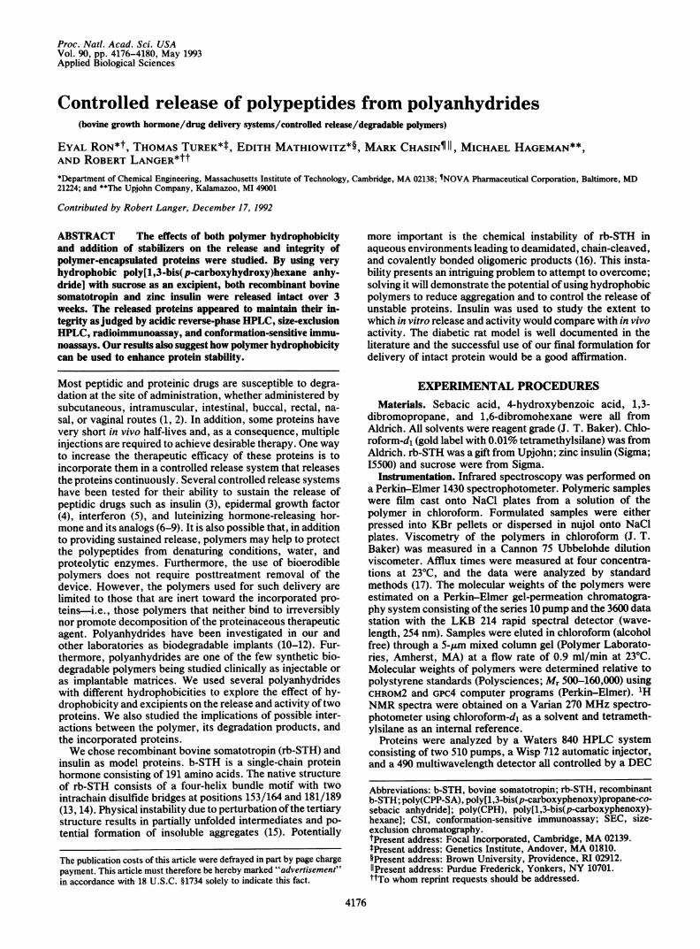
drides were placed in periodically changed phosphate buffer.There was no difference between the SEC-HPLC chromato-gram peak shape of the rb-STH prior to incorporation and thereleased protein, even after 288 h (Fig. 1 Left). No additionalpeaks were observed. Periodically, buffer samples wereconcentrated, dialyzed, and analyzed by isoelectric focusingand SDS gel electrophoresis. Preliminary analysis (data notshown) of these buffer samples did not reveal any change inthe pl pattern and the molecular weight (20,000 by SDS/PAGE) of the released protein, suggesting no alteration dueto interaction with the polymer. b-STH release determined byRIA closely matched the results obtained by SEC-HPLC(Table 1). Similarly, in a second study, a significant corre-lation ofrb-STH release, as determined by SEC-HPLC, RIA,and CSI, was achieved (Table 2). Correlation between SEC-HPLC, RIA, and CSI supports the release of rb-STH withmaintained conformational integrity.The postrelease devices were washed and lyophilized. The
remaining powder was analyzed for both sulfur and nitrogencontent. Since the polymer structure contains neither nitro-gen nor sulfur, postrelease analysis of the two elements isrelated to the unreleased protein content. A mass balance
Applied Biological Sciences: Ron et al.
4178 Applied Biological Sciences: Ron et al.
102.243 1 A
51.121
E
6.0 7.0 8.0 9.0 10.0
Time, min
I.
I
0.000
101.237 B ,
50.61 8 /
0.000 , _
3.676 C
1.838 / \
0.000 -4.0 5.0 6.0 7.0 8.0
Time, min
FIG. 1. (Left) SEC of rb-STH before fabrication (A), after 6 h (B), and after 288 h (C). (Right) SEC of zinc insulin before fabrication (A),after 12 h (B), and after 196 h (C).
calculation enabled quantitative determination of the rb-STHremaining within the device and direct comparison to thereleased protein. Incorporated rb-STH released as deter-mined by SEC-HPLC and RIA and confirmed by postreleaseelemental analysis was 92.6%, 88.8%, and 93.5%, respec-tively.We also wanted to examine whether rb-STH would decom-
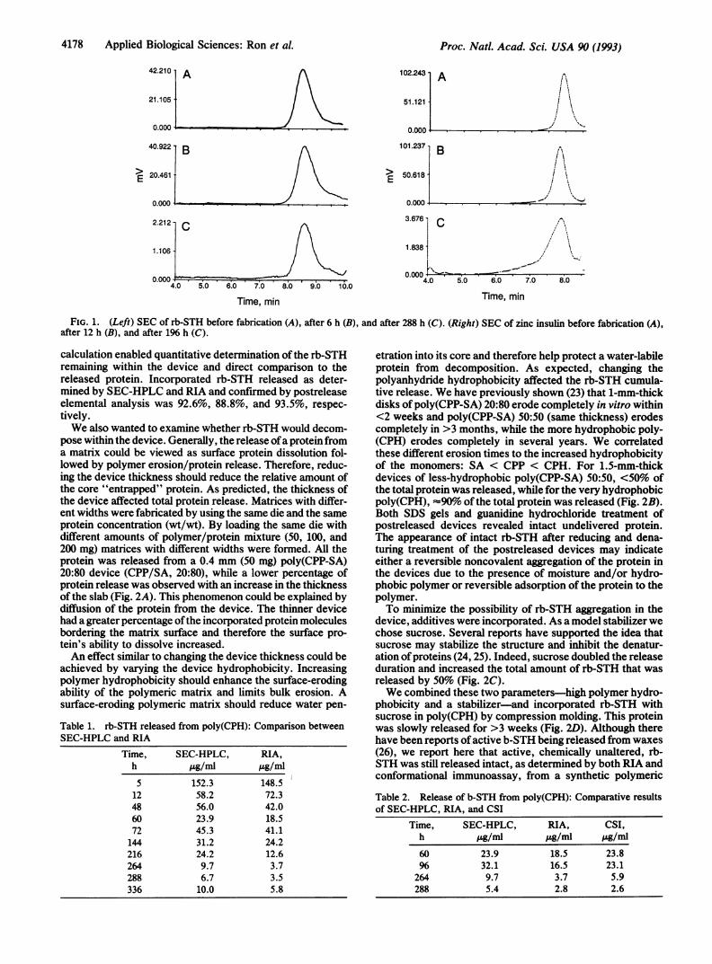
pose within the device. Generally, the release ofa protein froma matrix could be viewed as surface protein dissolution fol-lowed by polymer erosion/protein release. Therefore, reduc-ing the device thickness should reduce the relative amount ofthe core "entrapped" protein. As predicted, the thickness ofthe device affected total protein release. Matrices with differ-ent widths were fabricated by using the same die and the sameprotein concentration (wt/wt). By loading the same die withdifferent amounts of polymer/protein mixture (50, 100, and200 mg) matrices with different widths were formed. All theprotein was released from a 0.4 mm (50 mg) poly(CPP-SA)20:80 device (CPP/SA, 20:80), while a lower percentage ofprotein release was observed with an increase in the thicknessof the slab (Fig. 2A). This phenomenon could be explained bydiffusion of the protein from the device. The thinner devicehad a greater percentage ofthe incorporated protein moleculesbordering the matrix surface and therefore the surface pro-tein's ability to dissolve increased.An effect similar to changing the device thickness could be
achieved by varying the device hydrophobicity. Increasingpolymer hydrophobicity should enhance the surface-erodingability of the polymeric matrix and limits bulk erosion. Asurface-eroding polymeric matrix should reduce water pen-
Table 1. rb-STH released from poly(CPH): Comparison betweenSEC-HPLC and RIA
Time, SEC-HPLC, RIA,h ,ug/ml Ag/ml5 152.3 148.512 58.2 72.348 56.0 42.060 23.9 18.572 45.3 41.1144 31.2 24.2216 24.2 12.6264 9.7 3.7288 6.7 3.5336 10.0 5.8
etration into its core and therefore help protect a water-labileprotein from decomposition. As expected, changing thepolyanhydride hydrophobicity affected the rb-STH cumula-tive release. We have previously shown (23) that 1-mm-thickdisks of poly(CPP-SA) 20:80 erode completely in vitro within<2 weeks and poly(CPP-SA) 50:50 (same thickness) erodescompletely in >3 months, while the more hydrophobic poly-(CPH) erodes completely in several years. We correlatedthese different erosion times to the increased hydrophobicityof the monomers: SA < CPP < CPH. For 1.5-mm-thickdevices of less-hydrophobic poly(CPP-SA) 50:50, <50% ofthe total protein was released, while for the very hydrophobicpoly(CPH), -90% ofthe total protein was released (Fig. 2B).Both SDS gels and guanidine hydrochloride treatment ofpostreleased devices revealed intact undelivered protein.The appearance of intact rb-STH after reducing and dena-turing treatment of the postreleased devices may indicateeither a reversible noncovalent aggregation of the protein inthe devices due to the presence of moisture and/or hydro-phobic polymer or reversible adsorption of the protein to thepolymer.To minimize the possibility of rb-STH aggregation in the
device, additives were incorporated. As a model stabilizer wechose sucrose. Several reports have supported the idea thatsucrose may stabilize the structure and inhibit the denatur-ation of proteins (24, 25). Indeed, sucrose doubled the releaseduration and increased the total amount of rb-STH that wasreleased by 50% (Fig. 2C).We combined these two parameters-high polymer hydro-
phobicity and a stabilizer-and incorporated rb-STH withsucrose in poly(CPH) by compression molding. This proteinwas slowly released for >3 weeks (Fig. 2D). Although therehave been reports ofactive b-STH being released from waxes(26), we report here that active, chemically unaltered, rb-STH was still released intact, as determined by both RIA andconformational immunoassay, from a synthetic polymeric
Table 2. Release of b-STH from poly(CPH): Comparative resultsof SEC-HPLC, RIA, and CSI
Time, SEC-HPLC, RIA, CSI,h pg/ml pAg/ml "g/ml60 23.9 18.5 23.896 32.1 16.5 23.1264 9.7 3.7 5.9288 5.4 2.8 2.6
42.210 1 A
21.105
E
0.000 4 _e..
40.922 BB
20.461-
0.000
2.212-c
1.1061-
0.000 _4.0 5.0
Proc. Natl. Acad. Sci. USA 90 (1993)

Applied Biological Sciences: Ron et al.
)O
O)0a)
a)a)
E0
Time, h
)O
a)a)a)
a).2"
E0
24 36
Time, h
a)a)a).2a)
a)
E
10
a)5a)a)
E2032
Time, h
10 15 20
SORT
FIG. 2. (A) Effect of increased thickness of the device on cumulative protein release. (B) Effect of device hydrophobicity on cumulativeprotein release. (C) Effect ofexcipient (sucrose) on cumulative protein release. (D) Combined effect ofhigh polymer hydrophobicity and sucrose,as an additive, on the release of both rb-STH and insulin.
erodible device after exposure to aqueous media for >2weeks.To further investigate the ability of sucrose and hydropho-
bic polymer to stabilize proteins, we analyzed insulin. SECof insulin displayed a single peak before device fabricationand for up to 196 h of release (Fig. 1 Right). A small peak(<10%) indicative of oligomers was observed after 196 h ofrelease. Using reverse-phase HPLC, insulin prior to devicefabrication had a retention time of 6.8 min. The placebopoly(CPP-SA) 20:80 displayed, at first, hydrophobic degra-dation products at a retention time of 7.9 min and additionalhydrophilic degradation products at 4.8 min. The analysis ofthe release of the insulin from the polymeric matrix did notreveal any peaks in addition to the peaks of intact insulin andthe polymer degradation products (Fig. 3). Analysis of re-leased insulin by RIA and SEC-HPLC reveals an excellent
c0
CO)n
3 4 6 7 8 9 10 (11 12
Time (min)
C
.Q
oco
:
3 4 5 6 7 8 9 10 11 12
Time (min)
FIG. 3. Reverse-phase HPLC of released insulin before fabrica-tion (A) and of insulin released after 1 (B) and 3 h.
correlation between these two methods (Fig. 4A). Massbalance analysis using elemental analysis supported theHPLC results. Again, the data suggested that there wasminimal or no interaction between insulin and polyanhy-drides; furthermore, the data suggest conservation of integ-rity of the conformation of insulin for >198 h of release.
a)a1)a1)cnCa)aC
0 50 100 150 200 250 300 350Time, h
a)-o0)
a)0(3
-8 -3 2 7 12 17
Time, days
FIG. 4. (A) Comparison of released insulin by RIA and reverse-phase HPLC. (B) Glucose control in urine of diabetic rats by insulinreleased from poly(CPP-SA) 20:80 and poly(CPH).
Proc. Natl. Acad. Sci. USA 90 (1993) 4179
BInsulin aigomers

4180 Applied Biological Sciences: Ron et al.
As before, we combined these two parameters: high poly-mer hydrophobicity [poly(CPH)] and a model stabilizer (su-crose). The release of insulin was still noted after 1.5 weeks,with >90% ofthe insulin originally incorporated in the devicebeing released (Fig. 2D).To further confirm the results described above, the effect
of hydrophobicity of matrices made of biodegradable poly-mers as carriers for the controlled release ofinsulin in treatingdiabetes mellitus was assessed in streptozotocin-induceddiabetic rats. The rats were monitored for both blood andurine glucose levels. The high level of glucose was loweredto a normal level with the administration of insulin-impregnated polyanhydrides. Discs made of poly(CPH) con-taining insulin demonstrated almost 2 weeks of urine andblood glucose control in diabetic rats. On the other hand,devices made of poly(CPP-SA) 50:50, the less hydrophobicpolymer, with the same insulin content, demonstrated lessthan a week ofurine and blood glucose control in diabetic rats(Fig. 4B).At the end of study, the rats were sacrificed and analyzed
histologically. The histology was characterized by a smallfibrous connective tissue capsule surrounding the implantthat was infiltrated with lymphocytes and macrophages. Thiswas a normal mild foreign body reaction as seen previouslyfor similar polyanhydrides and other biocompatible implants(27).Other methods for protein integrity analysis may be used
(e.g., circular dichroism, basic reverse-phase HPLC, quasi-elastic light scattering, nonreduced SDS gel electrophoresis)to extend analysis ofthe integrity ofthe incorporated protein.The release of hydrophilic proteins from polyanhydrides,
formulated as described, may possibly be explained by amultiple-mechanism procedure. First, dissolution and diffu-sion of the surface protein occurs. Second, proteins arereleased by a combined mechanism of polymer erosion anddrug diffusion. Nonetheless, the surface proteins diffusefaster than the rate of polymer degradation. The proteinsleave channels at the device's surface and water penetratesthrough these channels (28). The trapped proteins at the coreofthe device, which contact water but cannot diffuse throughthe narrow channels, eventually aggregate. Thus, more hy-drophobic devices that prevent water entry and/or thindevices with a greater percentage of protein near the surfaceof the matrix should minimize aggregation and promote agreater percentage of the incorporated proteins to be re-leased.
CONCLUSIONSWe successfully used the polyanhydrides' changeable hydro-phobicity to explore the effect of hydrophobicity on releaseand activity of two proteins. We established that higherhydrophobicity is advantageous for moisture-labile proteins.This higher hydrophobicity could be achieved by using a veryhydrophobic biodegradable poly(CPH). We also demon-strated that different excipients/stabilizers can be an impor-tant part of the formulation and can be essential to supportand extend the life of sensitive proteins. Finally, the inertnessof both polyanhydrides and their degradation products to-ward specific proteins was established.
The authors would like to thank L. F. Krabill for running therb-STH RIAs, J. S. Bourdage for running the CSI assays (both ofUpjohn), and J. C. Murphy (Massachusetts Institute ofTechnology)for histological analysis. The authors would like to thank J. Schrier(Genetics Institute) for assistance in reviewing this paper. This workwas supported by National Institutes of Health Grant GM26698.
1. Hori, R., Komada, K. & Okumura, K. (1983) J. Pharm. Sci. 72,435-439.
2. Hussain, A., Faraj, J. & Aramaki, Y. (1985) Biochem. Biophys.Res. Commun. 133, 923-928.
3. Brown, L., Munoz, C., Siemer, L., Edelman, E. & Langer, R.(1986) Diabetes 35, 692-697.
4. Murray, J., Brown, L., Klagsburn, M. & Langer, R. (1983) InVitro 19, 743-748.
5. Langer, R., Hsieh, D. S. T., Brown, L. & Rhine, W. (1981) inBetter Therapy with Existing Drugs: New Uses and DeliverySystems, ed. Bearn, A. (Merck, New York), pp. 179-216.
6. Ogawa, Y., Yamamoto, M., Okada, H., Yashiki, T. & Shi-mamoto, T. (1988) Chem. Pharm. Bull. 36, 1095-1103.
7. Ogawa, Y., Yamamoto, M., Takada, S., Okada, H. & Shimam-oto, T. (1988) Chem. Pharm. Bull. 36, 1502-1507.
8. Ogawa, Y., Okada, H., Yamamoto, M. & Shimamoto, T. (1988)Chem. Pharm. Bull. 36, 2576-2581.
9. Okada, H., Heya, T., Ogawa, Y. & Shimamoyo, T. (1988) J.Pharmacol. Exp. Ther. 244, 744-750.
10. Brem, H., Mahaley, S., Vick, N., Black, K., Schold, S. C.,Burger, P., Friedman, A., Cinic, I., Eller, T., Cozzens, J. &Kenealy, J. (1991) J. Neurosurg. 74, 441-446.
11. Leong, K. W., D'Amore, P., Marletta, M. & Langer, R. (1986)J. Biomed. Mater. Res. 20, 51-64.
12. Laurencin, C., Domb, A., Morris, C., Brown, V., Chasin, M.,Lange, N. & Langer, R. (1990) J. Biomed. Mater. Res. 24,1463-1481.
13. Sntome, J. A., Dellscha, J. M., Paladini, A. C., Pena, C.,Biscoglio, M. J., Daurat, S. T., Poskus, E. & Wolfenstein,C. E. M. (1973) Eur. J. Biochem. 37, 164-170.
14. Wallis, M. (1973) FEBS Lett. 35, 11-14.15. Brems, D. N. (1988) Biochemistry 27, 4541-4546.16. Hageman, M. J., Bauer, J. M., Possert, P. L. & Danrington,
R. T. (1992) J. Agric. Food Chem. 40, 348-355.17. Coffins, E. A. & Bares, J. (1970) Experiments in Polymer
Chemistry (Wiley, New York), p. 152.18. Domb, A. J. & Langer, R. (1987) J. Polym. Chem. 25, 3373-
3386.19. Ron, E., Mathiowitz, E., Mathiowitz, G. & Langer, R. (1991)
Macromolecules 24, 2278-2282.20. Niswender, G. D., Reichert, L. E., Jr., Midgley, A. R., Jr., &
Nalbandov, A. V. (1969) Endocrinology 84, 1166-1173.21. Pfund, W. P. & Bourdage, J. S. (1990) Mol. Immunol. 27,
495-502.22. Moseley, W. M., Krabill, L. F. & Olsen, R. F. (1982) J. Anim.
Sci. 55, 1062-1070.23. Leong, K. W., Brott, B. C. & Langer, R. (1985) J. Biomed.
Mater. Res. 19, 941-955.24. Lee, J. C. & Timasheff, S. N. (1981) J. Biol. Chem. 256,
7193-7201.25. Back, J. F., Oakenfull, D. & Smith, M. B. (1979) Biochemistry
18, 5191-5196.26. Cady, S. M. & Langer, R. (1992) J. Agric. Food Chem. 40,
332-336.27. Langer, L. F., Brem, H. & Langer, R. (1991) Technol. Rev. 94,
62-71.28. Ron, E. & Langer, R. (1991) in Treatise on Controlled Drug
Delivery, ed. Kydonieus, A. (Dekker, New York), pp. 199-224.29. Hageman, M. J. (1988) Drug Dev. Ind. Pharm. 14, 2047-2070.
Proc. Natl. Acad. Sci. USA 90 (1993)
Related Documents


