REVIEW Open Access Contributions of Age-Related Thymic Involution to Immunosenescence and Inflammaging Rachel Thomas 1 , Weikan Wang 1 and Dong-Ming Su 2* Abstract Immune system aging is characterized by the paradox of immunosenescence (insufficiency) and inflammaging (over-reaction), which incorporate two sides of the same coin, resulting in immune disorder. Immunosenescence refers to disruption in the structural architecture of immune organs and dysfunction in immune responses, resulting from both aged innate and adaptive immunity. Inflammaging, described as a chronic, sterile, systemic inflammatory condition associated with advanced age, is mainly attributed to somatic cellular senescence-associated secretory phenotype (SASP) and age-related autoimmune predisposition. However, the inability to reduce senescent somatic cells (SSCs), because of immunosenescence, exacerbates inflammaging. Age-related adaptive immune system deviations, particularly altered T cell function, are derived from age-related thymic atrophy or involution, a hallmark of thymic aging. Recently, there have been major developments in understanding how age-related thymic involution contributes to inflammaging and immunosenescence at the cellular and molecular levels, including genetic and epigenetic regulation, as well as developments of many potential rejuvenation strategies. Herein, we discuss the research progress uncovering how age-related thymic involution contributes to immunosenescence and inflammaging, as well as their intersection. We also describe how T cell adaptive immunity mediates inflammaging and plays a crucial role in the progression of age-related neurological and cardiovascular diseases, as well as cancer. We then briefly outline the underlying cellular and molecular mechanisms of age-related thymic involution, and finally summarize potential rejuvenation strategies to restore aged thymic function. Keywords: Thymic aging, Age-related thymic involution, Central tolerance, Negative selection and regulatory T (Treg) cell generation, Immunosenescence and inflammaging, Rejuvenation Introduction The aged immune system has various characteristics. One of which is immunosenescence, which describes the vast and varied changes in the structure and function of the immune system as a result of age [1–4]. Many of the early observations, such as reduced ability to fight new infec- tions, diminished vaccine immunity [5], and reduced tumor clearance [6, 7] are generally categorized as im- mune insufficiencies. Immunosenescence is not due to the lack of immune cells, but due to reduced immune reper- toire diversity, attributed to insufficient production of naïve immune cells and amplified oligo-clonal expansion of memory immune cells. Immunosenescence is therefore linked to the thymus. Natural aging causes the thymus to progressively atrophy, a process called thymic involution. This phenomenon is readily observed in most vertebrates [8] and results in structural alterations, as well as functional decline, ultimately resulting in significantly decreased thymic output of naïve T cells [9–11] that reduces the diversity of the T cell antigen receptor (TCR) repertoire, culminating in disrupted T cell homeostasis (Fig. 1, #3 right side). The second characteristic of aged immunity is termed inflammaging. Inflammaging describes the elevated self- reactivity in the elderly, resulting in the typical chronic, low-grade, but above baseline, systemic inflammatory phenotype observed in the absence of acute infection [12–19]. Inflammaging was originally attributed to © The Author(s). 2020 Open Access This article is distributed under the terms of the Creative Commons Attribution 4.0 International License (http://creativecommons.org/licenses/by/4.0/), which permits unrestricted use, distribution, and reproduction in any medium, provided you give appropriate credit to the original author(s) and the source, provide a link to the Creative Commons license, and indicate if changes were made. The Creative Commons Public Domain Dedication waiver (http://creativecommons.org/publicdomain/zero/1.0/) applies to the data made available in this article, unless otherwise stated. * Correspondence: [email protected] 2 Department of Microbiology, Immunology, and Genetics, University of North Texas Health Science Center, Fort Worth, Texas 76107, USA Full list of author information is available at the end of the article Thomas et al. Immunity & Ageing (2020) 17:2 https://doi.org/10.1186/s12979-020-0173-8

Welcome message from author
This document is posted to help you gain knowledge. Please leave a comment to let me know what you think about it! Share it to your friends and learn new things together.
Transcript
REVIEW Open Access
Contributions of Age-Related ThymicInvolution to Immunosenescence andInflammagingRachel Thomas1, Weikan Wang1 and Dong-Ming Su2*
Abstract
Immune system aging is characterized by the paradox of immunosenescence (insufficiency) and inflammaging(over-reaction), which incorporate two sides of the same coin, resulting in immune disorder. Immunosenescencerefers to disruption in the structural architecture of immune organs and dysfunction in immune responses, resultingfrom both aged innate and adaptive immunity. Inflammaging, described as a chronic, sterile, systemic inflammatorycondition associated with advanced age, is mainly attributed to somatic cellular senescence-associated secretoryphenotype (SASP) and age-related autoimmune predisposition. However, the inability to reduce senescent somaticcells (SSCs), because of immunosenescence, exacerbates inflammaging. Age-related adaptive immune systemdeviations, particularly altered T cell function, are derived from age-related thymic atrophy or involution, a hallmarkof thymic aging. Recently, there have been major developments in understanding how age-related thymicinvolution contributes to inflammaging and immunosenescence at the cellular and molecular levels, includinggenetic and epigenetic regulation, as well as developments of many potential rejuvenation strategies. Herein, wediscuss the research progress uncovering how age-related thymic involution contributes to immunosenescenceand inflammaging, as well as their intersection. We also describe how T cell adaptive immunity mediatesinflammaging and plays a crucial role in the progression of age-related neurological and cardiovascular diseases, aswell as cancer. We then briefly outline the underlying cellular and molecular mechanisms of age-related thymicinvolution, and finally summarize potential rejuvenation strategies to restore aged thymic function.
Keywords: Thymic aging, Age-related thymic involution, Central tolerance, Negative selection and regulatory T(Treg) cell generation, Immunosenescence and inflammaging, Rejuvenation
IntroductionThe aged immune system has various characteristics. Oneof which is immunosenescence, which describes the vastand varied changes in the structure and function of theimmune system as a result of age [1–4]. Many of the earlyobservations, such as reduced ability to fight new infec-tions, diminished vaccine immunity [5], and reducedtumor clearance [6, 7] are generally categorized as im-mune insufficiencies. Immunosenescence is not due to thelack of immune cells, but due to reduced immune reper-toire diversity, attributed to insufficient production ofnaïve immune cells and amplified oligo-clonal expansion
of memory immune cells. Immunosenescence is thereforelinked to the thymus. Natural aging causes the thymus toprogressively atrophy, a process called thymic involution.This phenomenon is readily observed in most vertebrates[8] and results in structural alterations, as well asfunctional decline, ultimately resulting in significantlydecreased thymic output of naïve T cells [9–11] thatreduces the diversity of the T cell antigen receptor (TCR)repertoire, culminating in disrupted T cell homeostasis(Fig. 1, #3 right side).The second characteristic of aged immunity is termed
inflammaging. Inflammaging describes the elevated self-reactivity in the elderly, resulting in the typical chronic,low-grade, but above baseline, systemic inflammatoryphenotype observed in the absence of acute infection[12–19]. Inflammaging was originally attributed to
© The Author(s). 2020 Open Access This article is distributed under the terms of the Creative Commons Attribution 4.0International License (http://creativecommons.org/licenses/by/4.0/), which permits unrestricted use, distribution, andreproduction in any medium, provided you give appropriate credit to the original author(s) and the source, provide a link tothe Creative Commons license, and indicate if changes were made. The Creative Commons Public Domain Dedication waiver(http://creativecommons.org/publicdomain/zero/1.0/) applies to the data made available in this article, unless otherwise stated.
* Correspondence: [email protected] of Microbiology, Immunology, and Genetics, University ofNorth Texas Health Science Center, Fort Worth, Texas 76107, USAFull list of author information is available at the end of the article
Thomas et al. Immunity & Ageing (2020) 17:2 https://doi.org/10.1186/s12979-020-0173-8
somatic cell senescence-associated secretory phenotype(SASP) [20–22] and chronic innate immune activation.In recent years, however, the contribution of aged adap-tive immune components and specifically self-reactive Tlymphocytes, has been realized [13, 23], as a prob-able primary contributor to the age-related develop-ment of subclinical autoimmune predisposition. Although
immunosenescence and inflammaging appear to beopposing phenotypes, they comprise two sides of the samecoin [24] when attempting to holistically understand age-related immune dysfunction [3, 4, 24, 25]. It has beenproposed that the basal inflammatory state in the elderly,defined by inflammaging, greatly contributes to many age-related degenerative diseases [23], including metabolic
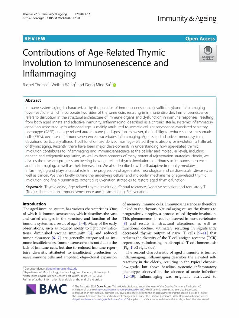
Fig. 1 Intersection of immunosenescence and inflammaging is associated with age-related thymic involution. The aged, involuted thymusexhibits ineffective central tolerance and declined thymopoiesis. The ineffective central tolerance includes (1) impaired negative selection, whichleads to the increased output of self-reactive T cells that attack self-tissues/organs, and (2) imbalanced generation of tTreg TCR repertoire, whichfails to sufficiently suppress self-reactive T cell–mediated autoimmune responses. Autoimmune responses lead to tissue damage and thus causechronic inflammation, which is one of the contributors to inflammaging. Reduced thymopoiesis leads to decreased output of naïve T cells for theclearance of senescent somatic cells (SSCs) and the expansion of oligo-clonal T cells in the aged periphery lack sufficient clearance capacity,which allows for SSC accumulation. SSCs are an important source of SASP, another contributor to inflammaging
Thomas et al. Immunity & Ageing (2020) 17:2 Page 2 of 17
diseases, such as Type-II Diabetes (as a complication ofpancreatitis), neurodegenerative diseases, such asAlzheimer’s disease, and cardiovascular diseases, such asatherosclerosis [18, 23, 26–28].T lymphocyte (T cell) development and selection occurs
in the thymus [29]. Included in this process is central tol-erance establishment (Fig. 1, #1 and #2 top left), which oc-curs via two mechanisms. First is thymocyte negativeselection, during which the majority of self (auto)-reactiveT cells are depleted from the repertoire via apoptosis [30].Second is the generation of CD4 single positive (CD4SP)FoxP3+ regulatory T (Treg) cells [31], whose primaryfunction is to suppress T cell-mediated self-reactivity andpreserve immune homeostasis in the periphery [32]. Thesearms of central T cell tolerance work in tandem, and Tregcells most likely compensate for imperfections of negativeselection, as some self-reactive T cells escape negative se-lection [33]. With age, however, the atrophied thymus de-clines in its capacity to establish central tolerance, thereby,causing increased self-reactive T cells to escape to theperiphery and participate in the process of inflammaging.Historically, there have been two schools of thought
regarding theoretical causes of age-related decreasedthymopoiesis. First is the idea of defective hematopoieticstem cells, since there are reduced numbers ofhematopoietic stem cell (HSC) progenitors produced byaged bone marrow (BM) [34]. It therefore follows thatthere are fewer early T-cell progenitors (ETP) enteringthe thymus from the BM, resulting in shrinkage of thethymus [35]. Second is the idea of a defect in stromalniches of the BM [36, 37] or thymus [38, 39]. Therefore,age-related hallmarks of thymic involution primarilyoccur within the thymic niche and then extend toimpact the development of ETPs.It is our belief that the latter theory is more substantiated
in light of recent advances, and that the extensive age-relatedalterations in thymic structure and microenvironment con-tribute most to the diminished thymopoiesis observed in theelderly [11, 38]. Thymic epithelial cells (TECs) are the pri-mary thymic stromal cells and include two sub-populations:medullary TECs (mTECs) and cortical TECs (cTECs). Thesetwo cell populations are distinct in their thymic localization,functions during thymocyte development, and molecular ex-pression patterns [40, 41]. Since the advent of cell-type spe-cific conditional gene knock-out (cKO) models, compellingevidence show that age-related thymic atrophy is tightly asso-ciated with postnatal TEC homeostasis, regulated by TECautonomous transcription factors (TFs), such as Forkheadbox N1 (FOXN1) [42]. To this end, rejuvenation of age-related thymic involution by developing FOXN1-TEC axis-based therapeutics is reasonable, although other strategiesare under investigation [43].In this review, we will discuss recent research progress
exploring how age-related thymic involution contributes
to inflammaging progression in conjunction with im-mune insufficiency, resulting in reduced clearance of thesenescent somatic cells (SSCs), coupled with increased Tcell-mediated self-reactivity and inflammation. We willoutline the differences in general senescence and immu-nosenescence as it pertains to inflammaging and age-related immune dysregulation. We will describe how theinvolvement of T cell adaptive immunity in mediatinginflammaging plays a crucial role in the progression ofage-related neurological and cardiovascular diseases, aswell as cancer. Finally, we will briefly outline the under-lying cellular and molecular mechanisms of age-relatedthymic involution and summarize potential rejuvenationstrategies to restore aged thymic function. Finding newways to attenuate the impacts of age-related thymic in-volution on inflammaging and immunosenescence is ofgreat clinical significance in an era refocusing medicinetoward healthy aging.
Contributions of Thymic Involution to T CellImmune System AgingSince T cell immune system aging mainly includes twoaspects: immunosenescence and inflammaging, in thissection we discuss recently published papers about howthey intersect, how they are induced, and how age-related thymic involution participates in these processes.We outline this intricate relationship between immuno-senescence and inflammaging associated with age-related thymic involution in Fig. 1.
Intersection of Immunosenescence and InflammagingWhen discussing hallmarks of biological aging, sevenoverarching pillars [44] are thought to collapse, namely:decreased adaptation to stress, loss of proteostasis, ex-haustion of stem cells, derangement of metabolism,macromolecular damage, epigenetic dysregulation, andintercellular communication disorder. These changes areintricately linked through the crossroads of immunose-nescence and inflammaging [23, 45], which characterizeimmunology of aging.Conventional senescence is a general term usually de-
noting somatic cellular senescence, referring to perman-ent or durable cell-cycle arrest first observed in culturedfibroblasts. The original observations leading to the dis-covery of senescence were not fully acknowledged by thescientific community because the initial observationswere described in in vitro cultured cells, although thisgroup believed there to be cell intrinsic factors leadingto the observed “degeneration” of the cells [46]. It waslater demonstrated that senescence occurs in vivo andhas since been more adequately defined as cells exhi-biting permanent cell cycle arrest, lack of proliferation,expression of corresponding anti-proliferation markers,such as p16INK4a and senescence-associated β
Thomas et al. Immunity & Ageing (2020) 17:2 Page 3 of 17
galactosidase (SA-β-gal), shortened telomeres, and acti-vation of DNA-damage signaling cascades. The charac-teristics of somatic cell senescence have recently beensignificantly reviewed elsewhere [47, 48].Somatic cellular senescence is believed to be advanta-
geous as an evolutionary protection against cancer devel-opment [47]. However, senescence of somatic cellsduring aging is thought to significantly contribute notonly to degeneration of aged tissue function if SSCs areaccumulated in certain organs, but also to the systemicinflammatory milieu via induction of SASP [18–23, 49].This largely pro-inflammatory cellular secretion patterninduces increased basal levels of serum IL-6 and IL-1, aswell as matrix metalloproteinases (MMPs) [18, 47].SASP has therefore been cited as a major contributor toinflammaging [18, 19, 23, 49]. Some of the mechanismssuggested to trigger cellular senescence are prolonged orchronic insults that accumulate over time, such as oxida-tive stress, gradual telomere shortening, and chronicinfections. One additional characteristic of senescentcells is that they actively resist apoptosis [47]. The anti-apoptotic pathways involve many factors includingdownregulation of Capsase-3 and increased Cyclin-dependent kinase inhibitors, p16 and p21 [50]. Morerecently, histone modification studies have implicatedaltered expression ratios of Bcl-2 and Bax family genesin mediating the anti-apoptotic phenotype of senescentfibroblasts [51].Immunosenescence is a much broader term that
encompasses all age-related changes to the immunesystem, both innate and adaptive [27, 52]. The primaryhallmarks of immunosenescence are dampened immuneresponses to new infection or vaccination, and dimin-ished anti-tumor immunosurveillance, including alteredimmune response phenotypes in activated T cells, in-creased memory T cell accumulation, and an inverted Tlymphocyte subset ratio [52]. Immunosenescence in Tcells [53] is commonly termed “cellular exhaustion”.This is usually characterized as loss of co-stimulatorysurface molecule CD28 and expression of Tim-3, inaddition to the other features of cellular senescence [54].T cell exhaustion differs from conventional senescencebecause of upregulation of surface markers such as PD-1and Tim-3. Additionally, this type of growth arrest is notpermanent, as blocking PD-1 can reverse T cell exhaus-tion, as demonstrated by recent clinical trials [54, 55].This unique type of growth arrest in T cells is primarilydue to prolonged or chronic TCR/antigen stimulation.Recently, a link between immunosenescence and som-
atic cellular senescence has been established [56, 57], inwhich the SSCs are no longer homeostatically reducedby the immune response. This results when natural killer(NK) cells, macrophages, astrocytes, and T cells undergodiminished chemotaxis toward accumulated SSCs for
targeted depletion [56–58]. The mechanisms by which Tcells deplete accumulated SSCs could include CD8+
cytotoxic T lymphocytes (CTLs), CD4+ Th1-like cellsproducing cytotoxic inflammatory cytokines (such asIFN-γ), and Th2-like cells producing IL-4 and TGF-β[56, 57]. In addition to diminished chemotaxis, there isalso dampened phagocytosis by neutrophils and macro-phages associated with age that facilitates SSC accumula-tion [59, 60]. This ultimately results in increasedproduction of SASP [21], which significantly contributesto inflammaging and subsequent development of age-related diseases [22, 61]. This intersection of inflamma-ging and immunosenescence with age-related diseasesremains unclear, but many groups are currently explor-ing various models to further elucidate the impact ofinflammaging and immunosenescence on age-relateddisease progression [23, 62].
Underlying Etiology of Immunosenescence andInflammagingThere are several proposed components underlying immu-nosenescence and inflammaging etiology. In addition tocellular SASP secretions which contribute to inflammagingas discussed above, chronic innate immune activation dueto long-term latent or persistent viral infection, for ex-ample, with members of the Herpesviridae family, havebeen proposed to contribute to low level pro-inflammatorycytokine production [17]. Most notably, cytomegalovirus(CMV), infection has been explored as a potential bio-marker in aging human patients [17, 63–65]. For example,several longitudinal studies of aging adults saw correlationswith CMV sero-positivity and increased morbidity [66, 67].Importantly, the role of the aged adaptive immune re-sponses to self-tissues (in the absence of acute infection),primarily induced by the T cell compartment, has beenfound to be a major player in the onset and progression ofinflammaging [12, 13] and associated with immunosens-cence [14, 68]. The aged, atrophied thymus, continues toselect T cells throughout the lifetime of the individual.However, the atrophied thymus is less able to negativelyselect self-reactive T cells, releasing these harmful, self-reactive T cells to the periphery, thereby, increasing sub-clinical autoimmune predisposition in the elderly [14].Additionally, age-related thymic atrophy results in reducedoutput of functional naïve T cells, or recent thymic emi-grant cells (RTEs) [9], over time [69]. Since peripheral Tcell numbers remain unchanged or relatively elevated inaged individuals [70–72], the reduced thymic output incombination with peripheral oligo-clonal expansion ofmemory T cells, which occupy immunological space in theperiphery [73–75], results in an overall contracted TCRrepertoire diversity [9, 76–78] thereby inducing immuneinsufficiency (immunosenescence).
Thomas et al. Immunity & Ageing (2020) 17:2 Page 4 of 17

Thymic involution directs Immunosenescence andInflammagingGiven both the altered output of naïve T cells anddisruption of central tolerance establishment, it followsthat thymic involution contributes to T cell-associatedimmunosenescence and inflammaging. Herein, wereview recently determined evidence in this field.As indicated above, an age-related subclinical auto-
immune predisposition induced by adaptive immunereaction to self-tissues by self-reactive T cells has re-cently been recognized as a potential factor under-lying inflammaging [13, 23]. This results mainly fromthat increased output of self-reactive T cells by theatrophied thymus, which should be depleted throughnegative selection as the first boundary for preventingself-reactivity. Treg cells suppress self-reactivity as thesecond frontier to prevent self-inflicted tissue damage.However, aged Treg cells usually are unable to do so[79], potentially due to the lack of Treg TCR diver-sity, as seen in an autoimmune diabetes model [80,81]. These changes are attributed to defects in centraltolerance establishment during the thymocyte devel-opment process, encompassing negative selection andthymic Treg (tTreg) cell generation.
Defective Negative SelectionUnder the current paradigm, negative selection is the sys-tematic removal of thymocytes expressing a TCR that ex-hibits high affinity for self-peptides presented by major
histocompatibility complex class II (MHC-II) on mTECs[30, 41, 82]. In support of this paradigm, it has been shownthat when these high affinity TCRs receive strong signaling,negative selection follows via apoptosis of the thymocyte[83, 84]. However, overall TCR signaling strength is a cul-mination of TCR affinity for the self-peptide and avidity, orthe combination the of affinity of TCR for self-peptide/MHC-II (self-pMHC-II) complexes and the number ofTCR/self-pMHC-II interactions that occur (Fig. 2). There-fore, if thymocyte-dependent factors (i.e. TCR affinity andnumber) of self-reactive thymocytes are unchanged, thenTCR signaling strength depends on the efficiency of self-pMHC-II expression by mTECs. Since aging inducesmTEC defects, such as decreased expression of auto-immune regulator (AIRE) and MHC-II, there is reducedcapacity for self-pMHC-II ligand expression [85, 86].Therefore, we suggest that a strong signaling strength shiftseither to an intermediate strength, which favorsCD4SPFoxP3+ tTreg cell generation (Fig. 2, arrow-a), or toan even lower (weak) strength, resulting in the release ofself-reactive thymocytes (Fig. 2, arrow-b) with the potentialto initiate self-reactivity and auto-inflammation.The FOXN1 cKO mouse model has proven to be a
beneficial model for studying the capacity for efficientself-pMHC-II ligand expression, because it maintainsyoung hematopoietic precursor cells and a young per-iphery in order to isolate the effects of TEC defects asso-ciated with thymic involution. We showed that thymicinvolution disrupts negative selection, as revealed by theenhanced release of self-reactive T cells recognizing
Fig. 2 TCR signaling strength decides self-reactive CD4sp T clone fates. Interaction between MHC-II/self peptide complex on mTEC and self-reactive TCR on CD4sp thymocyte produces three types of signaling strength: (1) a strong signal leads to negative selection, resulting inthymocyte depletion; (2) an intermediate signal leads to tTreg generation; (3) a weak signal results in thymocyte differentiation into Tconventional (Tcon) cells. We hypothesize that age-related thymic involution shifts signaling strength from strong to intermediate and relativelyenhances polyclonal tTreg generation (black arrow—a) ; while in some cases, antigen-specific interactions exhibit an even weaker signal, resultingin diminished antigen-specific tTreg cells and increased antigen-specific Tcon cells (black arrow—b)
Thomas et al. Immunity & Ageing (2020) 17:2 Page 5 of 17
interphotoreceptor retinoid-binding protein (IRBP) fromthe atrophied thymus of FOXN1 cKO mice compared tocontrol [13]. This result was confirmed using a mockself-antigen model in which expression of ovalbumin(under control of the AIRE-regulated rat insulin pro-moter) was decreased in the involuted thymus comparedto control [85].
Thymic-Derived Regulatory T Cell GenerationThe second arm of central tolerance induction is thegeneration of tTreg cells, which function to suppressautoimmune or chronic immune reactions in the periph-ery as peripheral Treg (pTreg) cells. It is proposed thatTreg cells compensate for imperfections in negative se-lection [33] that allow some self-reactive T cells to enterthe periphery. It is currently accepted that 80 - 95% ofpTreg cells are directly generated in the thymus, as op-posed to Treg cells that are induced in the periphery[87–89]. Under the current paradigm, the processes ofboth negative selection and tTreg generation in the thy-mus utilize the same set of agonist self-peptides [87, 90].In this setting, TCR signaling strength determineswhether developing thymocytes are deleted via negativeselection or enter the tTreg cell lineage. As described inthe previous section, TCR signaling strength is cumula-tive of TCR affinity and avidity, all other thymic micro-environmental variables, such as IL-2, being equal.Moreover, strong signaling induces apoptosis of highlyself-reactive thymocytes, while intermediately high sig-naling leads to tTreg generation (Fig. 2, arrow-a). Weaksignaling results in the survival of thymocytes that differ-entiate into conventional T (Tcon) cells (Schematicdiagram in Fig. 2, arrow-b) [41, 87].As mentioned earlier, the mTECs of the aged, invo-
luted thymus are less apt to express self-antigens, andthis could definitively dampen the overall avidity of TCRsignaling received by developing thymocytes. We haveobserved a relatively increased tTreg generation by theatrophied thymus, which showed no change in overalltTreg numbers, but an increased %tTreg:%tTcon cell ra-tio in the aged, atrophied thymus compared to controlswith the normal thymus [85]. This phenotype was alsoobserved in a mouse model with partial defects inMHC-II through microRNA inhibition [91]. We believethis to be a demonstration of the atrophied thymusattempting to compensate for defective negative selec-tion [13] in order to maintain central T cell tolerance inthe elderly.Additionally, several studies investigating the effects of
diminished ability for thymic self-antigen presentation inmTECs, such as AIRE gene knock, have shown similarresults of no change in overall Treg production. In com-bination with age-related accumulation of pTreg cells in
the periphery of mice and humans [73–75, 92], the rela-tive proportion of pTreg cells is increased in the elderly[93], though these aged pTregs exhibit unimpaired func-tionality [94–96]. Therefore, why is the increased or un-changed proportion of Treg cells unable to successfullymanage self-reactivity in the elderly? We hypothesizethat despite increased polyclonal Treg cells, certain self-tissue-specific Treg cells are reduced or imbalanced withself-reactive T effector (Teff) cells due to thymic atro-phy, creating holes in the Treg repertoire. There are sev-eral mouse models using AIRE gene alterations thatresult in similar defects in self-antigen presentation seenin the aged thymus that support our hypothesis.One study assessed the effects of AIRE knock-out thy-
mus on generation of a particular self-antigen-specifictTreg population, namely for the TCAF3 epitope of aprostate antigen, and saw a significant redirection of thisTCR-bearing thymocyte from the tTreg to the Tconlineage [97] (Fig. 2, arrow-b indicated). These redirectedTCAF3-specific Tcon cells were able to infiltrate theprostate tissue and cause visible lesions, but few TCAF3-specific Treg cells were observed [97, 98].Other studies investigating defects in mTEC self-
peptide expression due to specific self-protein knock-out[99] are beginning to indicate that similar impairmentsexhibited by the aged thymus may negatively impactantigen-specific (monoclonal) tTreg generation despitean unchanged or increased total (polyclonal) tTregpopulation. In contrast, these age-related thymic impair-ments seem to increase output of Tcon cells recognizingthe same self-antigens and may contribute to increasedperipheral self-tissue damage and inflammation [100].Further investigation will hopefully shed more light onhow these subtle deficits in central tolerance establish-ment by the aged thymus impact the aged Treg TCRrepertoire, in spite of a relatively increased aged poly-clonal Treg population [85].
Participation of Immunosenescence andInflammaging in Age-Related DiseasesImmunosenescence and inflammaging begin as largelysubclinical conditions, which eventually underlie age-related diseases. High-risk diseases in the elderly includ-ing neurodegenerative diseases, cardiovascular diseases,and late-life cancers [101–106] are associated with a per-sistent, chronic pro-inflammatory status and impairedregulation of aberrant pro-inflammatory cells due toimmunosenescence in aged individuals [56, 107, 108].
Age-related neurodegenerative diseasesMost age-related neurodegenerative diseases, such asAlzheimer’s disease (AD), are associated with immuno-senescence and/or inflammaging, which cause structural
Thomas et al. Immunity & Ageing (2020) 17:2 Page 6 of 17

and functional disturbances of the blood-brain barriers(BBB) [109, 110], thereby, leading to the infiltration ofimmune cells [101, 109, 111] into the central nervoussystem (CNS). However, whether these disease outcomesare a cause or an effect of the imbalanced pro-inflammatory and anti-inflammatory immune cells isunder investigation [112].Among these immune cells are IFN-γ-producing Th1
cells and IL-17A-producing Th17 cells that are pro-inflammatory. They interact with the CNS residentmicroglia and exacerbate AD [113–115]. Treg cells inAD play dual roles, either pathogenic or protective invarious animal studies [116, 117]. Conversely, Th2 cellsspecific for amyloid beta (Aβ), which is a peptide that isaccumulated in the AD brain [118], ameliorate AD inmice, showing improved cognition and reduced burdenof Aβ depositions.In addition to the BBB, the choroid plexus (CP) is also
an important barrier that maintains CNS homeostasis[111, 119, 120]. The CP harbors CD4+ Th1 cells produ-cing IFN-γ that stimulate CP epithelium to expressleukocyte trafficking factors and recruit small numbersof leukocytes, including T cells and monocyte-derivedmacrophages. In contrast to the pro-inflammatory Th1phenotype associated with disease exacerbation when inthe CNS parenchyma, the IFN-γ producing Th1 cells inthe CP support CNS tissue repair and maintain normalcognitive conditions [121, 122]. However, insufficientTh1 function occurs in the aged CP, leading to reducedIFN-γ and premature cognitive impairment in severalmouse models [119, 123]. This decreased Th1 immuneresponse might represent one of the profiles of immuno-senescence [124], but the definite mechanisms remain tobe explored.The role of infiltrating T cells in the CNS, including
effector T (Teff) and Treg cells, is another critical elem-ent in AD, which could be associated with age-relatedthymic involution. Teff cells, especially CNS-infiltratingTh1 cells, are recognized as pathogenic by multiple stud-ies [112]. For example, Th1 cells specific for Aβ-antigenin the AD brain was verified to promote the disease inmice [114]. Based on this fact, it remains an interestingquestion whether the altered negative selection ofantigen-specific T cells in the aged thymus is morefavorable for the development of Aβ-specific Teff cells,and thereby could potentially predispose aged indivi-duals to AD.Additionally, Treg cells could play dichotomous roles
in AD, either protective or pathogenic [101] likely de-pending on their location in the brain. The fundamentalfunction of Treg cells in immune response is to suppressthe activity of other immune cells including Teff andmyeloid leukocytes. Thus, they are generally believed toinhibit neuroinflammation inside the CNS [125].
However, Treg cells, which are residing at the CP, butnot infiltrating into the CNS, can be detrimental for AD,because the CP is an important gateway for leukocytetrafficking into the CNS to maintain its homeostasis[121, 122]. However, CP leukocyte trafficking can besuppressed by Treg cell-produced IL-10 [126]. Addition-ally, Treg cells can directly inhibit the expression ofleukocyte trafficking molecules by the CP, which wasverified to aggravate AD in an animal model [117]. Tregcells are accumulated in the periphery with advancingage in both mice and humans [73–75, 92, 93], partiallydue to the relatively enhanced Treg generation in theaged, atrophied thymus [85], and Treg cells also have anincreased suppressive function in aged mice [96]. There-fore, the negative effects of Treg cells in the CP andincreased Treg proportion and function in the aged per-iphery could be an important aspect for AD susceptibi-lity and provide a potential therapeutic target.
Age-related cardiovascular diseasesAge is also a predominant risk factor for cardiovasculardiseases [127, 128], the principal pathological conse-quences of which involves vascular endothelial dysfunc-tion and arterial stiffness. These basic pathologies areassociated with immunosenescence and inflammaging,particularly on the cardiovascular wall, and lead tohypertension, atherosclerosis, and ultimately heartfailure [129, 130].In recent years, accumulating evidence has implicated
the senescent T cell immune system in the pathogenesisof cardiovascular diseases, such as atherosclerosis, whichis proposed to be related to thymic involution [28], aswell as links to clonal expansion of senescent T cells andaccumulation of effector memory T cells in the elderly[130]. Apolipoprotein B (ApoB) is the major apolipopro-tein constituent of low-density lipoprotein (LDL), whichis a causal agent for atherosclerosis [131, 132]. AlthoughT cells are not the primary pathogenic cells in athero-sclerotic lesions, ApoB-100-specific T cells were re-ported in an atherogenic mouse model [133] and ApoBp18-specific Treg cells were found in healthy individuals[134]. This indicates that age-related thymic involutionmight predispose elderly individuals to atherosclerosisby biasing the balance of ApoB-specific Teff versus Tregcells.It is known that macrophages are the primary patho-
genic cells in atherosclerotic lesion onset. The accumu-lating CD8+ CD28null CD27- senescent T cells [135, 136]on the inflammatory cardiovascular wall constantly pro-duce IFN-γ, which activates macrophages to releaseMMPs for extracellular matrix degradation [137]. This isan important underlying mechanism of atherosclerosisetiology related to T cells. In addition, CD4+ CD28null
senescent T cells are relevant to the recurrence of acute
Thomas et al. Immunity & Ageing (2020) 17:2 Page 7 of 17
coronary events [138]. Circulating CD4+ effector mem-ory T cells were reported to be associated with athero-sclerosis in humans and in mouse models [139],providing further indication for the role of immunose-nescence in cardiovascular disease.
Late-Life CancersThere is substantial knowledge regarding aged immunefunction and cancer. Immunosenescence with advancedage is known to decrease cancer cell immunosurveillance[140, 141], and inflammaging creates a favorable cytokinemicroenvironment for tumorigenesis [106, 142]. However,knowledge about how age-related thymic involutiondirectly contributes to tumor development is insufficient.Declined immunosurveillance of cancer cells is related
to reduced thymopoiesis leading to an altered orcontracted TCR repertoire diversity [143]. If the range oftumor antigen recognition is narrowed by thymic involu-tion, the aged T cell immune system will be less apt toclear cancerous cells. Likewise, if the proportion of pro-tumorigenic TCRs is biased, the risk for cancer develop-ment is increased. For example, a pro-tumorigenic γδ-Tcell subset bearing Vγ6 and Vδ1 TCR chains, which isrelated to a higher risk of cancer development, was re-ported to be accumulated in aged mice [144, 145], but itremains undetermined whether this pro-tumorigenic γδ-T cell population is increased by the altered negativeselection in the aged thymus or by clonal expansion inthe aged periphery.Treg cells, on the other hand, contribute substantially
to the suppression of anti-tumor T cell responses, andthey frequently accumulate in the tumor microenviron-ment, dampening anti-tumor immunity [146, 147].Numerous studies have shown that cancer patients haveincreased Treg cells in peripheral blood and tumormicroenvironment [148–151]. For example, elderly lungcancer patients have more Treg cells in peripheral bloodthan age-matched controls [152]. This corresponds tothe peripheral accumulation of Treg cells and the poten-tially enhanced tTreg generation by the aged thymus[85], which could be an important factor predisposingelderly individuals to late-life cancer.An important aspect for cancer prognosis is metastatic
relapse, which typically occurs several years after re-moval of the primary tumor and treatment with adjuvanttherapy. The question is where the residual tumor cellshide during chemo- and/or radiotherapy. It has beenshown that lymphoid cancers can hide in the thymus inmice [153, 154]. Also, we recently reported that in micethe atrophied thymus can be a pre-metastatic cancerreservoir to protect non-lymphoid, solid cancer cellsfrom chemotherapy because the thymus provides an in-flammatory microenvironment favorable for solid tumorcell dormancy during chemotherapy [155].
Inflammation is a double-edged sword that is neces-sary for anti-tumor responses [156, 157], but it canalso induce drug resistance in the tumor cells [158,159]. Particularly, chronic inflammation is associatedwith increased risk of cancer, as supported by manystudies [160–162]. Inflammation-driven cancers areinduced by inflammatory cytokines, initiating or pro-moting multiple processes in tumorigenesis includingcellular mutations, metastasis, tumor growth, andangiogenesis [142, 163]. For example, macrophagesand T cells release TNFα which can exacerbate DNAdamage [164] and tumor-associated macrophages se-crete macrophage migration inhibitory factor thatdampens p53-dependent protection [165]. TNFα wasalso found to increase cancer metastasis to the lung[166] and the liver [167] in animal models. Addition-ally, tumor growth is promoted by IL-6 via the IL-6/JAK2/STAT3 pathway in kidney, lung and breast can-cer [168], and angiogenesis in prostate cancer patientswas found to be associated with TGFβ [169]. Theseexamples demonstrate the mutagenic potential ofseveral classic cytokines.One additional component contributing to age-related in-
creased cancer incidence is the skew toward myelopoesiscompared to lymphopoesis that is readily observed in bothanimal models and in humans when studying BM progeni-tor hematopoiesis [170–173]. A subset of these myeloidcells termed myeloid-derived suppressor cells (MDSCs) areincreased in aged individuals and are highly associated withcancer development and progression. For example, in astudy of colorectal cancer patients, a positive correlationwas observed for circulating MDSCs and overall tumorburden [174]. These cells suppress anti-tumor responsesthrough mechanisms that differ from Treg cell im-munosuppression, but nonetheless are correlated withage-related cancer incidence [175, 176]. MDSC induc-tion has been attributed to pro-inflammatory cyto-kines, such as IL-6, which we know to be increasedduring inflammaging [175, 177]. Therefore, perhaps ifthe thymic niche was rejuvenated for enhanced lym-phopoesis and the inflammatory environment duringinflammaging was dampened, rebalance of myeloid-to-lymphoid hematopoiesis could reduce MDSC induc-tion and alleviate their role in cancer progression.Taken together, the axis connecting age-related thymic
involution, T cell immunosenescence and chronicinflammatory environment, to tumorigenesis and tumormetastasis is intriguing, but the current knowledge isinsufficient, and more evidence is necessary.
Key Triggers Associated with Induction of Age-Related Thymic InvolutionAge-related thymic involution is characterized by a re-duction in thymic size and thymocyte numbers as well
Thomas et al. Immunity & Ageing (2020) 17:2 Page 8 of 17
as overt remodeling of the thymic microstructure [70].The thymus is a meshwork structure, in which thymo-cytes of hematopoietic origin undergo development andselection within various compartments containing TECsof non-hematopoietic origin [41, 91]. The aged, invo-luted thymus declines in both TECs and thymocytes.The initial question was which cellular compartmentcontained the primary defect that triggered thymicinvolution.It has been noted that BM hematopoietic stem cells
(HSCs) are decreased with age [34] and exhibit a skeweddevelopmental pathway resulting in a decreased ratio oflymphoid-to-myeloid cells [170–172, 178]. Since thethymocyte progenitor cells immigrate to the thymusfrom the BM, this raised a natural question of whetheraged BM-derived HSC lymphoid progenitors are suffi-ciently able to seed the thymus. Therefore, many studieshave investigated this aspect. The outcome was that agedHSCs contain defects [34] that could contribute to insuf-ficient thymic seeding by early T-cell progenitors (ETPs)[35], culminating in decreased thymic output with age[179]. The conclusion was largely based on BM trans-plantation experiments in mice [180] or in vitro fetalthymic organ culture experiments to assess ETP prolifer-ation [35]. Therefore, aged HSCs and ETPs wereregarded as having an intrinsic defect [181]. This conclu-sion was confirmed using BM aspirate samples fromyoung and elderly patients in which gene expressionprofiling of the HSCs showed differential gene expres-sion associated with skewed myeloid lineage determin-ation, however, it is possible that circulating factors inthe aged periphery, such as cytokines could be initiatingsuch lineage shifts [170].Importantly, the role of non-hematopoietic origin
TECs and BM stromal cells in age-related thymic involu-tion was neglected by these studies. We focused on therole of HSC/thymocyte niche cells by several experimen-tal designs [1]:. For BM transplantation, we avoided theusual whole body irradiation and reduced artifacts ofin vitro HSC manipulation [38, 182] by instead utilizingyoung or aged IL-7R knockout mice as recipients [38,183, 184], since these mice have a BM niche that is ac-cessible to seeding exogenous BM cells without irradi-ation [183, 185]. After BM cell engraftment, the youngBM cells exhibited a young phenotype in young recipi-ents, but the young BM cells exhibited an old phenotypein aged recipients [38]. This suggests that the microenvi-ronmental cells, rather than the HSCs, directs BM cellaging [39] [2]. .We also performed transplantation of“microenvironmental niche”, i.e. fetal mouse thymi, intoyoung or aged mice under the kidney capsule, in whichBM progenitors from the host mice directly seed theengrafted fetal thymus in vivo [182]. After engraftment,BM progenitors from young and aged mice developed
equally well in the young engrafted thymus [182]. Thesecomprehensive experiments provide substantial evidencedemonstrating that the aged non-hematopoietic micro-environment, rather than aged HSCs or ETPs [39], me-diates age-related thymic involution [11]. The result canbe explained by the “seed and soil” theory, which de-scribes how stem niches (soil) direct progenitor cell(seed) fate [186–188], and how thymocytes and the stro-mal microenvironment (TECs) cross-talk in the thymus[40], leading us to conclude that age-related thymic in-volution begins with defects in the TEC compartment.Therefore, it is possible that diminished thymic factors,such as IL-7 [189], in the aged, involuted thymus couldprovide signals to HSCs that facilitates the shift inlymphoid-to-myeloid lineage observed in aged HSCs.To identify which specific factors mediate cellular and
molecular TEC aging, many groups have performed sub-stantial work. They found many age-related TEC influ-encing factors, including sex steroids, cytokines,transcription factors, and microRNAs, but the singlemost predominant mechanistic factor currently acceptedas causal to thymic involution is the TEC autonomoustranscription factor FOXN1, which is uniquely expressedin epithelial cells of the thymus and skin to help regulateepithelial cell differentiation [190, 191]. It is required forthymic organogenesis and responsible for thymocyte de-velopment [42], as well as hair follicle development inthe skin [192, 193]. Many past and current studies utilizenude mice as a model, which exhibit a null mutation inFOXN1 resulting in the lack of hair and thymus, andtherefore lack of T cells [194, 195].FOXN1 expression is reduced in the aged thymus and
has even been described as one of the first markers ofthe onset of thymic involution [196, 197]. The questionof the cause-and-effect relationship of FOXN1 declineand thymic involution had been largely under debateuntil the advent of a FOXN1 cKO mouse model [198].In this model, the murine FOXN1 gene is loxP-floxedand the ubiquitous Cre-recombinase with tamoxifen(TM)-inducible fused estrogen receptor blocker(uCreERT) is introduced through crossbreeding [199], inwhich a low level of spontaneous activation takes placeover time, even without tamoxifen (TM) induction [200,201]. This causes a gradual excision of the FOXN1flox/flox
gene over time and results in a progressive loss ofFOXN1 with age. The thymic involution that results ispositively correlated with reduced FOXN1 levels [202].Furthermore, supplying exogenous FOXN1, such as viaplasmid [202] or transgene [203, 204], into the aged thy-mus greatly reduces thymic atrophy and improvesthymic function. Additionally, use of FOXN1 reportermice have enabled further elucidation of the timelineand kinetics of thymic atrophy with age [205]. It is nowlargely accepted that progressively decreased FOXN1
Thomas et al. Immunity & Ageing (2020) 17:2 Page 9 of 17
expression resulting from age introduces defects in TEChomeostasis, resulting in age-related thymic involution.
Trends for Rejuvenation of Age-Related ThymicInvolutionSince the T cell compartment is implicated in so manyaspects of inflammaging and immunosenescence, webelieve that one potential strategy for ameliorating theeffects of inflammaging is via rejuvenation of the aged,involuted thymus. By restoring thymic function, wewould repair the defects in negative selection and re-balance tTreg generation. Currently, there are severalstrategies for rejuvenation of thymic involution in theliterature, some of which target systemic T cell immunityand others focus on the thymus itself.
FOXN1-TEC axisSince the TEC-autonomous factor FOXN1 is heavily im-plicated in onset and progression of age-related thymicinvolution, several strategies attempt to target theFOXN1-TEC axis to specifically restore TEC function.
Cellular therapyFirst, some TEC stem cell-based strategies includeutilization of human embryonic/pluripotent stem cells[206–208], FOXN1eGFP/+ knock-in epithelial cells [209],and young TEC-based [210] or inducible TEC-based[211] strategies. These all involve engraftment of ex-ogenous FOXN1 producing cells into thymic tissue. Onesuch group directly transplanted TECs from newbornmice intrathymically into middle-aged recipients and ob-served renewed growth of the thymus as well as en-hanced T cell generation [210].Another group generated induced TECs (iTECs) from
exogenous FOXN1-overexpressing mouse embryonic fi-broblasts (MEF) cells by initiating the exogenous FOXN1expression that converted MEF cells into epithelial-likecells in vitro [211]. Engraftment of these iTECs underthe kidney capsule of syngenic adult mice created a denovo ectopic thymus. Host T cell progenitors seeded thede novo thymus-like organ generated by the transplantand normal thymocyte distributions were observed after4 weeks. Additionally, typical thymus microstructurewas seen in the de novo thymic engraftment [211].
Cytokine therapyThere are also some cytokine-to-TEC based therapies,such as keratinocyte growth factor [212, 213] and IL-22[214–216]. Many of these animal studies observedthymic regrowth and improved thymopoiesis, however,they largely used models of acute thymic insult, such asirradiation. As for chronic age-induced thymic atrophy,IL-22 may offer more benefits for improved thymicmicroenvironment since one study saw correlative up-
regulation of IL-22 and FOXN1 after acute thymic insultin mice [217]. Though promising, the extent of crosstalkbetween IL-22 and FOXN1 within the thymus remainsto be determined.Another cytokine under investigation is IL-7, which is
normally secreted by TECs, and helps mediate thymopo-esis. IL-7 is reduced in the aged thymus [189] but its rolein other aspects of immune system development andproliferation presents a challenge in approaching IL-7supplementation as a systemic therapy. One such ex-ample is a study administering recombinant IL-7 to agedrhesus macaques, which demonstrated little effect ofthymic function, but did result in enhanced peripheral Tcell proliferation [218]. Several clinical studies have beenconducted with systemic IL-7 treatment to boost periph-eral T cell proliferation after chemotherapy or after in-fection or vaccination to amplify immune responses, butthese were more focused on peripheral expansion(reviewed [219]). Importantly, peripheral T cell subsetsexpress differing levels of the IL-7 receptor, effecting theextent of IL-7-induced expansion (i.e. more CD8+ T cellsexpand compared to CD4+ T cells with minimal expan-sion of Treg cells) [219].However, IL-7 targeting to the aged thymus may re-
store more balanced T cell development in the elderly.For example, one study generated a plasmid-deliveredIL-7 fusion protein that combined IL-7 with the N-terminal extracellular domain of CCR9 to target thisprotein to the thymus and reduce adverse systemic ef-fects of increased IL-7 [220]. They observed restorationof thymic architecture and enhanced cellularity, similarto that of young animals, in the thymus of aged animalsthat received fusion protein treatment compared to un-altered IL-7 and control plasmid groups [220]. Thisstudy holds great promise as a targeted cytokine therapy.Finally, since TCR repertoire contraction is a contribu-
tor to immune insufficiency in aging, it is interesting tonote that systemic treatment with recombinant IL-7 re-sulted in increased TCR diversity in patients who hadundergone bone marrow transplant [221]. Again, giventhe other effects of systemic IL-7, this may not present arealistic therapy for thymic atrophy alone, but it doescompel further study into how some of these cytokinesand circulating factors may impact T cell developmentand selection independently and/or synergistically withage-related thymic involution.
Gene therapySimilar to the TEC-based cellular therapy, some groupshave utilized genetically-based methods to enhance ex-ogenous FOXN1 expression, either with FOXN1 cDNAplasmid or FOXN1 transgenes) [202–204]. One groupintrathymically injected plasmid vectors carryingFOXN1-cDNA into middle-aged and aged mice and
Thomas et al. Immunity & Ageing (2020) 17:2 Page 10 of 17
observed partial rescue of thymic size and thymocytenumbers compared to empty vector controls [202]. An-other group, utilizing an inducible FOXN1 overexpres-sion reporter gene system, showed in vivo upregulationof FOXN1 expression in middle-aged and aged mice re-sulted in increased thymic size and thymocyte numbers[204]. They also observed enhanced ETP cell numbers,and the mTECs:cTECs ratio was restored to normallevels [204]. Moreover, these targeted FOXN1 gene ther-apies also show great promise for rejuvenation of agedthymic structure and function.
Periphery – thymus axisGrowth hormonesDecline in growth hormone during aging has been sug-gested to contribute to age-related thymic involutionand animal studies using growth hormone supplementa-tion show rescue of thymic atrophy, increased T cellprogenitor recruitment into the thymus, as well as en-hanced thymic microenvironmental cytokine production[222–224]. Studies of growth hormone date back to theearly 1999s after the observations that TECs expressgrowth hormone receptors and that insulin-like growthfactor is expressed in the thymus [225–227]. Studies ofinsulin-like growth factor 1 (IGF-1), which is closely re-lated to growth hormone, show similar thymic func-tional and structural improvements upon increased IGF-1 levels in aged mice [222, 228]. Although, the effects ofcrosstalk between growth hormones and many otherneuroendocrine hormones with thymocytes and TECsare under investigation, these systemic pathways areextremely interwoven and thus difficult to compartmen-tally delineate [222, 228].
Sex hormonesThe effects of sex hormones on the thymus have longbeen characterized, with the earliest reports of thymicatrophy correlating with adolescence and reproductivehormones dating back to a 1904 study in cattle [229].Early studies using castration and sex steroid antagonistsin both male mice and male patients receiving androgenblockade for prostate cancer therapy demonstrated phe-notypes varying from delayed onset of thymic involutionto complete thymic regeneration [230–233]. Most ofthese early studies, however, focused primarily onphenotypic data, such as an increase of thymopoiesis,with insufficient mechanistic results. Generally, the reju-venation is thought to occur in the TEC compartmentbecause androgen receptors are expressed by TECs[234]. One of the potential mechanisms reported wasthat sex steroids inhibit cTEC expression of Notchligand Delta-like 4 (DLL4), shown in one study utilizinga luteinizing hormone-releasing hormone blockade thatsaw enhanced thymopoesis after blockade in mice [235].
DLL4 is an important factor for promoting T cell differ-entiation and development. It remains unclear whetherNotch ligands (there are four types) are decreased in theaged thymus and how this might play a role in decreasedthymopoiesis with age.In contrast, other studies of thymic rejuvenation
through sex steroid ablation exhibited in the least, only ashort-lived rejuvenation, and at most no influence what-soever on thymic involution in mice [236]. Others sug-gest that the observable thymic restoration can betransient (only 2 weeks) but harmful, asserting that the“rejuvenated” thymus potentially produces more harmfulT cells and increasing self-reactivity [237]. In support ofthe opinion that sex hormone ablation may cause detri-mental autoimmune implications, a human study, whichused medical castration resulted in a declined %CD4+CD25+ Treg cells and increased NK cells, whichmay compromise immune tolerance [238].Recently, studies on sex hormones and their impact on
thymocyte selection of the TCR repertoire via AIRE geneexpression by TECs in the thymus demonstrate thatthere are differences in males and females in both mouseand human samples [239–241]. Androgens from malespromote AIRE expression in mTECs to enhance thymo-cyte negative selection, while estrogens reduce AIRE ex-pression, dampening thymocyte negative selection andpotentially increasing autoimmunity [240, 241]. There-fore, these hormones may mediate thymic functionalityto a greater extent than simply structural atrophy. Inlight of this, sex steroid antagonists or castration-basedrejuvenation of thymic aging may have more disadvan-tages (inducing autoimmune predisposition in theelderly) than advantages.
Blood-borne factorsOf note, there are likely circulating factors that impactage-related thymic involution, including proteins,mRNAs, microRNAs and other signaling molecules. Onemethod to test this is a heterochronic parabiosis model,in which young and aged mice are surgically conjoinedresulting in mutual influence of blood-borne factors.These experiments, however, have not demonstrated re-juvenation of the aged thymus [242–250]. Conversely,when serum-derived extracellular vesicles, which carrycellular factors throughout the body, were taken fromyoung mice and given to aged hosts, partial thymic reju-venation with increased negative selection signaling wasobserved [251]. Interestingly, we also observed decreasedlevels of circulating pro-inflammatory IL-6, suggestingrescue from inflammaging following treatment withthese young serum-derived extracellular vesicles [251].Further work to elucidate the mechanism of amelioratedinflammaging phenotype is necessary, as it could be dueto increased targeted deletion of senescent cells in the
Thomas et al. Immunity & Ageing (2020) 17:2 Page 11 of 17
periphery causing less SASP secretion, enhanced Tregproduction, or other unknown mechanisms.
Life-Style/Physical ExerciseFinally, life-style habits should not be overlooked per-taining to immune health and healthy aging. Indeed, CTscans of patient thymus tissue demonstrate that ad-vanced fatty degeneration of the thymus is positivelycorrelated with increased BMI and with smoking [252].Additionally, physical exercise has demonstrated count-less benefits for immune health, some of which have re-cently been reported. One such study has documentedan intriguing correlation between physical exercise andimproved thymic function in elderly patients. This in-depth study compared numerous aspects of immunose-nescence and thymic output in aged adults who partici-pated in high levels of regular exercise for much of theiradult lives and aged adults who had been inactive [253].This study found that the aged individuals who main-tained physical exercise regimens exhibited reduction intypical decline in thymic output, decreased markers ofinflammaging, such as reduced serum IL-6, and in-creased serum IL-7 and IL-15, which may foster thymichealth and function [253]. The age-associated increase inTh17 phenotype was also significantly lessened in theaged cohort with physical exercise and lower peripheralTreg cell numbers were observed in these individualscompared to the inactive aged cohort [253]. Though notall aspects of immunosenescence were lessened in theexercising cohort, as both groups maintained the age-related accumulation of senescent T cells, this studydoes present some compelling findings. This group pub-lished a recent review and discussed the direct cross-talkbetween skeletal muscles during exercise and the im-mune compartment, even describing exercise as a poten-tial adjuvant to immunizations, as some studies havealso shown enhanced T cell priming and increased naïveT cell frequency [254]. Therefore, it is significant tomention the effects of physical exercise and overallhealthy life-style habits on immune health and directlyon thymic health over the lifespan.In sum, there are many varied avenues for restoration
of aged thymic structure and function as well as its in-fluences on inflammaging. Many of these rejuvenationstrategies focus on the TEC compartment, since declinein TECs and TEC-associated factors are implicated inthymic involution onset and progression, however, therole of other systemic players are still under investiga-tion. Additionally, each strategy has disadvantages. Forexample, intrathymic injection of newborn TECs can re-juvenate middle-aged thymus [210], but the source ofnewborn TECs is limited and may not be ideal as atranslational therapy. Additionally, generation of anectopic de novo thymus under the kidney capsule [211]
can generate naïve T cells, but this does not remedy theincreased self-reactive T cells released by the original at-rophied thymus remaining in the host. Also, the use ofthymus-targeted cytokines may be beneficial, but cautionis needed, as systemic cytokine therapies usually encom-pass adverse effects. Moreover, continued investigationis required for future development of practical and ef-fective interventions for age-related thymic involutionand inflammaging.
ConclusionAge-related thymic involution is a dynamic process thatimpacts overall T cell development and central T celltolerance establishment throughout life. Immunosens-cence and inflammaging describe two opposing arms ofthe aged immune system: immune insufficiency, with re-gard to infection, vaccination, and tumor surveillance,coupled with increased self-reactivity and chronic, sys-temic inflammation. The contributions of the aged thy-mus to the manifestations of immunosenscence andinflammaging have recently come to be appreciated.However, continued investigation into their synergy inthe aged immune system is needed. Additionally, as weshift our focus towards improving quality of life withage, research into potential avenues for reversing theadverse effects of age-related thymic involution on theaged T cell immune system is of paramount importance.Moreover, there are numerous areas still to explore inthis field with far-reaching applications.
AbbreviationsAβ: Amyloid beta; AD: Alzheimer’s disease; AIRE: Autoimmune regulator;BBB: Blood-brain barriers; BM: Bone marrow; cKO: conditional knock-out;CNS: Central nervous system; CP: Choroid plexus; CTL: Cytotoxic Tlymphocyte; ETP: Early T-cell progenitor; FOXN1: Forkhead box N1;MEF: Mouse embryonic fibroblasts; MDSC: Myeloid-derived suppressor cell;MHC-II: Major histocompatibility complex class II; RTE: Recent thymicemigrant; SASP: Senescence-associated secretory phenotype; SSC: Senescentsomatic cell; TCR: T cell antigen receptor; TEC: Thymic epithelial cell (mTEC:medullary TEC cTEC: cortical TEC); TF: Transcription factor; Tregcell: Regulatory T cell (tTreg cell: thymic Treg cell pTreg cell: peripheral Tregcell)
AcknowledgmentsWe thank Dr. Nan-Ping Peter Weng (Laboratory of Molecular Biology and Im-munology, NIH/NIA) for critical reading of the manuscript.
Competing of interestThe authors declare that they have no competing interests.
Authors’ contributionsConceptualization and supervision: D-MS; Writing original draft: RT, WW, andD-MS; Proofreading and revisions: RT; Visualization and figures: WW. All au-thors read and approved the final manuscript.
FundingSupported by NIH/NIAID grant R01AI121147 to D-M. S. The funder had norole in study design, data collection and analysis, decision to publish, orpreparation of the manuscript.
Availability of data and materialsNot applicable.
Thomas et al. Immunity & Ageing (2020) 17:2 Page 12 of 17
Ethics approval and consent to participateNot applicable.
Consent for publicationNot applicable.
Author details1Cell Biology, Immunology, and Microbiology Graduate Program, GraduateSchool of Biomedical Sciences, Fort Worth, Texas 76107, USA. 2Departmentof Microbiology, Immunology, and Genetics, University of North Texas HealthScience Center, Fort Worth, Texas 76107, USA.
Received: 3 October 2019 Accepted: 2 January 2020
References1. Pawelec G. Age and immunity: What is “immunosenescence”? Exp Gerontol.
2018;105:4–9.2. McElhaney JE, Effros RB. Immunosenescence: what does it mean to health
outcomes in older adults? Curr Opin Immunol. 2009;21(4):418–24.3. Pawelec G. Immunosenescence comes of age. Symposium on Aging
Research in Immunology: The Impact of Genomics. EMBO Rep.2007;8(3):220–3.
4. Pawelec G, Wagner W, Adibzadeh M, Engel A. T cell immunosenescencein vitro and in vivo. Exp Gerontol. 1999;34(3):419–29.
5. Nikolich-Zugich J. The twilight of immunity: emerging concepts in aging ofthe immune system. Nat Immun. 2018;19(1):10–9.
6. Zinger A, Cho WC, Ben-Yehuda A. Cancer and Aging - the InflammatoryConnection. Aging Dis. 2017;8(5):611–27.
7. Machiela MJ. Mosaicism, aging and cancer. Curr Opin Oncol. 2019;31(2):108–13.
8. Drabkin MJ, et al. Age-stratified Patterns of Thymic Involution onMultidetector CT. J Thorac Imaging. 2018;33(6):409. https://doi.org/10.1097/RTI.0000000000000349.
9. Hale JS, Boursalian TE, Turk GL, Fink PJ. Thymic output in aged mice. ProcNatl Acad Sci U S A. 2006;103(22):8447–52.
10. Rezzani R, Nardo L, Favero G, Peroni M, Rodella LF. Thymus and aging:morphological, radiological, and functional overview. Age (Dordr). 2014;36(1):313–51.
11. Palmer DB. The effect of age on thymic function. Front Immunol.2013;4:316.
12. Xia J, Wang H, Guo J, Zhang Z, Coder B, Su DM. Age-Related Disruption ofSteady-State Thymic Medulla Provokes Autoimmune Phenotype viaPerturbing Negative Selection. Aging Dis. 2012;3(3):248–59.
13. Coder BD, Wang H, Ruan L, Su DM. Thymic involution perturbs negativeselection leading to autoreactive T cells that induce chronic inflammation. JImmunol (Baltimore, Md : 1950). 2015;194(12):5825–37.
14. Coder B, Su DM. Thymic involution beyond T-cell insufficiency. Oncotarget.2015;6(26):21777–8.
15. De Martinis M, Franceschi C, Monti D, Ginaldi L. Inflamm-ageing andlifelong antigenic load as major determinants of ageing rate and longevity.FEBS Lett. 2005;579(10):2035–9.
16. Franceschi C, Bonafe M, Valensin S, Olivieri F, De Luca M, Ottaviani E, et al.Inflamm-aging. An evolutionary perspective on immunosenescence. Ann NY Acad Sci. 2000;908:244–54.
17. Brunner S, Herndler-Brandstetter D, Weinberger B, Grubeck-Loebenstein B.Persistent viral infections and immune aging. Ageing Res Rev. 2011;10(3):362–9.
18. Freund A, Orjalo AV, Desprez PY, Campisi J. Inflammatory networks duringcellular senescence: causes and consequences. Trends Mol Med.2010;16(5):238–46.
19. Franceschi C, Capri M, Monti D, Giunta S, Olivieri F, Sevini F, et al.Inflammaging and anti-inflammaging: a systemic perspective on aging andlongevity emerged from studies in humans. Mech Ageing Dev.2007;128(1):92–105.
20. Callender LA, Carroll EC, Beal RWJ, Chambers ES, Nourshargh S, Akbar AN,et al. Human CD8(+) EMRA T cells display a senescence-associated secretoryphenotype regulated by p38 MAPK. Aging Cell. 2018;17:1.
21. Coppe JP, Patil CK, Rodier F, Sun Y, Munoz DP, Goldstein J, et al.Senescence-associated secretory phenotypes reveal cell-nonautonomous
functions of oncogenic RAS and the p53 tumor suppressor. PLoS Biol. 2008;6(12):2853–68.
22. Coppe JP, Desprez PY, Krtolica A, Campisi J. The senescence-associatedsecretory phenotype: the dark side of tumor suppression. Annu Rev Pathol.2010;5:99–118.
23. Fulop T, Witkowski JM, Olivieri F, Larbi A. The integration of inflammaging inage-related diseases. Semin Immunol. 2018;40:17–35.
24. Fulop T, Larbi A, Dupuis G, Le Page A, Frost EH, Cohen AA, et al.Immunosenescence and Inflamm-Aging As Two Sides of the Same Coin:Friends or Foes? Front Immunol. 2017;8:1960.
25. Gui J, Mustachio LM, Su DM, Craig RW. Thymus Size and Age-relatedThymic Involution: Early Programming, Sexual Dimorphism, Progenitors andStroma. Aging Dis. 2012;3(3):280–90.
26. Palmer S, Albergante L, Blackburn CC, Newman TJ. Thymic involution andrising disease incidence with age. Proc Natl Acad Sci U S A. 2018;115(8):1883–8.
27. Fulop T, Dupuis G, Witkowski JM, Larbi A. The Role of Immunosenescencein the Development of Age-Related Diseases. Rev Investig Clin.2016;68(2):84–91.
28. Dai X, Zhang D, Wang C, Wu Z, Liang C. The Pivotal Role of Thymus inAtherosclerosis Mediated by Immune and Inflammatory Response. Int JMed Sci. 2018;15(13):1555–63.
29. Kurd N, Robey EA. T-cell selection in the thymus: a spatial and temporalperspective. Immunol Rev. 2016;271(1):114–26.
30. Palmer E. Negative selection--clearing out the bad apples from the T-cellrepertoire. Nat Rev Immunol. 2003;3(5):383–91.
31. Hsieh CS, Lee HM, Lio CW. Selection of regulatory T cells in the thymus. NatRev Immunol. 2012;12(3):157–67.
32. Cebula A, Seweryn M, Rempala GA, Pabla SS, McIndoe RA, Denning TL, et al.Thymus-derived regulatory T cells contribute to tolerance to commensalmicrobiota. Nature. 2013;497(7448):258–62.
33. Gallegos AM, Bevan MJ. Central tolerance: good but imperfect. ImmunolRev. 2006;209:290–6.
34. Sudo K, Ema H, Morita Y, Nakauchi H. Age-associated characteristics ofmurine hematopoietic stem cells. J Exp Med. 2000;192(9):1273–80.
35. Min H, Montecino-Rodriguez E, Dorshkind K. Reduction in thedevelopmental potential of intrathymic T cell progenitors with age. JImmunol (Baltimore, Md : 1950). 2004;173(1):245–50.
36. Kovtonyuk LV, Fritsch K, Feng X, Manz MG, Takizawa H. Inflamm-Aging ofHematopoiesis, Hematopoietic Stem Cells, and the Bone MarrowMicroenvironment. Front Immunol. 2016;7:502.
37. Latchney SE, Calvi LM. The aging hematopoietic stem cell niche: Phenotypicand functional changes and mechanisms that contribute to hematopoieticaging. Semin Hematol. 2017;54(1):25–32.
38. Sun L, Brown R, Chen S, Zhuge Q, Su DM. Aging induced decline in T-lymphopoiesis is primarily dependent on status of progenitor niches in thebone marrow and thymus. Aging. 2012;4(9):606–19.
39. Su DM, Aw D, Palmer DB. Immunosenescence: a product of theenvironment? Curr Opin Immunol. 2013;25(4):498–503.
40. Anderson G, Jenkinson EJ. Lymphostromal interactions in thymicdevelopment and function. Nat Rev Immunol. 2001;1(1):31–40.
41. Klein L, Kyewski B, Allen PM, Hogquist KA. Positive and negative selection ofthe T cell repertoire: what thymocytes see (and don't see). Nat RevImmunol. 2014;14(6):377–91.
42. Vaidya HJ, Briones Leon A, Blackburn CC. FOXN1 in thymus organogenesisand development. Eur J Immunol. 2016;46(8):1826–37.
43. Aspinall R, Lang PO. Interventions to restore appropriate immune functionin the elderly. Immun Ageing. 2018;15:5.
44. Kennedy BK, Berger SL, Brunet A, Campisi J, Cuervo AM, Epel ES, et al.Geroscience: linking aging to chronic disease. Cell. 2014;159(4):709–13.
45. Accardi G, Caruso C. Immune-inflammatory responses in the elderly: anupdate. Immun Ageing. 2018;15:11.
46. Hayflick L, Moorhead PS. The serial cultivation of human diploid cell strains.Exp Cell Res. 1961;25:585–621.
47. He S, Sharpless NE. Senescence in Health and Disease. Cell. 2017;169(6):1000–11.48. Hernandez-Segura A, Nehme J, Demaria M. Hallmarks of Cellular
Senescence. Trends Cell Biol. 2018;28(6):436–53.49. Campisi J. Cellular senescence: putting the paradoxes in perspective. Curr
Opin Genet Dev. 2011;21(1):107–12.50. Marcotte R, Lacelle C, Wang E. Senescent fibroblasts resist apoptosis by
downregulating caspase-3. Mech Ageing Dev. 2004;125(10-11):777–83.
Thomas et al. Immunity & Ageing (2020) 17:2 Page 13 of 17
51. Sanders YY, Liu H, Zhang X, Hecker L, Bernard K, Desai L, et al. Histonemodifications in senescence-associated resistance to apoptosis by oxidativestress. Redox Biol. 2013;1:8–16.
52. Chou JP, Effros RB. T cell replicative senescence in human aging. CurrPharm Des. 2013;19(9):1680–98.
53. Fukushima Y, Minato N, Hattori M. The impact of senescence-associated Tcells on immunosenescence and age-related disorders. Inflamm Regen.2018;38:24.
54. Crespo J, Sun H, Welling TH, Tian Z, Zou W. T cell anergy, exhaustion,senescence, and stemness in the tumor microenvironment. Curr OpinImmunol. 2013;25(2):214–21.
55. Topalian SL, Hodi FS, Brahmer JR, Gettinger SN, Smith DC, McDermott DF,et al. Safety, activity, and immune correlates of anti-PD-1 antibody in cancer.New Engl J Med. 2012;366(26):2443–54.
56. Prata L, Ovsyannikova IG, Tchkonia T, Kirkland JL. Senescent cell clearanceby the immune system: Emerging therapeutic opportunities. SeminImmunol. 2018;40:101275.
57. Burton DGA, Stolzing A. Cellular senescence: Immunosurveillance and futureimmunotherapy. Ageing Res Rev. 2018;43:17–25.
58. Sagiv A, Biran A, Yon M, Simon J, Lowe SW, Krizhanovsky V. Granuleexocytosis mediates immune surveillance of senescent cells. Oncogene.2013;32(15):1971–7.
59. Plowden J, Renshaw-Hoelscher M, Engleman C, Katz J, Sambhara S.Innate immunity in aging: impact on macrophage function. Aging Cell.2004;3(4):161–7.
60. Solana R, Tarazona R, Gayoso I, Lesur O, Dupuis G, Fulop T. Innateimmunosenescence: effect of aging on cells and receptors of theinnate immune system in humans. Semin Immunol. 2012;24(5):331–41.
61. Tchkonia T, Zhu Y, van Deursen J, Campisi J, Kirkland JL. Cellular senescenceand the senescent secretory phenotype: therapeutic opportunities. J ClinInvest. 2013;123(3):966–72.
62. Franceschi C, Zaikin A, Gordleeva S, Ivanchenko M, Bonifazi F, Storci G, et al.Inflammaging 2018: An update and a model. Semin Immunol. 2018;40:1–5.
63. Nikolich-Zugich J. Ageing and life-long maintenance of T-cell subsets in theface of latent persistent infections. Nat Rev Immunol. 2008;8(7):512–22.
64. High KP, Akbar AN, Nikolich-Zugich J. Translational research in immunesenescence: Assessing the relevance of current models. Semin Immunol.2012;24(5):373–82.
65. Lara J, Cooper R, Nissan J, Ginty AT, Khaw KT, Deary IJ, et al. A proposedpanel of biomarkers of healthy ageing. BMC Med. 2015;13:222.
66. Olsson J, Wikby A, Johansson B, Lofgren S, Nilsson BO, Ferguson FG.Age-related change in peripheral blood T-lymphocyte subpopulationsand cytomegalovirus infection in the very old: the Swedishlongitudinal OCTO immune study. Mech Ageing Dev. 2000;121(1-3):187–201.
67. Wikby A, Johansson B, Olsson J, Lofgren S, Nilsson BO, Ferguson F.Expansions of peripheral blood CD8 T-lymphocyte subpopulations and anassociation with cytomegalovirus seropositivity in the elderly: the SwedishNONA immune study. Exp Gerontol. 2002;37(2-3):445–53.
68. Bektas A, Schurman SH, Sen R, Ferrucci L. Human T cell immunosenescenceand inflammation in aging. J Leukoc Biol. 2017;102(4):977–88.
69. Petrie HT. Role of thymic organ structure and stromal composition insteady-state postnatal T-cell production. Immunol Rev. 2002;189:8–19.
70. Aspinall R, Andrew D. Thymic involution in aging. J Clin Immunol. 2000;20(4):250–6.
71. Hodes RJ. Telomere length, aging, and somatic cell turnover. J Exp Med.1999;190(2):153–6.
72. Franceschi C, Valensin S, Fagnoni F, Barbi C, Bonafe M. Biomarkers ofimmunosenescence within an evolutionary perspective: the challenge ofheterogeneity and the role of antigenic load. Exp Gerontol. 1999;34(8):911–21.
73. Tsukamoto H, Clise-Dwyer K, Huston GE, Duso DK, Buck AL, Johnson LL,et al. Age-associated increase in lifespan of naive CD4 T cells contributes toT-cell homeostasis but facilitates development of functional defects. ProcNatl Acad Sci U S A. 2009;106(43):18333–8.
74. Tsukamoto H, Huston GE, Dibble J, Duso DK, Swain SL. Bim dictates naiveCD4 T cell lifespan and the development of age-associated functionaldefects. J Immunol (Baltimore, Md : 1950). 2010;185(8):4535–44.
75. Chougnet CA, Tripathi P, Lages CS, Raynor J, Sholl A, Fink P, et al. A majorrole for Bim in regulatory T cell homeostasis. J Immunol (Baltimore, Md :1950). 2011;186(1):156–63.
76. Gui J, Zhu X, Dohkan J, Cheng L, Barnes PF, Su DM. The aged thymusshows normal recruitment of lymphohematopoietic progenitors but hasdefects in thymic epithelial cells. Int Immunol. 2007;19(10):1201–11.
77. Swain S, Clise-Dwyer K, Haynes L. Homeostasis and the age-associateddefect of CD4 T cells. Semin Immunol. 2005;17(5):370–7.
78. Goronzy JJ, Lee WW, Weyand CM. Aging and T-cell diversity. Exp Gerontol.2007;42(5):400–6.
79. Fessler J, Ficjan A, Duftner C, Dejaco C. The impact of aging on regulatoryT-cells. Front Immunol. 2013;4:231.
80. Ferreira C, Singh Y, Furmanski AL, Wong FS, Garden OA, Dyson J. Non-obesediabetic mice select a low-diversity repertoire of natural regulatory T cells.Proc Natl Acad Sci U S A. 2009;106(20):8320–5.
81. Ferreira C, Palmer D, Blake K, Garden OA, Dyson J. Reduced regulatory T celldiversity in NOD mice is linked to early events in the thymus. J Immunol(Baltimore, Md : 1950). 2014;192(9):4145–52.
82. Stritesky GL, Jameson SC, Hogquist KA. Selection of self-reactive T cells inthe thymus. Annu Rev Immunol. 2012;30:95–114.
83. Josefowicz SZ, Lu LF, Rudensky AY. Regulatory T cells: mechanisms ofdifferentiation and function. Annu Rev Immunol. 2012;30:531–64.
84. Li MO, Rudensky AY. T cell receptor signalling in the control of regulatory Tcell differentiation and function. Nat Rev Immunol. 2016;16(4):220–33.
85. Oh J, Wang W, Thomas R, Su DM. Capacity of tTreg generation is notimpaired in the atrophied thymus. PLoS Biol. 2017;15(11):e2003352.
86. Fletcher AL, Seach N, Reiseger JJ, Lowen TE, Hammett MV, Scott HS, et al.Reduced thymic Aire expression and abnormal NF-kappaB2 signaling in amodel of systemic autoimmunity. J Immunol. 2009;182(5):2690–9.
87. Klein L, Robey EA, Hsieh CS. Central CD4(+) T cell tolerance: deletion versusregulatory T cell differentiation. Nat Rev Immunol. 2019;19(1):7–18.
88. Pohar J, Simon Q, Fillatreau S. Antigen-Specificity in the ThymicDevelopment and Peripheral Activity of CD4 (+) FOXP3 (+) T RegulatoryCells. Front Immunol. 2018;9:1701.
89. Lathrop SK, Santacruz NA, Pham D, Luo J, Hsieh CS. Antigen-specificperipheral shaping of the natural regulatory T cell population. J Exp Med.2008;205(13):3105–17.
90. Wirnsberger G, Hinterberger M, Klein L. Regulatory T-cell differentiationversus clonal deletion of autoreactive thymocytes. Immunol Cell Biol. 2011;89(1):45–53.
91. Hinterberger M, Aichinger M. Prazeres da Costa O, Voehringer D, HoffmannR, Klein L. Autonomous role of medullary thymic epithelial cells in centralCD4(+) T cell tolerance. Nat Immun. 2010;11(6):512–9.
92. Raynor J, Lages CS, Shehata H, Hildeman DA, Chougnet CA.Homeostasis and function of regulatory T cells in aging. Curr OpinImmunol. 2012;24(4):482–7.
93. Jagger A, Shimojima Y, Goronzy JJ, Weyand CM. Regulatory T cells and theimmune aging process: a mini-review. Gerontology. 2014;60(2):130–7.
94. Nishioka T, Shimizu J, Iida R, Yamazaki S, Sakaguchi S. CD4+CD25+Foxp3+ Tcells and CD4+CD25-Foxp3+ T cells in aged mice. J Immunol (Baltimore,Md : 1950). 2006;176(11):6586–93.
95. Darrigues J, van Meerwijk JPM, Romagnoli P. Age-Dependent Changes inRegulatory T Lymphocyte Development and Function: A Mini-Review.Gerontology. 2018;64(1):28–35.
96. Garg SK, Delaney C, Toubai T, Ghosh A, Reddy P, Banerjee R, et al.Aging is associated with increased regulatory T-cell function. Aging Cell.2014;13(3):441–8.
97. Malchow S, Leventhal DS, Lee V, Nishi S, Socci ND, Savage PA. Aire EnforcesImmune Tolerance by Directing Autoreactive T Cells into the Regulatory TCell Lineage. Immunity. 2016;44(5):1102–13.
98. Malchow S, Leventhal DS, Savage PA. Organ-specific regulatory T cells ofthymic origin are expanded in murine prostate tumors. Oncoimmunology.2013;2(7):e24898.
99. Kieback E, Hilgenberg E, Stervbo U, Lampropoulou V, Shen P, Bunse M, et al.Thymus-Derived Regulatory T Cells Are Positively Selected on Natural Self-Antigen through Cognate Interactions of High Functional Avidity. Immunity.2016;44(5):1114–26.
100. Lages CS, Suffia I, Velilla PA, Huang B, Warshaw G, Hildeman DA, et al.Functional regulatory T cells accumulate in aged hosts and promotechronic infectious disease reactivation. J Immunol (Baltimore, Md : 1950).2008;181(3):1835–48.
101. Coder B, Wang W, Wang L, Wu Z, Zhuge Q, Su DM. Friend or foe: thedichotomous impact of T cells on neuro-de/re-generation during aging.Oncotarget. 2016;8(4):7116–37.
Thomas et al. Immunity & Ageing (2020) 17:2 Page 14 of 17
102. Paneni F, Diaz Canestro C, Libby P, Luscher TF, Camici GG. The AgingCardiovascular System: Understanding It at the Cellular and Clinical Levels. JAm Coll Cardiol. 2017;69(15):1952–67.
103. Sanada F, Taniyama Y, Muratsu J, Otsu R, Shimizu H, Rakugi H, et al. Sourceof Chronic Inflammation in Aging. Front Cardiovasc Med. 2018;5:12.
104. Wong BW, Meredith A, Lin D, McManus BM. The biological role ofinflammation in atherosclerosis. Can J Cardiol. 2012;28(6):631–41.
105. Costantini E, D'Angelo C, Reale M. The Role of Immunosenescence inNeurodegenerative Diseases. Mediat Inflamm. 2018;2018:6039171.
106. Bottazzi B, Riboli E, Mantovani A. Aging, inflammation and cancer. SeminImmunol. 2018;40:74–82.
107. Aw D, Silva AB, Palmer DB. Immunosenescence: emerging challenges for anageing population. Immunology. 2007;120(4):435–46.
108. Gruver AL, Hudson LL, Sempowski GD. Immunosenescence of ageing. JPathol. 2007;211(2):144–56.
109. Zenaro E, Piacentino G, Constantin G. The blood-brain barrier in Alzheimer'sdisease. Neurobiol Dis. 2017;107:41–56.
110. Chupel MU, Minuzzi LG, Furtado G, Santos ML, Hogervorst E, Filaire E,et al. Exercise and taurine in inflammation, cognition, and peripheralmarkers of blood-brain barrier integrity in older women. Appl PhysiolNutr Metab. 2018;43(7):733–41.
111. Erickson MA, Banks WA. Neuroimmune Axes of the Blood-Brain Barriers andBlood-Brain Interfaces: Bases for Physiological Regulation, Disease States,and Pharmacological Interventions. Pharmacol Rev. 2018;70(2):278–314.
112. Liang Z, Zhao Y, Ruan L, Zhu L, Jin K, Zhuge Q, et al. Impact of agingimmune system on neurodegeneration and potential immunotherapies.Prog Neurobiol. 2017;157:2–28.
113. Das R, Chinnathambi S. Microglial priming of antigen presentation andadaptive stimulation in Alzheimer’s disease. Cell Mol Life Sci. 2019;76(19):3681–94.
114. Browne TC, McQuillan K, McManus RM, O’Reilly JA, Mills KH, Lynch MA.IFN-gamma Production by amyloid beta-specific Th1 cells promotesmicroglial activation and increases plaque burden in a mouse model ofAlzheimer's disease. J Immunol. 2013;190(5):2241–51.
115. Zhang J, Ke KF, Liu Z, Qiu YH, Peng YP. Th17 cell-mediatedneuroinflammation is involved in neurodegeneration of abeta1-42-inducedAlzheimer's disease model rats. PLoS One. 2013;8(10):e75786.
116. Yang H, Yang H, Xie Z, Wei L, Bi J. Systemic transplantation of humanumbilical cord derived mesenchymal stem cells-educated T regulatory cellsimproved the impaired cognition in AbetaPPswe/PS1dE9 transgenic mice.PLoS One. 2013;8(7):e69129.
117. Baruch K, Rosenzweig N, Kertser A, Deczkowska A, Sharif AM, Spinrad A, et al.Breaking immune tolerance by targeting Foxp3(+) regulatory T cells mitigatesAlzheimer's disease pathology. Nat Commun. 2015;6:7967.
118. Cao C, Arendash GW, Dickson A, Mamcarz MB, Lin X, Ethell DW. Abeta-specific Th2 cells provide cognitive and pathological benefits to Alzheimer'smice without infiltrating the CNS. Neurobiol Dis. 2009;34(1):63–70.
119. Baruch K, Ron-Harel N, Gal H, Deczkowska A, Shifrut E, Ndifon W, et al. CNS-specific immunity at the choroid plexus shifts toward destructive Th2inflammation in brain aging. Proc Natl Acad Sci U S A. 2013;110(6):2264–9.
120. Ghersi-Egea JF, Strazielle N, Catala M, Silva-Vargas V, Doetsch F, EngelhardtB. Molecular anatomy and functions of the choroidal blood-cerebrospinalfluid barrier in health and disease. Acta Neuropathol. 2018;135(3):337–61.
121. Deczkowska A, Baruch K, Schwartz M. Type I/II Interferon Balance in the Regulationof Brain Physiology and Pathology. Trends Immunol. 2016;37(3):181–92.
122. Kunis G, Baruch K, Rosenzweig N, Kertser A, Miller O, Berkutzki T, et al. IFN-gamma-dependent activation of the brain's choroid plexus for CNSimmune surveillance and repair. Brain. 2013;136(Pt 11):3427–40.
123. Baruch K, Deczkowska A, David E, Castellano JM, Miller O, Kertser A, et al.Aging. Aging-induced type I interferon response at the choroid plexusnegatively affects brain function. Science. 2014;346(6205):89–93.
124. Sandmand M, Bruunsgaard H, Kemp K, Andersen-Ranberg K, Pedersen AN,Skinhoj P, et al. Is ageing associated with a shift in the balance between Type1 and Type 2 cytokines in humans? Clin Exp Immunol. 2002;127(1):107–14.
125. Sakaguchi S, Yamaguchi T, Nomura T, Ono M. Regulatory T cells andimmune tolerance. Cell. 2008;133(5):775–87.
126. Xie L, Choudhury GR, Winters A, Yang SH, Jin K. Cerebral regulatory T cellsrestrain microglia/macrophage-mediated inflammatory responses via IL-10.Eur J Immunol. 2015;45(1):180–91.
127. Heidenreich PA, Trogdon JG, Khavjou OA, Butler J, Dracup K, Ezekowitz MD,et al. Forecasting the future of cardiovascular disease in the United States: a
policy statement from the American Heart Association. Circulation. 2011;123(8):933–44.
128. North BJ, Sinclair DA. The intersection between aging and cardiovasculardisease. Circ Res. 2012;110(8):1097–108.
129. Del Pinto R, Ferri C. Inflammation-Accelerated Senescence and theCardiovascular System: Mechanisms and Perspectives. Int J Mol Sci. 2018;19:12.
130. Yu HT, Park S, Shin EC, Lee WW. T cell senescence and cardiovasculardiseases. Clin Exp Med. 2016;16(3):257–63.
131. Stock J. Triglycerides and cardiovascular risk: Apolipoprotein B holds the key.Atherosclerosis. 2019;284:221–2.
132. Summerhill VI, Grechko AV, Yet SF, Sobenin IA, Orekhov AN. The AtherogenicRole of Circulating Modified Lipids in Atherosclerosis. Int J Mol Sci. 2019;20:14.
133. Shaw MK, Tse KY, Zhao X, Welch K, Eitzman DT, Thipparthi RR, et al. T-CellsSpecific for a Self-Peptide of ApoB-100 Exacerbate Aortic Atheroma inMurine Atherosclerosis. Front Immunol. 2017;8:95.
134. Kimura T, Kobiyama K, Winkels H, Tse K, Miller J, Vassallo M, et al. Regulatory CD4(+)T Cells Recognize Major Histocompatibility Complex Class II Molecule-RestrictedPeptide Epitopes of Apolipoprotein B. Circulation. 2018;138(11):1130–43.
135. Fann M, Chiu WK, Wood WH 3rd, Levine BL, Becker KG, Weng NP. Geneexpression characteristics of CD28null memory phenotype CD8+ T cells andits implication in T-cell aging. Immunol Rev. 2005;205:190–206.
136. Suarez-Alvarez B, Rodriguez RM, Schlangen K, Raneros AB, Marquez-Kisinousky L, Fernandez AF, et al. Phenotypic characteristics of aged CD4(+)CD28(null) T lymphocytes are determined by changes in the whole-genome DNA methylation pattern. Aging Cell. 2017;16(2):293–303.
137. Leon ML, Zuckerman SH. Gamma interferon: a central mediator inatherosclerosis. Inflamm Res. 2005;54(10):395–411.
138. Liuzzo G, Biasucci LM, Trotta G, Brugaletta S, Pinnelli M, Digianuario G, et al.Unusual CD4+CD28null T lymphocytes and recurrence of acute coronaryevents. J Am Coll Cardiol. 2007;50(15):1450–8.
139. Ammirati E, Cianflone D, Vecchio V, Banfi M, Vermi AC, De Metrio M, et al.Effector Memory T cells Are Associated With Atherosclerosis in Humans andAnimal Models. J Am Heart Assoc. 2012;1(1):27–41.
140. Pawelec G. Immunosenescence and cancer. Biogerontology. 2017;18(4):717–21.141. Falci C, Gianesin K, Sergi G, Giunco S, De Ronch I, Valpione S, et al. Immune
senescence and cancer in elderly patients: results from an exploratory study.Exp Gerontol. 2013;48(12):1436–42.
142. Landskron G, De la Fuente M, Thuwajit P, Thuwajit C, Hermoso MA. Chronicinflammation and cytokines in the tumor microenvironment. J ImmunolRes. 2014;2014:149185.
143. Qi Q, Zhang DW, Weyand CM, Goronzy JJ. Mechanisms shaping the naive Tcell repertoire in the elderly - thymic involution or peripheral homeostaticproliferation? Exp Gerontol. 2014;54:71–4.
144. Prinz I, Sandrock I. Dangerous gammadelta T cells in aged mice. EMBO Rep.2019;20(8):e48678.
145. Chen HC, Eling N, Martinez-Jimenez CP, O'Brien LM, Carbonaro V, MarioniJC, et al. IL-7-dependent compositional changes within the gammadelta Tcell pool in lymph nodes during ageing lead to an unbalanced anti-tumourresponse. EMBO Rep. 2019;20(8):e47379.
146. Takeuchi Y, Nishikawa H. Roles of regulatory T cells in cancer immunity. IntImmunol. 2016;28(8):401–9.
147. Speiser DE, Ho PC, Verdeil G. Regulatory circuits of T cell function in cancer.Nat Rev Immunol. 2016;16(10):599–611.
148. Ichihara F, Kono K, Takahashi A, Kawaida H, Sugai H, Fujii H. Increasedpopulations of regulatory T cells in peripheral blood and tumor-infiltratinglymphocytes in patients with gastric and esophageal cancers. Clin CancerRes. 2003;9(12):4404–8.
149. Liyanage UK, Moore TT, Joo HG, Tanaka Y, Herrmann V, Doherty G, et al.Prevalence of regulatory T cells is increased in peripheral blood and tumormicroenvironment of patients with pancreas or breast adenocarcinoma. JImmunol. 2002;169(5):2756–61.
150. Ormandy LA, Hillemann T, Wedemeyer H, Manns MP, Greten TF, Korangy F.Increased populations of regulatory T cells in peripheral blood of patientswith hepatocellular carcinoma. Cancer Res. 2005;65(6):2457–64.
151. Woo EY, Chu CS, Goletz TJ, Schlienger K, Yeh H, Coukos G, et al. RegulatoryCD4 (+) CD25 (+) T cells in tumors from patients with early-stage non-smallcell lung cancer and late-stage ovarian cancer. Cancer Res. 2001;61(12):4766–72.
152. Hou PF, Zhu LJ, Chen XY, Qiu ZQ. Age-related changes in CD4+CD25+FOXP3+ regulatory T cells and their relationship with lung cancer. PLoSOne. 2017;12(3):e0173048.
Thomas et al. Immunity & Ageing (2020) 17:2 Page 15 of 17
153. Gilbert LA, Hemann MT. DNA damage-mediated induction of achemoresistant niche. Cell. 2010;143(3):355–66.
154. Bent EH, Gilbert LA, Hemann MT. A senescence secretory switch mediatedby PI3K/AKT/mTOR activation controls chemoprotective endothelialsecretory responses. Genes Dev. 2016;30(16):1811–21.
155. Sizova O, Kuriatnikov D, Liu Y, Su DM. Atrophied Thymus, a Tumor Reservoirfor Harboring Melanoma Cells. Mol Cancer Res. 2018;16(11):1652–64.
156. Yu H, Pardoll D, Jove R. STATs in cancer inflammation and immunity: aleading role for STAT3. Nat Rev Cancer. 2009;9(11):798–809.
157. Smyth MJ, Dunn GP, Schreiber RD. Cancer immunosurveillance andimmunoediting: the roles of immunity in suppressing tumor developmentand shaping tumor immunogenicity. Adv Immunol. 2006;90:1–50.
158. Kumari N, Dwarakanath BS, Das A, Bhatt AN. Role of interleukin-6 in cancerprogression and therapeutic resistance. Tumour Biol. 2016;37(9):11553–72.
159. Conze D, Weiss L, Regen PS, Bhushan A, Weaver D, Johnson P, et al.Autocrine production of interleukin 6 causes multidrug resistance in breastcancer cells. Cancer Res. 2001;61(24):8851–8.
160. Balkwill F, Mantovani A. Inflammation and cancer: back to Virchow? Lancet.2001;357(9255):539–45.
161. Coussens LM, Werb Z. Inflammation and cancer. Nature. 2002;420(6917):860–7.162. Itzkowitz SH, Yio X. Inflammation and cancer IV. Colorectal cancer in
inflammatory bowel disease: the role of inflammation. Am J PhysiolGastrointest Liver Physiol. 2004;287(1):G7–17.
163. Lu H, Ouyang W, Huang C. Inflammation, a key event in cancerdevelopment. Mol Cancer Res. 2006;4(4):221–33.
164. Pollard JW. Tumour-educated macrophages promote tumour progressionand metastasis. Nat Rev Cancer. 2004;4(1):71–8.
165. Hudson JD, Shoaibi MA, Maestro R, Carnero A, Hannon GJ, Beach DH. Aproinflammatory cytokine inhibits p53 tumor suppressor activity. J Exp Med.1999;190(10):1375–82.
166. Orosz P, Echtenacher B, Falk W, Ruschoff J, Weber D, Mannel DN.Enhancement of experimental metastasis by tumor necrosis factor. J ExpMed. 1993;177(5):1391–8.
167. Orosz P, Kruger A, Hubbe M, Ruschoff J, Von Hoegen P, Mannel DN.Promotion of experimental liver metastasis by tumor necrosis factor. Int JCancer. 1995;60(6):867–71.
168. Hodge DR, Hurt EM, Farrar WL. The role of IL-6 and STAT3 in inflammationand cancer. Eur J Cancer. 2005;41(16):2502–12.
169. Wikstrom P, Stattin P, Franck-Lissbrant I, Damber JE, Bergh A. Transforminggrowth factor beta1 is associated with angiogenesis, metastasis, and poorclinical outcome in prostate cancer. Prostate. 1998;37(1):19–29.
170. Pang WW, Price EA, Sahoo D, Beerman I, Maloney WJ, Rossi DJ, et al. Humanbone marrow hematopoietic stem cells are increased in frequency andmyeloid-biased with age. Proc Natl Acad Sci U S A. 2011;108(50):20012–7.
171. Morrison SJ, Wandycz AM, Akashi K, Globerson A, Weissman IL. The aging ofhematopoietic stem cells. Nat Med. 1996;2(9):1011–6.
172. Waterstrat A, Van Zant G. Effects of aging on hematopoietic stem andprogenitor cells. Curr Opin Immunol. 2009;21(4):408–13.
173. Chinn IK, Blackburn CC, Manley NR, Sempowski GD. Changes in primarylymphoid organs with aging. Semin Immunol. 2012;24(5):309–20.
174. Zhang B, Wang Z, Wu L, Zhang M, Li W, Ding J, et al. Circulating andtumor-infiltrating myeloid-derived suppressor cells in patients withcolorectal carcinoma. PLoS One. 2013;8(2):e57114.
175. Pawelec G, Verschoor CP, Ostrand-Rosenberg S. Myeloid-Derived SuppressorCells: Not Only in Tumor Immunity. Front Immunol. 2019;10:1099.
176. Veglia F, Perego M, Gabrilovich D. Myeloid-derived suppressor cells comingof age. Nat Immun. 2018;19(2):108–19.
177. Ruhland MK, Loza AJ, Capietto AH, Luo X, Knolhoff BL, Flanagan KC, et al.Stromal senescence establishes an immunosuppressive microenvironmentthat drives tumorigenesis. Nat Commun. 2016;7:11762.
178. Van Zant G, Liang Y. Concise review: hematopoietic stem cell aging, lifespan, and transplantation. Stem Cells Transl Med. 2012;1(9):651–7.
179. Linton PJ, Dorshkind K. Age-related changes in lymphocyte developmentand function. Nat Immun. 2004;5(2):133–9.
180. Tyan ML. Age-related decrease in mouse T cell progenitors. J Immunol(Baltimore, Md : 1950). 1977;118(3):846–51.
181. Min H, Montecino-Rodriguez E, Dorshkind K. Effects of aging on early B-and T-cell development. Immunol Rev. 2005;205:7–17.
182. Zhu X, Gui J, Dohkan J, Cheng L, Barnes PF, Su DM. Lymphohematopoieticprogenitors do not have a synchronized defect with age-related thymicinvolution. Aging Cell. 2007;6(5):663–72.
183. Peschon JJ, Morrissey PJ, Grabstein KH, Ramsdell FJ, Maraskovsky E, GliniakBC, et al. Early lymphocyte expansion is severely impaired in interleukin 7receptor-deficient mice. J Exp Med. 1994;180(5):1955–60.
184. Sun L, Hurez VJ, Thibodeaux SR, Kious MJ, Liu A, Lin P, et al. Agedregulatory T cells protect from autoimmune inflammation despite reducedSTAT3 activation and decreased constraint of IL-17 producing T cells. AgingCell. 2012;11(3):509–19.
185. Porritt HE, Rumfelt LL, Tabrizifard S, Schmitt TM, Zuniga-Pflucker JC, PetrieHT. Heterogeneity among DN1 prothymocytes reveals multiple progenitorswith different capacities to generate T cell and non-T cell lineages.Immunity. 2004;20(6):735–45.
186. Moore KA, Lemischka IR. Stem cells and their niches. Science. 2006;311(5769):1880–5.187. Wilson A, Trumpp A. Bone-marrow haematopoietic-stem-cell niches. Nat
Rev Immunol. 2006;6(2):93–106.188. Prockop SE, Petrie HT. Regulation of thymus size by competition for
stromal niches among early T cell progenitors. J Immunol. 2004;173(3):1604–11.
189. Aspinall R, Andrew D. Thymic atrophy in the mouse is a soluble problem ofthe thymic environment. Vaccine. 2000;18(16):1629–37.
190. Nehls M, Pfeifer D, Schorpp M, Hedrich H, Boehm T. New member of thewinged-helix protein family disrupted in mouse and rat nude mutations.Nature. 1994;372(6501):103–7.
191. Nehls M, Kyewski B, Messerle M, Waldschutz R, Schuddekopf K, Smith AJ,et al. Two genetically separable steps in the differentiation of thymicepithelium. Science. 1996;272(5263):886–9.
192. Schlake T. The nude gene and the skin. Exp Dermatol. 2001;10(5):293–304.193. Brissette JL, Li J, Kamimura J, Lee D, Dotto GP. The product of the mouse
nude locus, Whn, regulates the balance between epithelial cell growth anddifferentiation. Genes Dev. 1996;10(17):2212–21.
194. Flanagan SP. ‘Nude’, a new hairless gene with pleiotropic effects in themouse. Genet Res. 1966;8(3):295–309.
195. Pantelouris EM. Absence of thymus in a mouse mutant. Nature. 1968;217(5126):370–1.
196. Ortman CL, Dittmar KA, Witte PL, Le PT. Molecular characterization of themouse involuted thymus: aberrations in expression of transcription regulatorsin thymocyte and epithelial compartments. Int Immunol. 2002;14(7):813–22.
197. Rode I, Martins VC, Kublbeck G, Maltry N, Tessmer C, Rodewald HR. Foxn1Protein Expression in the Developing, Aging, and Regenerating Thymus. JImmunol. 2015;195(12):5678–87.
198. Cheng L, Guo J, Sun L, Fu J, Barnes PF, Metzger D, et al. Postnatal tissue-specific disruption of transcription factor FoxN1 triggers acute thymicatrophy. J Biol Chem. 2010;285(8):5836–47.
199. Zhang Z, Burnley P, Coder B, Su DM. Insights on FoxN1 biologicalsignificance and usages of the “nude” mouse in studies of T-lymphopoiesis.Int J Biol Sci. 2012;8(8):1156–67.
200. Matsuda T, Cepko CL. Controlled expression of transgenes introduced byin vivo electroporation. Proc Natl Acad Sci U S A. 2007;104(3):1027–32.
201. Bleul CC, Corbeaux T, Reuter A, Fisch P, Monting JS, Boehm T. Formation ofa functional thymus initiated by a postnatal epithelial progenitor cell.Nature. 2006;441(7096):992–6.
202. Sun L, Guo J, Brown R, Amagai T, Zhao Y, Su DM. Declining expression of asingle epithelial cell-autonomous gene accelerates age-related thymicinvolution. Aging Cell. 2010;9(3):347–57.
203. Zook EC, Krishack PA, Zhang S, Zeleznik-Le NJ, Firulli AB, Witte PL, et al.Overexpression of Foxn1 attenuates age-associated thymic involution andprevents the expansion of peripheral CD4 memory T cells. Blood. 2011;118(22):5723–31.
204. Bredenkamp N, Nowell CS, Blackburn CC. Regeneration of the aged thymusby a single transcription factor. Development (Cambridge, England). 2014;141(8):1627–37.
205. O’Neill KE, Bredenkamp N, Tischner C, Vaidya HJ, Stenhouse FH, Peddie CD,et al. Foxn1 Is Dynamically Regulated in Thymic Epithelial Cells duringEmbryogenesis and at the Onset of Thymic Involution. PLoS One. 2016;11(3):e0151666.
206. Parent AV, Russ HA, Khan IS, LaFlam TN, Metzger TC, Anderson MS,et al. Generation of functional thymic epithelium from humanembryonic stem cells that supports host T cell development. Cell StemCell. 2013;13(2):219–29.
207. Sun X, Xu J, Lu H, Liu W, Miao Z, Sui X, et al. Directed differentiation of humanembryonic stem cells into thymic epithelial progenitor-like cells reconstitutesthe thymic microenvironment in vivo. Cell Stem Cell. 2013;13(2):230–6.
Thomas et al. Immunity & Ageing (2020) 17:2 Page 16 of 17
208. Soh CL, Giudice A, Jenny RA, Elliott DA, Hatzistavrou T, Micallef SJ, et al.FOXN1 (GFP/w) reporter hESCs enable identification of integrin-beta4, HLA-DR, and EpCAM as markers of human PSC-derived FOXN1(+) thymicepithelial progenitors. Stem Cell Rep. 2014;2(6):925–37.
209. Barsanti M, Lim JM, Hun ML, Lister N, Wong K, Hammett MV, et al. A novelFoxn1(eGFP/+) mouse model identifies Bmp4-induced maintenance ofFoxn1 expression and thymic epithelial progenitor populations. Eur JImmunol. 2017;47(2):291–304.
210. Kim MJ, Miller CM, Shadrach JL, Wagers AJ, Serwold T. Young, proliferativethymic epithelial cells engraft and function in aging thymuses. J Immunol(Baltimore, Md : 1950). 2015;194(10):4784–95.
211. Bredenkamp N, Ulyanchenko S, O'Neill KE, Manley NR, Vaidya HJ, BlackburnCC. An organized and functional thymus generated from FOXN1-reprogrammed fibroblasts. Nat Cell Biol. 2014;16(9):902–8.
212. Erickson M, Morkowski S, Lehar S, Gillard G, Beers C, Dooley J, et al. Regulation ofthymic epithelium by keratinocyte growth factor. Blood. 2002;100(9):3269–78.
213. Alpdogan O, Hubbard VM, Smith OM, Patel N, Lu S, Goldberg GL, et al.Keratinocyte growth factor (KGF) is required for postnatal thymicregeneration. Blood. 2006;107(6):2453–60.
214. Bhandoola A, Artis D. Rebuilding the thymus. Science. 2012;336(6077):40–1.215. Dudakov JA, Hanash AM, Jenq RR, Young LF, Ghosh A, Singer NV, et al.
Interleukin-22 drives endogenous thymic regeneration in mice. Science.2012;336(6077):91–5.
216. Chaudhry MS, Velardi E, Dudakov JA, van den Brink MR. Thymus: the next(re) generation. Immunol Rev. 2016;271(1):56–71.
217. Pan B, Liu J, Zhang Y, Sun Y, Wu Q, Zhao K, et al. Acute ablation of DPthymocytes induces up-regulation of IL-22 and Foxn1 in TECs. Clin Immunol(Orlando, Fla). 2014;150(1):101–8.
218. Okoye AA, Rohankhedkar M, Konfe AL, Abana CO, Reyes MD, Clock JA, et al.Effect of IL-7 Therapy on Naive and Memory T Cell Homeostasis in AgedRhesus Macaques. J Immunol (Baltimore, Md : 1950). 2015;195(9):4292–305.
219. Mackall CL, Fry TJ, Gress RE. Harnessing the biology of IL-7 for therapeuticapplication. Nat Rev Immunol. 2011;11(5):330–42.
220. Henson SM, Snelgrove R, Hussell T, Wells DJ, Aspinall R. An IL-7 fusionprotein that shows increased thymopoietic ability. J Immunol (Baltimore,Md : 1950). 2005;175(6):4112–8.
221. Perales MA, Goldberg JD, Yuan J, Koehne G, Lechner L, Papadopoulos EB, et al.Recombinant human interleukin-7 (CYT107) promotes T-cell recovery afterallogeneic stem cell transplantation. Blood. 2012;120(24):4882–91.
222. Savino W, Postel-Vinay MC, Smaniotto S, Dardenne M. The thymus gland: atarget organ for growth hormone. Scand J Immunol. 2002;55(5):442–52.
223. Hirokawa K, Utsuyama M, Kikuchi Y. Trade off situation between thymus andgrowth hormone: age-related decline of growth hormone is a cause of thymicinvolution but favorable for elongation of lifespan. Biogerontology. 2016;17(1):55–9.
224. Chen BJ, Cui X, Sempowski GD, Chao NJ. Growth hormone acceleratesimmune recovery following allogeneic T-cell-depleted bone marrowtransplantation in mice. Exp Hematol. 2003;31(10):953–8.
225. De Mello-Coelho V, Savino W, Postel-Vinay MC, Dardenne M. Role of prolactinand growth hormone on thymus physiology. Dev Immunol. 1998;6(3-4):317–23.
226. Geenen V, Achour I, Robert F, Vandersmissen E, Sodoyez JC, Defresne MP, et al.Evidence that insulin-like growth factor 2 (IGF2) is the dominant thymicpeptide of the insulin superfamily. Thymus. 1993;21(2):115–27.
227. Ban E, Gagnerault MC, Jammes H, Postel-Vinay MC, Haour F, Dardenne M.Specific binding sites for growth hormone in cultured mouse thymicepithelial cells. Life Sci. 1991;48(22):2141–8.
228. Savino W, Mendes-da-Cruz DA, Lepletier A, Dardenne M. Hormonalcontrol of T-cell development in health and disease. Nat RevEndocrinol. 2016;12(2):77–89.
229. Paton DN. The relationship of the thymus to the sexual organs: II. Theinfluence of removal of the thymus on the growth of the sexual organs. JPhysiol. 1904;32(1):28–32.
230. Heng TS, Goldberg GL, Gray DH, Sutherland JS, Chidgey AP, Boyd RL. Effectsof castration on thymocyte development in two different models of thymicinvolution. J Immunol. 2005;175(5):2982–93.
231. Sutherland JS, Goldberg GL, Hammett MV, Uldrich AP, Berzins SP, Heng TS,et al. Activation of thymic regeneration in mice and humans followingandrogen blockade. J Immunol. 2005;175(4):2741–53.
232. Goldberg GL, Sutherland JS, Hammet MV, Milton MK, Heng TS, Chidgey AP,et al. Sex steroid ablation enhances lymphoid recovery followingautologous hematopoietic stem cell transplantation. Transplantation.2005;80(11):1604–13.
233. Gray DH, Seach N, Ueno T, Milton MK, Liston A, Lew AM, et al.Developmental kinetics, turnover, and stimulatory capacity of thymicepithelial cells. Blood. 2006;108(12):3777–85.
234. Olsen NJ, Olson G, Viselli SM, Gu X, Kovacs WJ. Androgen receptors inthymic epithelium modulate thymus size and thymocyte development.Endocrinology. 2001;142(3):1278–83.
235. Velardi E, Tsai JJ, Holland AM, Wertheimer T, Yu VW, Zakrzewski JL, et al. Sexsteroid blockade enhances thymopoiesis by modulating Notch signaling. JExp Med. 2014;211(12):2341–9.
236. Min H, Montecino-Rodriguez E, Dorshkind K. Reassessing the role of growthhormone and sex steroids in thymic involution. Clin Immunol. 2006;118(1):117–23.
237. Griffith AV, Fallahi M, Venables T, Petrie HT. Persistent degenerative changesin thymic organ function revealed by an inducible model of organregrowth. Aging Cell. 2012;11(1):169–77.
238. Page ST, Plymate SR, Bremner WJ, Matsumoto AM, Hess DL, Lin DW, et al.Effect of medical castration on CD4+ CD25+ T cells, CD8+ T cell IFN-gamma expression, and NK cells: a physiological role for testosterone and/or its metabolites. Am J Physiol Endocrinol Metab. 2006;290(5):E856–63.
239. Brown MA, Su MA. An Inconvenient Variable: Sex Hormones and TheirImpact on T Cell Responses. J Immunol. 2019;202(7):1927–33.
240. Bakhru P, Su MA. Estrogen turns down “the AIRE”. J Clin Invest. 2016;126(4):1239–41.241. Dragin N, Bismuth J, Cizeron-Clairac G, Biferi MG, Berthault C, Serraf A, et al.
Estrogen-mediated downregulation of AIRE influences sexual dimorphism inautoimmune diseases. J Clin Invest. 2016;126(4):1525–37.
242. Conboy IM, Conboy MJ, Wagers AJ, Girma ER, Weissman IL, Rando TA.Rejuvenation of aged progenitor cells by exposure to a young systemicenvironment. Nature. 2005;433(7027):760–4.
243. Pishel I, Shytikov D, Orlova T, Peregudov A, Artyuhov I, Butenko G.Accelerated aging versus rejuvenation of the immune system inheterochronic parabiosis. Rejuvenation Res. 2012;15(2):239–48.
244. Ruckh JM, Zhao JW, Shadrach JL, van Wijngaarden P, Rao TN, Wagers AJ,et al. Rejuvenation of regeneration in the aging central nervous system. CellStem Cell. 2012;10(1):96–103.
245. Brack AS, Conboy MJ, Roy S, Lee M, Kuo CJ, Keller C, et al. Increased Wntsignaling during aging alters muscle stem cell fate and increases fibrosis.Science. 2007;317(5839):807–10.
246. Loffredo FS, Steinhauser ML, Jay SM, Gannon J, Pancoast JR, Yalamanchi P,et al. Growth differentiation factor 11 is a circulating factor that reversesage-related cardiac hypertrophy. Cell. 2013;153(4):828–39.
247. Katsimpardi L, Litterman NK, Schein PA, Miller CM, Loffredo FS, Wojtkiewicz GR,et al. Vascular and neurogenic rejuvenation of the aging mouse brain byyoung systemic factors. Science. 2014;344(6184):630–4.
248. Villeda SA, Plambeck KE, Middeldorp J, Castellano JM, Mosher KI, Luo J, et al.Young blood reverses age-related impairments in cognitive function andsynaptic plasticity in mice. Nat Med. 2014;20(6):659–63.
249. Villeda SA, Luo J, Mosher KI, Zou B, Britschgi M, Bieri G, et al. The ageingsystemic milieu negatively regulates neurogenesis and cognitive function.Nature. 2011;477(7362):90–4.
250. Villeda SA, Wyss-Coray T. The circulatory systemic environment as a modulatorof neurogenesis and brain aging. Autoimmun Rev. 2013;12(6):674–7.
251. Wang W, Wang L, Ruan L, Oh J, Dong X, Zhuge Q, et al. Extracellular vesiclesextracted from young donor serum attenuate inflammaging via partiallyrejuvenating aged T-cell immunotolerance. FASEB J. 2018;1:fj201800059R.
252. Araki T, Nishino M, Gao W, Dupuis J, Hunninghake GM, Murakami T, et al.Normal thymus in adults: appearance on CT and associations with age, sex,BMI and smoking. Eur Radiol. 2016;26(1):15–24.
253. Duggal NA, Pollock RD, Lazarus NR, Harridge S, Lord JM. Major features ofimmunesenescence, including reduced thymic output, are ameliorated byhigh levels of physical activity in adulthood. Aging Cell. 2018;17:2.
254. Duggal NA, Niemiro G, Harridge SDR, Simpson RJ, Lord JM. Can physicalactivity ameliorate immunosenescence and thereby reduce age-relatedmulti-morbidity? Nat Rev Immunol. 2019;19(9):563–72.
Publisher’s NoteSpringer Nature remains neutral with regard to jurisdictional claims inpublished maps and institutional affiliations.
Thomas et al. Immunity & Ageing (2020) 17:2 Page 17 of 17
Related Documents