Contribution of Spores to the Ability of Clostridium difficile To Adhere to Surfaces Lovleen Tina Joshi, a Daniel S. Phillips, a Catrin F. Williams, b Abdullah Alyousef, a and Les Baillie a School of Pharmacy and Pharmaceutical Sciences, Cardiff University, Cardiff, United Kingdom, a and School of Biosciences, Cardiff University, Cardiff, United Kingdom b Clostridium difficile is the commonest cause of hospital-acquired infection in the United Kingdom. We characterized the abili- ties of 21 clinical isolates to form spores; to adhere to inorganic and organic surfaces, including stainless steel and human adeno- carcinoma cells; and to germinate. The composition of culture media had a significant effect on spore formation, as significantly more spores were produced in brain heart infusion broth (Student’s t test; P 0.018). The spore surface relative hydrophobicity (RH) varied markedly (14 to 77%) and was correlated with the ability to adhere to stainless steel. We observed no correlation between the ribotype and the ability to adhere to steel. When the binding of hydrophobic (DS1813; ribotype 027; RH, 77%) and hydrophilic (DS1748; ribotype 002; RH, 14%) spores to human gut epithelial cells at different stages of cell development was ex- amined, DS1813 spores adhered more strongly, suggesting the presence of surface properties that aid attachment to human cells. Electron microscopy studies revealed the presence of an exosporium surrounding DS1813 spores that was absent from spores of DS1748. Finally, the ability of spores to germinate was found to be strain and medium dependent. While the significance of these findings to the disease process has yet to be determined, this study has highlighted the importance of analyzing multiple isolates when attempting to characterize the behavior of a bacterial species. C lostridium difficile is an anaerobic, spore-forming, Gram-pos- itive bacterium implicated as the primary cause of antibiotic- associated diarrhea in the United Kingdom (http://www.statistics .gov.uk/hub/index.html). Alterations in colonic microbiota, usually due to broad-spectrum antibiotic treatment, increase sen- sitivity to C. difficile infection and enable the vegetative organism to produce cytotoxins that destroy host intestinal epithelial cells (35). The subsequent increase in vascular permeability leads to the formation of pseudomembranes composed of neutrophils, fibrin, mucosa, blood, and cellular debris (28, 32). Bloody diarrhea en- sues, resulting in the release of large numbers of spores into the environment. Spores are formed when the vegetative organism is subjected to stress, such as nutrient limitation, and they allow the pathogen to survive in a dormant state outside the anaerobic environment of the gut until a new host is colonized (34). Differences in the abil- ities of isolates to form spores have been proposed as a contribut- ing factor to the emergence of hypervirulent variants (e.g., B1/ NAP1/027) of the pathogen (1, 32). The ability to adhere to surfaces commonly found in the health care environment, such as bed linen and stainless steel, is also thought to play a role (12, 30). The attachment of bacterial spores to organic and inorganic surfaces is influenced by a number of factors, which include hy- drophobicity and the presence of surface structures, such as ap- pendages (7) and an outer spore layer known as the exosporium (9, 17, 19, 26, 34). This loose outer layer, which surrounds spores produced by some, but not all, Bacillus and Clostridium spp., is thought to play a key role in the attachment of spores to environ- mental and cellular surfaces (6, 9, 15, 16, 17). Furthermore, a correlation has been demonstrated between spore hydrophobicity and the ability of exosporium-positive pathogenic Bacillus cereus isolates to adhere to human colonic adenocarcinoma cells and to germinate (2). In a recent study of C. difficile spores, a similar correlation between spore hydrophobicity and cell surface bind- ing to undifferentiated 5-day-old Caco-2 cells has been reported (24). Once the spores have colonized a new host, they must germi- nate and express the pathogens’ various virulence factors (18). The mechanisms by which spores determine that the local envi- ronmental conditions are conducive to growth are currently un- determined (5, 21, 29). While specific germination receptors have not been identified, studies have shown that germination is trig- gered in vitro by the amino acids glycine, histidine, and cysteine and the bile salt sodium taurocholate (10, 14, 35). As has been proposed for spore formation, variations in the abilities of strains to germinate in vivo could impact the ability to initiate infection. In this study, we aimed to characterize the abilities of a diverse collection of hypervirulent (ribotype 027) and United Kingdom epidemic (ribotypes 001, 078, and 106) and endemic clinical iso- lates of C. difficile, selected to capture the genetic heterogeneity of the species, to form spores, to adhere to organic and inorganic surfaces, and to germinate (36). We specifically sought to deter- mine if a relationship existed between spore surface properties and the ability to adhere to surfaces. MATERIALS AND METHODS Strains and growth conditions. The clinical isolates of C. difficile used in this study are shown in Table 1 and were obtained from the National Anaerobic Reference Unit, Cardiff, Wales. Strain CD630 (33) was ob- tained from the National Collection of Type Cultures (NCTC), Health Protection Agency, United Kingdom. Bacillus atrophaeus ATCC 9372 was purchased from the American Type Culture Collection (ATCC), Manas- sas, VA. Unless otherwise stated, all organisms were stored as spores at 4°C. The following media were used to culture the organisms and to pro- duce spores: Columbia agar and broth (Difco, Franklin Lakes, NJ) and Received 8 June 2012 Accepted 17 August 2012 Published ahead of print 24 August 2012 Address correspondence to Les Baillie, [email protected]. Copyright © 2012, American Society for Microbiology. All Rights Reserved. doi:10.1128/AEM.01862-12 November 2012 Volume 78 Number 21 Applied and Environmental Microbiology p. 7671–7679 aem.asm.org 7671

Welcome message from author
This document is posted to help you gain knowledge. Please leave a comment to let me know what you think about it! Share it to your friends and learn new things together.
Transcript
Contribution of Spores to the Ability of Clostridium difficile ToAdhere to Surfaces
Lovleen Tina Joshi,a Daniel S. Phillips,a Catrin F. Williams,b Abdullah Alyousef,a and Les Bailliea
School of Pharmacy and Pharmaceutical Sciences, Cardiff University, Cardiff, United Kingdom,a and School of Biosciences, Cardiff University, Cardiff, United Kingdomb
Clostridium difficile is the commonest cause of hospital-acquired infection in the United Kingdom. We characterized the abili-ties of 21 clinical isolates to form spores; to adhere to inorganic and organic surfaces, including stainless steel and human adeno-carcinoma cells; and to germinate. The composition of culture media had a significant effect on spore formation, as significantlymore spores were produced in brain heart infusion broth (Student’s t test; P � 0.018). The spore surface relative hydrophobicity(RH) varied markedly (14 to 77%) and was correlated with the ability to adhere to stainless steel. We observed no correlationbetween the ribotype and the ability to adhere to steel. When the binding of hydrophobic (DS1813; ribotype 027; RH, 77%) andhydrophilic (DS1748; ribotype 002; RH, 14%) spores to human gut epithelial cells at different stages of cell development was ex-amined, DS1813 spores adhered more strongly, suggesting the presence of surface properties that aid attachment to human cells.Electron microscopy studies revealed the presence of an exosporium surrounding DS1813 spores that was absent from spores ofDS1748. Finally, the ability of spores to germinate was found to be strain and medium dependent. While the significance of thesefindings to the disease process has yet to be determined, this study has highlighted the importance of analyzing multiple isolateswhen attempting to characterize the behavior of a bacterial species.
Clostridium difficile is an anaerobic, spore-forming, Gram-pos-itive bacterium implicated as the primary cause of antibiotic-
associated diarrhea in the United Kingdom (http://www.statistics.gov.uk/hub/index.html). Alterations in colonic microbiota,usually due to broad-spectrum antibiotic treatment, increase sen-sitivity to C. difficile infection and enable the vegetative organismto produce cytotoxins that destroy host intestinal epithelial cells(35). The subsequent increase in vascular permeability leads to theformation of pseudomembranes composed of neutrophils, fibrin,mucosa, blood, and cellular debris (28, 32). Bloody diarrhea en-sues, resulting in the release of large numbers of spores into theenvironment.
Spores are formed when the vegetative organism is subjected tostress, such as nutrient limitation, and they allow the pathogen tosurvive in a dormant state outside the anaerobic environment ofthe gut until a new host is colonized (34). Differences in the abil-ities of isolates to form spores have been proposed as a contribut-ing factor to the emergence of hypervirulent variants (e.g., B1/NAP1/027) of the pathogen (1, 32). The ability to adhere tosurfaces commonly found in the health care environment, such asbed linen and stainless steel, is also thought to play a role (12, 30).
The attachment of bacterial spores to organic and inorganicsurfaces is influenced by a number of factors, which include hy-drophobicity and the presence of surface structures, such as ap-pendages (7) and an outer spore layer known as the exosporium(9, 17, 19, 26, 34). This loose outer layer, which surrounds sporesproduced by some, but not all, Bacillus and Clostridium spp., isthought to play a key role in the attachment of spores to environ-mental and cellular surfaces (6, 9, 15, 16, 17). Furthermore, acorrelation has been demonstrated between spore hydrophobicityand the ability of exosporium-positive pathogenic Bacillus cereusisolates to adhere to human colonic adenocarcinoma cells and togerminate (2). In a recent study of C. difficile spores, a similarcorrelation between spore hydrophobicity and cell surface bind-ing to undifferentiated 5-day-old Caco-2 cells has been reported(24).
Once the spores have colonized a new host, they must germi-nate and express the pathogens’ various virulence factors (18).The mechanisms by which spores determine that the local envi-ronmental conditions are conducive to growth are currently un-determined (5, 21, 29). While specific germination receptors havenot been identified, studies have shown that germination is trig-gered in vitro by the amino acids glycine, histidine, and cysteineand the bile salt sodium taurocholate (10, 14, 35). As has beenproposed for spore formation, variations in the abilities of strainsto germinate in vivo could impact the ability to initiate infection.
In this study, we aimed to characterize the abilities of a diversecollection of hypervirulent (ribotype 027) and United Kingdomepidemic (ribotypes 001, 078, and 106) and endemic clinical iso-lates of C. difficile, selected to capture the genetic heterogeneity ofthe species, to form spores, to adhere to organic and inorganicsurfaces, and to germinate (36). We specifically sought to deter-mine if a relationship existed between spore surface properties andthe ability to adhere to surfaces.
MATERIALS AND METHODSStrains and growth conditions. The clinical isolates of C. difficile used inthis study are shown in Table 1 and were obtained from the NationalAnaerobic Reference Unit, Cardiff, Wales. Strain CD630 (33) was ob-tained from the National Collection of Type Cultures (NCTC), HealthProtection Agency, United Kingdom. Bacillus atrophaeus ATCC 9372 waspurchased from the American Type Culture Collection (ATCC), Manas-sas, VA. Unless otherwise stated, all organisms were stored as spores at4°C. The following media were used to culture the organisms and to pro-duce spores: Columbia agar and broth (Difco, Franklin Lakes, NJ) and
Received 8 June 2012 Accepted 17 August 2012
Published ahead of print 24 August 2012
Address correspondence to Les Baillie, [email protected].
Copyright © 2012, American Society for Microbiology. All Rights Reserved.
doi:10.1128/AEM.01862-12
November 2012 Volume 78 Number 21 Applied and Environmental Microbiology p. 7671–7679 aem.asm.org 7671

brain heart infusion (BHI) agar and broth (Oxoid Ltd., Basingstoke,United Kingdom). To culture each organism, 10 �l of spore stock wasused to inoculate the appropriate degassed agar plate and streaked topurity following incubation. Cultures were incubated at 37�C in a BugBox Plus anaerobic workstation (Ruskinn Technology Ltd., Bridgend,United Kingdom) using an 85% nitrogen, 10% carbon dioxide, and 5%hydrogen gas mix and were examined at 48 h for the presence of charac-teristic colonies.
Medium composition. Columbia agar medium (Difco) contained thefollowing (approximate amount per liter): pancreatic digest of casein,12.0 g; peptic digest of animal tissue, 5.0 g; yeast extract, 3.0 g; beef extract,3.0 g; corn starch, 1.0 g; sodium chloride, 5.0 g; agar, 13.5 g; colistin, 10.0mg; nalidixic acid, 10.0 mg.
BHI agar (Oxoid Ltd., Basingstoke, United Kingdom) containedthe following (approximate amount per liter): calf brain infusion sol-ids, 12.5 mg; beef heart infusion solids, 5.0 mg; protease peptone, 10.0mg; sodium chloride, 5.0 mg; glucose, 2.0 mg; disodium phosphate,2.5 mg; agar, 10.0 mg.
Spore production. To produce spores, a single colony harvested fromthe appropriate agar plate was used to inoculate 10 ml of the correspond-ing degassed broth, which was then incubated for 10 days at 37�C in theanaerobic workstation (25). At the end of this period, the cultures werecentrifuged (Heraeus Biofuge Primo R; Fisher, United Kingdom) at5,000 � g for 15 min at room temperature, and the supernatant wasdiscarded. The retained pellet was then resuspended in 10 ml sterile de-ionized water (SDW) and centrifuged at 5,000 � g again. This washingcycle was repeated a further two times, at the end of which the spore pelletwas resuspended in 1 ml of SDW, heated to 80°C for 10 min to inactivateany remaining vegetative cells, and subsequently stored at 4°C.
Spore enumeration and viable-colony counts. The number of sporesproduced following broth culture was determined using a drop countmethod based on that of Miles et al. (20). An aliquot of 20 �l of extractedspores was serially diluted in degassed sterile broth medium (180 �l) usinga microtiter plate (Sterilin Ltd., Teddington, Middlesex, England) until adilution down to 10�10 was achieved. A total of three 10-�l aliquots from
each dilution were then spotted onto the surfaces of the appropriate de-gassed agar plates (neat to 10�10). To determine the effect of sodiumtaurocholate (Sigma-Aldrich, Dorset, England) on spore germination,plates with and without 1% sodium taurocholate were used to determinethe presence of viable colonies. Once inoculated, the spots were allowed todry at 25°C, and the plates were incubated anaerobically for 48 h at 37°C.CFU from each spot on the microtiter plate were then counted, and themean number of CFU per milliliter was determined by multiplying themean number of CFU by the dilution factor. The percent spore germina-tion was calculated by dividing the number of spores germinated withouttaurocholate in the medium by the number of spores germinated withtaurocholate in the medium. This value was then multiplied by 100 toobtain the percent germination.
Transmission electron microscopy (TEM) studies. Spore samples (1ml) were centrifuged at 2,000 � g for 4 min. The supernatant was re-moved, and the pelleted spores were fixed with 1 ml of a 2% glutaralde-hyde and 2% osmium tetroxide mixture. This was left for 1 h at 22°C, afterwhich the osmium tetroxide-spore mixture was centrifuged at 2,000 � gfor 2 min. BHI agar was prepared (3 g to 50 ml water), and molten agar wasadded to the stained spore pellet and left to set. The extra agar around thespores was removed, and the agar-embedded spore pellets were dehy-drated through an ethanol series from 50% ethanol to 100% alcohol for 10min each. The 100% dehydration was repeated 3 times, and then theethanol was replaced with propylene oxide (PO) for 15 min. Epoxy resinwas composed of a mixture of araldite CY212, dodecyl succinic anhydride(DDSA), and benzyldimethylamine (BDMA) (Sigma-Aldrich, Dorset,England). An equal quantity of PO was added to the resin and used tocover each spore pellet. The pellet was left for 36 h to allow resin infiltra-tion. The PO mixture was subsequently removed, and pure epoxy resin(Sigma-Aldrich, Dorset, England) was added to the samples, which werethen embedded in a flat molded tray and left in an oven at 60°C to hardenfor 48 h. Transverse sections were cut on a microtome (Reichert-Jung,Depew, NY) and stained on nickel grids with uranyl acetate and leadcitrate. Images were taken on a transmission electron microscope (PhilipsEM 208). Ten spores were viewed.
Negative-stain electron microscopy of whole spores. Spore sampleswere diluted in sterile distilled water, placed on Formvar-coated grids, andexamined after negative staining in 2% methylamine tungstate. After 15 to30 s, the excess stain was withdrawn using filter paper. The grids were thenwashed twice with water and dried with filter paper. Samples were exam-ined immediately using a Phillips EM 208 transmission electron micro-scope with an accelerating voltage of 80 kV, and magnifications between�20,000 and �55,000 were used.
MATH test. To determine the hydrophobic characteristics of individ-ual C. difficile spores from a range of isolates, we used a hexadecane hy-drocarbon-based microbial adhesion to hydrocarbon (MATH) test (31).Thus, C. difficile spores of each isolate were suspended in 9 ml of SDW toachieve an optical density at 600 nm (OD600) between 0.500 and 0.600(Ultrospec 1100 pro UV/Visible spectrophotometer; Biochrom, Cam-bridgeshire, United Kingdom). A 4-ml aliquot of spore suspension in aUniversal container was vortex mixed with 0.4 ml of the hydrocarbonn-hexadecane (Sigma-Aldrich, Dorset, England) at full speed for 1 min.The mixture was incubated at room temperature (22°C) for 15 min toallow the different phases to partition settle, at which time the opticaldensity of the lower aqueous phase was determined. Each assay was re-peated twice. Changes in hydrophobicity were expressed as a percentagechange by calculating the ratio between the original OD600 and the finalOD600 after hexadecane exposure.
Contact angle measurements. To ascertain the hydrophobic charac-teristics of the surfaces tested in this study, we employed a contact angletest on hospital grade stainless steel discs and agar. The angle of contactbetween the surface of the steel and the water was measured using a hor-izontal-projection technique. A droplet of water was put onto the stainlesssteel disc, and a light was used to project the image. The projection alloweda crude measurement of the contact angle between the droplet and the
TABLE 1 Strains of C. difficile used in this study
Strain PCR ribotype Source
C. difficileDS1759 001 MaidstoneDS1747 001 St. James, LeedsR8652 001 NCTCDS1750 001 St. James, LeedsDS1813 027 HinchingbrookeDS1801 027 LeicesterR20291 027 Stoke-MandevilleDS1807 027 SalfordR10459 106 DudleyDS1798 106 PooleDS1787 106 LeicesterDS1771 106 Bristol SouthmeadDS1742 014 Bristol FrenchayDS1748 002 LeedsDS1721 005 LeicesterDS1752 012 BradfordCD630 012 NCTCDS1723 078 LeicesterDS1724 020 LeicesterDS1684 010 BrightonDS1665 023 Bath
Other speciesB. atrophaeus ATCC 9372 ATCC
Joshi et al.
7672 aem.asm.org Applied and Environmental Microbiology
surface using a protractor, the hypothesis being that the contact anglebetween the water and a hydrophobic surface (steel) would be less thanthe contact angle between the water and a hydrophilic surface (agar).
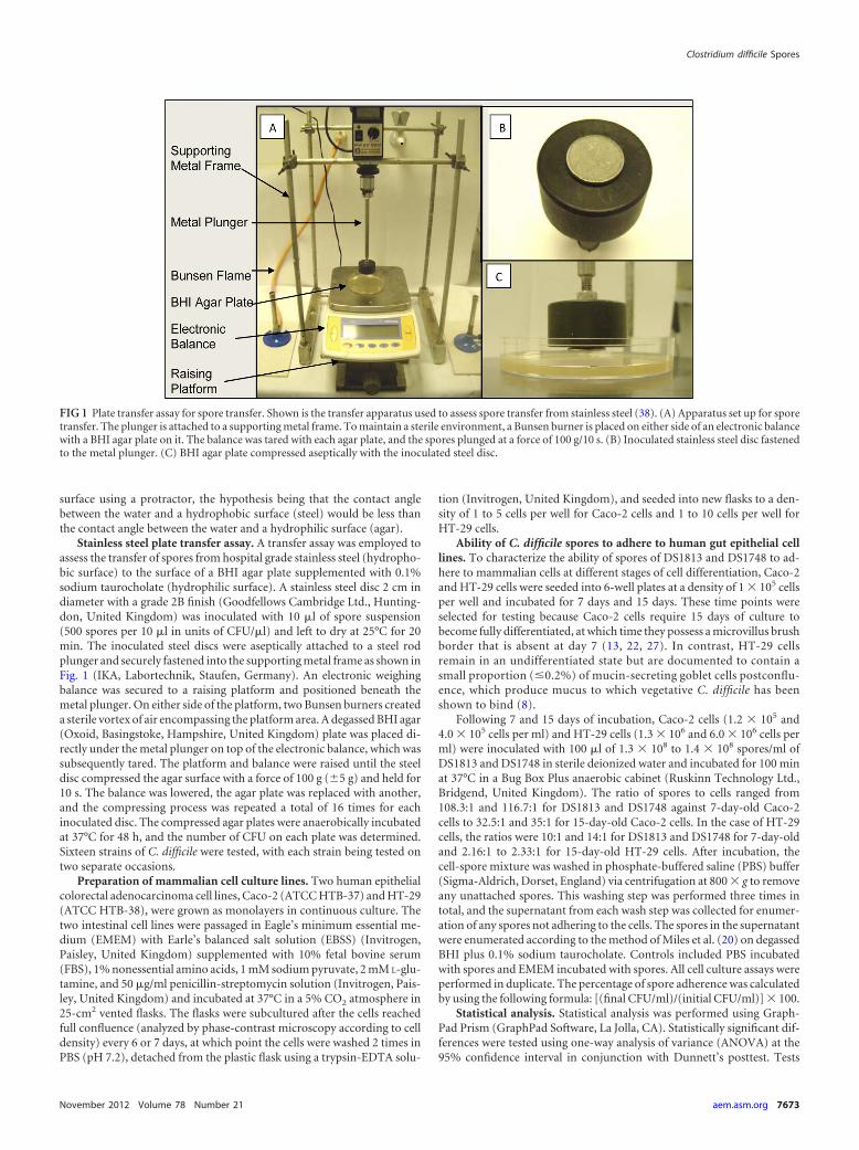
Stainless steel plate transfer assay. A transfer assay was employed toassess the transfer of spores from hospital grade stainless steel (hydropho-bic surface) to the surface of a BHI agar plate supplemented with 0.1%sodium taurocholate (hydrophilic surface). A stainless steel disc 2 cm indiameter with a grade 2B finish (Goodfellows Cambridge Ltd., Hunting-don, United Kingdom) was inoculated with 10 �l of spore suspension(500 spores per 10 �l in units of CFU/�l) and left to dry at 25°C for 20min. The inoculated steel discs were aseptically attached to a steel rodplunger and securely fastened into the supporting metal frame as shown inFig. 1 (IKA, Labortechnik, Staufen, Germany). An electronic weighingbalance was secured to a raising platform and positioned beneath themetal plunger. On either side of the platform, two Bunsen burners createda sterile vortex of air encompassing the platform area. A degassed BHI agar(Oxoid, Basingstoke, Hampshire, United Kingdom) plate was placed di-rectly under the metal plunger on top of the electronic balance, which wassubsequently tared. The platform and balance were raised until the steeldisc compressed the agar surface with a force of 100 g (�5 g) and held for10 s. The balance was lowered, the agar plate was replaced with another,and the compressing process was repeated a total of 16 times for eachinoculated disc. The compressed agar plates were anaerobically incubatedat 37°C for 48 h, and the number of CFU on each plate was determined.Sixteen strains of C. difficile were tested, with each strain being tested ontwo separate occasions.
Preparation of mammalian cell culture lines. Two human epithelialcolorectal adenocarcinoma cell lines, Caco-2 (ATCC HTB-37) and HT-29(ATCC HTB-38), were grown as monolayers in continuous culture. Thetwo intestinal cell lines were passaged in Eagle’s minimum essential me-dium (EMEM) with Earle’s balanced salt solution (EBSS) (Invitrogen,Paisley, United Kingdom) supplemented with 10% fetal bovine serum(FBS), 1% nonessential amino acids, 1 mM sodium pyruvate, 2 mM L-glu-tamine, and 50 �g/ml penicillin-streptomycin solution (Invitrogen, Pais-ley, United Kingdom) and incubated at 37°C in a 5% CO2 atmosphere in25-cm2 vented flasks. The flasks were subcultured after the cells reachedfull confluence (analyzed by phase-contrast microscopy according to celldensity) every 6 or 7 days, at which point the cells were washed 2 times inPBS (pH 7.2), detached from the plastic flask using a trypsin-EDTA solu-
tion (Invitrogen, United Kingdom), and seeded into new flasks to a den-sity of 1 to 5 cells per well for Caco-2 cells and 1 to 10 cells per well forHT-29 cells.
Ability of C. difficile spores to adhere to human gut epithelial celllines. To characterize the ability of spores of DS1813 and DS1748 to ad-here to mammalian cells at different stages of cell differentiation, Caco-2and HT-29 cells were seeded into 6-well plates at a density of 1 � 105 cellsper well and incubated for 7 days and 15 days. These time points wereselected for testing because Caco-2 cells require 15 days of culture tobecome fully differentiated, at which time they possess a microvillus brushborder that is absent at day 7 (13, 22, 27). In contrast, HT-29 cellsremain in an undifferentiated state but are documented to contain asmall proportion (�0.2%) of mucin-secreting goblet cells postconflu-ence, which produce mucus to which vegetative C. difficile has beenshown to bind (8).
Following 7 and 15 days of incubation, Caco-2 cells (1.2 � 105 and4.0 � 105 cells per ml) and HT-29 cells (1.3 � 106 and 6.0 � 106 cells perml) were inoculated with 100 �l of 1.3 � 108 to 1.4 � 108 spores/ml ofDS1813 and DS1748 in sterile deionized water and incubated for 100 minat 37°C in a Bug Box Plus anaerobic cabinet (Ruskinn Technology Ltd.,Bridgend, United Kingdom). The ratio of spores to cells ranged from108.3:1 and 116.7:1 for DS1813 and DS1748 against 7-day-old Caco-2cells to 32.5:1 and 35:1 for 15-day-old Caco-2 cells. In the case of HT-29cells, the ratios were 10:1 and 14:1 for DS1813 and DS1748 for 7-day-oldand 2.16:1 to 2.33:1 for 15-day-old HT-29 cells. After incubation, thecell-spore mixture was washed in phosphate-buffered saline (PBS) buffer(Sigma-Aldrich, Dorset, England) via centrifugation at 800 � g to removeany unattached spores. This washing step was performed three times intotal, and the supernatant from each wash step was collected for enumer-ation of any spores not adhering to the cells. The spores in the supernatantwere enumerated according to the method of Miles et al. (20) on degassedBHI plus 0.1% sodium taurocholate. Controls included PBS incubatedwith spores and EMEM incubated with spores. All cell culture assays wereperformed in duplicate. The percentage of spore adherence was calculatedby using the following formula: [(final CFU/ml)/(initial CFU/ml)] � 100.
Statistical analysis. Statistical analysis was performed using Graph-Pad Prism (GraphPad Software, La Jolla, CA). Statistically significant dif-ferences were tested using one-way analysis of variance (ANOVA) at the95% confidence interval in conjunction with Dunnett’s posttest. Tests
FIG 1 Plate transfer assay for spore transfer. Shown is the transfer apparatus used to assess spore transfer from stainless steel (38). (A) Apparatus set up for sporetransfer. The plunger is attached to a supporting metal frame. To maintain a sterile environment, a Bunsen burner is placed on either side of an electronic balancewith a BHI agar plate on it. The balance was tared with each agar plate, and the spores plunged at a force of 100 g/10 s. (B) Inoculated stainless steel disc fastenedto the metal plunger. (C) BHI agar plate compressed aseptically with the inoculated steel disc.
Clostridium difficile Spores
November 2012 Volume 78 Number 21 aem.asm.org 7673

were also used where there were less than three variables to compare. A Pvalue of �0.05 was considered significant (3). The Student t test was usedto compare the means of samples against a standard reference value (3).Furthermore, a linear regression line analysis was performed to determineif a linear relationship existed between the x and y variables within a plot(3). The equation is defined as follows: y � mx � C, where C is the yintercept and m is the gradient of the slope (3).
RESULTSEffect of medium composition on spore formation. To deter-mine if the ability of C. difficile isolates to form spores was influ-enced by the composition of the growth medium, the abilities ofeight clinical isolates of C. difficile to form spores in BHI andColumbia media were determined. Following incubation for 10days, heat-shocked cultures were diluted and plated onto eitherBHI or Columbia agar containing 0.1% sodium taurocholate totrigger germination. The abilities of isolates to form spores wereaffected by the composition of the media (Fig. 2). This was mostevident for strains R20291 (ribotype 027; Stoke Mandeville iso-late) and DS1801 (ribotype 027), which showed 3-log-unit and2-log-unit differences in spore formation ability, with the highernumber of spores being produced in BHI medium. Indeed, all ofthe strains showed significant differences in their abilities to formspores in BHI and Columbia media, as determined by Student’s ttest (P � 0.018).
The effect of medium composition on spore germinationwith and without the cogerminant sodium taurocholate. As canbe seen from Fig. 2, isolates of C. difficile varied in their abilities togerminate in different growth media in the presence and absenceof the cogerminant 1% sodium taurocholate. The percentage ofspores that germinated in the absence of sodium taurocholatediffered depending on the medium and the isolate (Fig. 3). Indeed,
the results suggest that Columbia medium contains more essentialcogerminants than BHI medium due to the observed variations ingermination ability. Strains DS1801 and DS1813, both of whichbelong to the hypervirulent 027 ribotype, germinated more effi-ciently in the presence of Columbia medium, producing 30% and7% more colonies on Columbia than on BHI medium (Fig. 3).The remaining strains showed no difference in spore germinationefficiency on either Columbia or BHI medium.
Hydrophobicity. The ability of a spore to adhere to a surface isinfluenced by its hydrophobicity. To characterize the relative hy-drophobicity (RH) of spores obtained from a range of clinicalisolates, we employed a MATH test (31) with hexadecane as theorganic solvent. Spores of B. atrophaeus ATCC 9372 (marker)were included as an internal control to enable comparisons toresults from other studies (37). As expected, the RH value for thestrain was similar to that reported by Wiencek et al. (37). As can beseen from Fig. 4, RH values ranged from 77% (DS1813; ribotype027) to 14% (DS1748; ribotype 002). While the most hydrophobicisolates belonged to the hypervirulent (027) and United Kingdomepidemic (001, 078, and 106) ribotypes, there was no significantdifference in the hydrophobicities of strains belonging to the sameribotype (Student’s t test; 027, P � 0.005; 106, P � 0.016; 001, P �0.0021; 078, P � 0.01), suggesting that there is no direct relation-ship between hydrophobicity and ribotype.
Ability of C. difficile spores to adhere to stainless steel. Stain-less steel is a material commonly used in the hospital environ-ment, and thus, we sought to determine if spores from a range ofclinical isolates varied in their ability to adhere to this material. Toachieve this, a plate transfer method was employed in which thepercentage of spores that remained attached to the surface of therelatively hydrophobic (45° contact angle) steel disc was deter-
FIG 2 Effect of medium composition on spore formation. The abilities of eight clinical isolates of C. difficile to form spores in Columbia or BHI broth weredetermined. To determine the number of spores formed in each medium, viable counts were performed on the agar form of the medium in the presence andabsence of 1% sodium taurocholate. A significant difference in the abilities of all strains to form spores in BHI and Columbia media was identified (Student’s ttest; P � 0.018; n � 4). The error bars indicate one standard deviation.
Joshi et al.
7674 aem.asm.org Applied and Environmental Microbiology

mined following consecutive impressions on the surfaces of 16hydrophilic (0° contact angle) BHI agar plates (Fig. 5). Regressionanalysis of the relationship between spore adherence and relativehydrophobicity revealed a significant correlation (P � 0.0247).
The effect was most pronounced for spores of DS1813 (81% bind-ing; RH, 77%), which adhered the most firmly to steel, comparedto those of DS1748 (15% binding; RH, 14%), which were the leastadherent. As was the case for the determination of RH, we ob-
FIG 3 Effect of medium composition on spore germination with and without the cogerminant sodium taurocholate. The abilities of spores of eight clinicalisolates of C. difficile to germinate on the corresponding agar form of the medium in the presence and absence of 1% sodium taurocholate were determined. Thepercentage of spore germination was calculated by dividing the number of spores germinated without taurocholate in the medium by the number of sporesgerminated with taurocholate in the medium. This value was then multiplied by 100 to obtain the percent germination. Each result is the mean of four repeats.CB, Columbia broth.
FIG 4 Relative hydrophobicities of spores formed by different ribotypes of C. difficile. The relative hydrophobicities of 21 clinical isolates of C. difficile belongingto different ribotypes were determined using a microbial adhesion to hydrocarbon test in which hexadecane was the organic solvent. Each value is the mean oftwo independent tests. The error bars indicate one standard deviation. Spores of B. atrophaeus ATCC 9372 were included as an internal control.
Clostridium difficile Spores
November 2012 Volume 78 Number 21 aem.asm.org 7675

served no statistical correlation between spore ribotype and sporeadherence to stainless steel.
Adherence of C. difficile spores to human gut epithelial celllines Caco-2 and HT-29. The abilities of spores that differed insurface hydrophobicity to adhere to human gut epithelial cells atdifferent stages of cell development were determined (Fig. 6).
While there was no significant difference in the ability of DS1813and DS1748 spores to adhere to 7-day Caco-2 cells, DS1813 sporesadhered significantly more strongly to 15-day-old Caco-2 cellsthan spores of DS1748 (one-way ANOVA; P � 0.0393). In the caseof the HT-29 cell line, we observed a significant difference in theabilities of the spores of both strains to adhere to 7-day HT-29
FIG 5 Ability of C. difficile spores to adhere to stainless steel. The percentages of C. difficile spores transferred from hydrophobic stainless steel to hydrophilic BHIagar were determined for 21 clinical isolates of C. difficile belonging to different ribotypes. Each assay was repeated twice. Regression analysis revealed a significantcorrelation (P � 0.0247) between spore adherence and relative hydrophobicity. There was no statistical correlation between spore ribotype and spore adherenceto stainless steel. The slope is defined as y � �0.4634x � 66.107.
FIG 6 Adherence of C. difficile spores to human gut epithelial cell monolayers. The spores of DS1813 (hydrophobic) and DS1748 (hydrophilic) were assessed forthe ability to adhere to monolayers of the human colon adenocarcinoma grade II cell line HT-29 and the Caco-2 human colorectal cell line that had beenincubated for 7 and 15 days. Spores were enumerated on 0.1% sodium taurocholate-supplemented BHI agar. The results are presented as percentages of the initialspore count. Each result represents the mean of two independent assays. D, days.
Joshi et al.
7676 aem.asm.org Applied and Environmental Microbiology

cells, with the DS1813 spores adhering more strongly (P � 0.0079)than those produced by DS1748. These results suggest that sporesof DS1813 have surface properties that allow better adhesion tothe cell lines than spores of DS1748. There was no difference in
spore adhesion to HT-29 cells that had been allowed to replicatefor 15 days.
Spore surface characterization by electron microscopy. TEMwas employed to characterize the surface architecture of spores of
FIG 7 Transmission electron microscopy of C. difficile spores. (A) TEM image of a cross section of a hydrophobic spore produced by C. difficile DS1813. Theexosporial layer can be seen. (B) Negative-stain electron microscopy of an exosporium sac surrounding a spore of DS1813. (C) DS1813 with exosporium visible.Subsequent strains did not exhibit an exosporial layer. (D) Strain DS1771; the concentric layers of the spore can be seen, but no exosporial layer is visible. (E)Strain DS1684 does not exhibit an exosporium layer. (F) Strain DS1748 exhibits the concentric spore structure but does not appear to possess an exosporium.Scale bars � 100 nm.
Clostridium difficile Spores
November 2012 Volume 78 Number 21 aem.asm.org 7677

clinical isolates of C. difficile that differed in their hydrophobicityand in their ability to adhere to surfaces. While the spores of all theisolates share common structural features, such as spore coat lay-ers, there were obvious differences (Fig. 7). The spores of the mosthydrophobic isolate, DS1813 (RH, 77%), comprised an electron-dense cortex surrounded by concentric rings of peptidoglycan; asecond, thinner coat layer; and an outer layer known as the exo-sporium (Fig. 7A, B, and C). In contrast, the exosporium wasmissing from spores of less hydrophobic isolates, such as DS1771(RH, 72.2%), DS1684 (RH, 45%), and DS1748 (RH, 14%) (Fig.7D, E, and F).
DISCUSSION
To avoid the toxic effects of oxygen and to survive the transit to anew host, a pathogen forms spores (35). There is considerabledebate as to whether clinical isolates, particularly hypervirulenttype BI/NAP1/027 strains, differ in their abilities to form spores(1, 4). While we are unable to comment on the rate of spore for-mation in this study, we did observe that certain 027 strains, suchas R20291, failed to achieve the same level of spore formationfollowing 10 days of incubation as other isolates and that thesedifferences were growth medium dependent. One can only spec-ulate as to whether a similar difference in spore formation occursin vivo.
Once a spore has been formed, it must escape from the infectedindividual and adopt strategies that enable it to survive and colo-nize an environment prior to finding a new host (35). We ob-served that spores produced by different clinical isolates in thesame culture medium varied in their relative hydrophobicities andin their abilities to adhere to stainless steel, a material commonlyfound in the health care environment. While it is possible thatspore surface properties are affected by the culture medium, thefact that a single medium was employed to determine RH andadherence suggests that the differences seen between strains arereal.
While it is tempting to speculate that hypervirulent ribotypesare better adapted to colonize environmental surfaces and therebyincrease their potential for person-to-person spread, we observedno significant correlation between hydrophobicity and ribotype.Indeed, while the most hydrophobic spores were predominantlyUnited Kingdom epidemic ribotypes, this may simply reflect therelatively high number of these isolates in our study population(11).
In addition to mediating binding to steel, the surface of thespore also facilitates attachment to colonic cells (23). A recentstudy of the binding characteristics of C. difficile spores to undif-ferentiated 5-day-old Caco-2 cells revealed a correlation betweenspore hydrophobicity and cell surface binding (24). While wefailed to observe any difference in the abilities of DS1813 andDS1748 spores to adhere to 7-day Caco-2 cells, we did see amarked difference with 15-day fully differentiated cells expressingthe apical microvilli and brush border proteins (8). To give a morecomprehensive picture of the cell populations encountered in thehuman gut, we also examined the ability of spores to adhere toHT-29 cells. In contrast to the results seen with Caco-2 cells, weobserved a significant difference in the abilities of DS1813 andDS1748 spores to adhere to 7-day HT-29 cells but saw no differ-ence with 15-day-old cells. Overall, these results suggest thatspores of DS1813 have surface properties that allow better adhe-sion to gut cell lines than spores of DS1748 and that the ability of
spores to bind is affected by the morphology and physiology ofspecific gut cells.
It had previously been observed that spores of C. difficile andClostridium sporogenes attach to the apical microvilli of humanepithelial cells (Caco-2 and MRC-5 cells) by means of an exospo-rium-like structure that, in addition to facilitating colonization,acted as an anchor during germination (22, 23). To determine ifthe observed differences in hydrophobicity and adherence weredue to variations in spore structure, we employed electron micros-copy to characterize the morphology of spores. The resulting TEMimages were similar to those presented by others (19, 24). Whilespores of each isolate shared a number of common features, anexosporium-like structure was seen surrounding only DS1813spores. It is thus tempting to speculate that the exosporium con-tributes to the hydrophobicity of the spore surface and plays a rolein mediating adherence to gut cells. Indeed, it was recently re-ported that sonication of C. difficile spores altered the ultrastruc-ture of the exosporium, which resulted in a significant reductionin spore hydrophobicity and adherence to Caco-2 cells (24).
While the precise factors that mediate spore adherence have yetto be defined, the results from this and other studies suggest thatcomponents of the spore surface play a central role in mediatingattachment to inorganic (stainless steel) and organic (human ad-enocarcinoma cells) surfaces. By fully understanding the processby which spores attach to clinically relevant surfaces, we hope tobe able to develop strategies that could inhibit attachment ofspores and thus block the infection cycle.
Finally, once a spore has attached to the surface of a gut cell, itmust transform into its biologically active vegetative form to ini-tiate infection (11). While there appears to be an association be-tween the 027 ribotype and the ability to germinate in Columbiamedium in the absence of taurocholate, the limited number ofisolates examined makes it impossible to draw any solid conclu-sions at this time. The results from this study suggest that furtherexamination of strain DS1801 could aid our understanding of thefactors that regulate germination.
In conclusion, we found that clinical isolates of C. difficile var-ied widely in their abilities to form spores, attach to clinicallyrelevant surfaces, and subsequently germinate in vitro. While thesignificance of these findings for the disease process has yet to bedetermined, this study has highlighted the importance of analyz-ing multiple isolates when attempting to characterize the behaviorof a bacterial species.
ACKNOWLEDGMENTS
We sincerely thank Jon Brazier and Val Hall for their kind donation ofstrains from the national anaerobic reference laboratory in Cardiff,United Kingdom. We also thank Anthony Hann from the Cardiff Schoolof Biosciences Electron Microscopy Department for his assistance withelectron microscopy.
We thank the Society for Applied Microbiology for funding this re-search.
REFERENCES1. Åkerlund DT, et al. 2008. Increased sporulation rate of epidemic Clos-
tridium difficile type 027/NAP1. J. Clin. Microbiol. 46:1530 –1533.2. Andersson A, Granum PE, Ronner U. 1998. The adhesion of Bacillus
cereus spores to epithelial cells might be an additional virulence mecha-nism. Int. J. Food Microbiol. 39:93–99.
3. Bowker DW, Randerson PF. 2007. Practical data analysis workbook forbiosciences, 3rd ed. Pearson Education Ltd., Harlow, Essex, UnitedKingdom.
Joshi et al.
7678 aem.asm.org Applied and Environmental Microbiology

4. Burns DA, Heap JT, Minton NP. 2010. The diverse sporulation charac-teristics of Clostridium difficile clinical isolates are not associated with type.Anaerobe 16:618 – 622.
5. Burns DA, Heap JT, Minton NP. 2010. SleC is essential for germinationof Clostridium difficile spores in nutrient-rich medium supplemented withthe bile salt taurocholate. J. Bacteriol. 192:657.
6. Charlton S, Moir AJG, Baillie L, Moir A. 1999. Characterization of theexosporium of Bacillus cereus. J. Appl. Microbiol. 87:241–245.
7. Doyle RJ, Rosenberg M. 1990. Microbial cell surface hydrophobicity.ASM Press, Washington, DC.
8. Eveillard M, et al. 1993. Identification and characterization of adhesivefactors of Clostridium difficile involved in adhesion to human colonic en-terocyte-like Caco-2 and mucus-secreting HT29 cells in culture. Mol. Mi-crobiol. 7:371–381.
9. Faille C, et al. 2002. Adhesion of Bacillus spores and Escherichia coli cellsto inert surfaces: role of surface hydrophobicity. Can. J. Microbiol. 48:728 –738.
10. Giel JL, Sorg JA, Sonenshein AL, Zhu J. 2010. Metabolism of bile salts inmice influences spore germination in Clostridium difficile. PLoS One5:e8740. doi:10.1371/journal.pone.0008740.
11. Heeg D, Burns DA, Cartman ST, Minton NP. 2012. Spores of Clostrid-ium difficile clinical isolates display a diverse germination response to bilesalts. PLoS One 7:e32381. doi:10.1371/journal.pone.0032381.
12. Hellickson LA, Owens KL. 2007. Cross-contamination of Clostridiumdifficile spores on bed linen during laundering. Am. J. Infect. Control35:32–33.
13. Howell S, Kenny AJ, Turner AJ. 1992. A survey of membrane peptidasesin two human colonic cell lines, Caco-2 and HT-29. Biochem. J. 284:595.
14. Howerton A, Ramirez N, Abel-Santos E. 2011. Mapping interactionsbetween germinants and Clostridium difficile spores. J. Bacteriol. 193:274.
15. Husmark U, Ronner U. 1992. The influence of hydrophobic, electrostaticand morphologic properties on the adhesion of Bacillus spores. Biofouling5:335–344.
16. Klavenes A, Stalheim T, Sjovold O, Josefsen K, Granum PE. 2002.Attachment of Bacillus cereus spores with and without appendages tostainless steel surfaces. Food Bioproducts Proc. 80:312–318.
17. Koshikawa T, et al. 1989. Surface hydrophobicity of spores of Bacillusspp. Microbiology 135:2717.
18. Kuehne SA, et al. 2010. The role of toxin A and toxin B in Clostridiumdifficile infection. Nature 467:711–713.
19. Lawley TD, et al. 2009. Proteomic and genomic characterization of highlyinfectious Clostridium difficile 630 spores. J. Bacteriol. 191:5377–5386.
20. Miles AA, Misra SS, Irwin JO. 1938. The estimation of the bactericidalpower of the blood. J. Hyg. 38:732–749.
21. Moir A. 2006. How do spores germinate? J. Appl. Microbiol. 101:526 –530.
22. Panessa-Warren BJ, Tortora GT, Warren JB. 1997. Exosporial mem-brane plasticity of Clostridium sporogenes and Clostridium difficile. TissueCell 29:449 – 461.
23. Panessa-Warren BJ, Tortora GT, Warren JB. 2007. High resolutionFESEM and TEM reveal bacterial spore attachment. Microsc. Microanal.13:251–266.
24. Paredes-Sabja D, Sarker MR. 2012. Adherence of Clostridium difficilespores to Caco-2 cells in culture. J. Med. Microbiol. 61:1208 –1218.
25. Perez J, Springthorpe VS, Sattar SA. 2005. Activity of selected oxidizingmicrobiocides against the spores of Clostridium difficile: relevance to en-vironmental control. Am. J. Infect. Control 33:320 –325.
26. Pinto M, et al. 1982. Enterocytic differentiation of cultured human coloncancer cells by replacement of glucose by galactose in the medium. Biol.Cell 44:193–196.
27. Pinto M, et al. 1983. Enterocyte-like differentiation and polarization ofthe human colon carcinoma cell line Caco-2 in culture. Biol. Cell 47:323–330.
28. Poutanen SM, Simor AE. 2004. Clostridium difficile-associated diarrheain adults. CMAJ. 171:51–58.
29. Ramirez N, Liggins M, Abel-Santos E. 2010. Kinetic evidence for thepresence of putative germination receptors in Clostridium difficile spores.J. Bacteriol. 192:4215.
30. Ronner U, Husmark U, Hendriksson A. 1990. Adhesion of Bacillusspores in relation to hydrophobicity. J. Appl. Microbiol. 69:550 –556.
31. Rosenburg M, Gutnik D, Rosenberg E. 1980. Adherence of bacteria tohydrocarbons: a simple method for measuring cell-surface hydrophobic-ity. FEMS Microbiol. Lett. 9:29 –33.
32. Rupnik M, Wilcox MH, Gerding DN. 2009. Clostridium difficile infec-tion: new developments in epidemiology and pathogenesis. Nat. Rev. Mi-crobiol. 7:526 –536.
33. Sebaihia M, et al. 2006. The multidrug-resistant human pathogen Clos-tridium difficile has a highly mobile, mosaic genome. Nat. Genet. 38:779 –786.
34. Setlow P. 2003. Spore germination. Curr. Opin. Microbiol. 6:550 –556.35. Sorg JA, Sonenshein AL. 2008. Bile salts and glycine as co-germinants for
Clostridium difficile spores. J. Bacteriol. 190:2505–2512.36. Stabler RA, et al. 2006. Comparative phylogenomics of Clostridium dif-
ficile reveals clade specificity and microevolution of hypervirulent strains.J. Bacteriol. 188:7297–7305.
37. Wiencek KM, Klapes NA, Foegeding PM. 1990. Hydrophobicity ofBacillus and Clostridium spores. Appl. Environ. Microbiol. 56:2600 –2605.
38. Williams GJ, Denyer SP, Hosein IK, Hill DW, Maillard JY. 2009.Limitations of the efficacy of surface disinfection in the healthcare setting.Infect. Control Hosp. Epidemiol. 30:570 –573.
Clostridium difficile Spores
November 2012 Volume 78 Number 21 aem.asm.org 7679
Related Documents