Cancer Biology and Signal Transduction Contribution of Hepatic Organic Anion- Transporting Polypeptides to Docetaxel Uptake and Clearance Hannah H. Lee 1 , Brenda F. Leake 1 , Wendy Teft 2 , Rommel G. Tirona 2 , Richard B. Kim 2 , and Richard H. Ho 1 Abstract The antimicrotubular agent docetaxel is a widely used che- motherapeutic drug for the treatment of multiple solid tumors and is predominantly dependent on hepatic disposition. In this study, we evaluated drug uptake transporters capable of trans- porting radiolabeled docetaxel. By screening an array of drug uptake transporters in HeLa cells using a recombinant vaccinia- based method, five organic anion–transporting polypeptides (OATP) capable of docetaxel uptake were identified: OATP1A2, OATP1B1, OATP1B3, OATP1C1, and Oatp1b2. Kinetic analysis of docetaxel transport revealed similar kinetic parameters among hepatic OATP1B/1b transporters. An assessment of polymorphisms (SNPs) in SLCO1B1 and SLCO1B3 revealed that a number of OATP1B1 and OATP1B3 variants were asso- ciated with impaired docetaxel transport. A Transwell-based vectorial transport assay using MDCKII stable cells showed that docetaxel was transported significantly into the apical com- partment of double-transfected (MDCKII-OATP1B1/MDR1 and MDCKII-OATP1B3/MDR1) cells compared with single- transfected (MDCKII-OATP1B1 and MDCKII-OATP1B3) cells (P < 0.05) or control (MDCKII-Co) cells (P < 0.001). In vivo docetaxel transport studies in Slco1b2 / mice showed approx- imately >5.5-fold higher plasma concentrations (P < 0.01) and approximately 3-fold decreased liver-to-plasma ratio (P < 0.05) of docetaxel compared with wild-type (WT) mice. The plasma clearance of docetaxel in Slco1b2 / mice was 83% lower than WT mice (P < 0.05). In conclusion, this study demon- strates the important roles of OATP1B transporters to the hepatic disposition and clearance of docetaxel, and supporting roles of these transporters for docetaxel pharmacokinetics. Mol Cancer Ther; 14(4); 994–1003. Ó2015 AACR. Introduction Docetaxel is a widely used chemotherapeutic drug for the treatment of multiple solid tumors, including breast, lung, head and neck, stomach, and prostate cancer. There is wide variability in the pharmacokinetics of docetaxel with up to 10-fold difference in drug clearance among patients receiving the same treatment regimens (1). Furthermore, studies have indicated that a mere 50% decrease in docetaxel clearance increased more than 4-fold the odds of developing grade 4 neutropenia (2). Accordingly, this substantial interindividual variability in docetaxel exposure may have important ramifications for clinical efficacy and drug-medi- ated toxicity. Recent studies suggest that drug disposition genes play a major role in the variability of docetaxel disposition (3–5). Furthermore, genetic variation, or polymorphisms, in drug disposition genes contributes to interindividual variability in chemotherapy response and toxicity (6–8). As the disposition of docetaxel is closely related to differences between an individual's ability to metabolize and eliminate this drug (3, 4, 6–8), a major cause of pharmacokinetic variability may be an attribute to differential expression of polymorphic drug-metabolizing enzymes and/or drug-eliminating transporters, but it has still been unclear. Doc- etaxel is mainly metabolized by the hepatic phase I drug-metab- olizing enzyme CYP3A4 and, to a lesser extent, by CYP3A5 (9) and eliminated by multidrug resistance protein 1 (MDR1, ABCB1, and P-glycoprotein; ref. 10). MDR1 is a membrane-localized and ATP- dependent drug efflux transporter with broad substrate specificity and primarily expressed in normal tissues such as the gastroin- testinal tract, the liver, the kidney, and the brain (11, 12). Expression of MDR1 in such tissues results in reduced drug absorption (gastrointestinal tract), enhanced elimination into the bile (liver) and urine (kidney), and reduced accumulation of toxic agents (the brain; ref. 11). Docetaxel is predominantly dependent on hepatic disposi- tion in humans; approximately 75% docetaxel is excreted in bile, but only approximately 5% in urine (13). Until recently, little was known regarding the hepatic uptake of docetaxel. Organic anion–transporting polypeptides (OATP) are primarily expressed in organs of importance to drug disposition such as the liver, intestine, kidney, and brain, and mediate the sodium- independent transport of a diverse range of amphipathic organ- ic compounds, including bile salts, steroid conjugates, thyroid hormones, anionic peptides, and numerous drugs (14, 15). Whereas OATP1A2, which is mainly expressed in brain, kidney, 1 Division of Hematology and Oncology, Department of Pediatrics, Vanderbilt University School of Medicine, Nashville,Tennessee. 2 Divi- sion of Clinical Pharmacology, Schulich School of Medicine and Den- tistry, Western University/University of Western Ontario, London, Ontario, Canada. Note: Supplementary data for this article are available at Molecular Cancer Therapeutics Online (http://mct.aacrjournals.org/). Corresponding Author: Richard Ho, Vanderbilt University Medical Center, 2220 Pierce Avenue, 338 PRB, Nashville, TN 37232-6310. Phone: 615-936-2802; Fax: 615-936-1767; E-mail: [email protected] doi: 10.1158/1535-7163.MCT-14-0547 Ó2015 American Association for Cancer Research. Molecular Cancer Therapeutics Mol Cancer Ther; 14(4) April 2015 994 on March 26, 2020. © 2015 American Association for Cancer Research. mct.aacrjournals.org Downloaded from Published OnlineFirst February 18, 2015; DOI: 10.1158/1535-7163.MCT-14-0547

Welcome message from author
This document is posted to help you gain knowledge. Please leave a comment to let me know what you think about it! Share it to your friends and learn new things together.
Transcript
Cancer Biology and Signal Transduction
Contribution of Hepatic Organic Anion-Transporting Polypeptides to Docetaxel Uptakeand ClearanceHannah H. Lee1, Brenda F. Leake1,Wendy Teft2, Rommel G. Tirona2,Richard B. Kim2, and Richard H. Ho1
Abstract
The antimicrotubular agent docetaxel is a widely used che-motherapeutic drug for the treatment of multiple solid tumorsand is predominantly dependent on hepatic disposition. In thisstudy, we evaluated drug uptake transporters capable of trans-porting radiolabeled docetaxel. By screening an array of druguptake transporters in HeLa cells using a recombinant vaccinia-based method, five organic anion–transporting polypeptides(OATP) capable of docetaxel uptake were identified: OATP1A2,OATP1B1, OATP1B3, OATP1C1, and Oatp1b2. Kinetic analysisof docetaxel transport revealed similar kinetic parametersamong hepatic OATP1B/1b transporters. An assessment ofpolymorphisms (SNPs) in SLCO1B1 and SLCO1B3 revealedthat a number of OATP1B1 and OATP1B3 variants were asso-ciated with impaired docetaxel transport. A Transwell-basedvectorial transport assay using MDCKII stable cells showed that
docetaxel was transported significantly into the apical com-partment of double-transfected (MDCKII-OATP1B1/MDR1and MDCKII-OATP1B3/MDR1) cells compared with single-transfected (MDCKII-OATP1B1 and MDCKII-OATP1B3) cells(P < 0.05) or control (MDCKII-Co) cells (P < 0.001). In vivodocetaxel transport studies in Slco1b2�/� mice showed approx-imately >5.5-fold higher plasma concentrations (P < 0.01) andapproximately 3-fold decreased liver-to-plasma ratio (P < 0.05)of docetaxel compared with wild-type (WT) mice. The plasmaclearance of docetaxel in Slco1b2�/� mice was 83% lowerthan WT mice (P < 0.05). In conclusion, this study demon-strates the important roles of OATP1B transporters to thehepatic disposition and clearance of docetaxel, and supportingroles of these transporters for docetaxel pharmacokinetics. MolCancer Ther; 14(4); 994–1003. �2015 AACR.
IntroductionDocetaxel is a widely used chemotherapeutic drug for the
treatment of multiple solid tumors, including breast, lung, headand neck, stomach, and prostate cancer. There is wide variabilityin the pharmacokinetics of docetaxel with up to 10-fold differencein drug clearance among patients receiving the same treatmentregimens (1). Furthermore, studies have indicated that a mere50% decrease in docetaxel clearance increased more than 4-foldthe odds of developing grade 4 neutropenia (2). Accordingly, thissubstantial interindividual variability in docetaxel exposure mayhave important ramifications for clinical efficacy and drug-medi-ated toxicity.
Recent studies suggest that drug disposition genes play a majorrole in the variability of docetaxel disposition (3–5). Furthermore,genetic variation, or polymorphisms, in drug disposition genes
contributes to interindividual variability in chemotherapyresponse and toxicity (6–8). As the disposition of docetaxel isclosely related to differences between an individual's ability tometabolize and eliminate this drug (3, 4, 6–8), a major cause ofpharmacokinetic variability may be an attribute to differentialexpression of polymorphic drug-metabolizing enzymes and/ordrug-eliminating transporters, but it has still been unclear. Doc-etaxel is mainly metabolized by the hepatic phase I drug-metab-olizing enzymeCYP3A4 and, to a lesser extent, byCYP3A5 (9) andeliminated bymultidrug resistance protein 1 (MDR1, ABCB1, andP-glycoprotein; ref. 10).MDR1 is amembrane-localized and ATP-dependent drug efflux transporter with broad substrate specificityand primarily expressed in normal tissues such as the gastroin-testinal tract, the liver, the kidney, and the brain (11, 12).Expression of MDR1 in such tissues results in reduced drugabsorption (gastrointestinal tract), enhanced elimination intothe bile (liver) and urine (kidney), and reduced accumulationof toxic agents (the brain; ref. 11).
Docetaxel is predominantly dependent on hepatic disposi-tion in humans; approximately 75% docetaxel is excreted inbile, but only approximately 5% in urine (13). Until recently,little was known regarding the hepatic uptake of docetaxel.Organic anion–transporting polypeptides (OATP) are primarilyexpressed in organs of importance to drug disposition such asthe liver, intestine, kidney, and brain, and mediate the sodium-independent transport of a diverse range of amphipathic organ-ic compounds, including bile salts, steroid conjugates, thyroidhormones, anionic peptides, and numerous drugs (14, 15).Whereas OATP1A2, which is mainly expressed in brain, kidney,
1Division of Hematology and Oncology, Department of Pediatrics,Vanderbilt University School of Medicine, Nashville, Tennessee. 2Divi-sion of Clinical Pharmacology, Schulich School of Medicine and Den-tistry, Western University/University of Western Ontario, London,Ontario, Canada.
Note: Supplementary data for this article are available at Molecular CancerTherapeutics Online (http://mct.aacrjournals.org/).
Corresponding Author: Richard Ho, Vanderbilt University Medical Center, 2220Pierce Avenue, 338 PRB, Nashville, TN 37232-6310. Phone: 615-936-2802; Fax:615-936-1767; E-mail: [email protected]
doi: 10.1158/1535-7163.MCT-14-0547
�2015 American Association for Cancer Research.
MolecularCancerTherapeutics
Mol Cancer Ther; 14(4) April 2015994
on March 26, 2020. © 2015 American Association for Cancer Research. mct.aacrjournals.org Downloaded from
Published OnlineFirst February 18, 2015; DOI: 10.1158/1535-7163.MCT-14-0547

and small intestine, is expressed in cholangiocytes of liver, butnot in hepatocytes (16), OATP1B1 and OATP1B3 are expressedhighly in the hepatocytes of the liver and their localization isrestricted to the basolateral membrane of hepatocytes (17).Accordingly, these transporters facilitate the hepatocellularaccumulation of compounds such as drugs before metabolismand biliary secretion, and thus may largely contribute to drugdisposition. As hepatic uptake appears to be an importantfactor in docetaxel clearance, it would be of interest to evaluatehepatic uptake transporters as determinants of docetaxel dis-position. Indeed, previous studies indicated that OATP1B3 iscapable of transporting docetaxel in vitro (3, 18). Recently, astudy also reported that differential expression of the OATP1Bfamily in the human liver regulates the initial step in theelimination of docetaxel, before metabolism (5).
In this study, we identified multiple OATPs capable of trans-porting docetaxel by screening an array of drug uptake transpor-ters in HeLa cells using a recombinant vaccinia-based method.Among them, we aimed to gain more insight into the importanceof hepaticOATPs to the disposition of docetaxel. Thus, we studiedthe roles of OATP1B transporters in the hepatic uptake, clearance,and plasma exposure of docetaxel in vivo and in vitro.
Materials and MethodsChemicals and reagents
Radiolabeled [3H]docetaxel (5 Ci/mmol, 15 Ci/mmol; >98%purity) was obtained from Moravek Biochemicals and Radio-chemicals.Unlabeled docetaxel (>97%purity)was obtained fromSigma-Aldrich. All other chemicals and reagents, unless statedotherwise, were obtained from Sigma-Aldrich research and wereof the highest grade available.
Cell culture and virus preparationHeLa cells were purchased from the ATCC (July 2012) and
cultured in DMEM supplemented with 10% FBS, penicillin (100U/mL), and streptomycin (100 mg/mL; Invitrogen). The polarizedMDCKII (Madin–Darby Canine Kidney II) cells were purchasedfrom the Sigma-Aldrich (January 2013), and we generated severalstable cell lines immediately after obtaining the cells. The originalbank of MDCKII cells is from Public Health England (PHE;London, UK) and PHE authenticates their cell line, includingtesting forMycoplasma by culture isolation, Hoechst DNA stainingandPCR, culture testing for contaminant bacteria, yeast and fungi,species verification by DNA barcoding, and identity verificationby DNA profiling. In addition, stable cell lines generated with theMDCKII cells for this study were confirmed by protein expressionand transport activities using known specific substrates([3H]estrone-3-sulfate for OATP1B1; [3H]cholecystokinin forOATP1B3). All MDCKII cells were cultured in DMEM containinghigh glucose and L-glutamine supplemented with 10% heat-inactivated FBS, penicillin (100 U/mL), and streptomycin (100mg/mL; Invitrogen) and all cells, including HeLa cells, weremaintained in a 5% CO2 atmosphere at 37�C in a humidifiedincubator. Preparation of viral stock of vtf-7 virus was prepared asdescribed previously (19). Briefly, HeLa cells grown to nearconfluence in 25-cm tissue culture plates were infected with 1plaque-forming unit (PFU)/10 cells. After an incubation period of48 h at 37�C, the infected cells were pelleted, homogenized, andrecovered through centrifugation, followed by tittering of viralstock as described by Blakely and colleagues (20).
Wild-type and variant transporter plasmid constructionPreparation of expression plasmids containing human and rat
uptake transporters, including pEF6/V5-His/OATP1B1 and pEF6/V5-His/OATP1B3, has been described previously (21). Briefly, thefull open reading frames (ORF) of human and rat transportercDNAs were obtained by PCR, using AmpliTaq DNA polymerase(PerkinElmer), from cDNA libraries synthesized from a variety ofhuman and rat tissue mRNA, respectively. A pEF6/V5-His/MDR1expression plasmid was also obtained by the same method asabove. Generation of OATP1B1 and OATP1B3 variant expressionplasmids used for in vitro functional studies has been describedpreviously (21, 22). For consistency of expression, all transporterswere packaged into the pEF6/V5-His-TOPO vector (Invitrogen).The expression plasmids for wild-type (WT) pcDNA3.1(þ)/OATP1B1 and pcDNA3.1(þ)/OATP1B3 were constructedby excising out the ORFs of the OATP1B1 and OATP1B3cDNA sequences, respectively, from the plasmids pEF6/V5-His/OATP1B1 and pEF6/V5-His/OATP1B3 with Nhe1 and Xho1, andsubcloning into pcDNA3.1(þ) vector (Invitrogen). To generate anexpression plasmid for pcDNA3.1-zeo(þ)/MDR1 (WT), PCR wasfirst performed with corresponding nucleotides containing startcodon and internal EcoR1 site of MDR1 cDNA sequences usingplasmid pEF6/V5-His/MDR1 as a template, and PCR products (1-1182nts) were then subcloned into pcDNA3.1-zeo(þ) vector(Invitrogen). Second, DNA fragments (1171-3843nts) wereobtained by digesting MDR1 cDNA with EcoR1 and Not1 fromplasmid pEF6/V5-His/MDR1. Finally, pcDNA3.1-zeo(þ)/MDR1(WT) plasmid containing the full ORFs (1-3843nts) ofMDR1wasgenerated by inserting the second DNA fragments (1171-3843nts) into the first pcDNA3.1-zeo(þ)/MDR1(1-1182nts)plasmid digested with the same enzymes described above. Allplasmids, includingWTs andvariants,were verifiedby sequencingin the DNA Sequencing Facility (VANTAGE) at Vanderbilt Uni-versity Medical Center.
Measurement of docetaxel transport kineticsDocetaxel transport kinetics were assessed in HeLa cells recom-
binantly expressing OATP1B/1b transporters. To measure trans-port kinetics, [3H]docetaxel uptake during the linear phase (first 3minutes)was assessed in thepresence of various concentrations ofunlabeled compound. Transporter-dependent uptake was deter-mined in parallel experiments as the difference in drug uptakebetween transporter and parental plasmid DNA-transfected HeLacells. Michaelis–Menten-type nonlinear curve fitting was used toestimate the maximal uptake rate (Vmax) and concentration atwhich half the maximal uptake occurs (Km) for OATP1B3 andOatp1b2. For OATP1B1 uptake kinetics, weighted, nonlinearcurve fitting was performed using a two-component model com-prising of a saturable process and a nonsaturable process. Allexperiments were carried out in duplicate on at least 2 to 3experimental days.
Generation of stably transfected MDCKII cellsMDCKII cell lines expressing hepatic uptake transporters or/
and an efflux transporter as well as control cell (MDCKII-Co, MDCKII-MDR1, MDCKII-OATP1B1, MDCKII-OATP1B3,MDCKII-OATP1B1/MDR1, andMDCKII-OATP1B3/MDR1) weregenerated as follows: To generate the single-transfected cells,MDCKII-OATP1B1 and MDCKII-OATP1B3 cells, MDCKII cellswere transfected with the plasmids pcDNA3.1(þ)/OATP1B1 or
OATPs and Docetaxel Disposition
www.aacrjournals.org Mol Cancer Ther; 14(4) April 2015 995
on March 26, 2020. © 2015 American Association for Cancer Research. mct.aacrjournals.org Downloaded from
Published OnlineFirst February 18, 2015; DOI: 10.1158/1535-7163.MCT-14-0547
pcDNA3.1(þ)/OATP1B3 using Lipofectamine 2000 reagent(Invitrogen) according to the manufacturer's instructions. At 18to 32 hours after transfection, the cells were split and divided into10-cm culture dishes with fresh media containing G-418 sulfate(800 mg/mL; Mediatech). After additional selection with G-418sulfate, single colonies of transfectants were screened forOATP1B1 and OATP1B3 protein expression by immunoblotanalysis to detect cell clones with the desired protein expression.Cell clones with highest protein expression comparable with theexpression of the control cell lines were chosen for further experi-ments. For generation of double-(MDCKII-OATP1B1/MDR1and MDCKII-OATP1B3/MDR1) transfected cell lines, single-(MDCKII-OATP1B1 and MDCKII-OATP1B3) transfected cellswere retransfected with the plasmid pcDNA3.1-zeo(þ)/MDR1using the same transfection reagent mentioned above. For gen-eration of MDCKII-MDR1 cells, MDCKII cells were also trans-fected with the plasmid pcDNA3.1-zeo(þ)/MDR1. After addi-tional selection with zeocin (800 mg/mL; Invitrogen), singlecolonies of all transfectants were screened for MDR1 expressionas well as OATP1B1 and OATP1B3 by immunoblot analysis todetect cell cloneswith thehighest expression. All expression valueswere normalized to the protein b-actin. Consequently, cell cloneswith the highest uptake transporters and/or an efflux transporterexpression were chosen and used for drug transport studies.Control cells (MDCKII-Co) lacking any insert were obtained byfirst transfecting theMDCKII cells with pcDNA3.1(þ) vector only,followed by retransfecting with pcDNA3.1-zeo (þ)vector only.
Preparation of crude cell membrane fractions and cell surfaceproteins
Protein expression from MDCKII stable cells was assessed byimmunoblot analysis of plasma membrane enriched prepara-tions and biotinylated cell surface proteins. Crude cell membranefractions were conducted as described previously with minormodification (23). Briefly, cells were scraped and collected inhypotonic lysis buffer (5 mmol/L Hepes, pH 7.2, 300 mmol/Lmannitol, and 1x protease inhibitor cocktail; Roche) and homog-enizedwith a pellet pestle (Thermo Fisher Scientific) and a syringewith 26G needle. The lysates were centrifuged at 800 � g for 10minutes to remove cell debris, and the supernatant was saved. Thesupernatant was then centrifuged at 30,000 � g at 4�C for 30minutes to obtain membrane pellets. The membrane pellets weredissolved in the same buffer without mannitol and proteinconcentration in pellets was determined using the BicinchoninicAcid Assay (BCA) Protein Assay Kit (Thermo Fisher Scientific).Each 10 mg of extracted membrane proteins was used for immu-noblot analysis.
For the isolation of cell surface fractions, cell surface biotinyla-tion was performed as described previously (21). Briefly, an equalnumber of eachMDCKII stable cell was plated into 6-well cultureplates. Twenty-four hours later, each well was washed in ice-coldPBS-Ca2þ/Mg2þ (138 mmol/L NaCl2, 2.7 mmol/L KCl, 1.5mmol/L KH2PO4, 1 mmol/L MgCl2, 0.1 mmol/L CaCl2, pH7.4) and treated with sulfo-N-hydroxysuccinimide-SS-biotin(ThermoFisher Scientific) for 1hour at 4�C. The cellswerewashedwith ice-cold PBS-Ca2þ/Mg2þ containing 100 mmol/L glycineand lysed with RIPA buffer (10 mmol/L Tris base, 150 mmol/LNaCl, 1 mmol/L EDTA, 0.1% SDS, 1% Triton X-100, pH 8.0)containing 1x protease inhibitor cocktail (Roche). Followingcentrifugation, 140 mL of streptavidin–agarose beads (Thermo
Fisher Scientific)were added to 600mL of cell lysate and incubatedfor 1 hour at room temperature. Beads were washed four timeswith ice-cold lysis buffer, and biotinylated proteins were releasedfrom the beads by adding Laemmli sample buffer (Bio-Rad).Consequently, eluted biotinylated (cell surface expressed) pro-teins were subjected to immunoblot analysis for detection ofOATP1B1, OATP1B3, or MDR1 by specific antibodies.
Immunoblot analysisTo confirm protein expressions from each stable cell line
generated, immunoblot analysis was performed as describedpreviously (14). Briefly, crude cell membrane proteins (each 10mg) and biotinylated fractions were loaded on 10% SDS poly-acrylamide gels and transferred to nitrocellulose membranes(PerkinElmer). After preincubation in PBS containing 0.05%Tween 20 and 5% nonfat dry milk, the blots were first incubatedwith the polyclonal rabbit anti-OATP1B3 antibody (1:2,500dilution) and reprobed with different antibodies later, such asthe polyclonal rabbit anti-OATP1B1 antibody (1:2,500 dilution)and the monoclonal anti-MDR1 antibody (1:1,000 dilution;Santa Cruz Biotechnology) at room temperature for 2 hours. Theblots were washed with PBS containing 0.05% Tween 20 for 15minutes (3� 5minutes) and then incubated for 1 hours with thesecondary antibody an anti-Rabbit IgG for OATP1B1 andOATP1B3 and an anti-mouse IgG antibody for MDR1 conjugatedwith horseradish peroxidase (HRP; 1:10,000 dilution; Promega).The protein bandswere detected using theWestern lightning plus-enhanced chemiluminescence ECL (PerkinElmer). As a loadingcontrol, the blots were stripped and reprobed with the monoclo-nal mouse anti–Naþ/Kþ-ATPase a antibody (1:5,000 dilution;Santa Cruz Biotechnology), followed by incubation with an anti-mouse IgG antibody conjugated with HRP (1:10,000 dilution;Promega).
Transport assaysRecombinant vaccinia-based uptake transport assay. Transportassays using a recombinant vaccinia virus were conducted asdescribed previously (19). Briefly, HeLa cells grown in 12-wellplates (0.8�106 cells/well)were infectedwith vaccinia (vtf-7) at amultiplicity of infection of 10 PFU/cell in Opti-MEM I medium(Invitrogen) and allowed to adsorb for 30 minutes at 37�C. Cellsin each well were then transfected with 1 mg transporter cDNApackaged in pEF6/V5-His-TOPO vector (Invitrogen), along withLipofectin (Invitrogen) and incubated at 37�C for 16 hours. Theparental plasmid lacking any insert was used as control. Uptake(radioactivity) of docetaxel was measured after an incubation of10minutes. Total radioactivity was determined after the additionof cell lysates to vials containing 5 mL of Biodegradable Scintil-lation Cocktail (Amersham Biosciences). Retained cellular radio-activity was quantified by liquid scintillation counter (PerkinEl-mer). Transport activities were expressed in the percentage com-pared with the vector control. All experiments were carried out induplicate on at least 2 to 3 experimental days. In each set ofexperiments, taurocholate uptake into cells transfected to expresssodium/taurocholate cotransporting polypeptide (NTCP) wasincluded as a positive control for transfection and expressionefficiency. Experience with this assay panel has demonstrated thatinclusion of a single positive control is sufficiency, as relativeactivities of each transporter for reference substrates are consistentbetween experiments. In each set of experiments, docetaxel uptake
Lee et al.
Mol Cancer Ther; 14(4) April 2015 Molecular Cancer Therapeutics996
on March 26, 2020. © 2015 American Association for Cancer Research. mct.aacrjournals.org Downloaded from
Published OnlineFirst February 18, 2015; DOI: 10.1158/1535-7163.MCT-14-0547

into the cells transfected with the vector only was included as anegative control to ensure that results were not confounded by aneffect of the transfection process.
Transwell-based vectorial transport assay. Vectorial transportassays were conducted as described previously with minor mod-ifications (24). MDCKII cells were seeded onto 12-Transwell(diameter 12 mm; pore size 0.4 mm; Corning Incorporated) atan initial density of 0.4� 106 cells per well and grown for 3 days.Twenty-four hours before transport experiments, the cells weretreated with 10 mmol/L sodium butyrate (Sigma-Aldrich) toincrease protein expression. Transepithelial resistance was mea-sured in each well using a millicell voltammeter (model ERS;Millipore); wells registering a resistance of 200 O or greater, aftercorrecting for the resistance obtained in control blank wells, wereused in the transport experiments (23). [3H]Docetaxel was dis-solved in the uptake bufferOpti-MEM1medium(Invitrogen) to afinal concentration of 0.1 mmol/L without addition of unlabeleddocetaxel. After washing cells with prewarmed uptake buffer, 0.8mL of uptake buffer without [3H]docetaxel was added to theapical compartments of the cell monolayers. The same amount(0.8 mL) of uptake buffer containing [3H]docetaxel was thenadded to the basolateral compartments of the cell monolayers.Cells were then incubated at 37�C for given time points. After 0.5,1, and 2 hours, aliquots (50 mL) were removed from both theapical and basolateral compartments and plates were placed backto the incubator. After 3 hours incubation, additional 50 mL weretaken from both compartments and docetaxel radioactivity inmedia obtained at each time point was then measured by liquidscintillation counter (PerkinElmer). Finally, the cells were washedthree timeswith ice-cold PBSbuffer andwere lysedwith 0.2%SDSsolution containing 1� protease inhibitor cocktail (Roche). Celllysates were then used to determine the protein concentrations bythe BCA Assay Kit (Thermo Fisher Scientific). To measure theactivity of intracellular docetaxel, cells in Transwell were incubat-ed for 0.5, 1, 2, and 3 hours after adding radiolabeled docetaxel(final conc. 0.1 mmol/L). The Transwell plates were taken out ofthe incubator at each time point. Cells were then washed, lysed,followed by measuring the radioactivity of docetaxel and deter-mining the protein concentration as described above. All experi-ments were performed more than three times in > triplicates.
Docetaxel distribution in Slco1b2�/� miceMale Slco1b2�/� mice, 21 to 24 weeks of age and male WT
littermate controls, were used to determine docetaxel distribu-tion. The radiolabeled [3H]docetaxel (1mg/kg, specific activity 30Ci/mmol) dissolved in ethanol/0.9% saline solution was injectedi.v. into the tail vein of groups of 4mice. Blood samples from eachmouse were drawn from the saphenous vein at 5 and 15 minutesafter injection. After 30 minutes, mice were anesthetized withisofluorane, blood was removed by cardiac puncture and liverswere harvested, weighed, and homogenized with PBS (1% w/vBSA). Total radioactivity was determined after the addition ofplasma (25 mL) or liver homogenates (500 mL) to vials containing5 mL of Ultima Gold Scintillation Cocktail (PerkinElmer LASCanada Inc.). Docetaxel clearance after i.v. injection was calcu-lated as dose/AUC, where AUC is the area under the plasmaconcentration-time profile from t ¼ 0 to ¥. The protocols for theanimal experiments were approved by The University of WesternOntario Animal Care Committee.
Data fitting and statistical analysisParameters for saturation kinetics (Vmax andKm)were estimated
by nonlinear curve fitting using Prism (GraphPad Software, Inc.)or Scientist (Micromath Scientific Software). Determination ofstatistical differences between group parameters was determinedusing the Student t test, Mann–Whitney U test, one way ANOVA(using Tukey–Kramer multiple comparison test), or Fisher exacttest, as appropriate. A P value of <0.05 was taken to be theminimum level of statistical significance.
ResultsDocetaxel uptake is mediated bymultiple OATP transporters invitro
A panel of uptake transporter expression plasmids wasexpressed individually in HeLa cells using recombinant vacciniaand evaluated for docetaxel transport. After 10 minutes incuba-tion, we identified several members of the human OATP familycapable of docetaxel uptake, including OATP1A2, OATP1B1,OATP1B3, and OATP1C1 (Fig. 1). Rat Oatp1b2, orthologous tohuman OATP1B1 and OATP1B3, also showed efficient docetaxeluptake. However, not all OATPs were capable of transportingdocetaxel as OATP2B1, OATP3A1, OATP4A1, and OATP5A1demonstrated no significant docetaxel uptake. In addition, otherknown drug uptake transporters such as the organic anion trans-porters (OAT), organic cation transporters, as well as bile aciduptake transporters such as NTCP and apical sodium-dependentbile acid transporter (ASBT), exhibited no docetaxel transport.
Time course experiments indicated significant accumulationof docetaxel over control in OATP1B1-, OATP1B3-, and ratOatp1b2-expressingHeLa cells up to 30minutes after incubation.To gain a better understanding of the pharmacokinetic parametersof docetaxel transport mediated by hepatic OATP transporters,kinetic analysis was performed. Radiolabeled docetaxel uptakewas assessed in the presence of varying concentrations of unla-beled docetaxel (0.1–50 mmol/L for OATP1B3; 0.1–300 mmol/LforOATP1B1; 0.1–100 mmol/L forOatp1b2) for estimation ofKm
Figure 1.Multiple OATPs transport docetaxel in vitro. A panel of OATP drug uptaketransporters was assessed for docetaxel (0.1 mmol/L) transport activity at 10minutes. Vector control (pEF6) was used as a reference. Multiple humanOATPs, including OATP1A2, OATP1B1, OATP1B3, and OATP1C1, are capable oftransporting docetaxel in vitro. Rat Oatp1b2 also transports docetaxel in vitro.Data, the percentage of cellular uptake compared with vector control (mean� SE, n ¼ 6); � , P < 0.05; ��� , P < 0.001.
OATPs and Docetaxel Disposition
www.aacrjournals.org Mol Cancer Ther; 14(4) April 2015 997
on March 26, 2020. © 2015 American Association for Cancer Research. mct.aacrjournals.org Downloaded from
Published OnlineFirst February 18, 2015; DOI: 10.1158/1535-7163.MCT-14-0547
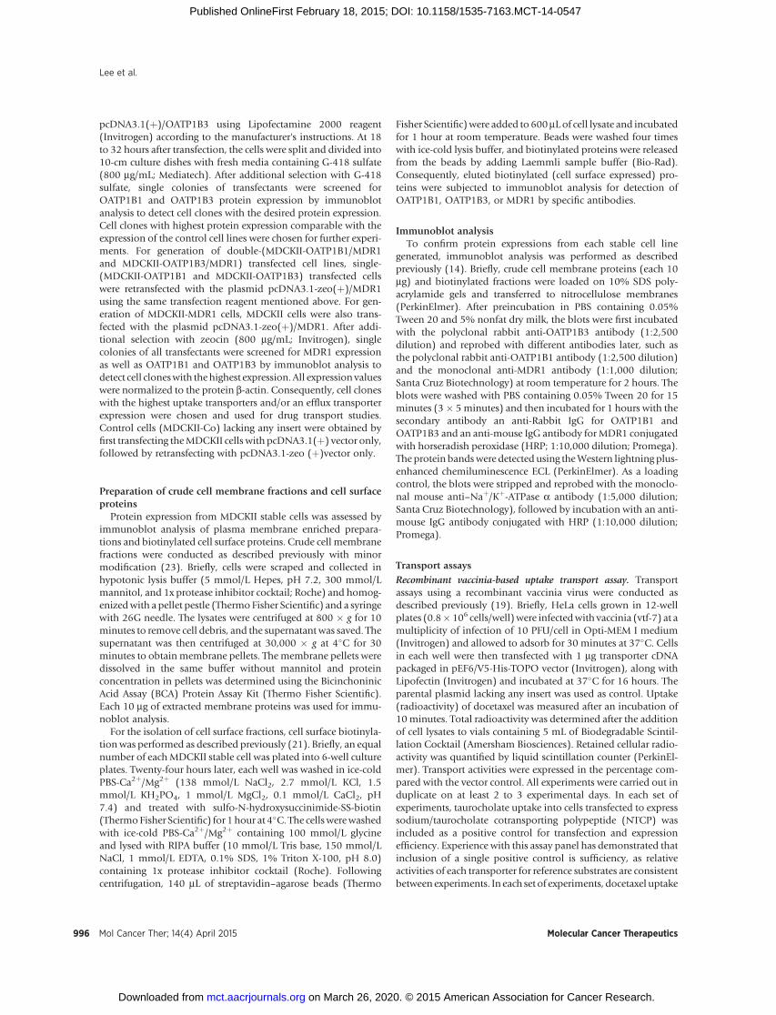
and Vmax. Interestingly, kinetic analysis for OATP1B1 suggestedtwo kinetic components for transport. An Eadie–Hofstee plottogether with curve fitting indicated that, for OATP1B1, transportkinetics at concentrations up to 300 mmol/L are described by ahigh-affinity, low-capacity component (Km 0.43 � 1.32 mmol/L,Vmax 37.7 � 47.2 pmol/mg/min) together with a nonsaturablecomponent that is low affinity, high capacity (Fig. 2A). Transportof docetaxel into HeLa cells transfected with OATP1B3 orOatp1b2 was time-dependent and saturable with an Km of 14� 6.5 mmol/L and 20.8 � 7.4 mmol/L, respectively, and a max-imum velocityVmax of 480� 90, and 1,548� 186 pmol/mg/min,respectively (Fig. 2B and C).
OATP1B1 and OATP1B3 variants differentially transportdocetaxel in vitro.
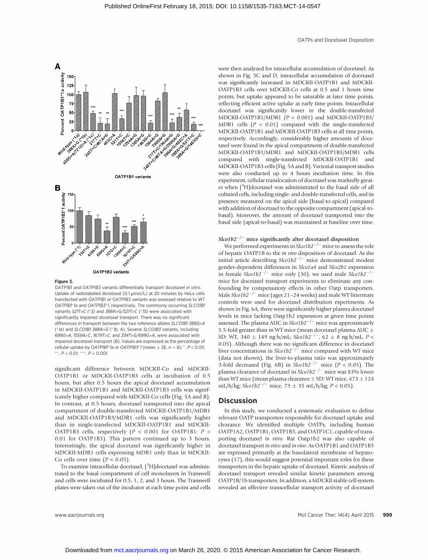
Each panel of OATP1B1 (Supplementary Table S1) andOATP1B3 (Supplementary Table S2) nonsynonymous variantswas expressed in HeLa cells and evaluated for differential trans-port of docetaxel in vitro. A number of variants were associatedwith significantly impaired docetaxel uptake when comparedwith the WT reference alleles, SLCO1B1�1a or SLCO1B3�1. Per-taining to SLCO1B1 encoding OATP1B1, most notably, the com-mon alleles, SLCO1B1 521T>C (�5) and 388A>G/521T>C (�15),demonstrated significantly reduced docetaxel uptake (Fig. 3A).The two reference alleles, SLCO1B1 388G>A (�1a) and SLCO1B1388A>G (�1b), exhibited equivalent docetaxel transport activity.Regarding SLCO1B3 encoding OATP1B3, several nonsynon-ymous SNPs were associated with impaired docetaxel transportin vitro, including SLCO1B3 699G>A, 1559A>C, 1679T>C, and334T>G/699G>A (Fig. 3B).
Protein expression of all stable MDCKII cell linesTo investigate the directional transport activity of docetaxel,
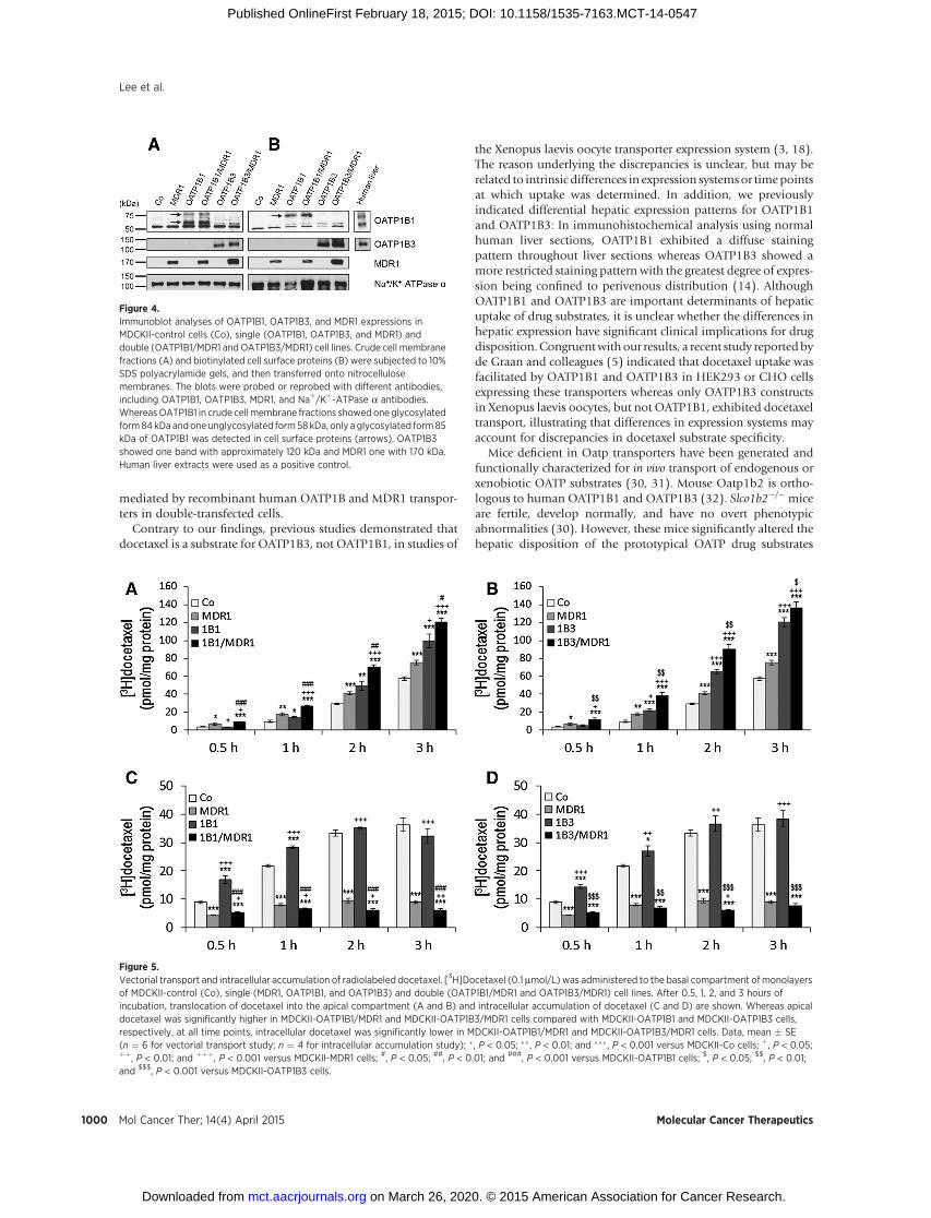
we generated MDCKII cell lines stably expressing hepaticuptake transporters OATP1B1 or OATP1B3 and/or an effluxtransporter MDR1. OATP1B1, OATP1B3, and MDR1 proteinexpression from stable cells are shown in Fig. 4. All cell linesexpressed the proteins of predicted molecular weights, whereasno significant amounts of any protein of interest were detect-able in control cells (MDCKII-Co) lacking any insert. OATP1B1in crude cell membrane fractions (Fig. 4A) showed one glyco-sylated form with an apparent molecular weight of 84 kDa andone unglycosylated form (58 kDa), but only glycosylated form85 kDa was detected in cell surface proteins (Fig. 4B). Theseresults support the previous reports that 58 kDa of OATP1B1 isan ER-attached immature form (14, 25). In this study, we didnot detect constitutive OATP1B1 or OATP1B3 protein expres-sion (Fig. 4A and B) as well as OATP1A2 in control cells byimmunoblot analysis, consistent with previous reports (26–28). Furthermore, transcriptional expression of constitutiveOATP1A2 and OATP1B1 had been previously noted in MDCKcells, but not in MDCKII cells (29).
Vectorial docetaxel transport is dependent on OATP1B uptakeand MDR1 efflux
For vectorial transport assay, [3H]docetaxel was administeredto the basal compartment of cell monolayers of control(MDCKII-Co), single (MDCKII-OATP1B1, MDCKII-OATP1B3,and MDCKII-MDR1)-, and double (MDCKII-OATP1B1/MDR1and MDCKII-OATP1B3/MDR1)-transfected cells. There was no
Figure 2.Docetaxel transport kinetics. An Eadie–Hofstee plot at a broadrange of concentrations (0.1–300 mmol/L; A) for OATP1B1 showed ahigh-affinity, low-capacity component (Km 0.43 � 1.32 mmol/L, Vmax
37.7 � 47.2 pmol/mg/min) together with a nonsaturable componentthat is low-affinity, high-capacity (V, reaction rate; S, substrateconcentration). Michaelis–Menten-type nonlinear curve fittingsare demonstrated for OATP1B3 (0.1–50 mmol/L; B) and Oatp1b2(0.1–100 mmol/L; C). Data, mean � SE (n ¼ 8 for OATP1B3 andOatp1b2;n ¼ 12 for OATP1B1).
Lee et al.
Mol Cancer Ther; 14(4) April 2015 Molecular Cancer Therapeutics998
on March 26, 2020. © 2015 American Association for Cancer Research. mct.aacrjournals.org Downloaded from
Published OnlineFirst February 18, 2015; DOI: 10.1158/1535-7163.MCT-14-0547
significant difference between MDCKII-Co and MDCKII-OATP1B1 or MDCKII-OATP1B3 cells at incubation of 0.5hours, but after 0.5 hours the apical docetaxel accumulationin MDCKII-OATP1B1 and MDCKII-OATP1B3 cells was signif-icantly higher compared with MDCKII-Co cells (Fig. 5A and B).In contrast, at 0.5 hours, docetaxel transported into the apicalcompartment of double-transfected MDCKII-OATP1B1/MDR1and MDCKII-OATP1B3/MDR1 cells was significantly higherthan in single-transfected MDCKII-OATP1B1 and MDCKII-OATP1B3 cells, respectively (P < 0.001 for OATP1B1; P <0.01 for OATP1B3). This pattern continued up to 3 hours.Interestingly, the apical docetaxel was significantly higher inMDCKII-MDR1 cells expressing MDR1 only than in MDCKII-Co cells over time (P < 0.05).
To examine intracellular docetaxel, [3H]docetaxel was adminis-trated to the basal compartment of cell monolayers in Transwelland cells were incubated for 0.5, 1, 2, and 3 hours. The Transwellplates were taken out of the incubator at each time point and cells
were then analyzed for intracellular accumulation of docetaxel. Asshown in Fig. 5C and D, intracellular accumulation of docetaxelwas significantly increased in MDCKII-OATP1B1 and MDCKII-OATP1B3 cells over MDCKII-Co cells at 0.5 and 1 hours timepoints, but uptake appeared to be saturable at later time points,reflecting efficient active uptake at early time points. Intracellulardocetaxel was significantly lower in the double-transfectedMDCKII-OATP1B1/MDR1 (P < 0.001) and MDCKII-OATP1B3/MDR1 cells (P < 0.01) compared with the single-transfectedMDCKII-OATP1B1 andMDCKII-OATP1B3 cells at all time points,respectively. Accordingly, considerably higher amounts of doce-taxel were found in the apical compartment of double-transfectedMDCKII-OATP1B1/MDR1 and MDCKII-OATP1B3/MDR1 cellscompared with single-transfected MDCKII-OATP1B1 andMDCKII-OATP1B3 cells (Fig. 5A and B). Vectorial transport studieswere also conducted up to 4 hours incubation time. In thisexperiment, cellular translocation of docetaxel wasmarkedly great-er when [3H]docetaxel was administrated to the basal side of allcultured cells, including single- anddouble-transfected cells, and itspresence measured on the apical side (basal-to-apical) comparedwith additionof docetaxel to the opposite compartment (apical-to-basal). Moreover, the amount of docetaxel transported into thebasal side (apical-to-basal) was maintained at baseline over time.
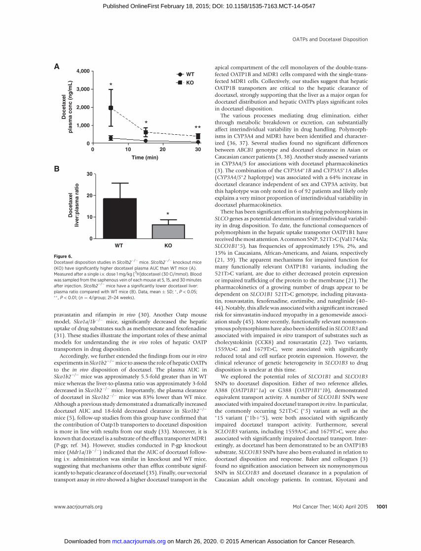
Slco1b2�/� mice significantly alter docetaxel dispositionWeperformed experiments in Slco1b2�/�mice to assess the role
of hepatic OATP1B to the in vivo disposition of docetaxel. As theinitial article describing Slco1b2�/� mice demonstrated modestgender-dependent differences in Slco1a4 and Slco2b1 expressionin female Slco1b2�/� mice only (30), we used male Slco1b2�/�
mice for docetaxel transport experiments to eliminate any con-founding by compensatory effects in other Oatp transporters.Male Slco1b2�/�mice (ages 21–24weeks) andmaleWT littermatecontrols were used for docetaxel distribution experiments. Asshown in Fig. 6A, there were significantly higher plasma docetaxellevels in mice lacking Oatp1b2 expression at given time pointsassessed. The plasma AUC in Slco1b2�/� mice was approximately5.5-fold greater than in WTmice (mean docetaxel plasma AUC�SD: WT, 340 � 149 ng�h/mL; Slco1b2�/�, 62 � 8 ng�h/mL, P <0.05). Although there was no significant difference in docetaxelliver concentrations in Slco1b2�/� mice compared with WT mice(data not shown), the liver-to-plasma ratio was approximately3-fold decreased (Fig. 6B) in Slco1b2�/� mice (P < 0.05). Theplasma clearance of docetaxel in Slco1b2�/� mice was 83% lowerthanWTmice (mean plasma clearance� SD:WTmice, 473� 124mL/h/kg; Slco1b2�/� mice, 73 � 35 mL/h/kg; P < 0.05).
DiscussionIn this study, we conducted a systematic evaluation to define
relevant OATP transporters responsible for docetaxel uptake andclearance. We identified multiple OATPs, including humanOATP1A2,OATP1B1,OATP1B3, andOATP1C1, capable of trans-porting docetaxel in vitro. Rat Oatp1b2 was also capable ofdocetaxel transport in vitro and in vivo. AsOATP1B1 andOATP1B3are expressed primarily at the basolateral membrane of hepato-cytes (17), this would suggest potential important roles for thesetransporters in the hepatic uptake of docetaxel. Kinetic analysis ofdocetaxel transport revealed similar kinetic parameters amongOATP1B/1b transporters. In addition, aMDCKII stable cell systemrevealed an effective transcellular transport activity of docetaxel
Figure 3.OATP1B1 and OATP1B3 variants differentially transport docetaxel in vitro.Uptake of radiolabeled docetaxel (0.1 mmol/L) at 20 minutes by HeLa cellstransfected with OATP1B1 or OATP1B3 variants was assessed relative to WTOATP1B1�1a and OATP1B3�1, respectively. The commonly occurring SLCO1B1variants 521T>C (�5) and 388A>G/521T>C (�15) were associated withsignificantly impaired docetaxel transport. There was no significantdifferences in transport between the two reference alleles SLCO1B1 388G>A(�1a) and SLCO1B1 388A>G (�1b; A). Several SLCO1B3 variants, including699G>A, 1559A>C, 1679T>C, and 334T>G/699G>A, were associated withimpaired docetaxel transport (B). Values are expressed as the percentage ofcellular uptake by OATP1B1�1a or OATP1B3�1 (mean � SE, n ¼ 6); � , P < 0.05;�� , P < 0.01; ��� , P < 0.001.
OATPs and Docetaxel Disposition
www.aacrjournals.org Mol Cancer Ther; 14(4) April 2015 999
on March 26, 2020. © 2015 American Association for Cancer Research. mct.aacrjournals.org Downloaded from
Published OnlineFirst February 18, 2015; DOI: 10.1158/1535-7163.MCT-14-0547
mediated by recombinant human OATP1B and MDR1 transpor-ters in double-transfected cells.
Contrary to our findings, previous studies demonstrated thatdocetaxel is a substrate for OATP1B3, not OATP1B1, in studies of
the Xenopus laevis oocyte transporter expression system (3, 18).The reason underlying the discrepancies is unclear, but may berelated to intrinsic differences in expression systemsor timepointsat which uptake was determined. In addition, we previouslyindicated differential hepatic expression patterns for OATP1B1and OATP1B3: In immunohistochemical analysis using normalhuman liver sections, OATP1B1 exhibited a diffuse stainingpattern throughout liver sections whereas OATP1B3 showed amore restricted staining pattern with the greatest degree of expres-sion being confined to perivenous distribution (14). AlthoughOATP1B1 and OATP1B3 are important determinants of hepaticuptake of drug substrates, it is unclear whether the differences inhepatic expression have significant clinical implications for drugdisposition. Congruentwithour results, a recent study reportedbyde Graan and colleagues (5) indicated that docetaxel uptake wasfacilitated by OATP1B1 and OATP1B3 in HEK293 or CHO cellsexpressing these transporters whereas only OATP1B3 constructsin Xenopus laevis oocytes, but not OATP1B1, exhibited docetaxeltransport, illustrating that differences in expression systems mayaccount for discrepancies in docetaxel substrate specificity.
Mice deficient in Oatp transporters have been generated andfunctionally characterized for in vivo transport of endogenous orxenobiotic OATP substrates (30, 31). Mouse Oatp1b2 is ortho-logous to human OATP1B1 and OATP1B3 (32). Slco1b2�/� miceare fertile, develop normally, and have no overt phenotypicabnormalities (30). However, these mice significantly altered thehepatic disposition of the prototypical OATP drug substrates
Figure 4.Immunoblot analyses of OATP1B1, OATP1B3, and MDR1 expressions inMDCKII-control cells (Co), single (OATP1B1, OATP1B3, and MDR1) anddouble (OATP1B1/MDR1 and OATP1B3/MDR1) cell lines. Crude cell membranefractions (A) and biotinylated cell surface proteins (B) were subjected to 10%SDS polyacrylamide gels, and then transferred onto nitrocellulosemembranes. The blots were probed or reprobed with different antibodies,including OATP1B1, OATP1B3, MDR1, and Naþ/Kþ-ATPase a antibodies.WhereasOATP1B1 in crude cellmembrane fractions showed oneglycosylatedform84kDa andoneunglycosylated form58kDa, only aglycosylated form85kDa of OATP1B1 was detected in cell surface proteins (arrows). OATP1B3showed one band with approximately 120 kDa and MDR1 one with 170 kDa.Human liver extracts were used as a positive control.
Figure 5.Vectorial transport and intracellular accumulation of radiolabeled docetaxel. [3H]Docetaxel (0.1 mmol/L) was administered to the basal compartment of monolayersof MDCKII-control (Co), single (MDR1, OATP1B1, and OATP1B3) and double (OATP1B1/MDR1 and OATP1B3/MDR1) cell lines. After 0.5, 1, 2, and 3 hours ofincubation, translocation of docetaxel into the apical compartment (A and B) and intracellular accumulation of docetaxel (C and D) are shown. Whereas apicaldocetaxel was significantly higher in MDCKII-OATP1B1/MDR1 and MDCKII-OATP1B3/MDR1 cells compared with MDCKII-OATP1B1 and MDCKII-OATP1B3 cells,respectively, at all time points, intracellular docetaxel was significantly lower in MDCKII-OATP1B1/MDR1 and MDCKII-OATP1B3/MDR1 cells. Data, mean � SE(n ¼ 6 for vectorial transport study; n ¼ 4 for intracellular accumulation study); � , P < 0.05; �� , P < 0.01; and ��� , P < 0.001 versus MDCKII-Co cells; þ, P < 0.05;þþ, P < 0.01; and þþþ, P < 0.001 versus MDCKII-MDR1 cells; #, P < 0.05; ##, P < 0.01; and ###, P < 0.001 versus MDCKII-OATP1B1 cells; $, P < 0.05; $$, P < 0.01;and $$$, P < 0.001 versus MDCKII-OATP1B3 cells.
Lee et al.
Mol Cancer Ther; 14(4) April 2015 Molecular Cancer Therapeutics1000
on March 26, 2020. © 2015 American Association for Cancer Research. mct.aacrjournals.org Downloaded from
Published OnlineFirst February 18, 2015; DOI: 10.1158/1535-7163.MCT-14-0547
pravastatin and rifampin in vivo (30). Another Oatp mousemodel, Slco1a/1b�/� mice, significantly decreased the hepaticuptake of drug substrates such as methotrexate and fexofenadine(31). These studies illustrate the important roles of these animalmodels for understanding the in vivo roles of hepatic OATPtransporters in drug disposition.
Accordingly, we further extended the findings from our in vitroexperiments in Slco1b2�/�mice to assess the role of hepaticOATPsto the in vivo disposition of docetaxel. The plasma AUC inSlco1b2�/� mice was approximately 5.5-fold greater than in WTmice whereas the liver-to-plasma ratio was approximately 3-folddecreased in Slco1b2�/� mice. Importantly, the plasma clearanceof docetaxel in Slco1b2�/� mice was 83% lower than WT mice.Although a previous study demonstrated a dramatically increaseddocetaxel AUC and 18-fold decreased clearance in Slco1b2�/�
mice (5), follow-up studies from this group have confirmed thatthe contribution of Oatp1b transporters to docetaxel dispositionis more in line with results from our study (33). Moreover, it isknown that docetaxel is a substrate of the efflux transporterMDR1(P-gp; ref. 34). However, studies conducted in P-gp knockoutmice (Mdr1a/1b�/�) indicated that the AUC of docetaxel follow-ing i.v. administration was similar in knockout and WT mice,suggesting that mechanisms other than efflux contribute signif-icantly tohepatic clearanceof docetaxel (35). Finally, our vectorialtransport assay in vitro showed a higher docetaxel transport in the
apical compartment of the cell monolayers of the double-trans-fected OATP1B and MDR1 cells compared with the single-trans-fected MDR1 cells. Collectively, our studies suggest that hepaticOATP1B transporters are critical to the hepatic clearance ofdocetaxel, strongly supporting that the liver as a major organ fordocetaxel distribution and hepatic OATPs plays significant rolesin docetaxel disposition.
The various processes mediating drug elimination, eitherthrough metabolic breakdown or excretion, can substantiallyaffect interindividual variability in drug handling. Polymorph-isms in CYP3A4 and MDR1 have been identified and character-ized (36, 37). Several studies found no significant differencesbetween ABCB1 genotype and docetaxel clearance in Asian orCaucasian cancer patients (3, 38). Another study assessed variantsin CYP3A4/5 for associations with docetaxel pharmacokinetics(3). The combination of the CYP3A4�1B and CYP3A5�1A alleles(CYP3A4/5�2 haplotype) was associated with a 64% increase indocetaxel clearance independent of sex and CYP3A activity, butthis haplotype was only noted in 6 of 92 patients and likely onlyexplains a very minor proportion of interindividual variability indocetaxel pharmacokinetics.
There has been significant effort in studying polymorphisms inSLCO genes as potential determinants of interindividual variabil-ity in drug disposition. To date, the functional consequences ofpolymorphism in the hepatic uptake transporter OATP1B1 havereceived themost attention. A commonSNP, 521T>C(Val174Ala;SLCO1B1�5), has frequencies of approximately 15%, 2%, and15% in Caucasians, African-Americans, and Asians, respectively(21, 39). The apparent mechanisms for impaired function formany functionally relevant OATP1B1 variants, including the521T>C variant, are due to either decreased protein expressionor impaired trafficking of the protein to the membrane (21). Thepharmacokinetics of a growing number of drugs appear to bedependent on SLCO1B1 521T>C genotype, including pitavasta-tin, rosuvastatin, fexofenadine, ezetimibe, and nateglinide (40–44).Notably, this allelewas associatedwith a significant increasedrisk for simvastatin-induced myopathy in a genomewide associ-ation study (45). More recently, functionally relevant nonsynon-ymous polymorphisms have also been identified in SLCO1B3 andassociated with impaired in vitro transport of substrates such ascholecystokinin (CCK8) and rosuvastatin (22). Two variants,1559A>C and 1679T>C, were associated with significantlyreduced total and cell surface protein expression. However, theclinical relevance of genetic heterogeneity in SLCO1B3 to drugdisposition is unclear at this time.
We explored the potential roles of SLCO1B1 and SLCO1B3SNPs to docetaxel disposition. Either of two reference alleles,A388 (OATP1B1�1a) or G388 (OATP1B1�1b), demonstratedequivalent transport activity. A number of SLCO1B1 SNPs wereassociatedwith impaired docetaxel transport in vitro. In particular,the commonly occurring 521T>C (�5) variant as well as the�15 variant (�1bþ�5), were both associated with significantlyimpaired docetaxel transport activity. Furthermore, severalSCLO1B3 variants, including 1559A>C and 1679T>C, were alsoassociated with significantly impaired docetaxel transport. Inter-estingly, as docetaxel has been demonstrated to be an OATP1B3substrate, SLCO1B3 SNPs have also been evaluated in relation todocetaxel disposition and response. Baker and colleagues (3)found no signification association between six nonsynonymousSNPs in SLCO1B3 and docetaxel clearance in a population ofCaucasian adult oncology patients. In contrast, Kiyotani and
4,000
3,000
2,000
1,000
00 10 20
WTA
B
KO
KOWT
*
*
*
**
Time (min)
30
Do
ceta
xel
pla
sma
con
c (n
g/m
L)
30
20
10
0
Do
ceta
xel
liver
:pla
sma
rati
o
Figure 6.Docetaxel disposition studies in Slco1b2�/� mice. Slco1b2�/� knockout mice(KO) have significantly higher docetaxel plasma AUC than WT mice (A).Measured after a single i.v. dose 1 mg/kg [3H]docetaxel (30 Ci/mmol). Bloodwas sampled from the saphenous vein of each mouse at 5, 15, and 30minutesafter injection. Slco1b2�/� mice have a significantly lower docetaxel liver:plasma ratio compared with WT mice (B). Data, mean � SD; � , P < 0.05;�� , P < 0.01; (n ¼ 4/group; 21–24 weeks).
OATPs and Docetaxel Disposition
www.aacrjournals.org Mol Cancer Ther; 14(4) April 2015 1001
on March 26, 2020. © 2015 American Association for Cancer Research. mct.aacrjournals.org Downloaded from
Published OnlineFirst February 18, 2015; DOI: 10.1158/1535-7163.MCT-14-0547
colleagues (46) identified a noncoding intronic SNP in SLCO1B3(rs11045585) that was associated with a significantly higher riskfor docetaxel-induced leucopenia/neutropenia in a population ofJapanese adult oncology patients. Finally, de Graan and collea-gues (5) found no significant differences in docetaxel clearanceamong a predominantly white adult oncology population (n ¼141) when assessed for commonly occurring polymorphisms inSLCO1B1 or 1B3. As OATP1B1 and OATP1B3 are both expressedat the basolateral membrane of hepatocytes and can facilitatedocetaxel uptake, it is possible that a potential loss of docetaxeluptake associatedwith polymorphic variant in eitherOATP1B1or1B3 may be compensated for by the other OATP1B transporter,thereby mitigating the influence of the polymorphism on hepaticclearance. However, additional clinical PK:PG correlative studiesin larger populations of patients may clarify the relevance ofOATP1B polymorphisms to the wide variability in docetaxelpharmacokinetics.
In conclusion, we describe important roles for hepatic OATPs,including OATP1B1 and OATP1B3, to the disposition of doce-taxel. Through a series of in vitro and in vivo experiments, wedemonstrate that hepatic OATP1B transporters play significantroles in the hepatic uptake, clearance, and plasma exposure ofdocetaxel. Moreover, we indicate that commonly occurringSLCO1B1 and SLCO1B3 variants differentially transport docetaxelin vitro, which may contribute to the oft-witnessed wide interin-dividual variability in docetaxel disposition and response.Accordingly, our findings reveal important insights into therelevance of hepatic OATPs to the clinical pharmacology ofdocetaxel. In addition, we suggest that docetaxel is transported
by multiple OATPs, which may also play important roles ondocetaxel disposition, pharmacokinetics, and toxicities.
Disclosure of Potential Conflicts of InterestNo potential conflicts of interest were disclosed.
Authors' ContributionsConception and design: H.H. Lee, W. Teft, R.G. Tirona, R.B. Kim, R.H. HoDevelopment of methodology: H.H. Lee, R.H. HoAcquisition of data (provided animals, acquired and managed patients,provided facilities, etc.): B.F. Leake, W. Teft, R.H. HoAnalysis and interpretation of data (e.g., statistical analysis, biostatistics,computational analysis): H.H. Lee, W. Teft, R.G. Tirona, R.B. Kim, R.H. HoWriting, review, and/or revision of the manuscript: H.H. Lee, R.G. Tirona,R.B. Kim, R.H. HoAdministrative, technical, or material support (i.e., reporting or organizingdata, constructing databases): B.F. LeakeStudy supervision: R.B. Kim, R.H. HoOther (conducted experiments): H.H. Lee
Grant SupportThis project was supported by grants from Hyundai Hope on Wheels
(to R.H. Ho), NIH R01 GM099924 (to R.H. Ho), and Canadian Institutesof Health Research (CIHR) grants (MOP-8975 and DSEN-PREVENT FRN-117588; to R.B. Kim).
The costs of publication of this articlewere defrayed inpart by the payment ofpage charges. This article must therefore be hereby marked advertisement inaccordance with 18 U.S.C. Section 1734 solely to indicate this fact.
Received June26, 2014; revised January21, 2015; accepted February 10, 2015;published OnlineFirst February 18, 2015.
References1. Baker SD, SparreboomA, Verweij J. Clinical pharmacokinetics of docetaxel:
recent developments. Clin Pharmacokinet 2006;45:235–52.2. Bruno R, Hille D, Riva A, Vivier N, ten Bokkel Huinnink WW, van
Oosterom AT, et al. Population pharmacokinetics/pharmacodynamicsof docetaxel in phase II studies in patients with cancer. J Clin Oncol1998;16:187–96.
3. Baker SD, Verweij J, Cusatis GA, vanSchaik RH, Marsh S, Orwick SJ, et al.Pharmacogenetic pathway analysis of docetaxel elimination. Clin Phar-macol Ther 2009;85:155–63.
4. Jabir RS, Naidu R, Annuar MA, Ho GF, Munisamy M, Stanslas J. Pharma-cogenetics of taxanes: impact of gene polymorphisms of drug transporterson pharmacokinetics and toxicity. Pharmacogenomics 2012;13:1979–88.
5. de Graan AJ, Lancaster CS, Obaidat A, Hagenbuch B, Elens L, Friberg LE,et al. Influence of polymorphic OATP1B-type carriers on the disposition ofdocetaxel. Clin Cancer Res 2012;18:4433–40.
6. Kim KP, Ahn JH, Kim SB, Jung KH, Yoon DH, Lee JS, et al. Prospectiveevaluation of the drug-metabolizing enzyme polymorphisms and toxicityprofile of docetaxel in Korean patients with operable lymph node-positivebreast cancer receiving adjuvant chemotherapy. Cancer Chemother Phar-macol 2012;69:1221–7.
7. Tsai SM, Lin CY, Wu SH, Hou LA, Ma H, Tsai LY, et al. Side effects afterdocetaxel treatment in Taiwanese breast cancer patients with CYP3A4,CYP3A5, and ABCB1 gene polymorphisms. Clin Chim Acta 2009;404:160–5.
8. Tran A, Jullien V, Alexandre J, Rey E, Rabillon F, Girre V, et al. Pharma-cokinetics and toxicity of docetaxel: role of CYP3A, MDR1, and GSTpolymorphisms. Clin Pharmacol Ther 2006;79:570–80.
9. van Herwaarden AE, Wagenaar E, van der Kruijssen CM, van WaterschootRA, Smit JW, Song JY, et al. Knockout of cytochrome P450 3A yields newmouse models for understanding xenobiotic metabolism. J Clin Invest2007;117:3583–92.
10. Wils P, Phung-Ba V, Warnery A, Lechardeur D, Raeissi S, Hidalgo IJ, et al.Polarized transport of docetaxel and vinblastine mediated by P-glycopro-
tein in human intestinal epithelial cell monolayers. Biochem Pharmacol1994;48:1528–30.
11. Gottesman MM, Pastan I. Biochemistry of multidrug resistance mediatedby the multidrug transporter. Annu Rev Biochem 1993;62:385–427.
12. Leveque D, Jehl F. P-glycoprotein and pharmacokinetics. Anticancer Res1995;15:331–6.
13. Cortes JE, Pazdur R. Docetaxel. J Clin Oncol 1995;13:2643–55.14. Ho RH, Tirona RG, Leake BF, Glaeser H, Lee W, Lemke CJ, et al. Drug and
bile acid transporters in rosuvastatin hepatic uptake: function, expression,and pharmacogenetics. Gastroenterology 2006;130:1793–806.
15. Hagenbuch B, Meier PJ. The superfamily of organic anion transportingpolypeptides. Biochim Biophys Acta 2003;1609:1–18.
16. Iusuf D, van de Steeg E, Schinkel AH. Functions of OATP1A and 1Btransporters in vivo: insights from mouse models. Trends Pharmacol Sci2012;33:100–8.
17. Hagenbuch B, Gui C. Xenobiotic transporters of the human organic aniontransporting polypeptides (OATP) family. Xenobiotica 2008;38:778–801.
18. Smith NF, AcharyaMR, Desai N, FiggWD, SparreboomA. Identification ofOATP1B3 as a high-affinity hepatocellular transporter of paclitaxel. CancerBiol Ther 2005;4:815–8.
19. Ho RH, Leake BF, Roberts RL, Lee W, Kim RB. Ethnicity-dependentpolymorphism in Naþtaurocholate cotransporting polypeptide(SLC10A1) reveals a domain critical for bile acid substrate recognition. JBiol Chem 2004;279:7213–22.
20. Blakely RD, Clark JA, Rudnick G, Amara SG. Vaccinia-T7 RNA polymeraseexpression system: evaluation for the expression cloning of plasma mem-brane transporters. Anal Biochem 1991;194:302–8.
21. Tirona RG, Leake BF, Merino G, Kim RB. Polymorphisms in OATP-C:identification of multiple allelic variants associated with altered transportactivity among European- and African-Americans. J Biol Chem 2001;276:35669–75.
22. Schwarz UI, Meyer zu Schwabedissen HE, Tirona RG, Suzuki A, Leake BF,Mokrab Y, et al. Identification of novel functional organic anion-
Lee et al.
Mol Cancer Ther; 14(4) April 2015 Molecular Cancer Therapeutics1002
on March 26, 2020. © 2015 American Association for Cancer Research. mct.aacrjournals.org Downloaded from
Published OnlineFirst February 18, 2015; DOI: 10.1158/1535-7163.MCT-14-0547

transporting polypeptide 1B3 polymorphisms and assessment of substratespecificity. Pharmacogenetics Genomics 2011;21:103–14.
23. Kim RB, Fromm MF, Wandel C, Leake B, Wood AJ, Roden DM, et al. Thedrug transporter P-glycoprotein limits oral absorption and brain entry ofHIV-1 protease inhibitors. J Clin Invest 1998;101:289–94.
24. Fahrmayr C, Konig J, Auge D, Mieth M, Munch K, Segrestaa J, et al. Phase Iand II metabolism and MRP2-mediated export of bosentan in a MDCKII-OATP1B1-CYP3A4-UGT1A1-MRP2 quadruple-transfected cell line. Br JPharmacol 2013;169:21–33.
25. Konig J, Cui Y, Nies AT, Keppler D. Localization and genomic organizationof a new hepatocellular organic anion transporting polypeptide. J BiolChem 2000;275:23161–8.
26. KopplowK, Letschert K, Konig J,Walter B, KepplerD.Humanhepatobiliarytransport of organic anions analyzed by quadruple-transfected cells. MolPharmacol 2005;68:1031–8.
27. Sasaki M, Suzuki H, Ito K, Abe T, Sugiyama Y. Transcellular transport oforganic anions across a double-transfected Madin-Darby canine kidney IIcell monolayer expressing both human organic anion-transporting poly-peptide (OATP2/SLC21A6) and Multidrug resistance-associated protein 2(MRP2/ABCC2). J Biol Chem 2002;277:6497–503.
28. Konig J, Cui Y, Nies AT, Keppler D. A novel human organic aniontransporting polypeptide localized to the basolateral hepatocyte mem-brane. Am J Physiol Gastrointest Liver Physiol 2000;278:G156–64.
29. Goh LB, Spears KJ, Yao D, Ayrton A, Morgan P, Roland Wolf C, et al.Endogenous drug transporters in in vitro and in vivomodels for the predictionof drug disposition in man. Biochem Pharmacol 2002;64:1569–78.
30. Zaher H, Meyer zu Schwabedissen HE, Tirona RG, Cox ML, Obert LA,Agrawal N, et al. Targeted disruption ofmurine organic anion-transportingpolypeptide 1b2 (Oatp1b2/Slco1b2) significantly alters disposition ofprototypical drug substrates pravastatin and rifampin. Mol Pharmacol2008;74:320–9.
31. van de Steeg E, Wagenaar E, van der Kruijssen CM, Burggraaff JE, deWaart DR, Elferink RP, et al. Organic anion transporting polypeptide1a/1b-knockout mice provide insights into hepatic handling of biliru-bin, bile acids, and drugs. J Clin Invest 2010;120:2942–52.
32. Fischer WJ, Altheimer S, Cattori V, Meier PJ, Dietrich DR, Hagenbuch B.Organic anion transporting polypeptides expressed in liver and brainmediate uptake ofmicrocystin. Toxicol Appl Pharmacol 2005;203:257–63.
33. Sparreboom A, Mathijssen RH. Hepatic uptake transporters and docetaxeldisposition in mice-letter. Clin Cancer Res 2014;20:4167.
34. Bardelmeijer HA, Ouwehand M, Buckle T, Huisman MT, Schellens JH,Beijnen JH, et al. Low systemic exposure of oral docetaxel inmice resulting
from extensive first-pass metabolism is boosted by ritonavir. Cancer Res2002;62:6158–64.
35. vanWaterschoot RA, Lagas JS, Wagenaar E, Rosing H, Beijnen JH, SchinkelAH. Individual and combined roles of CYP3A, P-glycoprotein (MDR1/ABCB1) and MRP2 (ABCC2) in the pharmacokinetics of docetaxel. Int JCancer 2010;127:2959–64.
36. Zhou SF, Di YM, Chan E, Du YM, Chow VD, Xue CC, et al. Clinicalpharmacogenetics and potential application in personalized medicine.Curr Drug Metab 2008;9:738–84.
37. Chinn LW, Kroetz DL. ABCB1 pharmacogenetics: progress, pitfalls, andpromise. Clin Pharmacol Ther 2007;81:265–9.
38. Cox MC, Low J, Lee J, Walshe J, Denduluri N, Berman A, et al. Influence ofgarlic (Allium sativum) on the pharmacokinetics of docetaxel. Clin CancerRes 2006;12:4636–40.
39. Ho RH, Choi L, Lee W, Mayo G, Schwarz UI, Tirona RG, et al. Effect ofdrug transporter genotypes on pravastatin disposition in European- andAfrican-American participants. Pharmacogenet Genomics 2007;17:647–56.
40. Chung JY, Cho JY, Yu KS, Kim JR, OhDS, Jung HR, et al. Effect of OATP1B1(SLCO1B1) variant alleles on the pharmacokinetics of pitavastatin inhealthy volunteers. Clin Pharmacol Ther 2005;78:342–50.
41. Lee E, Ryan S, Birmingham B, Zalikowski J, March R, Ambrose H, et al.Rosuvastatin pharmacokinetics and pharmacogenetics in white and Asiansubjects residing in the same environment. Clin Pharmacol Ther 2005;78:330–41.
42. Niemi M, Backman JT, Kajosaari LI, Leathart JB, Neuvonen M, Daly AK,et al. Polymorphic organic anion transporting polypeptide 1B1 is a majordeterminant of repaglinide pharmacokinetics. Clin Pharmacol Ther2005;77:468–78.
43. Oswald S, Giessmann T, Luetjohann D, Wegner D, Rosskopf D, WeitschiesW, et al. Disposition and sterol-lowering effect of ezetimibe are influencedby single-dose coadministration of rifampin, an inhibitor of multidrugtransport proteins. Clin Pharmacol Ther 2006;80:477–85.
44. Zhang W, He YJ, Han CT, Liu ZQ, Li Q, Fan L, et al. Effect of SLCO1B1genetic polymorphism on the pharmacokinetics of nateglinide. Br J ClinPharmacol 2006;62:567–72.
45. Link E, Parish S, Armitage J, Bowman L,Heath S,Matsuda F, et al. SLCO1B1variants and statin-induced myopathy—a genomewide study. N Engl JMed 2008;359:789–99.
46. Kiyotani K,Mushiroda T, KuboM, Zembutsu H, Sugiyama Y, Nakamura Y.Association of genetic polymorphisms in SLCO1B3 and ABCC2 withdocetaxel-induced leukopenia. Cancer Sci 2008;99:967–72.
www.aacrjournals.org Mol Cancer Ther; 14(4) April 2015 1003
OATPs and Docetaxel Disposition
on March 26, 2020. © 2015 American Association for Cancer Research. mct.aacrjournals.org Downloaded from
Published OnlineFirst February 18, 2015; DOI: 10.1158/1535-7163.MCT-14-0547

2015;14:994-1003. Published OnlineFirst February 18, 2015.Mol Cancer Ther Hannah H. Lee, Brenda F. Leake, Wendy Teft, et al. to Docetaxel Uptake and ClearanceContribution of Hepatic Organic Anion-Transporting Polypeptides
Updated version
10.1158/1535-7163.MCT-14-0547doi:
Access the most recent version of this article at:
Material
Supplementary
http://mct.aacrjournals.org/content/suppl/2015/02/18/1535-7163.MCT-14-0547.DC1
Access the most recent supplemental material at:
Cited articles
http://mct.aacrjournals.org/content/14/4/994.full#ref-list-1
This article cites 46 articles, 12 of which you can access for free at:
Citing articles
http://mct.aacrjournals.org/content/14/4/994.full#related-urls
This article has been cited by 2 HighWire-hosted articles. Access the articles at:
E-mail alerts related to this article or journal.Sign up to receive free email-alerts
Subscriptions
Reprints and
To order reprints of this article or to subscribe to the journal, contact the AACR Publications Department at
Permissions
Rightslink site. Click on "Request Permissions" which will take you to the Copyright Clearance Center's (CCC)
.http://mct.aacrjournals.org/content/14/4/994To request permission to re-use all or part of this article, use this link
on March 26, 2020. © 2015 American Association for Cancer Research. mct.aacrjournals.org Downloaded from
Published OnlineFirst February 18, 2015; DOI: 10.1158/1535-7163.MCT-14-0547
Related Documents