Contribution of cutaneous inputs from the hindpaw to the control of locomotion: 1. Intact cats. L.J.G. Bouyer* and S. Rossignol. Centre de Recherche en Sciences Neurologiques Faculté de Médecine, Université de Montréal, Montréal, Canada H3C 3J7. Running head: Role of cutaneous inputs during locomotion in intact cats Corresponding Author. Dr. Serge Rossignol Département de Physiologie, CRSN Faculté de Médecine, Université de Montréal C.P. 6128, Succursale Centre-Ville Montréal (Québec), Canada H3C 3J7 Tel: (514) 343-6366, Fax: (514) 343-6113 E-mail: [email protected] *Current address Laurent Bouyer, Ph.D. Department of Rehabilitation, Université Laval and Center for interdisciplinary research in rehabilitation and social integration (CIRRIS) IRDPQ-Site François-Charon 525, blvd Wilfrid-Hamel, Room 1318 Québec, Qc, Canada G1M 2S8 Email: [email protected] Copyright (c) 2003 by the American Physiological Society. Articles in PresS. J Neurophysiol (August 27, 2003). 10.1152/jn.00496.2003

Welcome message from author
This document is posted to help you gain knowledge. Please leave a comment to let me know what you think about it! Share it to your friends and learn new things together.
Transcript
Contribution of cutaneous inputs from the hindpaw to the control of locomotion: 1. Intact cats.
L.J.G. Bouyer* and S. Rossignol.
Centre de Recherche en Sciences Neurologiques Faculté de Médecine,
Université de Montréal, Montréal, Canada H3C 3J7.
Running head: Role of cutaneous inputs during locomotion in intact cats Corresponding Author. Dr. Serge Rossignol Département de Physiologie, CRSN Faculté de Médecine, Université de Montréal C.P. 6128, Succursale Centre-Ville Montréal (Québec), Canada H3C 3J7 Tel: (514) 343-6366, Fax: (514) 343-6113 E-mail: [email protected] *Current address Laurent Bouyer, Ph.D. Department of Rehabilitation, Université Laval and Center for interdisciplinary research in rehabilitation and social integration (CIRRIS) IRDPQ-Site François-Charon 525, blvd Wilfrid-Hamel, Room 1318 Québec, Qc, Canada G1M 2S8 Email: [email protected]
Copyright (c) 2003 by the American Physiological Society.
Articles in PresS. J Neurophysiol (August 27, 2003). 10.1152/jn.00496.2003
Page 2 of 2
ABSTRACT The goal of this study was to evaluate the role of hindpaw cutaneous feedback in the control of
locomotion, by cutting some (in one cat) or all (in two cats) cutaneous nerves bilaterally at ankle level.
Kinematic and EMG recordings were obtained before and for several weeks after denervation during
level and incline (15º up and down) treadmill walking. Ladder walking and ground reaction forces were
also documented sporadically. Early after the denervation (1-3 days), cats could not walk across a
ladder. Yet, deficits were small during level treadmill walking. Increased knee flexion velocity caused a
14% reduction in swing phase duration. EMG activity was consistently increased in knee, ankle and toe
flexors, and in at least one knee or ankle extensor. The adaptive changes during walking on the incline
were much reduced after denervation. Ladder walking gradually recovered within 3-7 weeks. By this
time, level treadmill walking kinematics had completely returned to normal, but EMG activity in flexors
remained above control. Incline walking improved but did not return to normal. Medio-lateral ground
reaction forces during overground walking were increased by 200%. It is concluded that in intact cats,
cutaneous inputs contribute more to demanding situations such as walking on a ladder or on inclines
than to level walking. Active adaptive mechanisms are likely involved since the EMG locomotor pattern
never returned to control level. The companion paper shows on the other hand that when the same cats
are spinalised, these cutaneous inputs become critical for foot placement during locomotion.
250 words
Page 3 of 3
INTRODUCTION
Experiments by Sherrington in the early 1900’s (Sherrington, 1910) suggested that cutaneous
inputs do not play a major role in the control of locomotion in quadrupeds. Indeed, having removed all
cutaneous nerves of the hind and fore paws, Sherrington concluded that this procedure impaired
“walking so little as to make it highly unlikely that the loss of receptivity of the feet destroys any large
factor in the reflex basis of these acts”. Sherrington also reported that, in decerebrate or chronic spinal
cats with similar cutaneous denervation, bilateral rhythmic movements of the hindlimbs could still be
induced by perineal stimulation or extension of the hips (air stepping).
That the generation of the fundamental rhythm of locomotion does not depend essentially on
peripheral afferent inputs (proprioceptive or cutaneous) has now been quite well established. Indeed, the
basic locomotor pattern can be expressed after neuromuscular paralysis which removes all movement
related phasic sensory inputs in spinal cats injected with L-DOPA (Grillner & Zangger, 1979), or after
dorsal rhizotomy in decerebrate cats walking on a treadmill (Grillner & Zangger, 1984).
However, several lines of evidence suggest that afferent inputs may play a regulatory role
(Rossignol et al 1988; Rossignol 1996). For instance, direct measurements (Stein et al., 2000) indicate
that the stretch reflex might contribute up to 30% of the ankle extensor activity during normal walking.
In an attempt to understand such a specific role of afferents, this paper and its companion will
concentrate specifically on the role of cutaneous inputs of the hindpaws in the control of locomotion in
intact and spinal cats.
It is known that stimulation of the foot skin or afferent nerves gives rise to complex phase-dependent
responses involving several muscles and several joints and which are well-integrated within the step
cycle so as not to impede locomotion, but yet efficiently modify the limb trajectory to avoid obstacles
(Rossignol, 1996). These responses not only recruit muscles active during a given phase but also
antagonist muscles at several joints to make these responses appropriate (Drew & Rossignol, 1987).
Work in humans (Zehr & Stein, 1999) not only corroborates these observations but also suggest quite
Page 4 of 4
specific roles for reflex responses evoked by stimulation of the various receptive fields so as to remove
the foot from potentially damaging obstacle. Such a fine “local sign” control is also suggested by work
in rats (Schouenborg & Kalliomaki, 1990), cats (Levinsson et al., 1999), and humans (Van Wezel et al.,
1997;Zehr & Stein, 1999). Therefore, cutaneous afferents are undoubtedly involved in compensating for
perturbations of the locomotor cycle (reviewed in (Rossignol, 1996;Rossignol et al., 1988).
Chronic recordings from dorsal root ganglia in freely walking cats (Loeb et al., 1977) show that
cutaneous inputs from the paw provide a variety of information to the CNS. Some of the recorded units
are phasic, discharging mainly around touch down and toe off, while others are tonic, discharging
throughout stance (Loeb et al., 1977). Moreover, studies performed during fictive locomotion show that
there also exists a sophisticated phase-dependent control of cutaneous afferents at presynaptic (Gossard
et al., 1990;Gossard et al., 1989) and pre-motoneuronal (Degtyarenko et al., 1996;Burke et al., 2001)
sites during locomotion suggesting again that these inputs might have some role to play in the regulation
of locomotion.
Given that cutaneous inputs give rise to elaborate responses to perturbation, that cutaneous receptors
are activated during normal locomotion and that there is a complex pre- and post- synaptic control of
cutaneous afferents, what are the consequences on locomotion of removing these cutaneous inputs?
Previous work using anesthesia of the pads (Engberg, 1964;Wand et al., 1980;Forssberg et al., 1977) or
partial neurectomy (Duysens & Stein, 1978) also confirms Sherrington’s observation that few deficits
are observed (Duysens et al., 2000). The question we ask here is whether these deficits become only
apparent when using more elaborate analysis methods and also more demanding locomotor situations in
which these cutaneous inputs may be crucial.
Another fundamental aspect that motivated the present investigation on the role of cutaneous
inputs of the paws during locomotion is our long-lasting interest in understand the mechanisms that
control the recovery and expression of locomotion in cats spinalised at T13 (Rossignol, 1996;Rossignol
et al., 2000). Indeed, spinal cats might depend even more critically than normal cats on sensory cues to
Page 5 of 5
bear weight and to adapt their walking speed to the treadmill speed, functions they execute with no
difficulty. The peripheral cutaneous neurectomy model used here makes it possible to quite readily
compare the same denervated cats in the intact condition and after spinalisation.
Our results are presented in two companion papers. This first paper describes the effects on
locomotion of depriving the hindpaws of some or all cutaneous inputs in the otherwise intact cat.
Chronic multi-muscle EMG recordings combined with movement kinematics are used to document
treadmill locomotion in cats before and over the weeks following a bilateral hindpaw cutaneous
denervation (complete or incomplete). More demanding locomotor situations such as walking on a
horizontal ladder, inclines and walkway with embedded force platforms complement our observations.
By comparing the details of the locomotor movements, motor patterns and force distribution to the
ground in the presence and absence of skin feedback from the hindpaws, we have obtained valuable
information on some aspects of the role of cutaneous inputs in the adaptation of locomotion. In this first
paper we will conclude that the deficits observed in the otherwise intact cats are minimal in
undemanding locomotor tasks (treadmill) but important in more demanding tasks (incline treadmill,
ladder walking) at least for several weeks after denervation. The companion paper will report the effect
of spinalisation at T13 on the same cats, performed several weeks after recovery from the denervation.
Cats that were well-adapted to the denervation before spinalisation, could not place the foot properly
after spinalisation suggesting a critical role of cutaneous inputs for the correct expression of spinal
locomotion. Parts of these results have been previously presented in abstract form (Bouyer & Rossignol,
1997;Bouyer & Rossignol, 1998a;Bouyer & Rossignol, 1998b).
METHODS
Animals and general procedures
Three adult cats of either sex (weight 3.7-4.5 kg) were used in this study, for a total of 101 recording
sessions. The animals were first selected for their ability to walk on a treadmill and then trained several
Page 6 of 6
times a week to walk at a constant speed on the treadmill. They were subsequently implanted with
chronic electromyographic electrodes, allowed to recover from the implantation, and then recorded to
obtain baseline data for EMG recordings and kinematics. After several controls were obtained, cats were
denervated (see below for specific protocol) and recorded again at different times after the cutaneous
denervation under several conditions: on the treadmill (with or without slopes), on a horizontal ladder
with rungs of different sizes, and on a walkway instrumented with force platforms. After a period of
recovery, the same animals were spinalised, as will be reported in the companion paper.
The experimental protocol was in accordance with the guidelines of the animal Ethics
Committee of the Université de Montréal. All surgical procedures were carried out under general
anesthesia and aseptic conditions in a dedicated operating room. After premedication (Atravet 0.1 mg/kg
S.C., Glycopyrrolate 0.01 mg/kg S.C., and Ketamine 5-10 mg/kg, S.C.), cats were intubated and
maintained under gaseous anesthesia (Isoflurane 2%), while monitoring heart rate and respiration. After
surgery, analgesic (Buprenorphine hydrochloride, 0.005-0.01 mg/kg S.C.) was given every 6-8 hours for
1-2 days. No recordings were made while cats were under analgesics.
Implantation of Electromyographic Electrodes
EMG electrodes were implanted chronically in selected muscles of the hindlimbs. Implantations
were not identical for all cats. DS1 and DS2 were implanted with an emphasis on extensors, and DS3 on
ankle flexors (see Results section). The different muscles implanted and main function were: Sartorius
Anterior (hip flexor/knee extensor), Semitendinosus (knee flexor/hip extensor), Vastus Lateralis (knee
extensor), Tibialis Anterior (ankle flexor), Extensor Digitorum Longus (ankle flexor), Medial
Gastrocnemius (ankle extensor), Lateral Gastrocnemius (ankle extensor), Soleus (ankle extensor), and
Extensor Digitorum Brevis (toe flexor). For each muscle, a pair of Teflon-insulated multistrain fine
wires (50µm diameter; Cooner Wire, Chatsworth, CA, AS633) was led subcutaneously from head
Page 7 of 7
mounted multipin connectors (CINCH Connectors, TTI inc., Pointe-Claire, Canada) and sown into the
belly of the muscle for bipolar EMG recordings.
Cutaneous Denervation
Below the ankle, the skin of the paw is innervated by 5 nerves that cover distinct,
complementary territories (Crouch, 1969). Their name, abbreviation, site of neurectomy and receptive
fields are listed in Table 1. Except for the Tibial and Deep Peroneal n., cutaneous nerves are purely
sensory below the ankle. Cutting cutaneous nerves at the level given in Table 1 therefore caused little
direct impairment in muscle function. Neurectomies consisted of making a small incision in the skin,
exposing the nerve by gently separating it from the adjacent connective and vascular tissues using
hemostats, suturing the proximal and distal parts using silk tread, cutting the nerve, and capping its
proximal end with flexible vinyl polysiloxane (Reprosil, Dentsply International, Milford, DE) to prevent
regrowth. The nerve was then put back in place and the opening closed.
In all cats, neurectomies were performed symmetrically in both hindlimbs. The neurectomies
were made according to the following protocols: for cat DS1, all 5 cutaneous nerves were cut bilaterally
in a single procedure, causing a complete denervation of the hindlimb paws. Cat DS2 was only partially
denervated, keeping the Deep Peroneal nerve intact on both sides. Cat DS3 was denervated in two
stages. Initially, the Deep Peroneal nerve was left intact, while all other nerves were cut. In a second
procedure 13 days later, the cutaneous branch of the Deep Peroneal nerve (DPc) was cut. Care was taken
not to damage the motor branch of this nerve, which innervates EDB. Therefore, 2 cats had eventually a
complete denervation (DS1 and DS3) and 2 cats had a partial denervation, one (DS2) for 76 days and
the other (DS3) for 13 days. This was seen as the optimal combination to reduce the number of
chronically implanted animals. The completeness of the denervation was also evaluated clinically by
gently pinching the skin at different locations on the paw.
Insert Table 1 near here.
Page 8 of 8
EMG and kinematic recordings during treadmill locomotion
Cats were trained to walk regularly on a motorized treadmill under 3 conditions: level, 15
degrees pitch up and 15 degrees pitch down, at speeds varying from 0.3 m/s to 0.7 m/s for level walking,
and 0.3 m/s to 0.5 m/s for incline walking. Reflective markers were placed on the left hindlimb over the
iliac crest, greater trochanter, lateral epicondyle, lateral malleolus, metatarso-phalangeal joint (MTP)
and the tip of the fourth toe. Recording sessions consisted of acquiring video images simultaneously
with the EMG data during treadmill locomotion, in order to fully describe the walking pattern. Filtered
and amplified EMG signals (100 Hz-3 kHz band pass, gains of 500 to 20K) from the implanted muscles
were recorded on VHS tape using a 16-channel Vetter Digital 4000a PCM recorder (1.25 kHz
bandwith/channel). Kinematic and electromyographic (EMG) data were synchronized using a SMPTE
time code generator (Skotel Inc.).
Ladder walking
Cats were also trained to walk regularly on the rungs of a 2 m long by 35 cm wide horizontal
ladder. Three types of rungs were used: round (2.5 cm diameter), small flat (2.5 cm width), and large flat
(6.5 cm width). Only video images were recorded during this task.
Ground reaction forces during overground walking.
Cats DS2 and DS3 were also trained to walk across a 3 m long by 0.6 m wide walkway. A 3-axis
force platform (AMTI, model ORS6-5-1) was installed flush with the surface of the walkway to record
vertical, antero-posterior and medio-lateral ground reaction forces generated by one paw during walking.
A thin opaque plastic mat was placed over the entire walkway, such that the cat could not see where the
force platform was located. Force and moment data were acquired at 1.2 KHz and synchronized to video
images of the cat walking across the walkway.
Page 9 of 9
Analyses
Kinematic analysis. Joint angles and foot lifts/contacts were reconstructed off-line frame-by-frame from
the video images using a Peak Performance motion analysis system (Englewood, CA) with a resolution
of 60 fields/s. The knee angle was mathematically corrected for skin slippage by triangulation using
post-mortem leg segment lengths (femur and tibia). The step cycle was divided into 4 sections,
according to Philippson (Philippson, 1905): the swing phase was subdivided into flexion (F) and the first
extension phase (E1) while the stance phase was divided in two extension phases, weight acceptance
(E2), and push off (E3) (See Fig. 1D).
Double hindlimb support was defined as the % of total cycle time spent with both hindlimbs in
contact with the treadmill belt and evaluated from the videotapes. In addition, the duty cycle, % of total
cycle time with two, three, or four paws on the ground was sometimes quantified. Video segments
where all four limbs were visible over about 10 consecutive steps were analyzed. These segments were
not necessarily the same as used for double hindlimb support analysis, as the camera was usually
zoomed onto the hindlimbs in order to have a better resolution for kinematic analysis.
Kinetic analysis: ground reaction forces were measured off-line from the data obtained during
locomotion on a walkway with embedded force plateforms. Trials were selected for analysis only when
walking speed across the walkway was constant and within the range of 0.35-0.55 m/s (measured using
the video images) and when the cat placed only one paw on the platform. Voltage.signals from the force
platforms where low-pass filtered at 50 Hz using a 2nd order Butterworth filter, calibrated using the
platform’s calibration matrix provided by the manufacturer (AMTI Inc) and converted to Newtons. Each
trial data was then normalized for walking direction, as reported before (Lavoie et al., 1995). Finally,
data for each limb where grouped, synchronized on paw contact and averaged using custom software.
EMG analysis. Electromyographic data recorded on tape were played back off-line and printed out using
an electrostatic plotter (Gould ES-2000), along with their SMPTE time stamp. Data segments consisting
of at least 10 consecutive steps at constant speed were chosen using the EMG printouts and videotapes
Page 10 of 10
of the experiments. Electromyographic data of these chosen segments were then digitized using custom
software at 1000 samples/sec per channel on a 200 MHz Pentium computer. Burst duration, amplitude,
and relative timing of chosen data segments were then measured by manually placing cursors using
custom analysis software.
To compare EMG patterns between sessions (e.g. Fig 1), EMGs were rectified, normalized to
256 points per step, and averaged over at least 10 consecutive steps. Data from two sessions were then
superimposed after normalizing to mean step duration.
The amplitude of EMG activity in extensors was quantified by integrating the area under each
rectified EMG burst and dividing it by the burst duration, a measure called mean amplitude. For flexors,
it was noticed when comparing averaged EMG activity, that the changes caused by the denervation in
activation amplitude were spiky (e.g. St in Fig. 1F-G). Therefore, as the increase in amplitude didn’t last
for the entire burst duration on most occasions, mean amplitude was not the ideal method to assess the
changes in amplitude related to the denervation. For this muscle, and also for the other flexors, peak
amplitude was therefore used to quantify changes in activity instead of mean amplitude. This method
gave very consistent measurements.
Individual time courses of compensation.
Kinematic and EMG recordings on the treadmill were continued until each cat had recovered the
ability to walk across the most challenging situation, i.e. the round rungs. This explains why the abscissa
on the time course graphs presented in this paper is different for each cat.
Statistics.
To measure statistical differences after the denervation, data from at least 3 separate controls
were pooled and compared to data obtained on each of the many post-denervation tests using a one-way
ANOVA followed by Dunnett’s post-hoc test for many comparisons against a control group. If data
Page 11 of 11
failed normality and equivariance tests, an ANOVA on rank (Kruskal-Wallis method) followed by
Dunn’s post-hoc test was applied instead (Glantz, 1992).
RESULTS
Overall, cats recovered very well from the denervation. Surprisingly, they showed very little
slippage of the feet, even on the laboratory floor, which was more slippery than the treadmill belt.
However, lateral stability was not as good as pre-denervation. In addition, in early days, the cats
presented a more cautious type of walking: while standing or walking, the hindlimbs tended to be
abducted and the hindpaws everted. Pinching of the skin of the paw produced no withdrawal response.
1. Level treadmill walking
Although the cats showed some obvious deficits in foot placement during ladder walking (see
below), one of the most striking observation was the absence of any major deficit during level treadmill
walking after the cutaneous denervation. All cats, partially or completely denervated, appeared, at least
to the naked eye, to walk normally on the treadmill. However, a more detailed analysis revealed several
consistent changes in the locomotor movements and EMG activity.
Kinematics and EMGs
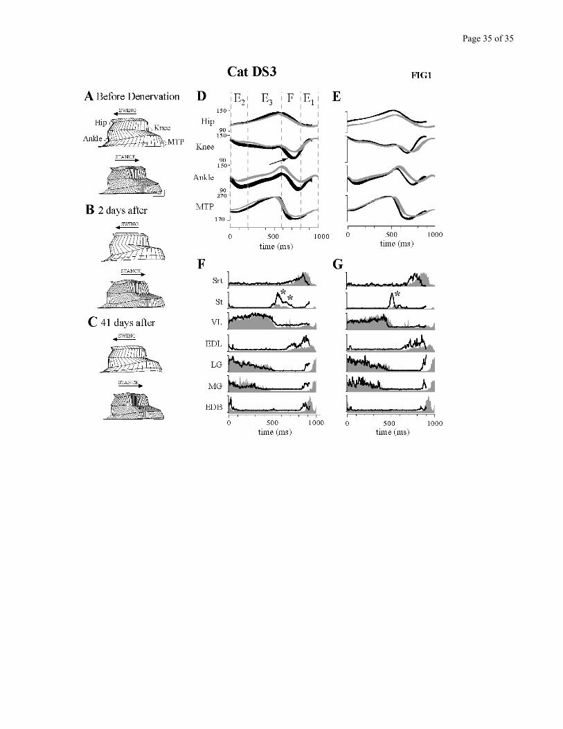
Figure 1 presents an overview of the kinematic and EMG changes observed during level
treadmill locomotion after the cutaneous denervation. This figure summarizes the main changes present
short term (2 days) and long term (41 days) post-denervation by using stick figures, angular excursion
plots and averaged EMGs of the control and the denervated conditions.
Two days after the complete denervation (Fig. 1B), swing was faster but stride length was
similar to the control before denervation (Fig. 1A). Using foot contact as the synchronizing event, Figure
1D represents angular displacements at the hip, knee, ankle and MTP joints, with a superposition of
Page 12 of 12
control and early post-denervation conditions. At the knee, flexion during the swing phase increased
both in amplitude and velocity, as shown by a steeper slope of the angular excursion (c.f. arrow). The
changes at the ankle result in part from a more crouched posture. This Figure also shows that the overall
cycle duration was reduced after denervation. This was mainly due to the more rapid swing phase, while
stance was hardly affected. So although stride length was maintained, cycle time was decreased.
Averaged rectified EMGs (Fig.1F) show that distal flexor activity was significantly increased, mainly in
amplitude for EDL (ankle flexor), and both in amplitude (see asterisks indicating the increase of both
bursts) and duration for St (knee flexor). These changes corroborate the changes in kinematics.
The recovery of the cats was monitored for several weeks after the denervation. Late after the
denervation (Fig. 1C), step duration remained shorter than control. Knee flexion excursion returned to
normal (Fig 1E) but knee flexion velocity remained above control (steeper slope). Figure 1G shows that
the increased activity persisted in St and EDL, concomitant with the maintained increased knee velocity
and shorter swing duration.
Figure 1 - near here
Time course of compensation
The 3 cats did not follow the same pattern of neurectomies, in order to serve as reciprocal
controls (see Methods). Cat DS1 was completely denervated in one surgical procedure, while cat DS2
was only partially denervated (cutaneous branch not cut from Deep Peroneal n.) and cat DS3 was first
partially denervated and 13 days later, the denervation was completed. During the several weeks that
followed the neurectomies, animals were periodically tested on the treadmill, to establish a time course
of compensation. Data obtained during this extended period, as well as interanimal differences will be
presented in Figures 2 to 7.
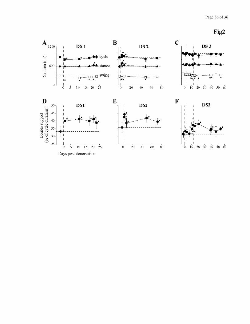
Figure 2 describes the changes in step cycle duration (A-C) and double hindlimb support (D-F).
The main change observed in the early days after the denervation was a reduction in swing duration, as
seen in all three cats. Note that in DS3 this change was more apparent when the denervation was
Page 13 of 13
completed (see data points after second vertical dashed line) at which time the changes were almost
identical to those observed in DS1. DS2 also showed a decrease in swing duration after the partial
denervation.
Stance duration, on the contrary was hardly affected. It was not statistically different in any
session for cat DS3 and only different in 1/5 session in cat DS1. DS2 showed a temporary increase in
stance duration on days 2, 3 and 7. These changes were only transitory, as at the 49 days session and
beyond, the situation was similar to the other cats.
The denervation significantly increased (P<0.05) the percentage of time spent in double hindlimb
support (Fig 2, D-F) by 5% to 10% of total cycle time. This increase in double support augments the
stability of walking. Not only was this increase present early after the denervation, but it also persisted
for the duration of the post-denervation testing period.
Figure 2 - near here
To measure the effects of increased double hindlimb support on quadrupedal walking, we also
measured the time spent with two, three, or four paws on the ground in cat DS3 (duty cycle; see
Methods). Segments were taken from a total of 5 walking sessions: 3 controls, 1 early (8 days) post
complete denervation, and 1 late (34 days) post complete denervation. The results are summarized in
Table 2, for a walking speed of 0.5 m/s. Before denervation, cat DS3 duty cycle was distributed the
following way: 0.6% of cycle duration was spent on four limbs, 70.4% on 3 limbs, and 29.0% on two
limbs. Early after the denervation, the time spent on 3 limbs was significantly increased to 79.4%
(P<0.05), and the time spent on 2 limbs significantly reduced to 20.4% (P<0.05). Time spent on 4 limbs
was unchanged, remaining essentially null (0.2 ± 0.4 %). Late after the denervation, part of these
changes in duty cycle remained: the time spent on 2 limbs was 25.0%, remaining significantly below
control (P<0.05), while the time spent on 3 limbs was 73.9%, which, although non-significantly higher
than control, nevertheless showed a tendency to be higher.
Page 14 of 14
Table 2 near here
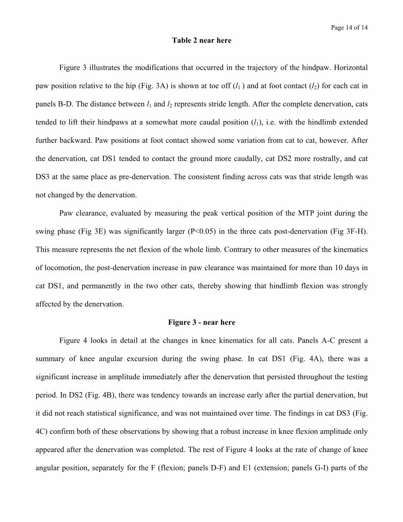
Figure 3 illustrates the modifications that occurred in the trajectory of the hindpaw. Horizontal
paw position relative to the hip (Fig. 3A) is shown at toe off (l1 ) and at foot contact (l2) for each cat in
panels B-D. The distance between l1 and l2 represents stride length. After the complete denervation, cats
tended to lift their hindpaws at a somewhat more caudal position (l1), i.e. with the hindlimb extended
further backward. Paw positions at foot contact showed some variation from cat to cat, however. After
the denervation, cat DS1 tended to contact the ground more caudally, cat DS2 more rostrally, and cat
DS3 at the same place as pre-denervation. The consistent finding across cats was that stride length was
not changed by the denervation.
Paw clearance, evaluated by measuring the peak vertical position of the MTP joint during the
swing phase (Fig 3E) was significantly larger (P<0.05) in the three cats post-denervation (Fig 3F-H).
This measure represents the net flexion of the whole limb. Contrary to other measures of the kinematics
of locomotion, the post-denervation increase in paw clearance was maintained for more than 10 days in
cat DS1, and permanently in the two other cats, thereby showing that hindlimb flexion was strongly
affected by the denervation.
Figure 3 - near here
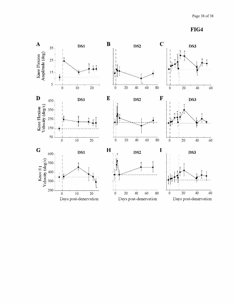
Figure 4 looks in detail at the changes in knee kinematics for all cats. Panels A-C present a
summary of knee angular excursion during the swing phase. In cat DS1 (Fig. 4A), there was a
significant increase in amplitude immediately after the denervation that persisted throughout the testing
period. In DS2 (Fig. 4B), there was tendency towards an increase early after the partial denervation, but
it did not reach statistical significance, and was not maintained over time. The findings in cat DS3 (Fig.
4C) confirm both of these observations by showing that a robust increase in knee flexion amplitude only
appeared after the denervation was completed. The rest of Figure 4 looks at the rate of change of knee
angular position, separately for the F (flexion; panels D-F) and E1 (extension; panels G-I) parts of the
Page 15 of 15
swing phase. This more detailed analysis reveals that the velocity of knee movements is significantly
increased in all cats after the denervation, regardless if it was partial or complete. The fact that mainly
velocity, not displacement, was increased explains why very little deficit was visible by the naked eye.
The changes in knee velocity persisted for several weeks.
Figure 4 - near here
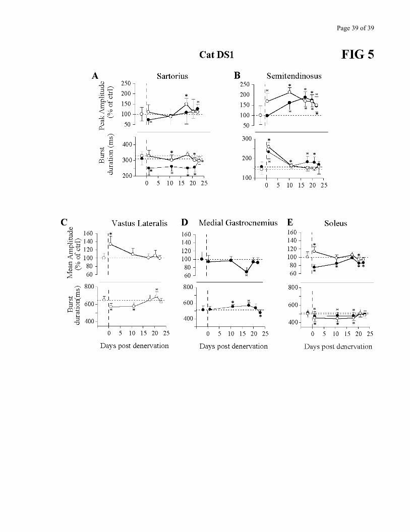
EMG changes after cutaneous denervation
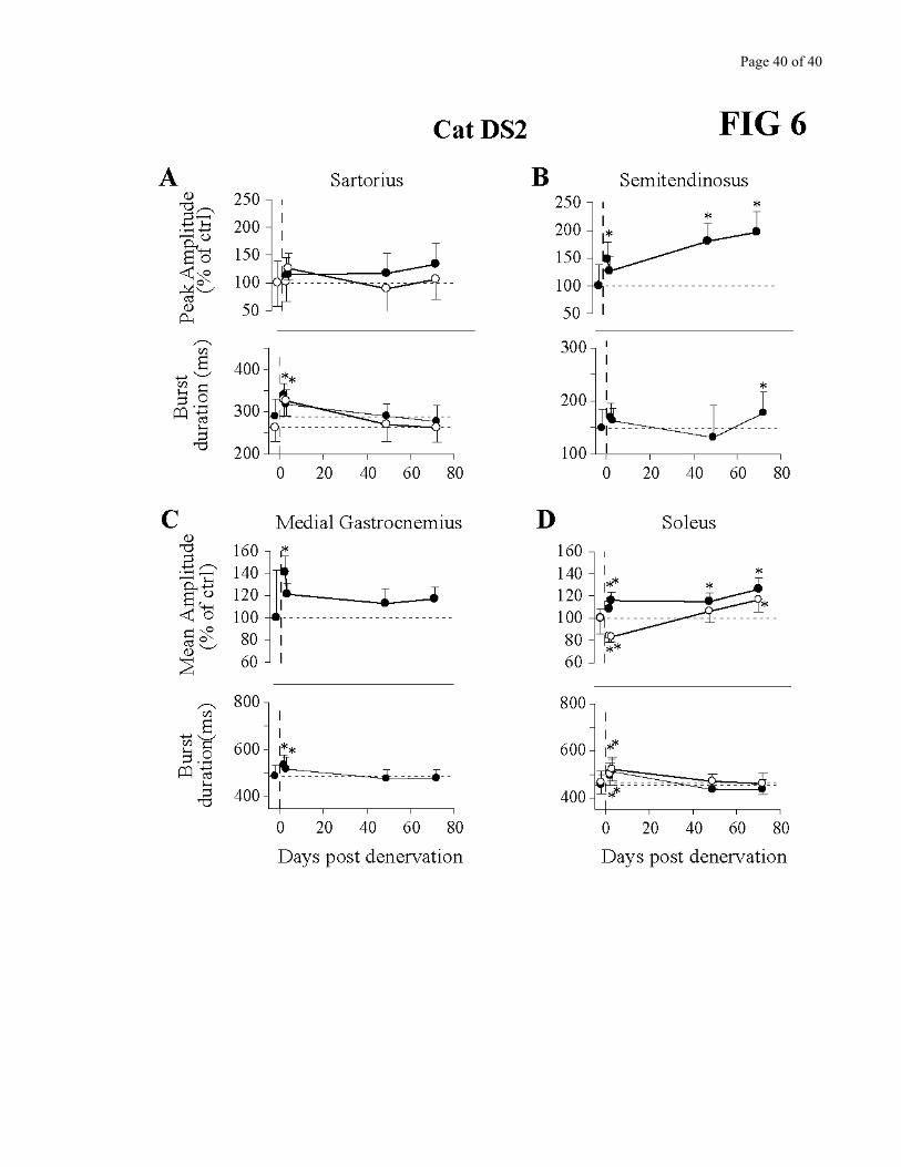
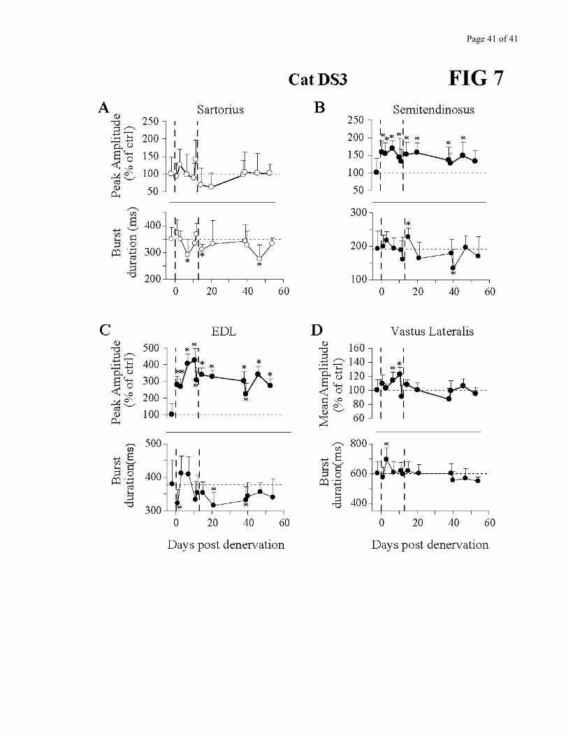
Figures 5 to 7 describe the changes in amplitude (peak for flexors, mean for extensors; c.f.
Methods) as well as burst duration in several muscles for the 3 cats. As the denervation was bilateral,
muscles from both limbs were superimposed when available. The most consistent change seen across all
cats was an increase in amplitude of the knee flexor Semitendinosus, which persisted throughout the
period of observation. This corroborates very well the kinematics in which knee flexion amplitude
and/or velocity were increased (Fig. 4) and paw clearance larger (Fig. 3). In cat DS3, EDL (recorded
only in this cat) also showed a persistent increase in amplitude. Burst duration of these flexor muscles
varied over time, with a larger duration early after denervation. The hip flexor/knee extensor Sartorius
activity did not significantly change except in the very early period after the denervation.
Regarding extensors, at least one prime extensor EMG activity was significantly increased in
each cat, and this increase was maintained for several days. However, it was not the same muscle in all
cats. For cat DS1, VL (knee extensor) EMG activity was increased for several days (Fig. 5C), while GM
(ankle extensor) activity remained essentially constant (Fig. 5D). SOL, another ankle extensor, showed
opposite changes in activity in the left and right hindlimbs on the first test post-denervation, followed by
a small reduction in activity long-term post-denervation (Fig.5 E). In cat DS2, overall, both GM and
SOL activity were increased (Fig. 6C-D). On the first test post-denervation however, the right SOL
presented a punctual decrease in activity (Fig. 6D). Finally, cat DS3 showed a significant increase in VL
activity after the partial denervation followed by a return to control amplitude (Fig. 7D).
Page 16 of 16
Figures 5, 6 and 7 - near here
Summarizing the evolution of locomotor parameters after partial and complete denervations
Inspection of Figures 1 through 7 shows that overall changes in the kinematics of locomotion
were larger after the complete than the partial denervation, but that in all cases most parameters
gradually returned statistically towards pre-denervation values after several days (depending on the cat).
The parameters that remained changed on a long term basis were: the percentage of double support, toe
clearance (a measure of total limb flexion during swing), and knee flexion amplitude and velocity during
the F phase (complete denervation). Factors that did return to pre-denervation values were: step cycle
and swing durations, toe position at foot contact, knee flexion amplitude and velocity during the F phase
(partial denervation).
2. Treadmill walking with pitch slopes
In addition to level walking, cats where also trained to walk with the treadmill belt inclined 15
degrees up and 15 degrees down. Although these are fairly easy slopes, walking on inclined surfaces
requires more subtle control of force amplitude and direction applied to the ground than level walking,
and can therefore be considered an additional challenge for the locomotor control capabilities of our
animals.
Before denervation, all cats could easily walk at 0.5 m/s in either condition. Early post-
denervation, the maximal speed was reduced to 0.4 m/s for cats DS1 and DS2. Above this speed, cats
could not maintain a regular pace but showed hesitation and stopped walking every 3-4 steps. As control
data was collected at 0.3, 0.4, and 0.5 m/s, data analysis was therefore performed on walking segments
obtained at 0.4 m/s.
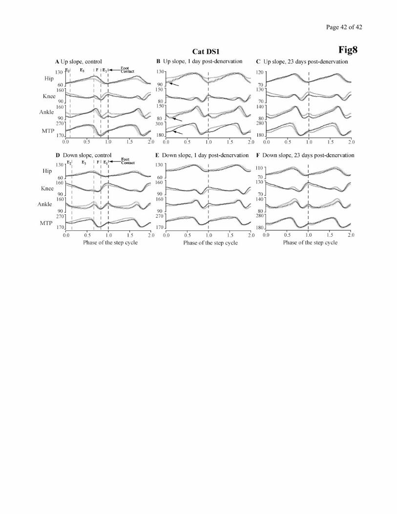
Figures 8 presents the kinematics of the locomotor adaptations that occurred in the control
situation, 1 day after, and 23 days after the denervation in cat DS1 for up and down slopes. Joint
excursion data obtained while walking on the incline (dark traces) are superimposed on data obtained
Page 17 of 17
during level walking on the same day (gray traces). Using this approach, the joint adaptations required
between level and incline walking are emphasized. By comparing the differences between control and
post-denervation, it is possible to identify the initial deficit and extent of recovery performed by the
animal. The data from this cat is representative of all 3 animals.
UPWARD SLOPE
Figure 8A-C summarizes the performance of cat DS1 walking 15º upwards. The kinematics are
presented as angular displacements separately for each hindlimb joint. In the control situation, when
comparing between walking with a 15º up slope and level walking (Fig 8A), modifications are present at
the hip, knee and ankle. The hip extension is increased at the end of stance. At the knee, changes are
more complex. There is a larger extension at the end of stance, followed by a more rapid flexion during
the F phase and a reduced extension during E1 leading to a more flexed knee posture at foot contact that
is gradually reduced through the rest of the stance phase. The situation is similar at the ankle as at the
knee: slightly larger extension at the end of stance, rapid, and in this case a larger joint flexion during
the F phase leading to a more flexed ankle posture that is gradually reduced over the first half of the
stance phase. The MTP joint displacements are very similar. As far as timing is concerned, flexion is
initiated later at all joints, and so is E1 at the knee and ankle.
One day after the denervation, the cat was not at ease during slope walking, and some important
differences were present when comparing level to upslope walking. Comparing Figures 8A and 8B, the
following observations can be made. The hip joint is more flexed throughout swing, resulting in an over
flexed hip at foot contact that gradually disappears during the first half of stance. The onset of swing is
no longer delayed compared to the level walking situation. At the knee, the onset of swing is no longer
delayed, E1 extension is exaggerated and the knee angle during stance is not more flexed than level
walking. At the ankle, the main difference with control is a lack of extra flexion during F, such that the
ankle joint angle is similar to level walking at foot contact. This joint however yields almost
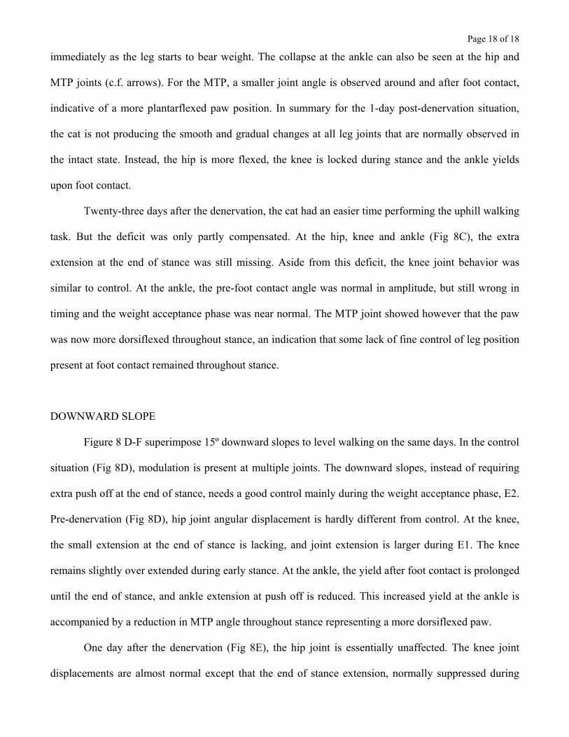
Page 18 of 18
immediately as the leg starts to bear weight. The collapse at the ankle can also be seen at the hip and
MTP joints (c.f. arrows). For the MTP, a smaller joint angle is observed around and after foot contact,
indicative of a more plantarflexed paw position. In summary for the 1-day post-denervation situation,
the cat is not producing the smooth and gradual changes at all leg joints that are normally observed in
the intact state. Instead, the hip is more flexed, the knee is locked during stance and the ankle yields
upon foot contact.
Twenty-three days after the denervation, the cat had an easier time performing the uphill walking
task. But the deficit was only partly compensated. At the hip, knee and ankle (Fig 8C), the extra
extension at the end of stance was still missing. Aside from this deficit, the knee joint behavior was
similar to control. At the ankle, the pre-foot contact angle was normal in amplitude, but still wrong in
timing and the weight acceptance phase was near normal. The MTP joint showed however that the paw
was now more dorsiflexed throughout stance, an indication that some lack of fine control of leg position
present at foot contact remained throughout stance.
DOWNWARD SLOPE
Figure 8 D-F superimpose 15º downward slopes to level walking on the same days. In the control
situation (Fig 8D), modulation is present at multiple joints. The downward slopes, instead of requiring
extra push off at the end of stance, needs a good control mainly during the weight acceptance phase, E2.
Pre-denervation (Fig 8D), hip joint angular displacement is hardly different from control. At the knee,
the small extension at the end of stance is lacking, and joint extension is larger during E1. The knee
remains slightly over extended during early stance. At the ankle, the yield after foot contact is prolonged
until the end of stance, and ankle extension at push off is reduced. This increased yield at the ankle is
accompanied by a reduction in MTP angle throughout stance representing a more dorsiflexed paw.
One day after the denervation (Fig 8E), the hip joint is essentially unaffected. The knee joint
displacements are almost normal except that the end of stance extension, normally suppressed during

Page 19 of 19
down slope, is now present. The main deficit is at the ankle joint. The gradual yield observed after foot
contact in the control situation is no longer present. This is confirmed by a lack of reduction of MTP
angle throughout stance.
Twenty-three days after the denervation (Fig 8F), the recovery was incomplete. The modulation
of ankle yield improved, but as shown by the MTP joint data, the paw remained more plantar flexed than
control. All 3 cats exhibited similar changes in slope adaptations.
In summary, although the cats could manage the slopes, and improved their performance over
time after the denervation, the slope adaptations were different from those in the pre-denervation
condition. In addition, cats DS1 and DS2 never recovered the ability to walk at 0.5 m/s on inclines.
Figure 8 - near here
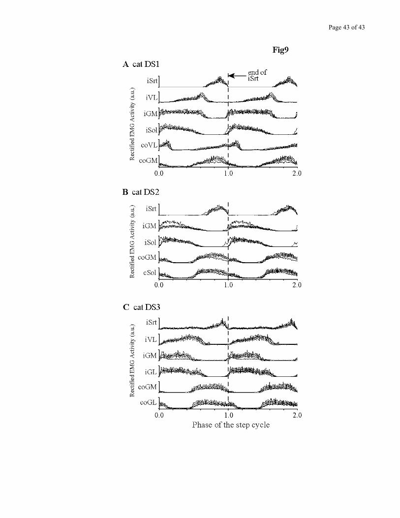
Changes in EMG patterns associated with incline walking.
EMG activity from several muscles was also obtained during these walking sessions. Figure 9
shows the superimposition of the control and early post denervation locomotor pattern for up slope,
separately for each of the 3 cats. These averages were obtained over many steps (>20) and synchronized
on the end of the Sartorius burst, a reliable timing cue close to foot contact. For post-denervation data to
be considered statistically different from control, it had to fall outside of the 95% confidence interval
boundaries of the control data. Contrary to the changes in kinematics, the changes in EMG activity of
the implanted muscles during incline walking were small, and differed from one cat to the next, and
even between the two legs in the same cat. For example, cat DS2 left GM increased its activity after
denervation, while its right GM decreased (Fig. 9B). Slope walking itself did greatly modulate activity
in extensor muscles, however. This normal modulation associated with incline walking caused much
greater changes in the EMG locomotor pattern than denervation itself. Data for down slopes presented
the same characteristics of large modulation due to treadmill incline, but no consistent deficit across cats
post-denervation (not shown).
Page 20 of 20
Figure 9 – near here
3. Walking on a horizontal ladder
To increase even more the demand for fine locomotor control, the cats were also tested on a
horizontal ladder. Contrary to treadmill walking, ladder walking was greatly impaired early after the
denervation. Most of the time, cats even refused to walk on the ladder. Thus, early post-denervation,
none of the 3 cats could stand on the rungs of the ladder, even if helped by the experimenter and allowed
to view their hindlimbs. The best that could be obtained was a temporary maintenance of posture if the
paws were manually placed on the rungs, and no external perturbation applied. After a few seconds in
this position, the cats would start to sway laterally and loose balance.
With time, ladder walking recovered almost completely, in each cat. After partial or complete
denervations, the paw placement strategy differed.
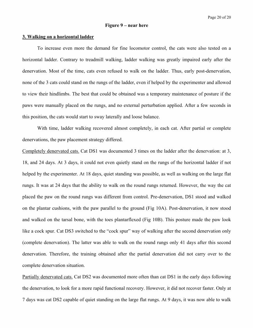
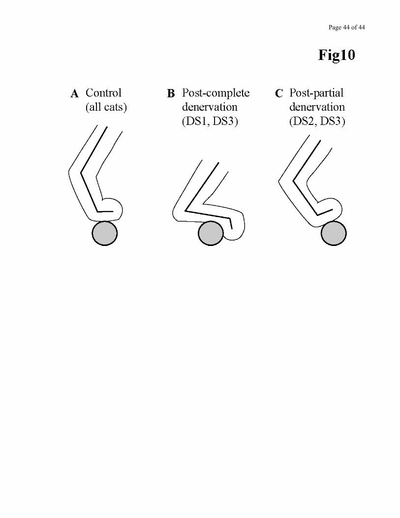
Completely denervated cats. Cat DS1 was documented 3 times on the ladder after the denervation: at 3,
18, and 24 days. At 3 days, it could not even quietly stand on the rungs of the horizontal ladder if not
helped by the experimenter. At 18 days, quiet standing was possible, as well as walking on the large flat
rungs. It was at 24 days that the ability to walk on the round rungs returned. However, the way the cat
placed the paw on the round rungs was different from control. Pre-denervation, DS1 stood and walked
on the plantar cushions, with the paw parallel to the ground (Fig 10A). Post-denervation, it now stood
and walked on the tarsal bone, with the toes plantarflexed (Fig 10B). This posture made the paw look
like a cock spur. Cat DS3 switched to the “cock spur” way of walking after the second denervation only
(complete denervation). The latter was able to walk on the round rungs only 41 days after this second
denervation. Therefore, the training obtained after the partial denervation did not carry over to the
complete denervation situation.
Partially denervated cats. Cat DS2 was documented more often than cat DS1 in the early days following
the denervation, to look for a more rapid functional recovery. However, it did not recover faster. Only at
7 days was cat DS2 capable of quiet standing on the large flat rungs. At 9 days, it was now able to walk
Page 21 of 21
on the flat rungs. Complete recovery was observed 49 days post-denervation. Contrary to completely
denervated cats, DS2 (and DS3 after the first denervation) walked with the paw dorsiflexed when
crossing on the round rungs (Fig 10C).
Figure 10 - near here
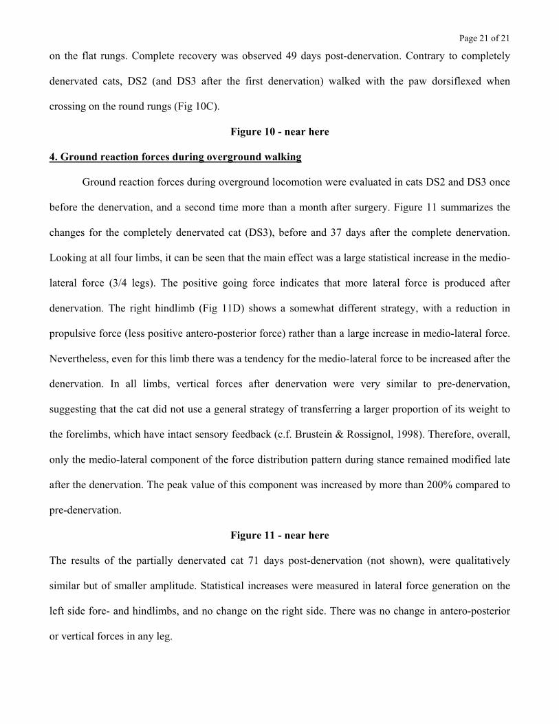
4. Ground reaction forces during overground walking
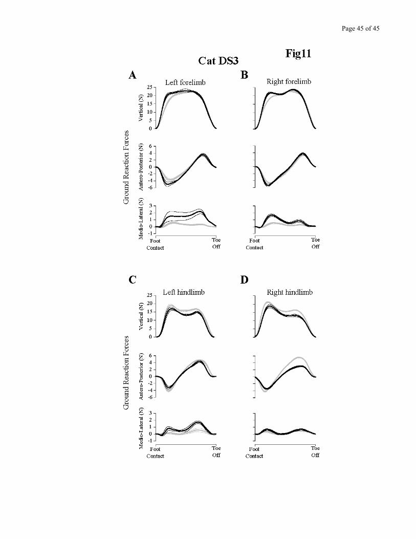
Ground reaction forces during overground locomotion were evaluated in cats DS2 and DS3 once
before the denervation, and a second time more than a month after surgery. Figure 11 summarizes the
changes for the completely denervated cat (DS3), before and 37 days after the complete denervation.
Looking at all four limbs, it can be seen that the main effect was a large statistical increase in the medio-
lateral force (3/4 legs). The positive going force indicates that more lateral force is produced after
denervation. The right hindlimb (Fig 11D) shows a somewhat different strategy, with a reduction in
propulsive force (less positive antero-posterior force) rather than a large increase in medio-lateral force.
Nevertheless, even for this limb there was a tendency for the medio-lateral force to be increased after the
denervation. In all limbs, vertical forces after denervation were very similar to pre-denervation,
suggesting that the cat did not use a general strategy of transferring a larger proportion of its weight to
the forelimbs, which have intact sensory feedback (c.f. Brustein & Rossignol, 1998). Therefore, overall,
only the medio-lateral component of the force distribution pattern during stance remained modified late
after the denervation. The peak value of this component was increased by more than 200% compared to
pre-denervation.
Figure 11 - near here
The results of the partially denervated cat 71 days post-denervation (not shown), were qualitatively
similar but of smaller amplitude. Statistical increases were measured in lateral force generation on the
left side fore- and hindlimbs, and no change on the right side. There was no change in antero-posterior
or vertical forces in any leg.

Page 22 of 22
DISCUSSION
These experiments show that the absence of cutaneous inputs from the hindpaws in otherwise
intact cats does not prevent the expression of the locomotor rhythm in undemanding situations such as
level walking on a treadmill. This corroborates the observations by Sherrington on hindpaw denervation
(Sherrington, 1910) and later works showing that nerve cuts(Duysens & Stein, 1978) or foot pad
anesthesia (Forssberg et al., 1977;Prochazka et al., 1978;Engberg, 1964;Wand et al., 1980) had
practically no effect on locomotion, at least to the naked eye. However, our results show that even in
such undemanding conditions there are consistent long-term changes such as increases in vertical paw
trajectory and knee flexion velocity accompanied by an increase in the peak amplitude of knee and ankle
flexor muscles. Furthermore, there is a significant increase in the percentage of the locomotor cycle
spent in double support. The deficits become more apparent in more demanding locomotor situations
and suggest that cutaneous afferents from the paw are used for fine control of foot placement. When cats
are required to walk on incline planes, the normal compensatory modifications of joints to accommodate
for the slopes are reduced. Furthermore, our results suggest that the cutaneous inputs are particularly
involved in locomotor conditions demanding a precise positioning of the foot such as ladder walking. In
the initial period post-denervation, cats are unable to walk on a horizontal ladder. Later on, they develop
a strategy of “cock spur” posture, which allows them to grasp the rungs and perform the task at a slower
pace. The normal elegant walk on the rungs seen before denervation never recovers. Finally, when
walking on a walkway with imbedded force platforms, cats tend to exert larger medio-lateral forces even
long term post-denervation, again suggesting that the loss of cutaneous inputs results in permanent
abnormalities of the walking pattern, even if subtle to the naked eye.
Two questions are raised by these observations. First, what locomotor deficits could have been
predicted from the known connectivity of cutaneous afferents with the spinal circuitry? Second, are the
changes specific to the cutaneous deprivation or do they represent a switch to a locomotor strategy that
Page 23 of 23
produces a more stable walking pattern and, as a result, offsets the insecurity introduced by the sensory
loss?
Predicted and observed deficits
The direct effect of the denervation.
As mentioned in the Introduction, experiments by Sherrington (Sherrington, 1910) suggested that
cutaneous inputs from the paws do not play a major role in the control of locomotion in quadrupeds.
However, in the further description by Sherrington, it is clear that some important functional locomotor
deficits were present in his cats after the cutaneous denervation. They included doubling of the toes
underneath the planta, foot drag during swing and a higher lift during stepping. Animals would not
correct this misplacement, as they seemed unaware of it. Sherrington did not come to a clear conclusion
on the reason for the doubling of the toes but discarded the possibility that cutting the innervation to
small intrinsic foot muscles such as EDB (innervated by the Deep Peroneal nerve) could be responsible,
as a specific denervation of this muscle did not result in doubling of the toes.
The cats presented in this study, despite having some deficits in foot placement on the ladder in
the first few days after denervation, could all perform quasi-normal locomotor movements on the
treadmill without foot drag or toe underturning. However, they exhibited a higher lift of the paw during
the swing phase, similar to Sherrigton’s cats. We have been careful in our own experiments to dissect
the cutaneous branch of the Deep Peroneal nerve free from its motor innervation to EDB before the
denervation, and only cut the cutaneous branch to insure that the deficits observed would not be due to a
direct damage to the muscle’s innervation. In addition, in cat DS3, EDB had been implanted with
chronic EMG electrodes to ensure that the motor innervation remained functional after the surgery. This
will become a crucial point in the companion paper where the same denervated cats are trained to walk
after spinalisation, as it will allow us to discard the possibility that the deficits observed in that condition
are due to a local damage to the innervation of the intrinsic foot musculature.
Page 24 of 24
The abolition of cutaneous reflex pathways
A challenging question in the field of locomotor control is to establish the role of specific
afferent feedback in the normal, unperturbed locomotion. One of the main findings in this report was
that the cutaneous denervation caused an increase in knee and ankle flexor activity during the flexion
phase of locomotion.
It is known from nerve recordings (Duysens & Stein, 1978;Popovic et al., 1993) that there are
discharges in the cutaneous nerves at different points in the step cycle. Chronic unit recordings in dorsal
root ganglia (Loeb et al., 1977) also clearly indicate discharges of single units that are not necessarily
linked to clear kinematic events such as foot contact, but may represent skin stretches during joint
movements. Are these discharges contributing to the locomotor pattern, such as has been suggested for
proprioceptive inputs from ankle extensors which may contribute to more than one third of the muscle
output during decerebrate walking (Stein et al., 2000)?
Regarding extensor muscles, before and after injecting a local anesthetic into the central foot
pad, Engberg (Engberg, 1964;Engberg & Lundberg, 1969) found no change in the EMG activity of
flexor digitorum brevis (FDB), a toe extensor acting during the stance phase of locomotion. The results
presented in this study further suggest that the load inputs from the cutaneous receptors contribute
relatively little essential feedback to the normal locomotor output of the ankle extensor muscles despite
the well-established excitatory connections between cutaneous afferents and ankle extensor
motoneurons (Wilson, 1963;LaBella et al., 1989b;LaBella et al., 1989a). Based on the latter work, the
cutaneous denervation was expected to produce a marked decrease in leg extensor activity. On the
contrary, activity of at least one prime extensor was significantly increased in each cat for several days
after denervation (Fig. 5-7). Whether this resulted from an increase in the gain of compensatory
proprioceptive reflexes of the ankle extensors or to an increase in central drive is unknown. Perhaps an
experiment similar to that reported by Stein et al (Stein et al., 2000) using an imposed force profile on
Page 25 of 25
the output of ankle extensors during decerebrate walking after a cutaneous denervation could shed some
light on this difference.
On the basis of cutaneous nerve stimulation we could have expected larger deficits in the
walking pattern of our cats after denervation. Early work (Duysens, 1977;Duysens & Pearson, 1976) had
shown that cutaneous stimuli could prolong the stance phase significantly. In normal cats, Halbertsma
(Halbertsma, 1983) showed that as speed of walking increases, the hindlimb paw is gradually lifted off
the ground at a distance closer to the hip, while foot contact position relative to the hip remains
remarkably constant across a wide range of speeds. After denervation, our cats did not show a reduction
in stride length nor an important modification of paw position relative to the hip at paw lift or contact.
The movement was simply executed faster, and sometimes with a greater flexion at the knee, which
would simply lift the paw higher off the ground without affecting lift or contact position. Although we
saw a decrease in cycle duration in all three cats, the shortening was due to a decrease in swing duration
(faster swing) while stance duration remained more or less the same. Similarly, the stride length was
preserved, suggesting that cutaneous inputs contribute little to the overall duration of the stance phase or
the overall metrics of the step cycle in the otherwise normal cat.
Another strategy that could have been used by the cats would have been to shift the weight control over
to the forelimbs, as previously reported after large lesions of the ventral and ventrolateral spinal tracts
(Brustein & Rossignol, 1998). This was not the case, as measurements of vertical forces in all four
limbs did not show such a weight shift (Fig.11).
Regarding flexor muscles, nerve stimulation studies during locomotion have shown that
stimulating the dorsum of the paw (SP n. receptive field) gives rise to a multi-articular response that
normally brings the paw up and around an obstacle hit during the swing movement(Drew & Rossignol,
1987;Forssberg, 1979). This response is inhibited during stance (Drew & Rossignol, 1987). Using
spatial facilitation during fictive locomotion Degtyarenko et al. (Degtyarenko et al., 1996), recording
from TA and EDL motoneurons, have shown the presence of an elaborate excitatory and inhibitory
Page 26 of 26
short-latency set of connections between cutaneous afferents from the paw and flexor motoneurons that
is powerfully modulated by the CPG during locomotion. According to these authors, during flexion,
EDL motoneurons receive disynaptic inhibitory PSPs from SP stimulation while TA motoneurons
receive no inhibition. On the other hand, stimulation of the medial plantar nerve, a cutaneous branch of
the Tibial nerve, produces essentially no response during swing. These results are compatible with the
neural organization needed to prevent claw protusion during the stumbling reaction (Degtyarenko et al.,
1996). However, we cannot easily predict how elimination of paw cutaneous feedback during
unperturbed swing would lead this circuitry to increase EDL peak EMG activity to 350% of control (c.f.
Fig 7C). Therefore, the increase in EMG activity of flexor muscles reported in this study could not have
been easily predicted by the removal of any of the known cutaneous pathways from the paw and
probably results from an increase in central drive.
Moreover, recent work in rats (Schouenborg et al., 1992;Schouenborg & Kalliomaki, 1990;Schouenborg
et al., 1994), cats (Levinsson et al., 1999;Degtyarenko et al., 1996;Burke et al., 2001) and humans (Van
Wezel et al., 1997) suggest that skin inputs may also be involved in a fine “local sign” control. Another
interpretation of our results could therefore be that the loss of such fine control probably induces cats to
adopt a safer walking strategy where the limb is lifted higher (in case there would be obstacles) and
faster to increase the percentage of time spent on the ground (increase in double-support). Such a change
in walking strategy would require an increase in flexor EMG activity. Furthermore, this interpretation is
supported by the increase in medio-lateral force measured during stance (c.f. Figure 11), which also
probably represents an additional way to increase stability in the absence of fine control of the distal
limb.
Page 27 of 27
Short-term and long-term compensations
One of our general objectives was to study the adaptive capacity of the locomotor system to a
permanent reduction in skin feedback. To address this objective, we studied the denervated animals for
several weeks after the denervation and measured functional improvements over time.
With respect to level treadmill walking, the small deficits in knee flexion amplitude were rapidly
compensated. The increased knee flexion velocity remained, and the ankle and knee flexor muscle
activity gradually diminished but remained significantly above control level. Therefore, careful
inspection of the kinematics of walking and motor pattern showed some permanent modifications. The
persistence of these modifications rules out their presence as being related to a change in motor strategy
associated with pain or other aspects related to the denervation procedure.
The second important result in this study was that the animals showed extensive compensation
during ladder and slope walking over the several weeks that followed the denervation. This
compensation was gradual, which is suggestive of motor learning rather than switching to an already
existing alternate motor strategy. But while functionally extensive, compensation was nevertheless
incomplete, as shown by: 1) the differences in joint modulation on slopes between control and 23 days
post-denervation; 2) the late ladder walking motor strategy (“cock spur” paw position); 3) the greater
than 200% increase in medio-lateral ground reaction force during overground locomotion.
Therefore, while the locomotor system of the adult cat is capable of compensating for the
removal of hindpaw cutaneous inputs, it cannot regain fine control of the movement in their absence. In
summary, the lack of long-term compensation for the deficits in lateral force production during level
walking, in joint adaptations during slope walking, and in paw placement during ladder walking
suggests that cutaneous inputs are essential for the fine control of locomotion and become more useful
as the locomotor task becomes more demanding.
Page 28 of 28
Partial versus complete denervation
There were more similarities than differences in motor deficit and adaptive strategy between
animals having undergone a partial or a complete cutaneous denervation. During ladder walking, time
course of recovery on the rungs were equivalent. During incline walking, the reduction in joint angular
modulation was also similar. Overground, the increase in medio-lateral force was of the same magnitude
and direction. The only difference between the partially and completely denervated cats was at the level
of the knee hyperflexion, which only appeared transiently after the complete cutaneous denervation. The
2-stage denervation in cat DS3 nicely shows the similarities and differences between the partial and
complete denervations and strengthens our findings.
Compensatory mechanisms
The extensive compensation after the complete hindpaw cutaneous denervation is in contrast to
cats having undergone a complete deafferentation through a dorsal rhizotomy (Wetzel et al.,
1976;Goldberger, 1983;Goldberger, 1977;Hiebert & Pearson, 1999). After such procedure, which
abolishes feedback from all sources of sensory information originating from the whole limb, cats tend
not to use the deafferented limb (see however (Grillner & Zangger, 1974) for acute decerebrate cats with
MLR stimulation). However, if a single dorsal root is spared, cats gradually recover the ability to use the
experimental limb. These experiments suggest that central reorganization can occur after small or large
reductions in sensory feedback, provided some feedback from the limb remains. In the case of the
cutaneous denervation, remaining feedback could be obtained from muscle and joint receptors, as
discussed above.
However, in addition to these peripheral signals, some central mechanisms could also participate
to compensation. The cortico-spinal system could be involved in the locomotor compensation after a
complete or incomplete cutaneous denervation and increase the central locomotor drive. Preliminary
data from experiments involving a cutaneous denervation performed on motor cortex-lesioned cats show
Page 29 of 29
no increase in St amplitude after the denervation and the presence of foot drag (Bouyer et al., 2000),
supporting a role for the motor cortex in the compensation process.
The limit of the denervation protocol is that the deficit observed even immediately after the
neurectomies is the outcome of a combination of 2 factors: 1) the missing contribution of the removed
input; 2) the immediate compensation from the remaining neural circuits. The long-term compensation
gradually occurring over the following weeks on the other hand results from the adaptive capacity of the
system. In this paper, we have used chronic recordings to compare early and late locomotor function to
address part of this question and showed specific deficits after the removal of cutaneous inputs from the
paw. To push the study of a role for cutaneous inputs in the control of locomotion even further, the
companion paper will address a complementary question: once compensated, what happens to these
animals if part of their compensatory capacity, the supraspinal pathways, are disconnected from the
basic locomotor circuits? Does the integrity of cutaneous inputs become even more critical then? It will
be shown that after spinalisation, the same denervated cats presented a severe, long-term impairment in
paw placement, deficit that is not seen in spinal non-denervated controls (Rossignol et al., 2000).
Page 30 of 30
ACKNOWLEDGMENTS
This work was supported by grants from the Canadian Institutes of Health Research (CIHR) and the
Province of Quebec Fonds pour la Formation de Chercheurs et l’Aide à la Recherche (FCAR). L.J.B.
was a fellow of the Canadian Neurotrauma Research Program-CIHR partnership. We thank Janyne
Provencher, France Lebel, Jeanne Faubert, Philippe Drapeau, and Claude Gagner for technical
assistance. A special thank to Dr Trevor Drew for providing access to his force platforms and for his
help with some of the computer analyses, and to Drs. Sylvain Lavoie and Edna Brustein without whose
help the walkway data could not have been collected.
Page 31 of 31
Reference List
Bouyer, L. & Rossignol, S. Hindfoot cutaneous input contribution to locomotion in intact and spinal cats. Society for Neuroscience Abstracts 23[298.2], 759. 1997.
Bouyer, L. & Rossignol, S. (1998a). The contribution of cutaneous inputs to locomotion in the intact and the spinal cat. In Neuronal mechanisms for generating locomotor activity, eds. Kiehn, O., Harris-Warrick, R. M., Jordan, L. M., Hultborn, H., & Kudo, N., pp. 508-512.
Bouyer, L. & Rossignol, S. The effects of clonidine on foot placement during locomotion in the spinal cat after partial or complete hindpaw. Society for Neuroscience Abstracts 24, 1154. 1998b.
Bouyer, L. J. G., Drew, T., & Rossignol, S. A cortical contribution to the functional recovery observed following a cutaneous neurectomy of the hindpaw. Society for Neuroscience Abstracts 26[459.13], 1232. 2000.
Brustein, E. & Rossignol, S. (1998). Recovery of locomotion after ventral and ventrolateral spinal lesions in the cat. I. Deficits and adaptive mechanisms. Journal of Neurophysiology 80, 1245-1267.
Burke, R. E., Degtyarenko, A. M., & Simon, E. S. (2001). Patterns of locomotor drive to motoneurons and last-order interneurons: clues to the structure of the CPG. Journal of Neurophysiology 86, 447-462.
Crouch, J. E. (1969). Text-atlas of cat anatomy Lea and Febiger, Philadelphia.
Degtyarenko, A. M., Simon, E. S., & Burke, R. E. (1996). Differential modulation of disynaptic cutaneous inhibition and excitation in ankle flexor motoneurons during fictive locomotion. Journal of Neurophysiology 76, 2972-2985.
Drew, T. & Rossignol, S. (1987). A kinematic and electromyographic study of cutaneous reflexes evoked from the forelimb of unrestrained walking cats. Journal of Neurophysiology 57, 1160-1184.
Duysens, J. (1977). Reflex control of locomotion as revealed by stimulation of cutaneous afferents in spontaneously walking premammilary cats. Journal of Neurophysiology 40, 737-751.
Duysens, J., Clarac, F., & Cruse, H. (2000). Load-regulating mechanisms in gait and posture: comparative aspects. Physiol Rev. 80, 83-133.
Duysens, J. & Pearson, K. G. (1976). The role of cutaneous afferents from the distal hindlimb in the regulation of the step cycle of thalamic cats. Experimental Brain Research 24, 245-255.
Duysens, J. & Stein, R. B. (1978). Reflexes induced by nerve stimulation in walking cats with implanted cuff electrodes. Experimental Brain Research 32, 213-224.
Engberg, I. (1964). Reflexes to foot muscles in the cat. Acta physiologica scandinavia 62, 1-64.
Engberg, I. & Lundberg, A. (1969). An electromyographic analysis of muscular activity in the hindlimb of the cat during unrestrained locomotion. Acta physiologica scandinavia 75, 614-630.
Page 32 of 32
Forssberg, H. (1979). Stumbling corrective reaction: a phase-dependent compensatory reaction during locomotion. Journal of Neurophysiology 42, 936-953.
Forssberg, H., Grillner, S., & Rossignol, S. (1977). Phasic gain control of reflexes from the dorsum of the paw during spinal locomotion. Brain Research 132, 121-139.
Glantz, A. G. (1992). Primer of Biostatistics, 3rd ed. McGraw-Hill, New York.
Goldberger, M. E. (1977). Locomotor recovery after unilateral hindlimb deafferentation in cats. Brain Research 123, 59-74.
Goldberger, M. E. (1983). Recovery of accurate limb movements after deafferentation in cats. In Spinal cord reconstruction, eds. Kao, C. C., Bunge, R. P., & Reier, P. J., pp. 455-463. Raven Press, New York.
Gossard, J.-P., Cabelguen, J.-M., & Rossignol, S. (1989). Intra-axonal recordings of cutaneous primary afferents during fictive locomotion in the cat. Journal of Neurophysiology 62, 1177-1188.
Gossard, J.-P., Cabelguen, J.-M., & Rossignol, S. (1990). Phase-dependent modulation of primary afferent depolarization in single cutaneous primary afferents evoked by peripheral stimulation during fictive locomotion in the cat. Brain Research 537, 14-23.
Grillner, S. & Zangger, P. (1974). Locomotor movements generated by the deafferented spinal cord. Acta physiologica scandinavia 91, 38-39A.
Grillner, S. & Zangger, P. (1979). On the central generation of locomotion in the low spinal cat. Experimental Brain Research 34, 241-261.
Grillner, S. & Zangger, P. (1984). The effect of dorsal root transection on the efferent motor pattern in the cat's hindlimb during locomotion. Acta physiologica scandinavia 120, 393-405.
Halbertsma, J. M. (1983). The stride cycle of the cat: the modelling of locomotion by computerized analysis of automatic recordings. Acta physiologica scandinavia Suppl. 521, 1-75.
Hiebert, G. W. & Pearson, K. G. (1999). Contribution of sensory feedback to the generation of extensor activity during walking in the decerebrate Cat. Journal of Neurophysiology 81, 758-770.
LaBella, L. A., Kehler, J. P., & McCrea, D. A. (1989a). A differential synaptic input to the motor nuclei of triceps surae from the caudal and lateral cutaneous sural nerves. Journal of Neurophysiology 61, 291-301.
LaBella, L. A., Kehler, J. P., & McCrea, D. A. Short-latency excitation from sural afferents predominates in the medial gastrocnemius portion of triceps surae motor nuclei. Society for Neuroscience Abstracts . 1989b.
Lavoie, S., McFadyen, B., & Drew, T. (1995). A kinematic and kinetic analysis of locomotion during voluntary gait modification in the cat. Experimental Brain Research 106, 39-56.
Levinsson, A., Garwicz, M., & Schouenborg, J. (1999). Sensorimotor transformation in cat nociceptive withdrawal reflex system. Eur.J.Neurosci. 11, 4327-4332.
Page 33 of 33
Loeb, G. E., Bak, M. J., & Duysens, J. (1977). Long-term unit recording from somatosensory neurons in the spinal ganglia of the freely walking cat. Science 197, 1192-1194.
Philippson, M. (1905). L'autonomie et la centralisation dans le système nerveux des animaux. Trav.Lab.Physiol.Inst.Solvay (Bruxelles.) 7, 1-208.
Popovic, D. B., Stein, R. B., Jovanovic, K. L., Dai, R., Kostov, A., & Armstrong, W. W. (1993). Sensory nerve recording for closed-loop control to restore motor functions. I.E.E.E.Trans Biomed.Eng. 40, 1024-1031.
Prochazka, A., Sontag, K. H., & Wand, P. (1978). Motor reactions to perturbations of gait: proprioceptive and somesthetic involvement. Neurosci.Lett. 7, 35-39.
Rossignol, S. (1996). Neural control of stereotypic limb movements. In Handbook of Physiology, Section 12. Exercise: Regulation and Integration of Multiple Systems, eds. Rowell, L. B. & Sheperd, J. T., pp. 173-216. Oxford University Press, New York.
Rossignol, S., Bélanger, M., Chau, C., Giroux, N., Brustein, E., Bouyer, L., Grenier, C.-A., Drew, T., Barbeau, H., & Reader, T. (2000). The spinal cat. In Neurobiology of spinal cord injury, eds. Kalb, R. G. & Strittmatter, S. M., pp. 57-87. Humana Press, Totowa.
Rossignol, S., Lund, J. P., & Drew, T. (1988). The role of sensory inputs in regulating patterns of rhythmical movements in higher vertebrates. A comparison between locomotion, respiration and mastication. In Neural control of rhythmic movements in vertebrates, eds. Cohen, A., Rossignol, S., & Grillner, S., pp. 201-283. Wiley and sons Co, New York.
Schouenborg, J., Holmberg, H., & Weng, H. R. (1992). Functional organization of the nociceptive withdrawal reflexes. II. Changes of excitability and receptive fields after spinalization in the rat. Experimental Brain Research 90, 469-478.
Schouenborg, J. & Kalliomaki, J. (1990). Functional organization of the nociceptive withdrawal reflexes. I.Activation of hindlimb muscles in the rat. Experimental Brain Research 83, 67-78.
Schouenborg, J., Weng, H. R., & Holmberg, H. (1994). Modular organization of spinal nociceptive reflexes: a new hypothesis. NIPS 9, 261.
Sherrington, C. S. (1910). Flexion-reflex of the limb, crossed extension-reflex, and reflex stepping and standing. Journal of physiology 40, 28-121.
Stein, R. B., Misiaszek, J. E., & Pearson, K. G. (2000). Functional role of muscle reflexes for force generation in the decerebrate walking cat. The Journal of Physiology Online 525, 781-791.
Van Wezel, B. M., Ottenhoff, F. A., & Duysens, J. (1997). Dynamic control of location-specific information in tactile cutaneous reflexes from the foot during human walking. Journal of Neuroscience 17, 3804-3814.
Wand, P., Prochazka, A., & Sontag, K. H. (1980). Neuromuscular responses to gait perturbations in freely moving cats. Experimental Brain Research 38, 109-114.
Wetzel, M. C., Atwater, A. E., Wait, J. V., & Stuart, D. G. (1976). Kinematics of locomotion by cats with a single hindlimb deafferented. Journal of Neurophysiology 39, 667-678.
Page 34 of 34
Wilson, V. J. (1963). Ipsilateral excitation of extensor motoneurones. Nature 198, 290-291.
Zehr, E. P. & Stein, R. B. (1999). What functions do reflexes serve during human locomotion? Prog.Neurobiol. 58, 185-205.
Page 35 of 35
Page 36 of 36
Page 37 of 37
Page 38 of 38
Page 39 of 39
Page 40 of 40
Page 41 of 41
Page 42 of 42
Page 43 of 43
Page 44 of 44
Page 45 of 45
Page 46 of 46
FIGURE LEGENDS
Figure 1- A-C. Stick figure reconstructions of left hindlimb movements from a frame-by-frame video
analysis of cat DS3 walking at 0.5 m/s. Control (A), 2 days (B), and 41 days (C) after the complete
denervation. Horizontal and vertical scales are 5 cm. D-E. Mean angular excursions at each of the four
limb joints. The thickness of the lines represents ± 1 standard error of the mean. In D, control (gray line)
and 2 days post-denervation (black line) are superimposed. Dashed lines represent the divisions of the
step cycle according to Philippson (1905) for the control walking situation. In E, the superimposition is
between control (gray line) and 41 days post-denervation (black line). Synchronization is on foot
contact. Arrow highlights increased knee flexion. F-G. Rectified averaged EMG activity of the main
muscles recorded simultaneously with the kinematics shown in D-E. See text for abbreviations. In F,
control EMG activity (gray area) and 2 days post-denervation (black line) are superimposed. In G, the
superimposition is between control (gray area) and 41 days post-denervation (black line). Abbreviations:
Srt: Sartorius Anterior; St: Semitendinosus; VL: Vastus Lateralis; EDL: Extensor Digitorum Longus;
LG: Lateral Gastrocnemius; MG: Medial Gastrocnemius; EDB: Extensor Digitorum Brevis.
Figure 2- A-C. Duration of the step cycle (black circles) and of its two components, stance (black
triangles) and swing (white squares) as a function of time post-denervation for cats DS1 (A), DS2 (B)
and DS3 (C) walking on the treadmill at 0.5 m/s. Horizontal dashed lines represent the mean pre-
denervation value. Vertical dashed lines represent time of denervation. For cat DS3, two denervations
were performed sequentially: the first at day 0 (partial denervation), and the second at day 13 (complete
denervation). D-F. Percentage of step cycle duration where both hindpaws were in contact with the
ground, calculated from a frame-by-frame video analysis of the walking sessions. D. Cat DS1. E. Cat
DS2. F. Cat DS3. For all graphs in this Figure, each point represents the mean +/- standard deviation. *=
P<0.05, one way ANOVA against pre-denervation control.
Page 47 of 47
Figure 3 – Details of the metrics of the left hindpaw during treadmill locomotion. A-D. Stride length
and paw position relative to the hip. A. Line drawing; l1 represents the horizontal distance between the
hip and toe markers at toe off; l2 represents the horizontal distance between the hip and toe markers at
foot contact. The distance between l1 and l2 is a measure of stride length. B. Cat DS1. C. Cat DS2. D.
Cat DS3. E-H. Vertical elevation of the paw during the swing phase. E. Line drawing representing how
the peak vertical position of the MTP joint with respect to the treadmill surface was measured. F. Cat
DS1. G. Cat DS2. H. Cat DS3. For all graphs in this Figure, each point represents the mean ± standard
deviation. Horizontal dashed lines represent the mean pre-denervation value. Vertical dashed lines
represent time of denervation. *= P<0.05, one way ANOVA against pre-denervation control.
Figure 4 - Kinematics of the knee joint. A-C. Maximal knee flexion amplitude during the swing phase
for cats DS1 (A), DS2 (B) and DS3 (C). D-F. Peak flexion velocity of the knee joint reached during the
F phase for cats DS1 (D), DS2 (E) and DS3 (F). G-I. Peak extension velocity of the knee joint reached
during the E1 phase for cats DS1 (G), DS2 (H) and DS3 (I). For all graphs, each point represents the
mean +/- standard deviation. Horizontal dashed lines represent the mean pre-denervation value. Vertical
dashed lines represent time of denervation. *= P<0.05, one way ANOVA against pre-denervation
control.
Figure 5– Summary of EMG activity for cat DS1. Black circles represent muscles of the left leg, and
white circles those of the right leg. A-B. Peak amplitude and burst duration for hip flexor Sartorius (A)
and knee flexor Semitendinosus (B). C-E. Mean amplitude and burst duration for knee extensor Vastus
Lateralis (C), ankle extensor Medial Gastrocnemius (D), and ankle extensor Soleus (E). For all graphs,
each point represents the mean +/- standard deviation. Horizontal dashed lines delineate the pre-
Page 48 of 48
denervation value. Vertical dashed lines represent the times of denervation. *= P<0.05, one way
ANOVA against pre-denervation control.
Figure 6– Summary of EMG activity for cat DS2. Black circles represent the muscles of the left leg, and
white circles those of the right leg. A-B. Peak amplitude and burst duration for hip flexor Sartorius (A)
and knee flexor Semitendinosus (B). C-D. Mean amplitude and burst duration for ankle extensors
Medial Gastrocnemius (C) and Soleus (D). For all graphs, each point represents the mean +/- standard
deviation. Horizontal dashed lines delineate the pre-denervation value. Vertical dashed lines represent
the times of denervation. *= P<0.05, one way ANOVA against pre-denervation control.
Figure 7– Summary of EMG activity for cat DS3. Black circles represent the muscles of the left leg, and
white circles those of the right leg. A-C. Peak amplitude and burst duration for hip flexor Sartorius (A),
knee flexor Semitendinosus (B), and ankle flexor Extensor Digitorum Longus (EDL; C). D. Mean
amplitude and burst duration for knee extensor Vastus Lateralis (D). For all graphs, each point
represents the mean +/- standard deviation. Horizontal dashed lines delineate the pre-denervation value.
Vertical dashed lines represent the times of denervation. *= P<0.05, one way ANOVA against pre-
denervation control.
Figure 8- A-C. Comparative kinematics of leveled and 15 degree pitch up slope treadmill walking for
cat DS1 at 0.4 m/s, before (A), 1 day (B), and 23 days (C) after the complete denervation. D-F.
Comparative kinematics of leveled and 15 degree pitch down slope treadmill walking for the same cat
on the same days. On each graph, the gray traces represent relative joint angles for leveled walking and
the black traces for slope walking. Dotted lines show +/- one standard error of the mean. All data are
synchronized relative to foot contact. Dashed lines represent the divisions of the step cycle according to
Philippson (1905) for the leveled walking situation.
Page 49 of 49
Figure 9- Rectified average EMG activity obtained before denervation (thin lines ± 95% confidence
intervals) and early after denervation (thick lines) during 15 degrees upslope walking. Data are
synchronized on the end of the Sartorius burst. A. Cat DS1, control and 1-day post complete
denervation. B. Cat DS2, control and 3 days post partial denervation. C. Cat DS3, control and 8 days
post complete denervation. See text for muscle abbreviations. i=left, co=right.
Figure 10- Line drawing of the hindpaw made from video images showing the characteristic paw
placement on the rungs during ladder walking. A. Before denervation. B. After the complete
denervation. C. After the partial denervation.
Figure 11- Ground reaction forces during the stance phase of overground walking on a walkway for
each of the four paws of cat DS3. Gray traces represent pre-denervation control, and black traces 37
days post-denervation. Thin lines represent 95% confidence intervals on the mean. A. Left forelimb. B.
Right forelimb. C. Left hindlimb. D. Right hindlimb.
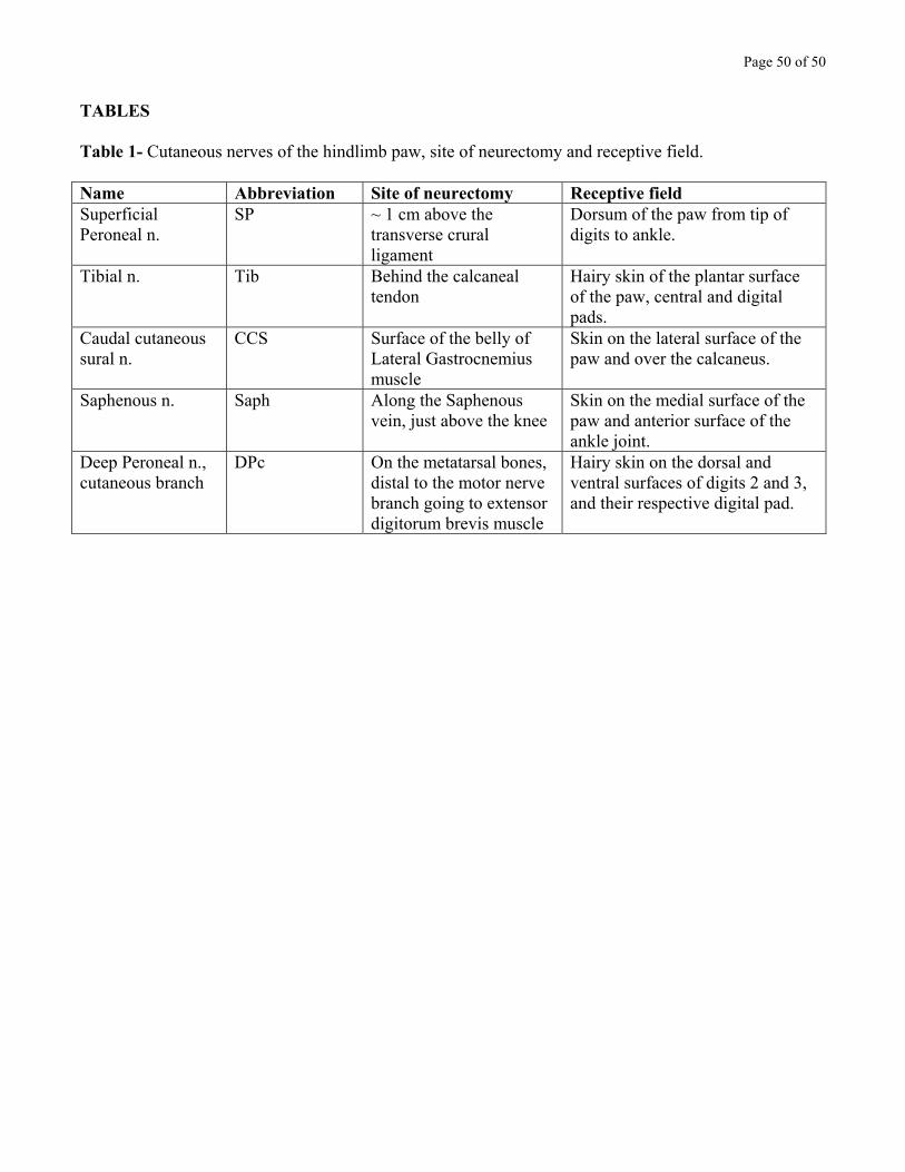
Page 50 of 50
TABLES Table 1- Cutaneous nerves of the hindlimb paw, site of neurectomy and receptive field. Name Abbreviation Site of neurectomy Receptive field Superficial Peroneal n.
SP ~ 1 cm above the transverse crural ligament
Dorsum of the paw from tip of digits to ankle.
Tibial n. Tib Behind the calcaneal tendon
Hairy skin of the plantar surface of the paw, central and digital pads.
Caudal cutaneous sural n.
CCS Surface of the belly of Lateral Gastrocnemius muscle
Skin on the lateral surface of the paw and over the calcaneus.
Saphenous n. Saph Along the Saphenous vein, just above the knee
Skin on the medial surface of the paw and anterior surface of the ankle joint.
Deep Peroneal n., cutaneous branch
DPc On the metatarsal bones, distal to the motor nerve branch going to extensor digitorum brevis muscle
Hairy skin on the dorsal and ventral surfaces of digits 2 and 3, and their respective digital pad.
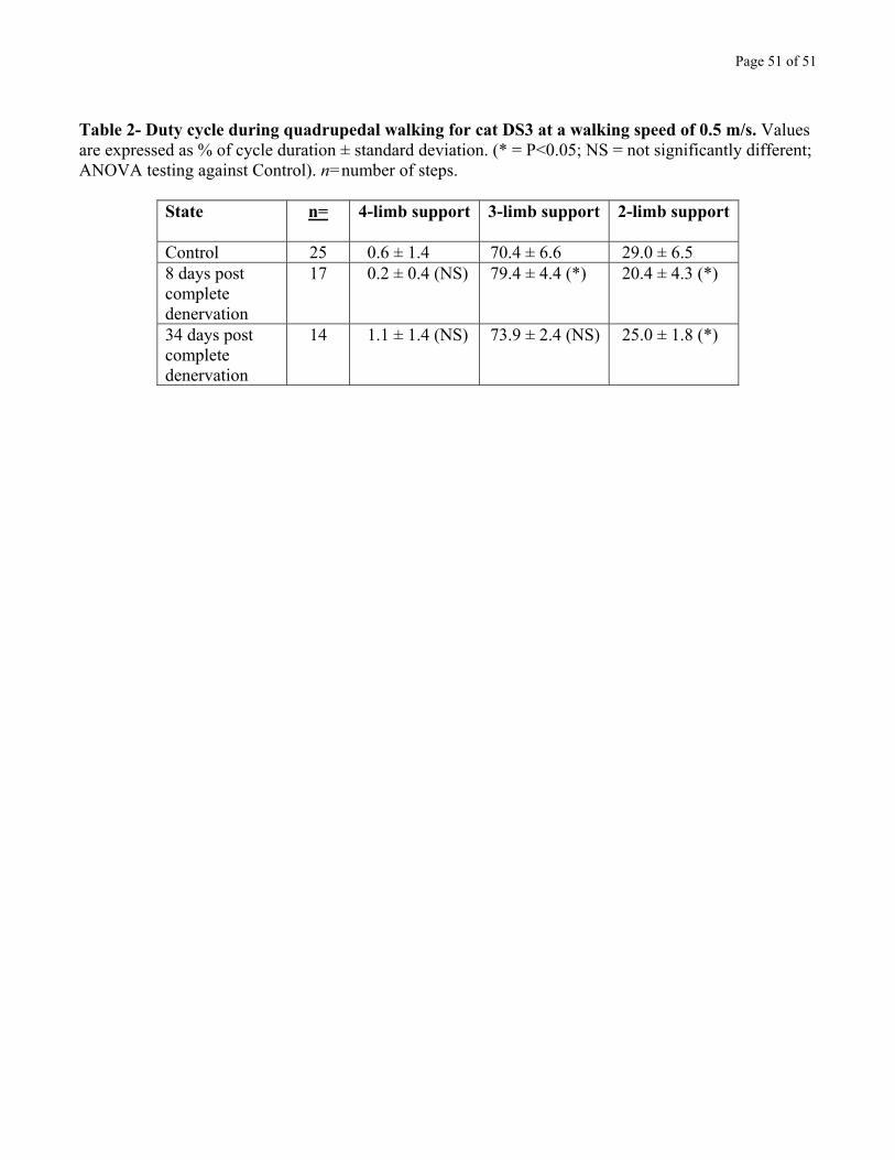
Page 51 of 51
Table 2- Duty cycle during quadrupedal walking for cat DS3 at a walking speed of 0.5 m/s. Values are expressed as % of cycle duration ± standard deviation. (* = P<0.05; NS = not significantly different; ANOVA testing against Control). n=number of steps.
State n= 4-limb support 3-limb support 2-limb support
Control 25 0.6 ± 1.4 70.4 ± 6.6 29.0 ± 6.5 8 days post complete denervation
17 0.2 ± 0.4 (NS) 79.4 ± 4.4 (*) 20.4 ± 4.3 (*)
34 days post complete denervation
14 1.1 ± 1.4 (NS) 73.9 ± 2.4 (NS) 25.0 ± 1.8 (*)
Related Documents