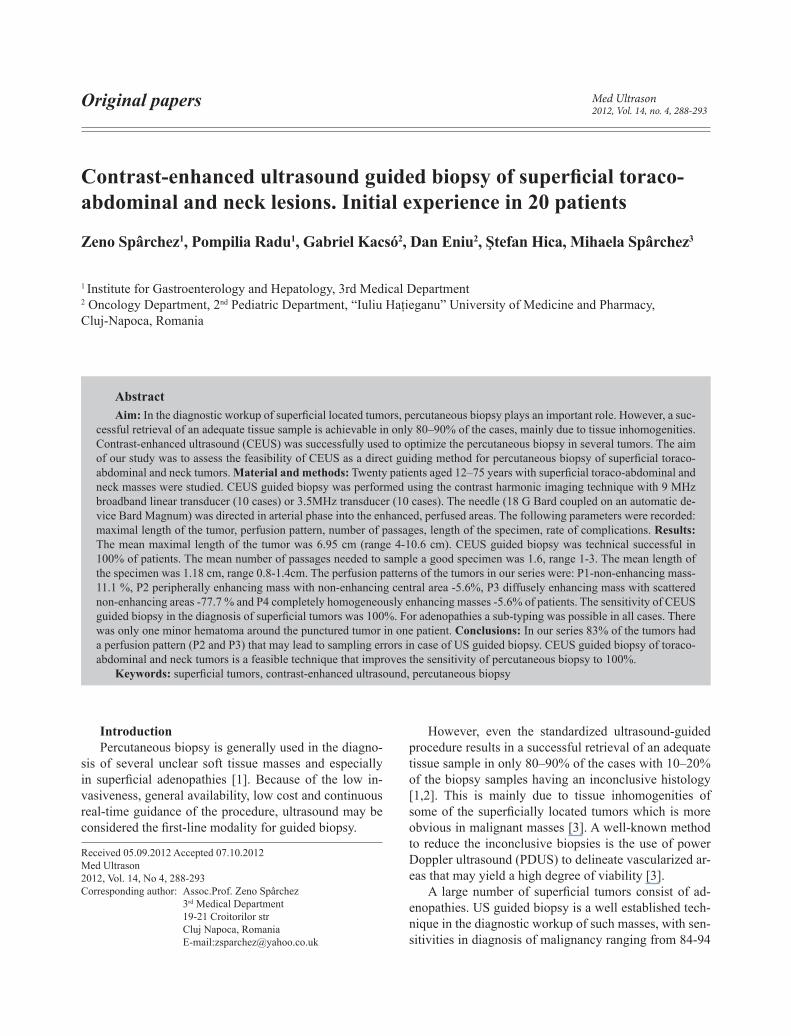
Original papers Med Ultrason 2012, Vol. 14, no. 4, 288-293 Abstract Aim: In the diagnostic workup of superficial located tumors, percutaneous biopsy plays an important role. However, a suc- cessful retrieval of an adequate tissue sample is achievable in only 80–90% of the cases, mainly due to tissue inhomogenities. Contrast-enhanced ultrasound (CEUS) was successfully used to optimize the percutaneous biopsy in several tumors. The aim of our study was to assess the feasibility of CEUS as a direct guiding method for percutaneous biopsy of superficial toraco- abdominal and neck tumors. Material and methods: Twenty patients aged 12–75 years with superficial toraco-abdominal and neck masses were studied. CEUS guided biopsy was performed using the contrast harmonic imaging technique with 9 MHz broadband linear transducer (10 cases) or 3.5MHz transducer (10 cases). The needle (18 G Bard coupled on an automatic de- vice Bard Magnum) was directed in arterial phase into the enhanced, perfused areas. The following parameters were recorded: maximal length of the tumor, perfusion pattern, number of passages, length of the specimen, rate of complications. Results: The mean maximal length of the tumor was 6.95 cm (range 4-10.6 cm). CEUS guided biopsy was technical successful in 100% of patients. The mean number of passages needed to sample a good specimen was 1.6, range 1-3. The mean length of the specimen was 1.18 cm, range 0.8-1.4cm. The perfusion patterns of the tumors in our series were: P1-non-enhancing mass- 11.1 %, P2 peripherally enhancing mass with non-enhancing central area -5.6%, P3 diffusely enhancing mass with scattered non-enhancing areas -77.7 % and P4 completely homogeneously enhancing masses -5.6% of patients. The sensitivity of CEUS guided biopsy in the diagnosis of superficial tumors was 100%. For adenopathies a sub-typing was possible in all cases. There was only one minor hematoma around the punctured tumor in one patient. Conclusions: In our series 83% of the tumors had a perfusion pattern (P2 and P3) that may lead to sampling errors in case of US guided biopsy. CEUS guided biopsy of toraco- abdominal and neck tumors is a feasible technique that improves the sensitivity of percutaneous biopsy to 100%. Keywords: superficial tumors, contrast-enhanced ultrasound, percutaneous biopsy Contrast-enhanced ultrasound guided biopsy of superficial toraco- abdominal and neck lesions. Initial experience in 20 patients Zeno Spârchez 1 , Pompilia Radu 1 , Gabriel Kacsó 2 , Dan Eniu 2 , Ştefan Hica, Mihaela Spârchez 3 1 Institute for Gastroenterology and Hepatology, 3rd Medical Department 2 Oncology Department, 2 nd Pediatric Department, “Iuliu Haţieganu” University of Medicine and Pharmacy, Cluj-Napoca, Romania Received 05.09.2012 Accepted 07.10.2012 Med Ultrason 2012, Vol. 14, No 4, 288-293 Corresponding author: Assoc.Prof. Zeno Spârchez 3 rd Medical Department 19-21 Croitorilor str Cluj Napoca, Romania E-mail:[email protected] Introduction Percutaneous biopsy is generally used in the diagno- sis of several unclear soft tissue masses and especially in superficial adenopathies [1]. Because of the low in- vasiveness, general availability, low cost and continuous real-time guidance of the procedure, ultrasound may be considered the first-line modality for guided biopsy. However, even the standardized ultrasound-guided procedure results in a successful retrieval of an adequate tissue sample in only 80–90% of the cases with 10–20% of the biopsy samples having an inconclusive histology [1,2]. This is mainly due to tissue inhomogenities of some of the superficially located tumors which is more obvious in malignant masses [3]. A well-known method to reduce the inconclusive biopsies is the use of power Doppler ultrasound (PDUS) to delineate vascularized ar- eas that may yield a high degree of viability [3]. A large number of superficial tumors consist of ad- enopathies. US guided biopsy is a well established tech- nique in the diagnostic workup of such masses, with sen- sitivities in diagnosis of malignancy ranging from 84-94

Welcome message from author
This document is posted to help you gain knowledge. Please leave a comment to let me know what you think about it! Share it to your friends and learn new things together.
Transcript
Original papers Med Ultrason2012, Vol. 14, no. 4, 288-293
AbstractAim: In the diagnostic workup of superficial located tumors, percutaneous biopsy plays an important role. However, a suc-
cessful retrieval of an adequate tissue sample is achievable in only 80–90% of the cases, mainly due to tissue inhomogenities. Contrast-enhanced ultrasound (CEUS) was successfully used to optimize the percutaneous biopsy in several tumors. The aim of our study was to assess the feasibility of CEUS as a direct guiding method for percutaneous biopsy of superficial toraco-abdominal and neck tumors. Material and methods: Twenty patients aged 12–75 years with superficial toraco-abdominal and neck masses were studied. CEUS guided biopsy was performed using the contrast harmonic imaging technique with 9 MHz broadband linear transducer (10 cases) or 3.5MHz transducer (10 cases). The needle (18 G Bard coupled on an automatic de-vice Bard Magnum) was directed in arterial phase into the enhanced, perfused areas. The following parameters were recorded: maximal length of the tumor, perfusion pattern, number of passages, length of the specimen, rate of complications. Results: The mean maximal length of the tumor was 6.95 cm (range 4-10.6 cm). CEUS guided biopsy was technical successful in 100% of patients. The mean number of passages needed to sample a good specimen was 1.6, range 1-3. The mean length of the specimen was 1.18 cm, range 0.8-1.4cm. The perfusion patterns of the tumors in our series were: P1-non-enhancing mass- 11.1 %, P2 peripherally enhancing mass with non-enhancing central area -5.6%, P3 diffusely enhancing mass with scattered non-enhancing areas -77.7 % and P4 completely homogeneously enhancing masses -5.6% of patients. The sensitivity of CEUS guided biopsy in the diagnosis of superficial tumors was 100%. For adenopathies a sub-typing was possible in all cases. There was only one minor hematoma around the punctured tumor in one patient. Conclusions: In our series 83% of the tumors had a perfusion pattern (P2 and P3) that may lead to sampling errors in case of US guided biopsy. CEUS guided biopsy of toraco-abdominal and neck tumors is a feasible technique that improves the sensitivity of percutaneous biopsy to 100%.
Keywords: superficial tumors, contrast-enhanced ultrasound, percutaneous biopsy
Contrast-enhanced ultrasound guided biopsy of superficial toraco-abdominal and neck lesions. Initial experience in 20 patients
Zeno Spârchez1, Pompilia Radu1, Gabriel Kacsó2, Dan Eniu2, Ştefan Hica, Mihaela Spârchez3
1 Institute for Gastroenterology and Hepatology, 3rd Medical Department2 Oncology Department, 2nd Pediatric Department, “Iuliu Haţieganu” University of Medicine and Pharmacy,Cluj-Napoca, Romania
Received 05.09.2012 Accepted 07.10.2012 Med Ultrason 2012, Vol. 14, No 4, 288-293 Corresponding author: Assoc.Prof. Zeno Spârchez 3rd Medical Department 19-21 Croitorilor str Cluj Napoca, Romania E-mail:[email protected]
IntroductionPercutaneous biopsy is generally used in the diagno-
sis of several unclear soft tissue masses and especially in superficial adenopathies [1]. Because of the low in-vasiveness, general availability, low cost and continuous real-time guidance of the procedure, ultrasound may be considered the first-line modality for guided biopsy.
However, even the standardized ultrasound-guided procedure results in a successful retrieval of an adequate tissue sample in only 80–90% of the cases with 10–20% of the biopsy samples having an inconclusive histology [1,2]. This is mainly due to tissue inhomogenities of some of the superficially located tumors which is more obvious in malignant masses [3]. A well-known method to reduce the inconclusive biopsies is the use of power Doppler ultrasound (PDUS) to delineate vascularized ar-eas that may yield a high degree of viability [3].
A large number of superficial tumors consist of ad-enopathies. US guided biopsy is a well established tech-nique in the diagnostic workup of such masses, with sen-sitivities in diagnosis of malignancy ranging from 84-94
289Med Ultrason 2012; 14(4): 288-293
tered intravenously followed by a 10 ml flush of saline via a 20G intravenous canula placed in an antecubital vein.
The ultrasound-guided biopsies were performed with an 18 G cutting needle (Bard) coupled on an automatic gun (Bard Magnum).
After the skin was sterilized the predicted needle path was anesthetized with 2% lidocaine. Prior to the intrave-nous injection of SonoVue, the needle was inserted into the skin entry. A free hand technique guiding method was used for both linear and convex probes.
When the first bubble arrived in the lesion following the contrast agent injection, the needle was directed in the arterial phase into the enhanced, perfused areas. In cases when the first specimen where judged to be unsuitable for a pathological diagnosis a second passage was done in the early venous phase. In cases where lymphoma was suspected, at least 3 specimens were obtained.
The following parameters were recorded: maximal length of the tumor, number of passages, length of the specimen, and rate of complications.
The perfusion patterns of the tumors were classified according to Loizides et al in: a) P1-non-enhancing mass or only rim-enhancement of the surrounding pseudo-capsule; b) P2- Peripherally enhancing mass with non-enhancing central area; c) P3-Diffusely enhancing mass with scattered non-enhancing areas and/or enhancement bridges; P4- Completely homogeneously enhancing masses [14]. We split the P3 tumors in 2 groups: P3a with low to moderate percentage of necrosis and P3b with high percentage of necrosis based on qualitative assessment of the non-enhanced tumoral areas in the biopsy plane.
For adenopathies we assessed the enhancement pat-tern according to Yu et al: Category 1 - intense homo-geneous enhancement; Category 2 - moderate homo-geneous enhancement: Category 3 - inhomogeneous enhancement; Category 4 - absent enhancement [15].
After the patients were informed about the benefits and risks of the procedure they provided an informal con-sent. The study was approved by the Ethical Commit-tee of the ”Iuliu Haţieganu” University of Medicine and Pharmacy, Cluj-Napoca, Romania.
Results
Among the 14 tumors 12 were malignant and 2 were benign (Table I). There were 3 metastatic adenopathies and 3 lymphomas (2 cases with Hodgkin and 1 with non Hodgkin lymphoma).
The mean maximal length of the tumors was 6.95 cm (range 4-10, 6 cm). For adenopathies the mean size was 5.5 cm, range 4-7.7 cm.
% and in sub-classification of malignant lymphomas be-tween 89-94% [4,5].
Ultrasound harmonic imaging with 2nd generation contrast agents (SonoVue) has the capacity of empha-sizing the macro and above all the microvascularization of various parenchyma and tumors. Beside the well-ac-cepted use in detection and characterization of various tumors, especially those located in the liver, contrast-en-hanced ultrasound (CEUS) has enabled the delimitation of the avascular, necrotic areas from the viable, active, vascularized regions of the tumors. By using this tech-nique for targeting, it becomes more likely to guarantee tissue viability [6].
The added value of CEUS in percutaneous biopsy was demonstrated in liver, pulmonary, kidney and soft tissue tumors [7-12]. However, in those studies CEUS was used prior to biopsy, an approach that may cause needle misplacement and thus an incorrect sampling, es-pecially in large tumors. In liver tumors CEUS was used also as a direct guiding method to target otherwise invis-ible areas [13].
The aim of our prospective pilot study was to assess the feasibility of CEUS as a direct guiding method for percutaneous biopsy of superficial lesions (tumors and adenopathies) in the trunk and neck.
Material and methods
Between January 2011 and October 2012, 20 subjects aged 12 –75 years with unclear superficial trunk and neck masses (Table I) were referred for percutaneous guided biopsy. There were 14 tumors (9 localized on the thoracic wall, 3 on the abdominal wall, 1 on the shoulder area, and 1 in the neck area) and 6 adenopathies (3 localized in the axilla, 2 in the supraclavicular space, and 1 in the laterocervical area).
One of the authors with experience in soft tissue ul-trasound performed a baseline grayscale and power Dop-pler US with a 9-12 MHz broadband linear transducer on a Logiq 7 and 9 (General Electric, Milwaukee USA) and S 2000 (Siemens, Erlangen, Germany) systems.
CEUS guided biopsy was performed using the con-trast harmonic imaging technique with a 9 MHz broad-band linear transducer. In 10 cases with large tumors a 3.5 MHz transducer was used. For the linear transducer a MI of 0.12 was set meanwhile for the 3.5 MHz transduc-er a 0.09 MI was used. Focus is positioned at the bottom of the screen to minimize microbubble destruction. Field of view and gain were optimized to provide the clearest depiction of the lesion.
A standard bolus of 2.4 ml of a second-generation contrast agent (SonoVue®, Bracco, Italy) was adminis-

290 Zeno Spârchez et al Contrast-enhanced ultrasound guided biopsy of superficial toraco-abdominal and neck lesions
CEUS guided biopsy was technically successful in 100% of the patients. The mean number of passages was 1.6, range 1-3. The mean length of the specimen was 1.18 cm, range 0.8-1.4cm.
The perfusion pattern of the tumors in our series was: P1 2 pts with hematomas (14.2 %) (fig 1), P2 -1 patient (7.1%), P3 -10 patients (71.5 %) (fig 2) and P4 -1 patient (7.1%) (fig 3). In the P3 group low to moderate necrosis was present in 70% of cases and extensive necrosis in 30%.
All adenopathies displayed an inhomogeneous en-hancement (category 3) (fig 4).
The sensitivity of CEUS guided biopsy in the diag-nosis of both superficial tumors and adenopathies was 100%. For adenopathies a sub-typing was possible in all cases.
There was only one minor hematoma around the punctured tumor in patient 1.
Patient Age Sex Location Largest diameter
Specimens (cm) Final diagnosis Enhance-
ment type
1 68 Male Thoracic wall, suprascapular 5 cm 1.4 Metastatic adenocarcinoma P3a
2 71 Male Neck, tyroid 10 cm 1.2; 1,2 Anaplastic carcinoma P3b
3 68 Female Abdominal wall, lombar 10cm 1;1.2 Metastatic adenocarcinoma P3b
4 40 Male Abdominal wall, inguinal 7 cm 1.2; 0.8 Metastatic carcinoma (keratiniz-ing squamous cell type)
P2
5 64 Male Right shoulder 5.5 cm 1.4 Chondrosarcoma P4
6 72 Female Abdominal wall, hypogastru 10.6 cm 1.2 Organized haematoma P1
7 55 Male Infraclavicular space 7cm 1; 1.2 Old haematoma P1
8 68 Female Thoracic wall 4 cm 1.2 Metastatic carcinoma (breast) P3a
9 51 Female Thoracic wall 4 cm 1.2; 1.3 Metastatic adenocarcinoma P3a
10 64 Male Thoracic wall 10 cm 1.2 Metastatic carcinoma (renal carcinom clear cell type)
P3a
11 57 Female Thoracic wall 4,6 cm 1.2 Metastatic carcinoma (breast carcinoma)
P3a
12 54 Male Thoracic wall 14 cm 1.4; 1.2 Metastatic carcinoma (diffuse type, undifferent)
P3b
13 60 Male Thoracic wall 10 cm 1.2 Metastatic carcinoma (renal carcinoma, clear cell type)
P3a
14 26 Male Thoracic wall 7.7 cm 1,4 Non small cell carcinoma (large cells)
P3a
15 70 Female Laterocervical 5 cm 0.8; 1.0 Metastatic carcinoma (medullary carcinoma)
C3
16 50 Male Supraclavicular space 4.8 cm 1.3 Metastatic carcinoma (renal carcinom clear cell type)
C3
17 31 Male Axilla 7 cm 1.2; 1 Metastatic melanoma C3
18 80 Female Axilla 4 cm 1.4; 1.2 ; 1 Hodgkin lymphoma C3
19 20 Male Supraclavicular space 4 cm 1.2 Hodgkin lymphoma C3
20 72 Female Axilla 5 cm 1.4; 1.5; 1 Non Hodgkin lymphoma C3

291Med Ultrason 2012; 14(4): 288-293
Fig 1. Abdominal wall hematoma in a patient with NH: a) US showed a well demarcated, ovalar mass; b) CEUS depicts a non-enhancing mass with a rim enhancement of the surrounding pseudo-capsule. Note the presence of the needle inside.
Fig 4. Metastatic adenopathy from renal cell carci-noma: a) US shows a 5 cm enlarged lymphnode in the supraclavicular region; b) CEUS guided biopsy arterial phase. The needle (<) is placed in an en-hancing area, avoiding a central area of necrosis.
Fig 2. Metastatic renal cell carcinoma in thoracic wall: a) US+color Doppler revealed a 10 cm tumor in the thoracic wall invading the pleural space; note the scarse vascularity in the tumor; b) CT depicts a thoracic wall tumor which invades the pleura and extends into the lung; c) CEUS guided biopsy, ar-terial phase; the tumor displays a P3a pattern. <- Needle
Fig 3. Chondrosarcoma of the shoulder: a) A 5.5 cm hipoechoic mass is seen near the right shoulder; b) CT image; c) CEUS in arterial phase. Note the relatively homogeneous enhancement of the tumor without necrotic areas (P4 pattern). The needle is not seen on this image.
Discussions
In superficial masses, the tumor diagnosis is based on the histological data offered by the tumor fragment, sam-pled by percutaneous needle biopsy or surgical biopsy. Compared to open surgical biopsy, image guided biopsy has certain benefits, such as well acceptance by the pa-tients and the possibility to be performed on an outpatient basis [16].
The performance of percutaneous guided puncture bi-opsy in superficial tumors is influenced by several factors such as needle type, the number/size/length of acquired samples, and the experience of the performing physician [3]. Among tumor characteristics the tumor type and size play an important role. It is known that the performances of needle biopsy are lower for large tumors due to the existence of tumor necrosis or fatty changes. Necrotic tissue cannot be identified on B-mode sonography, espe-cially before liquefaction has occurred, possibly leading to an unsuccessful biopsy or a false-negative diagnosis [17]. In larger lesions, biopsy is usually performed in the peripheral zone or a hypervascular area of the tumors.
The central region of malignant soft tissue masses of-ten has an increased interstitial pressure compared with the peripheral zone [14]. As a consequence, superficial malignant neoplasms exhibit often 2 distinct areas: a cen-tral region with reduced vascularization and subsequent necrosis and a highly vascularised outer rim of infiltra-tive tumor tissue.
The use of CEUS perfusion patterns of soft tissue masses was demonstrated to be a useful tool in differen-tiating benign from malignant tumors. Thus P2 and P3 patterns are associated with malignancy with good sensi-tivities (for P3) and specificities (for P2) [14]. In our se-ries of patients with relative large tumors, 83% had a P3 pattern which demonstrated the high frequency of tumors with reduced vascularization and appearance of central

292 Zeno Spârchez et al Contrast-enhanced ultrasound guided biopsy of superficial toraco-abdominal and neck lesions
necrosis. In such tumors US guided percutaneous biopsy may target a nonviable area resulting in an inconclusive result.
CEUS has a higher degree of diagnostic accuracy than conventional ultrasonography for evaluations of superficial lymphadenopathy. The contrast enhancement patterns and time intensity curves may add important clues for differential diagnosis of benign and malignant lymph nodes and may predict invasiveness in metastatic lymph nodes [15,18,19]. According to Yu et al 97% of metastatic adenopathies have an inhomogeneous en-hancement with areas of necrosis (category 3) finding that may emphasize the need of a CEUS guided biopsy in such cases [15]. Lymphomas have a more variable CEUS appearance, all the enhancement patterns (including the non-enhancement type found in B-cell lymphoma) being present at CEUS [15]. In our small number of biopsied adenopathies, both the metastatic and lymphomatous adenopaties displayed a category 3 pattern. This could be explained by the relatively large size of the involved lymph nodes (mean size 5.5 cm).
For both superficial tumors and adenopathies, by de-picting the perfused areas, CEUS can select the appropri-ate area for biopsy and guide the needle in that area.
The biopsy needles are clearly visible under CEUS conditions due to the fact that the needle causes tissue motion in the vicinity of the needle which generates har-monic signals, detected by the transducer. For automatic TruCut needle (i.e. Bard type) the presence of air in the side notch is easily visible after the automatic tissue re-trieval [20]. Sometimes, the bright contrast enhancement in the surrounding parenchyma masks the echogenic bi-opsy needle in the CEUS image [13].
One technical difficulty is related to the short period of arterial enhancement available for puncture. If the tip of the needle is lost it may take a few seconds to find it and to perform the biopsy without losing the arterial en-hancement. This limitation may be overcome by the use of needle guides, and availability of ultrasound systems that display both the CEUS and conventional images on the screen or by performing a planning CEUS before CEUS guided biopsy [6].
In our small number of patients, CEUS guided biopsy was technical feasible in all patients although in 12 cases we used a system that displays only the CEUS image for both linear and convex probes. For superficial tumors the needle path is very short so it is quite easy to target an enhanced area using only the CEUS image.
In soft tissue tumors many series have shown that al-though the accuracy of US guided biopsy in diagnosis of malignancy is very high (94-97%), in identifying tu-mor subtype and grading, the accuracy is only 76-80%
[11,21]. This can be explained by the fact that in large tumors there is a heterogeneous histopathology not only with necrotic and hemorrhagic areas but also with ar-eas with different morphology and grading. Moreover in mixed tumors there are areas with both high and low grade morphology, areas that may explain the lower ac-curacy. Those areas may be depicted and punctured using CEUS [11].
Using CEUS before biopsy to select a proper biopsy area, Loizides at al reached a sensitivity of 100% in the diagnosis of malignancy and in establishing the histolog-ical entity and tumor grading [11]. An important finding in that study was the discrepancy in some cases between power Doppler and CEUS in depicting hypervascular ar-eas, a discrepancy that may explain why power Doppler US is of less important value in optimizing the biopsy.
In a larger cohort of patients with soft tissue tumors, De Marchi et all using CEUS before biopsy and reached a sensitivity of 97.1% and a specificity of 92.5% [12]. The sensitivity and specificity in specific histopathologi-cal subgroups were 100%, in the grading definition the same indices being 100% and 96.8% [11]. In both studies no correlations were made between biopsy accuracy and enhancement patterns.
In our study CEUS guided biopsy yielded 100% sen-sitivity in the diagnosis of superficial tumors. 91.6% of those tumors displayed an inhomogeneous pattern with areas of necrosis and 33.3% of cases presented with an important necrosis. These findings support the manda-tory role of CEUS as guiding methods in the biopsy of those tumors.
In large adenopathies, the sensitivity was 100% for both diagnosis of malignancy and sub-typing. To our knowledge there are no other studies investigating the role of CEUS guided biopsy in the diagnosis of super-ficial adenopathies. This preliminary study shows prom-ising results that should be validated in larger studies. However, based on these findings it may be supposed that the use of CEUS guidance would increase the accuracy of biopsy in adenopathies to 100%.
There are several limitations of this study. Although it is a prospective pilot study, no control group was used and the number of patients was limited. These prelimi-nary results must be assessed in a large prospective com-parative study with conventional US guided biopsy for both soft tissue tumors and adenopathies. Secondly only 2 tumors were benign, a fact that may influence the final results.
In contrast with other papers, in the present study CEUS was used during the biopsy to target the en-hanced areas. This may be a debating issue as the same results could be obtained using CEUS before biopsy to
293Med Ultrason 2012; 14(4): 288-293
map the enhancement pattern inside the tumor. Moreo-ver, depicting a benign enhancement pattern by CEUS a biopsy might have been avoided. The results obtained in this study (an excellent feasibility of the method and high percentage of cases with various degrees of necro-sis among both soft tissue tumors and adenopathies) may support this approach.
Conflict of interest: none
Acknowledgement: This study forms part of the re-search project within BIOPTICON 2329/2008 PNCDI II 2007 – 2013.
References
1. Mitsuyoshi G, Naito N, Kawai A, et al. Accurate diagnosis of musculoskeletal lesions by core needle biopsy. J Surg Oncol 2006; 94: 21–27.
2. Chiou HJ, Chou YH, Chiu SY, et al. Differentiation of benign and malignant superficial soft-tissue masses using grayscale and color doppler ultrasonography. J Chin Med Assoc 2009; 72: 307–315.
3. Wu JS, Goldsmith JD, Horwich PJ, Shetty SK, Hochman MG. Bone and soft tissue lesions: what factors affect diag-nostic yield of image-guided core-needle biopsy. Radiology 2008; 248: 962–970.
4. Screaton NJ, Berman LH, Grant JW. Head and neck lym-phadenopathy: evaluation with US-guided cutting-needle biopsy. Radiology 2002; 224: 75-81.
5. Demharter J, Müller P, Wagner T, Schlimok G, Haude K, Bohndorf K. Percutaneous core-needle biopsy of enlarged lymph nodes in the diagnosis and subclassification of ma-lignant lymphomas. Eur Radiol 2001; 11: 276-283.
6. Sparchez Z, Radu P, Zaharia T, et al. Usefulness of contrast enhanced ultrasound guidance in percutaneous biopsies of liver tumors. J Gastrointestin Liver Dis 2011; 20: 191-196.
7. Wu W, Chen MH, Yin SS, et al. The role of contrast-en-hanced sonography of focal liver lesions before percutane-ous biopsy. AJR Am J Roentgenol 2006; 187: 752-761.
8. Grossjohann HS, Bachmann Nielsen M. Ultrasound con-trast agents may help in avoiding necrotic areas at biopsy. Ultraschall Med 2006; 27: 2-3.
9. Cao BS, Wu JH, Li XL, Deng J, Liao GQ. Sonographically guided transthoracic biopsy of peripheral lung and medias-tinal lesions: role of contrast-enhanced sonography. J Ultra-sound Med 2011; 30: 1479-1490.
10. Sparchez Z, Radu P, Kacso G, et al. Performance of CEUS guided biopsy in large renal and adrenal tumors. Ultrasound Med Biol 2011; 37: S33.
11. Loizides A, Widmann G, Freuis T, Peer S, Gruber H. Opti-mizing ultrasound-guided biopsy of musculoskeletal mass-es by application of an ultrasound contrast agent. Ultras-chall Med 2011; 32: 307–310.
12. De Marchi A, Brach del Prever AM, Linari A, et al. Accura-cy of core-needle biopsy after contrast-enhanced ultrasound in soft-tissue tumors. Eur Radiol 2010; 20: 2740-2748.
13. Yoon SH, Lee KH, Kim SY, et al. Real-time contrast-en-hanced ultrasound-guided biopsy of focal hepatic lesions not localised on B-mode ultrasound. Eur Radiol 2010; 20: 2047-2056.
14. Loizides A, Peer S, Plaikner M, Djurdjevic T, Gruber H. Perfusion pattern of musculo-skeletal masses using con-trast-enhanced ultrasound: a helpful tool for characterisa-tion? Eur Radiol 2012 ; 22: 1803-1811.
15. Yu M, Liu Q, Song HP, et al. Clinical application of con-trast-enhanced ultrasonography in diagnosis of superficial lymphadenopathy. J Ultrasound Med 2010; 29: 735-740.
16. Skrzynski MC, Biermann JS, Montag A, Simon MA. Diag-nostic accuracy and charge-savings of outpatient core nee-dle biopsy compared with open biopsy of musculoskeletal tumors. J Bone Joint Surg Am 1996; 78: 644–649.
17. Solbiati L, Ierace T, Tonolini M, Cova L. Guidance and moni-toring of radiofrequency liver tumor ablation with contrast-en-hanced ultrasound. Eur J Radiol 2004; 51(Suppl1): S19–S23.
18. Weskott HP. Ultrasound in the diagnostic management of malignant lymphomas. Radiologe 2012; 52: 347-359.
19. Ouyang Q, Chen L, Zhao H, Xu R, Lin Q. Detecting me-tastasis of lymph nodes and predicting aggressiveness in patients with breast carcinomas. J Ultrasound Med 2010; 29: 343-352.
20. Schlottmann K, Klebl F, Zorger N, Feuerbach S, Schölm-erich J. Contrast-enhanced ultrasound allows for interven-tions of hepatic lesions which are invisible on conventional B-mode. Z Gastroenterol 2004; 42: 303-310.
21. Strauss DC, Qureshi YA, Hayes AJ, Thway K, Fisher C, Thomas JM. The role of core needle biopsy in the diagnosis of suspected soft tissue tumours. J Surg Oncol 2010; 102: 523-529.
Related Documents