Contradictory Reasoning Network: An EEG and fMRI Study Camillo Porcaro 1 * . , Maria Teresa Medaglia 1,2. , Ngoc Jade Thai 3 , Stefano Seri 3 , Pia Rotshtein 4 , Franca Tecchio 1,5 1 LET’S-ISTC-CNR, Rome, Italy, 2 Phylosophy Department, Roma Tre University, Rome, Italy, 3 Aston Brain Centre, School of Life and Health Sciences, Aston University, Birmingham, United Kingdom, 4 Behavioural Brain Sciences Centre, School of Psychology, University of Birmingham, Birmingham, United Kingdom, 5 Department of Neuroimaging, IRCCS San Raffaele Pisana, Rome, Italy Abstract Contradiction is a cornerstone of human rationality, essential for everyday life and communication. We investigated electroencephalographic (EEG) and functional magnetic resonance imaging (fMRI) in separate recording sessions during contradictory judgments, using a logical structure based on categorical propositions of the Aristotelian Square of Opposition (ASoO). The use of ASoO propositions, while controlling for potential linguistic or semantic confounds, enabled us to observe the spatial temporal unfolding of this contradictory reasoning. The processing started with the inversion of the logical operators corresponding to right middle frontal gyrus (rMFG-BA11) activation, followed by identification of contradictory statement associated with in the right inferior frontal gyrus (rIFG-BA47) activation. Right medial frontal gyrus (rMeFG, BA10) and anterior cingulate cortex (ACC, BA32) contributed to the later stages of process. We observed a correlation between the delayed latency of rBA11 response and the reaction time delay during inductive vs. deductive reasoning. This supports the notion that rBA11 is crucial for manipulating the logical operators. Slower processing time and stronger brain responses for inductive logic suggested that examples are easier to process than general principles and are more likely to simplify communication. Citation: Porcaro C, Medaglia MT, Thai NJ, Seri S, Rotshtein P, et al. (2014) Contradictory Reasoning Network: An EEG and fMRI Study. PLoS ONE 9(3): e92835. doi:10.1371/journal.pone.0092835 Editor: Daniel Houser, George Mason University, United States of America Received July 17, 2013; Accepted February 26, 2014; Published March 25, 2014 Copyright: ß 2014 Porcaro et al. This is an open-access article distributed under the terms of the Creative Commons Attribution License, which permits unrestricted use, distribution, and reproduction in any medium, provided the original author and source are credited. Funding: The MRI scan costs of the study were supported by the Lord Dowding Fund for Humane Research. PR was supported by the Leverhulme Trust. The funders had no role in study design, data collection and analysis, decision to publish, or preparation of the manuscript. Competing Interests: The authors have declared that no competing interests exist. * E-mail: [email protected] . These authors contributed equally to this work. Introduction The ability to reason, crucial for effective social interactions and for the solution of common practical problems, is among the most advanced human intellectual abilities. Contradiction, as a sub-class of reasoning process is one of the cornerstones of human rational reasoning and is part of everyday life and communication. In debates or conversations, we usually examine a statement and evaluate the validity of its content before agreeing with or arguing against what is being said. In western culture, the elements normally used in a conversation derive from a framework based on the Aristotelian Square of Opposition (ASoO). Formally, the ASoO is based on categorical statements, either universal statements related to the totality, expressed by the logical operator (quantifier) All or particular statements related to a small subset of it, expressed by logical operator Some. When contradicting a generic statement, we tend to search for counter examples. For instance, to refute the statement ‘All swans are white’, we might argue that ‘The Australian swan is black’. In logic, the first statement (i.e. ‘All…’) is contradicted by the formulation ‘Some swans are black’ (i.e. ‘Some…’). Categorical propositions may be used in various logical relationships; in particular a premise- conclusion pair can be contradictory or non-contradictory [1]. In the ASoO the contradiction between a first categorical proposition (premise) with a second categorical proposition (conclusion) can be achieved by inverting the logical operators All with Some and the attribute, i.e. ‘white’ with ‘black’ in the example above. Two main types of logical reasoning can be distinguished: inductive when we derive a rule based on a series of observations, and deductive when we formulate an example based on a rule. When the premise is a particular example (‘Some’) and the conclusion is a universal rule (‘All’), their relation is assessed using inductive reasoning. In contrast deductive reasoning is needed to assess the reverse relation: how a universal rule (‘All’) applies to a particular example (‘Some’). In the past decade, studies on conditional reasoning [2] and syllogism [3–9] have contributed to shed light on the neural basis of logical reasoning. Using such logical structures, these studies have consistently reported activation of distributed cortical and sub-cortical networks, including those associated with language processing and semantic and visuo-spatial skills (see for review [10–15]). Lateralized effects have also been reported, with the left hemisphere playing a key role in linguistic processes and the right hemisphere mainly supporting logical reasoning [11,12,16,17], although other studies have found key linguistic and logical processes to be in different sub-areas in the left hemisphere [5,7,8]. To our knowledge, the neural correlates associated with different components of contradictory thinking, a simple but PLOS ONE | www.plosone.org 1 March 2014 | Volume 9 | Issue 3 | e92835

Welcome message from author
This document is posted to help you gain knowledge. Please leave a comment to let me know what you think about it! Share it to your friends and learn new things together.
Transcript
Contradictory Reasoning Network: An EEG and fMRIStudyCamillo Porcaro1*., Maria Teresa Medaglia1,2., Ngoc Jade Thai3, Stefano Seri3, Pia Rotshtein4,
Franca Tecchio1,5
1 LET’S-ISTC-CNR, Rome, Italy, 2 Phylosophy Department, Roma Tre University, Rome, Italy, 3 Aston Brain Centre, School of Life and Health Sciences, Aston University,
Birmingham, United Kingdom, 4 Behavioural Brain Sciences Centre, School of Psychology, University of Birmingham, Birmingham, United Kingdom, 5 Department of
Neuroimaging, IRCCS San Raffaele Pisana, Rome, Italy
Abstract
Contradiction is a cornerstone of human rationality, essential for everyday life and communication. We investigatedelectroencephalographic (EEG) and functional magnetic resonance imaging (fMRI) in separate recording sessions duringcontradictory judgments, using a logical structure based on categorical propositions of the Aristotelian Square ofOpposition (ASoO). The use of ASoO propositions, while controlling for potential linguistic or semantic confounds, enabledus to observe the spatial temporal unfolding of this contradictory reasoning. The processing started with the inversion ofthe logical operators corresponding to right middle frontal gyrus (rMFG-BA11) activation, followed by identification ofcontradictory statement associated with in the right inferior frontal gyrus (rIFG-BA47) activation. Right medial frontal gyrus(rMeFG, BA10) and anterior cingulate cortex (ACC, BA32) contributed to the later stages of process. We observed acorrelation between the delayed latency of rBA11 response and the reaction time delay during inductive vs. deductivereasoning. This supports the notion that rBA11 is crucial for manipulating the logical operators. Slower processing time andstronger brain responses for inductive logic suggested that examples are easier to process than general principles and aremore likely to simplify communication.
Citation: Porcaro C, Medaglia MT, Thai NJ, Seri S, Rotshtein P, et al. (2014) Contradictory Reasoning Network: An EEG and fMRI Study. PLoS ONE 9(3): e92835.doi:10.1371/journal.pone.0092835
Editor: Daniel Houser, George Mason University, United States of America
Received July 17, 2013; Accepted February 26, 2014; Published March 25, 2014
Copyright: � 2014 Porcaro et al. This is an open-access article distributed under the terms of the Creative Commons Attribution License, which permitsunrestricted use, distribution, and reproduction in any medium, provided the original author and source are credited.
Funding: The MRI scan costs of the study were supported by the Lord Dowding Fund for Humane Research. PR was supported by the Leverhulme Trust. Thefunders had no role in study design, data collection and analysis, decision to publish, or preparation of the manuscript.
Competing Interests: The authors have declared that no competing interests exist.
* E-mail: [email protected]
. These authors contributed equally to this work.
Introduction
The ability to reason, crucial for effective social interactions and
for the solution of common practical problems, is among the most
advanced human intellectual abilities. Contradiction, as a sub-class
of reasoning process is one of the cornerstones of human rational
reasoning and is part of everyday life and communication. In
debates or conversations, we usually examine a statement and
evaluate the validity of its content before agreeing with or arguing
against what is being said. In western culture, the elements
normally used in a conversation derive from a framework based on
the Aristotelian Square of Opposition (ASoO). Formally, the
ASoO is based on categorical statements, either universal
statements related to the totality, expressed by the logical operator
(quantifier) All or particular statements related to a small subset of
it, expressed by logical operator Some. When contradicting a
generic statement, we tend to search for counter examples. For
instance, to refute the statement ‘All swans are white’, we might
argue that ‘The Australian swan is black’. In logic, the first
statement (i.e. ‘All…’) is contradicted by the formulation ‘Some
swans are black’ (i.e. ‘Some…’). Categorical propositions may be
used in various logical relationships; in particular a premise-
conclusion pair can be contradictory or non-contradictory [1]. In
the ASoO the contradiction between a first categorical proposition
(premise) with a second categorical proposition (conclusion) can be
achieved by inverting the logical operators All with Some and the
attribute, i.e. ‘white’ with ‘black’ in the example above. Two main
types of logical reasoning can be distinguished: inductive when we
derive a rule based on a series of observations, and deductive when
we formulate an example based on a rule. When the premise is a
particular example (‘Some’) and the conclusion is a universal rule
(‘All’), their relation is assessed using inductive reasoning. In
contrast deductive reasoning is needed to assess the reverse
relation: how a universal rule (‘All’) applies to a particular example
(‘Some’).
In the past decade, studies on conditional reasoning [2] and
syllogism [3–9] have contributed to shed light on the neural basis
of logical reasoning. Using such logical structures, these studies
have consistently reported activation of distributed cortical and
sub-cortical networks, including those associated with language
processing and semantic and visuo-spatial skills (see for review
[10–15]). Lateralized effects have also been reported, with the left
hemisphere playing a key role in linguistic processes and the right
hemisphere mainly supporting logical reasoning [11,12,16,17],
although other studies have found key linguistic and logical
processes to be in different sub-areas in the left hemisphere [5,7,8].
To our knowledge, the neural correlates associated with
different components of contradictory thinking, a simple but
PLOS ONE | www.plosone.org 1 March 2014 | Volume 9 | Issue 3 | e92835
fundamental logical process, are still largely unexplored. To this
aim we assessed responses in the reasoning networks to logical
structures devoted to the identification of contradiction.
Based on a protocol used in our previous high-resolution EEG
study [18], we undertook a separate event-related fMRI experi-
ment and attempted to integrate EEG and fMRI data. The
purpose of the present study was to investigate the brain
recruitment and specific timing during the identification of
contradiction. We used a protocol based on ASoO premise-
conclusion pairs, since it allowed deconvolving the neuronal
responses reflecting contradiction identification and manipulation
of the two logical operators while controlling for linguistic and
semantic features of the sentences. To delineate processing related
to the contradictory judgment, we contrasted contradictory
(C, e.g. ‘All swan are white – Some swan are black’) to non-
contradictory (nC, e.g ‘All swan are white’ – ‘Some swan are
white’) premise-conclusion pairs. Furthermore, contrasting All-
Some (AS: deduction, ‘All swan are white – Some swan are black’)
with Some-All ( SA: induction, ‘Some swan are white – All swan
are black’) sentence pairs, we manipulated the order of universal/
particular logical operators in order to verify if inductive and
deductive reasoning are subtended by different neural structures.
We hypothesized that in this later case the order in which the two
sentences (AS vs. SA) are presented may affect the way in which
they are logically processed and that brain imaging could identify
the neural correlates of the logical inversion operation, which is
orthogonal to the content inversion operation.
Data of the experiment is available from the corresponding
author upon request.
Materials and Methods
ParticipantsEEG study. data for the EEG study was collected during a
previous study (reported in [18]) from eleven healthy native Italian
speakers (mean age: 30.5 years; range: 24–38 years; six women).
fMRI study. Thirteen healthy native Italian speakers (mean
age 31.1 range 25 – 42 years; 6 women) were recruited. Eight of
these participants had also participated the EEG study.
None of the participants had a formal background in logic and
none had prior history of neurological or psychiatric conditions.
All participants had normal or corrected-to-normal vision.
Participants gave their informed written consent after the nature
of the study was explained to them. The study was approved by
the Ethics Committee of Aston University, where the study was
conducted.
Stimuli and procedureStimuli were presented visually via a Dell PC using a paradigm
developed on the E-Prime programming software (Psychology
Software Tools, Inc.; http://www.pstnet.com). A 262 factorial
design was used with the following factors: Logical Operator
(INDUCTION: SOME-ALL, DEDUCTION: ALL-SOME) and Contradiction
(CONTRADICTORY, NON CONTRADICTORY). Sentences were in
Italian (the native language of the participants). The protocol
consisted of 140 sentence pairs, equally represented in the
following four forms: 35 All… Some… – Contradictory (AS-C),
35 Some… All… – Contradictory (SA-C), 35 All… Some… – Non-
contradictory (AS-nC) and 35 Some… All… – Non-contradictory
(SA-nC). The English translation of 4 typical sentences is provided
in Table 1. In the AS-C and AS-nC the premise is universal (A:
All) and the conclusion is particular (S: Some), whilst the conclusion
is contradictory for the first set (AS-C) and non-contradictory for
the other (AS-nC). Similar definitions apply for the SA-C and SA-
nC. The sentences were identical as far as the logical operator
manipulation; the only difference was the order in which the
logical operators were presented, eliciting inductive or deductive
reasoning. The C and nC sentences had identical grammatical
structure and were matched as much as possible for length and
complexity.
Prior to the experiment, participants received a training session
on a separate set of stimuli to help them to familiarize with the task
and the recording environment. Participants (Ps) were asked to
evaluate whether the conclusion was contradictory or not with
respect to the premise, independent of the correctness of the
sentence content. The trial sequence started with 0.5s fixation to
alert the Ps of the beginning of the trial, the premise sentence was
then presented for 2.5s followed by a 0.5s fixation and the
conclusion sentence for 2.5s (Figure 1). The trial ended with the
presentation of a question mark for 1.5s, during which participants
were asked to perform a button press (right hand = contradictory,
left hand = non-contradictory). Participants were instructed to
refrain from responding if unable to make a clear choice. Three
randomly selected inter trial intervals (5.5 or 7.5 or 10.5s) were
used for the fMRI experiment, to facilitate the estimation of the
Hemodynamic Response Function (HRF) in the MRI experiment
[19]. A fixed 1s inter trial interval was used for the EEG
procedure. Sentence pairs were presented in a random order.
Analysis of behavioral dataRepeated measures analyses of variance (ANOVAs) were
separately applied to the percentage of errors and the reaction
times of correct responses (dependent variables) with Logical
Operator (INDUCTION [SA], DEDUCTION [AS]) and Contradiction
(CONTRADICTORY [C], NON-CONTRADICTORY [nC]) as within-
subject factors using SPSS15 (SPSS Inc, Chicago IL, USA).
Threshold for significance was set at p,0.05.
Table 1. Stimuli Example.
UNIVERSAL – PARTICULAR PARTICULAR – UNIVERSAL
CONTRADICTORY All swans are white - Some swans are black Some swans are white - All swans are black
[AS-C] [SA-C]
NON-CONTRADICTORY All swans are white - Some swans are white Some swans are black - All swans are black
[AS-nC] [SA-nC]
An example of the four experimental conditions. The same number of premise-conclusion pairs was presented for each condition. Acronyms are indicated in squarebrackets.doi:10.1371/journal.pone.0092835.t001
Contradictory Reasoning Investigated by EEG & fMRI
PLOS ONE | www.plosone.org 2 March 2014 | Volume 9 | Issue 3 | e92835
EEG data acquisitionScalp EEG signals were recorded continuously during the
protocol using an EGI data acquisition system (Electrical
Geodesics, Eugene, OR, USA; http://www.egi.com) with the
third generation of EGI dense array net, the HydroCel Geodesic
Sensor Net VR (128-channel HCGSN [20]). Gain and zero
calibration were performed before the start of each EEG
recording; channel impedances were kept below 50 kV for all
net sensors [21]. Amplified EEG signals were sampled at 500 Hz
(anti-aliasing filter at 200 Hz) and stored for off-line processing. All
channels were referenced to the vertex (Cz) electrode. Source
analysis of this dataset was presented in [18].
EEG data analysisA semiautomatic Independent Component Analysis (ICA)
procedure was applied to characterize artifactual non-cerebral
activities, i.e. eye movements, muscular artifacts and environmen-
tal noise, and remove them without rejecting the contaminated
epochs [22–24]. EEG analysis was then performed after back-
projecting all the non-artifactual ICs in the original signal space.
To identify specific neural regions involved in different phases of
the task, the ‘‘cleaned’’ data were then averaged, triggered on the
presentation of the first sentence. A 7-second time window that
comprised the whole trial (premise 2.5 s + blank screen 0.5 s +conclusion 2.5 s + response 1.5 s) was chosen, and reasoning-
associated Global Field Power (GFP, [25]) was computed. We
assessed brain activation patterns performing the same contrasts
for EEG and fMRI. The ‘C vs. nC’ and ‘SA vs. AS’ difference
Event Related Potential (ERP) was subject to source analysis using
sLORETA algorithm [26] as implemented in CURRY 6
(Neuroscan, Hamburg, Germany, http://www.neuroscan.com/).
sLORETA was computed using a regular grid with a spacing of
4 mm throughout the brain region and a four shell spherical head
model. The results were projected onto the template brain of the
Montreal Neurological Institute (MNI). Anatomical sources of the
ERPs were identified blindly to the results of the fMRI data.
The current analysis extended the analysis presented in the
previous study on the same data set [18] now integrating the
dynamics of specific involved structures.
Statistical analysis of EEG dataTo identify which and when cerebral events were crucial for a
specific processing step, corresponding ANOVA designs were
performed on maximal channel ERPs including Logical Operator
(INDUCTION [SA], DEDUCTION [AS]), Contradiction (CONTRADICTORY
[C], NON-CONTRADICTORY [nC]) 2-levels within-subjects factors
and the n-level Activation latency (LAT1,LAT2,…, LATn) within-subjects
factor. The selection of latencies LAT1, LAT2 … LATn corresponding
to ‘relevant events’ was performed implementing a data-driven
procedure. We selected the two channels of maximal amplitude
(positive and negative polarity at earliest latency) of the grand
average ERP across the four conditions (SA, AS, C and nC).
Noteworthy, the same two channels (F9 and F10) displayed the
maximal amplitudes all along the task and also in the four
conditions separately (AS, SA, C, nC). ‘Relevant’ latencies LAT1,
LAT2 … LATn were those which the t-test analysis calculated
millisecond by millisecond indicated as differing for at least one
of two maximal channels ERPs in in either conditions’ com-
parison (INDUCTION vs. DEDUCTION or CONTRADICTORY vs. NON-
CONTRADICTORY).
Threshold for significance was set at p,0.05 and trends were
reported for p,0.10. Effects were reported as results only when
below significance. Post-hoc comparisons for each latency point
were reported for the channel with strongest effect.
fMRI data acquisitionAll data were acquired with a 3 Tesla Siemens Trio MRI
scanner (Siemens Medical Systems). Functional images were
acquired with an 8-channel birdcage phased-array head coil using
a gradient echo sequence with the following parameters, TR = 3.0 s,
TE = 30 ms, flip angle = 90u Matrix 64664, FOV 192 cm,
363 mm in-plane voxel resolution and 3 mm slice thickness. A total
of 44 slices per full brain volume and between 765 to 800 volumes
per participant was acquired in a single session.
fMRI data analysisThe fMRI data were analyzed using SPM8 (Wellcome
Department of Imaging Neuroscience, London; www.fil.ion.ucl.
ac.uk/spm). Pre-processing of the data included slice time
correction, spatial realignment to correct for movement artifacts
and motion by distortions interactions and normalized to the MNI
standard space. The data were re-sampled given a 26262 voxel
size and smoothed using 6 FWHM Gaussian Kernel to account for
residual inter-subject differences and to accommodate assumptions
of random field theory used for family wise error corrections [27].
We first estimated the effect size for each participant on each of
the four conditions (recall 262 design) using the general linear
model [28]. Each condition was modeled by a separate regressor;
the onsets of each trial corresponded to the time when a correct
response was given. In addition, response time and length of the
sentences were included as covariates to control for potential
stimulus and response confounds. Error and non-response trials
were modeled as separate regressors. All the regressors were
convolved with two bases functions: the canonical HRF [29] and
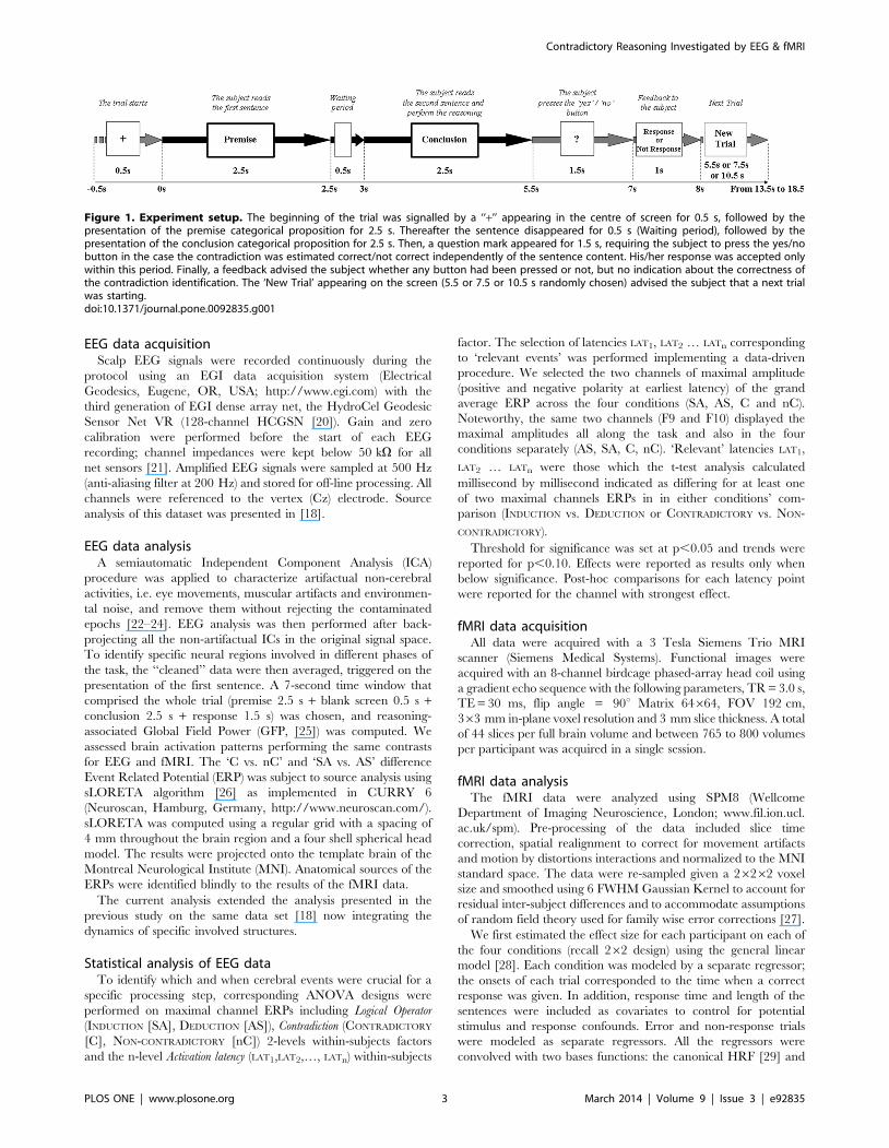
Figure 1. Experiment setup. The beginning of the trial was signalled by a ‘‘+’’ appearing in the centre of screen for 0.5 s, followed by thepresentation of the premise categorical proposition for 2.5 s. Thereafter the sentence disappeared for 0.5 s (Waiting period), followed by thepresentation of the conclusion categorical proposition for 2.5 s. Then, a question mark appeared for 1.5 s, requiring the subject to press the yes/nobutton in the case the contradiction was estimated correct/not correct independently of the sentence content. His/her response was accepted onlywithin this period. Finally, a feedback advised the subject whether any button had been pressed or not, but no indication about the correctness ofthe contradiction identification. The ‘New Trial’ appearing on the screen (5.5 or 7.5 or 10.5 s randomly chosen) advised the subject that a next trialwas starting.doi:10.1371/journal.pone.0092835.g001
Contradictory Reasoning Investigated by EEG & fMRI
PLOS ONE | www.plosone.org 3 March 2014 | Volume 9 | Issue 3 | e92835
its derivatives that capture fluctuations in response onset [30]. To
correct for signal changes due to head movement, the 6
realignment parameters were included in the design matrix. An
additional set of harmonic regressors was used to account for any
temporal low-pass frequency variance within the data that is
typical to fMRI signal with a cut-off of 1/128 Hz.
Consistent effects across participants (random-effects, second-
level analysis [31]) were then tested using the general linear model
framework. The data for the described model were the estimated
effect size of the 4 experimental conditions for each subject (AS-C,
AS-nC, SA-C, SA-nC). Plots of the averaged estimated HRF
response size represent the responses (the first Eigen-variant) of a
3 mm3 sphere centered on the maxima group response. We used a
mixed cluster and peak threshold [32]. We report clusters with a
peak at P,0.001 uncorrected and at least 75 contiguous voxels
showing Z . 2.58, unless specified otherwise. To cross validate the
reliability of our observations we tested for overlap between the
EEG sources and the regions of increased BOLD signal in the
fMRI experiment. This was done by using a 5 mm sphere around
the peak of the sources observed from the independent EEG study
and analysis (see above section) for small volume correction to the
fMRI regions.
Results
Behavioral performance during the fMRI and the EEGexperiments
Results of behavioral performance are summarized in Table 2.
Participants performed PARTICULAR - UNIVERSAL (SA: INDUCTION)
and UNIVERSAL - PARTICULAR (AS: DEDUCTION) trials with
comparable accuracy (. 95%), both for the contradictory and
non-contradictory conditions (p . 0.150). Such low error rate
suggests that logical inference based on the Aristotelian categorical
proposition structure is a natural process that is easily performed
by all participants, even without ever having had any formal
training in logic.
Reaction Times showed a strong Logical Operator effect [F(1, 12)
= 14.555, p,0.005], with longer reaction times seen for inductive
reasoning, when a UNIVERSAL rule had to be inferred based on an
example. No main effect for Contradiction or an interaction between
Logical Operator*Contradiction were observed [p.0.5 for both].
Observed longer RT for SA than AS (Logical Operator effect
p,0.005) but no RT difference between C and nC (Contradiction
effect p.0.5) in both the EEG [18] and the fMRI experiments. To
further test whether our participants developed the heuristic
strategy in the course of the experiment (i.e. produced the
identification based on the inversion of the attribute instead of
considering the whole sentences), we divided the experiment into
four parts and performed an ANOVA adding Time (1u, 2u, 3u, 4uperiod) as within-subjects factor to Logical Operator (AS, SA) and
Contradiction (C, nC). The results revealed that participants
improved their overall performance along the course of the
experiment (Time effect F(3,36) = 15, p,0.001) and were faster to
compute AS than SA logical operation (F(3,36) = 9.83, p = 0.011)
with no effect of Contradiction. Importantly, Time did not interact
with either of the manipulations (p . 0.200 for both Time*
Contradiction and Time*Logical Operator), indicating that participants
did not change their strategy during the course of the experiment.
Brain activation - EEG dataDynamics of Logical Operator and Contradiction
processing. When comparing the two channels of maximal
amplitude (positive polarity at the first peak F9, negative polarity
at F10), ERP peaks showed significant differences (p,.05, Figure 2
top row) at 400, 1200, 3300, 3500, 3800, 4400 ms in the
INDUCTION vs. DEDUCTION contrast, and at 3300, 4200, 4800 ms in
the CONTRADICTORY vs. NON-CONTRADICTORY contrast. Based on
this finding, these 8 latencies were included in the ANOVA design.
The full ANOVA design including the 3 within-subjects factors,
showed a significant Logical Operator*Contradiction*Activation latency
interaction factor [F(7,28) = 3.080, p = .016] revealing a different
behavior at different latencies when manipulating the logical
operator or recognizing the contradiction. Consequently, for each
latency reduced models were analyzed, and latencies were
considered of interest whenever at least one factor (Logical Operator
or Contradiction) was significant. As detailed in Table 3 a trend for
Logical Operator*Contradiction was found at 400 and 3300 ms. Logical
Operator factor reached statistical significance at 400, 1200, 3300,
3500, 3800 and 4400 ms and Contradiction factor was significant at
3300, 4200 ms and showed a trend at 4800 ms.
Structures crucial for identification of contradictionAs expected (since all premises are presented in the two
contrasting conditions) the analysis of ERP data contrasting C vs.
nC premise-conclusion pairs revealed no differences in GFP values
between 0 and 3000 ms during the whole premise processing
(Figure 2). The first GFP peak was seen 300 ms after the onset of
the conclusive sentence. The cerebral activation at that time point
localized to the left MFG (BA 11 - Figure 2, Table 4). The activity
at the next GFP peak, occurring around 1.2 s after the beginning
of the trial, was localized in the right IFG and corresponded to
processing of the conclusion.
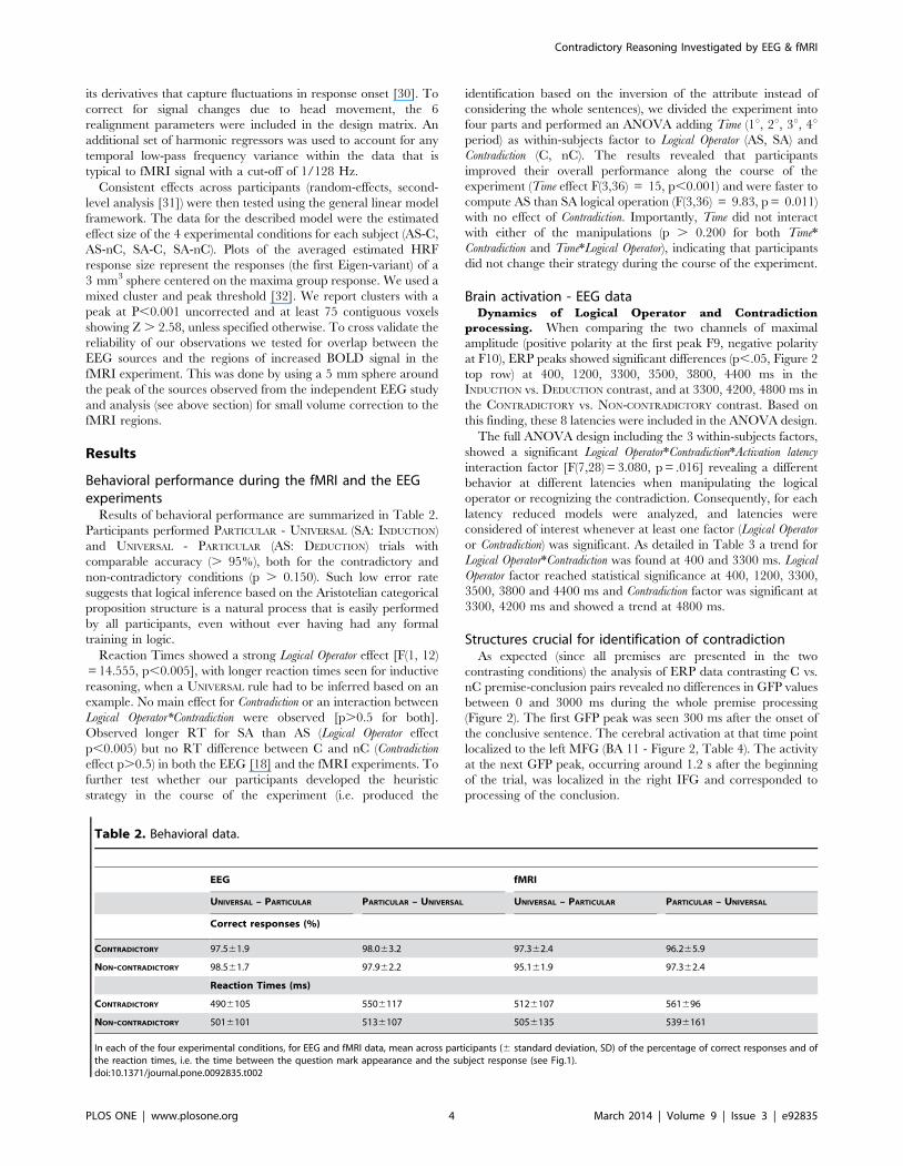
Table 2. Behavioral data.
EEG fMRI
UNIVERSAL – PARTICULAR PARTICULAR – UNIVERSAL UNIVERSAL – PARTICULAR PARTICULAR – UNIVERSAL
Correct responses (%)
CONTRADICTORY 97.561.9 98.063.2 97.362.4 96.265.9
NON-CONTRADICTORY 98.561.7 97.962.2 95.161.9 97.362.4
Reaction Times (ms)
CONTRADICTORY 4906105 5506117 5126107 561696
NON-CONTRADICTORY 5016101 5136107 5056135 5396161
In each of the four experimental conditions, for EEG and fMRI data, mean across participants (6 standard deviation, SD) of the percentage of correct responses and ofthe reaction times, i.e. the time between the question mark appearance and the subject response (see Fig.1).doi:10.1371/journal.pone.0092835.t002
Contradictory Reasoning Investigated by EEG & fMRI
PLOS ONE | www.plosone.org 4 March 2014 | Volume 9 | Issue 3 | e92835
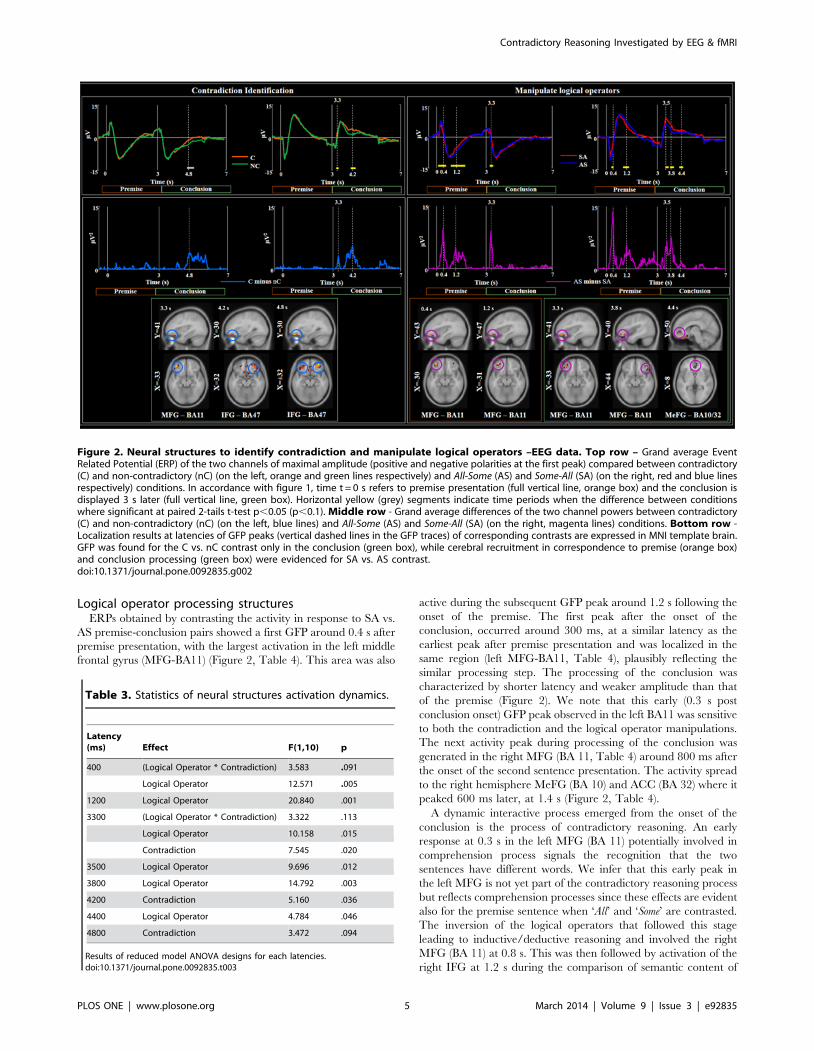
Logical operator processing structuresERPs obtained by contrasting the activity in response to SA vs.
AS premise-conclusion pairs showed a first GFP around 0.4 s after
premise presentation, with the largest activation in the left middle
frontal gyrus (MFG-BA11) (Figure 2, Table 4). This area was also
active during the subsequent GFP peak around 1.2 s following the
onset of the premise. The first peak after the onset of the
conclusion, occurred around 300 ms, at a similar latency as the
earliest peak after premise presentation and was localized in the
same region (left MFG-BA11, Table 4), plausibly reflecting the
similar processing step. The processing of the conclusion was
characterized by shorter latency and weaker amplitude than that
of the premise (Figure 2). We note that this early (0.3 s post
conclusion onset) GFP peak observed in the left BA11 was sensitive
to both the contradiction and the logical operator manipulations.
The next activity peak during processing of the conclusion was
generated in the right MFG (BA 11, Table 4) around 800 ms after
the onset of the second sentence presentation. The activity spread
to the right hemisphere MeFG (BA 10) and ACC (BA 32) where it
peaked 600 ms later, at 1.4 s (Figure 2, Table 4).
A dynamic interactive process emerged from the onset of the
conclusion is the process of contradictory reasoning. An early
response at 0.3 s in the left MFG (BA 11) potentially involved in
comprehension process signals the recognition that the two
sentences have different words. We infer that this early peak in
the left MFG is not yet part of the contradictory reasoning process
but reflects comprehension processes since these effects are evident
also for the premise sentence when ‘All’ and ‘Some’ are contrasted.
The inversion of the logical operators that followed this stage
leading to inductive/deductive reasoning and involved the right
MFG (BA 11) at 0.8 s. This was then followed by activation of the
right IFG at 1.2 s during the comparison of semantic content of
Figure 2. Neural structures to identify contradiction and manipulate logical operators –EEG data. Top row – Grand average EventRelated Potential (ERP) of the two channels of maximal amplitude (positive and negative polarities at the first peak) compared between contradictory(C) and non-contradictory (nC) (on the left, orange and green lines respectively) and All-Some (AS) and Some-All (SA) (on the right, red and blue linesrespectively) conditions. In accordance with figure 1, time t = 0 s refers to premise presentation (full vertical line, orange box) and the conclusion isdisplayed 3 s later (full vertical line, green box). Horizontal yellow (grey) segments indicate time periods when the difference between conditionswhere significant at paired 2-tails t-test p,0.05 (p,0.1). Middle row - Grand average differences of the two channel powers between contradictory(C) and non-contradictory (nC) (on the left, blue lines) and All-Some (AS) and Some-All (SA) (on the right, magenta lines) conditions. Bottom row -Localization results at latencies of GFP peaks (vertical dashed lines in the GFP traces) of corresponding contrasts are expressed in MNI template brain.GFP was found for the C vs. nC contrast only in the conclusion (green box), while cerebral recruitment in correspondence to premise (orange box)and conclusion processing (green box) were evidenced for SA vs. AS contrast.doi:10.1371/journal.pone.0092835.g002
Table 3. Statistics of neural structures activation dynamics.
Latency(ms) Effect F(1,10) p
400 (Logical Operator * Contradiction) 3.583 .091
Logical Operator 12.571 .005
1200 Logical Operator 20.840 .001
3300 (Logical Operator * Contradiction) 3.322 .113
Logical Operator 10.158 .015
Contradiction 7.545 .020
3500 Logical Operator 9.696 .012
3800 Logical Operator 14.792 .003
4200 Contradiction 5.160 .036
4400 Logical Operator 4.784 .046
4800 Contradiction 3.472 .094
Results of reduced model ANOVA designs for each latencies.doi:10.1371/journal.pone.0092835.t003
Contradictory Reasoning Investigated by EEG & fMRI
PLOS ONE | www.plosone.org 5 March 2014 | Volume 9 | Issue 3 | e92835

the conclusion with respect to the premise. Finally, re-evaluation
of the logical operators was associated with the right MeFG
activation at 1.4 s. It is interesting to note that within the reasoning
time-window, inversion of the logical operators (0.8 s at right
BA11) preceded the semantic comparison of the sentence content
(1.4 s, at the right BA47).
The EEG-derived activation data showed that the right
hemisphere contributed more than the left during the whole
reasoning time window (0.8 – 1.4 s from the presentation of the
conclusion) for both the identification of the contradiction (C vs.
nC, Figure 2 left, Table 4) and the logical operator manipulation
(SA vs. AS, Figure 2 right, Table 4). A left-hemisphere
involvement was present earlier 0.3 s in all contrasts (Figure 2 left).
Brain activations - fMRI dataOverall task relevant activation. In line with previous
fMRI studies, we first tested for the main effect of reasoning,
independent of the specific condition. i.e. cerebral responses across
all the four conditions when compared to the fixation baseline.
Note that this general contrast includes regions involved in
processing of visual stimuli, reading, comprehension, decision,
preparation of motor response, regions that are involved in non-
specific but task relevant cognitive demands as well as regions
directly performing the reasoning task. Given the non-specificity of
this contrast, we report only results that survived whole brain
family-wise error correction (FWE) at the cluster level p,0.05.
Overall, regions within the bilateral prefrontal, parietal and left
temporal cortices showed increased activations during task versus
baseline (Table 5; Figure 3). Interestingly, while performing the
task the early sensory structures (visual cortex and thalamus)
showed a decrease in activity relative to baseline (Table 5). This
suggests that while judging the logical reasoning, participants were
engaged in internal mental processing while attempting to ‘block’
external distractions [33].
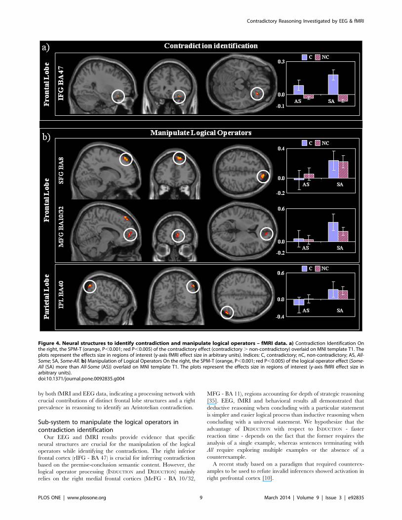
Structures crucial for identification of contradictionThe right inferior frontal gyrus (IFG, BA 47), and caudate
nuclei showed increase activation during contradictory compared
to non-contradictory trials (p,0.001, Figure 4a and Table 6).
Within these regions there was no effect of the logical operator
manipulation, suggesting that BA 47 responded in a similar way
irrespectively to the manipulation of All or Some. Even though the
size of these clusters was below our pre-specified threshold, the
involvement of the right BA 47 and the caudate in decision-
making, response selection and contradictory scenarios is consis-
tent with previous literature (see discussion for further details). As
expected, the fMRI responses of the right BA 47 overlapped with
the contradictory EEG-source peak at 1.2 s. This overlap was
again formally confirmed with small volume correction (SVC)
using a 5 mm sphere centered at the EEG-source peak to the
fMRI data. Using SVC, the response in right IFG was larger for
contradictory statements than no-contradictory ones (FWE-
corrected p = 0.008). The opposite contrast (nC. C) caused no
activation above threshold.
Logical operator processing structuresIn line with the behavioral findings, we observed a greater
BOLD signal change for inductive reasoning propositions ending
with a UNIVERSAL (SA) than for deductive reasoning - those ending
with a PARTICULAR statement (AS) in the right superior frontal
gyrus (SFG – BA 8), medial frontal gyrus (MeFG – BA 10) and the
right inferior parietal lobule (IPL – BA 40, angular gyrus) (Figure
4b, Table 5). Within the above foci there was no reliable effects for
the contradictory manipulation (p . 0.1), suggesting that
responses to contradictory and non-contradictory sentences
recruited these structures in a similar way. We note that the
EEG activation peak (at 1.4s) in the right MeFG for the inversion
of the logical operator overlapped with the fMRI activation at that
region. This overlap was confirmed by applying SVC using a
5 mm sphere centered at the EEG peak to the fMRI data, the
response in right MeFG was found to be FWE-corrected
(p = 0.010). There was no above threshold activation for increased
response to SA vs. AS.
Relationship between behavior and brain activationsAs detailed above, no effects of experimental conditions were
observed on error rates. Reaction times were longer when
contradiction identification was performed after a universal
(condition SA) rather than a particular conclusion (condition
AS). This finding lead us to investigate brain activation properties
Table 4. Neural structures to identify contradiction and manipulate logical operators – EEG data.
BA X Y Z Latency
(mm) (mm) (mm) (s)
CONTRADICTORY vs. NON-CONTRADICTORY Prem Conc
Frontal Lobe Middle Frontal Gyrus (L) 11 –33 41 –14 3.3 0.3
Inferior Frontal Gyrus (R) 47 32 30 –17 4.2 1.2
INDUCTION: PARTICULAR – UNIVERSAL (SA) vs. DEDUCTION: UNIVERSAL – PARTICULAR (AS) Prem Conc
Frontal Lobe Middle Frontal Gyrus (L) 11 –30 43 –15 0.4
11 –31 47 –15 1.2
11 –33 41 –14 3.3 0.3
Middle Frontal Gyrus (R) 11 44 40 –14 3.8 0.8
Medial Frontal Gyrus/ACC (R) 10/32 8 50 0 4.4 1.4
Localization results in MNI coordinate and latencies of GFP peaks of contrasted EEG data. Latencies are expressed relative to both the premise presentation (t = 0, firstlatency column, Prem) and the conclusion presentation (t = 0 at 3s after premise presentation, second latency column, Conc); this also means that latencies ,3s of thefirst latency column refer to premise processing and latencies . 3s relate to conclusion processing. Latencies in bold indicate reasoning steps occurring in the sameprocessing phase after premise and conclusion presentation. Dynamics of lateralization of brain recruitments is underlined by dedicated boxes for left (L) and right (R)activated areas. In italics are indicated cerebral processing of the premise.doi:10.1371/journal.pone.0092835.t004
Contradictory Reasoning Investigated by EEG & fMRI
PLOS ONE | www.plosone.org 6 March 2014 | Volume 9 | Issue 3 | e92835
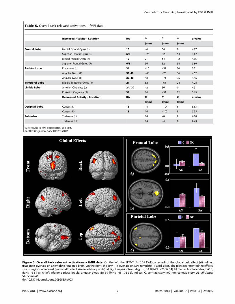
Table 5. Overall task relevant activations – fMRI data.
Increased Activity - Location BA X Y Z z-value
(mm) (mm) (mm)
Frontal Lobe Medial Frontal Gyrus (L) 10 –6 54 8 4.77
Superior Frontal Gyrus (L) 6/8 –26 32 54 4.67
Medial Frontal Gyrus (R) 10 2 54 –2 4.95
Superior Frontal Gyrus (R) 6/8 36 32 54 3.86
Parietal Lobe Precuneus (L) 31 –10 –54 30 3.71
Angular Gyrus (L) 39/40 –48 –76 36 4.52
Angular Gyrus (R) 39/40 48 –74 36 4.46
Temporal Lobe Middle Temporal Gyrus (R) 21 52 –64 24 4.28
Limbic Lobe Anterior Cingulate (L) 24/ 32 –2 36 0 4.51
Posterior Cingulate (R) 31 10 –52 22 3.63
Decreased Activity - Location BA X Y Z z-value
(mm) (mm) (mm)
Occipital Lobe Cuneus (L) 18 –8 –104 6 5.63
Cuneus (R) 18 16 –102 8 5.55
Sub-lobar Thalamus (L) 14 –8 8 6.28
Thalamus (R) 14 –4 6 6.23
fMRI results in MNI coordinates. See text.doi:10.1371/journal.pone.0092835.t005
Figure 3. Overall task relevant activations – fMRI data. On the left, the SPM-T (P,0.05 FWE-corrected) of the global task effect (stimuli vs.fixation) is overlaid on a template rendered brain. On the right, the SPM-T is overlaid on MNI template T1 axial slices. The plots represented the effectssize in regions of interest (y-axis fMRI effect size in arbitrary units). a) Right superior frontal gyrus, BA 8 [MNI: –26 32 54]; b) medial frontal cortex, BA10,(MNI: –6 54 8), c) left inferior parietal lobule, angular gyrus, BA 39 [MNI: –48 –76 36]. Indices: C, contradictory; nC, non-contradictory; AS, All-Some;SA, Some-All.doi:10.1371/journal.pone.0092835.g003
Contradictory Reasoning Investigated by EEG & fMRI
PLOS ONE | www.plosone.org 7 March 2014 | Volume 9 | Issue 3 | e92835
explaining the reaction difference in the two conditions (Reaction
Time delay, RTd = RT(SA)-RT(AS)). In particular, we focused
on the activity of brain regions emerging from the same SA vs. AS
contrast. After checking variables distribution for gaussianity with
Shapiro-Wilk statistics, a linear regression analysis was performed
to identify possible cerebral activation features predicting the RTd.
EEG and fMRI studies indicated that specific areas were
involved for manipulating the logical operator. We investigated if
activation latency or current strength of right BA11 or BA10/32
(see Table 4) or BOLD intensity in BA10/32, BA8 and BA40
(Table 6) were able to explain the longer reaction time which
occurred in SA vs. AS conditions (see Table 2). Investigating
preliminarily the Pearson’s correlations with the reaction time
delay (RTd), no association appeared with either EEG current
strengths or fMRI BOLD. Instead right BA11 and BA10/32
activation latencies correlated with RTd. We used regression
analysis to establish which of these displayed higher association.
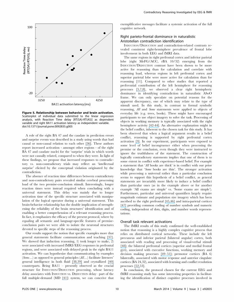
The regression analysis with RTd as dependent variable and right
BA11 and BA10/32 activation latencies as independent variables
(Stepwise method) indicated that only right BA11 activation
latency (rBA11al) entered the model, as expressed by
RTd = –307.6 + 0.090 rBA11al (Figure 5).
The 80% of RTd variance was explained by this model
[F(1,9) = 35,660, p,.001].
Discussion
Going beyond our previous study using the Aristotelian Square
of Oppositions [18], here we delineated the spatial temporal
envelope of two processes that are keys to reasoning thinking:
logical operators of deductive and inductive inferences and the
detection of contradictions. We found that inductive (SA: Some-
All ) reasoning was more difficult than deductive (AS: All-Some)
reasoning and it was associated with increase responses of the right
superior and medial prefrontal cortex (BA 8, 10, 32) and the
inferior parietal (BA 40), in the fMRI. The EEG revealed more
refined spatial-temporal properties of the reasoning processed.
Here 0.8 s after the onset of the conclusion sentence the right
MFG (BA 11) showed a different activity depending on the type of
reasoning made. Interestingly the timing of right MFG (BA 11)
peak predicted participants’ reaction time delay in inductive vs.
deductive reasoning. The responses of the MeFG (BA 10) that
were seen in the fMRI were observed later at 1.4 s. Participants
found it equally easy to detect contradictory as well as non-
contradictory sentences, however both the fMRI and EEG
revealed that responses of the right MFG (BA 47) arising at 1.2
s after the onset of the conclusion sentence showed stronger
responses to the contradictory than to non-contradictory sentenc-
es. Our data highlighted that specific spatial-temporal network
properties subtend deduction/induction and identification of
contradiction. We next discuss in more details each of the
observed effects and potential reasons of some apparent inconsis-
tencies between the fMRI and EEG data.
The limitation of our study is the relatively small simple size,
since the statistics are prone to false negative as well as false
positive results in this case. However, the reliability of the results
emerges from convergent evidence provided by the two multi-
modal brain measurements (EEG and fMRI), the correlation of
the behavioral reaction delays with the brain activation latencies,
of the region crucial to perform the step differing between the two
conditions, and the consistency of the findings with previous
literature.
Dynamics of reasoning network to identify anAristotelian contradiction
The analysis of the EEG data enabled to describe the temporal
sequence of the activation of structures within the prefrontal cortex
necessary to solve an Aristotelian contradiction. In our previous
EEG study [18] we focused on identifying the frontal network that
was involved in all the four conditions. This revealed a bilateral
temporo-polar cortices (BA 21, 38), bilateral Inferior Frontal
Gyrus (IFG - BA 47) cortex, the Medial Frontal Gyrus (MeFG -
BA 10, 11) and the Anterior Cingulate Cortex (ACC - BA 32) [18].
In this study we focused on identifying how the spatial layout of
the network changes dynamically as a function of the reasoning
conditions.
After the onset of the conclusion the left MFG was activated first
(0.3 s) and at the almost same latency as following the onset of the
premise (0.4 s). The involvement of the left MFG present in the
premise and not just in the conclusion suggests a P300-like
activation, reflecting semantic comprehension processing. This
area is in fact part of the P300 generator structure [34].
Only after the onset of the conclusive sentence, a second
response emerged about half a second after the initial P300 within
the right frontal lobe structures that were associated with the
logical operator manipulation (right MFG, 0.8 s after conclusion
presentation, Figure 2). Four hundred ms later (1.2 s after
conclusion presentation), contradiction identification involved the
right hemispheric IFG whereas 200 ms later (1.4 s) ACC is
activated.
While EEG-derived activation revealed the dynamics of the
reasoning process, these structures were observed independently
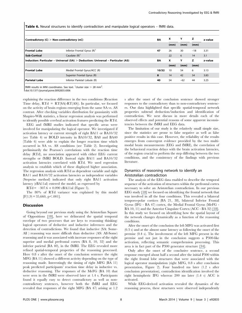
Table 6. Neural structures to identify contradiction and manipulate logical operators – fMRI data.
Contradictory (C) . Non-contradictory (nC) BA X Y Z z-value
(mm) (mm) (mm)
Frontal Lobe Inferior Frontal Gyrus (R)1 47 26 30 –18 3.31
Sub-Cortical Caudate (R)1 22 6 14 3.1
Induction: Particular – Universal (SA) . Deduction: Universal – Particular (AS) BA X Y Z z-value
(mm) (mm) (mm)
Frontal Lobe Medial Frontal Gyrus/ACC (R) 10/32 10 54 6 3.13
Superior Frontal Gyrus (R) 8 14 42 54 3.83
Parietal Lobe Inferior Parietal Lobule (R) 40 54 –62 44 3.25
fMRI results in MNI coordinates. See text. 1cluster size . 28 mm3.doi:10.1371/journal.pone.0092835.t006
Contradictory Reasoning Investigated by EEG & fMRI
PLOS ONE | www.plosone.org 8 March 2014 | Volume 9 | Issue 3 | e92835
by both fMRI and EEG data, indicating a processing network with
crucial contributions of distinct frontal lobe structures and a right
prevalence in reasoning to identify an Aristotelian contradiction.
Sub-system to manipulate the logical operators incontradiction identification
Our EEG and fMRI results provide evidence that specific
neural structures are crucial for the manipulation of the logical
operators while identifying the contradiction. The right inferior
frontal cortex (rIFG - BA 47) is crucial for inferring contradiction
based on the premise-conclusion semantic content. However, the
logical operator processing (INDUCTION and DEDUCTION) mainly
relies on the right medial frontal cortices (MeFG - BA 10/32,
MFG - BA 11), regions accounting for depth of strategic reasoning
[35]. EEG, fMRI and behavioral results all demonstrated that
deductive reasoning when concluding with a particular statement
is simpler and easier logical process than inductive reasoning when
concluding with a universal statement. We hypothesize that the
advantage of DEDUCTION with respect to INDUCTION - faster
reaction time - depends on the fact that the former requires the
analysis of a single example, whereas sentences terminating with
All require exploring multiple examples or the absence of a
counterexample.
A recent study based on a paradigm that required counterex-
amples to be used to refute invalid inferences showed activation in
right prefrontal cortex [10].
Figure 4. Neural structures to identify contradiction and manipulate logical operators – fMRI data. a) Contradiction Identification Onthe right, the SPM-T (orange, P,0.001; red P,0.005) of the contradictory effect (contradictory . non-contradictory) overlaid on MNI template T1. Theplots represent the effects size in regions of interest (y-axis fMRI effect size in arbitrary units). Indices: C, contradictory; nC, non-contradictory; AS, All-Some; SA, Some-All. b) Manipulation of Logical Operators On the right, the SPM-T (orange, P,0.001; red P,0.005) of the logical operator effect (Some-All (SA) more than All-Some (AS)) overlaid on MNI template T1. The plots represent the effects size in regions of interest (y-axis fMRI effect size inarbitrary units).doi:10.1371/journal.pone.0092835.g004
Contradictory Reasoning Investigated by EEG & fMRI
PLOS ONE | www.plosone.org 9 March 2014 | Volume 9 | Issue 3 | e92835
A role of the right BA 47 and the caudate in prediction errors
and surprise events was described in a study using words that had
causal or non-causal relation to each other [36]. These authors
report increased activation - amongst other regions - of the right
BA 47 and caudate nuclei for the ‘surprise’ trials in which words
were not causally related, compared to when they were. In light of
these findings, we propose that increased responses to contradic-
tory vs. non-contradictory trials may reflect an ‘intellectual
surprise’ elicited by the conceptual violation originated by the
contradiction.
The absence of reaction time differences between contradictory
and non-contradictory pairs revealed similar cerebral processing
load of the two premise-conclusion stimuli. Interestingly, longer
reaction times were instead required when concluding with a
universal statement. This delay was mainly due to longer
activation time of the specific structure associated with manipu-
lation of the logical operator during a universal statement. This
brain-behavior relationship has the double implication of strength-
ening the reliability of the brain structures’ identification and of
enabling a better comprehension of a relevant reasoning process.
In fact, it emphasizes the efficacy of the present protocol, where by
equaling all semantic and language-specific features of premise-
conclusion pairs we were able to isolate the neuronal structures
devoted to specific steps of the reasoning process.
Our results support the notion that specific examples more that
general statements facilitate communication and teaching [37].
We showed that induction reasoning, 1) took longer to make, 2)
were associated with increased fMRI/EEG responses in prefrontal
regions, and were associated with delayed peak in the right BA11
activation. We can hypothesize that communicating by examples
(Some…) as opposed to general principles (All…) facilitate listeners’
general intelligence in both fluid [38,39] and crystallized [40]
counterparts. Being BA11 – presently observed as the crucial
structure for INDUCTION/DEDUCTION processing, whose latency
delay associates with INDUCTION vs. DEDUCTION delay– part of the
full multiple-demand (MD [41]) system, we can conceive that
exemplificative messages facilitate a systemic activation of the full
cognitive network.
Right parieto-frontal dominance in naturalisticAristotelian contradiction identification
INDUCTION/DEDUCTION and contradiction-related contrasts re-
vealed consistent right-hemisphere prevalence of frontal lobe
involvement in both EEG and fMRI data.
The same regions in right prefrontal cortex and inferior parietal
lobe (right MeFG/ACC, rBA 10/32) emerging from the
INDUCTION/DEDUCTION contrast have been shown to be more
active for reasoning than for calculation and correlate with
reasoning load, whereas regions in left prefrontal cortex and
superior parietal lobe were more active for calculation than for
reasoning [11]. Compared to other studies that reported a
preferential contribution of the left hemisphere for reasoning
processes [5,7,8], we observed a clear right hemispheric
dominance in identifying contradiction in naturalistic ASoO
frame. We can only speculate on potential reasons for the
apparent discrepancy, one of which may relate to the type of
stimuli used. In this study, in contrast to formal symbolic
reasoning, All and Some statements were applied to objects of
everyday life (e.g. trees, books). These might have encouraged
participants to use object imagery to solve the task. Processing of
objects in working memory is typically associated with the right
hemisphere activity [42-44]. An alternative explanation might in
the belief conflict, inherent to the chosen task for this study. It has
been observed that when a logical argument results in a belief
conflict, reasoning is supported by right prefrontal cortex
activation [3]. In our experiment, participants were exposed to
some level of belief incongruence either when processing the
premise or the conclusion, even though they were instructed to
ignore the truthfulness of the statement. The construction of
logically contradictory statements implies that one of them is to
some extent in conflict with experience-based belief. For example
a statement that ‘All books are thick’ is in conflict with our prior
knowledge that ‘Some books are thin’. The stronger activation
while processing a universal rather than a particular conclusion
seems to support this hypothesis of a belief conflict, as general
statements are invariably more likely to induce a belief conflict
than particular ones (as in the example above or for another
example ‘All exams are simple’ vs. ‘Some exams are simple’).
Furthermore, particular and universal quantifiers largely require
magnitude estimate and population size imagery which has been
ascribed to the right prefrontal [45,46] and intra-parietal cortices
[47] providing common coding of number symbols and numeric
coding, independent of dots, digits, and number words represen-
tations.
Overall task relevant activationsThe fMRI results of this study confirmed the well-established
notion that reasoning is a highly complex cognitive process that
relies on distributed cortical networks. These include the left
precuneus and inferior parietal (bilateral angular) cortex, both
associated with reading and processing of visual-verbal stimuli
[48]; the bilateral prefrontal cortices (superior and medial frontal
gyri), associated with executive functions, working memory and
decision making processes [49–51]; premotor cortices (BA 6)
bilaterally, associated with motor response and anterior cingulate
cortices (BA 24,32), associated with learning and conflict resolution
processes [52,53].
In conclusion, the protocol chosen for the current EEG and
fMRI reasoning study has some interesting properties in facilitat-
ing the identification of distinct cognitive processes involved in
Figure 5. Relationship between behavior and brain activation.Scatterplot of individual data submitted to the linear regressionanalysis, with Reaction Time delay [RT(SA)-RT(AS)] as dependentvariable and right BA11 activation latency as independent variable.doi:10.1371/journal.pone.0092835.g005
Contradictory Reasoning Investigated by EEG & fMRI
PLOS ONE | www.plosone.org 10 March 2014 | Volume 9 | Issue 3 | e92835
deciding whether two statements are contradictory. The perfect
symmetry of the premise-conclusion logical structure used in this
study has the advantage of being sensitive to isolate the process
underlying the manipulation of the logical operators (All/Some)
minimizing the linguistic and semantic confounds. Using this we
have, for the first time, been able to identify the network involved
in determining contradiction in logic. Investigating brain-behavior
relationships we found that processing general statements is
associated with longer processing time. Significant brain-behavior
relationships strengthened the reliability of brain involvements in
the contradictory judgment as assessed by the present study and
honed the understanding of why specific cases are superior for
human communication than general declarations.
Author Contributions
Conceived and designed the experiments: CP MTM. Performed the
experiments: CP MTM NJT. Analyzed the data: CP MTM PR. Wrote the
paper: CP MTM SS PR FT.
References
1. Aristotle Organon (2003) Translated by Colli G, 3rd ed., Adelphi issn:9788845917554.
2. Noveck IA, Goel V, Smith KW (2004) The neural basis of conditional reasoning
with arbitrary content. Cortex 40: 613–622.
3. Goel V, Buchel C, Frith C, Dolan RJ (2000) Dissociation of mechanismsunderlying syllogistic reasoning. Neuroimage 12: 504–514.
4. Goel V, Dolan RJ (2003) Reciprocal neural response within lateral and ventral
medial prefrontal cortex during hot and cold reasoning. Neuroimage 20: 2314–2321.
5. Goel V, Dolan RJ (2004) Differential involvement of left prefrontal cortex in
inductive and deductive reasoning. Cognition 93: B109– B121.
6. Qiu J, Li H, Luo Y, Zhang Q, Tu S (2009) The neural basis of syllogisticreasoning: An event-related potential study. Brain Research 1273: 106–113.
7. Monti MM, Osherson DN, Martinez MJ, Parsons LM (2007) Functional
neuroanatomy of deductive inference: a language-independent distributednetwork. Neuroimage 3: 1005–1016.
8. Monti MM, Parsons LM, Osherson DN (2009) The boundaries of language and
thought in deductive inference. Proc Natl Acad Sci U S A 106: 12554–12559.
9. Abrusci VM, Casadio C, Medaglia MT, Porcaro C (2013) Universal vs.particular reasoning: a study with neuroimaging techniques. Logic Journal of
IGPL 21: 1017–1027.
10. Goel V (2007) Anatomy of deductive reasoning. Trends Cogn Sci 11: 435–441.
11. Kroger JK, Nystrom LE, Cohen JD, Johnson-Laird PN (2008) Distinct NeuralSubstrates for Deductive and Mathematical Processing. Brain Research 1243:
86–103.
12. Johnson-Laird PN (2010) Mental models and human reasoning. Proc Natl AcadSci U S A. 107: 18243–50.
13. Van Overwalle F (2011) A dissociation between social mentalizing and general
reasoning. Neuroimage 54: 1589–1599.
14. Prado J, Chadha A, Booth JR (2011) The brain network for deductive reasoning:a quantitative meta-analysis of 28 neuroimaging studies. J Cogn Neurosci 23:
3483–3497
15. Monti MM, Osherson DN (2012) Logic, language and the brain. Brain Research1428: 33–42.
16. Osherson DN, Perani D, Cappa S, Schnur T, Grassi F, et al. (1998) Distinct
brain loci in deductive versus probabilistic reasoning. Neuropsychologia 36:369–376.
17. Parsons LM, Osherson D (2001) New Evidence for Distinct Right and Left Brain
Systems for Deductive versus Probabilistic Reasoning. Cerebral Cortex 11: 954–965.
18. Medaglia MT, Tecchio F, Seri S, Di Lorenzo G, Rossini PM, et al. (2009)
Contradiction in universal and particular reasoning. Hum Brain Mapp 30:
4187–4197.
19. Josephs O, Henson RN (1999) Event-related functional magnetic resonance
imaging: modelling, inference and optimization. Philos Trans R Soc Lond B Biol
Sci 354: 1215–1228.
20. Tucker DM (1993) Spatial sampling of head electrical fields: the geodesic sensor
net. Electroencephalogr Clin Neurophysiol 87: 145–163.
21. Ferree T, Luu P, Russel JS, Tucker DM (2001) Scalp electrode impedance,
infection risk, and EEG data quality. Clin Neurophysiol 112: 536–544.
22. Barbati G, Porcaro C, Zappasodi F, Rossini PM, Tecchio F (2004) Optimization
of an independent component analysis approach for artifact identification and
removal in magnetoencephalographic signals. ClinNeurophysiol 115: 1220–1232.
23. Porcaro C, Zappasodi F, Barbati G, Salustri C, Pizzella V, et al. (2006) Fetal
auditory responses to external sounds and mother’s heart beat: Detection
improved by independent component analysis. Brain Research 1101: 51–58.
24. Porcaro C, Coppola G, Di Lorenzo G, Zappasodi F, Siracusano A, et al. (2009)Hand somatosensory subcortical and cortical sources assessed by functional
source separation: An EEG study. Hum Brain Mapping 30: 660–674.
25. Lehmann D, Skrandies W (1984) Spatial analysis of evoked potentials in man-areview. Prog Neurobiol 23: 227–50.
26. Pascual-Marqui RD (2002) Standardized low-resolution brain electromagnetic
tomography (sLORETA): Technical details. Methods Find Exp Clin Pharmacol24D: 5–12.
27. Worsley KJ, Friston KJ (1995) Analysis of fMRI time-series revisited - again.
Neuroimage 2: 173–181.
28. Kiebel S, Holmes A (2003). The General Linear Model. In: Human Brain
Function, Frackowiak RS, Friston KJ, Frith C, Dolan RJ, Price C, Zeki S,Ashburner J, Penny W. Academic Press. pp. 725–760.
29. Friston KJ, Glaser DE, Mechelli A, Turner R, Price C (2003) Hemodynamic
modelling. In: Human Brain Function, Frackowiak RS, Friston KJ, Frith C,Dolan RJ, Price C, Zeki S, Ashburner J, Penny W. Academic Press. pp. 823–
842.30. Friston KJ, Fletcher P, Josephs O, Holmes A, Rugg MD, et al. (1998) Event-
related fMRI: characterizing differential responses. Neuroimage 7: 30–40.31. Penny W, Holmes A, Friston KJ (2003) Random effects Analysis. In: Human
Brain Function, Frackowiak RS, Friston KJ, Frith C, Dolan R, Price C, Zeki S,
Ashburner J, Penny W. Oxford Academic Press. pp. 843–850.32. Poline JB, Worsley KJ, Evans AC, Friston KJ (1997) Combining spatial extent
and peak intensity to test for activations in functional imaging. Neuroimage 5:83–96.
33. Ojemann GA (1982) Models of the brain organization for higher integrative
functions derived with electrical stimulation techniques. Hum Neurobiol 1: 243–9.34. Polich J, Kok A (1995) Cognitive and biological determinants of P300: an
integrative review. Biol Psychol 41: 103–146.35. Coricelli G, Nagel R (2009) Neural correlates of depth of strategic reasoning in
medial prefrontal cortex. Proc Natl Acad Sci U S A 106: 9163–9168.
36. Fenker DB, Schoenfeld MA, Waldmann MR, Schuetze H, Heinze HJ, et al.(2010) Virus and epidemic: causal knowledge activates prediction error circuitry.
J Cogn Neurosci 22: 2151–2163.37. Atkinson RK, Derry SJ, Renkl A, Wortham D (2000) Learning from Examples:
Instructional Principles from the Worked Examples. Research review ofeducational research 70: 181–214.
38. Perfetti B, Tesse M, Varanese S, Saggino A, Onofrj M (2011) Irrelevant features
of a stimulus can either facilitate or disrupt performance in a working memorytask: the role of fluid intelligence. PLoS One 6: e26249.
39. Ebisch SJ, Perrucci MG, Mercuri P, Romanelli R, Mantini D, et al. (2012).Common and unique neuro-functional basis of induction, visualization, and
spatial relationships as cognitive components of fluid intelligence. Neuroimage
62: 331–342.40. Colom R, Burgaleta M, Roman FJ, Karama S, Alvarez-Linera J, et al. (2013)
Neuroanatomic overlap between intelligence and cognitive factors: Morphom-etry methods provide support for the key role of the frontal lobes. Neuroimage
72: 143–152.41. Duncan J (2013). The structure of cognition: attentional episodes in mind and
brain. Neuron 80: 35–50.
42. Corbetta M, Shulman GL (2002) Control of goal-directed and stimulus-drivenattention in the brain. Nature Reviews Neuroscience 3: 201–215.
43. Mort DJ, Malhotra P, Mannan SK, Rorden C, Pambakian A, et al. (2003) Theanatomy of visual neglect. Brain 126: 1986–1997.
44. Rossi S, Cappa SF, Babiloni C, Pasqualetti P, Miniussi C, et al. (2001) Prefrontal
cortex in long-term memory: an ‘interference’ approach using magneticstimulation. Nature Neuroscience 9: 948–952.
45. Shallice T, Stuss DT, Alexander MP, Picton TW, Derkzen D (2008). Themultiple dimensions of sustained attention. Cortex 44: 794–805.
46. Doricchi F, Guariglia P, Gasparini M, Tomaiuolo F (2005) Dissociation betweenphysical and mental number line bisection in right hemisphere brain damage.
Nat Neurosci 8: 1663–1665.
47. Piazza M, Pinel P, Le Bihan D, Dehaene S (2007) A magnitude code common tonumerosities and number symbols in human intraparietal cortex. Neuron 53:
293–305.48. Price CJ (2000) The anatomy of language: contributions from functional
neuroimaging. J Anat 197: 335–359.
49. Watanabe M (1998) Cognitive and motivational operations in primate prefrontalneurons. Rev Neurosci 9: 225–241.
50. Smith EE, Jonides J (1999) Storage and executive processes in the frontal lobes.Science 12: 1657–1661.
51. Duncan J, Owen AM (2000) Common regions of the human frontal loberecruited by diverse cognitive demands. Trends Neurosci 23: 475–483.
52. Swick D, Turken AU (2002) Dissociation between conflict detection and error
monitoring in the human anterior cingulate cortex. Proc Natl Acad Sci U S A99: 16354–16359.
53. Doya K (2008) Modulators of decision making. Nat Neurosci 11: 410–416.
Contradictory Reasoning Investigated by EEG & fMRI
PLOS ONE | www.plosone.org 11 March 2014 | Volume 9 | Issue 3 | e92835
Related Documents