Chemotherapeutic drugs, long the mainstay of cancer treatment, cause DNA damage and disrupt DNA repli- cation in proliferating cells. Drug regi- mens have been designed to kill as many tumor cells as possible by treating with “maximum tolerated doses” (MTDs) of these cytotoxic agents. Side effects such as neurotoxicity and dam- age to proliferating cells in healthy tis- sues pose serious constraints on the use of chemotherapy. In an effort to bal- ance toxicity with efficacy, a conven- tional dosing schedule calls for episod- ic application of a cytotoxic drug at or near the MTD, followed by periods of rest to allow normal tissues to recover. Many such chemotherapy regimens are initially efficacious, resulting in tumor regression or stabilization and pro- longed survival. In rare cases, cures are achieved. In general, however, respons- es are short-lived, with relapses often marked by aggressive cancers that are resistant to the cytotoxic drug. Fur- thermore, the standard MTD regimen as a rule seriously impairs quality of life. The harsh side effects and the ulti- mate failures of most chemotherapies have fueled broad investigation of alternatives, including drugs that tar- get not the transformed tumor cells themselves, but rather a genetically stable constituent cell type of tumors, the endothelial cells that form blood vessels. Angiogenesis, the process by which new blood vessels are formed, is a hallmark capability of cancer (1); a compelling body of evidence argues that tumor growth depends on the vas- culature, and, in particular, on contin- uing angiogenesis (2, 3). More than two dozen new drugs that are in or soon to enter clinical trials appear to interfere with tumor angiogenesis (3, 4); there is considerable anticipation about their benefits in treating cancer. Now, two studies suggest a potentially comple- mentary strategy of rescheduling the administration of classical cytotoxic drugs in order to target tumor endothelial cells. It is well established that tumor- associated endothelial cells proliferate during chronic angiogenesis in tumors, albeit at lower frequencies than the tumor cells themselves. Apparently because of their lower rate of cell divi- sion, replication of these endothelial cells is only weakly disrupted by the episodic regimens of standard chemotherapeutic protocols. In these two new studies, however, cytotoxic drugs were administered routinely, to target the slowly proliferating tumor endothelial cells and abrogate their apparent capability to repair and recov- er during the usual rest periods. Both groups worked with mice bearing sub- cutaneous tumors, and each presents data suggesting that “metronomic” dosing regimens—either continuous infusion or frequent administration without extended rest periods—could have real value in the clinic. Both also demonstrated combinatorial effects of such altered cytotoxic drug regimens with newer, more specific angiogenesis inhibitors. Metronomic drug delivery in immunodeficient mice Klement and colleagues, in this issue of the JCI, tested two agents on tumors arising from human neuroblastoma cell lines, inoculated into immunodefi- cient mice (5). The first agent, the mito- sis-blocking cytotoxic drug vinblastine, killed cultured endothelial cells at doses considerably below those required to affect the drug-sensitive neuroblastoma cells directly. Comparable doses, well below the MTD, impaired but did not abolish tumor growth in mice. Klement et al. (5) also explored the use of the mAb DC-101 (6), which disrupts the function of VEGF-R2/flk-1/KDR, one of two receptors for VEGF. VEGF sig- naling can induce endothelial cell pro- liferation and angiogenesis; in addition, accumulating data indicate that VEGF is a survival factor that protects endothelial cells from apoptosis (7). Treatment with DC101 impaired tumor growth, more so than vinblas- tine alone, but each agent alone pro- duced only a period of “stable disease,” after which lethal tumor growth resumed. Remarkably, regular administration of the two drugs in combination pro- duced regression of tumors, with no recurrence during 180 days of treat- ment. Histopathology revealed wide- spread apoptosis in tumors from all three treatment groups, notably in endothelial cells; cell death was most pronounced in the combination trial. The case for both agents having anti- angiogenic activity was bolstered with an angiogenesis bioassay involving ingrowth of capillaries into subcuta- neous matrix plugs containing an angiogenic growth factor: vinblastine and DC101 alone, and in combina- tion, inhibited angiogenesis. Another group has independently documented the antiangiogenic effects of low-dose vinblastine (8). In recent unpublished work, the combination of metronom- ic, low-dose chemotherapy and anti- VEGFR2 has been found to stabilize or regress tumors derived from several human breast cancer lines resistant to the cognate cytotoxic drug, supporting the generality of the strategy (G. Kle- ment and R. Kerbel, personal commu- nication). The Journal of Clinical Investigation | April 2000 | Volume 105 | Number 8 1045 Less is more, regularly: metronomic dosing of cytotoxic drugs can target tumor angiogenesis in mice Douglas Hanahan, 1,2 Gabriele Bergers, 1,2 and Emily Bergsland 2,3 1 Department of Biochemistry and Biophysics, 2 Hormone Research Institute, and 3 Department of Medicine, University of California San Francisco, San Francisco, California, USA Address correspondence to: Douglas Hanahan, Departments of Biochemistry and Biophysics, and Hormone Research Institute, University of California San Francisco, San Francisco, California 94143-0534, USA. Phone: (415) 476-9209; Fax: (415) 731-3612; E-mail: [email protected]. See related article, pages R15–R24. Commentary

Welcome message from author
This document is posted to help you gain knowledge. Please leave a comment to let me know what you think about it! Share it to your friends and learn new things together.
Transcript

Chemotherapeutic drugs, long themainstay of cancer treatment, causeDNA damage and disrupt DNA repli-cation in proliferating cells. Drug regi-mens have been designed to kill asmany tumor cells as possible by treatingwith “maximum tolerated doses”(MTDs) of these cytotoxic agents. Sideeffects such as neurotoxicity and dam-age to proliferating cells in healthy tis-sues pose serious constraints on the useof chemotherapy. In an effort to bal-ance toxicity with efficacy, a conven-tional dosing schedule calls for episod-ic application of a cytotoxic drug at ornear the MTD, followed by periods ofrest to allow normal tissues to recover.Many such chemotherapy regimens areinitially efficacious, resulting in tumorregression or stabilization and pro-longed survival. In rare cases, cures areachieved. In general, however, respons-es are short-lived, with relapses oftenmarked by aggressive cancers that areresistant to the cytotoxic drug. Fur-thermore, the standard MTD regimenas a rule seriously impairs quality of life.
The harsh side effects and the ulti-mate failures of most chemotherapieshave fueled broad investigation ofalternatives, including drugs that tar-get not the transformed tumor cellsthemselves, but rather a geneticallystable constituent cell type of tumors,the endothelial cells that form bloodvessels. Angiogenesis, the process bywhich new blood vessels are formed, isa hallmark capability of cancer (1); acompelling body of evidence arguesthat tumor growth depends on the vas-culature, and, in particular, on contin-uing angiogenesis (2, 3). More than twodozen new drugs that are in or soon toenter clinical trials appear to interferewith tumor angiogenesis (3, 4); there isconsiderable anticipation about their
benefits in treating cancer. Now, twostudies suggest a potentially comple-mentary strategy of rescheduling theadministration of classical cytotoxicdrugs in order to target tumorendothelial cells.
It is well established that tumor-associated endothelial cells proliferateduring chronic angiogenesis in tumors,albeit at lower frequencies than thetumor cells themselves. Apparentlybecause of their lower rate of cell divi-sion, replication of these endothelialcells is only weakly disrupted by the episodic regimens of standardchemotherapeutic protocols. In thesetwo new studies, however, cytotoxicdrugs were administered routinely, totarget the slowly proliferating tumorendothelial cells and abrogate theirapparent capability to repair and recov-er during the usual rest periods. Bothgroups worked with mice bearing sub-cutaneous tumors, and each presentsdata suggesting that “metronomic”dosing regimens—either continuousinfusion or frequent administrationwithout extended rest periods—couldhave real value in the clinic. Both alsodemonstrated combinatorial effects ofsuch altered cytotoxic drug regimenswith newer, more specific angiogenesisinhibitors.
Metronomic drug delivery in immunodeficient miceKlement and colleagues, in this issue ofthe JCI, tested two agents on tumorsarising from human neuroblastomacell lines, inoculated into immunodefi-cient mice (5). The first agent, the mito-sis-blocking cytotoxic drug vinblastine,killed cultured endothelial cells at dosesconsiderably below those required toaffect the drug-sensitive neuroblastomacells directly. Comparable doses, well
below the MTD, impaired but did notabolish tumor growth in mice. Klementet al. (5) also explored the use of themAb DC-101 (6), which disrupts thefunction of VEGF-R2/flk-1/KDR, oneof two receptors for VEGF. VEGF sig-naling can induce endothelial cell pro-liferation and angiogenesis; in addition,accumulating data indicate that VEGFis a survival factor that protectsendothelial cells from apoptosis (7).Treatment with DC101 impairedtumor growth, more so than vinblas-tine alone, but each agent alone pro-duced only a period of “stable disease,”after which lethal tumor growthresumed.
Remarkably, regular administrationof the two drugs in combination pro-duced regression of tumors, with norecurrence during 180 days of treat-ment. Histopathology revealed wide-spread apoptosis in tumors from allthree treatment groups, notably inendothelial cells; cell death was mostpronounced in the combination trial.The case for both agents having anti-angiogenic activity was bolstered withan angiogenesis bioassay involvingingrowth of capillaries into subcuta-neous matrix plugs containing anangiogenic growth factor: vinblastineand DC101 alone, and in combina-tion, inhibited angiogenesis. Anothergroup has independently documentedthe antiangiogenic effects of low-dosevinblastine (8). In recent unpublishedwork, the combination of metronom-ic, low-dose chemotherapy and anti-VEGFR2 has been found to stabilize orregress tumors derived from severalhuman breast cancer lines resistant tothe cognate cytotoxic drug, supportingthe generality of the strategy (G. Kle-ment and R. Kerbel, personal commu-nication).
The Journal of Clinical Investigation | April 2000 | Volume 105 | Number 8 1045
Less is more, regularly: metronomic dosing of cytotoxic drugs can target tumor angiogenesis in mice
Douglas Hanahan,1,2 Gabriele Bergers,1,2 and Emily Bergsland2,3
1Department of Biochemistry and Biophysics,2Hormone Research Institute, and3Department of Medicine, University of California San Francisco, San Francisco, California, USA
Address correspondence to: Douglas Hanahan, Departments of Biochemistry and Biophysics, and Hormone Research Institute, University of California San Francisco, San Francisco, California 94143-0534, USA. Phone: (415) 476-9209; Fax: (415) 731-3612; E-mail: [email protected].
See related article,pages R15–R24.
Commentary
Renewed promise for discarded drugsIn a parallel study, Browder et al. (9) grew cyclophosphamide-resistanttumors (Lewis lung carcinoma andEMT-6 breast carcinoma) in immuno-competent mice to focus more specifi-cally on the effects of a cytotoxic drugon tumor endothelium under differentdosing schedule. Cyclophosphamidetreatment in a conventional MTD regi-men only modestly delayed growth ofboth tumor types in mice. In contrast,when cyclophosphamide was insteadsupplied regularly (once every 6 days),tumor growth was significantlyimpaired, although the tumors eventu-ally prevailed. Provocatively, the relapseof drug-resistant Lewis Lung tumorscould be prevented by a combinationtherapy, this time involving similarmetronomic dosing with the experi-mental angiogenesis inhibitor TNP-470. TNP-470 had previously beenshown to impair but not regress subcu-taneous tumor growth in mice (10) and
to enhance high-dose episodicchemotherapy (11). In combination,cyclophosphamide and TNP-470 erad-icated aggressive drug-resistant tumorsin 32/38 tumor-bearing mice. Other tri-als assessed drug-sensitive Lewis Lungcarcinomas and L1210 leukemias, bothof which could be regressed withoutrelapse using metronomic dosing ofcyclophosphamide alone. The dose ofcyclophosphamide used in this studywas relatively high and resulted in sig-nificant toxicity; one wonders whetherlower, nontoxic levels of cyclophos-phamide would have sufficed, in com-bination with TNP-470, to induce theregression of these tumors. As predict-ed, metronomic cytotoxic dosing elicit-ed repeated waves of apoptosis oftumor endothelial cells. Using anangiogenesis bioassay in normal mice,Browder et al. (9) confirmed thatmetronomic dosing of cyclophos-phamide, as well as of a number ofother cytotoxic drugs (including 5-flu-orouracil [5-FU]), was antiangiogenic.
Collectively, these two studies (5, 9)clearly establish that metronomic regi-mens of cytotoxic drugs can be antian-giogenic, repositioning cytotoxic thera-pies as bi- or multifunctional againstdistinct heterotypic cell types in tumors(Figure 1). [The concept of antiangio-genic dosing was originated by Browderet al. (9), as noted by Klement et al. (5).]Both studies further demonstrated thevalue of combining modified chemo-therapeutic regimens with experimen-tal angiogenesis inhibitors. While thedata presented are compelling, directextrapolation to the clinical settingpresents several immediate challengesrelated to the choice of drug, dose, andschedule for maximum antiangiogenicactivity. In de-emphasizing the tumorcell as a target, this strategy requires afundamental change in our approachto therapy, one that potentially includesretreatment of refractory cancers withagents that have previously failed, orthe use of agents traditionally deemedinactive or ineffective in a particularcancer type. Second, identification of aMTD by standard toxicity criteria is rel-atively straightforward, whereas select-ing the optimum antiangiogenic dosethat is nontoxic yet efficacious may bedifficult; surrogate markers of responseand/or accurate preclinical models willbe important. Novel imaging modali-ties designed to monitor angiogenesismay prove instrumental in this regard.
Prospects for clinical applicationTo put these results in perspective, it isimportant to note that we already havesome experience with continuous ormetronomic dosing in the clinical set-ting. Dozens of chemotherapeuticagents have been delivered by continu-ous infusion (12), although the inter-pretation of these studies is hamperedby the lack of a standard definition ofcontinuous infusion (ranging, in dif-ferent studies, from 24 hours to severalmonths) and the paucity of random-ized clinical trials comparing bolus andcontinuous infusion. 5-FU stands outas the exception. Potentially consistentwith Browder’s observation (9) thatmetronomic dosing enhances theantiangiogenic activity of 5-FU, a recentmeta-analysis of several randomizedstudies involving patients with metasta-tic colon cancer demonstrated a higherresponse rate with continuous-infusiontherapy (22% vs. 14%); the impact onsurvival, however, was trivial (13), and
1046 The Journal of Clinical Investigation | April 2000 | Volume 105 | Number 8
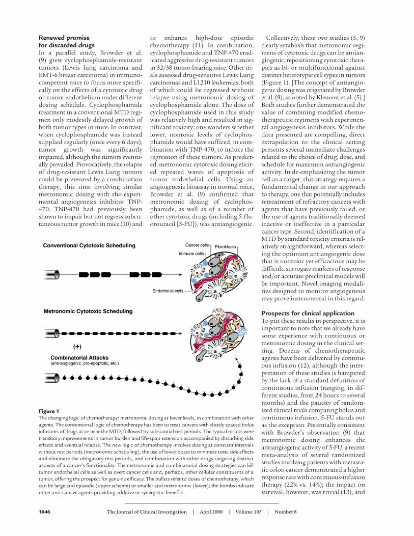
Figure 1The changing logic of chemotherapy: metronomic dosing at lower levels, in combination with otheragents. The conventional logic of chemotherapy has been to treat cancers with closely spaced bolusinfusions of drugs at or near the MTD, followed by substantial rest periods. The typical results weretransitory improvements in tumor burden and life-span extension accompanied by disturbing sideeffects and eventual relapse. The new logic of chemotherapy involves dosing at constant intervalswithout rest periods (metronomic scheduling), the use of lower doses to minimize toxic side effectsand eliminate the obligatory rest periods, and combination with other drugs targeting distinctaspects of a cancer’s functionality. The metronomic and combinatorial dosing strategies can killtumor endothelial cells as well as overt cancer cells and, perhaps, other cellular constituents of atumor, offering the prospect for genuine efficacy. The bullets refer to doses of chemotherapy, whichcan be large and episodic (upper scheme) or smaller and metronomic (lower); the bombs indicateother anti-cancer agents providing additive or synergistic benefits.

this regimen has not become standardpractice. Additional clinical trialsexploring chronic low-dose or continu-ous-infusion chemotherapy have metwith limited success (14–17).
Although these data are sobering inthe context of the current animal stud-ies, it is important to note that contin-uous or chronic chemotherapy admin-istration in the clinical setting hasnearly always been undertaken usingdoses at or near the MTD, resulting intoxicity and requisite breaks from ther-apy. Hence, the value of chemothera-pies administered at low doses onantiangiogenic schedules remains to befully assessed. There are, meanwhile,encouraging anecdotal results; forexample, from a study in which drug-resistant patients with breast cancerwere placed on a low-dose metronomicschedule involving the same cytotoxicdrug (18). If long-term administrationis to be achieved in practice, futurestudies should be aimed at identifyingthe optimal antiangiogenic agents,doses, and schedules, with special con-sideration to patient convenience, aswell as toxicity and efficacy.
Despite these complimentary reports(5, 9) on the eradication of subcuta-neous tumors in mice, it may be unreal-istic to expect such dramatic results inhumans. In particular, metronomic dos-ing with cytotoxic drugs, while demon-strably antiangiogenic, seem unlikely toprove efficacious in general as singleagents. Nevertheless, we believe thatmetronomic delivery of lowered doses ofcytotoxic drugs could be devised to min-imize often devastating side effects ofchemotherapy, while targeting endothe-lial and tumor cells. True efficacy may
come only with combinatorial thera-pies, wherein novel cytotoxic dosingschedules are used in conjunction withother drugs or radiation. Possible com-binations include other approved drugs,such as cox-2 inhibitors (19), thalido-mide (20), or IFN-α/β (3, 21), as well asexperimental drugs such as VEGF/VEGF-receptor inhibitors, other angio-genesis inhibitors (e.g., TNP-470),proapoptotic drugs (22), or biothera-peutic agents such as oncolytic viruses(ref. 23; also see other articles in the cur-rent JCI Perspective series on cancer bio-therapy). The possibilities raised bythese studies are provocative anddeserve further preclinical and clinicalinvestigation.
AcknowledgmentsWe thank Terry Schoop of Biomed Arts,San Francisco, for preparation of thefigure.
1. Hanahan, D., and Weinberg, R.A. 2000. The hall-marks of cancer. Cell. 100:57–70.
2. Hanahan, D., and Folkman, J. 1996. Patterns andemerging mechanisms of the angiogenic switchduring tumorigenesis. Cell. 86:353–364.
3. Folkman, J. 2000. Tumor angiogenesis. In Cancermedicine. Holland et al., editors. B.C. Decker Inc.Hamilton, Ontario, Canada. In press.
4. Cancer trials: news and features. Angiogenesisinhibitors in clinical trials. http://cancertrials.nci.nih.gov/news/angio/table.html.
5. Klement, G., et al. 2000. Continuous low-dose ther-apy with vinblastine and VEGF receptor-2 antibodyinduces sustained tumor regression without overttoxicity. J. Clin. Invest. 105:R15–R24.
6. Witte, L., et al. 1998. Monoclonal antibodies target-ing the VEGF receptor-2 (Flk1/KDR) as an anti-angiogenic therapeutic strategy. Cancer MetastasisRev. 17:155–161.
7. Benjamin, L.E., Golijanin, D., Itin, A., Pode, D., andKeshet, E. 1999. Selective ablation of immatureblood vessels in established human tumors followsvascular endothelial growth factor withdrawal. J.Clin. Invest. 103:159–165.
8. Vacca, A., et al. 1999. Antiangiogenesis is produced
by nontoxic doses of vinblastine. Blood.94:4143–4155.
9. Browder, T., et al. 2000. Antiangiogenic schedulingof chemotherapy improves efficacy against experi-mental drug-resistant cancer. Cancer Res.60:1878–1886.
10. Ingber, D., et al. 1990. Synthetic analogues offumagillin that inhibit angiogenesis and suppresstumor growth. Nature. 348:555–557.
11. Teicher, B.A., et al. 1994. Potentiation of cytotoxiccancer therapies by TNP-470 alone and with otheranti-angiogenic agents. Int. J. Cancer. 57:920–925.
12. Lokich, J., and Anderson, N. 1997. Dose intensity forbolus versus infusion chemotherapy administra-tion: review of the literature for 27 anti-neoplasticagents. Ann. Oncol. 8:15–25.
13. The Meta-analysis Group in Cancer. 1998. Efficacyof intravenous continuous infusion to fluorouracilcompared with bolus administration in advancedcolorectal cancer. J. Clin. Oncol. 16:301–308.
14. Blumenreich, M.S., et al. 1994. Inefficacy of low-dose continuous oral etoposide in non-small celllung cancer. Am. J. Clin. Oncol. 17:163–165.
15. Burris, H. 1998. Weekly schedules of docetaxel.Semin. Oncol. 25(Suppl. 13):21–23.
16. Sorensen, P., Andersen, L.J., Hansen, O., andBastholt, L. 1999. Long-term continuous 5-fluo-rouracil infusion in patients with advanced headand neck cancer. Acta Oncol. 38:1043–1045.
17. Regazzoni, S., Pesce, G., Marini, G., Cavalli, F., andGoldhirsch, A. 1996. Low-dose continuous intra-venous infusion of 5-fluorouracil for metastaticbreast cancer. Ann. Oncol. 7:807–813.
18. Rocca, A.M., et al. 1999. Low dose oral methotrexate(MTX) and cyclophosphamide (CTX) in metastaticbreast cancer (MBC): antitumor activity and corre-lation with serum vascular endothelial growth fac-tor (VEGF) levels. Proc. Am. Soc. Clin. Oncol. 18:121a.(Abstr.)
19. Masferrer, J.L., Koki, A., and Seibert, A. 1999. COX-2 inhibitor. A new class of antiangiogenic agents.Ann. NY Acad. Sci. 889:84–86.
20. D’Amato, R.J., Loughnan, M.S., Flynn, E., and Folk-man, J. 1994. Thalidomide is an inhibitor of angio-genesis. Proc. Natl. Acad. Sci. USA. 91:4082–4085.
21. Slaton, J.W., Perrotte, P., Inoue, K., Dinney, C.P.N.,and Fidler, I.J. 1999. Interferon-α–mediated down-regulation of angiogenesis-related genes and thera-py of bladder cancer are dependent on optimizationof biological dose and schedule. Clin. Cancer Res.5:2726–2734.
22. Ashkenazi, A., et al. 1999. Safety and antitumoractivity of recombinant soluble Apo2 ligand. J. Clin.Invest. 104:155–162.
23. Heise, C., and Kirn, D.H. 2000. Replication-sensitiveadenoviruses as oncolytic agents. J. Clin. Invest.105:847–851.
The Journal of Clinical Investigation | April 2000 | Volume 105 | Number 8 1047
IntroductionThe current interest in developing antiangiogenicdrugs to treat cancer—a concept first proposed byFolkman (1)—can be traced to a number of factors (2).One is the molecular elucidation of a number of angio-genic growth factors, such as VEGF, the angiopoietins,and the receptor tyrosine kinases expressed by activat-ed endothelial cells of newly formed vessels for suchgrowth factors (3). Such discoveries have provided anumber of targets for the rational development ofantiangiogenic drugs such as humanized anti-VEGFneutralizing antibodies (4) and agents that block recep-tors for VEGF or the angiopoietins (2, 5–8). Anotherfactor is that some antiangiogenic drugs may delay oreven circumvent the problem of acquired drug resist-ance (9, 10) because they target the genetically stableendothelial cells of newly formed tumor blood vesselsrather than genetically unstable tumor cells that areprone to mutate and develop resistance (11, 12).
In addition to such rationally designed antiangio-genic drugs, there is a surprisingly diverse and extensivelist of angiogenesis inhibitors that were not originallydesigned to function as such, e.g., thalidomide, IFN-α,and IL-12 (2). Intriguingly, this list may include manydifferent conventional cytotoxic chemotherapeuticdrugs (2), radiation (13), and hormonal ablation ther-apies (14, 15). With respect to chemotherapy, the pres-ence of dividing endothelial cells in newly formingtumor blood vessels (16–18) should render such ves-sels—in contrast to their mature, quiescent counter-parts found in normal adult tissues—sensitive to thecytotoxic effects of such drugs in a manner similar todividing bone marrow, hair follicle, or gut mucosal cells(11). This hypothesized “collateral damage” to thetumor vasculature could conceivably contribute to theantitumor efficacy of chemotherapy in vivo (11). If so,it should follow, as we first proposed in 1991 (11), thateven tumors consisting of tumor cells resistant to a par-
The Journal of Clinical Investigation | April 2000 | Volume 105 | Number 8 R15
Continuous low-dose therapy with vinblastine and VEGF receptor-2 antibody induces sustained tumor regression without overt toxicity
Giannoula Klement,1 Sylvain Baruchel,2 Janusz Rak,1 Shan Man,1 Katherine Clark,1
Daniel J. Hicklin,3 Peter Bohlen,3 and Robert S. Kerbel1
1Sunnybrook and Women's College Health Sciences Centre, Biological Sciences Program, Division of Cancer Biology Research,and Toronto-Sunnybrook Regional Cancer Centre, Toronto, Ontario M4N 3M5, Canada; Department of Medical Biophysics,University of Toronto, Ontario, Canada
2Hospital for Sick Children, Department of Pediatrics, Division of Hematology/Oncology, New Agent and Innovative Therapy Program, Toronto, Ontario M5G 1X8, Canada
3ImClone Systems Inc., New York, New York 10014, USA
Address correspondence to: Robert S. Kerbel, Sunnybrook and Women’s College Health Sciences Centre, Biological Sciences,Division of Cancer Biology Research, Room S-218, 2075 Bayview Avenue, Toronto, Ontario M4N 3M5, Canada. Phone: (416) 480-5711; Fax: (416) 480-5703; E-mail: [email protected].
Received for publication November 2, 1999, and accepted in revised form February 25, 2000.
Various conventional chemotherapeutic drugs can block angiogenesis or even kill activated, divid-ing endothelial cells. Such effects may contribute to the antitumor efficacy of chemotherapy invivo and may delay or prevent the acquisition of drug-resistance by cancer cells. We have imple-mented a treatment regimen that augments the potential antivascular effects of chemotherapy,that is devoid of obvious toxic side effects, and that obstructs the development of drug resistanceby tumor cells. Xenografts of 2 independent neuroblastoma cell lines were subjected to either con-tinuous treatment with low doses of vinblastine, a monoclonal neutralizing antibody (DC101) tar-geting the flk-1/KDR (type 2) receptor for VEGF, or both agents together. The rationale for thiscombination was that any antivascular effects of the low-dose chemotherapy would be selective-ly enhanced in cells of newly formed vessels when survival signals mediated by VEGF are blocked.Both DC101 and low-dose vinblastine treatment individually resulted in significant but transientxenograft regression, diminished tumor vascularity, and direct inhibition of angiogenesis.Remarkably, the combination therapy resulted in full and sustained regressions of large estab-lished tumors, without an ensuing increase in host toxicity or any signs of acquired drug resist-ance during the course of treatment, which lasted for >6 months.
This article may have been published online in advance of the print edition. The date of publication is available from the JCI website, http://www.jci.org.
J. Clin. Invest. 105:R1–R8 (2000).
ticular drug might still respond to that drug throughsuch antivasculature “side effects.” However, this pre-sumably occurs infrequently, or at least not to any ther-apeutically meaningful extent, as most human cancersare intrinsically resistant to chemotherapy, or initiallyrespond, only to recur as a result of overgrowth of drug-resistant subpopulations. This is all the more perplex-ing given recent reports from several groups showingthat a variety of conventional chemotherapeutic drugscan bring about significant antiangiogenic or antivas-cular effects in vivo in a number of assays of angiogen-esis. These drugs include tubulin-inhibiting taxanes,camptothecin analogues, antimetabolites, anthracy-clines, and platinum drugs (19–25).
Recent results from Folkman’s laboratory havehelped shed some light on this paradox (26).Chemotherapy is normally given acutely, usually in theform of bolus infusions at maximum tolerated doses(MTDs) with long rest periods (e.g., 3 weeks) betweensuccessive drug exposures. It was suggested that theserest periods provide the endothelial cell compartmentof a tumor an opportunity to repair some of the dam-age inflicted by the chemotherapy (26). Browder et al.proposed that this repair process could be partiallycompromised by administering lower doses of achemotherapeutic drug, such as cyclophosphamide,more frequently, e.g., weekly. In this regard, it is inter-esting to note that continuous low-dose administra-tion of methotrexate for the treatment of arthritis mayhave an antiangiogenic basis (27). This hypothetical“antiangiogenic scheduling” of chemotherapy (26)optimizes antitumor/vascular side effects so that evena subline of the Lewis lung carcinomas previouslyselected in vivo for acquired resistance to the MTD ofcyclophosphamide can be rendered sensitive again tothe drug in vivo by using continuous low-dose therapyof the same drug (26).
We decided to test the effects of low-dose continuouschemotherapy as a possible antiangiogenic strategy with1 major additional modification, namely, combinationwith an agent that blocks the function of VEGF receptor-2 (flk-1/KDR) and, hence, VEGF itself. The rationale fortesting this particular combination is based on the find-ing that a major function of VEGF is now recognized tobe promotion of survival of endothelial cells comprisingnewly formed vessels (15, 28, 29). Hence, the ability ofsuch cells to cope with the damage inflicted by continu-ous low-dose exposure to a chemotherapeutic drug couldbe selectively and significantly impaired, given the high-ly restricted pattern of expression of VEGF receptors toactivated endothelial cells (3, 6, 7). This could reduce hosttoxicity and thereby allow for longer-term administrationof the chemotherapeutic agent without sacrificing, andperhaps even improving, antitumor efficacy.
To test the rationale of this combination strategyreported here, we used vinblastine, a monoclonalanti–flk-1 neutralizing antibody called DC101 (5, 6), andpoor prognosis-related human neuroblastoma cell linesgrown as xenografts in SCID mice. Poor prognosis neu-
roblastoma in children is usually treated with aggressivecombination chemotherapy at MTDs, with or without abone marrow transplant (30). However, such therapiesare associated with severe side effects and are seldomeffective. The purpose of these experiments was to devel-op a more acceptable alternative treatment and one thatcould also be applied in principle to other types of cancer.
MethodsCells and culture conditions. Neuroblastoma cell lines SK-N-MC and SK-N-AS were obtained from AmericanType Culture Collection (ATCC; Rockville, Maryland,USA) and expanded as a monolayer culture by serialpassage on tissue culture plates (Nunc A/S, Roskilde,Denmark) in DMEM, 5% FBS (GIBCO BRL, GrandIsland, New York, USA). Human umbilical veinendothelial cells (HUVECs; Clonetics, San Diego, Cali-fornia, USA) were expanded on 1% gelatin-coated tissueculture plates in MCDB 131 culture medium (JRH Bio-sciences, Lenexa, Kansas, USA) supplemented with 5ng/mL basic fibroblast growth factor (bFGF) (R&DSystems, Inc., Minneapolis, Minnesota, USA), 10µM/mL heparin (Wyeth-Ayerst, St. Laurent, Quebec,Canada), 10 ng/mL EGF (Upstate Biotechnology Inc.,Lake Placid, New York, USA), and 10% FBS.
In vitro determination of drug sensitivity. Three thousandcells in 200 µL growth media per well were plated in 96-well flat-bottom tissue culture plates (Nunc A/S) andincubated at 37°C, 5% CO2 for 24 hours before initia-tion of treatment. The cells were then washed with PBSand treated with 1–500 ng/mL vinblastine sulfate (Cal-biochem/Novabiochem Corp., San Diego, California,USA) for 24 hours, in groups of 8 wells per dose. Thecells were then pulsed for 6 hours with 2 µCi/well ofmethyl-[3H]thymidine (Amersham Life Science, Buck-inghamshire, United Kingdom). The plates were frozenand thawed, and the DNA was harvested onto a filter-mat using a Titertek Cell Harvester (Flow LaboratoriesLtd., Irvine, Ayr, UK). Incorporated radioactivity wasmeasured on Wallac 1205 BetaPlate ScintillationCounter (Wallac Oy, Turku, Finland), and proliferationwas expressed as a percentage of [3H]thymidine intreated cells versus that in controls.
In vivo tumor growth assessment. SK-N-MC cells wereharvested using 1% trypsin EDTA (GIBCO BRL), and asingle-cell suspension of 2 × 10⁶ cells in 0.2 mL ofgrowth medium was injected subcutaneously into theflanks of 4- to 6-week-old CB-17 SCID mice (CharlesRiver, St. Constant, Quebec, Canada). Approximately 3weeks later, most tumors had grown to approximately0.75 cm3, and mice were randomized into groups of 5animals. Two independent experiments were per-formed, each totaling 20 animals in 4 groups. Thetreatment was as follows: group I (control), 0.4 mL ofPBS (DC101 vehicle) intraperitoneally every 3 days and0.15 mL injectable saline (Vinblastine vehicle) intraperi-toneally every 3 days; group II, 0.4 mL of 2 mg/mLDC101 antibody (800 µg/mouse) (31) every 3 days and0.15 mL of injectable saline intraperitoneally every 3
R16 The Journal of Clinical Investigation | April 2000 | Volume 105 | Number 8
days; group III, vinblastine sulfate 0.75 mg/m2 (∼ 0.25mg/kg) intraperitoneal bolus at the start of therapy,followed by 1 mg/m2 per day (∼ 0.33 mg/kg per day) viasubcutaneous Alzet osmotic pumps (Alza Corp, PaloAlto, California, USA) for 3 weeks, followed by mainte-nance therapy with 0.15 mL of 0.067 mg/mL vinblas-tine sulfate (1.5 mg/m2) intraperitoneally every 3 days,and 0.4 mL of PBS intraperitoneally every 3 days; andgroup IV, combination of DC101 and vinblastine atdoses identical to the single-agent groups.
SK-N-AS cells were harvested using 1% trypsin EDTA,and a single-cell suspension of 2 × 10⁶ cells in 0.2 mL ofgrowth medium was injected subcutaneously into theflanks of 4- to 6-week-old CB-17 SCID mice. Approxi-mately 3 weeks later (average tumor size, 0.25 cm3), themice were randomized into groups of 5 animals andtreated as follows: controls, 0.4 mL of PBS and 0.15 mLof saline twice weekly; DC101 group, 800 µg of DC101mAb/mouse and 0.15 mL of saline intraperitoneallytwice weekly; vinblastine group, 0.4 mL of PBS and 1.5mg/m2 (∼ 0.5 mg/kg) vinblastine twice weekly; andcombination therapy group, treated with DC101 andvinblastine at doses identical to the single-agent group.
The body weight, tumor size, and general clinical sta-tus of the animals were recorded every 2–3 days. Per-pendicular tumor diameters were measured using aVernier scale caliper, and tumor volume was estimatedusing the formula for ellipsoid: (width2 × length)/2.Growth curves were statistically analyzed using repeat-ed measures ANOVA.
The mice were sacrificed when tumor size reached 1.5cm3 or 7.5–10% of their body weight as required byinstitutional guidelines. All other animal care was inaccordance with institutional guidelines as well.
Histology. All tumors were excised, fixed in 10%(vol/vol) formalin, and processed for immunohisto-chemical analysis. To obtain adequate tissue for thecombination treatment group, 2 mice were sacrificedat 7.5 weeks of treatment. For single-agent and controlgroups, the tissues were collected and analyzed at theconclusion of the treatment, i.e., when the mice had tobe sacrificed. Paraffin blocks were cut to 5-µm sectionsand stained with hematoxylin and eosin (H&E) formorphology evaluation and with the Apoptosis Detec-tion System (Promega Corp., Madison, Wisconsin,USA) for assessment of programmed cell death.
Relative tumor vascularity assessed by an FITC-dextranperfusion assay. This method was designed to assess therelative functionality of the tumor vasculature. A sus-pension of 2 × 10⁶ SK-N-AS neuroblastoma cells in 0.2mL of media was injected into the flanks of CB-17SCID mice. Tumors were allowed to grow to approxi-mately 0.25 cm3, at which point tumor bearing micewere then treated with 1 mg/m2 (∼ 0.33 mg/kg) vin-blastine intraperitoneally every 3 days, or 800 µgDC101 intraperitoneally every 3 days, a combinationof the 2 agents, or saline. At 14 days, when divergencein tumor growth between the treatment groups wasclearly evident, 0.2 ml of 25 mg/mL FITC-dextran in
PBS (Sigma Chemical Co., St. Louis, Missouri, USA;average molecular weight of 148 kDa) was injectedsystemically into the lateral tail vein of each mouseand allowed to circulate for 20–30 minutes. Mice werethen sacrificed by cervical dislocation, and blood sam-ples collected by cardiac puncture into heparinizedtubes for assessment of systemic fluorescein levels.Tumors were resected from the surrounding connec-tive tissue, being careful to avoid spillage of intravas-cular contents; weighed; and placed into tubes con-taining 1:10 dispase (Collaborative Research, TwoOaks, Bedford, Massachusetts, USA). To normalize fordilution caused by the difference in tumor sizes, 1 mLof 1:10 dispase was added per 0.5 g of tissue. Tumorswere incubated in a dark 37°C shaker overnight. Thetissue was homogenized, centrifuged at 3,000 g for 10minutes, and supernatant was collected and stored inthe dark until further analysis. Similarly, the bloodsamples were centrifuged immediately after collection,and plasma was separated and protected from light at4°C until analysis. Fluorescence readings wereobtained on a FL600 Fluorescence Plate Reader (Bio-tek Instruments Inc., Winooski, Vermont, USA) froma standard curve created by serial dilution of the FITC-dextran used for injection. The ratio of tumor fluores-cence/plasma fluorescence was assumed to be reflec-tive of the degree of tumor perfusion.
In vivo angiogenesis assessment by the Matrigel plug assay.Matrigel (Collaborative Biomedical Products, Bedford,Massachusetts, USA; refs. 5 and 32) stored at –20°C, wasthawed at 4°C overnight, and mixed with 500 ng/mLbFGF. A total of 0.5 mL of this mixture was then injectedsubcutaneously into the shaved flanks of twenty 6- to 8-week-old female Balb/cJ mice (The Jackson Laboratory,Bar Harbor, Maine, USA). Five mice, used as negative con-trols, were injected with Matrigel alone. Three days later,treatment mice were randomized into 4 groups as fol-lows: group I, saline intraperitoneally; group II, 800 µgDC101 intraperitoneally; group III, 1 mg/m2 vinblastineintraperitoneally; and group IV, combination therapy. All25 mice were treated on days 4 and 7 and sacrificed onday 10. On day 10, 0.2 mL of 25 mg/mL FITC-dextran inPBS was injected systemically into the lateral tail vein ofeach mouse and allowed to circulate for 20–30 minutes.Mice were then sacrificed by cervical dislocation, bloodsamples collected by cardiac puncture into heparinizedtubes, centrifuged immediately after their collection, andplasma was separated and protected from light at 4°C.The Matrigel plugs were resected from surrounding con-nective tissues, placed into tubes containing 1 mL of 1:10dispase, and incubated in the dark in a 37°C shakerovernight. The following day, the plugs were homoge-nized and centrifuged at 3,000 g for 10 minutes, andsupernatant was saved in the dark for analysis of fluores-cence. Fluorescence readings were obtained on FL600Fluorescence Plate Reader using a standard curve creat-ed by serial dilution of FITC-dextran used for injection.Angiogenic response was expressed as a ratio of Matrigelplug fluorescence/plasma fluorescence.
The Journal of Clinical Investigation | April 2000 | Volume 105 | Number 8 R17
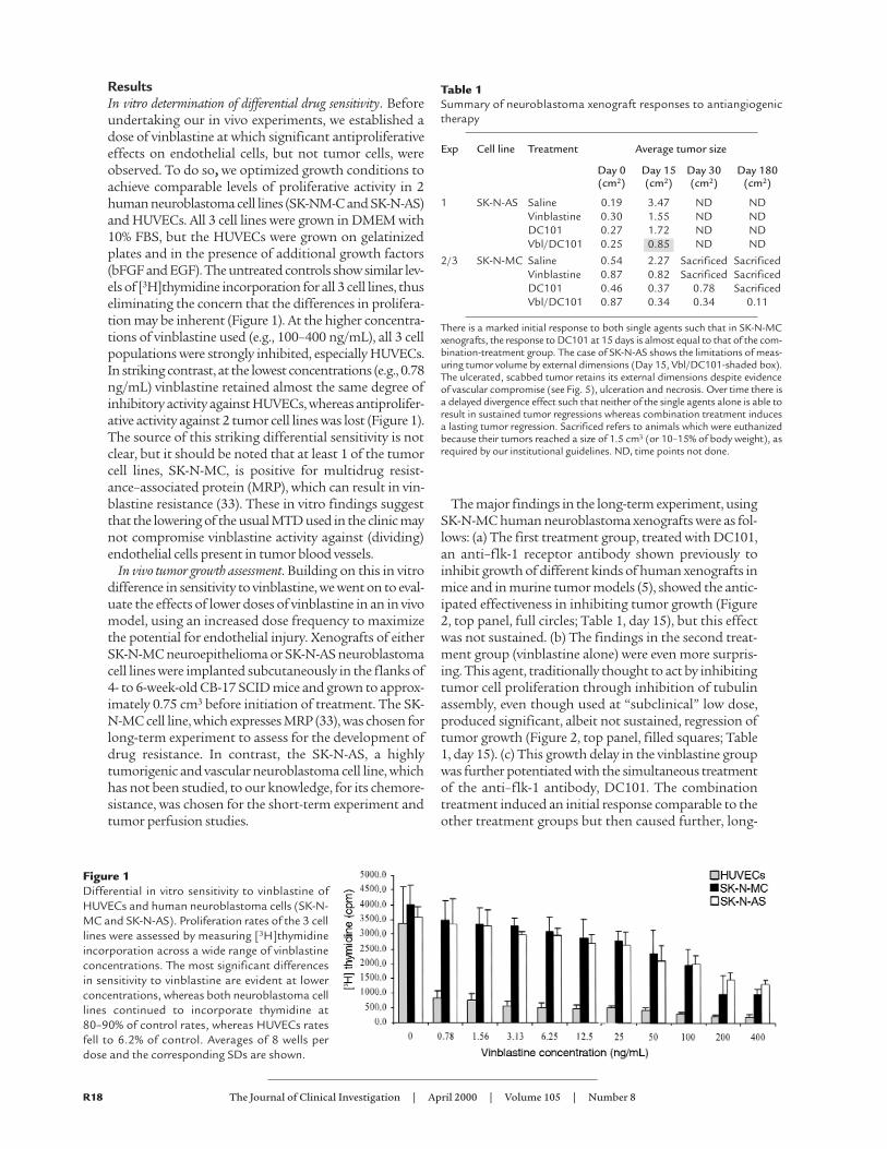
ResultsIn vitro determination of differential drug sensitivity. Beforeundertaking our in vivo experiments, we established adose of vinblastine at which significant antiproliferativeeffects on endothelial cells, but not tumor cells, wereobserved. To do so, we optimized growth conditions toachieve comparable levels of proliferative activity in 2human neuroblastoma cell lines (SK-NM-C and SK-N-AS)and HUVECs. All 3 cell lines were grown in DMEM with10% FBS, but the HUVECs were grown on gelatinizedplates and in the presence of additional growth factors(bFGF and EGF). The untreated controls show similar lev-els of [3H]thymidine incorporation for all 3 cell lines, thuseliminating the concern that the differences in prolifera-tion may be inherent (Figure 1). At the higher concentra-tions of vinblastine used (e.g., 100–400 ng/mL), all 3 cellpopulations were strongly inhibited, especially HUVECs.In striking contrast, at the lowest concentrations (e.g., 0.78ng/mL) vinblastine retained almost the same degree ofinhibitory activity against HUVECs, whereas antiprolifer-ative activity against 2 tumor cell lines was lost (Figure 1).The source of this striking differential sensitivity is notclear, but it should be noted that at least 1 of the tumorcell lines, SK-N-MC, is positive for multidrug resist-ance–associated protein (MRP), which can result in vin-blastine resistance (33). These in vitro findings suggestthat the lowering of the usual MTD used in the clinic maynot compromise vinblastine activity against (dividing)endothelial cells present in tumor blood vessels.
In vivo tumor growth assessment. Building on this in vitrodifference in sensitivity to vinblastine, we went on to eval-uate the effects of lower doses of vinblastine in an in vivomodel, using an increased dose frequency to maximizethe potential for endothelial injury. Xenografts of eitherSK-N-MC neuroepithelioma or SK-N-AS neuroblastomacell lines were implanted subcutaneously in the flanks of4- to 6-week-old CB-17 SCID mice and grown to approx-imately 0.75 cm3 before initiation of treatment. The SK-N-MC cell line, which expresses MRP (33), was chosen forlong-term experiment to assess for the development ofdrug resistance. In contrast, the SK-N-AS, a highlytumorigenic and vascular neuroblastoma cell line, whichhas not been studied, to our knowledge, for its chemore-sistance, was chosen for the short-term experiment andtumor perfusion studies.
The major findings in the long-term experiment, usingSK-N-MC human neuroblastoma xenografts were as fol-lows: (a) The first treatment group, treated with DC101,an anti–flk-1 receptor antibody shown previously toinhibit growth of different kinds of human xenografts inmice and in murine tumor models (5), showed the antic-ipated effectiveness in inhibiting tumor growth (Figure2, top panel, full circles; Table 1, day 15), but this effectwas not sustained. (b) The findings in the second treat-ment group (vinblastine alone) were even more surpris-ing. This agent, traditionally thought to act by inhibitingtumor cell proliferation through inhibition of tubulinassembly, even though used at “subclinical” low dose,produced significant, albeit not sustained, regression oftumor growth (Figure 2, top panel, filled squares; Table1, day 15). (c) This growth delay in the vinblastine groupwas further potentiated with the simultaneous treatmentof the anti–flk-1 antibody, DC101. The combinationtreatment induced an initial response comparable to theother treatment groups but then caused further, long-
R18 The Journal of Clinical Investigation | April 2000 | Volume 105 | Number 8
Figure 1Differential in vitro sensitivity to vinblastine ofHUVECs and human neuroblastoma cells (SK-N-MC and SK-N-AS). Proliferation rates of the 3 celllines were assessed by measuring [3H]thymidineincorporation across a wide range of vinblastineconcentrations. The most significant differencesin sensitivity to vinblastine are evident at lowerconcentrations, whereas both neuroblastoma celllines continued to incorporate thymidine at80–90% of control rates, whereas HUVECs ratesfell to 6.2% of control. Averages of 8 wells perdose and the corresponding SDs are shown.
Table 1Summary of neuroblastoma xenograft responses to antiangiogenictherapy
Exp Cell line Treatment Average tumor size
Day 0 Day 15 Day 30 Day 180(cm2) (cm2) (cm2) (cm2)
1 SK-N-AS Saline 0.19 3.47 ND NDVinblastine 0.30 1.55 ND NDDC101 0.27 1.72 ND NDVbl/DC101 0.25 ND ND
2/3 SK-N-MC Saline 0.54 2.27 Sacrificed SacrificedVinblastine 0.87 0.82 Sacrificed SacrificedDC101 0.46 0.37 0.78 SacrificedVbl/DC101 0.87 0.34 0.34 0.11
There is a marked initial response to both single agents such that in SK-N-MCxenografts, the response to DC101 at 15 days is almost equal to that of the com-bination-treatment group. The case of SK-N-AS shows the limitations of meas-uring tumor volume by external dimensions (Day 15, Vbl/DC101-shaded box).The ulcerated, scabbed tumor retains its external dimensions despite evidenceof vascular compromise (see Fig. 5), ulceration and necrosis. Over time there isa delayed divergence effect such that neither of the single agents alone is able toresult in sustained tumor regressions whereas combination treatment inducesa lasting tumor regression. Sacrificed refers to animals which were euthanizedbecause their tumors reached a size of 1.5 cm3 (or 10–15% of body weight), asrequired by our institutional guidelines. ND, time points not done.
0.85
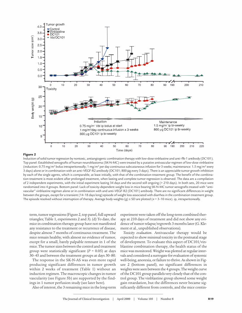
term, tumor regressions (Figure 2, top panel, full upwardtriangles; Table 1, experiments 2 and 3). (d) To date, themice in combination therapy group have not manifestedany resistance to the treatment or recurrence of disease,despite almost 7 months of continuous treatment. Themice remain healthy, with almost no evidence of tumor,except for a small, barely palpable remnant in 1 of themice. The tumor sizes between the control and treatmentgroup were statistically significant (P = 0.05) at days30–45 and between the treatment groups at days 30–80.
The response in the SK-N-AS was even more rapidproducing significant differences in tumor growthwithin 2 weeks of treatment (Table 1) without aninduction regimen. The macroscopic changes in tumorvascularity (see Figure 5b) are supported by the find-ings in 1 tumor perfusion study (see later here).
Also of interest, the 3 remaining mice in the long-term
experiment were taken off the long-term combined ther-apy at 210 days of treatment and did not show any evi-dence of tumor relapse/regrowth 3 months later (G. Kle-ment et al., unpublished observations).
Toxicity evaluation. Antivascular therapy would beexpected to show minimal toxicity in the postnatal stageof development. To evaluate this aspect of DC101/vin-blastine combination therapy, the health status of themice was monitored. Weight was plotted at regular inter-vals and considered a surrogate for evaluation of systemicwell-being, anorexia, or failure to thrive. As shown in Fig-ure 2 (bottom panel), no significant differences inweights were seen between the 4 groups. The weight curveof the DC101 group parallels very closely that of the con-trol group. The vinblastine group showed some weightgain retardation, but the differences never became sig-nificantly different from controls, and the mice contin-
The Journal of Clinical Investigation | April 2000 | Volume 105 | Number 8 R19
Figure 2Induction of solid tumor regression by nontoxic, antiangiogenic combination therapy with low-dose vinblastine and anti–flk-1 antibody (DC101).Top panel: Established xenografts of human neuroblastoma (SK-N-MC) were treated by a putative antivascular regimen of low-dose vinblastine(induction: 0.75 mg/m2 bolus intraperitoneally; 1 mg/m2 per day continuous subcutaneous infusion for 3 weeks; maintenance: 1.5 mg/m2 every3 days) alone or in combination with an anti–VEGF-R2 antibody (DC101; 800 µg every 3 days). There is an appreciable tumor growth inhibitionby each of the single agents, which is comparable, at least initially, with that of the combination treatment group. The benefit of the combina-tion treatment is most evident after prolonged treatment, when lasting and complete tumor regression is observed. The data are a compilationof 2 independent experiments, with the initial experiment lasting 34 days and the second still ongoing (> 210 days). In both sets, 20 mice wererandomized into 4 groups. Bottom panel: Lack of toxicity-dependent weight loss in mice bearing SK-N-MC tumor xenografts treated with “anti-vascular” vinblastine regimen alone or in combination with and anti–VEGF-R2 (DC101) antibody. There are no significant differences in weightbetween the groups, except for a transient (14–18 days long) episode of weight loss associated with diarrhea in the combination treatment group.The episode resolved without interruption of therapy. Average body weights (g) ± SD are plotted (n = 3–10 mice). ip, intraperitoneally.

ued to grow. Similarly, the toxicity profile in the combi-nation treatment group was very similar to those in thesingle-agent groups, with the exception of a transientepisode of weight loss associated with diarrhea. Theepisode lasted approximately 2–3 weeks and was unlike-ly to be due to the therapy as the mice recovered withoutinterruption of treatment.
Other usual signs of drug toxicity in mice, such as ruf-fled fur, anorexia, cachexia, skin tenting (due to dehy-dration), skin ulcerations, or toxic deaths (21), were notseen at the doses used in our experiments. Diarrhea, acommon sign of vinblastine toxicity when doses of 10
mg/kg (21) are used, was generally not observed, exceptfor the previously mentioned transient episode.
Histopathological analysis. To elucidate further the mech-anisms involved in the tumor regression after treatmentwith vinblastine, DC101, or the combined therapy, tissuehistopathology assessment was undertaken. The typicaltissue architecture of untreated SK-N-MC tumors isshown in Figure 3 (control panels). Cancer cells with highnuclear to cytoplasmic ratio form cuffs around centralvessels, and apoptotic cells characterized by pyknoticnuclei and cytoplasmic blebbing, are only evident as a thinrim at the periphery of the cuffs. The nuclei of these cells
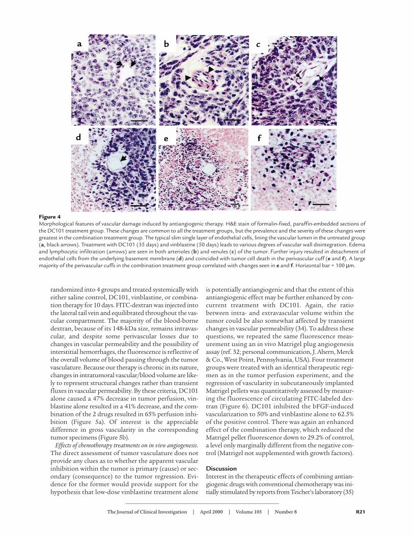
stain strongly for terminal deoxynucleotidyltransferase (TUNEL) reactivity, as expectedfor cells undergoing apoptosis. Vinblastinealone or DC101 treatment alone both showan increase in the width of the apoptotic rims(Figure 3), suggesting the cells most distal tothe tumor vasculature are primarily affected,but a large percentage of viable tumor cellsstill survive in the center of the cuff. In bothsingle-agent groups, specimens were collectedat the time mice had to be sacrificed for ethi-cal concerns regarding tumor burden. Despitethe evident increase in apoptosis, there was anet tumor growth. In contrast, histology ofthe combined therapy group, as would be pre-dicted by the regression in tumor size in thistreatment group at the time of analysis (7.5weeks of treatment), shows overwhelmingloss of both cell viability and preexistingtumor architecture (Figure 3, bottom panels).There is a close similarity of the appearance ofH&E and TUNEL stain. Interestingly, weobserved signs of endothelial cell toxicity in allof the treatment groups (Figure 4). Ratherthan a typical single layer of flattenedendothelial cells surrounding the vascularlumen in untreated group (Figure 4a), weobserved edema (Figure 4, b and c) anddetachment from surrounding basementmembrane (Figure 4, c–e) leading to completevascular wall disintegration and tumor celldeath (Figure 4f). Even though the actualexamples depicted in Figure 4 are those in theDC101 group, their appearance is sharedbetween the groups, and it is only the degreeof tumor vessel regression that results in theseen differences in tumor size.
Tumor perfusion by assessment of intravascularfluorescence. To explore further the possibili-ty that tumor regression induced with treat-ment using DC101 and/or vinblastine wasindeed due to the vascular injury, rather thana direct antitumor cell effect, we assessedtumor perfusion directly by using a FITC-dextran fluorescence method. Mice carryingestablished subcutaneous SK-N-AS humanneuroblastoma xenografts (∼ 0.25 cm3) were
R20 The Journal of Clinical Investigation | April 2000 | Volume 105 | Number 8
Figure 3Vinblastine, DC101, or combination therapy induces tumor cell apoptosis inperivascular cuffs of SK-N-MC tumor xenografts. H&E stain of formalin-fixed,paraffin-embedded sections. The typical tissue architecture (control, top two pan-els) shows perivascular cuffing by neoplastic cells and normal endothelial cell lin-ing (arrows). Apoptotic cells (ap) are seen only at the periphery of the cuff, andtheir presence is confirmed by TUNEL staining (right-sided panels). In both sin-gle-treatment groups (vinblastine and DC101), widening of the apoptotic rims,and extension of the apoptotic figures into the cuff can be observed after 35 and50 days of treatment, respectively. Viable malignant cells are still present withinthe tumor cuff in both single-agent groups. In contrast, histology of the combinedtherapy group reveals diffuse tumor cell death and total loss of preexisting archi-tecture (bottom left-hand panel), a finding supported by the diffuse TUNEL stainin corresponding specimens (bottom right-hand panel).
randomized into 4 groups and treated systemically witheither saline control, DC101, vinblastine, or combina-tion therapy for 10 days. FITC-dextran was injected intothe lateral tail vein and equilibrated throughout the vas-cular compartment. The majority of the blood-bornedextran, because of its 148-kDa size, remains intravas-cular, and despite some perivascular losses due tochanges in vascular permeability and the possibility ofinterstitial hemorrhages, the fluorescence is reflective ofthe overall volume of blood passing through the tumorvasculature. Because our therapy is chronic in its nature,changes in intratumoral vascular/blood volume are like-ly to represent structural changes rather than transientfluxes in vascular permeability. By these criteria, DC101alone caused a 47% decrease in tumor perfusion, vin-blastine alone resulted in a 41% decrease, and the com-bination of the 2 drugs resulted in 65% perfusion inhi-bition (Figure 5a). Of interest is the appreciabledifference in gross vascularity in the correspondingtumor specimens (Figure 5b).
Effects of chemotherapy treatments on in vivo angiogenesis.The direct assessment of tumor vasculature does notprovide any clues as to whether the apparent vascularinhibition within the tumor is primary (cause) or sec-ondary (consequence) to the tumor regression. Evi-dence for the former would provide support for thehypothesis that low-dose vinblastine treatment alone
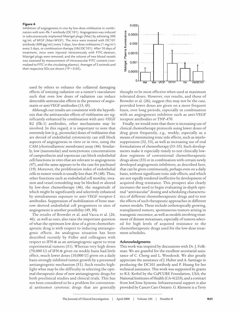
is potentially antiangiogenic and that the extent of thisantiangiogenic effect may be further enhanced by con-current treatment with DC101. Again, the ratiobetween intra- and extravascular volume within thetumor could be also somewhat affected by transientchanges in vascular permeability (34). To address thesequestions, we repeated the same fluorescence meas-urement using an in vivo Matrigel plug angiogenesisassay (ref. 32; personal communication, J. Ahern, Merck& Co., West Point, Pennsylvania, USA). Four treatmentgroups were treated with an identical therapeutic regi-men as in the tumor perfusion experiment, and theregression of vascularity in subcutaneously implantedMatrigel pellets was quantitatively assessed by measur-ing the fluorescence of circulating FITC-labeled dex-tran (Figure 6). DC101 inhibited the bFGF-inducedvascularization to 50% and vinblastine alone to 62.5%of the positive control. There was again an enhancedeffect of the combination therapy, which reduced theMatrigel pellet fluorescence down to 29.2% of control,a level only marginally different from the negative con-trol (Matrigel not supplemented with growth factors).
DiscussionInterest in the therapeutic effects of combining antian-giogenic drugs with conventional chemotherapy was ini-tially stimulated by reports from Teicher’s laboratory (35)
The Journal of Clinical Investigation | April 2000 | Volume 105 | Number 8 R21
Figure 4Morphological features of vascular damage induced by antiangiogenic therapy. H&E stain of formalin-fixed, paraffin-embedded sections ofthe DC101 treatment group. These changes are common to all the treatment groups, but the prevalence and the severity of these changes weregreatest in the combination treatment group. The typical slim single layer of endothelial cells, lining the vascular lumen in the untreated group(a, black arrows). Treatment with DC101 (35 days) and vinblastine (50 days) leads to various degrees of vascular wall disintegration. Edemaand lymphocytic infiltration (arrows) are seen in both arterioles (b) and venules (c) of the tumor. Further injury resulted in detachment ofendothelial cells from the underlying basement membrane (d) and coincided with tumor cell death in the perivascular cuff (e and f). A largemajority of the perivascular cuffs in the combination treatment group correlated with changes seen in e and f. Horizontal bar = 100 µm.

showing that the antitumor effects of MTDs of variouschemotherapeutic cytotoxic drugs can be augmented, atleast in preclinical experiments, when they are combinedwith angiogenesis inhibitors (36). Counterintuitively,such effects have been attributed to greater delivery of thecytotoxic drugs into tumor masses (36). Increasing theefficacy of cyclic high-dose chemotherapy in such a man-ner, however, would unlikely alter 2 of the major prob-lems associated with the use of chemotherapy given inthis way: induction of moderate to severe side effects (e.g.,myelosuppression, nausea, and hair loss), and the devel-
opment of acquired resistance to the cytotoxic drugs. Incontrast, our results suggest that these problems can besignificantly attenuated, and perhaps even avoided, byusing much more frequent administration of signifi-cantly lower doses of a chemotherapeutic drug, e.g., vin-blastine, when given in combination with anti–VEGFreceptor-2 neutralizing antibodies, without sacrificingpotent antitumor efficacy. Thus, large (0.75 cm3) estab-lished human neuroblastoma xenografts in SCID micecould be induced to regress completely with this combi-nation strategy, whereas either agent alone caused onlypartial and temporary regressions, with relapses observedin all treated animals 30–50 days after initiation of theindividual therapy treatments. In striking contrast, a fullyregressed state could be induced and maintained for aslong as the combination therapy was maintained, whichin our case was 210 days, in the absence of any significanttoxicity, as assessed by lack of weight loss. No myelosup-pression has been observed either (Klement et al., unpub-lished observations).
The dose of vinblastine used in our experiments wasin the range of 1.5 mg/m2, every 3 days, which isapproximately 1/4 of the MTD of this drug in humans,and 1/16–1/20 of the MTD in mice, given the fact thatthe MTD of vinblastine in mice is 4–5 times higherthan in humans (37, 38). Similar to Browder et al., whoused low-dose cyclophosphamide therapy (26), wefound evidence, in our case using the Matrigel plugassay, that continuous low-dose vinblastine adminis-tration can cause a direct antiangiogenic effect in vivo.
The decision to use anti–VEGF receptor-2 antibodiesin our combination strategy was based on 3 main con-siderations. First, dividing endothelial cells in particularmay be damaged or killed by exposure to the vinblastinetherapy. Second, the extent of this damage may beenhanced in a highly selective manner given the cellulardistribution of VEGF receptors, which is mainly, thoughnot exclusively, restricted to activated endothelial cells ofnewly formed vessels (2, 3, 5). Third, 1 of the major func-tions of VEGF is to prevent apoptosis of such endothe-lial cells during the process of vasculogenesis and angio-genesis (14, 28). The prosurvival function of VEGF foractivated endothelial cells appears due to its ability toactivate PI3 kinase/Akt/PKB (39), and/or upregulateexpression of several genes and/or proteins known tohave antiapoptotic function such as bcl-2 (40), as well asXIAP (41) and survivin (41, 42), the latter 2 being mem-bers of the IAP family of apoptosis inhibitors (43). Forexample, survivin protein expression can be massivelyupregulated, e.g., 15- to 20-fold (41, 42), and XIAP 4-fold,in human endothelial cells by in vitro by VEGF (41)Moreover, XIAP and survivin (especially XIAP) can con-fer resistance to chemotherapeutic drugs in tumor cells(44). Thus, blockade of VEGF function could conceiv-ably render surviving endothelial cells more vulnerableto the damaging effects of chemotherapy, especiallywhen the cytotoxic drug is given at low doses, by the pos-sible downregulation of multiple antiapoptotic proteinsnormally induced by VEGF. A similar reasoning has been
R22 The Journal of Clinical Investigation | April 2000 | Volume 105 | Number 8
Figure 5Impact of antiangiogenic treatment regimen (Vbl+DC101) on integri-ty of tumor vasculature. (a) The decrease in intravascular FITC-dex-tran fluorescence reflects changes in tumor perfusion in establishedSK-N-AS neuroblastoma xenografts subjected to a 2-week course oftreatment with anti–flk-1 (DC101) antibody, low-dose vinblastine,or combination of the 2. Both single-drug treatments caused a sig-nificant decrease in tumor perfusion, and this effect was enhancedby the combination therapy. Averages of 5 animals (bearing bilater-al tumor xenografts) and their respective SDs are shown (*P = 0.05).(b) Tumor appearance in treated and untreated animals at the timeof excision. Notable is the change in tumor vascularity in the single-treatment groups even before an appreciable change in tumor size.Groups were treated for 14 days before specimen collection.
used by others to enhance the collateral damagingeffects of ionizing radiation on a tumor’s vasculaturesuch that even low doses of radiation can inducedetectable antivascular effects in the presence of angio-statin or anti-VEGF antibodies (13, 45).
Although our results are consistent with the hypoth-esis that the antivascular effects of vinblastine are sig-nificantly enhanced by combination with anti–VEGFR2 (flk-1) antibodies, other mechanisms may beinvolved. In this regard, it is important to note thatextremely low (e.g., picomolar) doses of vinblastine thatare devoid of endothelial cytotoxicity can still blockaspects of angiogenesis in vitro or in vivo, using theCAM (chorioallantoic membrane) assay (46). Similar-ly, low (nanomolar) and noncytotoxic concentrationsof camptothecin and topotecan can block endothelialcell functions in vitro that are relevant to angiogenesis(47), and the same appears to be the case for paclitaxel(19). Moreover, the proliferation index of endothelialcells in tumor vessels is usually less than 3% (48). Thus,other functions such as endothelial cell motility, inva-sion and vessel remodeling may be blocked or alteredby low-dose chemotherapy (46), the magnitude ofwhich might be significantly and selectively enhancedby simultaneous exposure to anti–VEGF receptor-2antibodies. Suppression of mobilization of bone mar-row–derived endothelial cell progenitors to sites ofangiogenesis is another possibility (49, 50).
The results of Browder et al. and Vacca et al. (26,46), as well as ours, also raise the important questionof what the optimum low dose of a given chemother-apeutic drug is with respect to inducing antiangio-genic effects. An analogous situation has beendescribed recently by Fidler and colleagues withrespect to IFN-α as an antiangiogenic agent to treatexperimental tumors (51). Whereas very high doses(70,000 U) of IFN-α given on weekly basis had littleeffect, much lower doses (10,000 U) given on a dailybasis strongly inhibited tumor growth by a presumedantiangiogenic mechanism (51). Such results high-light what may be the difficulty in selecting the opti-mal therapeutic dose of new antiangiogenic drugs forboth preclinical studies and clinical trials. This hasnot been considered to be a problem for convention-al antitumor cytotoxic drugs that are generally
thought to be most effective when used at maximumtolerated doses. However, our results, and those ofBrowder et al. (26), suggest this may not be the case,provided lower doses are given on a more frequentbasis, over long periods, especially in combinationwith an angiogenesis inhibitor such as anti-VEGFreceptor antibodies or TNP-470.
Finally, we would note that there is increasing use ofclinical chemotherapy protocols using lower doses ofdrug given frequently, e.g., weekly, especially as ameans of minimizing toxic side effects, such as myelo-suppression (52, 53), as well as increasing use of oralformulations of chemotherapy (53–55). Such develop-ments make it especially timely to test clinically low-dose regimens of conventional chemotherapeuticdrugs alone (53) or in combination with certain newlydeveloped angiogenesis inhibitors, as described here,that can be given continuously, perhaps even on a dailybasis, without significant toxic side effects, and whichare not rapidly rendered ineffective by development ofacquired drug resistance. This prospect also clearlyincreases the need to begin evaluating in-depth opti-mal “antivascular” dosing and scheduling characteris-tics of different chemotherapeutic drugs and to testthe effects of such therapeutic approaches in differenttumor models. These include orthotopically growing,transplanted tumors, spontaneous tumors arising intransgenic oncomice, as well as models involving treat-ment of distant metastases, especially of tumors select-ed for high levels of acquired resistance to thechemotherapeutic drugs used for the low-dose treat-ment schedules.
AcknowledgmentsThis work was inspired by discussions with Dr. J. Folk-man. We are grateful for the excellent secretarial assis-tance of C. Cheng and L. Woodcock. We also greatlyappreciate the assistance of J. Huber and A. Santiago inproducing the DC101 antibody and P. Huang for hertechnical assistance. This work was supported by grantsto R.S. Kerbel by the CaPCURE Foundation, USA; theNational Institutes of Health (CA-41233), and a contractfrom ImClone Systems. Infrastructural support is alsoprovided by Cancer Care Ontario. G. Klement is a Terry
The Journal of Clinical Investigation | April 2000 | Volume 105 | Number 8 R23
Figure 6Inhibition of angiogenesis in vivo by low-dose vinblastine in combi-nation with anti–flk-1 antibody (DC101). Angiogenesis was inducedin subcutaneously implanted Matrigel plugs (Mat) by admixing 500ng/mL of bFGF (Mat+bFGF). The mice were treated with DC101antibody (800 µg/mL) every 3 days, low-dose vinblastine (1 mg/m2)every 3 days, or combination therapy (Vbl/DC101). After 10 days oftreatment, mice were injected intravenously with FITC-dextran;Matrigel plugs were removed; and the volume of new blood vesselswas assessed by measurement of intravascular FITC content (nor-malized to FITC in the circulating plasma). Averages of 5 animals andtheir respective SDs are shown (AP = 0.05).

Fox Fellow of the National Cancer Institute of Canada.
1. Folkman, J. 1972. Anti-angiogenesis: new concept for therapy of solidtumors. Ann. Surg. 175:409–416.
2. Kerbel, R.S. 2000. Tumor angiogenesis: past, present, and the near future.Carcinogenesis. 21:505–515.
3. Ferrara, N., and Alitalo, K. 1999. Clinical applications of angiogenic growthfactors and their inhibitors. Nat. Med. 5:1359–1364.
4. Presta, L.G., et al. 1997. Humanization of an anti-vascular endothelialgrowth factor monoclonal antibody for the therapy of solid tumors andother disorders. Cancer Res. 57:4593–4599.
5. Witte, L., et al. 1998. Monoclonal antibodies targeting the VEGF receptor-2(Flk1/KDR) as an anti-angiogenic therapeutic strategy. Cancer Metastasis Rev.17:155–161.
6. Prewett, M., et al. 1999. Antivascular endothelial growth factor receptor (fetalliver kinase 1) monoclonal antibody inhibits tumor angiogenesis andgrowth of several mouse and human tumors. Cancer Res. 59:5209–5218.
7. Fong, T.A., et al. 1999. SU5416 is a potent and selective inhibitor of the vas-cular endothelial growth factor receptor (Flk-1/KDR) that inhibits tyrosinekinase catalysis, tumor vascularization, and growth of multiple tumor types.Cancer Res. 59:99–106.
8. Lin, P., et al. 1997. Inhibition of tumor angiogenesis using a soluble recep-tor establishes a role for Tie2 in pathologic vascular growth. J. Clin. Invest.100:2072–2078.
9. Boehm, T., Folkman, J., Browder, T., and O’Reilly, M.S. 1997. Antiangiogenictherapy of experimental cancer does not induce acquired drug resistance.Nature. 390:404–407.
10. Kaban, L.B., et al. 1999. Antiangiogenic therapy of a recurrent giant celltumor of the mandible with interferon Alfa-2a. Pediatrics. 103:1145–1149.
11. Kerbel, R.S. 1991. Inhibition of tumor angiogenesis as a strategy to circum-vent acquired resistance to anti-cancer therapeutic agents. BioEssays.13:31–36.
12. Kerbel, R.S. 2000. Acquired drug resistance driven by tumor cell geneticinstability: circumvention by direct acting anti-angiogenic vascular target-ing agents. In DNA alterations in cancer: genetic and epigenetic changes. M. Ehrlich,editor. Biotechniques Books. Natick, MA. 489–501.
13. Gorski, D.H., et al. 1999. Blockage of the vascular endothelial growth factorstress response increases the antitumor effects of ionizing radiation. CancerRes. 59:3374–3378.
14. Jain, R.K., et al. 1998. Endothelial cell death, angiogenesis, and microvascu-lar function after castration in an androgen-dependent tumor: role of vas-cular endothelial growth factor. Proc. Natl. Acad. Sci. USA. 95:10820–10825.
15. Benjamin, L.E., Golijanin, D., Itin, A., Pode, D., and Keshet, E. 1999. Selec-tive ablation of immature blood vessels in established human tumors fol-lows vascular endothelial growth factor withdrawal. J. Clin. Invest.103:159–165.
16. Denekamp, J. 1993. Angiogenesis, neovascular proliferation and vascularpathophysiology as targets for cancer therapy. Br. J. Radiol. 66:181–196.
17. Tannock, I.F. 1970. Population kinetics of carcinoma cells, capillaryendothelial cells, and fibroblasts in a transplanted mouse mammary tumor.Cancer Res. 30:2470–2476.
18. Tannock, I.F., and Hayashi, S. 1972. The proliferation of capillary endothe-lial cells. Cancer Res. 32:77–82.
19. Belotti, D., et al. 1996. The microtubule-affecting drug paclitaxel has antian-giogenic activity. Clin. Cancer Res. 2:1843–1849.
20. O’Leary, J.J., et al. 1999. Antiangiogenic effects of camptothecin analogues9-amino-20(S)-camptothecin, topotecan, and CPT-11 studied in the mousecornea model. Clin. Cancer Res. 5:181–187.
21. Baguley, B.C., Holdaway, K.M., Thomsen, L.L., Zhuang, L., and Zwi, L.J. 1991.Inhibition of growth of colon 38 adenocarcinoma by vinblastine andcolchicine: evidence for a vascular mechanism. Eur. J. Cancer. 27:482–487.
22. Schirner, M., Hoffmann, J., Menrad, A., and Schneider, M.R. 1998. Antian-giogenic chemotherapeutic agents: characterization in comparison to theirtumor growth inhibition in human renal cell carcinoma models. Clin. Can-cer Res. 4:1331–1336.
23. Presta, M., et al. 1999. Purine analogue 6-methylmercaptopurine ribosideinhibits early and late phases of the angiogenesis process. Cancer Res.59:2417–2424.
24. Steiner, R. 1992. Angiostatic activity of anticancer agents in the chickembryo chorioallantoic membrane (CHE-CAM) assay. In Angiogenesis: keyprinciples–science–technology–medicine. R. Steiner, P.S. Weiss, and R. Langer, edi-tors. Birkhauser Verlag. Basel, Switzerland. 449–454.
25. Yoshikawa, A., Saura, R., Matsubara, T., and Mizuno, K. 1997. A mechanismof cisplatin action: antineoplastic effect through inhibition of neovascular-ization. Kobe. J. Med. Sci. 43:109–120.
26. Browder, T., et al. 2000. Antiangiogenic scheduling of chemotherapyimproves efficacy against experimental drug-resistant cancer. Cancer Res. Inpress.
27. Hirata, S., Matsubara, T., Saura, R., Tateishi, H., and Hirohata, K. 1989. Inhi-bition of in vitro vascular endothelial cell proliferation and in vivo neovas-cularization by low-dose methotrexate. Arthritis Rheum. 32:1065–1073.
28. Alon, T., et al. 1995. Vascular endothelial growth factor acts as a survival fac-tor for newly formed retinal vessels and has implications for retinopathy ofprematurity. Nat. Med. 1:1024–1028.
29. Yuan, F., et al. 1996. Time-dependent vascular regression and permeabilitychanges in established human tumor xenografts induced by an anti-vascu-lar endothelial growth factor/vascular permeability factor antibody. Proc.Natl. Acad. Sci. USA. 93:14765–14770.
30. Stram, D.O., et al. 1996. Consolidation chemoradiotherapy and autologousbone marrow transplantation versus continued chemotherapy for metasta-tic neuroblastoma: a report of two concurrent Children’s Cancer Groupstudies. J. Clin. Oncol. 14:2417–2426.
31. Rockwell, P., Neufeld, G., Glassman, A., Caron, D., and Goldstein, N. 1995.In vitro neutralization of vascular endothelial growth factor activation offlk-1 by a monoclonal antibody. Mol. Cell. Different. 3:91–109.
32. Passaniti, A., et al. 1992. A simple, quantitative method for assessing angio-genesis and antiangiogenic agents using reconstituted basement membrane,heparin, and fibroblast growth factor. Lab. Invest. 67:519–528.
33. Bordow, S.B., et al. 1994. Expression of the multidrug resistance-associatedprotein (MRP) gene correlates with amplification and overexpression of theN-myc oncogene in childhood neuroblastoma. Cancer Res. 54:5036–5040.
34. Jain, R.K. 1990. Vascular and interstitial barriers to delivery of therapeuticagents in tumors. Cancer Metastasis Rev. 9:253–266.
35. Teicher, B.A., Sotomayor, E.A., and Huang, Z.D. 1992. Antiangiogenic agentspotentiate cytotoxic cancer therapies against primary and metastatic dis-ease. Cancer Res. 52:6702–6704.
36. Kakeji, Y., and Teicher, B.A. 1997. Preclinical studies of the combination ofangiogenic inhibitors with cytotoxic agents. Invest. New Drugs. 15:39–48.
37. Tashiro, T., et al. 1989. Responsiveness of human lung cancer/nude mouseto antitumor agents in a model using clinically equivalent doses. CancerChemother. Pharmacol. 24:187–192.
38. Inaba, M., et al. 1989. Evaluation of antitumor activity in a human breasttumor/nude mouse model with a special emphasis on treatment dose. Can-cer. 64:1577–1582.
39. Gerber, H.P., et al. 1998. Vascular endothelial growth factor regulatesendothelial cell survival through the phosphatidylinositol 3′-kinase/Akt sig-nal transduction pathway. Requirement for Flk-1/KDR activation. J. Biol.Chem. 273:30336–30343.
40. Nor, J.E., Christensen, J., Mooney, D.J., and Polverini, P.J. 1999. Vascularendothelial growth factor (VEGF)-mediated angiogenesis is associated withenhanced endothelial cell survival and induction of Bcl-2 expression. Am. J.Pathol. 154:375–381.
41. Tran, J., et al. 1999. Marked induction of the IAP family anti-apoptotic pro-teins survivin and XIAP by VEGF in vascular endothelial cells. Biochem. Bio-phys. Res. Commun. 264:781–788.
42. O’Connor, D.S., et al. 2000. Control of apoptosis during angiogenesis by sur-vivin expression in endothelial cells. Am. J. Pathol. 156:393–398.
43. LaCasse, E.C., Baird, S., Korneluk, R.G., and MacKenzie, A.E. 1998. Theinhibitors of apoptosis (IAPs) and their emerging role in cancer. Oncogene.17:3247–3259.
44. Tamm, I., et al. 1998. IAP-family protein survivin inhibits caspase activityand apoptosis induced by Fas (CD95), Bax, caspases, and anticancer drugs.Cancer Res. 58:5315–5320.
45. Mauceri, H.J., et al. 1998. Combined effects of angiostatin and ionizing radi-ation in antitumour therapy. Nature. 394:287–291.
46. Vacca, A., et al. 1999. Antiangiogenesis is produced by nontoxic doses of vin-blastine. Blood. 94:4143–4155.
47. Clements, M.K., Jones, C.B., Cumming, M., and Daoud, S.S. 1999. Antian-giogenic potential of camptothecin and topotecan. Cancer Chemother. Phar-macol. 44:411–416.
48. Vartanian, R.K., and Weidner, N. 1995. Endothelial cell proliferation in pro-static carcinoma and prostatic hyperplasia: correlation with Gleason’s score,microvessel density, and epithelial cell proliferation. Lab. Invest. 73:844–850.
49. Asahara, T., et al. 1999. VEGF contributes to postnatal neovascularizationby mobilizing bone marrow-derived endothelial progenitor cells. EMBO J.18:3964–3972.
50. Ito, H., et al. 1999. Endothelial progenitor cells as putative targets for angio-statin. Cancer Res. 59:5875–5877.
51. Slaton, J.W., Perrotte, P., Inoue, K., Dinney, C.P., and Fidler, I.J. 1999. Inter-feron-alpha-mediated down-regulation of angiogenesis-related genes andtherapy of bladder cancer are dependent on optimization of biological doseand schedule. Clin. Cancer Res. 5:2726–2734.
52. Hainsworth, J.D., Burris, H.A., Erland, J.B., Thomas, M., and Greco, F.A.1998. Phase I trial of docetaxel administered by weekly infusion in patientswith advanced refractory cancer. J.Clin. Oncol. 16:2164–2168.
53. Rocca, A., et al. 1999. Low dose oral Methotrexate (MTX) and Cyclophos-phamide (CTX) in metastatic breast cancer (MBC): antitumor activity andcorrelation with serum vascular endothelial growth factor (VEGF) levels.Proc. Am. Soc. Clin. Oncol. 18:121a. (Abstr.)
54. McLeod, H.L., and Evans, W.E. 1999. Oral cancer chemotherapy: the prom-ise and the pitfalls. Clin. Cancer Res. 5:2669–2671.
55. Lamont, E.B., and Schilsky, R.L. 1999. The oral fluoropyrimidines in cancerchemotherapy. Clin. Cancer Res. 5:2289–2296.
R24 The Journal of Clinical Investigation | April 2000 | Volume 105 | Number 8
Related Documents