Content & Objectives In this lecture you are expected to learn: Types of body tissues. The histological structure of epithelial tissue Types , distribution, and function of each type:

Content & Objectives In this lecture you are expected to learn: Types of body tissues. The histological structure of epithelial tissue Types, distribution,
Dec 13, 2015
Welcome message from author
This document is posted to help you gain knowledge. Please leave a comment to let me know what you think about it! Share it to your friends and learn new things together.
Transcript

Content & ObjectivesIn this lecture you are expected to
learn:Types of body tissues.The histological structure of epithelial tissueTypes , distribution, and function of each
type:
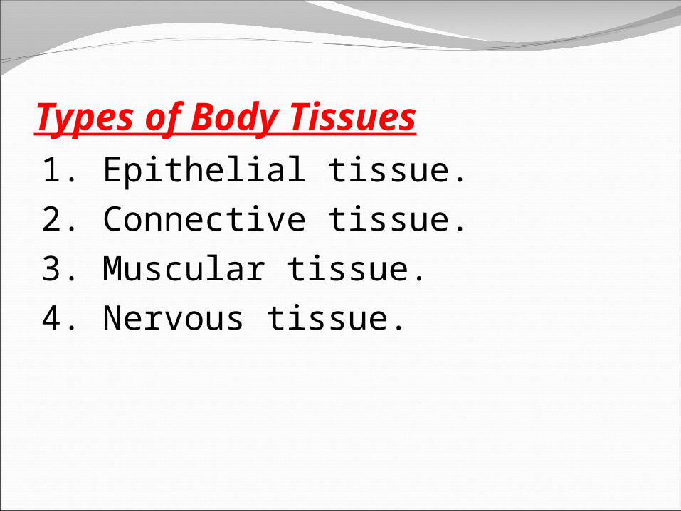
Types of Body Tissues1. Epithelial tissue.2. Connective tissue.3. Muscular tissue.4. Nervous tissue.
Epithelial tissueIt is present in two forms: 1-Epithelia or
epithelial membrane (contiguous cells covering external body surfaces and lines internal cavities).
2-Glands (from invaginated epithelial cells).Functions of epithelial tissue:A-Protection.B-Transcellular transport.C-Secretion of mucus, hormones and enzymes.D-Absorption.E-Detection of sensation as taste buds and retina .F-Selective permeability.

Characteristics of epithelium:1.The cells are tightly bound together by junction
complex.2.Is avascular (nourishment by diffusion from CT.)3.Rests on basal lamina (formed by epithelial cells)
that separate epithelial cells from underlying CT.
4.Has little intercellular space and little extracellular matrix.
5.Its cells exhibit a high turnover rate and constant cell renewal for a particular epithelium
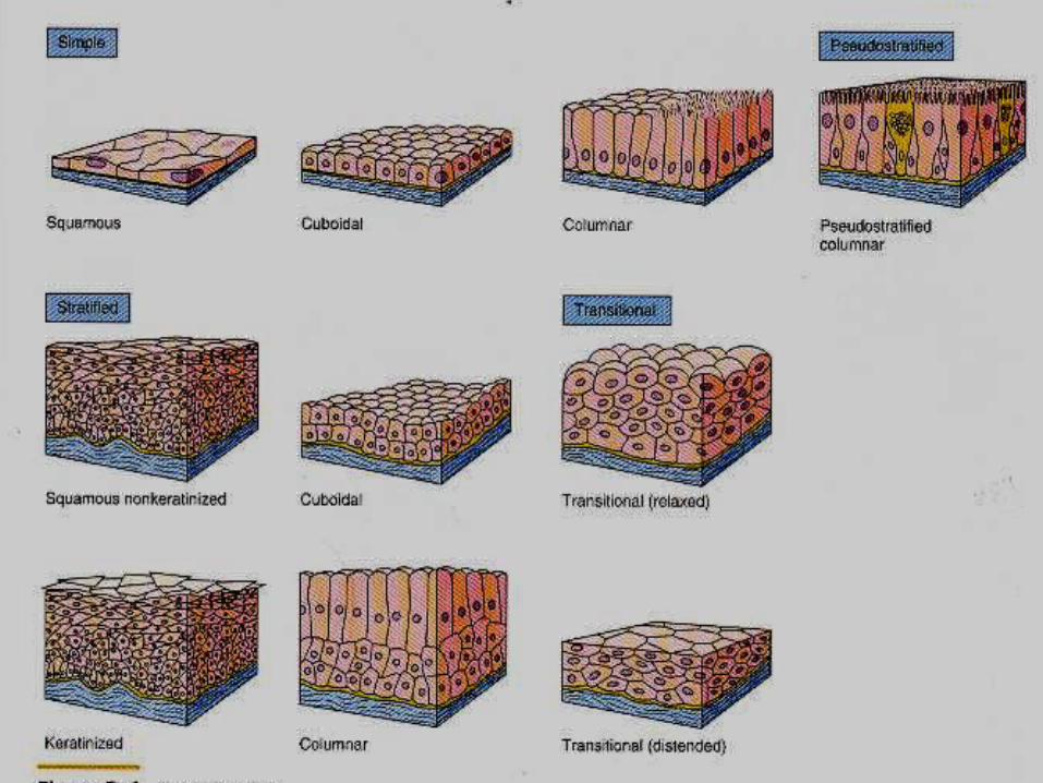
Classification of epithelial membranesAccording to the number of cell layers:
I. Simple (formed of one layer of cells)II. Stratified (formed of more than one layer of
cells)According to the morphology of the cells:
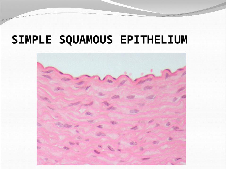
1-Simple squamous epithelium: thin low (flat) profile polygonal cells with central flat nuclei. (Ex. pulmonary alveoli, loop of Henle, endothelium of blood vessels, pleura and peritoneal cavities).

Simple squamous epithelium

2-Simple cuboidal epithelium: single layer of cuboidal polygonal cells with central round nuclei (Ex. ducts of many glands, covering of the ovary, follicular cells of thyroid follicles and some kidney
tubules).

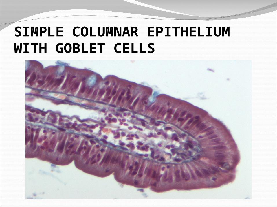
3-Simple columnar epithelium: single layer of columnar cells with ovoid nuclei located in basal half of cells (Ex. lining of stomach, gall bladder and large ducts of glands)
. Or may have cilia as in oviduct, uterus, small bronchi and ductuli efferentes.

* Simple columnar epith. may exhibit goblet cells
and microvilli as in intestine
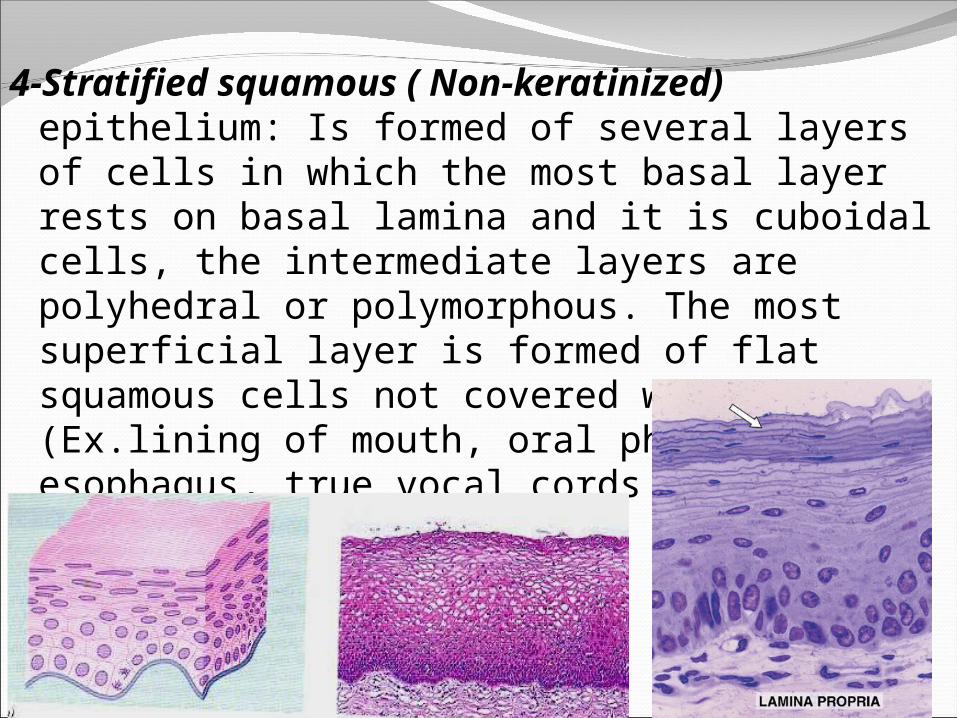
4-Stratified squamous ( Non-keratinized) epithelium: Is formed of several layers of cells in which the most basal layer rests on basal lamina and it is cuboidal cells, the intermediate layers are polyhedral or polymorphous. The most superficial layer is formed of flat squamous cells not covered with keratin (Ex.lining of mouth, oral pharynx, esophagus, true vocal cords and vagina).

5-Stratified squamous (Keratinized) epithelium: similar to non keratinized type but the superficial layer is covered with keratin (Ex. epidermis of skin specially in soles and palms)
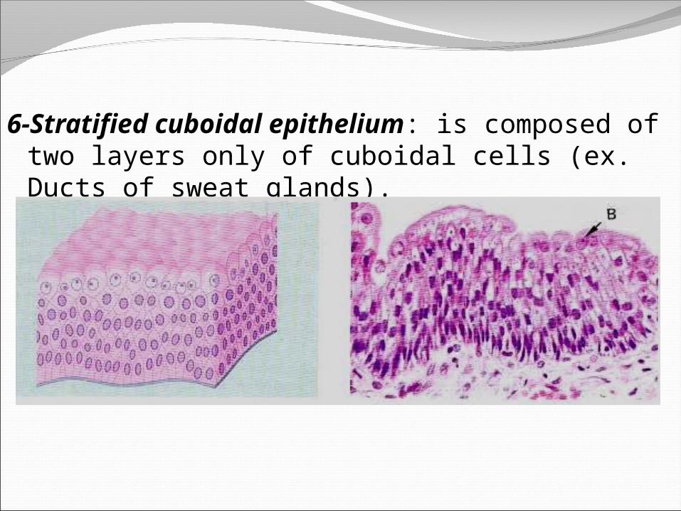
6-Stratified cuboidal epithelium: is composed of two layers only of cuboidal cells (ex. Ducts of sweat glands).

7-Stratified columnar epithelium: Is formed of more than one layer of cells with superficial columnar cells (ex. conjunctiva of the eye, large excretory ducts and regions of male urethra).
8-Transitional epithalium: is formed of many layers of cells (3-6), the basal layer is formed of low columnar or cuboidal cells, while the superficial layer is formed of large dome shaped binucleated cells (in empty bladder).
*In full bladder the dome-shaped cells become flattened and the epithelium becomes thinner.

9-Pseudostratified columnar epithelium9-Pseudostratified columnar epithelium
It appears to be stratified but it is composed of single layer of cells that all are resting on the basal lamina but only some of cells reach the surface of epithelium. Theses tall cells have narrow base and broad apical surface. Cells not extending to the surface have broad base and narrow apical end. The nuclei are located at different levels (ex. Male urethra, epididymis and large excretory ducts).Ciliated Pseudostratified epithelium with goblet cells (respiratory epith), has ciliated tall cells that reach the free border (ex. trachea, primary bronchia and nasal cavity)

Polarity and cell-surface specializationsApical domain. As microvilli, cilia, stereocilia
and flagella.Basolateral domain: a. Lateral plasma membrane specializations, as
junctional complexes and intercellular interdigitations.
b. Basal plasma membrane specializations, as enfolding and hemidesmosomes.
I.P. * Stratified epithelium dose NOT have goblet
cells, cilia or microvilli.* Keratin is found in str. sq. epith. only.
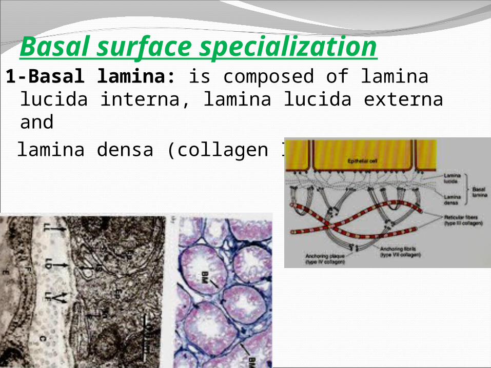
Basal surface specialization1-Basal lamina: is composed of lamina lucida
interna, lamina lucida externa and lamina densa (collagen IV) inbetween.

2- Basal enfolding: They increase the surface area of the basal membrane. The basal cytoplasm and mitochondria form finger like projections (striated appearance).They are involved in ion transport and are found in striated ducts of salivary glands.

3-Hemidesosomes: resemble half desmosomes and attached to the basal cell membrane on the basal lamina. The cytoplasmic aspect of cell membrane contains Attachment plaques in which keratin tonofilaments are inserted.

Glands1-Exocrine glands: secrete their
products via ducts2-Endocrine glands: are ductless,
their products pass into the blood or lymph.
* Each gland is formed of stroma and parenchyma. Stroma is, C.T. that support and invade the parenchyma, formed of capsule, septa and supporting background, and parenchyma is formed of secretory units and ducts.
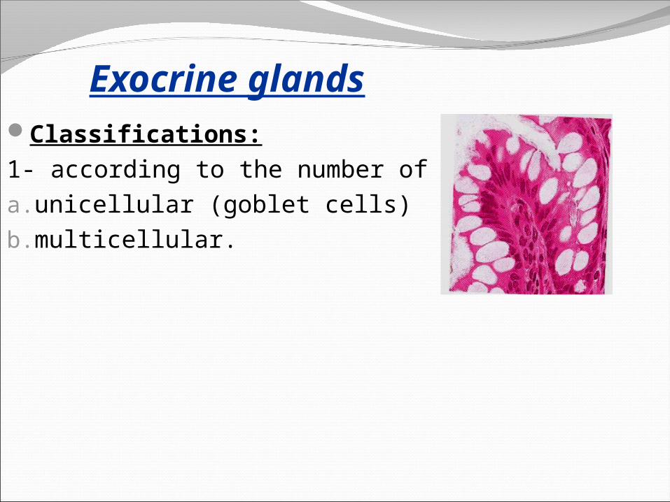
Exocrine glandsClassifications:1- according to the number of cells: a.unicellular (goblet cells) b.multicellular.
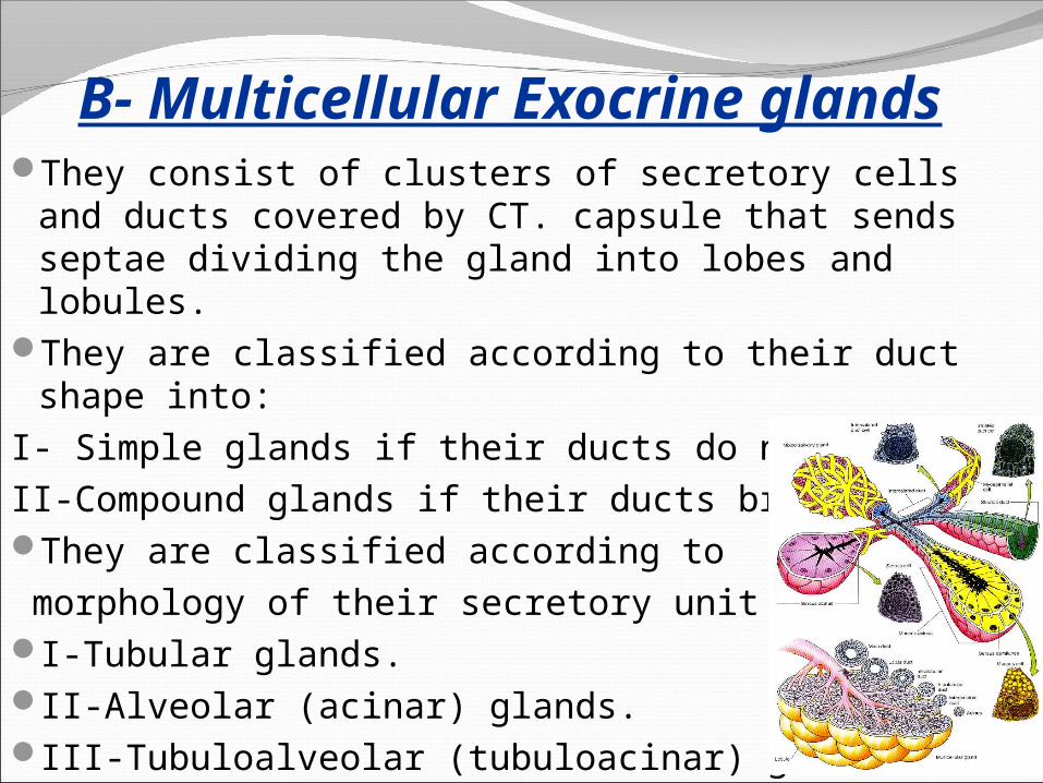
B- Multicellular Exocrine glandsThey consist of clusters of secretory cells and
ducts covered by CT. capsule that sends septae dividing the gland into lobes and lobules.
They are classified according to their duct shape into:
I- Simple glands if their ducts do not branch.II-Compound glands if their ducts branch.They are classified according to morphology of their secretory unit into:I-Tubular glands.II-Alveolar (acinar) glands.III-Tubuloalveolar (tubuloacinar) glands

2- According to their mechanism of release of secretion: a. holocrine b. merocrine c. apocrine.

3-According to the type of secretion: a. Serous glands. b. mucous glands
Serous cellsAre pyramidal, have single, round, basally located nuclei,
RER and Golgi complex numerous basal mitochondria and abundant apical secretory granules.
Mucous secreting cellsAre similar in shape to the serous cellsbut, their nuclei are flattened, few mitochondria, less RER, the apical part is rich in secretory carbohydrate granules.

Myoepithelial cellsThey share the basal lamina of acinar cells and small
ducts of many multicellular exocrine glands such as sweat and major salivary glands.
They are epithelial in origin, but have some characteristics of smooth muscle cells (contractility).
They have small nuclei and fibrillar cytoplasm radiating from the body wrapping
around the acini and small ducts.They help squeezing and expressing secretions from the acini and small ducts.
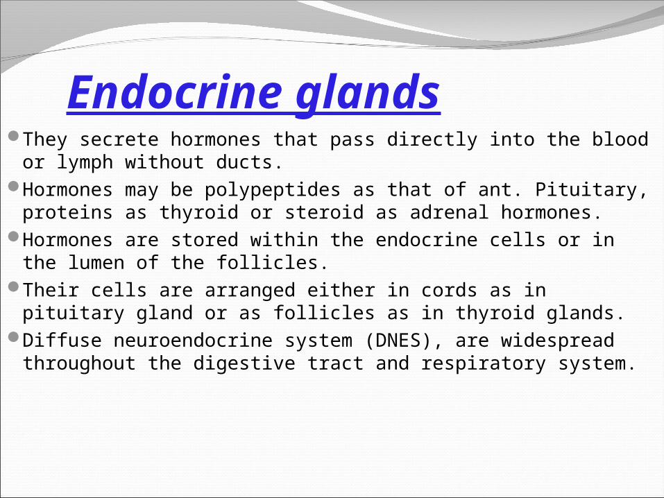
Endocrine glandsThey secrete hormones that pass directly into the blood
or lymph without ducts.Hormones may be polypeptides as that of ant. Pituitary,
proteins as thyroid or steroid as adrenal hormones.Hormones are stored within the endocrine cells or in the
lumen of the follicles.Their cells are arranged either in cords as in pituitary
gland or as follicles as in thyroid glands.Diffuse neuroendocrine system (DNES), are widespread
throughout the digestive tract and respiratory system.
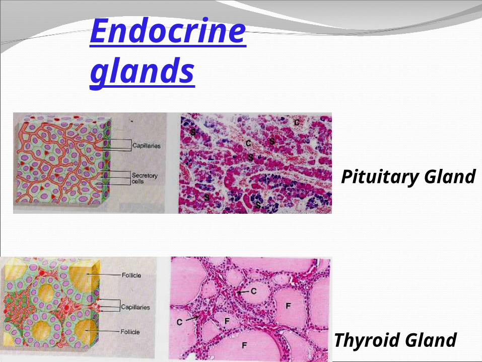
Pituitary Gland
Thyroid Gland
Endocrine glands

Pictures
SIMPLE SQUAMOUS EPITHELIUM

SIMPLE CUBOIDAL EPITHELIUM
SIMPLE COLUMNAR EPITHELIUMWITH GOBLET CELLS

PSEUDO-STRATUIFIED CILIATED COLUMNAREPITHELIUM WITH GOBLET CELLS

NON-KERATINIZED STRATIFIED SQUAMOUSEPITHELIUM

KERATINIZED STRATIFIED SQUAMOUSEPITHELIUM

TRANSITIONAL EPITHELIUM
Related Documents