Containing a Lassa fever epidemic in a resource-limited setting: outbreak description and lessons learned from Abakaliki, Nigeria (January–March 2012) Nnennaya A. Ajayi a , Chinedu G. Nwigwe b , Ben N. Azuogu c , Benson N. Onyire d , Elizabeth U. Nwonwu c , Lawrence U. Ogbonnaya c , Francis I. Onwe e , Tobin Ekaete f , Stephan Gu ¨ nther g , Kingsley N. Ukwaja a, * a Department of Internal Medicine, Federal Teaching Hospital Abakaliki, Ebonyi State, Nigeria b Department of Surgery, Federal Teaching Hospital Abakaliki, Ebonyi State, Nigeria c Department of Community Medicine, Federal Teaching Hospital Abakaliki, Ebonyi State, Nigeria d Department of Pediatrics, Federal Teaching Hospital Abakaliki, Ebonyi State, Nigeria e Epidemiology Unit, Ministry of Health, Ebonyi State, Nigeria f Institute of Lassa Fever Research and Control, Irrua Specialist Teaching Hospital, Edo State, Nigeria g Department of Virology, Bernhard-Nocht-Institute for Tropical Medicine, Hamburg, Germany 1. Introduction Lassa fever (LF) is caused by a single-stranded RNA virus of the family Arenaviridae. It is endemic in Guinea, Sierra Leone, Liberia, and parts of Nigeria, and also in other countries in the West African sub-region. 1 It is also a zoonotic disease, whose animal reservoir is a rat of the genus Mastomys (the ‘multimammate’ rat). People become infected through direct exposure to the excreta of infected rats, or by transmission from person to person via body fluids. 1 Lassa infection is asymptomatic in about 80% of cases, but causes an acute illness in the rest. Fever and general weakness are followed by headache, chest pain, vomiting, diarrhea, cough, pleural effusion, bleeding from orifices, and in the late stages sometimes disorientation and coma. 2 Deafness occurs in 25% of cases. In fatal cases, it kills rapidly – usually within 14 days. The overall case fatality rate is around 1%, rising to 15% of hospitalized cases. 1 Worldwide, an estimated two million persons are affected every year, resulting in 5000 to 10 000 deaths. 3 It is estimated that 300 000 to 500 000 cases of LF occur annually across West Africa. 4 LF is endemic in West Africa and high rates of seroprevalence have been reported in Nigeria, Sierra Leone, Guinea, and Liberia. 5–8 In Nigeria, the seroprevalence of LF is about 21%. 8 It is an epidemic-prone disease dreaded by health workers and the community. The threat of LF as an epidemic- prone disease in Nigeria is indicated by an alert threshold of a International Journal of Infectious Diseases 17 (2013) e1011–e1016 A R T I C L E I N F O Article history: Received 6 February 2013 Received in revised form 28 May 2013 Accepted 30 May 2013 Corresponding Editor: Eskild Petersen, Aarhus, Denmark Keywords: Lassa fever Outbreak report Case report Epidemiology Infection control Nigeria S U M M A R Y Objectives: Despite the epidemic nature of Lassa fever (LF), details of outbreaks and response strategies have not been well documented in resource-poor settings. We describe the course of a LF outbreak in Ebonyi State, Nigeria, during January to March 2012. Methods: We analyzed clinical, epidemiological, and laboratory data from surveillance records and hospital statistics during the outbreak. Fisher’s exact tests were used to compare proportions and t-tests to compare differences in means. Results: The outbreak response consisted of effective coordination, laboratory testing, active surveillance, community mobilization, contact and suspected case evaluation, and case management. Twenty LF cases (10 confirmed and 10 suspected) were recorded during the outbreak. Nosocomial transmission to six health workers occurred through the index case. Only 1/110 contacts had an asymptomatic infection. Overall, there was high case fatality rate among all cases (6/20; 30%). Patients who received ribavirin were less likely to die than those who did not (p = 0.003). The mean delay to presentation for patients who died was 11 3.5 days, while for those who survived was 6 2.6 days (p < 0.001). Conclusions: The response strategies contained the epidemic. Challenges to control efforts included poor local laboratory capacity, inadequate/poor quality of protective materials, fear among health workers, and inadequate emergency preparedness. ß 2013 International Society for Infectious Diseases. Published by Elsevier Ltd. All rights reserved. * Corresponding author. Tel.: +234 08036243196. E-mail addresses: [email protected], [email protected] (K.N. Ukwaja). Contents lists available at SciVerse ScienceDirect International Journal of Infectious Diseases jou r nal h o mep ag e: w ww .elsevier .co m /loc ate/ijid 1201-9712/$36.00 – see front matter ß 2013 International Society for Infectious Diseases. Published by Elsevier Ltd. All rights reserved. http://dx.doi.org/10.1016/j.ijid.2013.05.015

Welcome message from author
This document is posted to help you gain knowledge. Please leave a comment to let me know what you think about it! Share it to your friends and learn new things together.
Transcript
International Journal of Infectious Diseases 17 (2013) e1011–e1016
Containing a Lassa fever epidemic in a resource-limited setting:outbreak description and lessons learned from Abakaliki, Nigeria(January–March 2012)
Nnennaya A. Ajayi a, Chinedu G. Nwigwe b, Ben N. Azuogu c, Benson N. Onyire d,Elizabeth U. Nwonwu c, Lawrence U. Ogbonnaya c, Francis I. Onwe e, Tobin Ekaete f,Stephan Gunther g, Kingsley N. Ukwaja a,*a Department of Internal Medicine, Federal Teaching Hospital Abakaliki, Ebonyi State, Nigeriab Department of Surgery, Federal Teaching Hospital Abakaliki, Ebonyi State, Nigeriac Department of Community Medicine, Federal Teaching Hospital Abakaliki, Ebonyi State, Nigeriad Department of Pediatrics, Federal Teaching Hospital Abakaliki, Ebonyi State, Nigeriae Epidemiology Unit, Ministry of Health, Ebonyi State, Nigeriaf Institute of Lassa Fever Research and Control, Irrua Specialist Teaching Hospital, Edo State, Nigeriag Department of Virology, Bernhard-Nocht-Institute for Tropical Medicine, Hamburg, Germany
A R T I C L E I N F O
Article history:
Received 6 February 2013
Received in revised form 28 May 2013
Accepted 30 May 2013
Corresponding Editor: Eskild Petersen,
Aarhus, Denmark
Keywords:
Lassa fever
Outbreak report
Case report
Epidemiology
Infection control
Nigeria
S U M M A R Y
Objectives: Despite the epidemic nature of Lassa fever (LF), details of outbreaks and response strategies
have not been well documented in resource-poor settings. We describe the course of a LF outbreak in
Ebonyi State, Nigeria, during January to March 2012.
Methods: We analyzed clinical, epidemiological, and laboratory data from surveillance records and
hospital statistics during the outbreak. Fisher’s exact tests were used to compare proportions and t-tests
to compare differences in means.
Results: The outbreak response consisted of effective coordination, laboratory testing, active surveillance,
community mobilization, contact and suspected case evaluation, and case management. Twenty LF cases
(10 confirmed and 10 suspected) were recorded during the outbreak. Nosocomial transmission to six health
workers occurred through the index case. Only 1/110 contacts had an asymptomatic infection. Overall,
there was high case fatality rate among all cases (6/20; 30%). Patients who received ribavirin were less likely
to die than those who did not (p = 0.003). The mean delay to presentation for patients who died was 11 � 3.5
days, while for those who survived was 6 � 2.6 days (p < 0.001).
Conclusions: The response strategies contained the epidemic. Challenges to control efforts included poor
local laboratory capacity, inadequate/poor quality of protective materials, fear among health workers,
and inadequate emergency preparedness.
� 2013 International Society for Infectious Diseases. Published by Elsevier Ltd. All rights reserved.
Contents lists available at SciVerse ScienceDirect
International Journal of Infectious Diseases
jou r nal h o mep ag e: w ww .e lsev ier . co m / loc ate / i j id
1. Introduction
Lassa fever (LF) is caused by a single-stranded RNA virus of thefamily Arenaviridae. It is endemic in Guinea, Sierra Leone, Liberia,and parts of Nigeria, and also in other countries in the West Africansub-region.1 It is also a zoonotic disease, whose animal reservoir isa rat of the genus Mastomys (the ‘multimammate’ rat). Peoplebecome infected through direct exposure to the excreta of infectedrats, or by transmission from person to person via body fluids.1
Lassa infection is asymptomatic in about 80% of cases, but causesan acute illness in the rest. Fever and general weakness are
* Corresponding author. Tel.: +234 08036243196.
E-mail addresses: [email protected], [email protected]
(K.N. Ukwaja).
1201-9712/$36.00 – see front matter � 2013 International Society for Infectious Disea
http://dx.doi.org/10.1016/j.ijid.2013.05.015
followed by headache, chest pain, vomiting, diarrhea, cough,pleural effusion, bleeding from orifices, and in the late stagessometimes disorientation and coma.2 Deafness occurs in 25% ofcases. In fatal cases, it kills rapidly – usually within 14 days. Theoverall case fatality rate is around 1%, rising to 15% of hospitalizedcases.1
Worldwide, an estimated two million persons are affectedevery year, resulting in 5000 to 10 000 deaths.3 It is estimatedthat 300 000 to 500 000 cases of LF occur annually across WestAfrica.4 LF is endemic in West Africa and high rates ofseroprevalence have been reported in Nigeria, Sierra Leone,Guinea, and Liberia.5–8 In Nigeria, the seroprevalence of LF isabout 21%.8 It is an epidemic-prone disease dreaded by healthworkers and the community. The threat of LF as an epidemic-prone disease in Nigeria is indicated by an alert threshold of a
ses. Published by Elsevier Ltd. All rights reserved.

N.A. Ajayi et al. / International Journal of Infectious Diseases 17 (2013) e1011–e1016e1012
single suspected case and an epidemic threshold of a singleconfirmed case, by the Nigeria Federal Ministry of Health.9
Since the identification of the virus in 1969, some outbreaks ofthe disease have been reported in various parts of Nigeriaincluding Ekpoma, Aboh-Mbaise, Onitsha, Jos, and Lafia,7,10,11
and more recently in Ebonyi, Taraba, Nasarawa, Yobe, Rivers,Ondo, and Edo states.12–14
Despite the epidemic nature of LF in Nigeria and other Africancountries, the details of outbreaks and subsequent responses tocontain it have not been well documented in these places, and it isdifficult to learn from these experiences to improve themanagement of future outbreaks. Available Nigerian reportshave focused mainly on a nosocomial outbreak that occurredalmost two decades ago,7 or more recently on laboratorydiagnosis of blood samples of suspected cases sent to a nationalreference laboratory.12,13 Thus limited information exists regard-ing the containment and management of cases and suspectsduring outbreak situations.
We report on the investigation and management of the LFoutbreak that occurred from January 1 to March 25, 2012 inEbonyi State, Nigeria. Our specific objectives were: to describe thesocio-demographic characteristics, clinical characteristics, andthe management outcome of the cases seen, to describe theoutbreak response, and to attempt to draw lessons for futureoutbreaks.
2. Methods
2.1. Study area and population
Ebonyi State is located in the southeastern geopoliticalzone of Nigeria. It has 13 local government areas and anestimated population of 2.5 million people.15 About 80% of thepopulation are subsistence farmers and a similar proportionresides in rural areas.15 The patients studied were managed atthe Federal Teaching Hospital, Abakaliki (FETHA), Ebonyi State,in southeastern Nigeria. The hospital services the urban andsemi-urban agrarian communities in Ebonyi State. It is the onlytertiary referral center serving an estimated three millionpopulation in the southeastern region of Nigeria and receivesreferrals from neighboring states including Cross River andAkwa Ibom.
Table 1Definitions used in the study
Variable Definition
Lassa fever diagnosisa
Alert case Unexplained fever for 5 days or more not re
community or hospital
Action to be taken: invite the Lassa fever te
Suspected casea Illness with gradual onset with one or more
diarrhea, myalgia, chest pain, hearing loss, b
suspected case of Lassa fever. And has recei
Action to be taken: declare ‘suspected’ case
Laboratory confirmed case A suspected case that is laboratory confirme
Action to be taken: for Lassa fever treatmen
Epidemiologically confirmed case A suspected case that is epidemiologically l
confirmed case, with signs and symptoms c
unavailable or tested negative)
Action to be taken: for Lassa fever treatmen
Final outcome
Dead Confirmed/suspected cases who died from L
Survived Confirmed/suspected cases who recovered f
Contacta
Contact Someone who slept in the same household a
touched his/her linens or body fluids
a Definitions based on: Centers for Disease Control and Prevention and World Health O
the African region. Atlanta, GA: CDC; 2010, p. 1–416.16
2.2. Data sources and methods
We retrospectively analyzed surveillance data, hospital records,and the outbreak response activities. Additional information aboutthe affected LF patients was collected from the individual clinicalrecords. We collected data on the onset of the outbreak and themanagement of individual patients. For suspected and confirmedLF cases, we collected data on their socio-demographic and clinicalcharacteristics (age, sex, residence, occupation, most likely sourceof exposure, clinical presentation, and laboratory data), as well asthe final outcome of their management. Additional informationrelated to timing of events was also collected.
2.3. Diagnosis
All patients/contacts with features suggestive of LF (alert cases)were evaluated clinically and were classified using the WorldHealth Organization (WHO)/Integrated Disease Surveillance andResponse (IDSR) guidelines as suspected cases16 (Table 1).Confirmation was based on a positive test using Lassa virus-specific reverse-transcriptase PCR (RT-PCR), or epidemiologically(Table 1).16 Laboratory confirmation was performed at theInstitute of Lassa Fever Research and Control, Irrua SpecialistTeaching Hospital, Edo State, Nigeria. The Lassa virus-specificRT-PCR was performed as described previously.17
2.4. Data analysis
The data analysis was conducted using Epi Info. Continuousvariables were summarized using means � standard deviations andmedians, and categorical variables were summarized as proportions.Categorical group comparisons were made using Fisher’s exact tests,and continuous variables were compared using the t-tests fordifferences in means.
3. Results
3.1. Outbreak detection
On January 9, 2012, the FETHA notified the Ebonyi State andFederal Ministry of Health (MoH) of a laboratory confirmed LFcase. The individual was a 25-year-old university graduate
sponding to antimalarial/antibiotics (for malaria, typhoid fever) from the
chnical committee to review the case
of the following: malaise, fever, headache, sore throat, cough, nausea, vomiting,
leeding tendency, and a history of contact with excreta of rodents or with a
ved standard treatment for fever (above) without response
, send for laboratory confirmation, and may start Lassa fever treatment
d (positive IgM antibody, PCR, or virus isolation)
t
inked to a laboratory confirmed case (for example, contacts of a laboratory
onsistent with Lassa fever, whose confirmatory laboratory results were either
t
assa fever and/or its complications
rom Lassa fever following treatment
s the case within 1 month, or had direct contact with the case (dead or alive), or
rganization. Technical guidelines for integrated disease surveillance and response in
The Inde x case
1 Laboratory con firmed
3 La boratory con firmed
10 Suspected cases
Total of 20 cases
10 S uspected
10 Confirmed
Community cases
24 Ale rt cases
6 No socomial cases
5 La boratory con firmed
1 Epidemiologicall y con firmed
13 Suspectedcases
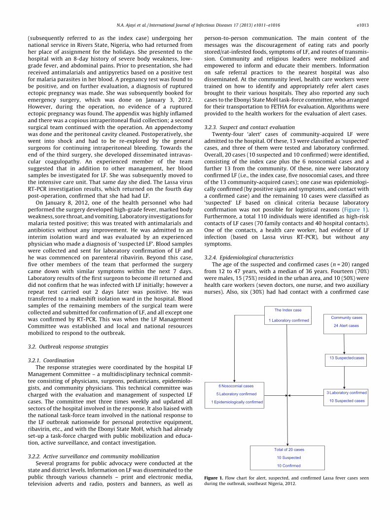
Figure 1. Flow chart for alert, suspected, and confirmed Lassa fever cases seen
during the outbreak, southeast Nigeria, 2012.
N.A. Ajayi et al. / International Journal of Infectious Diseases 17 (2013) e1011–e1016 e1013
(subsequently referred to as the index case) undergoing hernational service in Rivers State, Nigeria, who had returned fromher place of assignment for the holidays. She presented to thehospital with an 8-day history of severe body weakness, low-grade fever, and abdominal pains. Prior to presentation, she hadreceived antimalarials and antipyretics based on a positive testfor malaria parasites in her blood. A pregnancy test was found tobe positive, and on further evaluation, a diagnosis of rupturedectopic pregnancy was made. She was subsequently booked foremergency surgery, which was done on January 3, 2012.However, during the operation, no evidence of a rupturedectopic pregnancy was found. The appendix was highly inflamedand there was a copious intraperitoneal fluid collection; a secondsurgical team continued with the operation. An appendectomywas done and the peritoneal cavity cleaned. Postoperatively, shewent into shock and had to be re-explored by the generalsurgeons for continuing intraperitoneal bleeding. Towards theend of the third surgery, she developed disseminated intravas-cular coagulopathy. An experienced member of the teamsuggested that in addition to other management, her bloodsamples be investigated for LF. She was subsequently moved tothe intensive care unit. That same day she died. The Lassa virusRT-PCR investigation results, which returned on the fourth daypost-operation, confirmed that she had had LF.
On January 8, 2012, one of the health personnel who hadperformed the surgery developed high-grade fever, marked bodyweakness, sore throat, and vomiting. Laboratory investigations formalaria tested positive; this was treated with antimalarials andantibiotics without any improvement. He was admitted to aninterim isolation ward and was evaluated by an experiencedphysician who made a diagnosis of ‘suspected LF’. Blood sampleswere collected and sent for laboratory confirmation of LF andhe was commenced on parenteral ribavirin. Beyond this case,five other members of the team that performed the surgerycame down with similar symptoms within the next 7 days.Laboratory results of the first surgeon to become ill returned anddid not confirm that he was infected with LF initially; however arepeat test carried out 2 days later was positive. He wastransferred to a makeshift isolation ward in the hospital. Bloodsamples of the remaining members of the surgical team werecollected and submitted for confirmation of LF, and all except onewas confirmed by RT-PCR. This was when the LF ManagementCommittee was established and local and national resourcesmobilized to respond to the outbreak.
3.2. Outbreak response strategies
3.2.1. Coordination
The response strategies were coordinated by the hospital LFManagement Committee – a multidisciplinary technical commit-tee consisting of physicians, surgeons, pediatricians, epidemiolo-gists, and community physicians. This technical committee wascharged with the evaluation and management of suspected LFcases. The committee met three times weekly and updated allsectors of the hospital involved in the response. It also liaised withthe national task-force team involved in the national response tothe LF outbreak nationwide for personal protective equipment,ribavirin, etc., and with the Ebonyi State MoH, which had alreadyset-up a task-force charged with public mobilization and educa-tion, active surveillance, and contact investigation.
3.2.2. Active surveillance and community mobilization
Several programs for public advocacy were conducted at thestate and district levels. Information on LF was disseminated to thepublic through various channels – print and electronic media,television adverts and radio, posters and banners, as well as
person-to-person communication. The main content of themessages was the discouragement of eating rats and poorlystored/rat-infested foods, symptoms of LF, and routes of transmis-sion. Community and religious leaders were mobilized andempowered to inform and educate their members. Informationon safe referral practices to the nearest hospital was alsodisseminated. At the community level, health care workers weretrained on how to identify and appropriately refer alert casesbrought to their various hospitals. They also reported any suchcases to the Ebonyi State MoH task-force committee, who arrangedfor their transportation to FETHA for evaluation. Algorithms wereprovided to the health workers for the evaluation of alert cases.
3.2.3. Suspect and contact evaluation
Twenty-four ‘alert’ cases of community-acquired LF wereadmitted to the hospital. Of these, 13 were classified as ‘suspected’cases, and three of them were tested and laboratory confirmed.Overall, 20 cases (10 suspected and 10 confirmed) were identified,consisting of the index case plus the 6 nosocomial cases and afurther 13 from the community. Of these, nine were laboratoryconfirmed LF (i.e., the index case, five nosocomial cases, and threeof the 13 community-acquired cases); one case was epidemiologi-cally confirmed (by positive signs and symptoms, and contact witha confirmed case) and the remaining 10 cases were classified as‘suspected’ LF based on clinical criteria because laboratoryconfirmation was not possible for logistical reasons (Figure 1).Furthermore, a total 110 individuals were identified as high-riskcontacts of LF cases (70 family contacts and 40 hospital contacts).One of the contacts, a health care worker, had evidence of LFinfection (based on Lassa virus RT-PCR), but without anysymptoms.
3.2.4. Epidemiological characteristics
The age of the suspected and confirmed cases (n = 20) rangedfrom 12 to 47 years, with a median of 36 years. Fourteen (70%)were males, 15 (75%) resided in the urban area, and 10 (50%) werehealth care workers (seven doctors, one nurse, and two auxiliarynurses). Also, six (30%) had had contact with a confirmed case

Table 2Demographic and clinical characteristics of Lassa fever cases, southeast Nigeria,
2012
Variable Frequency (N = 20) Percentage (%)
Gender
Male 14 70
Female 6 30
Age group, years
10–19 2 10
20–29 2 10
30–39 12 60
�40 4 20
Residence
Urban 15 75
Rural 5 25
Occupation
Health care worker 10 50
Student 3 15
Cleric 2 10
Laborer 2 10
Youth corps 1 5
Other (farmer, artisan, etc.) 2 10
Case type
Confirmed 10 50
Suspected 10 50
Source of infection
Exposure to confirmed case 6 30
Unknown 14 70
Table 3Clinical presentation of Lassa fever cases, southeast Nigeria, 2012
Clinical presentation Frequency (N = 20) Percentage (%)
Fever 20 100
Sore throat 14 70
Abdominal pain 17 85
Headache 7 35
Vomiting 10 50
Bloody vomiting 3 15
Bloody stool 3 15
Body pains 5 25
Body weakness 5 25
Prolonged menstruation 2 10
Spontaneous abortion 2 10
N.A. Ajayi et al. / International Journal of Infectious Diseases 17 (2013) e1011–e1016e1014
(Table 2). The mean incubation period was 10 (range 5–18) days.The initial case occurred in the first epidemiological week of 2012in the hospital (Figure 2). Epidemiological investigations werecommenced in the first epidemiological week of 2012 with theevaluation of the index case. In the following weeks, there was aninitial rapid increase in cases (nosocomial cases) reaching a plateauin the second week of 2012, and subsequently a gradual decrease.The number of cases reported declined from the sixth epidemio-logical week to the thirteenth epidemiological week of 2012.Laboratory confirmation was made in the first epidemiologicalweek of 2012.
3.2.5. Clinical characteristics
The most common symptom in this outbreak was fever, whichoccurred in 20 (100%) of the cases. This was followed by abdominalpain (17; 85%), sore throat (14; 70%), and vomiting (10; 50%)(Table 3). The admission temperature of the cases ranged from38 to 40 8C with a median of 39 8C. Hemorrhagic symptoms such
0
1
2
3
4
5
6
1 2 3 4 5 6 7 8 9 10 11 12 13
Num
ber o
f cas
es s
een
Epidemiologic al w eeks 2012
Suspec ted Confirme d
Figure 2. Suspected and confirmed Lassa fever cases by week of presentation during
the outbreak, southeast Nigeria, 2012.
as bloody vomiting and bloody stools were less common, occurringin three (15%) of the cases. Also, two female cases presented withfever and prolonged menstruation (more than 10 days, unlike theirusual 3–4 days of menstrual flow). Furthermore, two pregnantwomen who presented with fever with spontaneous abortion andwho died within 2 h of presentation were later confirmed to havebeen infected with LF. At presentation, only a few of the patientshad a low hemoglobin level (15%); 25% had low platelets, 25% hadlymphocytosis, 35% had lymphopenia, 30% had neutropenia, and15% had leukopenia (Table 4). Liver transaminases were raised in45% of cases for alanine aminotransferase (ALT), 40% of cases foraspartate aminotransferase (AST), and 35% had both enzymeselevated. Furthermore, 30% had raised creatinine and 35% hadraised urea. All the patients (100%) had proteinuria.
3.2.6. Case management
No isolation ward was available at FETHA at the onset of theoutbreak. However, parts of the private wards (consisting of 12private rooms) were not in use and were converted to an interimisolation ward exclusively for suspected and confirmed LF patients.All except the index case and three other cases who died within afew hours of presentation were treated with ribavirin, and allreceived supportive therapy (Table 5). Patients who receivedribavirin were less likely to die than those who did not (Fisher’sexact test; p = 0.003). The mean delay to presentation for patientswho died was 11 � 3.5 days, while for those who survived was6 � 2.6 days (t-statistic 4.3; p < 0.001). The mean duration oftreatment before the patients became afebrile was 8.2 � 3.8 days.Overall, there was high case fatality rate among all cases (6/20; 30%)and among confirmed cases (4/10; 40%). Sudden progression to
Table 4Interpretations from basic investigations on Lassa fever cases, southeast Nigeria,
2012
Parameter Frequency (N = 20) Percentage (%)
Low hemoglobin 3 15
Low platelets 5 25
Leukocytosis 2 10
Leukopenia 3 15
Neutrophilia 7 35
Neutropenia 6 30
Lymphocytosis 5 25
Lymphopenia 7 35
Raised creatinine 6 30
Raised urea 7 35
Raised urea and creatinine 4 20
Raised ALT 9 45
Raised AST 8 40
Raised ALT and AST 7 35
Raised total bilirubin 6 30
Proteinuria 20 100
ALT, alanine aminotransferase; AST, aspartate aminotransferase.
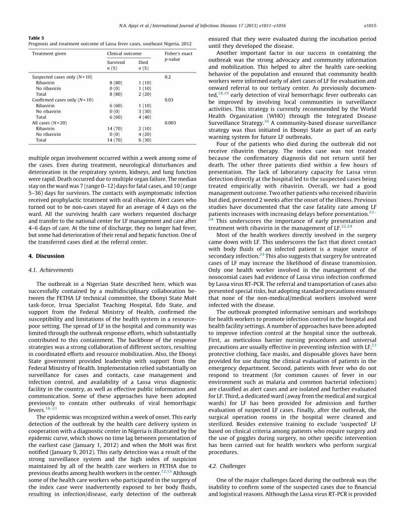
Table 5Prognosis and treatment outcome of Lassa fever cases, southeast Nigeria, 2012
Treatment given Clinical outcome Fisher’s exact
p-valueSurvived
n (%)
Died
n (%)
Suspected cases only (N = 10) 0.2
Ribavirin 8 (80) 1 (10)
No ribavirin 0 (0) 1 (10)
Total 8 (80) 2 (20)
Confirmed cases only (N = 10) 0.03
Ribavirin 6 (60) 1 (10)
No ribavirin 0 (0) 3 (30)
Total 6 (60) 4 (40)
All cases (N = 20) 0.003
Ribavirin 14 (70) 2 (10)
No ribavirin 0 (0) 4 (20)
Total 14 (70) 6 (30)
N.A. Ajayi et al. / International Journal of Infectious Diseases 17 (2013) e1011–e1016 e1015
multiple organ involvement occurred within a week among some ofthe cases. Even during treatment, neurological disturbances anddeterioration in the respiratory system, kidneys, and lung functionwere rapid. Death occurred due to multiple organ failure. The medianstay on the ward was 7 (range 0–12) days for fatal cases, and 10 (range5–36) days for survivors. The contacts with asymptomatic infectionreceived prophylactic treatment with oral ribavirin. Alert cases whoturned out to be non-cases stayed for an average of 4 days on theward. All the surviving health care workers requested dischargeand transfer to the national center for LF management and care after4–6 days of care. At the time of discharge, they no longer had fever,but some had deterioration of their renal and hepatic function. One ofthe transferred cases died at the referral center.
4. Discussion
4.1. Achievements
The outbreak in a Nigerian State described here, which wassuccessfully contained by a multidisciplinary collaboration be-tween the FETHA LF technical committee, the Ebonyi State MoHtask-force, Irrua Specialist Teaching Hospital, Edo State, andsupport from the Federal Ministry of Health, confirmed thesusceptibility and limitations of the health system in a resource-poor setting. The spread of LF in the hospital and community waslimited through the outbreak response efforts, which substantiallycontributed to this containment. The backbone of the responsestrategies was a strong collaboration of different sectors, resultingin coordinated efforts and resource mobilization. Also, the EbonyiState government provided leadership with support from theFederal Ministry of Health. Implementation relied substantially onsurveillance for cases and contacts, case management andinfection control, and availability of a Lassa virus diagnosticfacility in the country, as well as effective public information andcommunication. Some of these approaches have been adoptedpreviously to contain other outbreaks of viral hemorrhagicfevers.18–21
The epidemic was recognized within a week of onset. This earlydetection of the outbreak by the health care delivery system incooperation with a diagnostic center in Nigeria is illustrated by theepidemic curve, which shows no time lag between presentation ofthe earliest case (January 1, 2012) and when the MoH was firstnotified (January 9, 2012). This early detection was a result of thestrong surveillance system and the high index of suspicionmaintained by all of the health care workers in FETHA due toprevious deaths among health workers in the center.12,13 Althoughsome of the health care workers who participated in the surgery ofthe index case were inadvertently exposed to her body fluids,resulting in infection/disease, early detection of the outbreak
ensured that they were evaluated during the incubation perioduntil they developed the disease.
Another important factor in our success in containing theoutbreak was the strong advocacy and community informationand mobilization. This helped to alter the health care-seekingbehavior of the population and ensured that community healthworkers were informed early of alert cases of LF for evaluation andonward referral to our tertiary center. As previously documen-ted,18,19 early detection of viral hemorrhagic fever outbreaks canbe improved by involving local communities in surveillanceactivities. This strategy is currently recommended by the WorldHealth Organization (WHO) through the Integrated DiseaseSurveillance Strategy.16 A community-based disease surveillancestrategy was thus initiated in Ebonyi State as part of an earlywarning system for future LF outbreaks.
Four of the patients who died during the outbreak did notreceive ribavirin therapy. The index case was not treatedbecause the confirmatory diagnosis did not return until herdeath. The other three patients died within a few hours ofpresentation. The lack of laboratory capacity for Lassa virusdetection directly at the hospital led to the suspected cases beingtreated empirically with ribavirin. Overall, we had a goodmanagement outcome. Two other patients who received ribavirinbut died, presented 2 weeks after the onset of the illness. Previousstudies have documented that the case fatality rate among LFpatients increases with increasing delays before presentation.22–
24 This underscores the importance of early presentation andtreatment with ribavirin in the management of LF.22,24
Most of the health workers directly involved in the surgerycame down with LF. This underscores the fact that direct contactwith body fluids of an infected patient is a major source ofsecondary infection.24 This also suggests that surgery for untreatedcases of LF may increase the likelihood of disease transmission.Only one health worker involved in the management of thenosocomial cases had evidence of Lassa virus infection confirmedby Lassa virus RT-PCR. The referral and transportation of cases alsopresented special risks, but adopting standard precautions ensuredthat none of the non-medical/medical workers involved wereinfected with the disease.
The outbreak prompted informative seminars and workshopsfor health workers to promote infection control in the hospital andhealth facility settings. A number of approaches have been adoptedto improve infection control at the hospital since the outbreak.First, as meticulous barrier nursing procedures and universalprecautions are usually effective in preventing infection with LF,23
protective clothing, face masks, and disposable gloves have beenprovided for use during the clinical evaluation of patients in theemergency department. Second, patients with fever who do notrespond to treatment (for common causes of fever in ourenvironment such as malaria and common bacterial infections)are classified as alert cases and are isolated and further evaluatedfor LF. Third, a dedicated ward (away from the medical and surgicalwards) for LF has been provided for admission and furtherevaluation of suspected LF cases. Finally, after the outbreak, thesurgical operation rooms in the hospital were cleaned andsterilized. Besides extensive training to exclude ‘suspected’ LFbased on clinical criteria among patients who require surgery andthe use of goggles during surgery, no other specific interventionhas been carried out for health workers who perform surgicalprocedures.
4.2. Challenges
One of the major challenges faced during the outbreak was theinability to confirm some of the suspected cases due to financialand logistical reasons. Although the Lassa virus RT-PCR is provided
N.A. Ajayi et al. / International Journal of Infectious Diseases 17 (2013) e1011–e1016e1016
for free, the national reference laboratory for confirmatory testingwas located approximately 300 km from the epicenter of theoutbreak. Confirmatory diagnosis and screening of contactsrequired collection of blood samples, storage, and transportationto the reference laboratory. We had difficulties during theseprocesses. There is a need to site a diagnostic laboratory in all of thestates where LF outbreaks have occurred. This will improve earlydiagnosis and confirmation of cases and ensure appropriatetreatment.
Secondly, inadequate quality and/or unavailability of personalprotective materials, especially at the onset of the outbreak, weremajor problems. This probably contributed to the nosocomialtransmission of the Lassa virus to one of the health careprofessionals involved in the management of the LF patientsduring the outbreak. There is need to ensure the availability ofquality personal protective materials, especially masks, goggles,and gloves, and for these to be worn during outbreak situations.
Thirdly, inadequate knowledge of the disease among thecommunity and health workers resulted in fear of contractingthe disease. This led to a high rate of apprehension and anxietyamong the clinical staff involved in the management of the cases. Ittook several education seminars and clear evidence that the barriernursing currently being used for the management of cases iseffective, for the confidence of some of the clinical staff to return.
Fourthly, another major challenge was the inability to trace theorigin. Investigation of contacts of the cases did not yield evidenceof transmission of LF between family members and close relatives.It is likely that the cases were infected through the consumption ofinfected foods.25 Education of the community on safe handling offoods and to stop eating rats may play a role in curtailing futureoutbreaks.
Finally, the lack of epidemic preparedness for LF affected theearly containment of the outbreak. There is a need for a dedicatedward for suspected LF cases in the State. Also, for post-outbreakcontrol interventions, there is a need to improve the epidemicpreparedness and response in case of a resurgence or a new LFepidemic. Furthermore, there is a need to update the registration ofvital statistics (birth and deaths) in Ebonyi State. In the future thiswill complement the efforts of ongoing active surveillance for theearly warning of epidemics.16,19 The data collected will show thepattern of the disease burden in the community by identifyingsudden changes in the epidemiology of diseases in the State.
5. Conclusions
Several lessons learned in this outbreak could be useful intackling future outbreaks. These lessons include: (1) the need toensure effective coordination and early mobilization of resourcesat the local level, involving the media and local communities inoutbreak control strategies; (2) providing efficient laboratorycapacity for confirmation of LF; (3) empowerment of health workerson knowledge and preventative measures of LF; (4) ensuring thatactive surveillance, a high index of suspicion, and infection controlmeasures are emphasized on hospital wards and in surgicaltheatres; and (5) the need to sustain epidemic preparedness inLF-prone states. There is also a need to address legal issues,especially in relation to health workers and others involved inoutbreak control, in terms of insurance and compensation of healthworkers who contract the disease.
Acknowledgements
We thank the management of the Federal Teaching HospitalAbakaliki, Ebonyi State, The Ebonyi State Ministry of Health, and
the Nigeria Federal Ministry of Health for their support incontrolling the outbreak. The confirmatory laboratory testingwas supported by grant GU 883/1-1 from the German ResearchFoundation (DFG). The Department of Virology of the Bernhard-Nocht-Institute is a WHO Collaborating Centre for Arbovirus andHaemorrhagic Fever Reference and Research (DEU-000115).
Ethical approval: Approval for the study was obtained from theFederal Teaching Hospital Abakaliki Health Research and EthicsCommittee. Patients and health care workers who were affectedwere informed that a report would be published with their privacyprotected.
Conflict of interest: All authors confirm that they have no conflictof interest to declare.
References
1. Viral haemorrhagic fevers consortium: Lassa fever. VHFC; 2013. Available at:http://www.vhfc.org/lassa_fever (accessed April 9, 2013).
2. Centers for Disease Control and Prevention. Imported Lassa fever—New Jersey,2004. MMWR Morb Mortal Wkly Rep 2004;53:894–7.
3. McCormick JB. Lassa fever. In: Saluzzo JF, Dodet B, editors. Emergence andcontrol of rodent-borne viral diseases. France: Elsevier; 1999. p. 177–95.
4. Ogbu O, Ajuluchukwu E, Uneke CJ. Lassa fever in West Africa sub region: anoverview. J Vector Borne Dis 2007;44:1–11.
5. Kerneis S, Koivogui L, Magassouba N, Koulemou K, Lewis R, Aplogan A, et al.Prevalence and risk factors of Lassa seropositivity in inhabitants of the forestregion of Guinea: a cross-sectional study. PLoS Negl Trop Dis 2009;3:e548.
6. Monath TP, Newhouse VF, Kemp GE, Setzer HW, Cacciapnoti A. Lassa virusisolation from Mastomys natalensis rodents during an epidemic in Sierra Leone.Science 1974;185:263–5.
7. Fisher-Hoch SP, Tomori O, Nasidi A, Perez-Oronoz GI, Fakile Y, Hutwagner L,et al. Review of cases of nosocomial Lassa fever in Nigeria: the high price of poormedical practice. BMJ 1995;311:857–9.
8. Tomori O, Fabiyi A, Sorungbe A, Smith A, McCormick JB. Viral hemorrhagic feverantibodies in Nigerian populations. Am J Trop Med Hyg 1988;38:407–10.
9. Federal Ministry of Health. National policy on Integrated Disease Surveillanceand Response (IDSR). Abuja: FMOH; 2005.
10. Inegbenebor U, Okosun J, Inegbenebor J. Prevention of Lassa fever in Nigeria.Trans R Soc Trop Med Hyg 2010;4:51–4.
11. McCormick JB. Epidemiology and control of Lassa fever. Curr Top MicrobiolImmunol 1987;134:69–78.
12. Omilabu SA, Badaru SO, Okokhere P, Asogun D, Drosten C, Emmerich P, et al.Lassa fever, Nigeria, 2003 and 2004. Emerg Infect Dis 2005;11:1642–4.
13. Ehichioya DU, Hass M, Olschlager S, Becker-Ziaja B, Chukwu CO, Coker J, et al.Lassa fever, Nigeria, 2005–2008. Emerg Infect Dis 2010;16:1040–1.
14. Nigeria Center for Disease Control (NCDC). Weekly epidemiological report.WER – Nigeria 2012; 2(18). Available at: http://www.fmh.gov.ng/images/stories/documents/WeeklyEpidemiologyReport_FMOH_11th_May_2012.pdf(accessed May 14, 2012).
15. The National Population Commission. 2006 Population and housing census ofthe Federal Republic of Nigeria. Priority tables (Volume I). Abuja, The NationalPopulation Commission; 2009.
16. Centers for Disease Control and Prevention and the World Health Organization.Technical guidelines for integrated disease surveillance and response in theAfrican region. Atlanta, GA: CDC; 2010. p. 1–416.
17. Asogun DA, Adomeh DI, Ehimuan J, Odia I, Hass M, Gabriel M, et al. Moleculardiagnostics for Lassa fever at Irrua Specialist Teaching Hospital, Nigeria: lessonslearnt from two years of laboratory operation. PLoS Negl Trop Dis 2012;6:e1839.
18. Borchert M, Mutyaba I, Van Kerkhove MD, Lutwama J, Luwaga H, Bisoborwa G,et al. Ebola haemorrhagic fever outbreak in Masindi District, Uganda: outbreakdescription and lessons learned. BMC Infect Dis 2011;11:357.
19. Lamunu M, Lutwama JJ, Kamugisha J, Opio A, Nambooze J, Ndayimirije N, et al.Containing a haemorrhagic fever epidemic: the Ebola experience in Uganda(October 2000–January 2001). Int J Infect Dis 2004;8:27–37.
20. Yilmaz GR, Buzgan T, Irmak H, Safran A, Uzun R, Cevik MA, et al. The epidemi-ology of Crimean-Congo hemorrhagic fever in Turkey, 2002–2007. Int J Infect Dis2009;13:380–6.
21. Ali F, Saleem T, Khalid U, Mehmood SF, Jamil B. Crimean-Congo hemorrhagicfever in a dengue-endemic region: lessons for the future. J Infect Dev Ctries2010;4:459–63.
22. Asogun D, Okokhere P, Tobin E, Okogbenin SA, Akpede G, Happi C, et al. Lassafever practice challenges in Nigeria. Int J Infect Dis 2012;16(Suppl 1):e69.
23. Holmes GP, McCormick JB, Trock SC, Chase RA, Lewis SM, Mason CA, et al. Lassafever in the United States. Investigation of a case and new guidelines formanagement. N Engl J Med 1990;323:1120–3.
24. McCormick JB, King IJ, Webb PA, Scribner CL, Craven RB, Johnson KM, et al. Lassafever: effective therapy with ribavirin. N Engl J Med 1986;314:20–6.
25. Solomon T, Thomson G. Viral infection. In: Cook GC, Zumla AI, editors. Manson’stropical diseases. 22nd ed., China: Saunders–Elsevier; 2009. p. 771–2.
Related Documents


![[Nearly] 50 years of Lassa fever: The road ahead › wp-content › ... · Lassa fever is a zoonosis Photo credits: Lina Moses, PhD Tulane Lassa fever is acquired through contact](https://static.cupdf.com/doc/110x72/5f21de1063ce4b7cac66e87f/nearly-50-years-of-lassa-fever-the-road-ahead-a-wp-content-a-lassa.jpg)


