© 2015 Wichg Publishing IJAO Int J Arf Organs 2015; 38 (2): 96-104 ISSN 0391-3988 ORIGINAL ARTICLE Xenotransplantaon using pig organs has become a prom- ising field and could be a potenal source of organ transplant for humans. The challenge of xenotransplantaon is to de- ceive the human immune system to accept an animal organ or ssue without any immune response aſter implantaon (3). Although the introducon of genecally-modified ani- mals such as the development of pigs expressing transgenes for human complement regulatory proteins and alpha 1,3- galactosyltransferase (α 1,3-Gal)-deficient pigs has decreased the suscepbility of organs to the xenoreacve immune re- sponse, other immunological and other non-immunological hurdles remain for xenotransplantaon (4). In addion to the immunological aspects, there are many crucial prerequisites that should be addressed for successful xenotransplantaon, including compability of the donor or- gan and prevenng disease transmission from the donor ani- mal to the human recipient. Pigs contain a retrovirus called porcine endogenous retrovirus (PERV) and this virus can in- fect human cell lines (5, 6). Many researchers are interested in developing three- dimensional (3D) organs for regeneraon using an autologous source of funconal human cells, which could be readily de- rived from paent ssue, natural plaorms, or scaffolds. De- cellularizaon of animal livers holds enormous potenal as an approach to create a renewable source of livers that can be DOI: 10.5301/ijao.5000394 Construcon of a biocompable decellularized porcine hepac lobe for liver bioengineering Kamal H. Hussein 1,2 , Kyung M. Park 1,2 , Hyun M. Kim 1,2 , Pankaj K. Teoa 1,2 , Jinn H. Ghim 1 , Heung M. Woo 1-3 1 Stem Cell Instute, Kangwon Naonal University, Chuncheon, Gangwon - Korea 2 College of Veterinary Medicine, Kangwon Naonal University, Chuncheon, Gangwon - Korea 3 Harvard Stem Cell Instute, Renal Division, Brigham and Women’s Hospital, Harvard Medical School, Cambridge, Massachuses - USA Introducon Liver transplantaon is a well-established therapy for paents with severe liver failure. The shortage of donors is sll a serious and growing problem, while the number of pa- ents who are wait-listed for liver transplantaon connues to increase (1). In 2009, 15 641 paents were waing for liver transplant in the United States and 2396 of these paents died before receiving a liver transplant (2). Therefore, gen- erang a ssue engineered liver graſt could be a soluon to close the growing gap between demand and availability of appropriate organs. ABSTRACT Objecve: One of the major obstacles in applying decellularized organs for clinical use is the recellularizaon step, during which huge numbers of cells are required to develop whole livers. We established a simple protocol for construcng a bioarficial hepac lobe and invesgated its biocompability. Methods: The right lateral lobe of porcine liver was decellularized using 0.1% sodium dodecyl sulfate through the right branch of the portal vein. Decellularized lobes were evaluated by histological and biochemical analyses. DNA content was quanfied to validate the decellularizaon protocol. The presence of immunogenic and pathogenic angens was checked to exclude potenal rejecon and thrombosis aſter xenotransplantaon. Xeno-reacvity of decellularized ssue against human peripheral blood mononuclear cells was examined. Cytotoxicity was evalu- ated against hepatocarcinoma cells. Finally, scaffolds were incubated in collagenase for biodegradaon tesng. Results: The decellularized lobe preserved the three-dimensional architecture, ultrastructure, extracellular ma- trix components, and vasculature. Scaffolds were almost depleted of DNA in addion to angenic and pathogenic angens, which are considered barriers to xenotransplantaon. The human immune response against scaffolds was considered non-significant. Our matrices were biocompable and biodegradable. Conclusions: We successfully developed a non-cytotoxic, non-immunogenic, and biodegradable porcine hepac lobe for future liver regeneraon and bioengineering. Keywords: Scaffold, Decellularizaon, Biocompatbility, Cytotoxicity, Biodegradability, Immunogenicity Accepted: January 18, 2015 Published online: March 1, 2015 Corresponding author: Heung-Myong Woo, DVM, PhD Department of Veterinary Surgical Science College of Veterinary Medicine and Stem Cell Instute Kangwon Naonal University Hyojadong 192-1 Chuncheon Gangwon, Korea [email protected]

Welcome message from author
This document is posted to help you gain knowledge. Please leave a comment to let me know what you think about it! Share it to your friends and learn new things together.
Transcript

© 2015 Wichtig Publishing
IJAO Int J Artif Organs 2015; 38 (2): 96-104
ISSN 0391-3988 Original article
Xenotransplantation using pig organs has become a prom-ising field and could be a potential source of organ transplant for humans. The challenge of xenotransplantation is to de-ceive the human immune system to accept an animal organ or tissue without any immune response after implantation (3). Although the introduction of genetically-modified ani-mals such as the development of pigs expressing transgenes for human complement regulatory proteins and alpha 1,3- galactosyltransferase (α 1,3-Gal)-deficient pigs has decreased the susceptibility of organs to the xenoreactive immune re-sponse, other immunological and other non-immunological hurdles remain for xenotransplantation (4).
In addition to the immunological aspects, there are many crucial prerequisites that should be addressed for successful xenotransplantation, including compatibility of the donor or-gan and preventing disease transmission from the donor ani-mal to the human recipient. Pigs contain a retrovirus called porcine endogenous retrovirus (PERV) and this virus can in-fect human cell lines (5, 6).
Many researchers are interested in developing three- dimensional (3D) organs for regeneration using an autologous source of functional human cells, which could be readily de-rived from patient tissue, natural platforms, or scaffolds. De-cellularization of animal livers holds enormous potential as an approach to create a renewable source of livers that can be
DOI: 10.5301/ijao.5000394
Construction of a biocompatible decellularized porcine hepatic lobe for liver bioengineeringKamal H. Hussein1,2, Kyung M. Park1,2, Hyun M. Kim1,2, Pankaj K. Teotia1,2, Jinn H. Ghim1, Heung M. Woo1-3
1 Stem Cell Institute, Kangwon National University, Chuncheon, Gangwon - Korea2 College of Veterinary Medicine, Kangwon National University, Chuncheon, Gangwon - Korea3 Harvard Stem Cell Institute, Renal Division, Brigham and Women’s Hospital, Harvard Medical School, Cambridge, Massachusetts - USA
Introduction
Liver transplantation is a well-established therapy for patients with severe liver failure. The shortage of donors is still a serious and growing problem, while the number of pa-tients who are wait-listed for liver transplantation continues to increase (1). In 2009, 15 641 patients were waiting for liver transplant in the United States and 2396 of these patients died before receiving a liver transplant (2). Therefore, gen-erating a tissue engineered liver graft could be a solution to close the growing gap between demand and availability of appropriate organs.
aBStractObjective: One of the major obstacles in applying decellularized organs for clinical use is the recellularization step, during which huge numbers of cells are required to develop whole livers. We established a simple protocol for constructing a bioartificial hepatic lobe and investigated its biocompatibility.Methods: The right lateral lobe of porcine liver was decellularized using 0.1% sodium dodecyl sulfate through the right branch of the portal vein. Decellularized lobes were evaluated by histological and biochemical analyses. DNA content was quantified to validate the decellularization protocol. The presence of immunogenic and pathogenic antigens was checked to exclude potential rejection and thrombosis after xenotransplantation. Xeno-reactivity of decellularized tissue against human peripheral blood mononuclear cells was examined. Cytotoxicity was evalu-ated against hepatocarcinoma cells. Finally, scaffolds were incubated in collagenase for biodegradation testing.Results: The decellularized lobe preserved the three-dimensional architecture, ultrastructure, extracellular ma-trix components, and vasculature. Scaffolds were almost depleted of DNA in addition to antigenic and pathogenic antigens, which are considered barriers to xenotransplantation. The human immune response against scaffolds was considered non-significant. Our matrices were biocompatible and biodegradable.Conclusions: We successfully developed a non-cytotoxic, non-immunogenic, and biodegradable porcine hepatic lobe for future liver regeneration and bioengineering.Keywords: Scaffold, Decellularization, Biocompatbility, Cytotoxicity, Biodegradability, Immunogenicity
Accepted: January 18, 2015Published online: March 1, 2015
Corresponding author:Heung-Myong Woo, DVM, PhDDepartment of Veterinary Surgical ScienceCollege of Veterinary Medicine and Stem Cell InstituteKangwon National UniversityHyojadong 192-1Chuncheon Gangwon, [email protected]
Hussein et al 97
© 2015 Wichtig Publishing
used for patients with hepatic failure (7). Decellularization re-moves a maximal amount of cellular and nuclear content leav-ing behind a 3-D support system of the extracellular matrix (ECM) without significantly affecting biological and mechani-cal properties (8, 9). This native ECM, which is composed of a various proteins, glycosaminoglycans (GAGs) and growth factors, can support attachment, proliferation, differentia-tion, and functioning of seeded cells (10). Several studies have considered blood vessel preservation and DNA removal, but retention of bioactive substances and ECM components are the most important characteristics for an ideal scaffold (11-13). Simple techniques for rapid decellularization are required to efficiently remove cellular components from native tissue without detrimental effects on the ECM and vasculature.
Although understanding the host immune response against liver scaffolds is crucial before xenotransplantation, studies on immunogenicity of these scaffolds are limited. In 2001, Si-mon et al transplanted decellularized porcine heart valves into 4 children. Three of the children died 7 days, 6 weeks, and 1 year postoperatively, respectively. A major finding in the histology of the explants was severe inflammation and degeneration, indi-cating a potential immune response against the xenogenic de-cellularized valves (14). The aim of this study was to establish a simplified protocol to prepare a bioartificial porcine hepatic lobe and investigate its immunogenicity, biodegradability, and cytotoxicity.
Materials and Methods
Liver harvest and decellularization
Fifteen porcine livers were obtained from 40 kg to 50 kg, mixed breed, adult pigs directly after slaughtering and evis-ceration in a slaughterhouse. The right branch of the portal vein was cannulated, and the right lateral lobe was perfused with heparinized phosphate buffered saline (PBS) to flush out blood. Then, the lobe was perfused with 0.1% sodium dodec-yl sulfate (SDS) for 9 ± 2.5 h at a flow rate of 60 ml/100 g of liver lobe/min. Next, the lobe was perfused with PBS for 12 h to flush out the SDS residue. The lobe was separated followed by suturing of the cut surface. The lobe was immersed and perfused with 0.1% peracetic acid (PAA) for 2 h for steriliza-tion then perfused again with PBS for 6 h.
Histological and immunohistochemical examination
To confirm the efficiency of our technique for removing nuclear and cytoplasmic materials, representative samples of both native and acellular right lateral lobe tissues were fixed in 10% neutral buffered formalin, embedded in paraf-fin, and cut into 4-mm sections for staining. Hematoxylin and eosin (H&E) staining was performed according to a standard protocol. Verhoeff-Van Gieson stain (VVG) was used to check whether the scaffolds retained collagen and elastin after de-cellularization or not.
For actin staining, sections of native and decellularized livers were deparaffinized in xylene, rehydrated, and rinsed in PBS. Antigen epitopes were retrieved with citrate buffer (10 mM sodium citrate, 0.05% Tween 20, pH 6.0) in a micro-wave oven. After permeabilization of the sections using 0.2%
Triton-X 100, non-specific binding sites were blocked using 1% bovine serum albumin for 1 h at room temperature. The sections were incubated overnight at 4°C with anti-actin (I-19): sc-1616 antibody (Santa Cruz Biotechnology, Santa Cruz, CA, USA) as a primary antibody. Then, sections were incubat-ed for 1 h with mouse anti-goat IgG-FITC: sc-2356 (Santa Cruz Biotechnology) as a secondary antibody. Nuclei were coun-terstained using a mounting medium containing DAPI (Vector Laboratories, Burlingame, CA, USA) and photographed under an immunofluorescent microscope (Olympus, Tokyo, Japan).
Scanning electron microscopy (SEM)
Samples from decellularized and native liver lobe were fixed in cold 4% (v/v) glutaraldehyde in PBS for 2 h then washed in PBS to remove the residual glutaraldehyde fol-lowed by dehydration in graded concentrations of etha-nol at room temperature. After drying the samples using a critical-point dryer, they were gold sputtered and ob-served under a scanning electron microscope (Carl Zeiss, Oberkochen, Germany).
Vascular integrity
Because of the importance of blood vessels for nutrient transportation, the vascular network was evaluated by in-fusing iohexol (Omnipaque 350; GE Healthcare, Piscataway, NJ, USA) through a cannula connected to the right branch of the portal vein. We also performed water-bath scanning for vascular structures using ultrasonography (HD11XE; Philips, Bothell, WA, USA) with a 12 to 5 MHz linear and trapezoidal transducer (Philips).
Sterility verification
For sterilization testing, the decellularized lobe scaffolds were placed in Dulbecco’s Modified Eagle’s Medium (DMEM; Invitrogen, Carlsbad, CA, USA) supplemented with 10% fetal bovine serum (FBS; Hyclone, Logan, UT, USA) at 37°C and on Columbia blood agar (BBL; Becton Dickinson, Sparks, MD, USA) for 4 days. Native livers and untreated scaffolds were used as positive controls, whereas medium without scaffolds was used as the negative control. Presence of turbidity in the media or growth on Columbia blood agar were considered as signs of infection.
Quantitative biochemistry (sulfated GAGs, collagen, and elastin quantification)
Equal wet weight biopsy samples were excised from native and decellularized liver tissues and digested in PBS containing 50 mg/ml Proteinase K at 56°C overnight. Af-ter digestion, the lysates were heat-inactivated at 90°C for 10 min and cleared twice by centrifugation at 13 000 × g for 10 min. The lysates were collected and assayed for protein concentration using the BCA Protein Assay Reagent (Thermo Scientific, Rockford, IL, USA). GAGs, collagen, and elastin were quantified in the lysate using Blyscan, Sircol, and Fastin assay kits (Biocolor, Newtownabbey, UK), respectively, ac-cording to the manufacturer’s instructions.
Decellularized liver for bioengineering98
© 2015 Wichtig Publishing
DNA quantification
Samples of lyophilized native and decellularized livers were digested with Proteinase K buffer at 56°C for 24 h. After treating the digest with AL buffer and ethanol, DNA was elut-ed using AE buffer. The concentration of extracted DNA from each sample was measured with a NanoDrop spectropho-tometer ND-1000 (PeqLab, Erlangen, Germany). The amount of double-stranded DNA was expressed as the weight of DNA per tissue dry weight (ng/mg).
Polymerase chain reaction (PCR) analysis
After extracting genomic DNA from the native and decel-lularized livers, 50 ng was used for PCR analysis with a TProfes-sional standard 96 gradient machine (Biometra, Goettingen, Germany). The primer sequences used for analysis are sum-marized in Table I. The PCR conditions were: 94°C for 3 min, followed by 34 cycles of 94°C for 30 s, annealing temperature for 30 s and 72°C for 45 s, and a final extension at 72°C for 10 min. The PCR products were analyzed on 1% agarose gels stained with ethidium bromide followed by imaging with ultra-violet transillumination (G:BOX F3, Syngene, Cambridge, UK) using a reference 100 bp DNA ladder (GeneRuler, Fermentas, Burlington, ON, Canada).
Xenoreactive response against the scaffold
We checked the xeno-reactivity of human peripheral blood mononuclear cells (PBMCs) against the decellularized
hepatic lobe matrices as previously described (7, 12, 15). Briefly, a total of 2 × 10⁴ human PBMCs (Zenbio, Research Tri-angle Park, NC, USA) were seeded in a 96-well plate and in-cubated for 24 h in RPMI-1640 medium (Gibco, Grand Island, NY, USA) supplemented with 1% penicillin-streptomycin and 10% FBS at 37°C in 5% CO2. Then, 5 mg of lyophilized native or decellularized liver (sterilized with ethylene oxide gas after lyophilization) was dispersed in the wells. Wells without any stimulus were used as negative controls, whereas cells cul-tured with 20 ng/ml recombinant human interleukin-2 (rhIL-2; Prospec, Rehovot, Israel) served as positive controls. The plate was incubated for 72 h at 37°C in 5% CO2, then 10 mL of 5 mg/ml 3-[4,5-dimethylthiazol-2-yl]-2,5- diphenyltetrazoium bromide (MTT; Sigma, St Louis, MO, USA) was added to each well. After a 4 h incubation, the media were removed and 200 µL dimethyl sulfoxide was added. After a 10 minute in-cubation, 100 µL aliquots from the wells were pipetted into another 96-well plate. Finally, absorbance was read using a spectrophotometer at a 570 nm wavelength.
The expression of inducible nitric oxide synthase (iNOS); as an immune activation marker (16), in the different groups was also determined using RT-PCR. Briefly, total RNA was extracted from PBMCs using TRIzol solution (Invitrogen) after trypsiniza-tion according to manufacturer’s instructions. One ug of total RNA was used to synthesize cDNA using random primers and GoScript reverse transcriptase (Promega, Seoul, Korea). The sequences of human iNOS and GAPDH are shown in Table I. The PCR protocol applied for iNOS consisted of 3 min at 94°C, 34 cycles of 94°C for 30 s, 56 °C for 30 s, 72°C for 45 s, and 72°C for 10 min, and for GAPDH of 3 min at 94°C, 34 cycles of
taBle i - Primers used for PCR analysis
Primer Primer sequence Annealing temperature Product size (bp)α 1,3 gal F: 5’-GCTCCACCTGGCAGTCATAG-3’ 54.95 361
R: 5’-GTCCTGGAGGATTCCCTTGA-3’
SLA-2 F: 5’-GTCACCTTGAGGTGCTGGG-3’ 55.04 185R: 5’-TGGCAGGTGTAGCTCTGCTC-3’
SLA-DRA F: 5’-CGAGAAGAGGTGGCAAGACA-3’ 54.5 220R: 5’-GTCCTGGAGGATTCCCTTGA-3’
PvWF F: 5’-GCCCCTTTGCAGGAGAAGAT-3’ 60.03 375R: 5’-ATACAGCCCTTTGCTGGCAT-3’
PERV F: 5’-CTACCCCGAGATTGAGGAGC-3’ 54.9 317R: 5’-GGGGGATGGTTAGTTTTCCA-3’
SN F: 5’-CACAGGTTTAGCTGCTGAGGT-3’ 60.8 515R: 5’-CTCGCTGATCTCAAAGCGGA-3’
β-actin F: 5’-TCCCTGGAGAAGAGCTACG-3’ 60.5 280R: 5’-TGTTGGCGTAGAGGTCCTTC-3’
Human iNOS F: 5’-GTTCTCAAGGCACAGGTCTC-3’ 56.3 210R: 5’-GCAGGTCACTTATGTCACTTATC-3’
GAPDH F: 5’-ACAGTCAGCCGCATCTTCTT-3’ 59.7 259R: 5’-GACAAGCTTCCCGTTCTCAG-3’
α-1,3 gal = alpha 1,3 galactosyltransferase; SLA-2 = swine leukocyte antigen 2; SLA-DRA = swine leukocyte antigen DR alpha; PERV = porcine endogenous retrovirus-gag; PvWF = porcine von Willebrand factor; SN = Sialoadhesin; β-actin = beta actin; iNOS = inducible nitric oxide synthase; GAPDH = glyceraldehyde-3-phosphate dehydrogenase transcript variant 1; F = forward; R = reverse.

Hussein et al 99
© 2015 Wichtig Publishing
94°C for 30 s, 59.5 °C for 30 s and 72°C for 45 s, and 72°C for 10 min. PCR products were electrophoresed on 1.5% agarose gel stained with ethidium bromide. We analyzed the relative expression of the genes in a semi-quantitative fashion with Im-age J software using GAPDH as an internal reference.
Cytotoxicity of the decellularized lobe ECM
Conditioned medium was prepared by perfusing the ster-ilized scaffold with DMEM supplemented with 1% antibiotic for 12 h to determine biocompatibility of the decellularized ECM. Next, hepatocarcinoma cells (HepG2) were cultured using preconditioned media after adding 10% FBS for 24 h. HepG2 cells cultured in standard complete DMEM were used as the negative control. HepG2 cells incubated in medium containing minced powdered latex gloves were used as posi-tive controls. Cell viability was assessed via a metabolic activ-ity assay using MTT as mentioned before.
Biodegradation assay
In vitro biodegradation test of decellularized scaffolds was performed by using collagenase digestion (7, 17). A hydroxy-proline analysis was performed to quantitatively detect the amount of collagen degraded from scaffolds and to predict scaffold susceptibility to enzymatic remodeling after trans-plantation. The scaffolds were placed in PBS (pH 7.4, 0.01% sodium azide) containing 265 U mg−1 collagenase at 37°C. Degradation was discontinued every 24 h by incubating the samples in an ice bath. The samples were centrifuged at 10 000 rpm for 10 min, the clear supernatant was hydrolyzed with 6 M HCl at 110°C for 18 h, and then reacted with 2% ninhydrin (Sigma-Aldrich) at 100°C for 10 min. The amount of hydroxyproline released from the scaffolds was determined at an absorbance of 570 nm on a spectrometer using a stan-dard curve.
Statistical analysis
Data are expressed as mean ± standard deviation. The statistical analysis was performed using SPSS 19.0 software (Chicago, IL, USA). Student’s t-test was used to identify signifi-cant differences. A P value <0.05 was considered statistically significant.
Results
Right lateral lobe was successfully decellularized after SDS perfusion.
The livers appeared white and translucent after perfusion with 0.1% SDS at a flow rate of 60 ml/100 g of liver lobe/min for 9 h ± 2.5 h. The livers maintained the volume and shape of the native liver by the end of the decellularization procedure (Figs. 1A and B).
The histological examination using hematoxylin and eo-sin showed preserved native liver architecture; the liver was organized into lobules with the absence of both cytoplasmic and nuclear staining. Each lobule was typically hexagonal in cross-section with a central vein in the middle and portal tri-ads at the corners between adjacent lobules (Figs. 1C and D).
Fig. 1 - Morphologic analysis of decellularized liver lobe. Macro-scopic view of native liver (A) and decellularized right lateral lobe (B) demonstrates the whitish coloration and preservation of the shape and size of the right lateral lobe after the decellulariza-tion process. Histologic appearance after hematoxylin and eosin staining of decellularized liver lobe (D) showed no cellular mate-rial compared to native liver (C) with preservation of the hex-agonal shape of the hepatic lobules. Verhoeff-Van Gieson (VVG) staining for elastin and collagen in native (E) and decellularized lobes (F) showed the presence of elastin (black) in the periar-terioles, whereas collagen (pink) was observed in the interlobu-lar areas and inside the lobule where it appeared as a network (Scale bar = 100 µm). Immunofluorescent staining of native (G) and decellularized (H) liver tissues using anti-actin and DAPI as a counterstain for the same sections reveals total removal of actin from decellularized liver tissues (Scale bar = 50 µm). Scanning electron microscopy image of decellularized lobe (J) showing the honeycomb structure of extracellular matrix with spaces previ-ously occupied by hepatic parenchymal cells compared to native liver (I) (Scale bar = 100 µm).
Decellularized liver for bioengineering100
© 2015 Wichtig Publishing
Darkly stained elastic tissue was observed in the peri-arterioles with a network of bright red-stained collagen under VVG stain (Figs. 1E and F). This staining showed a reduction in elastin content after decellularization, but no difference was noticed for collagen content compared to native liver. Moreover, no evidence of cytoplasmic materi-als was detected in decellularized tissues after staining for actin compared to that in native tissue (Figs. 1G and H).
The SEM analysis of native and decellularized livers was performed to evaluate the 3-D ultrastructure after decellular-ization and to confirm that the decellularization process did not alter ECM structure. SEM showed that the resulting ECM scaffold was a dense interconnective porous mesh of woven fibers with no residual cells compared with the native tissue (Figs. 1I and J). The high porosity will help in transportation of nutrients from culture media as well as will provide an ad-equate space for cell attachment and growth.
Decellularized lobe retains an intact vascular bed
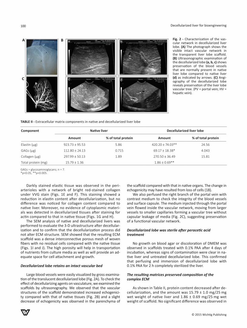
Large blood vessels were easily visualized by gross examina-tion of the translucent decellularized lobe (Fig. 2A). To check the effect of decellularizing agents on vasculature, we examined the scaffolds by ultrasonography. We observed that the vascular structures of the scaffold demonstrated increased echogenici-ty compared with that of native tissues (Fig. 2B) and a slight decrease of echogenicity was observed in the parenchyma of
the scaffold compared with that in native organs. The change in echogenicity may have resulted from loss of cells (18).
We also perfused the right branch of the portal vein with contrast medium to check the integrity of the blood vessels and surface capsule. The medium injected through the portal vein flowed inside the vascular network, moving from larger vessels to smaller capillaries forming a vascular tree without capsular leakage of media (Fig. 2C), suggesting preservation of a functional vascular network.
Decellularized lobe was sterile after peracetic acid treatment
No growth on blood agar or discoloration of DMEM was observed in scaffolds treated with 0.1% PAA after 4 days of incubation, whereas signs of contamination were clear in na-tive liver and untreated decellularized lobe. This confirmed that perfusing and immersion of decellularized lobe with 0.1% PAA for 2 h completely sterilized the liver.
The resulting matrices preserved composition of the complex ECM
As shown in Table II, protein content decreased after de-cellularization, and the amount was 15.79 ± 1.0 mg/25 mg wet weight of native liver and 1.86 ± 0.69 mg/25 mg wet weight of scaffold. No significant difference was observed in
Fig. 2 - Characterization of the vas-cular network in decellularized liver lobe. (A) The photograph shows the visible intact vascular network in the transparent liver lobe scaffold. (B) Ultrasonographic examination of the decellularized lobe (a, b, c) shows preservation of the blood vessels that are normally present in native liver lobe compared to native liver (d) as indicated by arrows. (C) Angi-ography of the decellularized lobe reveals preservation of the liver lobe vascular tree. (PV = portal vein; HV = hepatic vein).
taBle ii - Extracellular matrix components in native and decellularized liver lobe
Component Native liver Decellularized liver lobe
Amount % of total protein Amount % of total protein
Elastin (µg) 923.73 ± 95.53 5.86 420.20 ± 74.03** 24.56
GAGs (µg) 112.80 ± 24.13 0.715 69.17 ± 18.38* 4.043
Collagen (µg) 297.99 ± 50.13 1.89 270.50 ± 36.49 15.81
Total protein (mg) 15.79 ± 1.36 1.86 ± 0.69**
GAGs = glycosaminoglycans; n = 7.*p<0.05, **p<0.005.
Hussein et al 101
© 2015 Wichtig Publishing
the amount of collagen before and after decellularization; the amounts were 297.99 ± 50.13 µg/25 mg wet weight in native livers and 270.50 ± 36.49 µg/25 mg wet weight in decellularized livers. GAG content decreased partially af-ter decellularization, during which approximately 61% of GAG was preserved in decellularized tissues (decellularized livers: 112.80 ± 24.13 µg/25mg wet weight; native liver: 69.17 ± 18.3 µg/25 mg wet weight). Elastin was 420.20 ± 74.03 µg/25 mg wet weight of decellularized tissue which represented 45% of that in native tissue (923.73 ± 95.53 µg/ 25 mg wet weight).
Decellularized scaffolds were almost devoid of DNA
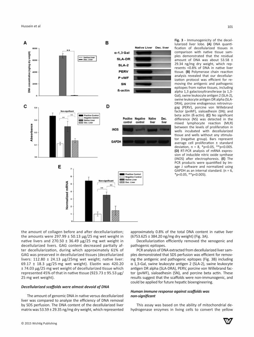
The amount of genomic DNA in native versus decellularized liver was compared to analyze the efficiency of DNA removal by SDS perfusion. The DNA content of the decellularized liver matrix was 53.59 ± 29.35 ng/mg dry weight, which represented
approximately 0.8% of the total DNA content in native liver (6753.625 ± 384.20 ng/mg dry weight) (Fig. 3A).
Decellularization efficiently removed the xenogenic and pathogenic epitopes.
PCR analysis of DNA extracted from decellularized liver sam-ples demonstrated that SDS perfusion was efficient for remov-ing the antigenic and pathogenic epitopes (Fig. 3B) including α 1,3-Gal, swine leukocyte antigen 2 (SLA-2), swine leukocyte antigen DR alpha (SLA-DRA), PERV, porcine von Willebrand fac-tor (pvWF), sialoadhesin (SN), and porcine beta actin. These results suggest that the scaffolds were non-immunogenic, and could be applied for future hepatic bioengineering.
Human immune response against scaffolds was non-significant
This assay was based on the ability of mitochondrial de-hydrogenase enzymes in living cells to convert the yellow
Fig. 3 - Immunogenicity of the decel-lularized liver lobe. (A) DNA quanti-fication of decellularized tissues in comparison with native tissue sam-ples demonstrated that the residual amount of DNA was about 53.58 ± 29.34 ng/mg dry weight, which rep-resents <0.8% of DNA in native liver tissue. (B) Polymerase chain reaction analysis revealed that our decellular-ization protocol was efficient for re-moving the antigenic and pathogenic epitopes from native tissues, including alpha 1,3 galactosyltransferase (α 1,3-Gal), swine leukocyte antigen 2 (SLA-2), swine leukocyte antigen DR alpha (SLA-DRA), porcine endogenous retrovirus-gag (PERV), porcine von Willebrand factor (pvWF), sialoadhesin (SN); and beta actin (ß-actin). (C) No significant difference (NS) was detected in the mixed lymphocyte reaction (MLR) between the levels of proliferation in wells incubated with decellularized tissue and wells without any stimula-tor (negative group). Bars represent average cell proliferation ± standard deviation, n = 8, *p<0.05, **p<0.005. (D) RT-PCR analysis of mRNA expres-sion of inducible nitric oxide synthase (iNOS) after electrophoresis. (E) The PCR products were quantified by Im-age J software and normalized using GAPDH as an internal standard. (n = 6, *p<0.05, **p<0.005).
Decellularized liver for bioengineering102
© 2015 Wichtig Publishing
water-soluble substrate MTT into a blue formazan, which is insoluble in water. The amount of formazan produced is di-rectly proportional to the number of live cells. Therefore, higher optical density (OD) values reflect the proliferation rate of PBMCs. PBMCs cultured in the presence of rhIL-2 ex-hibited the highest OD (0.333 ± 0.020), indicating a strong proliferative response (Fig. 3C). The OD values measured in wells co-cultured with native liver (0.248 ± 0.041) displayed significantly higher values than those of the negative group (0.111 ± 0.011). Importantly, no significant difference was de-tected between OD values obtained from the decellularized liver (0.134 ± 0.0159) and negative groups, indicating that the prepared ECM had lost its antigencity to induce human PBMC proliferation.
In order to get more insight into the potential human immune response against scaffolds, we have checked whether decellularized livers could induce nitric oxide pro-duction or not. As shown in Figs. 3D and E, iNOS expression was strongly up-regulated in human PBMCs stimulated by rh-IL-2 (5.42 ± 0.5 folds) and cells cultured with native liver (3.53 ± 0.4 folds) compared to the negative control. Con-sistent with the MTT proliferation assay, there was no sig-nificant difference in iNOS expression in PBMCs cultured in presence of decellularized liver (1.46 ± 0.6 folds) compared to the negative control group. Considering these evidences, it is clear that the decellularized liver scaffold is inert to the human PBMCs.
Cytotoxicity of decellularized liver ECM
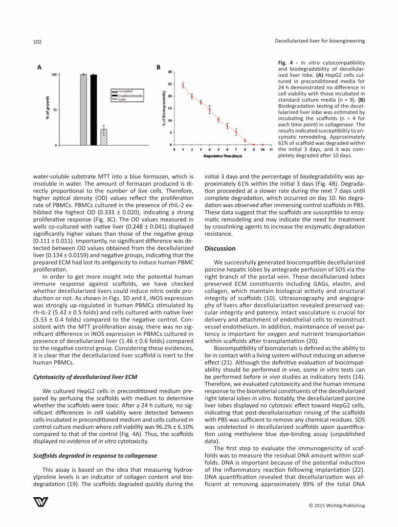
We cultured HepG2 cells in preconditioned medium pre-pared by perfusing the scaffolds with medium to determine whether the scaffolds were toxic. After a 24 h culture, no sig-nificant differences in cell viability were detected between cells incubated in preconditioned medium and cells cultured in control culture medium where cell viability was 96.2% ± 6.10% compared to that of the control (Fig. 4A). Thus, the scaffolds displayed no evidence of in vitro cytotoxicity.
Scaffolds degraded in response to collagenase
This assay is based on the idea that measuring hydrox-ylproline levels is an indicator of collagen content and bio-degradation (19). The scaffolds degraded quickly during the
initial 3 days and the percentage of biodegradability was ap-proximately 61% within the initial 3 days (Fig. 4B). Degrada-tion proceeded at a slower rate during the next 7 days until complete degradation, which occurred on day 10. No degra-dation was observed after immersing control scaffolds in PBS. These data suggest that the scaffolds are susceptible to enzy-matic remodeling and may indicate the need for treatment by crosslinking agents to increase the enzymatic degradation resistance.
Discussion
We successfully generated biocompatible decellularized porcine hepatic lobes by antegrade perfusion of SDS via the right branch of the portal vein. These decellularized lobes preserved ECM constituents including GAGs, elastin, and collagen, which maintain biological activity and structural integrity of scaffolds (10). Ultrasonography and angiogra-phy of livers after decellularization revealed preserved vas-cular integrity and patency. Intact vasculature is crucial for delivery and attachment of endothelial cells to reconstruct vessel endothelium. In addition, maintenance of vessel pa-tency is important for oxygen and nutrient transportation within scaffolds after transplantation (20).
Biocompatibility of biomaterials is defined as the ability to be in contact with a living system without inducing an adverse effect (21). Although the definitive evaluation of biocompat-ability should be performed in vivo, some in vitro tests can be performed before in vivo studies as indicatory tests (14). Therefore, we evaluated cytotoxicity and the human immune response to the biomaterial constituents of the decellularized right lateral lobes in vitro. Notably, the decellularized porcine liver lobes displayed no cytotoxic effect toward HepG2 cells, indicating that post-decellularization rinsing of the scaffolds with PBS was sufficient to remove any chemical residues. SDS was undetected in decellularized scaffolds upon quantifica-tion using methylene blue dye-binding assay (unpublished data).
The first step to evaluate the immunogenicity of scaf-folds was to measure the residual DNA amount within scaf-folds. DNA is important because of the potential induction of the inflammatory reaction following implantation (22). DNA quantification revealed that decellularization was ef-ficient at removing approximately 99% of the total DNA
Fig. 4 - In vitro cytocompatibility and biodegradability of decellular-ized liver lobe. (A) HepG2 cells cul-tured in preconditioned media for 24 h demonstrated no difference in cell viability with those incubated in standard culture media (n = 8). (B) Biodegradation testing of the decel-lularized liver lobe was estimated by incubating the scaffolds (n = 4 for each time point) in collagenase. The results indicated susceptibility to en-zymatic remodeling. Approximately 61% of scaffold was degraded within the initial 3 days, and it was com-pletely degraded after 10 days.
Hussein et al 103
© 2015 Wichtig Publishing
in native tissues. Rejection of xenotransplanted porcine native organs and tissues is a major problem, which lim-its the wide use of porcine organs in xenotransplantation. Alpha-Gal is a porcine epitope that is responsible for elic-iting acute rejection in humans after xenotransplantation. Many researchers are interested in generating pigs in which the α 1,3-Gal gene has been knocked out to overcome the barrier of hyperacute rejection (4). According to previous studies, SLA-2 and SLA-DRA play a role in rejection of xe-notranplanted organs (23). In addition, vWF is involved in xenograft dysfunction by binding to xenoreactive antibod-ies and activating primate platelets (24). In 2012, Brock et al explained the role of sialoadhesin, a highly conserved protein expressed on porcine Kupffer cells, in mediating the destruction of human red blood cells in an extracorpo-real porcine liver xenoperfusion model (25). Waldman et al confirmed that adding anti-porcine sialoadhesin antibody to an extracorporeal porcine liver system results in reduced human red blood cell destruction (26). Decellularization was advantageous and overcame many of these barriers, as it removed these antigenic epitopes. We also confirmed that the decellularized lobes were depleted from the pos-sible infection risk from PERV after xenotransplantation. Moreover, the results of the mixed lymphocyte reaction indicated that the decellularized liver lobes were inert to human PBMCs and lacked the ability to stimulate cell pro-liferation and iNOS activity compared to that in the posi-tive control and native tissue. Therefore, we claim that the decellularized liver lobe obtained by our decellularization technique will not elicit an immune response in human hosts.
Biodegradability of the ECM is another important crite-rion that should be addressed in organs or tissue prepared for xenotransplantation (27, 28). Degradation of scaffolds results in the release of matricryptic molecules that have angiogenic, mitogenic, chemoattractant, and antimicrobial properties (29). In vivo implantation of urinary bladder ma-trix after a segmental resection of the Achilles tendon in a mouse model was accompanied by degradation and recruit-ment of progenitor cells to the remodeling site. Degradation products were suggested to be the reason for this recruit-ment (30). Here, in vitro degradation testing revealed com-plete degradation of the scaffolds after 10 days, suggesting the susceptibility of the scaffolds to enzymatic remodeling after implantation.
In summary, we showed that our decellularization tech-nique was successful in removing the cellular and nuclear components from the right lateral lobe of native porcine livers. The resulting natural scaffolds preserved the vascula-ture, native architecture, and most extracellular proteins. In addition, these scaffolds were devoid of the antigenic and pathogenic-related epitopes, which involve infection risk and rejection after xenotransplantation. Our biodegradable ma-trices behave like an inert matrix towards human immune cells. Interestingly, development of a xenoantigen-free de-cellularized liver lobe is a good first step toward generating recellularized tissue for xenotranplantation or temporary hepatic support using extracorporeal perfusion in patients awaiting liver transplantation.
Abbreviations
DAPI 4’,6-diamidino-2-phenylindoleECM extracellular matrixGAGs glycosaminoglycansiNOS inducible nitric oxide synthaseMTT 3-[4,5-dimethylthiazol-2-yl]-2,5- diphenyltetra-
zoium bromidePAA peracetic acidPBMCs peripheral blood mononuclear cellsPERV porcine endogenous retrovirus-gagPvWF porcine von Willebrand factorSDS sodium dodecyl sulfateSEM scanning electron microscopeSLA-2 swine leukocyte antigen 2SLA-DRA swine leukocyte antigen DR alphaSN sialoadhesinVVG Verhoeff-Van Giesonα-1,3 gal alpha 1,3 galactosyltransferase.
DisclosuresFinancial support: This research was supported by grants from iPET, Ministry of Food, Agriculture, Forestry, and Fisheries and the Next-Generation BioGreen 21 program (No. PJ009627), Rural Develop-ment Administration, Korea. This study was also supported by a grant (Project Code No. Z-1541745-2013-14-01, Z-1541745-2013-14-02) from Animal and Plant Quarantine Agency, Ministry of Agri-culture, Food and Rural Affairs (MAFRA), Korea.Conflict of interest: The authors declare that they have no compet-ing interests.Meeting presentations: Part of this study has been presented as posters:1. Kamal Hany Hussein, Kyung-Mee Park, Pankaj Kumar Teotia, Seok-Ho Hong, Se-Ran Yang, Sung-Min Park, Cha Sang, and Heung-Myong Woo. Construction of a bioartificial immunogen-reduced porcine he-patic lobe for tissue engineering. 13th Congress of the Asian Society of Transplantation (CAST), Kyoto, Japan, September 2-6, 2013. (PC1-069)2. Kamal Hany Hussein, Kyung-Mee Park, Pankaj Kumar Teotia, Hyun-Min Kim, Seok-Ho Hong, Se-Ran Yang, Sung-Min Park, and Heung-Myong Woo. Development of recellularized three-dimensional scaf-fold using porcine liver scaffold. 12th Congress of the International Xenotransplantation Association (IXA), Osaka, Japan, November 10-13, 2013. (Poster no. 523).
References1. Soltys KA, Soto-Gutiérrez A, Nagaya M, et al. Barriers to the
successful treatment of liver disease by hepatocyte transplan-tation. J Hepatol. 2010;53(4):769-774.
2. Hoshiba T, Lu H, Kawazoe N, Chen G. Decellularized matrices for tissue engineering. Expert Opin Biol Ther. 2010;10(12): 1717-1728.
3. Sprangers B, Waer M, Billiau AD. Xenotransplantation: where are we in 2008? Kidney Int. 2008;74(1):14-21.
4. Ekser B, Gridelli B, Tector AJ, Cooper DK. Pig liver xenotrans-plantation as a bridge to allotransplantation: which patients might benefit? Transplantation. 2009;88(9):1041-1049.
5. Mattiuzzo G, Takeuchi Y, Scobie L. Potential zoonotic infection of porcine endogenous retrovirus in xenotransplantation. Meth-ods Mol Biol. 2012;885:263-279.
6. Fishman JA, Scobie L, Takeuchi Y. Xenotransplantation-associ-ated infectious risk: a WHO consultation. Xenotransplantation. 2012;19(2):72-81.
Decellularized liver for bioengineering104
© 2015 Wichtig Publishing
7. Hussein KH, Park KM, Teotia PK, et al. Fabrication of a biode-gradable xenoantigen-free rat liver scaffold for potential drug screening applications. Transplant Proc. 2013;45(8):3092-3096.
8. Booth C, Soker T, Baptista P, et al. Liver bioengineering: cur-rent status and future perspectives. World J Gastroenterol. 2012;18(47):6926-6934.
9. Hussein KH, Park KM, Teotia PK, et al. Sterilization using electrolyzed water highly retains the biological properties in tissue-engineered porcine liver scaffold. Int J Artif Organs. 2013;36(11):781-792.
10. Soto-Gutierrez A, Zhang L, Medberry C, et al. A whole-organ regenerative medicine approach for liver replacement. Tissue Eng Part C Methods. 2011;17(6):677-686.
11. Uygun BE, Soto-Gutierrez A, Yagi H, et al. Organ reengineer-ing through development of a transplantable recellularized liver graft using decellularized liver matrix. Nat Med. 2010; 16(7):814-820.
12. Zhao L, Zhao J, Wang S, et al. Evaluation of immunocompat-ibility of tissue-engineered periosteum. Biomed Mater. 2011; 6(1):015005.
13. Crapo PM, Gilbert TW, Badylak SF. An overview of tissue and whole organ decellularization processes. Biomaterials. 2011; 32(12):3233-3243.
14. Simon P, Kasimir MT, Seebacher G, et al. Early failure of the tis-sue engineered porcine heart valve SYNERGRAFT in pediatric patients. Eur J Cardiothorac Surg. 2003;23(6):1002-1006.
15. Eitan Y, Sarig U, Dahan N, Machluf M. Acellular cardiac extra-cellular matrix as a scaffold for tissue engineering: in vitro cell support, remodeling, and biocompatibility. Tissue Eng Part C Methods. 2010;16(4):671-683.
16. Saluja R, Jyoti A, Chatterjee M, et al. Molecular and biochemi-cal characterization of nitric oxide synthase isoforms and their intracellular distribution in human peripheral blood mononu-clear cells. Biochim Biophys Acta. 2011;1813(10):1700-1707.
17. Baptista PM, Siddiqui MM, Lozier G, Rodriguez SR, Atala A, Soker S. The use of whole organ decellularization for the gen-eration of a vascularized liver organoid. Hepatology. 2011; 53(2):604-617.
18. Park KM, Woo HM. Porcine bioengineered scaffolds as new frontiers in regenerative medicine. Transplant Proc. 2012;44 (4):1146-1150.
19. Sang L, Luo D, Xu S, Wang X, Li X. Fabrication and evaluation of biomimetic scaffolds by using collagen–alginate fibrillar gels for potential tissue engineering applications. Mater Sci Eng C. 2011;31(2):262-271.
20. Mirmalek-Sani SH, Sullivan DC, Zimmerman C, Shupe TD, Petersen BE. Immunogenicity of decellularized porcine liver for bioengineered hepatic tissue. Am J Pathol. 2013;183(2):558-565.
21. Rhodes N. Biocompatibility testing of tissue engineered prod-ucts. Vox Sang. 2004;87(Suppl 2):161-163.
22. Gilbert TW, Freund JM, Badylak SF. Quantification of DNA in biologic scaffold materials. J Surg Res. 2009;152(1):135-139.
23. Park KM, Park SM, Yang SR, Hong SH, Woo HM. Preparation of immunogen-reduced and biocompatible extracellular matrices from porcine liver. J Biosci Bioeng. 2013;115(2):207-215.
24. Lin CC, Cooper DK, Dorling A. Coagulation dysregulation as a barrier to xenotransplantation in the primate. Transpl Immunol. 2009;21(2):75-80.
25. Brock LG, Delputte PL, Waldman JP, Nauwynck HJ, Rees MA. Porcine sialoadhesin: a newly identified xenogeneic innate im-mune receptor. Am J Transplant. 2012;12(12):3272-3282.
26. Waldman JP, Vogel T, Burlak C, et al. Blocking porcine sialo-adhesin improves extracorporeal porcine liver xenoperfusion with human blood. Xenotransplantation. 2013;20(4):239-251.
27. Gilbert TW, Stewart-Akers AM, Badylak SF. A quantitative method for evaluating the degradation of biologic scaffold ma-terials. Biomaterials. 2007;28(2):147-150.
28. Babensee JE, Anderson JM, McIntire LV, Mikos AG. Host re-sponse to tissue engineered devices. Adv Drug Deliv Rev. 1998;33(1-2):111-139.
29. Wolf MT, Daly KA, Brennan-Pierce EP, et al. A hydrogel derived from decellularized dermal extracellular matrix. Biomaterials. 2012;33(29):7028-7038.
30. Beattie AJ, Gilbert TW, Guyot JP, Yates AJ, Badylak SF. Chemoat-traction of progenitor cells by remodeling extracellular matrix scaffolds. Tissue Eng Part A. 2009;15(5):1119-1125.
Related Documents