BioMed Central Page 1 of 14 (page number not for citation purposes) BMC Genomics Open Access Research article Conservation of DNA-binding specificity and oligomerisation properties within the p53 family Tobias Brandt 1 , Miriana Petrovich 1 , Andreas C Joerger 1 and Dmitry B Veprintsev* 2 Address: 1 MRC Centre for Protein Engineering, Cambridge, CB2 0QH, UK and 2 MRC Laboratory of Molecular Biology, Cambridge, CB2 0QH, UK Email: Tobias Brandt - [email protected]; Miriana Petrovich - [email protected]; Andreas C Joerger - acj2@mrc- lmb.cam.ac.uk; Dmitry B Veprintsev* - [email protected] * Corresponding author Abstract Background: Transcription factors activate their target genes by binding to specific response elements. Many transcription factor families evolved from a common ancestor by gene duplication and subsequent divergent evolution. Members of the p53 family, which play key roles in cell-cycle control and development, share conserved DNA binding and oligomerisation domains but exhibit distinct functions. In this study, the molecular basis of the functional divergence of related transcription factors was investigated. Results: We characterised the DNA-binding specificity and oligomerisation properties of human p53, p63 and p73, as well as p53 from other organisms using novel biophysical approaches. All p53 family members bound DNA cooperatively as tetramers with high affinity. Despite structural differences in the oligomerisation domain, the dissociation constants of the tetramers was in the low nanomolar range for all family members, indicating that the strength of tetramerisation was evolutionarily conserved. However, small differences in the oligomerisation properties were observed, which may play a regulatory role. Intriguingly, the DNA-binding specificity of p53 family members was highly conserved even for evolutionarily distant species. Additionally, DNA recognition was only weakly affected by CpG methylation. Prediction of p53/p63/p73 binding sites in the genome showed almost complete overlap between the different homologs. Conclusion: Diversity of biological function of p53 family members is not reflected in differences in sequence-specific DNA binding. Hence, additional specificity factors must exist, which allowed the acquisition of novel functions during evolution while preserving original roles. Background Sequence-specific transcription factors are responsible for processing environmental and developmental signals, and initiating the appropriate cellular response. The total number of transcription factors of an organism increases with its complexity: it is estimated to be around 300 for yeast, 1000 for worms and 3000 for humans [1]. Besides a DNA-binding domain, another common feature of many transcription factors, such as basic helix-loop-helix (bHLH) factors and basic-region leucine zipper (bZIP) factors, is an additional oligomerisation domain (OD) [2,3]. A functional role for oligomerisation is easy to Published: 23 December 2009 BMC Genomics 2009, 10:628 doi:10.1186/1471-2164-10-628 Received: 26 August 2009 Accepted: 23 December 2009 This article is available from: http://www.biomedcentral.com/1471-2164/10/628 © 2009 Brandt et al; licensee BioMed Central Ltd. This is an Open Access article distributed under the terms of the Creative Commons Attribution License (http://creativecommons.org/licenses/by/2.0 ), which permits unrestricted use, distribution, and reproduction in any medium, provided the original work is properly cited.

Welcome message from author
This document is posted to help you gain knowledge. Please leave a comment to let me know what you think about it! Share it to your friends and learn new things together.
Transcript

BioMed CentralBMC Genomics
ss
Open AcceResearch articleConservation of DNA-binding specificity and oligomerisation properties within the p53 familyTobias Brandt1, Miriana Petrovich1, Andreas C Joerger1 and Dmitry B Veprintsev*2Address: 1MRC Centre for Protein Engineering, Cambridge, CB2 0QH, UK and 2MRC Laboratory of Molecular Biology, Cambridge, CB2 0QH, UK
Email: Tobias Brandt - [email protected]; Miriana Petrovich - [email protected]; Andreas C Joerger - [email protected]; Dmitry B Veprintsev* - [email protected]
* Corresponding author
AbstractBackground: Transcription factors activate their target genes by binding to specific responseelements. Many transcription factor families evolved from a common ancestor by gene duplicationand subsequent divergent evolution. Members of the p53 family, which play key roles in cell-cyclecontrol and development, share conserved DNA binding and oligomerisation domains but exhibitdistinct functions. In this study, the molecular basis of the functional divergence of relatedtranscription factors was investigated.
Results: We characterised the DNA-binding specificity and oligomerisation properties of humanp53, p63 and p73, as well as p53 from other organisms using novel biophysical approaches. All p53family members bound DNA cooperatively as tetramers with high affinity. Despite structuraldifferences in the oligomerisation domain, the dissociation constants of the tetramers was in thelow nanomolar range for all family members, indicating that the strength of tetramerisation wasevolutionarily conserved. However, small differences in the oligomerisation properties wereobserved, which may play a regulatory role. Intriguingly, the DNA-binding specificity of p53 familymembers was highly conserved even for evolutionarily distant species. Additionally, DNArecognition was only weakly affected by CpG methylation. Prediction of p53/p63/p73 binding sitesin the genome showed almost complete overlap between the different homologs.
Conclusion: Diversity of biological function of p53 family members is not reflected in differencesin sequence-specific DNA binding. Hence, additional specificity factors must exist, which allowedthe acquisition of novel functions during evolution while preserving original roles.
BackgroundSequence-specific transcription factors are responsible forprocessing environmental and developmental signals,and initiating the appropriate cellular response. The totalnumber of transcription factors of an organism increaseswith its complexity: it is estimated to be around 300 for
yeast, 1000 for worms and 3000 for humans [1]. Besidesa DNA-binding domain, another common feature ofmany transcription factors, such as basic helix-loop-helix(bHLH) factors and basic-region leucine zipper (bZIP)factors, is an additional oligomerisation domain (OD)[2,3]. A functional role for oligomerisation is easy to
Published: 23 December 2009
BMC Genomics 2009, 10:628 doi:10.1186/1471-2164-10-628
Received: 26 August 2009Accepted: 23 December 2009
This article is available from: http://www.biomedcentral.com/1471-2164/10/628
© 2009 Brandt et al; licensee BioMed Central Ltd. This is an Open Access article distributed under the terms of the Creative Commons Attribution License (http://creativecommons.org/licenses/by/2.0), which permits unrestricted use, distribution, and reproduction in any medium, provided the original work is properly cited.
Page 1 of 14(page number not for citation purposes)
BMC Genomics 2009, 10:628 http://www.biomedcentral.com/1471-2164/10/628
rationalize: it combines the DNA-binding specificity ofindividual monomeric domains, leading to a substantialincrease in binding affinity. Divergence of transcriptionfactor function within a family could originate from evo-lutionary changes in the DNA-binding specificity and inthe oligomerisation properties.
A highly important family of transcription factors thatplay a key role in cell-cycle control and development isthat of p53, p63 and p73. p53 is at the centre of a tumoursuppressor network [4,5], and, as such, is essential for theprevention of cancer [6,7]. Both p63 and p73 are involvedin developmental processes. p63 is essential for epidermalmorphogenesis and limb development, whereas p73 isinvolved in the development of neural structures and thepheromone detection system, among its other roles. Nev-ertheless, p63 and p73 are also involved in processes con-trolled by p53 [8]. Interestingly, different functions arealso observed even for closely related p53 orthologs. Forexample, genes encoding proteins involved in DNAmetabolism are responsive to p53 in humans but not inmice [9]. All three family members consist of a structuredDNA-binding domain (DBD), an oligomerisationdomain and intrinsically disordered N-terminal transacti-vation and C-terminal regulatory domains [10]. Addition-ally, p63 and p73 also contain a structured sterile alphamotif (SAM) and an inhibitory domain at the C-terminus[11]. The majority of cancer-associated p53 mutations arefound in the DNA-binding domain [6,7], highlighting theimportance of correct DNA recognition. p53 specificallybinds to a 20 base pair (bp) consensus DNA sequence,also called a response element (RE), consisting of tworepeats of 5'-RRRCWWGYYY-3' (where R = A or G; Y = Cor T; W = A or T), separated by 0-13 bp [12,13]. In addi-tion, p53 also recognises a large number of sequences thatdeviate from this consensus site definition [14,15]. Sev-eral studies have shown that p53, p63 and p73 can recog-nise the same sites [16-18]. Additionally, each protein hasdifferent isoforms [19], which, in most cases, have identi-cal DNA-binding domains but exhibit differences in tran-scriptional activity, adding an additional layer ofcomplexity [17].
Despite a high degree of sequence conservation, particu-larly in the DNA-binding and tetramerisation domains,p53, p63 and p73 fulfil at least partially different roles.The molecular basis of how closely related transcriptionfactors differentiate between their respective target genesis only poorly understood. Here, we characterised the oli-gomerisation and DNA-binding properties of several p53family members. Firstly, we determined the dissociationconstants for dimers and tetramers of p53 family mem-bers using analytical ultracentrifugation. We then com-pared the DNA-binding specificity of full-length humanp53 (Hsp53) with that of its paralogs p63 and p73,
including the isoforms ΔNp63α, ΔNp63β, ΔNp63γ,ΔNp73β and an engineered truncated version of p73 con-taining DNA-binding and parts of the oligomerisationdomain only (p73CT, residues 104-383). We also com-pared the DNA-binding specificity of human p53 withthat of its orthologs from a number of species at varyingevolutionary distances from humans: mouse (Mus muscu-lus, Mmp53), frog (Xenopus laevis, Xlp53), zebrafish(Danio rerio, Drp53) and fruit fly (Drosophila melanogaster,Dmp53). In these measurements, we included effects ofCpG methylation as an additional factor potentially influ-encing DNA-binding specificity. We used a method forquantification of DNA-binding specificity which we haverecently developed [15,20]. Using fluorescence anisotropytitrations, we measured the effect of every possible singlebase pair substitution of a consensus sequence on theaffinity of the proteins for DNA. The DNA-binding datawere then used to identify putative binding sites withinthe human genome to assess the impact of the differencesin DNA-binding specificity.
ResultsOligomerisation equilibriaWe have shown previously that full-length human p53dissociates into dimers at nanomolar concentration, andthat oligomerisation is essential for high-affinity DNAbinding [21,22]. Here, we studied the oligomerisationproperties of members of the p53 family, namely Dmp53,Drp53, Hsp53, Mmp53, and Xlp53, as well as humanΔNp63β and ΔNp73β. The p63 and p73 isoforms containintact DNA-binding and tetramerisation domains. Weused sedimentation velocity analytical ultracentrifugation(SV-AUC) experiments with a fluorescence detection sys-tem [23], which allows measurements to be made at lownanomolar concentrations. To specifically incorporate afluorophore, we expressed proteins with a C-terminalCCPGCC tetra-cysteine tag and labelled them with FlAsH-EDT2, an arsenic derivative of fluorescein [24].
The sedimentation profile of Hsp53 at 22.5 μM monomerconcentration, measured using absorbance detection(data not shown), showed only one peak at ~2.9 S, whichwe assigned to a tetramer, because the protein has beenshown to be tetrameric at this concentration [22]. Subse-quently, we measured the sedimentation profiles oflabelled proteins at different concentrations using the flu-orescence detection system (Figure 1 and Additional file1). At lower concentrations, a second peak appeared at 1.8to 2.0 S. In order to improve the resolution of the sedi-mentation profiles in the range between 0.5 and 3 S, werepeated our experiments at higher rotor speeds (60 krpm). In addition to the tetramer peak, we were able toresolve two peaks at 1.1 S and 1.9 S, which correspond tomonomers and dimers, respectively.
Page 2 of 14(page number not for citation purposes)
BMC Genomics 2009, 10:628 http://www.biomedcentral.com/1471-2164/10/628
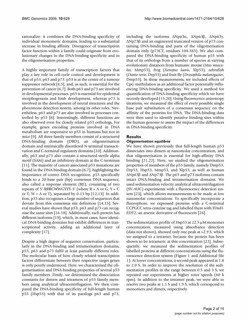
All proteins studied formed tetramers which dissociateinto dimers. For some proteins, these dimers dissociatedinto monomers. It was possible to determine their sedi-mentation profiles with well-resolved peaks, and thus tocalculate the dissociation constants Kd for the monomer-dimer and dimer-tetramer equilibria (Additional file 2,Figure 2). The dissociation constants for the dimer-tetramer equilibria of Dmp53, Drp53 and ΔNp73β were
in the low nanomolar range. The self-association wasabout 5 times weaker for ΔNp63β and about 10 timesweaker for Mmp53, Hsp53 and Xlp53. At low nanomolarconcentrations, tetramers of human p53 dissociated intodimers, whereas those of human p63 and p73 readily dis-sociated into dimers and monomers. In the case of p63,the dissociation constants for momomer-dimer anddimer-tetramer equlibria were similar. For p73, the dimer-monomer Kd was even larger than the dimer-tetramer Kd.This indicates that only small amounts of p73 dimers arepresent in solution. For human p53, this is not the case, asthe monomer-dimer Kd is about 20 times lower than thedimer-tetramer Kd. No monomers were observed forDmp53 and Drp53.
DNA-binding specificity of p53 family members is highly conservedIt is often assumed that diverging transcription factorshave differences in their DNA-binding specificity, whichresult in preferential recognition of a different responseelement sequence and an associated change in function.To answer the question of whether the p53 response ele-ment sequence is evolving and diverging, we comparedthe DNA-binding specificity of human p53, p63 and p73,and p53 from different species. We used a fluorescenceanisotropy assay, which we had developed earlier forquantifying the DNA-binding specificity of Hsp53[15,21].
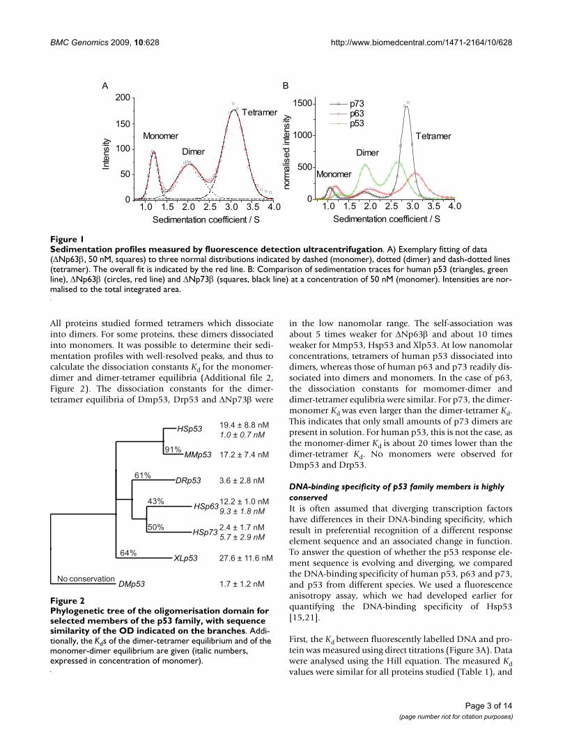
First, the Kd between fluorescently labelled DNA and pro-tein was measured using direct titrations (Figure 3A). Datawere analysed using the Hill equation. The measured Kdvalues were similar for all proteins studied (Table 1), and
Sedimentation profiles measured by fluorescence detection ultracentrifugationFigure 1Sedimentation profiles measured by fluorescence detection ultracentrifugation. A) Exemplary fitting of data (ΔNp63β, 50 nM, squares) to three normal distributions indicated by dashed (monomer), dotted (dimer) and dash-dotted lines (tetramer). The overall fit is indicated by the red line. B: Comparison of sedimentation traces for human p53 (triangles, green line), ΔNp63β (circles, red line) and ΔNp73β (squares, black line) at a concentration of 50 nM (monomer). Intensities are nor-malised to the total integrated area.
1.0 1.5 2.0 2.5 3.0 3.5 4.00
50
100
150
200Tetramer
Dimer
A
Inte
nsity
Sedimentation coefficient / S
Monomer
1.0 1.5 2.0 2.5 3.0 3.5 4.00
500
1000
1500
Tetramer
Dimer
Monomer
B
norm
alis
ed in
tens
ity
Sedimentation coefficient / S
p73 p63 p53
Phylogenetic tree of the oligomerisation domain for selected members of the p53 family, with sequence similarity of the OD indicated on the branchesFigure 2Phylogenetic tree of the oligomerisation domain for selected members of the p53 family, with sequence similarity of the OD indicated on the branches. Addi-tionally, the Kds of the dimer-tetramer equilibrium and of the monomer-dimer equilibrium are given (italic numbers, expressed in concentration of monomer).
HSp53
MMp53
DRp53
HSp63
HSp73
XLp53
DMp53
91%
61%
64%
43%
50%
No conservation
19.4 ± 8.8 nM 1.0 ± 0.7 nM
12.2 ± 1.0 nM9.3 ± 1.8 nM
2.4 ± 1.7 nM5.7 ± 2.9 nM
17.2 ± 7.4 nM
3.6 ± 2.8 nM
27.6 ± 11.6 nM
1.7 ± 1.2 nM
Page 3 of 14(page number not for citation purposes)
BMC Genomics 2009, 10:628 http://www.biomedcentral.com/1471-2164/10/628
observed differences were within the error range of themethod. The only exception was p73CT, which boundabout 4-5 times more weakly than the ΔNp73β isoform.Weaker binding of p73CT can be attributed to impairedself-oligomerisation due to a truncation of the tetrameri-sation domain, which has been shown to destabilise thetetramer [25]. The Hill coefficient n was averaged over allmeasured datasets (n = 1.64), which was in close agree-ment with the value we have previously reported forhuman p53 [22]. In combination with analytical ultracen-trifugation data, we can conclude that for all the proteinsstudied two dimers bind DNA cooperatively and form atetramer, similarly to human p53 [21].
The driving force for recognition of a specific DNAsequence surrounded by non-specific seqences is not theabsolute affinity but rather specificity, or the relative affin-ity for specific vs. non-specific sequences. To define theDNA-binding specificity of the members of the p53 fam-ily, we measured the affinity of the proteins to all possiblepermutations of a reference consensus binding sequence(Additional file 3) using a fluorescence competition titra-tion assay (Figure 3B). This sequence contains two identi-cal copies of a GGACATGTCC half-site and is one of thetightest-binding sequences for human p53 [15]. Theresults for all the p53 homologs analysed are summarisedin Figure 4. For every nucleotide substitution, the differ-ence of the logarithm of the dissociation constants for themutated sequence and the reference sequence (ΔlogKd)was determined. High positive values of ΔlogKd indicatehigh affinity penalties and low probability of observing
this substitution in the binding site. The effects of nucle-otide substitution are also presented as a sequence logo(Figure 5), which depicts the most preferred nucleotide ata position as the largest letter, and the relative selectivityat this position as the height of the bar. Based on the affin-ity differences, we calculated expected relative nucleotidefrequencies for each position, and a corresponding bitscore ranging from 0 to 2 [26,27]. Key features of theresponse element are highly conserved between all theproteins studied. The largest decrease in affinity wascaused by nucleotide changes at positions 4 and 7, whichcorrespond to the invariant C and G in the RRRCWW-GYYY consensus sequence. Nucleotide changes at posi-tions 5 and 6, corresponding to the central WW element,and positions 3 and 8, also caused significant changes inthe affinity. Generally, changes at the outer positions 1, 2and 9, 10 did not significantly affect binding. Accordingly,the largest contributions to the overall DNA-binding spe-cificity are made by positions 4 and 7, followed by 3, 5, 6,and 8. The observed changes can be alternativelyexpressed as a consensus sequence definition (Table 1).Selecting the nucleotide changes resulting in the highestaffinity at each position defines the highest affinitysequence. A better reflection of DNA-binding specificity isto apply a cut-off value representing the error of the meas-urement. All nucleotides at a particular position that causea lower affinity change, ΔlogKd, than the cut-off value aretreated as having equal binding properties. Depending onthe cut-off value, a number of different nucleotides can bepresent at a given position. For example, Dmp53 recog-nises the highest affinity sequence GAACATGTCC, which
Anisotropy titration assayFigure 3Anisotropy titration assay. A) Typical direct titration curve of a solution of 1.25 μ M Xlp53 titrated into 20 nM labelled Ref_Alexa488. Measured anisotropy values were fitted to a Hill equation allowing the calculation of the dissociation constant Kd for the binding event between the labelled DNA and the protein. B) Displacement of a labelled reporter oligonucleotide from the complex by an unlabelled competitor oligonucleotide, reflected by a decrease in the anisotropy, allows accurate measurement of the difference in the Kd between two sequences. Shown are typical titration curves for a competition experi-ment, in this case Xlp53: a 50 μM solution of competitor DNA was titrated into a solution of 20 nM labelled DNA and 400 nM Xlp53. Measured anisotropies are shown for a tight (Ref, logKd = -7.08, squares, straight line), an average (A5G, logKd = -6.64, circles, dashed line) and a weak (G7C, logKd = -5.70, triangles, dotted line) binding sequence with corresponding fits.
0.0 2.0x10⁻⁷ 4.0x10⁻⁷ 6.0x10-70.10
0.11
0.12
0.13
0.14
0.15
Anis
otro
py
c (p53) / mol/L
A
0.0 4.0x10⁻⁶ 8.0x10⁻⁶ 1.2x10-50.10
0.11
0.12
0.13
0.14
0.15
Anis
otro
py
c (competitor DNA) / mol/L
B
Page 4 of 14(page number not for citation purposes)
BMC Genomics 2009, 10:628 http://www.biomedcentral.com/1471-2164/10/628
becomes NRACATGTMB at a cut-off value of 0.1 logKdunits, and NDACRTGTHN at 0.2 logKd units, where N =any nucleotide; R = G or A; M = A or C; B = G, C or T; H =A, C or T; and W = A or T. As was shown for human p53[14,15], the observed DNA-binding specificity for all theproteins studied is less stringent than the originally pro-posed definition of the p53 consensus sequence RRRCW-WGYYY [12].
Despite the overall similarities of the DNA-specificity pro-files, there are also some notable differences. The magni-tude of the penalties with respect to the ΔlogKd associatedwith nucleotide changes and the corresponding contribu-tion to the overall specificity of binding varies for differentproteins. Both mammalian (human and mouse) p53 pro-teins, which had the lowest bit score (Table 1), showedthe lowest specificity. Evolutionarily more distant verte-
brate proteins (zebrafish Drp53 and frog Xlp53) exhibiteda selectivity pattern very similar to the mammalian pro-teins but showed higher bit score values of 10.8 and 12.8.Approximately 40 to 50% of the overall specificity camefrom positions 4 and 7. These positions were even moreimportant for human p63 and p73 and invertebrate p53(Dmp53), because they contributed 50 to 70% to theoverall specificity. It is interesting to note that while mostproteins prefered the C(A/T)(T/A)G motif at the centre ofthe half-site, p63, p73 and Dmp53 had a slight preferencefor G compared to T at position 5, recognising the motifC(A/G)(T/A)G or C(A/G/T)(T/A)G, depending on theselected cut-off. This observation resonates with findingsof Osada et al. that p63 preferentially recognises RRRCGT-GYYY [17], although A at position 5 resulted in strongerbinding in our experiments. The other interesting featureis that p73 favoured G over A in position 3. This is in con-
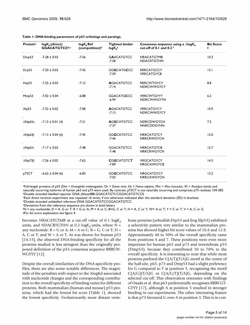
Table 1: DNA-binding parameters of p53 orthologs and paralogs.
Proteina logKd (direct)GGACATGTCCb,c
logKd Ref(competition)d
Tightest binderlogKd
eConsensus sequence using a ΔlogKd cut-off of 0.1 and 0.2 f
Bit Scoreg
Dmp53 -7.28 ± 0.03 -7.56 GAACATGTCC-7.58
NRACATGTMBNDACRTGTHN
10.3
Drp53 -7.20 ± 0.03 -7.42 GGGCATGCCC-7.59
RRRCATGCCYVRRCATGYCB
13.1
Hsp53 -7.25 ± 0.03 -7.12 AGACATGTCC-7.14
RRRCWTGYCYNDRCWWGYCY
8.8
Mmp53 -7.02 ± 0.04 -6.88 GGACATGCCC-6.94
VRRCWTGYYYNDRCWWGYYN
6.2
Xlp53 -7.32 ± 0.02 -7.08 AGACATGTCC-7.13
VRRCATGYCYNDRCWWGYCY
10.9
ΔNp63α -7.12 ± 0.01 (4) -7.21 AGGCATGTCC-7.37
NDRCDWGYCHNNRCDDGYHN
7.2
ΔNp63β -7.13 ± 0.04 (6) -7.45 GGGCATGTCC-7.46
NRRCATGTCYNRRCRWGYCN
12.0
ΔNp63γ -7.17 ± 0.02 -7.48 GGACATGTCC-7.48
NRRCATGTCBNRRCRWGYCN
12.7
ΔNp73β -7.26 ± 0.02 -7.62 CGGCATGTCT-7.89
VRGCATGYCYNRGCAWGYCB
14.5
p73CT -6.62 ± 0.04 (6) -6.83 GGGCATGTCC-7.03
NRGCATGYCYNRRCAWGYCN
13.2
aFull-length proteins of p53 (Dm = Drosophila melanogaster, Dr = Danio rerio, Hs = Homo sapiens, Mm = Mus musculus, Xl = Xenopus laevis) and naturally occurring isoforms of human p63 and p73 were used. By contrast, p73CT is not naturally occurring and comprises p73 residues 104-383.bDouble stranded labelled reporter DNA (Alexa488-GGACATGTCCGGACATGTCCf)cEach direct titration experiment was repeated 16 times, if not otherwise indicated after the standard deviation (SD) in bracketsdDouble stranded unlabelled reference DNA GGACATGTCCGGACATGTCCeDeviations from the reference sequence are shown in bold lettersfN = any nucleotide, D = A, G or T; R = G or A; M = A or C; B=G, C or T; H = A, C or T; W= A or T; Y = C or T; V = A, C or GgFor bit score explanation see figure 4.
Page 5 of 14(page number not for citation purposes)

BMC Genomics 2009, 10:628 http://www.biomedcentral.com/1471-2164/10/628
Page 6 of 14(page number not for citation purposes)
DNA-binding specificity of p53 family membersFigure 4DNA-binding specificity of p53 family members. ΔlogKd plot for all competitor DNA sequences. Affinity penalties with respect to the reference sequence caused by base pair substitutions are shown for all nucleotides (A = black, T = green, G = red, C = blue). The reference sequence is indicated below the axis. Only the analysed half-site is shown. A positive value indi-cates weaker binding of the competitor sequence than the reference sequence, whereas a negative value indicates that the sub-stitution leads to tighter protein-DNA binding.
-0.4
0
0.4
0.8
1.2
1.6
G G A C A T G T C C
Hsp53 Mmp53
0.4
0
0.4
0.8
1.2
1.6
G G A C A T G T C C
-0.4
0
0.4
0.8
1.2
1.6
G G A C A T G T C C 0.4
0
0.4
0.8
1.2
1.6
G G A C A T G T C C
-0.4
0
0.4
0.8
1.2
1.6
G G A C A T G T C C
Xlp53
-0.4
0
0.4
0.8
1.2
1.6
G G A C A T G T C C
ΔNp63α
-0.4
0
0.4
0.8
1.2
1.6
G G A C A T G T C C
ΔNp63β
-0.4
0
0.4
0.8
1.2
1.6
G G A C A T G T C C
ΔNp63γ
-0.4
0
0.4
0.8
1.2
1.6
G G A C A T G T C C
ΔNp73β
-0.4
0
0.4
0.8
1.2
1.6
G G A C A T G T C C
p73CT
Drp53Dmp53
Δlog
K d
Δlog
K d
Δlog
K d
Δlog
K d
Δlog
K d
Δlog
K d
Δlog
K d
Δlog
K d
Δlog
K d
Δlog
K d
-
-
G
AT
C

BMC Genomics 2009, 10:628 http://www.biomedcentral.com/1471-2164/10/628
Page 7 of 14(page number not for citation purposes)
Sequence logos for all p53 family proteins studiedFigure 5Sequence logos for all p53 family proteins studied. The complete response element is shown, and bit values are plotted against sequence position. A value of 0 means all four nucleotides bind with the same affinity and there is no selectivity, whereas the value 2 stands for absolute selectivity for one nucleotide, with the other three being highly penalised. A measure of the total information content (or selectivity) of the protein can be calculated by summing up all individual bit scores at every position. The maximum theoretical value of information content for a 20-bp response element is 40 bit.
2 4 6 8 10 12 14 16 18 20
2
1.5
1
0.5
0
2 4 6 8 10 12 14 16 18 20
2
1.5
1
0.5
0
2 4 6 8 10 12 14 16 18 20
2
1.5
1
0.5
0
2 4 6 8 10 12 14 16 18 20
2
1.5
1
0.5
02 4 6 8 10 12 14 16 18 20
2
1.5
1
0.5
0
2 4 6 8 10 12 14 16 18 20
2
1.5
1
0.5
0
Hsp532 4 6 8 10 12 14 16 18 20
2
1.5
1
0.5
0
Dmp53 Drp53
Mmp53
2 4 6 8 10 12 14 16 18 20
2
1.5
1
0.5
0
Xlp53
2 4 6 8 10 12 14 16 18 20
2
1.5
1
0.5
0
ΔNp63β
ΔNp63α
ΔNp63γ
ΔNp73β
2 4 6 8 10 12 14 16 18 20
2
1.5
1
0.5
0
p73CT
BMC Genomics 2009, 10:628 http://www.biomedcentral.com/1471-2164/10/628
trast to findings which suggest an A preceding the CWWGfollowed by a T forms the most stable complexes with p73[18]. It is worth noting that the overall effects of nucle-otide substitutions at positions 3 and 5 were relativelysmall compared to the effects at the positions 4 and 7.
While the isoforms ΔNp63β and ΔNp63γ behaved almostidentically, the isoform ΔNp63α showed considerablysmaller affinity penalties, meaning it is less specific. Inter-estingly, the DNA-binding affinities in the direct titrationsand affinities for the reference sequence in competitionexperiments were similar for all isoforms. This suggeststhat the presence of the extreme C-terminal post-SAMdomain in ΔNp63α may affect its DNA-binding specifi-city. Despite the significantly weaker binding of p73CTcompared to ΔNp73β to DNA, the DNA-binding specifi-city of both p73 proteins was identical. This suggests thatthe DNA-binding specificity of tetrameric p73 is deter-mined by the DNA-binding properties of individual DNA-binding domains, whereas the absolute affinity dependson the oligomerisation equilibrium.
DNA methylation does not alter the specificity of p53 family membersCpG methylation has been shown to affect DNA recogni-tion of transcription factors [28-30]. To investigate theeffects of CpG methylation on DNA recognition of p53family proteins, we used a method that we have previ-ously applied to human p53 [20]. We systematically intro-duced a CpG dinucleotide at each position in theconsensus p53 DNA binding sequence and identified sub-stitutions tolerated by p53 family proteins. We then com-pared the binding affinities of methylated versus non-methylated sequences containing CpG (Additional file 4).Vertebrate p53 proteins (Mmp53, Xlp53 and Drp53)behaved similarly to human p53 and were mildly affectedby substitutions at positions 2, 4 and 6. Interestingly,methylated sequences bound somewhat more tightly thannon-methylated, although the effect of a single methyla-tion was small. p63 and p73, along with invertebrateDmp53, also tolerated CpG nucleotides at these posi-tions. In particular, substitution at position 4 hardlychanged the affinity, confirming that the CGTC centralelement of the binding site is recognised equally well asCATG, which is preferred by p53.
Computational genome analysisTranscription factors recognise a range of sequences whichdeviate from the highest affinity sequence. As a result ofthis deviation, the affinity of these sequences can be sig-nificantly weaker than that of the highest affinitysequence. We have previously shown that most of thereported p53 binding sites have affinity values up to 1.5logKd units weaker than the highest affinity sequence, andthat there is a very large number of potential binding sites
in the genome [15]. In this study, the highest affinitysequence was practically identical for all the proteins stud-ied, but the relative penalties for nucleotide substitutionswere different. Such differential penalties may result inselection of non-overlapping sets of binding sites by dif-ferent p53 family members.
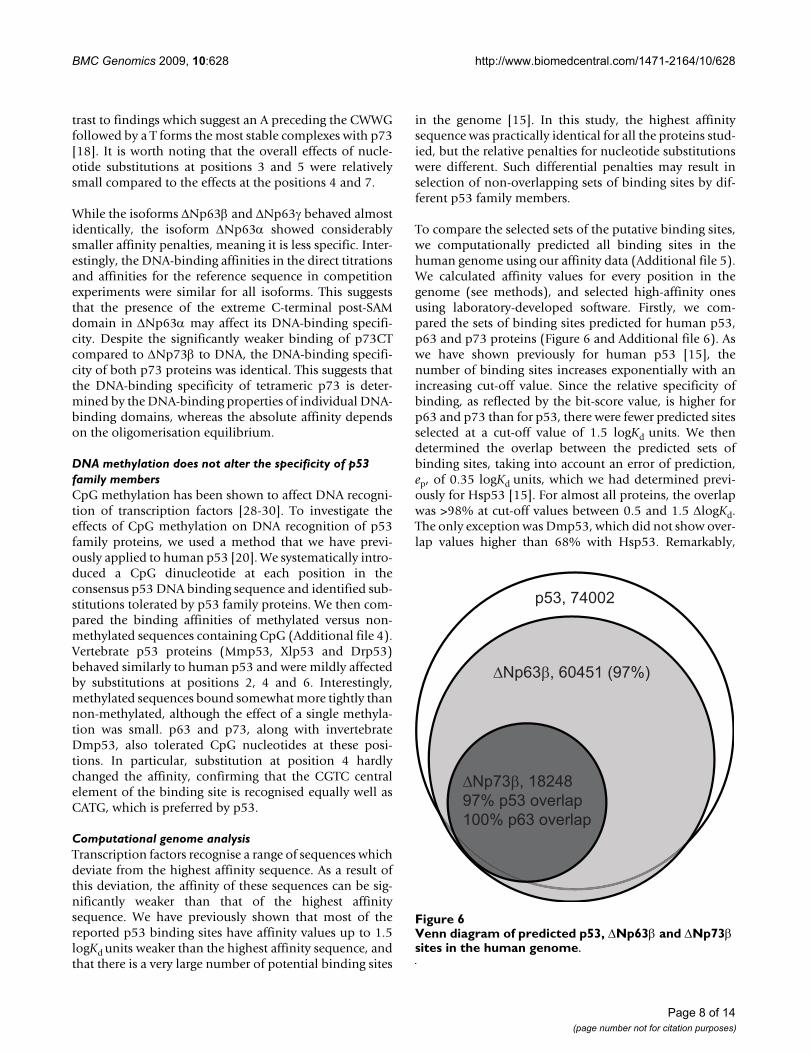
To compare the selected sets of the putative binding sites,we computationally predicted all binding sites in thehuman genome using our affinity data (Additional file 5).We calculated affinity values for every position in thegenome (see methods), and selected high-affinity onesusing laboratory-developed software. Firstly, we com-pared the sets of binding sites predicted for human p53,p63 and p73 proteins (Figure 6 and Additional file 6). Aswe have shown previously for human p53 [15], thenumber of binding sites increases exponentially with anincreasing cut-off value. Since the relative specificity ofbinding, as reflected by the bit-score value, is higher forp63 and p73 than for p53, there were fewer predicted sitesselected at a cut-off value of 1.5 logKd units. We thendetermined the overlap between the predicted sets ofbinding sites, taking into account an error of prediction,ep, of 0.35 logKd units, which we had determined previ-ously for Hsp53 [15]. For almost all proteins, the overlapwas >98% at cut-off values between 0.5 and 1.5 ΔlogKd.The only exception was Dmp53, which did not show over-lap values higher than 68% with Hsp53. Remarkably,
Venn diagram of predicted p53, ΔNp63β and ΔNp73β sites in the human genomeFigure 6Venn diagram of predicted p53, ΔNp63β and ΔNp73β sites in the human genome.
p53, 74002
ΔNp63β, 60451 (97%)
ΔNp73β, 1824897% p53 overlap100% p63 overlap
Page 8 of 14(page number not for citation purposes)

BMC Genomics 2009, 10:628 http://www.biomedcentral.com/1471-2164/10/628
Dmp53 showed overlaps close to 100% with ΔNp63α.Overall, the results of computational analysis suggest that,based on DNA-binding preferences alone, all members ofp53 family bind the same set of putative sites in thehuman genome. The observed quantitative differences inthe binding preferences may result in different affinitiestoward specific binding site sequences, but not in diverg-ing sets of target sites within a given genome.
DiscussionOligomerisation properties of p53 family proteinsThe tetramerisation domain of Hsp53 (residues 325-356)is highly conserved in all vertebrate proteins of the p53family [31]. A sequence alignment of the tetramerisationdomain region of proteins used in this study is shown inAdditional file 7. The Hsp53 tetramerisation domainforms a dimer of dimers and is composed of short mono-meric building blocks consisting of a β-strand followed byan α-helix [32-34]. The primary dimers are stabilized byan intermolecular β-sheet and mainly hydrophobic helixpacking interactions. In addition, the primary-dimerinterface is stabilised by a salt bridge, which is typical forp53 orthologs but not found in its paralogs (Figure 7,Additional file 7). The tetrameric interface is formed byhydrophobic helix packing interactions. The hydrophobicinterfaces are largely conserved in all the proteins studiedexcept for Dmp53, which shows no significant sequenceconservation and has a dimer-dimer interface that featuresa cluster of charged residues at its centre [31]. Importantly,recent structural studies have shown that the p73 tetram-erisation domain contains an additional C-terminal helix,which is essential for the structural integrity and stabilityof the tetramer (Figure 7A). This helix is conserved in p63and presumably has a similar structural role [25,35].
We determined dissociation constants for the monomer-dimer and dimer-tetramer equilibria of seven members ofthe p53 family (Figure 2, Additional file 2). Hsp53,Mmp53 and Xlp53 showed very similar Kd values, consist-ent with the high conservation of contact residues. p63and p73 form tighter tetramers than human p53, which,at least in the case of p73, can be attributed to extensiveinter-dimer contacts made by the additional C-terminalhelix (Figure 7A). Drp53, which, phylogenetically, can beplaced somewhere between mammalian p53 and thep63/p73 paralogs [25], also forms more stable tetramers.What is most surprising is that Dmp53 forms tetramerswith a comparable Kd, while having a completely differentdimer-dimer interface, suggesting that, despite structuraldivergence, the strength of the tetramer has been con-served through evolution.
Interestingly, the primary-dimer interface is tighter in p53than in p73 (6-fold) and p63 (9-fold). Comparison of theHsp53 and Drp53 sequences with p63 and p73 suggests
that this difference in dimer stability may be attributed tothe R337-D352 salt bridge that stabilizes the helix packingin the p53 primary dimer and large-to-small substitutionsof hydrophobic residues in p63 and p73. The salt bridgeis highly conserved in p53 across different species, and itsdisruption by a germline mutation (R337H) has beenlinked with adrenocortical carcinomas in children andother cancer forms [36,37]. p63 and p73 lack this inter-molecular salt bridge and have a threonine (p63) andglutamine (p73) instead of the arginine in p53. As a resultof the weakened dimer interface in p63 and p73, the dim-ers formed by tetramer dissociation are more likely to dis-
The tetramerisation domain of human p53 and p73Figure 7The tetramerisation domain of human p53 and p73. A) Superposition of the crystal structures of the tetramerisa-tion domain of human p53 (green) [57] and human p73 (pur-ple) [25], showing that p73 contains an additional C-terminal helix. This helix is conserved in the p63 sister protein. B) Pri-mary-dimer interface of human p53. The side chains of Y327, L330, I332, R333, F338, L344 (green) and the salt bridge between R337 and D352 (orange) are shown as stick models. The salt bridge is not conserved in human p63 and p73.
Arg337 / Asp352
B
A
Page 9 of 14(page number not for citation purposes)

BMC Genomics 2009, 10:628 http://www.biomedcentral.com/1471-2164/10/628
sociate directly into monomers. Since key features of theprimary dimer interface are highly conserved among dif-ferent species for each paralog, it is likely that they exhibitdissociation equilibria similar to their human orthologs.The only exceptions are Cavia porcellus and Pteropusvampyrus, whose p53 lacks the paralog-specific salt bridgeand may, therefore, also have weakened primary dimers.The observed differences in dissociation equilibria of thehuman paralogs may have important biological implica-tions for interactions with regulatory proteins, such asmembers of the S100 family, which have been shown todifferentially bind different oligomeric states of p53[38,39]. Taken together, our results show that the overallstrength of oligomerisation was conserved during the evo-lution of members of the p53 family, while subtle differ-ences in the equilibria may play a role in fine-tuning theirbiological activity.
DNA-contact residues are highly conserved in vertebratesThe sequence identity of the DNA-binding domain of p53family members varies and is highest between p53 fromclosely related species, e.g. 86% identity between mouseand human proteins and ~60% between Drp53/Xlp53and Hsp53. Hsp53 makes direct sequence-specific con-tacts with bases in the major groove of DNA via the sidechains of K120, A276, C277 and R280. Contacts with thephosphate backbone are made by the side chains of S241,R248 and R273, and the backbone amides of K120 andA276 [40,41]. All DNA-contact residues are conserved inthe vertebrate proteins studied (Additional file 8). Uponbinding to a DNA half-site, two DBDs form a self-comple-mentary protein-protein interface, mediated by residuesP177, H178, R181, M243 and G244, which are conservedin vertebrate p53 [40,41]. In human p63 and p73 (~60%sequence identity with Hsp53), however, there are keysubstitutions in this region, indicating differences in theinter-DBD interactions. Dmp53 shows only 24%sequence identity to human p53 [42], with significant dif-ferences in the various DNA-binding motifs. K120 in theflexible L1 loop of Hsp5 binds to two purine bases in posi-tion 2 and 3 of the response element. The equivalent loopin Dmp53 is shortened and more rigid, making it unlikelythat the lysine (K102 in Dmp53) forms the same DNAcontacts as in Hsp53. In addition, the alanine (A276)making sequence-specific hydrophobic contacts in Hsp53[40] is replaced by a threonine in Dmp53 (T262). Further-more, the DNA-backbone contact residue R273 in Hsp53is replaced by a lysine (K259). The L3 loop, which docksto the DNA minor groove via R248 in Hsp53, is also sig-nificantly different. It has a deletion and lacks the equiva-lent of R249, which plays a key role in stabilizing thisregion in Hsp53 [43]. Moreover, the L2/L3-loop regionthat forms the self-complementary DBD-DBD interfacealso shows variations, similarly to p63 and p73. Taken
together, it would be reasonable to expect that the DNA-binding properties of Dmp53 differ from those of Hsp53.
Conservation of the p53 response element and DNA-binding specificityWe quantified the DNA-binding properties of severalmembers of the p53 family and investigated their abilityto recognise methylated DNA. We found that the DNA-binding specificity of both orthologs and paralogs of p53was conserved. Human and mouse p53 proteins showedalmost identical specificity, consistent with their highestsequence conservation. It is also interesting to note thatthey exhibited the lowest absolute specificity, as reflectedby the lowest bit score of the derived motif. Evolutionarilymore distant vertebrate p53 proteins (Xlp53 and Drp53)showed a very similar specificity profile but somewhathigher specificity. There seems to be a very interestingunderlying correlation: the more complex the organismand the more complex the p53 pathway, the lower theabsolute specificity. p63 and p73 showed slightly differentDNA-binding specificity compared with p53. This differ-ence may be the result of the different residues in p63 andp73 being responsible for the interaction between twoDBDs upon binding to a half-site motif. Despite the lowsequence similarity of Dmp53 and human p53, and theiraforementioned differences in key DNA-binding motifs,the DNA-binding specificity of Dmp53 is preserved and issimilar to that of vertebrate p53 family members, in par-ticular the more ancestral p63 and p73 proteins. The long-est p63 isoform tested, ΔNp63α, has a significantlyreduced DNA-binding specificity compared to other iso-forms. It is possible that the additional post-SAM domainpresent in this isoform is directly or indirectly involved inregulation of its sequence-specific binding.
Using the affinity prediction, we identified all putativebinding sites in the human genome for p53, p63 and p73proteins. Despite quantitative differences in their DNA-binding specificity, all transcription factors studied selectoverlapping sets of binding sites. We found many moreputative binding sites than have been previously identi-fied in genome-wide experiments for p53/p63/p73 pro-teins [44-46]. The vast majority (95%) of experimentallyidentified p53 binding sites [44] contains a site predictedusing our affinity data. The published dataset for p63 [45]consists of 5000 sites, which is significantly more than the1700 sites reported for p53. Less than 20% of these 5000sites contain a predicted high-affinity p63 site within a500 bp window, perhaps reflecting different stringencycriteria in peak calling in these two studies. Despite thesedifferences, analysis of all in vivo binding-site sequences inthese studies generated positional weight matrices, repre-sented as sequence logos, which are very similar to thesequence logos derived by us based on in vitro bindingaffinity. This strongly suggests that the driving force for
Page 10 of 14(page number not for citation purposes)

BMC Genomics 2009, 10:628 http://www.biomedcentral.com/1471-2164/10/628
localisation of p53/p63/p73 to their respective sites in thegenome is their sequence-specific binding. A recent studyusing a novel microsphere assay showed that the DNA-binding specificity of endogenous p53 in cell lysate is thesame as that of the purified recombinant p53 from ourwork [47]. Nevertheless, several validated p53 responseelements contain non-canonical sequences [48,49]. It wasshown, that p53 acts weakly to moderately on responseelements that contain only a half or a three quarter site ofthe canonical consensus sequence [50]. This is in accord-ance with our results, as we observed considerable bind-ing to DNA with a mutated quarter or half site, which defacto represents a non-canonical p53 response element.Binding to non-canonical response elements may be facil-itated by co-activating transcription factors. A comprehen-sive comparison between in vivo and in vitro binding canbe found in an excellent recent review [51].
How can transcription factors with virtually identicalDNA-binding specificity elicit different biologicalresponses? There is also the closely related question ofhow transcription factors select their binding site in thegenome, among many potential sites of comparable affin-ity? The "chromatin structure" and "DNA accessibility"concepts may at least partially answer the second ques-tion, although the mechanism controlling the chromatinstructure with the specificity required is presentlyunknown. Different expression patterns of transcriptionfactors and/or their abundance in the nucleus can alsocontribute to their specificity. The involvement of addi-tional specificity factors would answer both questions.Such additional specificity factors should also bind DNAin a sequence-specific manner, and are likely to be tran-scription factors.
ConclusionsTaken together, our data show that tetramerisation of p53family members, which is important for high-affinityDNA binding, was established very early in the evolutionof the p53 family and has been functionally conservedever since. Despite significant differences in the contactsurfaces involved, the strength of oligomerisation was pre-served. Intriguingly, the DNA-binding specificity of differ-ent p53 family members is highly conserved even forevolutionarily distant species. This suggests that originalfunctions were preserved while new functions wereacquired during evolution, utilising the same DNA-bind-ing specificity. The "core function" DNA-binding specifi-city of the p53 transcription factor network did notsubstantially change during evolution. Instead, there isaccumulating evidence that functional divergence of thep53 family evolved through changes in the connectivitywithin the network, for example by interactions of p53family members with different sets of co-activating tran-scription factors.
MethodsProtein cloningFor human full-length p53 we used wild type protein forDNA-binding experiments and a super-stable mutant,which has four mutations in the core domain (QM-Hsp53, M133L/V203A/N239Y/N268D) [52,53], for ana-lytical ultracentrifugation experiments. A plasmid encod-ing Mmp53 was kindly provided by Geoffrey Wahl.Dmp53 was amplified from a cDNA library kindly pro-vided by Simon Bullock. Coding sequences encoding forother studied proteins were amplified from clonesobtained from the Mammalian Gene Collection (MGC),distributed via Geneservice (UK). For the ΔNp63γ iso-form, parts of the gene were amplified from a genomicDNA library (Geneservice). Additionally, we made a p73construct containing the DBD and parts of the OD(p73CT, residues 104-383). All inserts were subclonedinto a pET24a-HLTEV plasmid containing an N-terminal6xHis purification tag, a lipoyl domain [54] for improvedsolubility and a TEV-protease cleavage site. Constructscontaining a C-terminal FlAsH-tag CCPGCC [24] weredesigned in a similar manner.
Small scale expression screeningSmall-scale screening for soluble expression in differentcell lines was performed in 2 ml cultures on microplatesin 2xTY media following induction with 1 mM IPTG. Pro-teins were purified using His-Fusion magnetic beads (Bio-Clone Inc) on a BioSprint15 robot (Qiagen). Purifiedfractions were analysed by SDS-PAGE pre- and post-diges-tion with TEV-protease.
Expression and purificationLarge-scale expression and purification was carried outlargely as described earlier [20,22]. All proteins were over-expressed in E. coli BL21 or B834 cells (Novagen) at 18°Cfor 16-20 h and purified using standard Ni-affinity chro-matography protocols. Subsequently, the N-terminal tagswere cleaved off by TEV-protease digestion. As a secondpurification step for p53 orthologs, heparin affinity chro-matography was used. Solutions were diluted to reducethe salt concentration to about 30 mM NaCl. Proteinswere eluted using a 20 column volume NaCl gradient (0to 1 M NaCl). The final purification step was gel filtrationchromatography using a Superdex 200 16/60 preparativegel filtration column (GE Healthcare) in 225 mM NaCl,25 mM sodium phosphate pH 7.2, 10% glycerol and 5mM DTT. Protein purity of >95% was determined by SDS-gel electrophoresis. Samples were flash frozen in liquidnitrogen and stored at -80°C until used.
Labelling proteins with FlAsHLabelling of C-terminally FlAsH-tagged (CCPGCC) pro-teins [24] was performed in 150 mM NaCl, 25 mM phos-phate (pH 7.2), 10% glycerol, and 1 mM β-
Page 11 of 14(page number not for citation purposes)

BMC Genomics 2009, 10:628 http://www.biomedcentral.com/1471-2164/10/628
mercaptoethanol. 200 μL of 10 μM FlAsH-tagged proteinwere incubated with 1.5 equivalents of FlAsH-EDT2(Lumio Green, Invitrogen) at 8°C for 2.5 h. We estimatedthat the stock solution was supplied at a concentration ofapproximately 1 mM. Excess label was removed by dialy-sis into the above buffer. Labelled proteins could be fro-zen and stored for at least a few months. The labellingreaction could easily be reversed by adding DTT, so carehad to be taken to avoid DTT in buffers.
Sedimentation velocity experimentsWe used a XL-I analytical ultracentrifuge (Beckman)equipped with an AVIV fluorescence detection system(AVIV Biomedical). Experiments with C-terminallyFlAsH-tagged proteins and unlabelled QM-Hsp53 (usingan absorbance detection system) were done in 150 mMNaCl, 25 mM phosphate (pH 7.2), 10% glycerol, BSA (0.2mg/mL) and 1 mM β-mercaptoethanol at 10°C. For fluo-rescence measurements, cells were pre-treated with a con-centrated (1 mg/ml) solution of BSA and allowed to drybefore loading samples. Sample volume was 80-90 μL atconcentrations of 5-500 nM in SedVel60K fluorescencevelocity cells (Spin Analytical). At least 15 measurementswere done for each protein. Buffer density and viscositywere calculated using SEDNTERP software. Data analysisto obtain sedimentation coefficient traces was done withSEDFIT software [55]. Since only the tetramer peak at 3 Swas detected in experiments with Hsp53 without theFlAsH-tag, we ignored peaks at higher sedimentation coef-ficients found for FlAsH-tagged proteins as artefactscaused by cross-linking of oxidised cysteines of the tag.Fitting of sedimentation profiles to normal distributionsand Kd calculation was done with our own laboratory soft-ware to estimate the relative amount of dimers andtetramers. The reported values for human p53 are some-what lower than the values we have reported previously[56]. Most likely, a change in the cell design resulting insignificantly lower surface area of exposed epoxy materialand pre-treatment of the cells with concentrated BSA solu-tion minimised the adsorption of p53 proteins to the cellwall, thereby increasing the fraction of material present insolution.
Fluorescence anisotropy spectroscopyAll experiments were carried out in 96-well plates using aPherastar plate reader (BMG Labtech) equipped with aBravo 96-channel pipetting robot (Velocity 11) as previ-ously described [15]. Buffer conditions for all experimentswere 25 mM NaPi, 225 mM NaCl, 10% v/v glycerol, 5 mMDTT and 0.2 mg/mL BSA. Titrations were done at 22°Cand repeated at least three times. Direct titrations weredone as previously described [22] using 20 nM 5'-Alexa488-GGACATGTCCGGACATGTCC labelled DNA(Operon). The stock solution of 1.25 μM protein wastitrated in small amounts, which allows calculation of the
Kd for the binding of labelled DNA to protein [21]. Forcompetition experiments, a mixture of protein (at a con-centration four times above the Kd value, measured bydirect titrations) and 20 nM labelled DNA were used asanalytes, and competitor DNA (50 μM) was titrated insmall steps. Over 3000 titrations were performed in total.Data were analysed according to cooperative binding andcompetition models using laboratory developed software[15].
Computational search for putative binding sitesThe putative binding sites in the genome were locatedusing p53BindingSite software [15], available at http://www.mrc-lmb.cam.ac.uk/dbv. In short, the DNA-bindingaffinity was predicted for each position in the genomeusing binding affinity positional matrices measured foreach protein studied, and positions with predicted affinityhigher than the cut-off value were selected. We usedhuman genome release 36.3, zebrafish genome release10/06/2008 (International Human Genome SequencingConsortium), fruit fly genome release 5 (The FlyBase Con-sortium/Berkeley Drosophila Genome Project) andmouse genome release 37 (Mouse Genome SequencingConsortium). Instead of Xenopus laevis we used the Xeno-pus tropicalis genome (release 4.1, DOE Joint GenomeInstitute), as it is complete. We set the gap between bothhalf-sites of the RE to be 0 and 1.
Authors' contributionsDBV conceived research; TB, MP and DBV performedexperiments, TB, MP, ACJ and DBV analysed and inter-preted the results; TB and MP prepared figures; TB, ACJand DBV wrote the manuscript. All authors read andapproved the final manuscript.
Additional material
Additional file 1Figure S1. Example of raw fluorescence data from analytical ultracentrif-ugation experiments.Click here for file[http://www.biomedcentral.com/content/supplementary/1471-2164-10-628-S1.PDF]
Additional file 2Table S1. Summary of the calculated dissociation constants, observed sed-imentation coefficients and protein concentration ranges used in analyti-cal ultracentrifugation experiments.Click here for file[http://www.biomedcentral.com/content/supplementary/1471-2164-10-628-S2.PDF]
Additional file 3Table S2. DNA sequences used for anisotropy experiments.Click here for file[http://www.biomedcentral.com/content/supplementary/1471-2164-10-628-S3.PDF]
Page 12 of 14(page number not for citation purposes)
BMC Genomics 2009, 10:628 http://www.biomedcentral.com/1471-2164/10/628
AcknowledgementsWe thank Caroline Blair for initial cloning experiments and advice in molec-ular biology as well as Roger Williams, Sarah Teichmann, Caroline Blair and Joel Kaar for critical reading of the manuscript. Discussions about structural properties with Antonina Andreeva proved to be very helpful. TB is sup-ported by Cambridge European Trust and Medical Research Council. This research was supported by Cancer Research UK, the Medical Research Council, and by EC FP6 funding. This publication reflects the authors' views and not necessarily those of the EC. The Community is not liable for any use that may be made of the information.
References1. Wilson D, Charoensawan V, Kummerfeld SK, Teichmann SA: DBD-
-taxonomically broad transcription factor predictions: newcontent and functionality. Nucleic Acids Res 2008:D88-92.
2. Amoutzias GD, Veron AS, Weiner J, Robinson-Rechavi M, Brnberg-Bauer E, Oliver SG, Robertson DL: One billion years of bZIPtranscription factor evolution: conservation and change indimerization and DNA-binding site specificity. Molecular biol-ogy and evolution 2007, 24(3):827-835.
3. Massari ME, Murre C: Helix-loop-helix proteins: regulators oftranscription in eucaryotic organisms. Mol Cell Biol 2000,20(2):429-440.
4. Vogelstein B, Lane D, Levine AJ: Surfing the p53 network. Nature2000, 408(6810):307-310.
5. Vousden KH, Lu X: Live or let die: the cell's response to p53.Nat Rev Cancer 2002, 2(8):594-604.
6. Joerger AC, Fersht AR: Structure-function-rescue: the diversenature of common p53 cancer mutants. Oncogene 2007,26(15):2226-2242.
7. Petitjean A, Mathe E, Kato S, Ishioka C, Tavtigian SV, Hainaut P,Olivier M: Impact of mutant p53 functional properties onTP53 mutation patterns and tumor phenotype: lessons fromrecent developments in the IARC TP53 database. Hum Mutat2007, 28(6):622-629.
8. Moll UM, Slade N: p63 and p73: roles in development andtumor formation. Mol Cancer Res 2004, 2(7):371-386.
9. Jegga AG, Inga A, Menendez D, Aronow BJ, Resnick MA: Functionalevolution of the p53 regulatory network through its targetresponse elements. Proc Natl Acad Sci USA 2008, 105(3):944-949.
10. Joerger AC, Fersht AR: Structural biology of the tumor sup-pressor p53. Annual review of biochemistry 2008, 77:557-582.
11. Scoumanne A, Harms KL, Chen X: Structural basis for gene acti-vation by p53 family members. Cancer Biol Ther 2005,4(11):1178-1185.
12. El-Deiry WS, Kern SE, Pietenpol JA, Kinzler KW, Vogelstein B: Defi-nition of a consensus binding site for p53. Nat Genet 1992,1(1):45-49.
13. Funk WD, Pak DT, Karas RH, Wright WE, Shay JW: A transcrip-tionally active DNA-binding site for human p53 protein com-plexes. Mol Cell Biol 1992, 12(6):2866-2871.
14. Tomso DJ, Inga A, Menendez D, Pittman GS, Campbell MR, Storici F,Bell DA, Resnick MA: Functionally distinct polymorphicsequences in the human genome that are targets for p53transactivation. Proc Natl Acad Sci USA 2005, 102(18):6431-6436.
15. Veprintsev DB, Fersht AR: Algorithm for prediction of tumoursuppressor p53 affinity for binding sites in DNA. Nucleic AcidsRes 2008, 36(5):1589-1598.
16. Schavolt KL, Pietenpol JA: p53 and Delta Np63 alpha differen-tially bind and regulate target genes involved in cell arrestDNA repair and apoptosis. Oncogene 2007, 26(42):6125-6132.
17. Osada M, Park HL, Nagakawa Y, Yamashita K, Fomenkov A, Kim MS,Wu G, Nomoto S, Trink B, Sidransky D: Differential recognitionof response elements determines target gene specificity forp53 and p63. Mol Cell Biol 2005, 25(14):6077-6089.
18. Lokshin M, Li Y, Gaiddon C, Prives C: p53 and p73 display com-mon and distinct requirements for sequence specific bindingto DNA. Nucleic Acids Res 2007, 35(1):340-352.
19. Murray-Zmijewski F, Lane DP, Bourdon JC: p53/p63/p73 isoforms:an orchestra of isoforms to harmonise cell differentiationand response to stress. Cell Death Differ 2006, 13(6):962-972.
20. Petrovich M, Veprintsev DB: Effects of CpG methylation on rec-ognition of DNA by the tumour suppressor p53. J Mol Biol2009, 386(1):72-80.
21. Weinberg RL, Veprintsev DB, Fersht AR: Cooperative binding oftetrameric p53 to DNA. J Mol Biol 2004, 341(5):1145-1159.
22. Veprintsev DB, Freund SM, Andreeva A, Rutledge SE, Tidow H, Cana-dillas JM, Blair CM, Fersht AR: Core domain interactions in full-length p53 in solution. Proc Natl Acad Sci USA 2006,103(7):2115-2119.
23. MacGregor IK, Anderson AL, Laue TM: Fluorescence detectionfor the XLI analytical ultracentrifuge. Biophys Chem 2004,108(1-3):165-185.
24. Adams SR, Campbell RE, Gross LA, Martin BR, Walkup GK, Yao Y,Llopis J, Tsien RY: New biarsenical ligands and tetracysteinemotifs for protein labeling in vitro and in vivo: synthesis andbiological applications. J Am Chem Soc 2002, 124(21):6063-6076.
25. Joerger AC, Rajagopalan S, Natan E, Veprintsev DB, Robinson CV,Fersht AR: Structural evolution of p53, p63, and p73: implica-tion for heterotetramer formation. Proc Natl Acad Sci USA 2009,106(42):17705-17710.
26. Schneider TD, Stephens RM: Sequence logos: a new way to dis-play consensus sequences. Nucleic Acids Res 1990,18(20):6097-6100.
27. Schneider TD, Stormo GD, Gold L, Ehrenfeucht A: Informationcontent of binding sites on nucleotide sequences. J Mol Biol1986, 188(3):415-431.
28. Bird AP, Wolffe AP: Methylation-induced repression--belts,braces, and chromatin. Cell 1999, 99(5):451-454.
29. Jaenisch R, Bird A: Epigenetic regulation of gene expression:how the genome integrates intrinsic and environmental sig-nals. Nat Genet 2003, 33:245-254.
Additional file 4Figure S2. Results of fluorescence anisotropy binding assays using meth-ylated DNA.Click here for file[http://www.biomedcentral.com/content/supplementary/1471-2164-10-628-S4.PDF]
Additional file 5Table S3. DNA binding specificity positional weight matrices for p53 family members.Click here for file[http://www.biomedcentral.com/content/supplementary/1471-2164-10-628-S5.XLS]
Additional file 6Table S4. Results of computational prediction of putative binding sites within the genome.Click here for file[http://www.biomedcentral.com/content/supplementary/1471-2164-10-628-S6.PDF]
Additional file 7Figure S3. Sequence alignment of the tetramerisation domains of the p53 family members.Click here for file[http://www.biomedcentral.com/content/supplementary/1471-2164-10-628-S7.PDF]
Additional file 8Figure S4. Sequence alignment of the DNA-binding domains of the p53 family members.Click here for file[http://www.biomedcentral.com/content/supplementary/1471-2164-10-628-S8.PDF]
Page 13 of 14(page number not for citation purposes)
http://www.ncbi.nlm.nih.gov/entrez/query.fcgi?cmd=Retrieve&db=PubMed&dopt=Abstract&list_uids=1301998
http://www.ncbi.nlm.nih.gov/entrez/query.fcgi?cmd=Retrieve&db=PubMed&dopt=Abstract&list_uids=1301998
http://www.ncbi.nlm.nih.gov/entrez/query.fcgi?cmd=Retrieve&db=PubMed&dopt=Abstract&list_uids=1588974
http://www.ncbi.nlm.nih.gov/entrez/query.fcgi?cmd=Retrieve&db=PubMed&dopt=Abstract&list_uids=1588974
http://www.ncbi.nlm.nih.gov/entrez/query.fcgi?cmd=Retrieve&db=PubMed&dopt=Abstract&list_uids=1588974
http://www.ncbi.nlm.nih.gov/entrez/query.fcgi?cmd=Retrieve&db=PubMed&dopt=Abstract&list_uids=2172928
http://www.ncbi.nlm.nih.gov/entrez/query.fcgi?cmd=Retrieve&db=PubMed&dopt=Abstract&list_uids=2172928
http://www.ncbi.nlm.nih.gov/entrez/query.fcgi?cmd=Retrieve&db=PubMed&dopt=Abstract&list_uids=3525846
BMC Genomics 2009, 10:628 http://www.biomedcentral.com/1471-2164/10/628
Publish with BioMed Central and every scientist can read your work free of charge
"BioMed Central will be the most significant development for disseminating the results of biomedical research in our lifetime."
Sir Paul Nurse, Cancer Research UK
Your research papers will be:
available free of charge to the entire biomedical community
peer reviewed and published immediately upon acceptance
cited in PubMed and archived on PubMed Central
yours — you keep the copyright
Submit your manuscript here:http://www.biomedcentral.com/info/publishing_adv.asp
BioMedcentral
30. Watt F, Molloy PL: Cytosine methylation prevents binding toDNA of a HeLa cell transcription factor required for optimalexpression of the adenovirus major late promoter. Genes Dev1988, 2(9):1136-1143.
31. Ou HD, Lohr F, Vogel V, Mantele W, Dotsch V: Structural evolu-tion of C-terminal domains in the p53 family. EMBO J 2007,26(14):3463-3473.
32. Jeffrey PD, Gorina S, Pavletich NP: Crystal structure of thetetramerization domain of the p53 tumor suppressor at 1.7angstroms. Science 1995, 267(5203):1498-1502.
33. Clore GM, Ernst J, Clubb R, Omichinski JG, Kennedy WM, SakaguchiK, Appella E, Gronenborn AM: Refined solution structure of theoligomerization domain of the tumour suppressor p53. NatStruct Biol 1995, 2(4):321-333.
34. Lee W, Harvey TS, Yin Y, Yau P, Litchfield D, Arrowsmith CH: Solu-tion structure of the tetrameric minimum transformingdomain of p53. Nat Struct Biol 1994, 1(12):877-890.
35. Coutandin D, Lohr F, Niesen FH, Ikeya T, Weber TA, Schafer B,Zielonka EM, Bullock AN, Yang A, Guntert P, Knapp S, McKeon F, OuHD, Dotsch V: Conformational stability and activity of p73require a second helix in the tetramerization domain. CellDeath Differ 2009, 16(12):1582-1589.
36. DiGiammarino EL, Lee AS, Cadwell C, Zhang W, Bothner B, RibeiroRC, Zambetti G, Kriwacki RW: A novel mechanism of tumori-genesis involving pH-dependent destabilization of a mutantp53 tetramer. Nat Struct Biol 2002, 9(1):12-16.
37. Achatz MI, Olivier M, Le Calvez F, Martel-Planche G, Lopes A, RossiBM, Ashton-Prolla P, Giugliani R, Palmero EI, Vargas FR, Da Rocha JC,Vettore AL, Hainaut P: The TP53 mutation, R337H, is associ-ated with Li-Fraumeni and Li-Fraumeni-like syndromes inBrazilian families. Cancer Lett 2007, 245(1-2):96-102.
38. Fernandez-Fernandez MR, Veprintsev DB, Fersht AR: Proteins ofthe S100 family regulate the oligomerization of p53 tumorsuppressor. Proc Natl Acad Sci USA 2005, 102(13):4735-4740.
39. van Dieck J, Fernandez-Fernandez MR, Veprintsev DB, Fersht AR:Modulation of the oligomerization state of p53 by differentialbinding of proteins of the S100 family to p53 monomers andtetramers. J Biol Chem 2009, 284(20):13804-13811.
40. Kitayner M, Rozenberg H, Kessler N, Rabinovich D, Shaulov L, HaranTE, Shakked Z: Structural basis of DNA recognition by p53tetramers. Mol Cell 2006, 22(6):741-753.
41. Cho Y, Gorina S, Jeffrey PD, Pavletich NP: Crystal structure of ap53 tumor suppressor-DNA complex: understanding tumor-igenic mutations. Science 1994, 265(5170):346-355.
42. Jin S, Martinek S, Joo WS, Wortman JR, Mirkovic N, Sali A, YandellMD, Pavletich NP, Young MW, Levine AJ: Identification and char-acterization of a p53 homologue in Drosophila mela-nogaster. Proc Natl Acad Sci USA 2000, 97(13):7301-7306.
43. Joerger AC, Ang HC, Veprintsev DB, Blair CM, Fersht AR: Struc-tures of p53 cancer mutants and mechanism of rescue bysecond-site suppressor mutations. J Biol Chem 2005,280(16):16030-16037.
44. Smeenk L, van Heeringen SJ, Koeppel M, van Driel MA, Bartels SJ,Akkers RC, Denissov S, Stunnenberg HG, Lohrum M: Characteri-zation of genome-wide p53-binding sites upon stressresponse. Nucleic Acids Res 2008, 36(11):3639-3654.
45. Yang A, Zhu Z, Kapranov P, McKeon F, Church GM, Gingeras TR,Struhl K: Relationships between p63 Binding, DNA SequenceTranscription Activity and Biological Function in HumanCells. Mol Cell 2006, 24(4):593-602.
46. Wei CL, Wu Q, Vega VB, Chiu KP, Ng P, Zhang T, Shahab A, YongHC, Fu Y, Weng Z, Liu J, Zhao XD, Chew JL, Lee YL, Kuznetsov VA,Sung WK, Miller LD, Lim B, Liu ET, Yu Q, Ng HH, Ruan Y: A GlobalMap of p53 Transcription-Factor Binding Sites in the HumanGenome. Cell 2006, 124(1):207-219.
47. Noureddine MA, Menendez D, Campbell MR, Bandele OJ, HorvathMM, Wang X, Pittman GS, Chorley BN, Resnick MA, Bell DA: Prob-ing the functional impact of sequence variation on p53-DNAinteractions using a novel microsphere assay for protein-DNA binding with human cell extracts. PLoS genetics 2009,5(5):e1000462.
48. Okorokov AL, Orlova EV: Structural biology of the p53 tumoursuppressor. Curr Opin Struct Biol 2009, 19(2):197-202.
49. Riley T, Sontag E, Chen P, Levine A: Transcriptional control ofhuman p53-regulated genes. Nat Rev Mol Cell Biol 2008,9(5):402-412.
50. Jordan JJ, Menendez D, Inga A, Noureddine M, Bell DA, Resnick MA:Noncanonical DNA motifs as transactivation targets by wildtype and mutant p53. PLoS Genet 2008, 4(6):e1000104.
51. Menendez D, Inga A, Resnick MA: The expanding universe of p53targets. Nat Rev Cancer 2009, 9(10):724-737.
52. Joerger AC, Allen MD, Fersht AR: Crystal structure of a super-stable mutant of human p53 core domain. Insights into themechanism of rescuing oncogenic mutations. J Biol Chem 2004,279(2):1291-1296.
53. Nikolova PV, Henckel J, Lane DP, Fersht AR: Semirational designof active tumor suppressor p53 DNA binding domain withenhanced stability. Proc Natl Acad Sci USA 1998,95(25):14675-14680.
54. Hipps DS, Packman LC, Allen MD, Fuller C, Sakaguchi K, Appella E,Perham RN: The peripheral subunit-binding domain of thedihydrolipoyl acetyltransferase component of the pyruvatedehydrogenase complex of Bacillus stearothermophilus:preparation and characterization of its binding to the dihy-drolipoyl dehydrogenase component. Biochem J 1994, 297(Pt1):137-143.
55. Schuck P, Perugini MA, Gonzales NR, Howlett GJ, Schubert D: Size-distribution analysis of proteins by analytical ultracentrifuga-tion: strategies and application to model systems. Biophys J2002, 82(2):1096-1111.
56. Rajagopalan S, Jaulent AM, Wells M, Veprintsev DB, Fersht AR: 14-3-3 activation of DNA binding of p53 by enhancing its associa-tion into tetramers. Nucleic Acids Res 2008, 36(18):5983-5991.
57. Jeffrey PD, Gorina S, Pavletich NP: Crystal structure of thetetramerization domain of the p53 tumor suppressor at 1.7angstroms. Science 1995, 267(5203):1498-1502.
Page 14 of 14(page number not for citation purposes)
http://www.ncbi.nlm.nih.gov/entrez/query.fcgi?cmd=Retrieve&db=PubMed&dopt=Abstract&list_uids=3192075
http://www.ncbi.nlm.nih.gov/entrez/query.fcgi?cmd=Retrieve&db=PubMed&dopt=Abstract&list_uids=3192075
http://www.ncbi.nlm.nih.gov/entrez/query.fcgi?cmd=Retrieve&db=PubMed&dopt=Abstract&list_uids=3192075
http://www.ncbi.nlm.nih.gov/entrez/query.fcgi?cmd=Retrieve&db=PubMed&dopt=Abstract&list_uids=7878469
http://www.ncbi.nlm.nih.gov/entrez/query.fcgi?cmd=Retrieve&db=PubMed&dopt=Abstract&list_uids=7878469
http://www.ncbi.nlm.nih.gov/entrez/query.fcgi?cmd=Retrieve&db=PubMed&dopt=Abstract&list_uids=7878469
http://www.ncbi.nlm.nih.gov/entrez/query.fcgi?cmd=Retrieve&db=PubMed&dopt=Abstract&list_uids=7796267
http://www.ncbi.nlm.nih.gov/entrez/query.fcgi?cmd=Retrieve&db=PubMed&dopt=Abstract&list_uids=7796267
http://www.ncbi.nlm.nih.gov/entrez/query.fcgi?cmd=Retrieve&db=PubMed&dopt=Abstract&list_uids=7773777
http://www.ncbi.nlm.nih.gov/entrez/query.fcgi?cmd=Retrieve&db=PubMed&dopt=Abstract&list_uids=7773777
http://www.ncbi.nlm.nih.gov/entrez/query.fcgi?cmd=Retrieve&db=PubMed&dopt=Abstract&list_uids=7773777
http://www.ncbi.nlm.nih.gov/entrez/query.fcgi?cmd=Retrieve&db=PubMed&dopt=Abstract&list_uids=8023157
http://www.ncbi.nlm.nih.gov/entrez/query.fcgi?cmd=Retrieve&db=PubMed&dopt=Abstract&list_uids=8023157
http://www.ncbi.nlm.nih.gov/entrez/query.fcgi?cmd=Retrieve&db=PubMed&dopt=Abstract&list_uids=8023157
http://www.ncbi.nlm.nih.gov/entrez/query.fcgi?cmd=Retrieve&db=PubMed&dopt=Abstract&list_uids=9843948
http://www.ncbi.nlm.nih.gov/entrez/query.fcgi?cmd=Retrieve&db=PubMed&dopt=Abstract&list_uids=9843948
http://www.ncbi.nlm.nih.gov/entrez/query.fcgi?cmd=Retrieve&db=PubMed&dopt=Abstract&list_uids=9843948
http://www.ncbi.nlm.nih.gov/entrez/query.fcgi?cmd=Retrieve&db=PubMed&dopt=Abstract&list_uids=8280091
http://www.ncbi.nlm.nih.gov/entrez/query.fcgi?cmd=Retrieve&db=PubMed&dopt=Abstract&list_uids=8280091
http://www.ncbi.nlm.nih.gov/entrez/query.fcgi?cmd=Retrieve&db=PubMed&dopt=Abstract&list_uids=8280091
http://www.ncbi.nlm.nih.gov/entrez/query.fcgi?cmd=Retrieve&db=PubMed&dopt=Abstract&list_uids=7878469
http://www.ncbi.nlm.nih.gov/entrez/query.fcgi?cmd=Retrieve&db=PubMed&dopt=Abstract&list_uids=7878469
Related Documents