Connective Tissue *The most abundant of the tissue types* I. Consists of: A. Cells B. Matrix ~ amorphus substance - ground substances (glycosaminoglycad ~ repeating units of hexose(6C) sugar with nitrogen; glycoproteins - fibers (3) - provide strength and support

Connective Tissue *The most abundant of the tissue types* I. Consists of: A. Cells B. Matrix ~ amorphus substance - ground substances (glycosaminoglycad.
Dec 17, 2015
Welcome message from author
This document is posted to help you gain knowledge. Please leave a comment to let me know what you think about it! Share it to your friends and learn new things together.
Transcript

Connective Tissue
*The most abundant of the tissue types*
I. Consists of:
A. Cells
B. Matrix ~ amorphus substance
- ground substances (glycosaminoglycad ~ repeating units of hexose(6C) sugar with nitrogen; glycoproteins
- fibers (3) - provide strength and support

3 types of fibers:
1. Collagen Fibers: 60% of protein in bone and cartilage; 50-90% of dry weight of skin, ligaments, tendons, and dentine (stain pink)
tough fibers found in bone,
cartilage, tendons, and ligaments
2. Elastic Fibers (stain purple)
-can stretch
-found in skin, blood vessels, lungs
3. Reticular Fibers (stain purple)
-provide support
-found in blood vessels, fat cells, nerve fibers, and muscle

II. Connective Tissue features
- abundant matrix with few cells
- usually does not occur on free surfaces
- has a nerve supply (except cartilage)
- vascular (except cartilage)
- may be fluid, semifluid, gelatinous, fibrous, or calcified matrix. Cells of connective tissue secrete the matrix of the tissue.

III. Connective Tissue Cells- mature cells end in -cyte
Ex. osteocyte, chondrocyte- immature cells end in -blast
Ex. Fibroblast, chondroblast, osteoblast- destroying cells end in -clast
Ex. osteoclastCT Cell Types:1. fibroblasts – secrete fibers; most numerous2. macrophages – phagocytic WBC
- wanders through CT to fight infections3. Plasma Cells – secrete antibodies to aid
in immunity

Cell Types, cont.
4. Mast cells – produce histamine to dilate blood vessels in reaction to injury and infection; secrete heparin to prevent excessive
blood clotting
5. Adipocytes – store energy in form of fat
6. Leukocytes – white blood cells
- increase in number to fight infection

IV. Connective Tissue TypesA. Loose Connective Tissue
1. Areolar Connective: 2. Adipose Connective3. Reticular Connective
B. Dense Connective Tissue1. Dense Regular2. Dense Irregular3. Elastic Connective
C. Cartilage1. Hyaline cartilage2. Fibrocartilage3. Elastic cartilage
D. Osseous (bone)E. Vascular (blood)

Areolar
•gel-like matrix with fibroblasts, macrophages, mast cells and collagen and elastic fibers; •beneath epithelium, covering ventral organs; •functions in diffusion of nutrients and gases; wraps and cushions organs


Adipose
•Closely packed adipocytes (fat cells) with nuclei pushed to one side within matrix•Location=under skin and around kidneys and eyeballs, breasts•Functions: energy store, insulation, protection


Reticular
•Network of reticular fibers within loose ground substance and reticulocytes•Location = basement membranes and lymphatic organs (lymph nodes, thymus, spleen)•Function = support


Dense Regular
•Primarily collagen fibers (pink) with few fibroblasts (you can only see nuclei)•Location = tendons, ligaments•Functions = attachment, tensile strength•Poor blood supply=slow to no healing


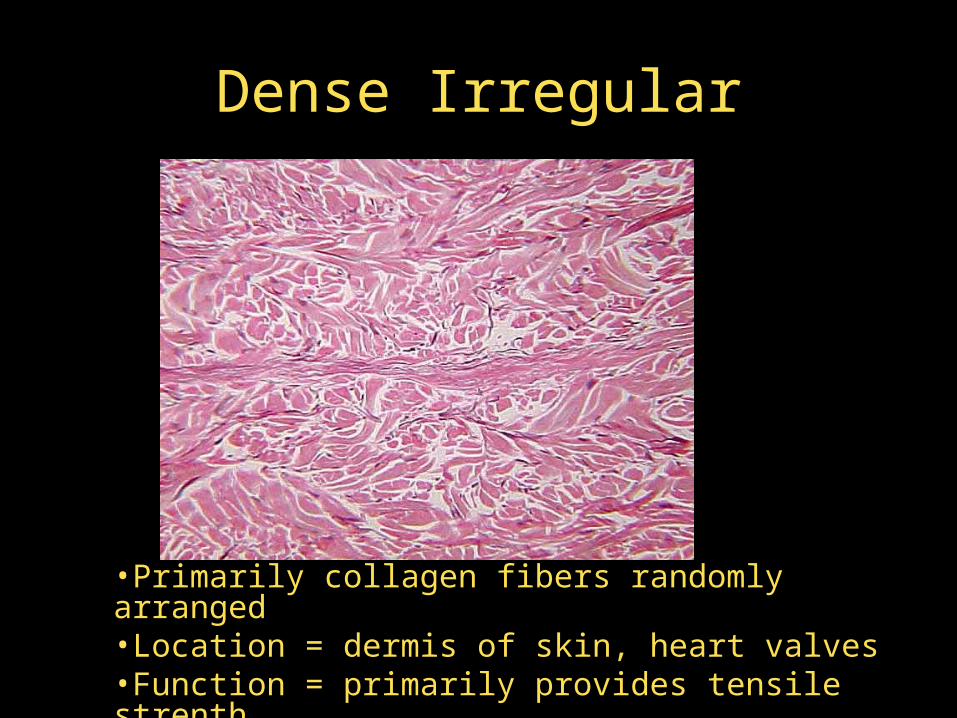
Dense Irregular
•Primarily collagen fibers randomly arranged•Location = dermis of skin, heart valves•Function = primarily provides tensile strenth

Elastic Connective
•Primarily elastin fibers (purple)•Location – lung tissue, wall of aorta, •Funciton = durability with stretch


Hyaline Cartilage
•Amorphous (chondroitin and glucosamine) matrix that surround cells = chondrocytes (within lacunae)•Locations = embryonic skeleton, costal cartilages, cartilage of the nose, trachea, and larynx•Function = support•Avascular = no healing


Fibrocartilage
•Less firm than elastic cartilage•Locations = intervertebral discs, pubic symphysis•Functions = tensile strength plus shock absorber

Elastic Cartilage
•Amorphous matrix that surrounds cells called chondrocytes; elastic fibers present•Locations = external ear, epiglottis•Functions = maintenance of shape plu flexibility

Osseus (bone)
•Hard, calcified matrix with collagen fibers and cells = osteocytes within lacunae•Location = bones of skeleton•Functions = protection, support, movement, calcium store, and hematopoiesis•Highly vascular = fast healing
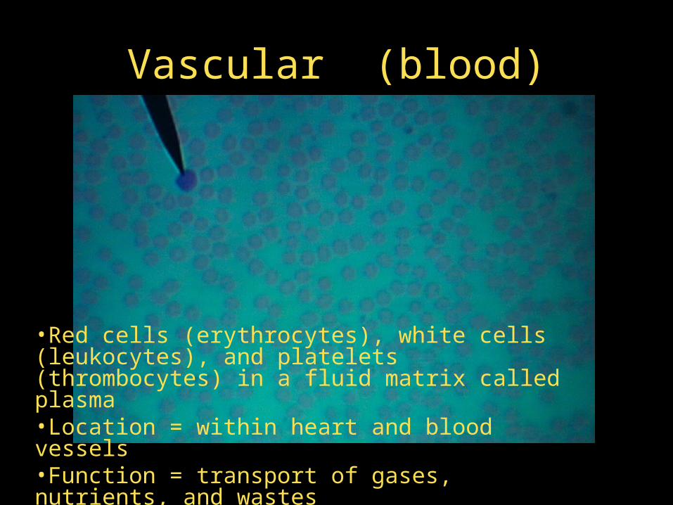
Vascular (blood)
•Red cells (erythrocytes), white cells (leukocytes), and platelets (thrombocytes) in a fluid matrix called plasma•Location = within heart and blood vessels•Function = transport of gases, nutrients, and wastes

Skeletal Muscle Tissue
• Structure: long thin cells (fibers) with many nuclei; alternating areas of light and dark (striations)
• Locations: attached to bones
• Function:
move bones of skeleton
• Control:
voluntary = conscious
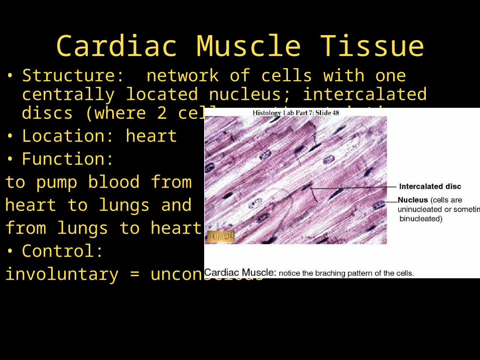
Cardiac Muscle Tissue• Structure: network of cells with one centrally
located nucleus; intercalated discs (where 2 cells meet); striations
• Location: heart• Function: to pump blood from heart to lungs and from lungs to heart• Control: involuntary = unconscious

Smooth Muscle Tissue• Structure: spindle-shaped cells with one centrally
located nucleus; no striations• Location: walls of hollow visceral organs; walls of
blood vessels; attached to hair follicles in the dermis• Function: movement of food through digestive tract; vasoconstriction• Control: involuntary = unconscious

Nervous Tissue• Structure: Primary cells = neurons which
respond to changes in their surroundings (stimuli)
• Neurons are surrounded by neuroglia (supporting cells)
• Locations:Brain, Spinal Cord, Nerves• Function: Coordination or integration of body
parts (to transmit signals from body parts to brain and from brain back to body parts)
• No reproduction of neurons, only neuroglia can divide
Related Documents