Conjugation of multivalent ligands to gold nanoshells and designing a dual modality imaging probe† Mathieu B ´ edard,‡ a Pramod K. Avti,‡ abcd Tina Lam, a L ´ eonie Rouleau, e Jean-Claude Tardif, cf ´ Eric Rh ´ eaume, * cf Fr ´ ed ´ eric Lesage * bc and Ashok Kakkar * a Design and synthesis of branched tetraethylene glycol (TEG) based ligands for subsequent conjugation to gold nanoshells are reported. TEG enhances the aqueous solubility of hollow gold nanoshells (HAuNShs), and the branched architecture provides stability. An examination of the supernatant of the surface displacement reaction shows that the structure of the ligand plays an important role in the functionalization of HAuNShs. The binding of multivalent ligands leads to rupturing of the gold nanoshell architecture; most probably due to the large dendron not compensating the replacement of small citrate capping agents. The construction of a probe with dual imaging capabilities is demonstrated by covalent linking of a dendron containing Cy5.5 A dye to gold nanoshells. It leads to fluorescence quenching of Cy5.5 A by the gold nanoshells, as evidenced in solution and in cellular internalization studies with J774 and bEnd.3 cells. 1. Introduction Among the repertoire of metal nanoparticles, gold nanoshells (AuNShs) have shown great potential in designing novel mate- rials for biomedical applications. 1 Core–shell based AuNShs, especially those built on silica nanoparticles, have been exten- sively explored as immunoassays, for thermoablation, cancer therapy, cell labelling, etc. 2 Gold nanoshells without any core (hollow, HAuNShs) have also become a topical area of research due to their increased absorption cross-section. 3 HAuNShs provide an attractive venue for developing near-infrared contrast agents for biological imaging. During their synthesis, HAuNShs are stabilised using citrate as a capping agent. 4 The scope of these nanomaterials can be further enhanced by their surface func- tionalization, leading to increased solubility, stability, targeting, and for delivering active therapeutic agents. 5 This is generally achieved by a displacement reaction using an appropriate thiol terminated ligand, which binds strongly to gold on the nanoshell surface. 6 Multivalent ligands such as dendrons provide oppor- tunities to enhance the efficacy of surface coverage by intro- ducing a greater number of desired moieties in fewer displacement reactions. 7 We report the design of such branched architectures containing tetraethylene glycol (TEG) in the back- bone and the surface, which are subsequently conjugated to gold nanoshells through the thiol moiety at their core. Oligoethylene glycols such as TEG provide a hydrophilic environment which enhances the aqueous solubility and dispersion of nanoparticles. We demonstrate that the shape of the ligand (linear vs. branched) plays a signicant role in the routinely used surface coating process, and a careful examination of the supernatant in ligand displacement reactions on metal nanoparticles is essential to determine the efficiency of this reaction. The combination of surface plasmon resonance of gold nanoshells with the uorescence of dyes operating in the thera- peutic window constitutes an important platform in developing dual imaging probes for improved diagnostics. 8 We designed a dendron based ligand containing an analog of Cy5.5 dye (Cy5.5 A ) which upon conjugation to the gold nanoshell surface leads to their easy dispersion in an aqueous medium, and quenching of the uorescence of Cy5.5 A . Our dendron based methodology provides a simple, efficient and a versatile route to introduce desired functionalities onto gold nanomaterials, and in devel- oping integrated dual imaging modalities. a Department of Chemistry, McGill University, 801 Sherbrooke St. West, Montreal, Quebec H3A 0B8, Canada. E-mail: [email protected]; Fax: +514-398-3797; Tel: +514-398-6912 b Institute of Biomedical Engineering, ´ Ecole Polytechnique de Montr´ eal, 2900, boul ´ Edouard-Montpetit, Montreal, H3C 3A7, Canada. E-mail: frederic.lesage@polymtl. ca; Fax: +514-340-4611; Tel: +514-340-4711 c Montreal Heart Institute, Research Center, 5000 B´ elanger Est, Montr´ eal, Quebec H1T 1C8, Canada. E-mail: [email protected]; Tel: +514-376-3330 ext. 3091 d Universit´ e de Montr´ eal, Institute of Biomedical Engineering, Department of Physiology, P.O. Box 6128, Station Centre-Ville, Montreal, Quebec, H3C 3J7, Canada e D´ epartment de g´ enie chimique et de g´ enie biotechnologique, 2500, Boulevard Universit´ e, Universit´ e de Sherbrooke, Sherbrooke, Qu´ ebec, J1K 2R1, Canada. E-mail: [email protected]; Tel: +1-514-821-8000 ext. 62758 f D´ epartment of Medicine, Universit´ e de Montr´ eal, Montreal, Quebec, Canada † Electronic supplementary information (ESI) available: Additional UV-Vis spectral, TEM and centrifugation speed data, and confocal images. See DOI: 10.1039/c4tb01811g ‡ These two authors contributed equally. Cite this: J. Mater. Chem. B, 2015, 3, 1788 Received 3rd November 2014 Accepted 3rd January 2015 DOI: 10.1039/c4tb01811g www.rsc.org/MaterialsB 1788 | J. Mater. Chem. B, 2015, 3, 1788–1800 This journal is © The Royal Society of Chemistry 2015 Journal of Materials Chemistry B PAPER Published on 06 January 2015. Downloaded by Ecole Polytechnique de Montreal on 20/02/2015 16:05:13. View Article Online View Journal | View Issue

Welcome message from author
This document is posted to help you gain knowledge. Please leave a comment to let me know what you think about it! Share it to your friends and learn new things together.
Transcript
Journal ofMaterials Chemistry B
PAPER
Publ
ishe
d on
06
Janu
ary
2015
. Dow
nloa
ded
by E
cole
Pol
ytec
hniq
ue d
e M
ontr
eal o
n 20
/02/
2015
16:
05:1
3.
View Article OnlineView Journal | View Issue
Conjugation of m
aDepartment of Chemistry, McGill Univers
Quebec H3A 0B8, Canada. E-mail: ashok
Tel: +514-398-6912bInstitute of Biomedical Engineering, Ecole
Edouard-Montpetit, Montreal, H3C 3A7, C
ca; Fax: +514-340-4611; Tel: +514-340-4711cMontreal Heart Institute, Research Center, 5
1C8, Canada. E-mail: Eric.Rheaume@icm-mdUniversite de Montreal, Institute of B
Physiology, P.O. Box 6128, Station Centre-VeDepartment de genie chimique et de ge
Universite, Universite de Sherbrooke, Sherb
[email protected]; Tel: +1-5fDepartment of Medicine, Universite de Mon
† Electronic supplementary informationspectral, TEM and centrifugation speed10.1039/c4tb01811g
‡ These two authors contributed equally.
Cite this: J. Mater. Chem. B, 2015, 3,1788
Received 3rd November 2014Accepted 3rd January 2015
DOI: 10.1039/c4tb01811g
www.rsc.org/MaterialsB
1788 | J. Mater. Chem. B, 2015, 3, 1788
ultivalent ligands to goldnanoshells and designing a dual modality imagingprobe†
Mathieu Bedard,‡a Pramod K. Avti,‡abcd Tina Lam,a Leonie Rouleau,e
Jean-Claude Tardif,cf Eric Rheaume,*cf Frederic Lesage*bc and Ashok Kakkar*a
Design and synthesis of branched tetraethylene glycol (TEG) based ligands for subsequent conjugation to
gold nanoshells are reported. TEG enhances the aqueous solubility of hollow gold nanoshells (HAuNShs),
and the branched architecture provides stability. An examination of the supernatant of the surface
displacement reaction shows that the structure of the ligand plays an important role in the
functionalization of HAuNShs. The binding of multivalent ligands leads to rupturing of the gold nanoshell
architecture; most probably due to the large dendron not compensating the replacement of small citrate
capping agents. The construction of a probe with dual imaging capabilities is demonstrated by covalent
linking of a dendron containing Cy5.5A dye to gold nanoshells. It leads to fluorescence quenching of
Cy5.5A by the gold nanoshells, as evidenced in solution and in cellular internalization studies with J774
and bEnd.3 cells.
1. Introduction
Among the repertoire of metal nanoparticles, gold nanoshells(AuNShs) have shown great potential in designing novel mate-rials for biomedical applications.1 Core–shell based AuNShs,especially those built on silica nanoparticles, have been exten-sively explored as immunoassays, for thermoablation, cancertherapy, cell labelling, etc.2 Gold nanoshells without any core(hollow, HAuNShs) have also become a topical area of researchdue to their increased absorption cross-section.3 HAuNShsprovide an attractive venue for developing near-infrared contrastagents for biological imaging. During their synthesis, HAuNShsare stabilised using citrate as a capping agent.4 The scope of these
ity, 801 Sherbrooke St. West, Montreal,
[email protected]; Fax: +514-398-3797;
Polytechnique de Montreal, 2900, boul
anada. E-mail: frederic.lesage@polymtl.
000 Belanger Est, Montreal, Quebec H1T
hi.org; Tel: +514-376-3330 ext. 3091
iomedical Engineering, Department of
ille, Montreal, Quebec, H3C 3J7, Canada
nie biotechnologique, 2500, Boulevard
rooke, Quebec, J1K 2R1, Canada. E-mail:
14-821-8000 ext. 62758
treal, Montreal, Quebec, Canada
(ESI) available: Additional UV-Visdata, and confocal images. See DOI:
–1800
nanomaterials can be further enhanced by their surface func-tionalization, leading to increased solubility, stability, targeting,and for delivering active therapeutic agents.5 This is generallyachieved by a displacement reaction using an appropriate thiolterminated ligand, which binds strongly to gold on the nanoshellsurface.6 Multivalent ligands such as dendrons provide oppor-tunities to enhance the efficacy of surface coverage by intro-ducing a greater number of desired moieties in fewerdisplacement reactions.7 We report the design of such branchedarchitectures containing tetraethylene glycol (TEG) in the back-bone and the surface, which are subsequently conjugated to goldnanoshells through the thiol moiety at their core. Oligoethyleneglycols such as TEG provide a hydrophilic environment whichenhances the aqueous solubility and dispersion of nanoparticles.We demonstrate that the shape of the ligand (linear vs. branched)plays a signicant role in the routinely used surface coatingprocess, and a careful examination of the supernatant in liganddisplacement reactions on metal nanoparticles is essential todetermine the efficiency of this reaction.
The combination of surface plasmon resonance of goldnanoshells with the uorescence of dyes operating in the thera-peutic window constitutes an important platform in developingdual imaging probes for improved diagnostics.8 We designed adendron based ligand containing an analog of Cy5.5 dye (Cy5.5A)which upon conjugation to the gold nanoshell surface leads totheir easy dispersion in an aqueous medium, and quenching ofthe uorescence of Cy5.5A. Our dendron based methodologyprovides a simple, efficient and a versatile route to introducedesired functionalities onto gold nanomaterials, and in devel-oping integrated dual imaging modalities.
This journal is © The Royal Society of Chemistry 2015
Paper Journal of Materials Chemistry B
Publ
ishe
d on
06
Janu
ary
2015
. Dow
nloa
ded
by E
cole
Pol
ytec
hniq
ue d
e M
ontr
eal o
n 20
/02/
2015
16:
05:1
3.
View Article Online
2. Experimental2.1. Materials and methods
The compounds trisodium citrate, sodium borohydride, gold-(III) chloride hydrate, tetraethylene glycol, p-toluenesulfonylchloride, sodium hydroxide, sodium hydride, thioacetic acid, 4-dimethylaminopyridine, methylsulfonyl chloride, triethyl-amine, sodium azide, tetrabutylammonium iodide, lithiumhydroxide, pentynoic acid, sodium ascorbate, copper(II) sulfatehexahydrate, borane-THF, bis(triphenylphosphine)palladiu-m(II) dichloride, propargyl bromide, (triisopropylsilyl) acety-lene, (trimethylsilyl) acetylene, potassium thioacetate, methyl3,4,5-trihydroxybenzoate, sodium thiomethoxide (SigmaAldrich), 1-ethyl-3-(3-dimethylaminopropyl)carbodiimide$98% (Fluka), cobalt(II) chloride hexahydrate (Fisher Scien-tic), hydrochloric and nitric acid (ACP Chemicals), PEG750 (AlfaAesar), and 3-bromo-5-iodobenzoic acid 98% (AK ScienticInc.), were used as received. A 400 square-mesh copper grid waspurchased from Electron Microscopy Science. Ultrapure waterwas doubly distilled by reverse osmosis though a Millipore RiOS8, followed by ltration through a MilliQ Academic A10 unit.Acetonitrile was dried over calcium hydride.
NMR acquisition was carried out on a 300 or 400 MHz VarianMercury instrument and operated using VNMRJ 2.2D soware.Mass spectral analyses (MS and MALDI-TOF) were performedon a Kratos MS25RFA high resolution mass spectrometer.UV-Vis and uorescence spectra were obtained on a Cary 500UV-Vis and Cary Eclipse Fluorimeter, respectively. TEM analyseswere carried out on a Tecnai 12 TEM with an AMT XR80C CCDCamera, and the TEM images were obtained in bright-eldmode at an accelerating voltage of 120 kV. Samples wereprepared by placing a small droplet of AuNSh aqueous solutionand letting it dry on a CF400-Cu carbon lm side of a 400square-mesh copper grid, and the image acquired from TEMwas processed using ImageJ soware. Thermogravimetricanalysis was performed on a TGA Q500 V6.7 Build 203 instru-ment. Samples for TGA were prepared by freezing the conju-gated gold nanoshell solution at �78 �C, followed bylyophilization overnight. The solid (approximately 0.25–0.35mg) was then collected, weighed onto a platinum pan, andsubjected to TGA under a ow of air. The temperature wasramped at a rate of 20 �C min�1 to 100 �C and held at isothermfor 2 min. It was then increased to 500 �C at the same rate, atwhich point a ow of nitrogen replaced air and the temperaturewas increased to 700 �C. Air and N2 ow rates were set at 40 mLmin�1 and 60 mL min�1, respectively. The primary weight lossis attributed to the thiolated ligand. Calculations were per-formed3a using the percentage of weight lost due to the organicligand obtained from TGA to approximate the amount of ligandoriginally present on the surface of the HAuNSh.
2.2. Gold nanoshell synthesis
Hollow gold nanoshells were prepared by adapting a procedurereported earlier,3a,4a and is briey described here. The glasswareused in the synthesis was soaked in a 3 : 1 mixture ofHCl : HNO3 (aqua regia) for 1 h, rinsed with ultrapure water 3
This journal is © The Royal Society of Chemistry 2015
times, and dried in an oven overnight. In a 2 L 3-necked roundbottom ask, 1.5 L of MilliQ H2O were degassed and lled withAr by repeating the cycle of evacuation and lling with Ar 4times. To this ask kept under a positive ow of Ar, trisodiumcitrate (1.2345 g, 4.20 mmol) was added, and the cycle of evac-uating and lling with Ar was repeated once more. Whileretaining a positive ow of Ar, cobalt(II) chloride hexahydrate(150 mg, 0.630 mmol) was added and the mixture was stirredvigorously. Sodium borohydride (NaBH4, 142.5 mg, 3.77 mmol)was dissolved in approximately 2 mL of degassed MilliQ water,and added via syringe injection to the above cobalt solutionwhile stirring vigorously. The reduction of the cobalt saltleading to the formation of cobalt nanoparticles was indicatedby a rapid color change from faint purple to near-black. Theformation of H2 gas bubbles was the visual cue for monitoringthe completion of the reaction. In another 3 L 3-neck roundbottom ask, 900 mL of MilliQ H2O was degassed and lledwith Ar in four cycles. Gold(III) chloride hydrate (HAuCl4 – 12mg, 0.0353 mmol) was dissolved in 2 mL of degassed MilliQwater, and added to this 3 L ask via syringe injection. Thecobalt solution was rapidly transferred to the 3-neck ask undera positive Ar ow. The mixture was le to react for 10 min andthen opened to air for 4 hours. Its green/grey color indicated theformation of gold nanoshells. The solution was concentrated bycentrifugation at 4600 rpm at 4 �C for 2 hours in separate tubes.The supernatants were recombined, followed by a second cycleof centrifugation for 3 hours at 4600 rpm at 4 �C.
The monofunctional ligand containing a protected thiolentity, tetraethylene glycol thioacetate (G0-TEG-SAc), wasprepared using an adaptation of the literature procedure.9
2.3. Synthesis of multivalent ligands
2.3.1 TEG-Ts (1). TEG-Ts (1) was prepared by adapting aprocedure reported by Ashton et al.9d TEG (17.56 g, 90.4 mmol)was dissolved in THF (2.5 mL) in a 500 mL three-necked roundbottom ask under an Ar atmosphere. NaOH (1.085 g, 27.12mmol) was dissolved in a minimal amount of water and addedto the TEG solution. The resulting mixture was stirred for 2minutes and then cooled to 0 �C. Subsequently, freshly puriedTsCl (3.45 g, 18.08 mmol) was dissolved in THF (10 mL) andadded dropwise (via an addition funnel under an Ar atmo-sphere) to the TEG solution while stirring vigorously. Once theaddition was complete, the reaction mixture was stirred for 2 h.THF was removed under reduced pressure and an ice-waterslurry (20 mL) was added. The product was extracted with DCM(60 mL) and the organic phase was washed with brine (10 mL)and dried over MgSO4. The crude product was concentrated invacuo, and was puried using column chromatography withEtOAc to give a colorless, transparent oil (4.08 g, 65%). 1H NMR(300 MHz, CDCl3): d 2.44 (s, 3H), 3.75–3.51 (m, 14H), 4.21–4.11(m, 2H), 7.34 (d, 2H), 7.79 (d, 2H) ppm.
2.3.2 TEG-SAc (2). TEG-SAc (2) was prepared by adapting aprocedure reported earlier.10 TEG-Ts (0.70 g, 2.00 mmol) wasdissolved in dry DMF (3 mL) in a 50 mL Schlenk under an Aratmosphere. Potassium thioacetate (0.69 g, 6.03 mmol) wasadded aer cooling at 4 �C and the mixture was stirred
J. Mater. Chem. B, 2015, 3, 1788–1800 | 1789
Journal of Materials Chemistry B Paper
Publ
ishe
d on
06
Janu
ary
2015
. Dow
nloa
ded
by E
cole
Pol
ytec
hniq
ue d
e M
ontr
eal o
n 20
/02/
2015
16:
05:1
3.
View Article Online
vigorously for 20 hours. HCl (5% v/v, 20 mL) was added drop-wise while stirring. The product was extracted with EtOAc (4 �40 mL) and the combined organic phases were washed withH2O (3 � 10 mL), brine (1 � 10 mL) and then dried over MgSO4.The crude product was concentrated in vacuo and puried usingash chromatography with Et2O/MeOH (100 : 0, 95 : 5, 90 : 10,85 : 15, 80 : 20) as the eluent, with the majority of the producteluting with 85 : 15. The product was concentrated underreduced pressure to give an orange viscous oil (0.260 g, 51%)which was stored under an Ar atmosphere at 4 �C. 1H NMR (400MHz, CDCl3): d 2.34 (s, 3H), 3.10 (t, 2H), 3.58–3.67 (m, 12H), 3.73(t, 2H) ppm.
2.3.3 Methyl 3,4,5-tris(prop-2-ynyloxy)benzoate (3). Amixture of methyl 3,4,5-trihydroxybenzoate (6.42 g, 34.86mmol), K2CO3 (19.28 g, 174.3 mmol) and propargyl bromide(20.73 g, 174.3 mmol) in DMF (50 mL) was stirred vigorously for5 days. Excess propargyl bromide was removed in vacuo. H2O(125 mL) was added and the product was extracted with EtOAc(4 � 75 mL). The collected organic phases were washed withH2O (3 � 75 mL) and brine (3 � 75 mL), dried over K2CO3, andthe solvent was removed to give a brittle off-white solid (9.56 g,92%). 1H NMR (300 MHz, CDCl3): d 2.46 (t, 1H), 2.53 (t, 2H), 3.91(s, 3H), 4.81 (2d, 6H), 7.47 (s, 2H) ppm. 13C NMR (300 MHz,CDCl3): d 52.37, 57.03, 60.31, 76.59, 78.66, 109.78, 125.74,141.01, 151.26, 166.24 ppm.
2.3.4 3,4,5-Tris(prop-2-ynyloxy)benzoic acid (dendron–acid-trip, 4). A solution of 3 (1 g, 3.35 mmol) in THF wasvigorously stirred with 4 M KOH(aq) (20 mL) for 2 days. Thesolution was acidied to pH ¼ 3 with HCl (37% v/v), and theproduct was extracted with THF (3 � 10 mL). The collectedorganic phases were washed with brine (2 � 8 mL), dried overMgSO4 and the solvent was removed in vacuo to give a brittle off-white solid (0.81 g, 85%). 1H NMR (300 MHz, DMSO): d 3.48 (t,1H), 3.62 (t, 2H), 4.71 (d, 2H), 4.89 (d, 4H), 7.38 (s, 2H), 13.06 (s,1H) ppm. 13C NMR (300 MHz, CDCl3): d 56.90, 59.92, 78.80,79.53, 109.29, 126.70, 140.10, 151.37, 167.09 ppm.
2.3.5 Dendron–TEG-SAc-trip (5). Compound 4 (0.234 g,0.826 mmol) and TEGSAc (0.250 g, 0.991 mmol) were dissolvedin dry DCM (8 mL) in a 50 mL Schlenk ask under an Aratmosphere. DMAP (0.151 g, 1.24 mmol) was added and themixture was stirred for 5 minutes. EDC (0.141 g, 0.908 mmol)was added and the mixture was stirred for 2 days. The solventwas removed in vacuo, and the crude product was puried usingash chromatography with DCM/MeOH (99 : 1). The solventwas removed in vacuo to give an orange-brown oil (0.31 g, 72%).1H NMR (300 MHz, CDCl3): d 2.32 (s, 3H), 2.52 (t, 1H), 2.55(t, 2H), 3.09 (t, 2H), 3.57–3.70 (m, 12H), 3.83 (t, 2H), 4.47 (t, 2H),4.80 (d, 4H), 4.82 (d, 2H), 7.49 (s, 2H) ppm. 13C NMR (300 MHz,CDCl3): d 28.71, 30.55, 57.16, 60.32, 64.52, 69.22, 69.74, 70.30,70.58, 70.12, 70.77, 75.58, 76.26, 110.14, 125.74, 141.23, 151.30,165.69, 195.50 ppm. Expected mass: 518.58 g mol�1, ESIm/z ¼ 519.17 g mol�1.
2.3.6 G1-3-TEGOH (6). Dendron–TEG-SAc-trip (5, 0.106 g,0.204 mmol), N3TEG3a (0.134 g, 0.613 mmol) and sodiumascorbate (0.012 g, 0.061 mmol) were dissolved in THF (1 mL) ina 50 mL Schlenk ask under an Ar atmosphere. CuSO4 wasadded and the mixture was stirred vigorously for 2 days at 40 �C.
1790 | J. Mater. Chem. B, 2015, 3, 1788–1800
The solvent was removed in vacuo. The product was extracted inDCM (5 mL) and stirred with EDTA (100 mg) for 2 hours. Thisprocess was repeated twice. The mixture was dried over Na2SO4
and ltered. The solvent was almost entirely removed in vacuo(leaving only approximately 2 mL). Et2O was added dropwise toprecipitate the oily product. When the orange solution ceasedbeing cloudy, the mixture was decanted. The orange oil wasdissolved in DCM (2 mL) and the precipitation was repeated.The remaining DCMwas removed in vacuo to yield an orange oil(0.095 g, 39%). 1H NMR (400 MHz, CDCl3): d 2.32 (s, 3H), 3.07 (t,2H), 3.32 (s, broad, 7H), 3.56–3.62 (m, 46H), 3.68–3.72 (m, 14H),3.84 (s, 3H), 3.92 (s, 8H), 4.46 (s, 3H), 4.60 (s, 8H), 5.25 (s, 6H),7.46 (s, 2H), 8.06 (s, 3H) ppm. 13C NMR (300 MHz, CDCl3): d28.76, 30.52, 50.31, 61.47, 63.13, 64.32, 69.11–69.66, 70.17–70.62, 72.45, 109.39, 124.92–125.02, 125.54, 141.76, 151.97,165.82, 195.49 ppm. Expected mass: 1176.29 g mol�1, ESIm/z ¼1198.52 g mol�1 [M + Na+].
2.3.7 G2-9-trip (7). G1-3-TEGOH (6, 0.459 g, 1.615 mmol)and 4 (0.380 g, 0.323 mmol) were dissolved in dry CH3CN (8 mL)in a 50 mL Schlenk ask under an Ar atmosphere. DMAP (0.178g, 1.454 mmol) was added and the mixture was stirred for 5minutes. EDC (0.166 g, 1.070 mmol) was added and the mixturewas stirred for 2 days. The crude product was extracted in DCM(30 mL) and dried over NaSO4. The solvent was removed invacuo. The crude product was puried using ash chromatog-raphy with DCM/MeOH (95 : 5). The solvent was removed invacuo to give a clear yellow oil (0.019 g, 3%). 1H NMR (300 MHz,CDCl3): d 2.32 (s, 3H), 2.47 (s, 3H), 2.57 (s, 6H), 3.07 (t, 2H), 3.58–3.70 (m, 45H), 3.78–3.88 (m, 16H), 4.42–4.54 (m, 9H), 4.52–4.54(m, 7H), 4.78–4.83 (m, 24H), 5.22 (s, 6H), 7.46 (s, 10H), 7.96 (s,3H) ppm. 13C NMR (300 MHz, CDCl3): d 28.80, 29.68, 50.25,53.42, 57.17, 60.31, 64.42, 69.14–69.72, 70.27–70.70, 78.00,78.67, 110.11, 125.69, 141.21, 151.28, 165.64 ppm. Expectedmass: 1975.04 g mol�1, ESI m/z ¼ 1996.69 g mol�1 [M + Na+].
2.3.8 G2-9-TEGOH (8). G1-9-trip (7, 0.017 g, 0.0086 mmol),N3-TEG3a (0.019 g, 0.086 mmol) and sodium ascorbate (0.015 g,0.077 mmol) were dissolved in THF (1 mL) in a 10 mL Schlenkask under an Ar atmosphere. CuSO4 (0.001 g, 0.0039 mmol)was added and the mixture was stirred vigorously for 2 days at40 �C. The solvent was removed in vacuo, and the product wasextracted into 5 mL of DCM and stirred with EDTA (40 mg) for 2hours. This process was repeated twice, and the solutionmixture was dried over Na2SO4 and ltered. The solution wasconcentrated to 1 mL in vacuo, and Et2O was added dropwise toprecipitate the oily product. The orange oil was dissolved inDCM (1mL) and the precipitation was repeated another 2 times.The remaining DCM was removed in vacuo to yield a yellow oilylm (0.034 g, 61%). 1H NMR (300 MHz, CDCl3): d 3.58 (s, broad,186H), 4.48 (s, broad, 8H), 4.58 (s, broad, 12H), 5.28 (s, broad,24H), 7.48 (s, broad, 8H), 8.08 (s, broad, 12H) ppm. Expectedmass: 3948.18 g mol�1, MALDI m/z ¼ 3955.47 g mol�1.
2.3.9 G1-3-Cy5.5A (9). G1-3-TEGOH (6, 0.0946 g, 0.0804mmol) and Cy5.5A analogue1d (0.170 g, 0.246 mmol) were dis-solved in dry DCM (8 mL) in a 50 mL Schlenk ask under an Aratmosphere in the dark. DMAP (0.0442 g, 0.362 mmol) wasadded and the mixture was stirred for 5 minutes. EDC (0.0412 g,0.265 mmol) was added and the mixture was stirred for 2 days.
This journal is © The Royal Society of Chemistry 2015
Paper Journal of Materials Chemistry B
Publ
ishe
d on
06
Janu
ary
2015
. Dow
nloa
ded
by E
cole
Pol
ytec
hniq
ue d
e M
ontr
eal o
n 20
/02/
2015
16:
05:1
3.
View Article Online
The crude product was extracted in DCM (30 mL) and washedwith water (1 � 10 mL) and brine (1 � 10 mL) and dried overNaSO4. The solvent was removed in vacuo. The crude productwas puried using neutral alumina for liquid chromatographywith DCM/MeOH (99 : 1) to remove the rst band and DCM/MeOH (90 : 10) to obtain the product. The mixture wasconcentrated in vacuo to give a blue-green powder (0.255 g,99%). 1H NMR (300 MHz, CDCl3): d 1.19–1.22 (m, 20H), 1.49 (s,9H), 1.68–1.74 (m, 12H), 1.83 (s, 22H), 1.95 (s, 3H), 2.04 (s, 16H),2.20 (s, 12H), 2.28–2.37 (m, 18H), 3.11 (s, 6H), 3.20–3.26 (m, 8H),3.55–3.67 (m, 42H), 3.81–3.87 (d, 9H), 4.02–4.16 (d, 6H), 4.41–4.54 (t, 9H), 4.63 (s, 5H), 5.18 (s, 6H), 5.27 (s, 2H), 6.06 (d, 5H),6.47 (s, 25H), 7.18 (t, 2H), 7.26–7.30 (m, 6H), 7.42 (s, 11H), 7.55(s, 6H), 7.71 (d, 2H), 7.88 (s, 13H), 8.02 (t, 8H), 8.16 (s, 20H) ppm.13C NMR (300 MHz, CDCl3): d 23.66, 24.42, 26.28, 27.27–27.57,29.63, 33.68, 43.98, 45.45, 49.96, 51.14, 63.45, 69.29, 70.39,102.39, 109.34, 110.24–110.64, 122.04, 124.94, 126.27, 127.63,128.17, 129.88, 130.69, 131.76, 139.25, 139.88, 143.07, 149.30,152.01, 154.11, 173.20. Expected mass: 3194.92 g mol�1, ESIm/z ¼ 3195.81 g mol�1.
2.4. Conjugation of ligands to gold nanoshells
The thiol entity in the desired ligand was deprotected using thegeneral procedure3a outlined here for G1-3-TEGOH: the thio-acetated ligand (0.118 g, 0.1 mmol) was dissolved in dry MeOH(2 mL) and sodium thiomethoxide (0.007 g, 0.1 mmol, 1 Msolution in MeOH) was added. The reaction was stirred for 3hours. HCl(aq) (2 mL, 0.1 M) was added to the mixture. Theproduct was extracted in DCM and washed with H2O and brine,dried over MgSO4 and concentrated in vacuo to yield the freethiol terminated ligand (0.072 g, 63%). The deprotected ligandwas reacted in situ for conjugating to the HAuNSh as follows. Afreshly sonicated and Ar-purged concentrated solution ofHAuNSh (3 mL) was mixed with the thioacetate-deprotectedligand under an Ar atmosphere. MeOH (1 mL) was added toincrease the solubility of the ligand. The mixture was stirredovernight in the dark. The solution of the conjugated HAuNShwas diluted with a solution of trisodium citrate (1.7 mM) andthen concentrated by centrifugation at 4600 rpm at 4 �C for 3hours before removing the supernatants and recombining theseparate pellets. The conjugated HAuNShs were diluted onceagain with the trisodium citrate solution, and the process wasrepeated twice. In the case of highly colored ligands, thesupernatant became progressively clearer with each wash andwas completely colourless aer the third and nal wash, indi-cating that all unbound ligands had been removed. Theconcentrated conjugated HAuNSh solution (approx. 3 mL) wasstored in the dark at 4 �C.
2.5. Photoacoustic tomography
Solutions of HAuNShs and their functionalized analogues werelled in a polyethylene tube (0.4 � 0.2 � 0.1 mm; O.D � I.D �W.T) which was sealed on both ends and immersed in water at adepth of �3 mm. Ultrasound (US) and photoacoustic (PA)imaging of the nanoparticle samples were performed usingVevo LAZR Photoacoustic Imaging System (Visual Sonics, ON,
This journal is © The Royal Society of Chemistry 2015
Canada) with an LZ250 imaging probe (16 MHz centralfrequency). US and PA images were acquired using a 680 nmwavelength laser and analyzed using Vevo workstation soware.
2.6. Cellular imaging
2.6.1 Cell culture. J774 Mouse monocyte/macrophages andbrain endothelial cells (bEnd.3) were cultured (at a density of 3� 104 cells per cm2) in DMEMmedium supplemented with 10%fetal bovine serum and incubated at 37 �C, 5% CO2 in ahumidied atmosphere. Aer overnight incubation the cellswere treated for 4 h with G1-3-Cy5.5A, G1-3-Cy5.5A–HAuNSh orHAuNSh. At the end of the incubation time, the plates werethoroughly washed thrice with PBS (pH-7.4) to remove anyunbound HAuNSh samples and xed with 2% para-formaldehyde (in PBS pH 7.4) for 30 min at room temperature.The cells were again washed three times with PBS and mountedusing 0.2% DABCO/glycerol (Sigma Aldrich, D2522, R6513).
2.6.2 Confocal microscopy. Cellular imaging was carriedout using a LSM 710 AxioObserver confocal microscope (CarlZeiss, Oberkochen, Germany) in the visible excitation wave-lengths (excitation wavelengths ¼ 633 nm). The uorescenceemissions were collected using a 697 nm long pass lter, totransmit only Cy5.5 emission. The images were acquired usingPlan Apochromatic 40 � 1.3 oil immersion objectives.
2.7. Cellular uptake quantication
The cellular uptake was quantied using inductively coupledplasma-optical emission spectroscopy (ICP-OES). Aer 4 htreatment with HAuNSh and their conjugates, the cell plateswere thoroughly washed 3 times with PBS to remove unboundnanoparticles, trypsinized and pelleted. The cell pellets weredigested using concentrated HNO3 and diluted with NanoQwater to make a nal 5% HNO3 concentration and then lteredthrough a 0.22 mm lter. ICP-OES was performed using anAgilent Technology 5100 ICP-OES spectrometer (Agilent Tech-nology Inc, CA, USA). The cellular internalized [Au] wasmeasured using a standard curve plotted by taking variousconcentration of Au standard (#13881, Alfa Aesar) using lmax ¼242.795 nm.
3. Results and discussion3.1. Ligand synthesis
Design of the branched architectures employed in this study isbased on our continued efforts to develop nanocarriers whichimpart aqueous solubility, biocompatibility, as well asenhanced efficiency in introducing multiple functional groupsinto a single platform.3a,11 We have now developed a method-ology to synthesize dendrons with a thiol entity at the core andvariable TEG groups at the periphery. One of the hydroxylgroups of TEG was rst selectively converted to a thioacetate. Toachieve this, it was tosylated by reacting p-toluenesulphonylchloride with a large excess of TEG under high dilution and atlow temperature. The resulting monotosylated TEG (TEG-Ts,(1)) is activated towards nucleophilic substitution, and wasreplaced with thioacetate using the one-step procedure reported
J. Mater. Chem. B, 2015, 3, 1788–1800 | 1791
Journal of Materials Chemistry B Paper
Publ
ishe
d on
06
Janu
ary
2015
. Dow
nloa
ded
by E
cole
Pol
ytec
hniq
ue d
e M
ontr
eal o
n 20
/02/
2015
16:
05:1
3.
View Article Online
by Sato et al.10 TEG-Ts (1) was reacted with commercially avail-able potassium thioacetate in dry DMF at room temperature for12 h under an Ar atmosphere. Although the original procedurestates that no purication is needed besides a short work up,column chromatography was necessary to purify TEG-SAc (2).The thioacetate was found to degrade if le for long periods oftime on a column, most probably due to the acidic nature ofsilica. Therefore, it was important that the purication of allsubsequent compounds containing a thioacetate group iscarried out relatively quickly, with column chromatography notlasting more than 3 h. TEG-SAc was isolated as light brown-redoil and either used immediately or stored for a short period oftime under an inert atmosphere at 4 �C.
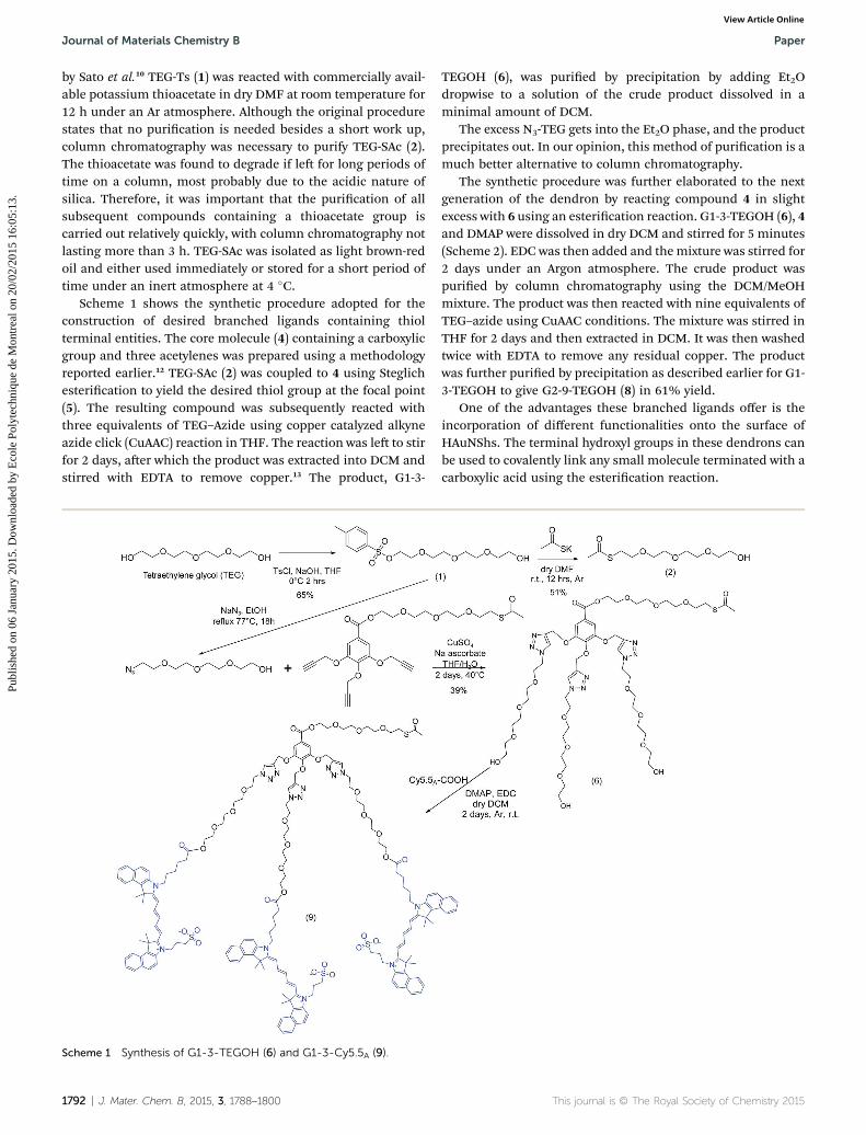
Scheme 1 shows the synthetic procedure adopted for theconstruction of desired branched ligands containing thiolterminal entities. The core molecule (4) containing a carboxylicgroup and three acetylenes was prepared using a methodologyreported earlier.12 TEG-SAc (2) was coupled to 4 using Steglichesterication to yield the desired thiol group at the focal point(5). The resulting compound was subsequently reacted withthree equivalents of TEG–Azide using copper catalyzed alkyneazide click (CuAAC) reaction in THF. The reaction was le to stirfor 2 days, aer which the product was extracted into DCM andstirred with EDTA to remove copper.13 The product, G1-3-
Scheme 1 Synthesis of G1-3-TEGOH (6) and G1-3-Cy5.5A (9).
1792 | J. Mater. Chem. B, 2015, 3, 1788–1800
TEGOH (6), was puried by precipitation by adding Et2Odropwise to a solution of the crude product dissolved in aminimal amount of DCM.
The excess N3-TEG gets into the Et2O phase, and the productprecipitates out. In our opinion, this method of purication is amuch better alternative to column chromatography.
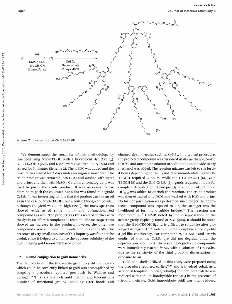
The synthetic procedure was further elaborated to the nextgeneration of the dendron by reacting compound 4 in slightexcess with 6 using an esterication reaction. G1-3-TEGOH (6), 4and DMAP were dissolved in dry DCM and stirred for 5 minutes(Scheme 2). EDC was then added and themixture was stirred for2 days under an Argon atmosphere. The crude product waspuried by column chromatography using the DCM/MeOHmixture. The product was then reacted with nine equivalents ofTEG–azide using CuAAC conditions. The mixture was stirred inTHF for 2 days and then extracted in DCM. It was then washedtwice with EDTA to remove any residual copper. The productwas further puried by precipitation as described earlier for G1-3-TEGOH to give G2-9-TEGOH (8) in 61% yield.
One of the advantages these branched ligands offer is theincorporation of different functionalities onto the surface ofHAuNShs. The terminal hydroxyl groups in these dendrons canbe used to covalently link any small molecule terminated with acarboxylic acid using the esterication reaction.
This journal is © The Royal Society of Chemistry 2015
Scheme 2 Synthesis of G2-9-TEGOH (8).
Paper Journal of Materials Chemistry B
Publ
ishe
d on
06
Janu
ary
2015
. Dow
nloa
ded
by E
cole
Pol
ytec
hniq
ue d
e M
ontr
eal o
n 20
/02/
2015
16:
05:1
3.
View Article Online
We demonstrated the versatility of this methodology byfunctionalizing G1-3-TEGOH with a uorescent dye (Cy5.5A).G1-3-TEGOH, Cy5.5A and DMAP were dissolved in dry DCM andstirred for 5 minutes (Scheme 2). Then, EDC was added and themixture was stirred for 2 days under an Argon atmosphere. Thecrude product was extracted into DCM and washed with waterand brine, and then with NaSO4. Column chromatography wasused to purify the crude product. It was necessary to usealumina to pack the column since silica was found to degradeCy5.5A. It was interesting to note that the product was not an oilas in the case of G1-3-TEGOH, but a brittle blue-green powder.Although the yield was quite high (99%), the mass spectrumshowed evidence of some mono- and di-functionalizedcompounds as well. The product was thus reacted further withthe dye in an effort to complete the reaction. Themass spectrumshowed an increase in the product; however, the other twocompounds were still noted in minute amounts in the MS. Thepresence of very small amounts of this impurity was found to beuseful, since it helped to enhance the aqueous solubility of thedual imaging gold nanoshell based probe.
3.2. Ligand conjugation to gold nanoshells
The deprotection of the thioacetate group to yield the ligandswhich could be covalently linked to gold was accomplished byadapting a procedure reported previously by Wallace andSpringer.14 This is a relatively mild method and tolerant of anumber of functional groups including ester bonds and
This journal is © The Royal Society of Chemistry 2015
charged dye molecules such as Cy5.5A. In a typical procedure,the protected compound was dissolved in dry methanol, cooledto 0 �C, and one molar solution of sodium thiomethoxide in drymethanol was added. The reaction mixture was le to stir for 3–4 hours depending on the ligand. The monodentate ligand G0-TEGOH required 3 hours, while the G1-3-TEGOH (6), G2-9-TEGOH (8) and the G1-3-Cy5.5A (9) ligands required 4 hours forcomplete deprotection. Subsequently, a solution of 0.1 molarHCl(aq) was added to quench the reaction. The crude productwas then extracted into DCM and washed with H2O and brine.No further purication was performed since longer the depro-tected compound was exposed to air, the stronger was thelikelihood of forming disulde bridges.15 The reaction wasmonitored by 1H NMR noted by the disappearance of theacetate group (typically found at 2.31 ppm). It should be notedthat the G1-3-TEGOH ligand is difficult to solubilize aer pro-longed storage at 4 �C under an inert atmosphere since it yieldsa gel-like consistency. For compound 9, 1H NMR and UV-Visconrmed that the Cy5.5A dye did not degrade under thedeprotection conditions. The resulting deprotected compoundswere immediately reacted in situ with a solution of HAuNShs,due to the sensitivity of the thiol group to dimerization onexposure to air.
Gold nanoshells utilized in this study were prepared usingthe procedure reported earlier,3a,4a and it involved cobalt as asacricial template. In brief, cobalt(II) chloride hexahydrate wasreduced with sodium borohydride (NaBH4) in the presence oftrisodium citrate. Gold (aurochloric acid) was then reduced
J. Mater. Chem. B, 2015, 3, 1788–1800 | 1793
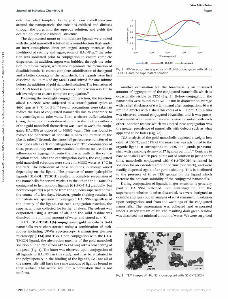
Fig. 1 UV-Vis absorbance spectra of HAuNSh, conjugated with G1-3-TEGOH, and the supernatant solution.
Journal of Materials Chemistry B Paper
Publ
ishe
d on
06
Janu
ary
2015
. Dow
nloa
ded
by E
cole
Pol
ytec
hniq
ue d
e M
ontr
eal o
n 20
/02/
2015
16:
05:1
3.
View Article Online
onto this cobalt template. As the gold forms a shell structurearound the nanoparticle, the cobalt is oxidized and diffusesthrough the pores into the aqueous solution, and yields thedesired hollow gold nanoshell structure.
The deprotected mono or multidentate ligands were mixedwith the gold nanoshell solution in a round-bottom ask underan inert atmosphere. Since prolonged storage increases thelikelihood of settling and aggregation of HAuNShs,3a the solu-tion was sonicated prior to conjugation to ensure completedispersion. In addition, argon was bubbled through the solu-tion to remove oxygen, which would promote the formation ofdisulde bonds. To ensure complete solubilisation of the ligandand a better coverage of the nanoshells, the ligands were rstdissolved in 1–2 mL of dry MeOH and stirred for one minutebefore the addition of gold nanoshell solution. The formation ofthe Au–S bond is quite rapid; however the reaction was le tostir overnight to ensure complete conjugation.16
Following the overnight conjugation reaction, the function-alized HAuNShs were subjected to 3 centrifugation cycles at4600 rpm at 4 �C for 3 h.3a Several precautions were taken toreduce the loss of conjugated nanoshells due to adherence tothe centrifugation tube walls. First, a citrate buffer solution(using the same concentration of citrate as during the synthesisof the gold nanoshell themselves) was used to wash the conju-gated HAuNSh as opposed to MilliQ water. This was found toreduce the adherence of nanoshells onto the surface of theplastic tubes.3a Second, the nanoshell pellets were transferred tonew tubes aer each centrifugation cycle. The combination ofthese precautionary measures resulted in almost no loss due toadherence or aggregation onto the plastic walls of the centri-fugation tubes. Aer the centrifugation cycles, the conjugatedgold nanoshell solutions were stored in MilliQ water at 4 �C inthe dark. The behaviour of these solutions in storage varieddepending on the ligand. The presence of more hydrophilicligands (G1-3-OH, TEGOH) resulted in complete suspension ofthe nanoshells for several weeks. On the other hand, HAuNShsconjugated to hydrophobic ligands (G1-3-Cy5.5A) gradually (butnever completely) separated from the aqueous supernatant overthe course of a few days. However, gentle shaking resulted inimmediate resuspension of conjugated HAuNSh regardless ofthe identity of the ligand. For each conjugation reaction, thesupernatant was collected for further analysis. The solvent wasevaporated using a stream of air, and the solid residue wasdissolved in a minimal amount of water and stored at 4 �C.
3.2.1 G1-3-TEGOH (6) conjugation to gold nanoshells. Goldnanoshells were characterized using a combination of tech-niques including UV-Vis spectroscopy, transmission electronmicroscopy (TEM) and TGA. Upon conjugation with the G1-3-TEGOH ligand, the absorption maxima of the gold nanoshellsolution blue shied (from 742 to 714 nm) with a broadening ofthe peak (Fig. 1). The latter was observed upon conjugation ofall ligands to HAuNSh in this study, and may be attributed tothe polydispersity in the binding of the ligands, i.e., not all ofthe nanoshells will have the same amount of ligands bound totheir surface. This would result in a population that is notuniform.
1794 | J. Mater. Chem. B, 2015, 3, 1788–1800
Another explanation for the broadness is an increasedamount of aggregation of the conjugated nanoshells which isoccasionally visible by TEM (Fig. 2). Before conjugation, thenanoshells were found to be 52 � 7 nm in diameter on averagewith a shell thickness of 6� 2 nm, and aer conjugation, 50� 6nm in diameter with a shell thickness of 6 � 2 nm. A thin lmwas observed around conjugated HAuNShs, and it was partic-ularly visible when several nanoshells were in contact with eachother. Another feature which was noted post-conjugation wasthe greater prevalence of nanoshells with defects such as whatappeared to be holes (Fig. 2c).
TGA analysis of the gold nanoshells depicted a weight lossonset at 150 �C, and 51% of the mass loss was attributed to theorganic ligand. It corresponds to �226 387 ligands per nano-shell with a packing density of 27 ligands per nm2.3a Contrary tobare nanoshells which precipitate out of solution in just a shorttime, nanoshells conjugated with G1-3-TEGOH remained insolution for an extended amount of time (one week), and werereadily dispersed again aer gentle shaking. This is attributedto the presence of three TEG groups on the ligand whichincrease the aqueous solubility of the conjugated nanoshells.
During conjugation of ligands, major attention is generallypaid to HAuNShs collected upon centrifugation, and thesupernatant solution is oen discarded. We were intrigued toexamine and carry out an analysis of what remained in solutionupon conjugation, and from the washings of the conjugatednanoshells. The supernatant was collected and evaporatedunder a steady stream of air. The resulting dark green residuewas dissolved in a minimal amount of water. We were surprised
Fig. 2 TEM images of HAuNShs conjugated with G1-3-TEGOH.
This journal is © The Royal Society of Chemistry 2015
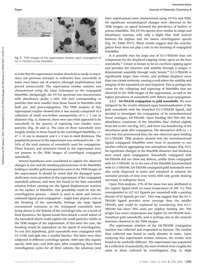
Fig. 3 TEM images of the supernatant residue upon conjugation ofG1-3-TEGOH to the HAuNShs.
Paper Journal of Materials Chemistry B
Publ
ishe
d on
06
Janu
ary
2015
. Dow
nloa
ded
by E
cole
Pol
ytec
hniq
ue d
e M
ontr
eal o
n 20
/02/
2015
16:
05:1
3.
View Article Online
to note that the supernatant residue dissolved so easily in water,since our previous attempts to redissolve bare nanoshells inwater once taken out of solution (through lyophilisation) hadproved unsuccessful. The supernatant residue solution wascharacterized using the same techniques as the conjugatedHAuNShs. Intriguingly, the UV-Vis spectrum was characteristicwith absorbance peaks (�409, 509 nm) corresponding toparticles that were smaller than those found in HAuNShs withboth pre- and post-conjugation. The TEM analysis of thissupernatant residue showed that it was mainly comprised of acollection of small non-hollow nanoparticles of 5 � 1 nm indiameter (Fig. 3). However, there were also what appeared to benanoshells in the process of rupturing into smaller nano-particles (Fig. 3b and c). The sizes of these nanoshells wereroughly similar to those found in the centrifuged HAuNShs, i.e.47 � 11 nm in diameter and 6 � 9 nm in shell thickness. Thepopulation present in the supernatant was estimated to be�10–20% of the total amount of nanoshells used for conjugation.These features and structures found in the supernatant werenot present in the TEM images of centrifuged conjugatednanoshells.
Several hypotheses were considered to explain the observedchanges in size and the breaking phenomenon of the HAuNShsleading to smaller gold nanoparticles seen in the TEM images ofthe supernatant. It should be noted that the damaged nano-shells were more prevalent in the supernatant of the conjugatednanoshell solution, and were not found in the bare nanoshellsolution before carrying out the ligand displacement reactionon the surface of HAuNShs. One possibility could be that thecentrifugation process – which is used to wash and removeunbound ligand post-conjugation – might have played a role inthe breaking of the nanoshells. Perhaps the large ligandencountered resistance as the conjugated nanoshells werebeing driven to the bottom of the centrifuge tube. As a result ofuid dynamics, the ligand would then detach a small subset ofthe nanoshell which could explain the small particles visible inthe TEM images of the supernatant. If this were the case, thebreaking would be dependent on the speed of centrifugation.To test this hypothesis, gold nanoshells were conjugated withG1-3-OH, and split into 3 smaller batches. The latter were thensubjected to different centrifuge speeds: 4600 rpm (the regularspeed), 3600 rpm, and 2600 rpm. Aer completing these threecentrifugation cycles for all three subsets, the solutions (and
This journal is © The Royal Society of Chemistry 2015
their supernatants) were characterized using UV-Vis and TEM.No signicant morphological changes were observed in theTEM images; no speed favoured the prevalence of broken orporous HAuNShs. The UV-Vis spectra were similar in shape andabsorbance maxima, with only a slight blue shi noticedbetween the highest and the lowest centrifugation speeds(Fig. S1, Table ST1†). These results suggest that the centrifu-gation force does not play a role in the breaking of conjugatedHAuNShs.
It is possible that the large size of G1-3-TEGOH does notcompensate for the displaced capping citrate agent on the barenanoshells.17 Citrate is known to be an excellent capping agentand provides the structure and stability through a unique 3-dimensional assembly through ionic bonds.18 G1-3-TEGOH issignicantly larger than citrate, and perhaps displaces morethan one citrate molecule, causing a void where the stability andintegrity of the nanoshell are now impaired. This is perhaps thecause for the collapsing and rupturing of HAuNShs that areobserved in the TEM images of the supernatant, as well as thehigher prevalence of nanoshells with defects post-conjugation.
3.2.2 G0-TEGOH conjugation to gold nanoshells. We wereintrigued by the results obtained upon functionalization of thegold nanoshells with the branched G1-3-TEGOH ligand, anddecided to investigate the conjugation of its monofunctionallinear analogue, G0-TEGOH. Upon binding HO–TEG–SH, theabsorbance maximum of the HAuNShs blue shied slightlyfrom 604 to 601 nm (Fig. S2†), and there was broadening of theabsorbance peak aer conjugation. The absorption shi (D ¼ 3nm) was less pronounced than the one observed upon bindingG1-3-TEGOH. TEM analysis showed that the monofunctionalligand conjugated HAuNShs were close in proximity to oneanother without aggregating into amorphous shapes (Fig. S3†).No signicant changes in the HAuNSh diameter and thicknesswere noted upon conjugation. Nanoshells conjugated withG0-TEGOH did not show any defects, unlike those conjugatedwith G1-3-TEGOH. As in the case of the HAuNSh functionalizedwith G1-3-TEGOH, G0-TEGOH conjugated gold nanoshells werealso easily dispersed in water and remained in solution forextended periods of time (one week) with only gentle shakingnecessary to redisperse them.
Upon TGA analysis, 37% of the mass loss was attributed tothe organic ligand (with an onset temperature of 200 �C). Thiscorresponded to 147 415 ligands per nanoshell with a packingdensity of 63 ligands per nm2. This may suggest that the G1-3-TEGOH ligand provides more coverage than the smallerTEGOH, and could be explained by considering that G1-3-TEGOH has three TEG units per sulphur binding site. Theweight loss onset temperature was higher for G0-TEGOH func-tionalized gold nanoshells, and is perhaps due to the networkstructure observed in the TEM images.
The supernatant solution of the G0-TEGOH conjugationreaction was collected and evaporated to dryness. The residuethus collected was found to easily dissolve in water. Uponanalyzing this supernatant residue solution, the results werefound to be markedly different. The supernatant was populatedby a collection of nanoshells, the sizes of which were roughly thesame as those collected by centrifugation (Fig. 4). High
J. Mater. Chem. B, 2015, 3, 1788–1800 | 1795
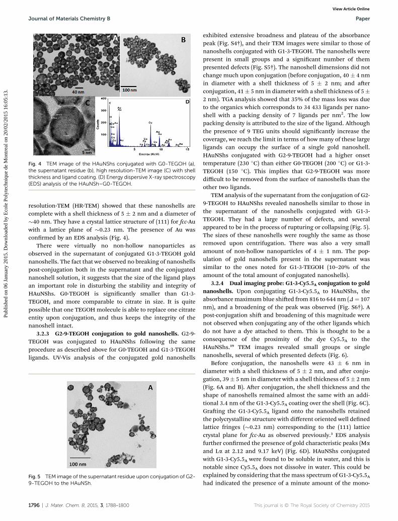
Fig. 4 TEM image of the HAuNShs conjugated with G0-TEGOH (a),the supernatant residue (b), high resolution-TEM image (C) with shellthickness and ligand coating. (D) Energy dispersive X-ray spectroscopy(EDS) analysis of the HAuNSh–G0-TEGOH.
Journal of Materials Chemistry B Paper
Publ
ishe
d on
06
Janu
ary
2015
. Dow
nloa
ded
by E
cole
Pol
ytec
hniq
ue d
e M
ontr
eal o
n 20
/02/
2015
16:
05:1
3.
View Article Online
resolution-TEM (HR-TEM) showed that these nanoshells arecomplete with a shell thickness of 5 � 2 nm and a diameter of�40 nm. They have a crystal lattice structure of (111) for fcc-Auwith a lattice plane of �0.23 nm. The presence of Au wasconrmed by an EDS analysis (Fig. 4).
There were virtually no non-hollow nanoparticles asobserved in the supernatant of conjugated G1-3-TEGOH goldnanoshells. The fact that we observed no breaking of nanoshellspost-conjugation both in the supernatant and the conjugatednanoshell solution, it suggests that the size of the ligand playsan important role in disturbing the stability and integrity ofHAuNShs. G0-TEGOH is signicantly smaller than G1-3-TEGOH, and more comparable to citrate in size. It is quitepossible that one TEGOHmolecule is able to replace one citrateentity upon conjugation, and thus keeps the integrity of thenanoshell intact.
3.2.3 G2-9-TEGOH conjugation to gold nanoshells. G2-9-TEGOH was conjugated to HAuNShs following the sameprocedure as described above for G0-TEGOH and G1-3-TEGOHligands. UV-Vis analysis of the conjugated gold nanoshells
Fig. 5 TEM image of the supernatant residue upon conjugation of G2-9-TEGOH to the HAuNSh.
1796 | J. Mater. Chem. B, 2015, 3, 1788–1800
exhibited extensive broadness and plateau of the absorbancepeak (Fig. S4†), and their TEM images were similar to those ofnanoshells conjugated with G1-3-TEGOH. The nanoshells werepresent in small groups and a signicant number of thempresented defects (Fig. S5†). The nanoshell dimensions did notchange much upon conjugation (before conjugation, 40 � 4 nmin diameter with a shell thickness of 5 � 2 nm; and aerconjugation, 41� 5 nm in diameter with a shell thickness of 5�2 nm). TGA analysis showed that 35% of the mass loss was dueto the organics which corresponds to 34 433 ligands per nano-shell with a packing density of 7 ligands per nm2. The lowpacking density is attributed to the size of the ligand. Althoughthe presence of 9 TEG units should signicantly increase thecoverage, we reach the limit in terms of howmany of these largeligands can occupy the surface of a single gold nanoshell.HAuNShs conjugated with G2-9-TEGOH had a higher onsettemperature (230 �C) than either G0-TEGOH (200 �C) or G1-3-TEGOH (150 �C). This implies that G2-9-TEGOH was moredifficult to be removed from the surface of nanoshells than theother two ligands.
TEM analysis of the supernatant from the conjugation of G2-9-TEGOH to HAuNShs revealed nanoshells similar to those inthe supernatant of the nanoshells conjugated with G1-3-TEGOH. They had a large number of defects, and severalappeared to be in the process of rupturing or collapsing (Fig. 5).The sizes of these nanoshells were roughly the same as thoseremoved upon centrifugation. There was also a very smallamount of non-hollow nanoparticles of 4 � 1 nm. The pop-ulation of gold nanoshells present in the supernatant wassimilar to the ones noted for G1-3-TEGOH (10–20% of theamount of the total amount of conjugated nanoshells).
3.2.4 Dual imaging probe: G1-3-Cy5.5A conjugation to goldnanoshells. Upon conjugating G1-3-Cy5.5A to HAuNShs, theabsorbancemaximum blue shied from 816 to 644 nm (D¼ 107nm), and a broadening of the peak was observed (Fig. S6†). Apost-conjugation shi and broadening of this magnitude werenot observed when conjugating any of the other ligands whichdo not have a dye attached to them. This is thought to be aconsequence of the proximity of the dye Cy5.5A to theHAuNShs.19 TEM images revealed small groups or singlenanoshells, several of which presented defects (Fig. 6).
Before conjugation, the nanoshells were 43 � 6 nm indiameter with a shell thickness of 5 � 2 nm, and aer conju-gation, 39� 5 nm in diameter with a shell thickness of 5� 2 nm(Fig. 6A and B). Aer conjugation, the shell thickness and theshape of nanoshells remained almost the same with an addi-tional 3.4 nm of the G1-3-Cy5.5A coating over the shell (Fig. 6C).Graing the G1-3-Cy5.5A ligand onto the nanoshells retainedthe polycrystalline structure with different oriented well denedlattice fringes (�0.23 nm) corresponding to the (111) latticecrystal plane for fcc-Au as observed previously.3 EDS analysisfurther conrmed the presence of gold characteristic peaks (Ma
and La at 2.12 and 9.17 keV) (Fig. 6D). HAuNShs conjugatedwith G1-3-Cy5.5A were found to be soluble in water, and this isnotable since Cy5.5A does not dissolve in water. This could beexplained by considering that the mass spectrum of G1-3-Cy5.5Ahad indicated the presence of a minute amount of the mono-
This journal is © The Royal Society of Chemistry 2015
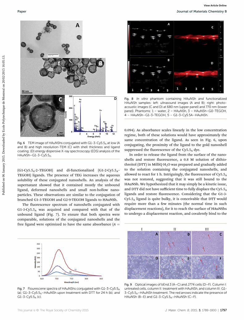
Fig. 8 In vitro phantom containing HAuNSh and functionalizedHAuNSh samples: left: ultrasound images (A and B); right: photo-acoustic images (C and D) at 680 nm (upper panel) and 770 nm (lowerpanel). Phantoms: 1 – water, 2 – HAuNSh, 3 – HAuNSh–G0-TEGOH,4 – HAuNSh–G1-3-TEGOH, 5 – G1-3-Cy5.5A–HAuNSh.
Fig. 6 TEM image of HAuNShs conjugated with G1-3-Cy5.5A at low (Aand B) and high resolution-TEM (C) with shell thickness and ligandcoating; (D) energy dispersive X-ray spectroscopy (EDS) analysis of theHAuNSh–G1-3-Cy5.5A.
Paper Journal of Materials Chemistry B
Publ
ishe
d on
06
Janu
ary
2015
. Dow
nloa
ded
by E
cole
Pol
ytec
hniq
ue d
e M
ontr
eal o
n 20
/02/
2015
16:
05:1
3.
View Article Online
(G1-Cy5.5A-2–TEGOH) and di-functionalized (G1-2-Cy5.5A–TEGOH) ligands. The presence of TEG increases the aqueoussolubility of these conjugated nanoshells. An analysis of thesupernatant showed that it contained mostly the unboundligand, deformed nanoshells and small non-hollow nano-particles. These observations are similar to the conjugation ofbranched G1-3-TEGOH and G2-9-TEGOH ligands to HAuNSh.
The uorescence spectrum of nanoshells conjugated withG1-3-Cy5.5A was acquired and compared with that of theunbound ligand (Fig. 7). To ensure that both spectra werecomparable, solutions of the conjugated nanoshells and thefree ligand were optimized to have the same absorbance (A ¼
Fig. 7 Flourescene spectra of HAuNShs conjugated with G1-3-Cy5.5A(a); G1-3-Cy5.5A–HAuNSh upon treatment with DTT for 24 h (b); andG1-3-Cy5.5A (c).
This journal is © The Royal Society of Chemistry 2015
0.094). As absorbance scales linearly in the low concentrationregime, both of these solutions would have approximately thesame concentration of the ligand. As seen in Fig. 6, uponconjugating, the proximity of the ligand to the gold nanoshellsuppressed the uorescence of the Cy5.5A dye.
In order to release the ligand from the surface of the nano-shells and restore uorescence, a 0.8 M solution of dithio-threitol (DTT) in MilliQ H2O was prepared and gradually addedto the solution containing the conjugated nanoshells, andallowed to react for 1 h. Intriguingly, the uorescence of Cy5.5Awas not restored, suggesting that it was still bound to theHAuNSh. We hypothesized that it may simply be a kinetic issue,and DTT did not have sufficient time to fully displace the Cy5.5Aligands and restore uorescence. Considering that the G1-3-Cy5.5A ligand is quite bulky, it is conceivable that DTT wouldrequire more than a few minutes (the normal time in suchdisplacement reactions), for it to reach the surface of HAuNShsto undergo a displacement reaction, and covalently bind to the
Fig. 9 Optical images of bEnd.3 (A–C) and J774 cells (D–F). Column I:untreated cells, column II: treatment with HAuNSh, and column III: G1-3-Cy5.5A–HAuNSh treatment. The red arrows indicate the presence ofHAuNSh (B–E) and G1-3-Cy5.5A–HAuNSh (C–F).
J. Mater. Chem. B, 2015, 3, 1788–1800 | 1797
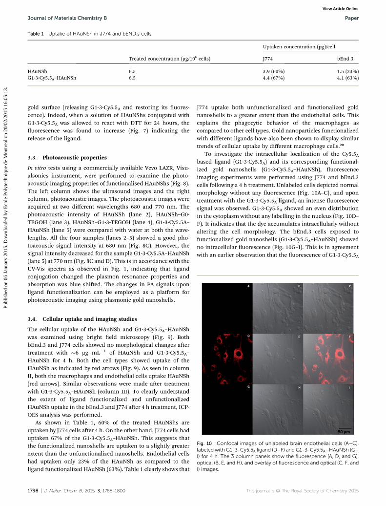
Table 1 Uptake of HAuNSh in J774 and bEND.s cells
Treated concentration (mg/106 cells)
Uptaken concentration (pg)/cell
J774 bEnd.3
HAuNSh 6.5 3.9 (60%) 1.5 (23%)G1-3-Cy5.5A–HAuNSh 6.5 4.4 (67%) 4.1 (63%)
Journal of Materials Chemistry B Paper
Publ
ishe
d on
06
Janu
ary
2015
. Dow
nloa
ded
by E
cole
Pol
ytec
hniq
ue d
e M
ontr
eal o
n 20
/02/
2015
16:
05:1
3.
View Article Online
gold surface (releasing G1-3-Cy5.5A and restoring its uores-cence). Indeed, when a solution of HAuNShs conjugated withG1-3-Cy5.5A was allowed to react with DTT for 24 hours, theuorescence was found to increase (Fig. 7) indicating therelease of the ligand.
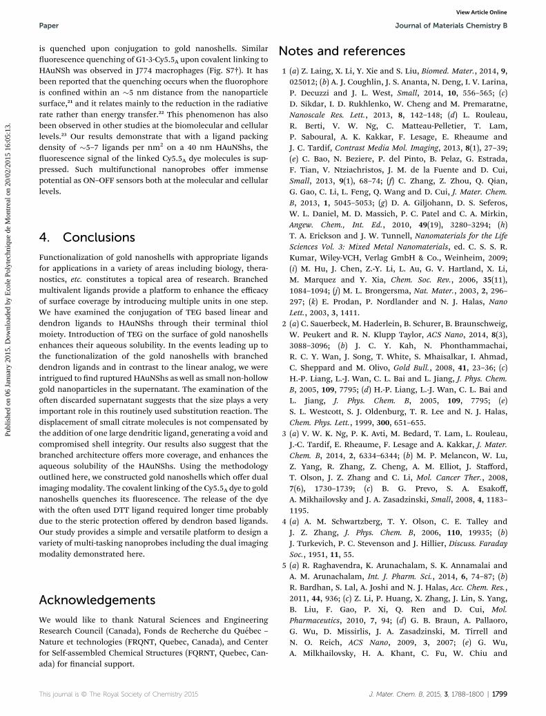
3.3. Photoacoustic properties
In vitro tests using a commercially available Vevo LAZR, Visu-alsonics instrument, were performed to examine the photo-acoustic imaging properties of functionalised HAuNShs (Fig. 8).The le column shows the ultrasound images and the rightcolumn, photoacoustic images. The photoacoustic images wereacquired at two different wavelengths 680 and 770 nm. Thephotoacoustic intensity of HAuNSh (lane 2), HAuNSh–G0-TEGOH (lane 3), HAuNSh–G1-3-TEGOH (lane 4), G1-3-Cy5.5A–HAuNSh (lane 5) were compared with water at both the wave-lengths. All the four samples (lanes 2–5) showed a good pho-toacoustic signal intensity at 680 nm (Fig. 8C). However, thesignal intensity decreased for the sample G1-3-Cy5.5A–HAuNSh(lane 5) at 770 nm (Fig. 8C and D). This is in accordance with theUV-Vis spectra as observed in Fig. 1, indicating that ligandconjugation changed the plasmon resonance properties andabsorption was blue shied. The changes in PA signals uponligand functionalization can be employed as a platform forphotoacoustic imaging using plasmonic gold nanoshells.
Fig. 10 Confocal images of unlabeled brain endothelial cells (A–C),labeled with G1-3-Cy5.5A ligand (D–F) and G1-3-Cy5.5A–HAuNSh (G–I) for 4 h. The 3 column panels show the fluorescence (A, D, and G),optical (B, E, and H), and overlay of fluorescence and optical (C, F, andI) images.
3.4. Cellular uptake and imaging studies
The cellular uptake of the HAuNSh and G1-3-Cy5.5A–HAuNShwas examined using bright eld microscopy (Fig. 9). BothbEnd.3 and J774 cells showed no morphological changes aertreatment with �6 mg mL�1 of HAuNSh and G1-3-Cy5.5A–HAuNSh for 4 h. Both the cell types showed uptake of theHAuNSh as indicated by red arrows (Fig. 9). As seen in columnII, both the macrophages and endothelial cells uptake HAuNSh(red arrows). Similar observations were made aer treatmentwith G1-3-Cy5.5A–HAuNSh (column III). To clearly understandthe extent of ligand functionalized and unfunctionalizedHAuNSh uptake in the bEnd.3 and J774 aer 4 h treatment, ICP-OES analysis was performed.
As shown in Table 1, 60% of the treated HAuNShs areuptaken by J774 cells aer 4 h. On the other hand, J774 cells haduptaken 67% of the G1-3-Cy5.5A–HAuNSh. This suggests thatthe functionalized nanoshells are uptaken to a slightly greaterextent than the unfunctionalized nanoshells. Endothelial cellshad uptaken only 23% of the HAuNSh as compared to theligand functionalized HAuNSh (63%). Table 1 clearly shows that
1798 | J. Mater. Chem. B, 2015, 3, 1788–1800
J774 uptake both unfunctionalized and functionalized goldnanoshells to a greater extent than the endothelial cells. Thisexplains the phagocytic behavior of the macrophages ascompared to other cell types. Gold nanoparticles functionalizedwith different ligands have also been shown to display similartrends of cellular uptake by different macrophage cells.20
To investigate the intracellular localization of the Cy5.5Abased ligand (G1-3-Cy5.5A) and its corresponding functional-ized gold nanoshells (G1-3-Cy5.5A–HAuNSh), uorescenceimaging experiments were performed using J774 and bEnd.3cells following a 4 h treatment. Unlabeled cells depicted normalmorphology without any uorescence (Fig. 10A–C), and upontreatment with the G1-3-Cy5.5A ligand, an intense uorescencesignal was observed. G1-3-Cy5.5A showed an even distributionin the cytoplasm without any labelling in the nucleus (Fig. 10D–F). It indicates that the dye accumulates intracellularly withoutaltering the cell morphology. The bEnd.3 cells exposed tofunctionalized gold nanoshells (G1-3-Cy5.5A–HAuNSh) showedno intracellular uorescence (Fig. 10G–I). This is in agreementwith an earlier observation that the uorescence of G1-3-Cy5.5A
This journal is © The Royal Society of Chemistry 2015
Paper Journal of Materials Chemistry B
Publ
ishe
d on
06
Janu
ary
2015
. Dow
nloa
ded
by E
cole
Pol
ytec
hniq
ue d
e M
ontr
eal o
n 20
/02/
2015
16:
05:1
3.
View Article Online
is quenched upon conjugation to gold nanoshells. Similaruorescence quenching of G1-3-Cy5.5A upon covalent linking toHAuNSh was observed in J774 macrophages (Fig. S7†). It hasbeen reported that the quenching occurs when the uorophoreis conned within an �5 nm distance from the nanoparticlesurface,21 and it relates mainly to the reduction in the radiativerate rather than energy transfer.22 This phenomenon has alsobeen observed in other studies at the biomolecular and cellularlevels.23 Our results demonstrate that with a ligand packingdensity of �5–7 ligands per nm2 on a 40 nm HAuNShs, theuorescence signal of the linked Cy5.5A dye molecules is sup-pressed. Such multifunctional nanoprobes offer immensepotential as ON–OFF sensors both at the molecular and cellularlevels.
4. Conclusions
Functionalization of gold nanoshells with appropriate ligandsfor applications in a variety of areas including biology, thera-nostics, etc. constitutes a topical area of research. Branchedmultivalent ligands provide a platform to enhance the efficacyof surface coverage by introducing multiple units in one step.We have examined the conjugation of TEG based linear anddendron ligands to HAuNShs through their terminal thiolmoiety. Introduction of TEG on the surface of gold nanoshellsenhances their aqueous solubility. In the events leading up tothe functionalization of the gold nanoshells with brancheddendron ligands and in contrast to the linear analog, we wereintrigued to nd ruptured HAuNShs as well as small non-hollowgold nanoparticles in the supernatant. The examination of theoen discarded supernatant suggests that the size plays a veryimportant role in this routinely used substitution reaction. Thedisplacement of small citrate molecules is not compensated bythe addition of one large dendritic ligand, generating a void andcompromised shell integrity. Our results also suggest that thebranched architecture offers more coverage, and enhances theaqueous solubility of the HAuNShs. Using the methodologyoutlined here, we constructed gold nanoshells which offer dualimaging modality. The covalent linking of the Cy5.5A dye to goldnanoshells quenches its uorescence. The release of the dyewith the oen used DTT ligand required longer time probablydue to the steric protection offered by dendron based ligands.Our study provides a simple and versatile platform to design avariety of multi-tasking nanoprobes including the dual imagingmodality demonstrated here.
Acknowledgements
We would like to thank Natural Sciences and EngineeringResearch Council (Canada), Fonds de Recherche du Quebec –
Nature et technologies (FRQNT, Quebec, Canada), and Centerfor Self-assembled Chemical Structures (FQRNT, Quebec, Can-ada) for nancial support.
This journal is © The Royal Society of Chemistry 2015
Notes and references
1 (a) Z. Laing, X. Li, Y. Xie and S. Liu, Biomed. Mater., 2014, 9,025012; (b) A. J. Coughlin, J. S. Ananta, N. Deng, I. V. Larina,P. Decuzzi and J. L. West, Small, 2014, 10, 556–565; (c)D. Sikdar, I. D. Rukhlenko, W. Cheng and M. Premaratne,Nanoscale Res. Lett., 2013, 8, 142–148; (d) L. Rouleau,R. Berti, V. W. Ng, C. Matteau-Pelletier, T. Lam,P. Saboural, A. K. Kakkar, F. Lesage, E. Rheaume andJ. C. Tardif, Contrast Media Mol. Imaging, 2013, 8(1), 27–39;(e) C. Bao, N. Beziere, P. del Pinto, B. Pelaz, G. Estrada,F. Tian, V. Ntziachristos, J. M. de la Fuente and D. Cui,Small, 2013, 9(1), 68–74; (f) C. Zhang, Z. Zhou, Q. Qian,G. Gao, C. Li, L. Feng, Q. Wang and D. Cui, J. Mater. Chem.B, 2013, 1, 5045–5053; (g) D. A. Giljohann, D. S. Seferos,W. L. Daniel, M. D. Massich, P. C. Patel and C. A. Mirkin,Angew. Chem., Int. Ed., 2010, 49(19), 3280–3294; (h)T. A. Erickson and J. W. Tunnell, Nanomaterials for the LifeSciences Vol. 3: Mixed Metal Nanomaterials, ed. C. S. S. R.Kumar, Wiley-VCH, Verlag GmbH & Co., Weinheim, 2009;(i) M. Hu, J. Chen, Z.-Y. Li, L. Au, G. V. Hartland, X. Li,M. Marquez and Y. Xia, Chem. Soc. Rev., 2006, 35(11),1084–1094; (j) M. L. Brongersma, Nat. Mater., 2003, 2, 296–297; (k) E. Prodan, P. Nordlander and N. J. Halas, NanoLett., 2003, 3, 1411.
2 (a) C. Sauerbeck, M. Haderlein, B. Schurer, B. Braunschweig,W. Peukert and R. N. Klupp Taylor, ACS Nano, 2014, 8(3),3088–3096; (b) J. C. Y. Kah, N. Phonthammachai,R. C. Y. Wan, J. Song, T. White, S. Mhaisalkar, I. Ahmad,C. Sheppard and M. Olivo, Gold Bull., 2008, 41, 23–36; (c)H.-P. Liang, L.-J. Wan, C. L. Bai and L. Jiang, J. Phys. Chem.B, 2005, 109, 7795; (d) H.-P. Liang, L.-J. Wan, C. L. Bai andL. Jiang, J. Phys. Chem. B, 2005, 109, 7795; (e)S. L. Westcott, S. J. Oldenburg, T. R. Lee and N. J. Halas,Chem. Phys. Lett., 1999, 300, 651–655.
3 (a) V. W. K. Ng, P. K. Avti, M. Bedard, T. Lam, L. Rouleau,J.-C. Tardif, E. Rheaume, F. Lesage and A. Kakkar, J. Mater.Chem. B, 2014, 2, 6334–6344; (b) M. P. Melancon, W. Lu,Z. Yang, R. Zhang, Z. Cheng, A. M. Elliot, J. Stafford,T. Olson, J. Z. Zhang and C. Li, Mol. Cancer Ther., 2008,7(6), 1730–1739; (c) B. G. Prevo, S. A. Esakoff,A. Mikhailovsky and J. A. Zasadzinski, Small, 2008, 4, 1183–1195.
4 (a) A. M. Schwartzberg, T. Y. Olson, C. E. Talley andJ. Z. Zhang, J. Phys. Chem. B, 2006, 110, 19935; (b)J. Turkevich, P. C. Stevenson and J. Hillier, Discuss. FaradaySoc., 1951, 11, 55.
5 (a) R. Raghavendra, K. Arunachalam, S. K. Annamalai andA. M. Arunachalam, Int. J. Pharm. Sci., 2014, 6, 74–87; (b)R. Bardhan, S. Lal, A. Joshi and N. J. Halas, Acc. Chem. Res.,2011, 44, 936; (c) Z. Li, P. Huang, X. Zhang, J. Lin, S. Yang,B. Liu, F. Gao, P. Xi, Q. Ren and D. Cui, Mol.Pharmaceutics, 2010, 7, 94; (d) G. B. Braun, A. Pallaoro,G. Wu, D. Missirlis, J. A. Zasadzinski, M. Tirrell andN. O. Reich, ACS Nano, 2009, 3, 2007; (e) G. Wu,A. Milkhailovsky, H. A. Khant, C. Fu, W. Chiu and
J. Mater. Chem. B, 2015, 3, 1788–1800 | 1799
Journal of Materials Chemistry B Paper
Publ
ishe
d on
06
Janu
ary
2015
. Dow
nloa
ded
by E
cole
Pol
ytec
hniq
ue d
e M
ontr
eal o
n 20
/02/
2015
16:
05:1
3.
View Article Online
J. A. Zasadzinski, J. Am. Chem. Soc., 2008, 130, 8175; (f)W. Cai, T. Gao, H. Hong and J. Sun, Nanotechnol., Sci.Appl., 2008, 1, 1–25; (g) P. K. Avti, D. Maysinger andA. Kakkar, Molecules, 2013, 18, 9531.
6 (a) B. Moses and Y. You, Med. Chem., 2013, 3, 192–198; (b)Z. J. Deng, M. Liang, I. Toth, M. J. Monteiro andR. F. Minchin, ACS Nano, 2012, 6, 8962; (c) C. Freese,M. I. Gibson, H. A. Klok, R. E. Unger and C. J. Kirkpatrick,Biomacromolecules, 2012, 13, 1533; (d) E. C. Dreaden,M. A. Mackey, X. Huang, B. Kang and M. A. El-Sayed,Chem. Soc. Rev., 2011, 40, 3391–3404; (e) Z. Liang, Y. Liu,X. Li, Q. Wu, J. Yu, S. Luo, L. Lai and S. Liu, J. Biomed.Mater. Res., Part A, 2011, 98, 479.
7 (a) J. P. Hermes, F. Sander, U. Fluch, T. Peterle,D. Thompson, R. Urbani, T. Pfohl and M. Mayor, J. Am.Chem. Soc., 2012, 134, 14674–14677; (b) C. Peng, K. Li,X. Cao, T. Xiao, W. Hou, L. Zheng, R. Guo, M. Shen,G. Zhang and X. Shi, Nanoscale, 2012, 4, 6768–6778; (c)J. R. Baker Jr, Hematology, 2009, 1, 708–719.
8 (a) H. Chen, M. Zhang, H. Yang, W. Xu, Y. Ma and Y. Gu, RSCAdv., 2014, 4, 8191–8199; (b) M. Kacenka, O. Kaman, J. Kotek,L. Falteisek, J. Cerny, D. Jirak, V. Herynek, K. Zacharovova,Z. Berkova, P. Jendelova, J. Kupcik, E. Pollert, P. Veverkaand I. Lukes, J. Mater. Chem., 2011, 21, 157–164; (c)Z. Wang, S. Zong, J. Yang, J. Li and Y. Cui, Biosens.Bioelectron., 2011, 26, 2883–2889; (d) W. Chen, R. Bardhan,M. Bartels, C. Perez-Torres, R. G. Pautler, N. J. Halas andA. Joshi, Mol. Cancer Ther., 2010, 9, 1028; (e) S. Lee andX. Chen, Mol. Imaging, 2009, 8, 1536–0121.
9 (a) M.-C. Daniel, M. E. Grow, H. Pan, M. Bednarek,W. E. Ghann, K. Zabetakis and J. Cornish, New J. Chem.,2011, 35, 2366; (b) H. Gao and K. Matyjaszewski, J. Am.Chem. Soc., 2007, 129, 6633; (c) A. Sharma, G. M. Soliman,N. Al-Hajaj, R. Sharma, D. Maysinger and A. Kakkar,Biomacromolecules, 2011, 13, 239; (d) P. R. Ashton, J. Huff,S. Menzer, I. W. Parsons, J. A. Preece, J. F. Stoddart,M. S. Tolley, A. J. P. White and D. J. Williams, Chem.–Eur.J., 1996, 2, 31.
10 Y. Sato, K. Yoshioka, M. Tanaka, T. Murakami, M. N. Ishidaand O. Niwa, Chem. Commun., 2008, 40, 4909.
11 (a) L. Albertazzi, F. M. Mickler, G. M. Pavan, F. Salomone,G. Bardi, M. Panniello, E. Amir, T. Kang, K. L. Killops,C. Brauchle, R. Amir and C. J. Hawker, Biomacromolecules,2012, 13, 4089; (b) Y. Umeda, C. Kojima, A. Harada,H. Horinaka and K. Kono, Bioconjugate Chem., 2010, 21,
1800 | J. Mater. Chem. B, 2015, 3, 1788–1800
1559; (c) X. Huan, D. Wang, R. Dong, C. Tu, B. Zhu, D. Yanand X. Zhu, Macromolecules, 2012, 45, 5941.
12 J. Camponovo, J. Ruiz, E. Cloutet and D. Astruc, Chem.–Eur.J., 2009, 15, 2990.
13 L. Wang, D. J. Kiemle, C. J. Boyle, E. L. Connors and I. Gitsov,Macromolecules, 2014, 47, 2199.
14 O. B. Wallace and D. M. Springer, Tetrahedron Lett., 1998, 39,2693.
15 X.-B. Li, Z.-J. Li, Y.-J. Gao, Q.-Y. Meng, S. Yu, R. G. Weiss,C.-H. Tung and L.-Z. Wu, Angew. Chem., Int. Ed., 2014, 53,2085.
16 R. L. Whetten and R. C. Price, Science, 2007, 318, 407.17 (a) D.-H. Tsai, M. P. Shelton, F. W. DelRio, S. Elzey, S. Guha,
M. R. Zachariah and V. A. Hackley, Anal. Bioanal. Chem.,2012, 404, 3015; (b) S. V. Kumar and S. Ganesan, Int. J.Green Nanotechnol., 2011, 3, 47.
18 J.-W. Park and J. S. Shumaker-Parry, J. Am. Chem. Soc., 2014,136, 1907.
19 (a) F. Zhang, J. Zhu, J.-J. Li and J.-W. Zhao, Appl. Phys. Lett.,2013, 103, 193703; (b) A.-M. Gabudean, F. Lerouge,T. Gallavardin, M. Iosin, S. Zaiba, O. Maury, P. L. Baldeck,C. Andraud and S. Parola, Opt. Mater., 2011, 33, 1377.
20 (a) R. Shukla, V. Bansal, M. Chaudhary, A. Basu,R. R. Bhonde and M. Sastry, Langmuir, 2005, 21, 10644–10654; (b) K. Ye, J. Qin, Z. Peng, X. Yang, L. Huang,F. Yuan, C. Peng, M. Jiang and X. Lu, Nanoscale Res. Lett.,2014, 9, 529; (c) T. A. Larson, P. P. Joshi and K. Sokolov,ACS Nano, 2012, 6(10), 9182.
21 (a) E. Dulkeith, A. C. Morteani, T. Niedereichholz, T. A. Klar,J. Feldmann, S. A. Levi, F. C. J. M. van Veggel,D. N. Reinhoudt, M. Moller and D. I. Gittins, Phys. Rev.Lett., 2002, 89, 203002; (b) K. Aslan and V. H. Perez-Luna, J.Fluoresc., 2004, 14, 401–405.
22 E. Dulkeith, M. Ringler, T. A. Klar, J. Feldmann, A. M. Javierand W. J. Parak, Nano Lett., 2005, 5, 585–589.
23 (a) A. E. Prigodich, D. S. Seferos, M. D. Massich,D. A. Giljohann, B. C. Lane and C. A. Mirkin, ACS Nano,2009, 3, 2147–2152; (b) C. C. You, O. R. Miranda, B. Gider,P. S. Ghosh, I. B. Kim, B. Erdogan, S. A. Krovi,U. H. F. Bunz and V. M. Rotello, Nat. Nanotechnol., 2007, 2,318–323; (c) A. Bajaj, O. R. Miranda, I. B. Kim,R. L. Phillips, D. J. Jerry, U. H. F. Bunz and V. M. Rotello,Proc. Natl. Acad. Sci. U. S. A., 2009, 106, 10912–10916; (d)D. J. Maxwell, J. R. Taylor and S. M. Nie, J. Am. Chem. Soc.,2002, 124, 9606–9612.
This journal is © The Royal Society of Chemistry 2015
Related Documents


![Multivalent Targeting Based Delivery of Therapeutic ...Multivalent Targeting Based Delivery of Therapeutic ... ... 10), . p)]] ...](https://static.cupdf.com/doc/110x72/5fe28d7a524ece466e32b4fb/multivalent-targeting-based-delivery-of-therapeutic-multivalent-targeting-based.jpg)


