Congo red and thioflavin-T analogs detect Ab oligomers Izumi Maezawa,* Hyun-Seok Hong,* Ruiwu Liu, Chun-Yi Wu, R. Holland Cheng,à Mei-Ping Kung,§ Hank F. Kung,§ Kit S. Lam, Salvatore Oddo,¶ Frank M. LaFerla¶ and Lee-Way Jin* *M.I.N.D. Institute and Department of Pathology, University of California Davis, Sacramento, California, USA Division of Hematology and Oncology, Department of Internal Medicine, University of California Davis, Sacramento, California, USA àDepartment of Molecular and Cellular Biology, University of California Davis, Davis, California, USA §Department of Radiology, University of Pennsylvania, Philadelphia, Pennsylvania, USA ¶Department of Neurobiology and Behavior, University of California Irvine, Irvine, California, USA Alzheimer’s disease (AD) is characterized by deposition of Ab fibrils in brain. The diagnosis and investigation of AD has been greatly facilitated by organic dyes that specifically bind Ab fibrils. In particular, Congo red (CR) and thioflavins are widely used to stain amyloid in biological samples. Because both molecules are charged and hydrophilic, they do not pass the blood–brain barrier. However, they have provided a starting point for the development of lipophilic derivatives that are able to penetrate the brain. A few CR- and thioflavin-T (ThT)-analogs, collectively called amyloid ligands here, have been developed as candidate probes for amyloid imaging using positron emission tomography (PET) or single photon emission computed tomography (Mathis et al. 2004; Kung et al. 2004; Cai et al. 2007). Recently, the build-up of Ab oligomers (AbO) in brain starting at early disease stages preceding fibril formation has been recognized as an additional neuropathological hallmark of AD (Klein et al. 2001; Ferreira et al. 2007). Accumulating evidence indicates that AbO, rather than Ab fibrils, can induce severe neurodegeneration and cognitive deficits (Klein et al. 2001; Hardy and Selkoe 2002; Glabe 2006; Cole and Frautschy 2006; Ferreira et al. 2007). Although AbO were Submitted August 9, 2007; Revised manuscript received September 6, 2007; accepted September 10, 2007. Address correspondence and reprint requests to Lee-Way Jin, MD PhD, The M.I.N.D. Institute and Department of Medical Pathology, UC Davis Health System, 2805 50th Street, Sacramento, CA 95817, USA. E-mail: [email protected] Abbreviations used: AD, Alzheimer’s disease; APP-C99, the car- boxyl-terminal 99 residues of the amyloid-b precursor protein; Ab, amyloid-b protein; AbO, Ab oligomers; BSB, (trans, trans)-1-bromo- 2,5-bis-(3-hydroxycarbonyl-4-hydroxy)styrylbenzene; CR, Congo red; DMSO, dimethylsulfoxide; EM, electron microscopy; IBOX, (2-4¢- dimethylaminophenyl)-6-iodobenzoxazole; KD, dissociation constant; PET, positron emission tomography; PIB, Pittsburgh Compound-B; RU, response unit; SPR, surface plasmon resonance spectroscopy; TC, tet- racycline; ThT, thioflavin-T. Abstract Several small molecule ligands for amyloid-b (Ab) fibrils deposited in brain have been developed to facilitate radio- logical diagnosis of Alzheimer’s disease (AD). Recently, the build-up of Ab oligomers (AbO) in brain has been recognized as an additional hallmark of AD and may play a more signifi- cant role in early stages. Evidence suggests that quantitative assessment of AbO would provide a more accurate index of therapeutic effect of drug trials. Therefore, there is an urgent need to develop methods for efficient identification as well as structural analysis of AbO. We found that some well estab- lished amyloid ligands, analogs of Congo red and thioflavin-T (ThT), bind AbO with high affinity and detect AbO in vitro and in vivo. Binding studies revealed the presence of binding sites for Congo red- and thioflavin-T-analogs on AbO. Furthermore, these ligands can be used for imaging intracellular AbO in living cells and animals and as positive contrast agent for ul- trastructural imaging of AbO, two applications useful for structural analysis of AbO in cells. We propose that by improving the binding affinity of current ligands, in vivo imaging of AbO is feasible by a ‘signal subtraction’ procedure. This approach may facilitate the identification of individuals with early AD. Keywords: Alzheimer, amyloid, imaging, ligand, oligomer, small molecule. J. Neurochem. (2008) 104, 457–468. d JOURNAL OF NEUROCHEMISTRY | 2008 | 104 | 457–468 doi: 10.1111/j.1471-4159.2007.04972.x ȑ 2007 The Authors Journal Compilation ȑ 2007 International Society for Neurochemistry, J. Neurochem. (2008) 104, 457–468 457

Welcome message from author
This document is posted to help you gain knowledge. Please leave a comment to let me know what you think about it! Share it to your friends and learn new things together.
Transcript

Congo red and thioflavin-T analogs detect Ab oligomers
Izumi Maezawa,* Hyun-Seok Hong,* Ruiwu Liu,� Chun-Yi Wu,� R. Holland Cheng,�Mei-Ping Kung,§ Hank F. Kung,§ Kit S. Lam,� Salvatore Oddo,¶ Frank M. LaFerla¶ andLee-Way Jin*
*M.I.N.D. Institute and Department of Pathology, University of California Davis, Sacramento, California, USA
�Division of Hematology and Oncology, Department of Internal Medicine, University of California Davis, Sacramento,
California, USA
�Department of Molecular and Cellular Biology, University of California Davis, Davis, California, USA
§Department of Radiology, University of Pennsylvania, Philadelphia, Pennsylvania, USA
¶Department of Neurobiology and Behavior, University of California Irvine, Irvine, California, USA
Alzheimer’s disease (AD) is characterized by deposition ofAb fibrils in brain. The diagnosis and investigation of ADhas been greatly facilitated by organic dyes that specificallybind Ab fibrils. In particular, Congo red (CR) and thioflavinsare widely used to stain amyloid in biological samples.Because both molecules are charged and hydrophilic, they donot pass the blood–brain barrier. However, they haveprovided a starting point for the development of lipophilicderivatives that are able to penetrate the brain. A few CR-and thioflavin-T (ThT)-analogs, collectively called amyloidligands here, have been developed as candidate probes foramyloid imaging using positron emission tomography (PET)or single photon emission computed tomography (Mathiset al. 2004; Kung et al. 2004; Cai et al. 2007).
Recently, the build-up of Ab oligomers (AbO) in brainstarting at early disease stages preceding fibril formation hasbeen recognized as an additional neuropathological hallmarkof AD (Klein et al. 2001; Ferreira et al. 2007). Accumulating
evidence indicates that AbO, rather than Ab fibrils, caninduce severe neurodegeneration and cognitive deficits (Kleinet al. 2001; Hardy and Selkoe 2002; Glabe 2006; Cole andFrautschy 2006; Ferreira et al. 2007). Although AbO were
Submitted August 9, 2007; Revised manuscript received September 6,2007; accepted September 10, 2007.Address correspondence and reprint requests to Lee-Way Jin, MD
PhD, The M.I.N.D. Institute and Department of Medical Pathology, UCDavis Health System, 2805 50th Street, Sacramento, CA 95817, USA.E-mail: [email protected] used: AD, Alzheimer’s disease; APP-C99, the car-
boxyl-terminal 99 residues of the amyloid-b precursor protein; Ab,amyloid-b protein; AbO, Ab oligomers; BSB, (trans, trans)-1-bromo-2,5-bis-(3-hydroxycarbonyl-4-hydroxy)styrylbenzene; CR, Congo red;DMSO, dimethylsulfoxide; EM, electron microscopy; IBOX, (2-4¢-dimethylaminophenyl)-6-iodobenzoxazole; KD, dissociation constant;PET, positron emission tomography; PIB, Pittsburgh Compound-B; RU,response unit; SPR, surface plasmon resonance spectroscopy; TC, tet-racycline; ThT, thioflavin-T.
Abstract
Several small molecule ligands for amyloid-b (Ab) fibrils
deposited in brain have been developed to facilitate radio-
logical diagnosis of Alzheimer’s disease (AD). Recently, the
build-up of Ab oligomers (AbO) in brain has been recognized
as an additional hallmark of AD and may play a more signifi-
cant role in early stages. Evidence suggests that quantitative
assessment of AbO would provide a more accurate index of
therapeutic effect of drug trials. Therefore, there is an urgent
need to develop methods for efficient identification as well as
structural analysis of AbO. We found that some well estab-
lished amyloid ligands, analogs of Congo red and thioflavin-T
(ThT), bind AbO with high affinity and detect AbO in vitro and
in vivo. Binding studies revealed the presence of binding sites
for Congo red- and thioflavin-T-analogs on AbO. Furthermore,
these ligands can be used for imaging intracellular AbO in
living cells and animals and as positive contrast agent for ul-
trastructural imaging of AbO, two applications useful for
structural analysis of AbO in cells. We propose that by
improving the binding affinity of current ligands, in vivo
imaging of AbO is feasible by a ‘signal subtraction’ procedure.
This approach may facilitate the identification of individuals
with early AD.
Keywords: Alzheimer, amyloid, imaging, ligand, oligomer,
small molecule.
J. Neurochem. (2008) 104, 457–468.
d JOURNAL OF NEUROCHEMISTRY | 2008 | 104 | 457–468 doi: 10.1111/j.1471-4159.2007.04972.x
� 2007 The AuthorsJournal Compilation � 2007 International Society for Neurochemistry, J. Neurochem. (2008) 104, 457–468 457
initially considered transient intermediates during Ab fibril-lization (Caughey and Lansbury 2003), they were later shownto constitute an alternative pathway independent of fibrilli-zation (Necula et al. 2007a). Indeed, AbO deposits in ADbrains do not co-localize with the Ab fibril plaques (Kayedet al. 2003). Recent evidence suggests that therapeuticinterventions that reduce Ab fibrils at the cost of augmentingnon-fibrillar Ab assemblies including AbO could be harmful(Cheng et al. 2007). Therefore, currently developed PETimaging approaches to quantify Ab fibrils only may not fullyreflect the therapeutic effect of drug trials. Taken together, it isstrongly indicated that methods for in vivo detection andquantification of AbO may (i) help identify individuals withearly stages of AD; (ii) provide a pathological correlate ofcognitive decline not obligatorily related to fibril formation;and (3) provide a better index of therapeutic effects.
However, AbO are small and metastable, making themdifficult to identify in biological samples, and their structuralfeatures difficult to determine (Chromy et al. 2003; Bitan2006). The only useful probes for their identification are therecently developed oligomer-specific antibodies (Kayedet al. 2003; Lambert et al. 2007). However, antibody-basedPET imaging has not been successful as a result of poor brainpenetration of the probes (Mathis et al. 2004). To gain abetter understanding of AbO, we set out to search smallmolecule compounds that can be used as probes for AbO.One of the approaches is to examine the existing amyloidligands, which have the advantage of excellent pharmacoki-netics and well-known toxicology properties. CR and ThTwere shown to bind Ab protofibrils, the immediate aggre-gation products of late oligomers (Walsh et al. 1999; Ferreiraet al. 2007), but whether they bind AbO has not beenexamined. Based on the assumption that AbO do not bindthioflavins, several studies have used the failure to bindthioflavins as one of the criteria for oligomer identification(for example, see Lacor et al. 2004; Caspersen et al. 2005;Oddo et al. 2006). However, at least two lines of AD modelmice showed thioflavin-positive intraneuronal, presumablynon-fibrillar Ab deposits (Casas et al. 2004; Oakley et al.2006), suggesting that thioflavins may bind AbO. In thisstudy, we critically examined the binding of amyloid ligandsto AbO and found evidence supporting the presence of ThT-and CR-binding sites on AbO. Our results would heraldmany applications of amyloid ligands to the studies of AbO,including structural analysis and in vivo imaging.
Materials and methods
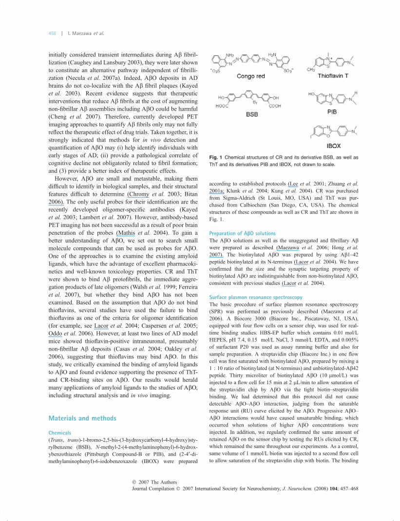
Chemicals(Trans, trans)-1-bromo-2,5-bis-(3-hydroxycarbonyl-4-hydroxy)sty-
rylbenzene (BSB), N-methyl-2-(4-methylaminophenyl)-6-hydrox-
ybenzothiazole (Pittsburgh Compound-B or PIB), and (2-4¢-di-methylaminophenyl)-6-iodobenzoxazole (IBOX) were prepared
according to established protocols (Lee et al. 2001; Zhuang et al.2001a; Klunk et al. 2004; Kung et al. 2004). CR was purchased
from Sigma-Aldrich (St Louis, MO, USA) and ThT was pur-
chased from Calbiochem (San Diego, CA, USA). The chemical
structures of these compounds as well as CR and ThT are shown in
Fig. 1.
Preparation of AbO solutionsThe AbO solutions as well as the unaggregated and fibrillary Abwere prepared as described (Maezawa et al. 2006; Hong et al.2007). The biotinylated AbO was prepared by using Ab1–42peptide biotinylated at its N-terminus (Lacor et al. 2004). We have
confirmed that the size and the synaptic targeting property of
biotinylated AbO are indistinguishable from non-biotinylated AbO,consistent with previous studies (Lacor et al. 2004).
Surface plasmon resonance spectroscopyThe basic procedure of surface plasmon resonance spectroscopy
(SPR) was performed as previously described (Maezawa et al.2006). A Biocore 3000 (Biacore Inc., Piscataway, NJ, USA),
equipped with four flow cells on a sensor chip, was used for real-
time binding studies. HBS-EP buffer which contains 0.01 mol/L
HEPES, pH 7.4, 0.15 mol/L NaCl, 3 mmol/L EDTA, and 0.005%
of surfactant P20 was used as assay running buffer and also for
sample preparation. A streptavidin chip (Biacore Inc.) in one flow
cell was first saturated with biotinylated AbO, prepared by mixing a
1 : 10 ratio of biotinylated (at N-terminus) and unbiotinylated-Ab42peptide. Thirty microliter of biotinylated AbO (10 lmol/L) was
injected to a flow cell for 15 min at 2 lL/min to allow saturation of
the streptavidin chip by AbO via the tight biotin–streptavidin
binding. We had determined that this protocol did not cause
detectable AbO–AbO interaction, judging from the saturable
response unit (RU) curve elicited by the AbO. Progressive AbO–AbO interactions would have caused unsaturable binding, which
occurred when solutions of higher AbO concentrations were
injected. In addition, we regularly confirmed the same amount of
retained AbO on the sensor chip by testing the RUs elicited by CR,
which remained the same throughout our experiments. As a control,
same volume of 1 mmol/L biotin was injected to a second flow cell
to allow saturation of the streptavidin chip with biotin. The binding
Fig. 1 Chemical structures of CR and its derivative BSB, as well as
ThT and its derivatives PIB and IBOX, not drawn to scale.
Journal Compilation � 2007 International Society for Neurochemistry, J. Neurochem. (2008) 104, 457–468� 2007 The Authors
458 | I. Maezawa et al.

of injected compounds in the flow phase to immobilized AbO was
measured by RU elicited. One RU represents about 1 pg/mm2 of the
analytes on the surface matrix of the sensor chip. The RUs elicited
by the compounds injected into the biotin control flow cell was set
as reference response, which represented the bulky refractive index
signals caused by solvent in the injected samples and was subtracted
from the RUs elicited by the same compounds injected to the AbOsaturated flow cell. To perform a binding kinetics analysis, 60 lL of
serial fivefold diluted compounds ranging from 50 lmol/L to
3.2 nmol/L with 0.5% dimethylsulfoxide (DMSO) in the HBS-EP
buffer were injected and flowed onto the sensor chip for 3 min at
20 lL/min, and the relationships between each RU obtained at the
steady state of binding (plateau of the binding curve) and each
concentration of the compounds were plotted. After the analyte
injection was stopped, HBS-EP buffer was flowed over the chip for
5 min at 20 lL/min to allow the bound analytes to dissociate from
the immobilized AbO and the dissociation curves were obtained.
The RU elicited by injecting HBS-EP buffer alone with 0.5% of
DMSO was used as the blank. As DMSO elicited very small
responses which were different in the reference biotin flow cell and
in the AbO flow cell, we carried out a calibration procedure by
injecting DMSO at concentrations ranging from 0.45% to 0.58% to
depict a calibration plot by which the correction factor at 0.5%
DMSO was calculated. The binding curves were then calibrated
with the correction factor. With this correction, we tested a known
non-Ab binding small molecule compounds named TP17 (Maezawa
et al. 2006) in 0.5% DMSO and showed no detectable response.
After the dissociation phase, 20 lL of the regeneration solution,
1 mol/L NaCl in 50 mmol/L NaOH, was injected and flowed over
the chip for 1 min at 20 lL/min to remove the residual bound
analytes from the immobilized AbO. A Biacore 3000 control
software Ver 4.1 provided by Biacore was used to record the
changing of the RUs and plot the binding curve. Scrubber Ver 2.0
purchased from BioLogic Software (Campbell, Australia) was used
to analyze the curves obtained from the SPR experiments including
the DMSO effect calibration, plot the relationship between each RU
at the steady state and each concentration of analytes, fit the plot,
and calculate the dissociation equilibrium constant, KD of the
analytes to the immobilized AbO. The concentration of the
compound which elicited the maximum RU, Rmax, was estimated
by the Scrubber and the concentration of the compound that elicited
one-half of the Rmax (1/2 Rmax) was calculated and assigned as KD.If the plateau could not be reached, Scrubber could also fit binding
curves to a 1 : 1 binding model to calculate several kinetic constants
such as ka and kd, which are association- and dissociation-rate
constant, respectively. The dissociation equilibrium constant, KD(M), was then obtained by calculating kd (s)1)/ka (M)1s)1), which
was also performed by Scrubber.
ThT fluorescence assayThe assay was performed according to a standard protocol
(LeVine III, 1993). Briefly, 50 lL of Ab samples (unaggregated,
oligomers, and fibrils) were mixed with 150 lL ThT solution
(5 lmol/L in 50 mmol/L glycine-NaOH at pH 8.5). Immediately
after mixing, the ThT fluorescence of samples was measured using
excitation at 437 nm and emission at 485 nm with 455 nm cut-off,
by a Spectra MAXgeminiXPS (Molecular Devices, Sunnyvale,
CA, USA).
Electron microscopyThe negative stain was performed by using a mixture of methyl-
amine tungstate (Nano-W) (Nanoprobes Inc., Upton, NY, USA) and
1% trehalose as we previously described (Hong et al. 2007). Forpositive stain using IBOX, the grids were stained with 5 lL of
100 lmol/L IBOX at 25�C for 30 min. The solution was blotted off
with a piece of filter paper and grids were washed with ddH2O
twice. The samples were observed by a JEOL1230 electron
microscope (JEOL USA Inc., Pleasanton, CA, USA).
Cell Culture models showing intraneuronal AbOTwo neuronal culture models, showing intracellular accumulation of
Ab40- and Ab42-oligomers, respectively, were utilized as previ-
ously described (Jin et al. 2004; Maezawa et al. 2006; Hong et al.2007). The first model, MC65 cells, is a human neuroblastoma line
with conditional expression of the carboxyl-terminal 99 residues of
the amyloid-b precursor protein (APP-C99). The expression of
APP-C99 transgene is under the negative regulation of the
suppressor, tetracycline (TC), in the culture medium. MC65 cells
were routinely grown without expression of APP-C99 transgene in
the presence of 1 lg/mL TC. To induce transgene production, the
cells were washed extensively, and plated at a density of 1.2–
1.5 · 105 cells/cm2 in Opti-MEM (without phenol-red) from Gibco/
BRL (Carlsbad, CA, USA) without serum and without TC. Ab was
subsequently generated from APP-C99 after proteolysis by cellular
c-secretase. Intracellular AbO started to accumulate as early as 4 h
after TC removal and reached maximal levels after 24 h. Removal
of TC induces cell death on day 3 which was found to be intimately
related to intracellular AbO formation, but not related to the small
amount of secreted Ab (Maezawa et al. 2006). The second model
used primary cultures of cortical neurons from newborn mice. These
neurons were infected with adenovirus that drove the expression of
APP bearing the Swedish mutation (APPsw). To induce AbOformation and accumulation, neurons were treated with U18666A, a
class 2 amphiphile that impairs intracellular cholesterol trafficking
by a direct inhibition of the function of Niemann–Pick type C
disease protein 1. This treatment induced endosomal accumulation
of Ab42 oligomers (Jin et al. 2004). In some experiments, we used a
variation of this model by applying U18666A to the N2a-APPsw
neuroblastoma cells (a gift from Dr Sangram Sisodia) expressing
APP with the Swedish mutation. Similar results were obtained using
the primary neurons and the N2a-APPsw cells.
Immunofluorescent stainingImmunofluorescence labeling of AbO was performed according to
our published protocols (Hong et al. 2007).
Soluble AbO from human brain tissueWe obtained hippocampal tissues from four AD subjects and two
cognitively and pathologically normal control subject from the
University of California Davis Alzheimer’s Disease Center. The use
of the tissue samples has been approved by UC Davis Institutional
Review Board Administration. These subjects had comparable
postmortem intervals averaging 5.5 h. Soluble extracts from brain
tissues were prepared as described (Gong et al. 2003; Lacor et al.2004). Molecular weight fractionation of oligomeric species was
performed using Centricon YM-100 and YM-10 concentrators
(Millipore, Bedford, MA, USA). The relative abundance of AbO in
� 2007 The AuthorsJournal Compilation � 2007 International Society for Neurochemistry, J. Neurochem. (2008) 104, 457–468
Amyloid ligands bind Ab oligomers | 459

the resulting solutions was determined by western blots using the
6E10 antibody and dot blots using the A11 antibody (Kayed et al.2003). While the AD samples contained various amounts of AbO,the two control samples showed no detectable AbO on western blots
and almost background levels on dot blots. The mean signal in AD
samples was elevated �6- to 15-fold, and this is an imprecise
estimate as the signal in control samples was close to background
(Lacor et al. 2004).
Detection of synapse-bound AbOHippocampal neurons, maintained on cover slides for at least
21 days, were cultured as described (Gong et al. 2003). To
demonstrate that AbO bind to synapses, we followed the procedures
described in Lacor et al. (2004). Briefly, neurons were incubated
with either the 10–100 kDa Centricon fraction of AD hippocampal
extracts or equivalent amount of control extracts for 5 min to 1 h at
37�C. Alternatively, neurons were treated with biotinylated AbO.After washes, neurons were fixed in 4%p-formaldehyde for 30 min
and the non-specific binding sites blocked with 5% bovine serum
albumin. The AbO bound to synapses were then detected by
incubation for 1 h with either A11 antibodies (1 : 500 dilution, for
AbO from brain extracts) or with Alex594-conjugated Strepavidin
(1 : 3000 dilution, for biotinylated AbO). For detection by amyloid
ligands, neurons were incubated with BSB, PIB, or IBOX of
indicated concentrations for 30 min at 37�C. The synaptic targetingof bound AbO was confirmed using separate cultures by their co-
localization with the post-synaptic marker PSD-95 (Lacor et al.2004).
The densities of AbO on dendrites were quantified assisted by the
Image J Program (NIH, Bethesda, MD, USA). For each condition,
the density of AbO-bound synaptic puncta was obtained by
averaging the densities of 50 randomly selected dendrites.
Results
Amyloid ligands bind soluble AbOTo test if amyloid ligands bind soluble AbO, we preparedsamples of soluble AbO devoid of protofibrils and fibrilsfrom synthetic Ab1–42 peptide by a standard protocol(Chromy et al. 2003; Hong et al. 2007). The size andbiological activities of our AbO preparations were confirmedby electron microscopy (EM), atomic force microscopy, andAbO-specific toxicity assays as previously described (Maez-awa et al. 2006; Hong et al. 2007). To be able to quantify thebinding of amyloid ligands to AbO, we devised an SPRmethod, which allows for the qualitative and quantitativemeasurements of interactions between the small moleculecompounds in the flow phase and the AbO immobilized onthe sensor chip (Myszka 2004; Hong et al. 2007). Themethod is described in details in the Materials and methods.The advantage of this method includes the highly sensitivereal-time detection of interactions and not requiring alabeling procedure which might change the property of thecompounds. SPR has been used to study the binding kineticsof molecules interacting with Ab (Tjernberg et al. 1996;
Maezawa et al. 2006; Yan et al. 2007). Using the sameconcentrations of compounds in this assay, we found that CRshowed the highest amount of binding (reflected by the RUvalue), followed by BSB (a CR analog, Lee et al. 2001), PIB(a ThT analog, Klunk et al. 2004), and ThT (Fig. 2).However, binding affinity indicated by the dissociationconstant (KD) is highest for PIB (KD = 50.3 nmol/L,average from two independent measurements), followedby ThT (498.0 nmol/L), BSB (3.2 lmol/L), and CR(19.5 lmol/L). These results indicate that AbO containnanomolar-affinity binding sites for ThT and its analog PIB,and micromolar-affinity binding sites for CR and its analogBSB. The higher binding capacities (higher RUs at the steady
(a)
(b)
Fig. 2 SPR responses following the binding of amyloid ligands to
AbO. (a) Typical SPR response curves elicited by 10 lmol/L of indi-
cated compounds. RU, response unit. (b) SPR analysis of binding
kinetics. Typical SPR response curves elicited by indicated com-
pounds with a series of concentrations from 3.2 nmol/L to 50 lmol/L
(CR, PIB, and BSB) or from 2 to 50 lmol/L (ThT, which elicited no
detectable response at below 2 lmol/L). See text for calculated KD
values. The small responses elicited by PIB and ThT are better
appreciated here by enlargement of the vertical axis.
Journal Compilation � 2007 International Society for Neurochemistry, J. Neurochem. (2008) 104, 457–468� 2007 The Authors
460 | I. Maezawa et al.

state of binding) of AbO towards CR and BSB, despite theirlower binding affinities, may reflect higher numbers ofbinding sites for these two molecules. By comparison, thelower binding capacities of AbO towards ThT and PIB,despite their higher binding affinities, may reflect lowernumbers of binding sites. It appears that the previouslyperceived inability of ThT to detect AbO is because of AbOsrelatively low binding capacity rather than a lack of bindingsites for ThT. As a negative control, a known negativecompound named TP17 (Maezawa et al. 2006) under thesame assay condition showed no binding activity.
The above SPR data suggest that the widely used ThTfluorescence assay for quantification of Ab fibrils (LeVineIII, 1993) may also detect AbO. To examine this possibility,we applied a standard protocol of this assay to confirmedAbO samples (devoid of protofibrils or fibrils). Both Ab1–40and 1–42 oligomers bound ThT (Fig. 3). In general, Ab1–40oligomers showed a slightly lower binding capacity thanfibrils made of equal molar concentrations of Ab1–40peptide, but Ab1–42 oligomers had a binding capacitycomparable with fibrils made of equal molar concentrationsof Ab1–42 peptide.
Negative contrast agents have been successfully used toimage AbO under EM (Kayed et al. 2004). However,selective stain with positive contrast in imaging specificsmall particles, such as AbO, has very limited success. To
test if we can directly visualize the binding, we employed aThT-analog named IBOX as a positive contrast reagent inEM, taking advantage of the electron dense iodine (Zhuanget al. 2001a) (Fig. 1). Similar to PIB, IBOX bound AbOwith nanomolar affinity (KD = 173.3 nmol/L, by SPR mea-surement). While the contrast of the oligomers was almostnon-existent in the absence of a staining agent (Fig. 4a, rightcolumn), AbO appeared with a substantially increasedcontrast after IBOX was added in lieu of a negative stain
Fig. 3 ThT fluoprescence assay of aggregates of both Ab1–40 and
Ab1–42. Error bars represent standard error. n = 4, +++p < 0.001 and++p < 0.01 for comparison between unaggregated and fibril forms, and
**p < 0.01 for comparison between unaggregated and oligomer forms
(one-way ANOVA with post hoc Tukey test). a.u., artificial unit.
(a)
(b)
Fig. 4 The use of IBOX as a positive contrast agent in EM imaging of
AbO. (a) AbO and Ab fibrils made of Ab1–42 were stained with Nano-
W (a negative stain dye, left panel), IBOX in lieu of the negative stain
dye (middle panel), or solvent only without any staining agents (right
panel). For testing the specificity of IBOX binding, the high density
lipoprotein (HDL) particles were similarly examined. The tobacco
mosaic virus (TMV) was added in all samples as an internal calibration
standard. For negative control, grids without adsorbed samples (no
sample) and stained with IBOX were also examined. Scale bar:
100 nm. (b) A gallery of AbO of different sizes positively stained by
IBOX. Scale bar: 20 nm.
� 2007 The AuthorsJournal Compilation � 2007 International Society for Neurochemistry, J. Neurochem. (2008) 104, 457–468
Amyloid ligands bind Ab oligomers | 461

(Fig. 4a, middle column and Fig. 4b). The specificity ofIBOX is supported by the lack of enhanced contrast whenapplied to the tobacco mosaic virus or the high densitylipoprotein (which may bind lipophilic molecules non-specifically). Fibrils were decorated by IBOX as expected,but apparently at a density lower than the heavily decoratedAbO. This is consistent with previous data showing ratherlow binding stoichiometry of Ab fibrils to amyloid ligands,with binding sites occurring only every hundreds tothousands of Ab monomers (Agdeppa et al. 2001; Mathiset al. 2003).
Amyloid ligands bind soluble AbO derived from AD brainsand block the binding of AbO to synapsesIt was shown that soluble AbO extracted from AD brainsbound specifically to dendritic arbors and were detected as
synaptic puncta (Lacor et al. 2004). We were able to confirmthis observation using cultured mouse hippocampal neurons(data not shown and Fig. 5). To determine if the aboveamyloid ligands also bind soluble AbO derived from humanbrains, we incubated the soluble human AbO (10–100 kDaspecies extracted from AD hippocampi, a final concentrationequivalent to 5.5 nmol/L of Ab peptides) with culturedhippocampal neurons, and stained the bound AbO with bothA11 and BSB. Fig. 5a shows substantial overlap betweenA11- and BSB-stained puncta. The less than complete co-localization of these two probes probably reflects theheterogeneity of AbO in biological samples (Oddo et al.2006; Necula et al. 2007a) or the differential mask ofbinding sites after AbO adhered to synapses. In contrast, theapplication of soluble extracts prepared from control hippo-campi showed neither A11- nor BSB-positive puncta,
(a)
(c) (d)
(b)
Fig. 5 Amyloid ligands bind soluble AbO from human AD hippocampi
and block AbO binding to synapses. (a) Cultured mouse hippocampal
neurons were incubated for 1 h with soluble AD extracts containing
AbO (AD) or with similarly prepared extracts from control subjects
(Cont). Unbound species were washed away, and synapse-bound
AbO were stained by A11 (red) in combination with BSB (green). The
nuclei were stained with Hoechst (blue). The insets show a magnified
randomly selected field demonstrating the overlap of A11 and BSB
staining. (b) Primary hippocampal neurons were incubated for 1 h with
(+) or without ()) biotinylated AbO (equivalent to 20 nmol/L of Ab
peptides). Unbound species were washed away, and synapse-bound
AbO were stained by StrepAvidin-Alexa594 (red) in combination with
BSB (2 lmol/L, green). The nuclei were stained with Hoechst (blue). (c
and d) Soluble AD extracts containing AbO were incubated with
amyloid ligands of indicated concentrations for 5 min, and the mixtures
were then applied to primary hippocampal neurons for further 5 min.
Unbound materials were washed away, and synapse-bound AbO were
stained by A11. (c) Representative phase contrast (showing dendrites)
with overlaid fluorescent images (showing synaptic AbO puncta) after
blockage of AbO binding by BSB of indicated concentrations. (d) The
density of A11-stained puncta in each condition was quantified and
expressed as mean percentage density with 0 lmol/L controls set at
100% density. Data from treatment with equivalent amount of DMSO
vehicle (without amyloid ligands) were also presented as negative
control. Error bars represent standard error. n = 2, *p < 0.01 and
**p < 0.001 compared with the 0 lmol/L control (one-way ANOVA with
post hoc Tukey test). Compounds known not to bind AbO (Maezawa
et al. 2006; Hong et al. 2007) were also tested and showed no sig-
nificant synaptic binding inhibitory effect (data not shown).
Journal Compilation � 2007 International Society for Neurochemistry, J. Neurochem. (2008) 104, 457–468� 2007 The Authors
462 | I. Maezawa et al.

excluding the possibility that BSB bound to non-Ab cellulartargets. To further confirm that the binding targets for BSBwere indeed AbO but not contaminants associated with AbO,we incubated hippocampal neurons with defined oligomersmade of biotinylated Ab1–42 peptide (Lacor et al. 2004).Double labeling of bound AbO by strepoavidin-containingsecondary reagent and BSB again showed substantial overlapbetween the two stains (Fig. 5b). Similar results wereobtained by using IBOX and PIB.
The binding of AbO to synapses was proposed to be amolecular basis for loss of connectivity in AD (Lacor et al.2007). Previously, we and others proposed a neuroprotectivemechanism by which AbO-binding compounds hinder theinteractions between AbO and cell surface substrates thatmediate AbO toxicity (Liu and Schubert 2006; Townsendet al. 2006; Hong et al. 2007). If amyloid ligands bind AbOin solution, they may hinder the subsequent harmfulinteractions between AbO and synapses. To test thispossibility, we mixed and incubated ligands with humansoluble AbO preparations for 5 min before applying themixture to hippocampal neurons. The presence of ligands insolution dose-dependently diminished the quantity of AbObound to synapses (Fig. 5c and d). The degree of inhibitionappeared to correlate with the AbO-binding capacities(Fig. 2a) of the compounds (CR > BSB > ThT). This effectwas not because of toxicity of amyloid ligands to neurons, asprior treatment of neurons by these ligands had no effect onsubsequent AbO-synapse binding. Taken together, theseresults suggest that amyloid ligands bind soluble AbO in ADbrains. In addition, they may offer a synaptoprotective effect.
Amyloid ligands stain intraneuronal AbO in the brains of3xTg-AD mice and in cultured neuronsThe neurotoxic AbO have been identified in both solubleforms and intraneuronal forms (Takahashi et al. 2004; Oddoet al. 2006). Intraneuronal AbO are associated with mem-branous organelles and tend to be insoluble (Takahashi et al.2004; Jin et al. 2004; Maezawa et al. 2006). They may havedifferent binding properties from soluble AbO. To test ifamyloid ligands also bind intraneuronal AbO, we tested theirability to detect intraneuronal AbO in well-establishedanimal and neuronal culture models. The 3xTg-AD micewere established as a model of AD-like early cognitivedeficits secondary to intraneuronal Ab (Billings et al. 2005;Oddo et al. 2006). In these mice, oligomerization of Ab firstoccurs intraneuronally between 4 and 6 months of age,presented as punctate staining in an intracellular compart-ment (Oddo et al. 2006). We confirmed the presence ofintraneuronal AbO using an oligomer-specific antibody A11(Kayed et al. 2003) and subsequently determined if thestaining by amyloid ligands co-localized with A11-immuno-reactive puncta. The punctate intraneuronal staining patternwas clearly demonstrated by BSB. When used in combina-tion, A11 and BSB stained almost completely same areas
(Fig. 6a). BSB did not bind A11, judging from the lack ofbinding activity of BSB onto A11 derivatized chip, measuredusing SPR (data not shown). Wild-type mice did not showany positive BSB or A11 staining. Additional ligands studiedinclude PIB and IBOX, ThT analogs, and these gave similarstaining patterns (data not shown). As the AbO in the 3xTg-AD mice are mainly made of Ab42 (Oddo et al. 2006), thisresult suggests that these amyloid ligands bind intraneuronalAb42 oligomers.
(a)
(b)
(c)
Fig. 6 BSB detects intraneuronal AbO in 3xTg-AD mice and cultured
neurons. (a) Frontal cortical sections of 8-month-old homozygous
3xTg-AD mice were stained with both A11 (red) and 0.5 lmol/L BSB
(+BSB, green) on the same sections or with A11 alone ()BSB). The
nuclei were stained with Hoechst (blue). The insets show magnified
images of a neuron demonstrating a complete overlap of A11 and BSB
staining. (b) N2a cells expressing APPsw (amyloid-b precursor protein
with Swedish mutation) were treated with (+) or without ()) U18666A
for 24 h. Only U18666A-treated cells accumulated AbO, mainly made
of Ab42 (Jin et al. 2004). Cells were co-stained by A11 (red) and BSB
(0.5 lmol/L, green). The nuclei were stained with Hoechst (blue). The
insets show magnified images of a neuron demonstrating an almost
complete overlap of A11 and BSB staining. (c) Primary cortical neu-
rons were transduced to express APPsw by an adenovirus (Jin et al.
2004). Neurons were treated with or without U18666A and stained as
described in (b).
� 2007 The AuthorsJournal Compilation � 2007 International Society for Neurochemistry, J. Neurochem. (2008) 104, 457–468
Amyloid ligands bind Ab oligomers | 463
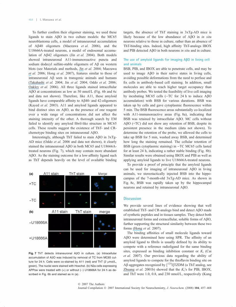
To further confirm their oligomer staining, we used theseligands to stain AbO in two culture models: the MC65neuroblastoma cells, a model of intraneuronal accumulationof Ab40 oligomers (Maezawa et al. 2006), and theU18666A-treated neurons, a model of endosomal accumu-lation of Ab42 oligomers (Jin et al. 2004). Both modelsshowed intraneuronal A11-immunoreactive puncta andsodium dodecyl sulfate-stable oligomers of Ab on westernblots (see Materials and methods, Jin et al. 2004; Maezawaet al. 2006; Hong et al. 2007), features similar to those ofintraneuronal Ab seen in transgenic animals and humans(Takahashi et al. 2004; Jin et al. 2004; Oddo et al. 2006;Oakley et al. 2006). All three ligands stained intracellularAbO at concentrations as low as 50 nmol/L (Fig. 6b and 6cand data not shown). Therefore, like A11, these amyloidligands have comparable affinity to Ab40- and 42-oligomers(Kayed et al. 2003). A11 and amyloid ligands appeared tobind distinct sites on AbO, as the presence of one reagentover a wide range of concentrations did not affect thestaining intensity of the other. A thorough search by EMfailed to identify any amyloid fibril-like structure in MC65cells. These results suggest the existence of ThT- and CR-chemotype binding sites on intraneuronal AbO.
Interestingly, although ThT failed to stain AbO in 3xTg-AD mice (Oddo et al. 2006 and data not shown), it clearlystained the intraneuronal AbO in both MC65 and U18666A-treated neurons (Fig. 7), which may contain more abundantAbO. As the staining outcome for a low-affinity ligand suchas ThT depends heavily on the level of available binding
targets, the absence of ThT staining in 3xTg-AD mice islikely because of the low abundance of AbO in in situneurons relative to those in culture, rather than an absence ofThT-binding sites. Indeed, high affinity ThT-analogs IBOXand PIB detected AbO in both neurons in situ and in culture.
The use of amyloid ligands for imaging AbO in living celland animalsBSB, PIB, and IBOX are able to penetrate cells, and may beused to image AbO in their native states in living cells,avoiding possible deformations from the need to perfuse andfix cells in antibody-based cell staining. In addition, smallmolecules are able to reach higher target occupancy thanantibody probes. We tested the feasibility of live cell imagingby incubating MC65 cells ()TC for 24 h to induce AbOaccumulation) with BSB for various durations. BSB wastaken up by cells and gave cytoplasmic fluorescence within5 min. The BSB fluorescence areas substantially co-localizedwith A11-immunoreactive areas (Fig. 8a), indicating thatBSB was retained by intracellular AbO. MC cells withoutAbO (+TC) did not show any retention of BSB, despite itspersistent presence in the medium (data not shown). Todetermine the retention of the probe, we allowed the cells totake up BSB for 5 min, washed away BSB, and determinedhow long the staining remained. The cellular retention ofBSB (green cytoplasmic staining) in )TC MC65 cells lastedfor at least 24 h, indicating a rather stable binding (Fig. 8b).Similar results were obtained using IBOX and PIB as well asapplying amyloid ligands to live U18666A-treated neurons.
To provide a proof of principle that the amyloid ligandscan be used for imaging of intraneuronal AbO in livinganimals, we stereotactically injected BSB into the hippo-campus of the 7-month-old 3xTg-AD mice. As shown inFig. 8c, BSB was rapidly taken up by the hippocampalneurons and retained by intraneuronal AbO.
Discussion
We provide several lines of evidence showing that wellestablished ThT- and CR-analogs bind and detect AbO madeof synthetic peptides and in tissues samples. They detect bothintraneuronal forms and extracellular, soluble forms of AbO,further supporting the structural similarity between these twoforms (Hong et al. 2007).
The binding affinities of small molecule ligands towardAbO were determined here using SPR. The affinity of anamyloid ligand to fibrils is usually defined by its ability tocompete with a reference radioligand for the same bindingsites, expressed as binding inhibition constant or Ki (Caiet al. 2007). Our previous data regarding the ability ofamyloid ligands to compete for the thioflavin binding site onAb aggregates recognized by [125I]TZDM (a ThT analog, seeZhuang et al. 2001b) showed that the Ki’s for PIB, IBOX,and ThT were 1.0, 0.8, and 230 nmol/L, respectively (Kung
(a)
(b)
Fig. 7 ThT detects intraneuronal AbO in culture. (a) Intracellular
accumulation of AbO was induced by removal of TC from MC65 cul-
ture for 24 h. Cells were co-stained by A11 (red) and ThT (2 lmol/L,
green). The nuclei were stained with Hoechst. (b) N2a cells expressing
APPsw were treated with (+) or without ()) U18666A for 24 h as de-
scribed in Fig. 6b and stained as in (a).
Journal Compilation � 2007 International Society for Neurochemistry, J. Neurochem. (2008) 104, 457–468� 2007 The Authors
464 | I. Maezawa et al.

et al. 2002 and unpublished data). This appears parallel tothe rank order we demonstrated for AbO binding, in whichPIB and IBOX showed higher binding affinities than ThT. Inaddition, our SPR results regarding AbO binding sites alsodifferentiate characteristic binding activities of CR- and ThT-analogs, suggesting the presence of at least two types ofbinding sites on AbO: the high-capacity, micromolar-affinityCR-chemotype sites, and the low-capacity, nanomolar-affin-ity ThT-chemotype sites. Notably, low-capacity, high-affinitybinding sites for ThT-analogs were previously identified onAb1–40 fibrils (Lockhart et al. 2005). Our results raise aninteresting question as to whether binding sites on AbO aresimilar to or are a subset of those on fibrils, which may reflectelements of shared structure. Further ligand-oligomer bind-ing studies are required to address this question. Based onprevious competition assay data, three types of binding siteson Ab fibrils were distinguished: those for CR, ThT, andFDDNP, respectively (Agdeppa et al. 2001; Zhuang et al.2001b; Cai et al. 2007). However, recent studies showedthree classes of ThT binding sites that are able to bind a widerange of chemotype structures, including BSB (Lockhartet al. 2005; Ye et al. 2005), attesting to the complex bindingsite types existing in various Ab aggregates (Cai et al. 2007).
If the binding sites on AbO represent only a subset of ligandbinding sites on Ab fibrils, in-depth analyses of ligand-AbOinteractions would be highly informative as it would be less achallenge to tease out individual profiles of the binding sites.
An important conclusion from our results and results fromWalsh et al. (1999) is that the widely used ThT or CR assayshould not be considered amyloid fibril specific. Rather,caution should be taken when this assay is applied toheterogeneous samples of Ab aggregates or biologicalsamples, as pre-fibrillar Ab aggregates including AbO maycontribute substantially to the fluorescence readouts. Fur-thermore, based on our EM visualization of binding usingIBOX, at least a subpopulation of AbO has a larger bindingcapacity per unit mass than Ab fibrils. Therefore, in additionto the ThT or CR assay, parallel assays specific for fibrils andoligomers, respectively, should be employed, for example, acombination of a filter assay to isolate only filaments and adot blot assay for oligomer quantification (Necula et al.2007b).
Compared with antibodies, which are large molecules,amyloid ligands demonstrate excellent brain uptake and cellpermeability, and will likely have wide applications in futurein vitro and in vivo studies of AbO. We have demonstrated,
(a) (b)
(c)
Fig. 8 Amyloid ligands are rapidly taken up by neurons and retained
by intraneuronal AbO. (a) MC65 cells were cultured without TC for
24 h to induce the intracellular accumulation of AbO. BSB (2 lmol/L)
or PIB (2 lmol/L) was added to the media. After indicated lengths of
time, the cells were fixed and stained with A11. Merged pictures
demonstrate the co-localization of BSB or PIB retention (green) with
A11-immunoreactive AbO (red). Parallel control cultures (+TC, without
AbO accumulation) did not show any A11 or amyloid ligand staining
(not shown). (b) MC65 cells were cultured with (+) or without ()) TC for
24 h. Cells were then exposed to BSB (2 lmol/L) for 5 min, washed
extensively, and fixed at the indicated time after the last wash. (c) BSB
was injected stereotactically into the hippocampus of 7-month-old
homozygous 3xTg-AD mice (0.12 lL of 10 lmol/L solution over 2 min
injection, Bregma Coordinates: anterioposterior )1.58; lateral )2;
dorsoventral )0.5). Mice were killed 30 min later and the brains fixed,
sectioned and stained by A11. Representative fluorescent images of
hippocampal pyramidal neurons from the injected side and the con-
tralateral non-injected side are shown.
� 2007 The AuthorsJournal Compilation � 2007 International Society for Neurochemistry, J. Neurochem. (2008) 104, 457–468
Amyloid ligands bind Ab oligomers | 465

for the first time, two useful applications: (i) enhancing thecontrast of AbO in EM imaging by positive staining and (ii)‘live’-imaging of intraneuronal AbO. The ability to traceAbO in living cells and animals would help our understand-ing of the metabolism and toxicity of AbO. With its dualability to give fluorescence and electron scattering, IBOX canbe used for high resolution correlative fluorescence and EM,especially when applied to ultrathin cryosections whichmaximally preserve the structure of macromolecules (Rob-inson et al. 2001). The combination – imaging AbO in livingcells under fluorescence microscopy and performing highresolution analysis of the same labeled AbO under EM –should provide detailed structural information and subcellu-lar localization of AbO in their native cellular environment.
[11C]-PIB has been used in clinical imaging and givesstrong signal in the cortex of AD patients, which has beenattributed to the presence of fibrillar amyloid (Klunk et al.2004; Mathis et al. 2004; Cai et al. 2007; Holtzman 2007).Our results raise a question of whether AbO may contributeto the signal of [11C]-PIB if AbO reach locally highconcentrations, such as in the intraneuronal compartments(Takahashi et al. 2004; Oddo et al. 2006) or in the synapticbinding sites in neuropil (Lacor et al. 2004). AbO in ADfrontal cortex may reach levels up to 70-fold over controlfrontal cortex (Gong et al. 2003), which might provideenough contrast between AD and controls for clinicalimaging. Intriguingly, it has been noticed that the highretention of [11C]-PIB in the frontal cortex in vivo differsfrom plaque quantities detected in vitro in autopsy paraffin-fixed tissue, raising suspicion of an artifact (Klunk et al.2004; Nordberg 2004). Because routine post-mortem neuro-pathological methods rarely detect AbO, our results suggest apossibility that focally high levels of AbO might contributeto the higher PIB retention in the frontal cortex. Futurestudies are needed to clarify this issue.
Evidence from animal studies suggest that AbO arecausally related to memory deficits independently of amyloidplaques or neuronal loss, and therefore may play a significantrole in the pre-dromal or early phase of AD, when plaquesare not yet formed (Takahashi et al. 2004; Billings et al.2005; Cole and Frautschy 2006; Oddo et al. 2006;Lesne et al. 2006). In addition, AbO may also makecontributions to dementia independent of Ab fibrils in anystages of the disease because they are not obligate interme-diates in fibril formation (Necula et al. 2007a). However,these notions remain untested in human because of the lackof methods to detect AbO in vivo. Although AbO and Abfibrils may have comparable unit binding capacities to someligands, substantial challenges reside in the diffuse distribu-tion and much smaller sizes of AbO, compared with theconspicuous Ab fibrils in amyloid plaques and congophilicangiopathy. For compounds with affinities comparable withPIB, the overall intensity of signal as a result of AbO bindingwould be markedly lower than that associated with fibrils,
except in perhaps a few cases with very high focal levels ofAbO.
To meet these challenges, compounds with substantiallyhigher AbO-binding affinities need to be developed for useas probes. Such high affinity ligands would significantlyenhance the relative contribution of AbO to the overallsignal. As the deposit of AbO in AD brains distributes in aperineuronal manner reminiscent of the synaptic-type depositobserved with prion-associated diseases (Lacor et al. 2004),probes with affinities that are at least comparable with thosesuitable for neurotransmitter receptor or transporter imagingagents (KDs of £ 1 nmol/L) would be required for AbOsignal to be manifest (Frost 1982). To realize this goal, animportant implication of our results is that some welldeveloped amyloid ligands can be used as lead compoundsfor the development of imaging agents with higher AbO-binding affinity. The methods developed in our study wouldprovide efficient means to screen focused libraries (madebased on the structure of amyloid ligands) and selectoptimized ligands under high stringency conditions (Penget al. 2006). Such new ligands would provide morecomprehensive quantifications of aggregated Ab species.Given that these compounds are likely to have high affinityto AbO, other pre-fibrillar aggregates and fibrils, it may benecessary to modify current imaging techniques. For exam-ple, when applying a high affinity probe to the early stages ofAD, the signal from the small amount of fibrillar amyloidwill likely need to be extracted (for example, by first imagingwith a fibril ligand such as PIB that would give little signalfrom diffuse AbO) and subsequently subtracted from thetotal signal. Such subtraction approach has been used todetect individual subtypes of related receptors (Frey andHowland 1992). This approach might be useful for quanti-fying cerebral AbO in patients with mild cognitive impair-ment or early AD and might be extremely informativeregarding the pathophysiological mechanisms of earlydegeneration (i.e. are AbO or pre-fibrillar aggregates toxic?).In addition, it might provide a more accurate index oftherapeutic effect in drug trials.
Acknowledgments
We are grateful to Dr George Martin, Dr Charles DeCarli for critical
readings of this manuscript, and Dr Duy Hua for providing TP17 for
negative control in SPR experiments. This work was supported by
grants from the UC Davis Health Science Research Fund, the UC
Davis Department of Pathology NIH Roadmap Grant, and the UC
Davis startup funds for Drs Jin and Lam.
References
Agdeppa E. D., Kepe V., Liu J., Flores-Torres S., Satyamurthy N., PetricA., Cole G. M., Small G. W., Huang S. C. and Barrio J. R (2001)Binding characteristics of radiofluorinated 6-dialkylamino-2-naphthylethylidene derivatives as positron emission tomography
Journal Compilation � 2007 International Society for Neurochemistry, J. Neurochem. (2008) 104, 457–468� 2007 The Authors
466 | I. Maezawa et al.

imaging probes for b-amyloid plaques in Alzheimer’s disease.J. Neurosci. 21, RC189.
Billings L. M., Oddo S., Green K. N., McGaugh J. L. and LaFerla F. M.(2005) Intraneuronal Ab causes the onset of early Alzheimer’sdisease-related cognitive deficits in transgenic mice. Neuron 45,675–688.
Bitan G. (2006) Structural study of metastable amyloidogenic proteinoligomers by photo-induced cross-linking of unmodified proteins.Methods Enzymol. 413, 217–236.
Cai L., Innis R. B. and Pike V. W. (2007) Radioligand development forPET imaging of b-amyloid (Ab) – current status. Curr. Med. Chem.14, 19–52.
Casas C., Sergeant N., Itier J. M. et al. (2004) Hippocampal neuron lossexceeds amyloid plaque load in a transgenic mouse model ofAlzheimer’s disease. Am. J. Pathol. 165, 1289–1300.
Caspersen C., Wang N., Yao J., Sosunov A., Chen X., Lustbader J. W.,Xu H. W., Stern D., McKhann G. and Yan S. D. (2005) Mito-chondrial Ab: a potential focal point for neuronal metabolic dys-function in Alzheimer’s disease. FASEB J. 19, 2040–2041.
Caughey B. and Lansbury P. T. (2003) Protofibrils, pores, fibrils, andneurodegeneration: separating the responsible protein aggregatesfrom the innocent bystanders. Ann. Rev. Neurosci. 26, 267–298.
Cheng I. H., Scearce-Levie K., Legleiter J. et al. (2007) Acceleratingamyloid-b fibrillization reduces oligomer levels and functionaldeficits in Alzheimer disease mouse models. J. Biol. Chem. 282,23818–23828 (Epub ahead of print).
Chromy B. A., Nowak R. J., Lambert M. P. et al. (2003) Self-assemblyof Ab(1-42) into globular neurotoxins. Biochemistry 42, 12749–12760.
Cole G. M. and Frautschy S. A. (2006) Alzheimer’s amyloid story findsits star. Trends Mol. Med. 12, 395–396.
Ferreira S. T., Vieira M. N. and De Felice F. G. (2007) Soluble proteinoligomers as emerging toxins in Alzheimer’s and other amyloiddiseases. IUBMB Life 59, 332–345.
Frey K. A. and Howland M. M. (1992) Quantitative autoradiography ofmuscarinic cholinergic receptor binding in the rat brain: distinctionof receptor subtypes in antagonist competition assays. J. Phar-macol. Exp. Ther. 263, 1391–1400.
Frost J. J. (1982) Pharmacokinetic aspects of the in vivo non-invasivestudy of neuroreceptors in man.CRC Press, Boca Raton, FL.
Glabe C. G. (2006) Common mechanisms of amyloid oligomer patho-genesis in degenerative disease. Neurobiol. Aging 27, 570–575.
Gong Y., Chang L., Viola K. L., Lacor P. N., Lambert M. P., Finch C. E.,Krafft G. A. and Klein W. L. (2003) Alzheimer’s disease-affectedbrain: presence of oligomeric Ab ligands (ADDLs) suggests amolecular basis for reversible memory loss. Proc. Natl Acad. Sci.USA 100, 10417–10422.
Hardy J. and Selkoe D. J. (2002) The amyloid hypothesis of Alzheimer’sdisease: progress and problems on the road to therapeutics. Science297, 353–356.
Holtzman D. M. (2007) Pittsburgh compound B retention and verifica-tion of amyloid deposition. Arch. Neurol. 64, 315–316.
Hong H. S., Maezawa I., Yao N., Xu B., Diaz-Avalos R., Rana S., HuaD. H., Cheng R. H., Lam K. S. and Jin L-W. (2007) Combining therapid MTT formazan exocytosis assay and the MC65 protectionassay led to the discovery of carbazole analogs as small moleculeinhibitors of Ab oligomer-induced cytotoxicity. Brain Res. 1130,223–234.
Jin L. W., Maezawa I., Vincent I. and Bird T. (2004) Intracellularaccumulation of amyloidogenic fragments of amyloid-b precursorprotein in neurons with Niemann–Pick type C defects is associatedwith endosomal abnormalities. Am. J. Pathol. 164, 975–985.
Kayed R., Head E., Thompson J. L., McIntire T. M., Milton S. C.,Cotman C. W. and Glabe C. G. (2003) Common structure of sol-
uble amyloid oligomers implies common mechanism of patho-genesis. Science 300, 486–489.
Kayed R., Sokolov Y., Edmonds B., McIntire T. M., Milton S. C., HallJ. E. and Glabe C. G. (2004) Permeabilization of lipid bilayers is acommon conformation-dependent activity of soluble amyloidoligomers in protein misfolding diseases. J. Biol. Chem. 279,46363–46366.
Klein W. L., Krafft G. A. and Finch C. E. (2001) Targeting small Aboligomers: the solution to an Alzheimer’s disease conundrum?Trends Neurosci. 24, 219–224.
Klunk W. E., Engler H., Nordberg A. et al. (2004) Imaging brainamyloid in Alzheimer’s disease with Pittsburgh compound-B. Ann.Neurol. 55, 306–319.
Kung M., Hou C., Zhuang Z. P., Zhang B., Skovronsky D., TrojanowskiJ. Q., Lee V. M. and Kung H. F. (2002) IMPY: an improvedthioflavin-T derivative for in vivo labeling of beta-amyloid pla-ques. Brain Res. 956, 202–210.
Kung M. P., Zhuang Z. P., Hou C. and Kung H. F. (2004) Developmentand evaluation of iodinated tracers targeting amyloid plaques forSPECT imaging. J. Mol. Neurosci. 24, 49–53.
Lacor P. N., Buniel M. C., Chang L. et al. (2004) Synaptic targeting byAlzheimer’s-related amyloid b oligomers. J. Neurosci. 24, 10191–10200.
Lacor P. N., Buniel M. C., Furlow P. W., Clemente A. S., Velasco P. T.,Wood M., Viola K. L. and Klein W. L. (2007) Ab oligomer-in-duced aberrations in synapse composition, shape, and densityprovide a molecular basis for loss of connectivity in Alzheimer’sdisease. J. Neurosci. 27, 796–807.
Lambert M. P., Velasco P. T., Chang L. et al. (2007) Monoclonal anti-bodies that target pathological assemblies of Ab. J. Neurochem.100, 23–35.
Lee C. W., Zhuang Z. P., Kung M. P., Plossl K., Skovronsky D., Gur T.,Hou C., Trojanowski J. Q., Lee V. M. and Kung H. F. (2001)Isomerization of (Z,Z) to (E,E)1-bromo-2,5-bis-(3-hydroxycar-bonyl-4-hydroxy)styrylbenzene in strong base: probes for amyloidplaques in the brain. J. Med. Chem. 44, 2270–2275.
Lesne S., Koh M. T., Kotilinek L., Kayed R., Glabe C. G., Yang A.,Gallagher M. and Ashe K. H. (2006) A specific amyloid-b proteinassembly in the brain impairs memory. Nature 440, 352–357.
LeVine III H. (1993) Thioflavine T interaction with synthetic Alzhei-mer’s disease beta-amyloid peptides: detection of amyloid aggre-gation in solution. Protein Sci. 2, 404–410.
Liu Y. and Schubert D. (2006) Treating Alzheimer’s disease by inacti-vating bioactive amyloid beta peptide. Curr. Alzheimer Res. 3,129–135.
Lockhart A., Ye L., Judd D. B., Merritt A. T., Lowe P. N., MorgensternJ. L., Hong G., Gee A. D. and Brown J. (2005) Evidence for thepresence of three distinct binding sites for the thioflavin T class ofAlzheimer’s disease PET imaging agents on b-amyloid peptidefibrils. J. Biol. Chem. 280, 7677–7684.
Maezawa I., Hong H. S., Wu H. C. et al. (2006) A novel tricyclic pyronecompound ameliorates cell death associated with intracellularamyloid-b oligomeric complexes. J. Neurochem. 98, 57–67.
Mathis C. A., Wang Y., Holt D. P., Huang G. F., Debnath M. L. andKlunk W. E. (2003) Synthesis and evaluation of 11C-labeled 6-substituted 2-arylbenzothiazoles as amyloid imaging agents.J. Med. Chem. 46, 2740–2754.
Mathis C. A., Wang Y. and Klunk W. E. (2004) Imaging b-amyloidplaques and neurofibrillary tangles in the aging human brain. Curr.Pharm. Des. 10, 1469–1492.
Myszka D. G. (2004) Analysis of small-molecule interactions usingBiacore S51 technology. Anal. Biochem. 329, 316–323.
Necula M., Kayed R., Milton S. and Glabe C. G. (2007a) Small moleculeinhibitors of aggregation indicate that amyloid b oligomerization
� 2007 The AuthorsJournal Compilation � 2007 International Society for Neurochemistry, J. Neurochem. (2008) 104, 457–468
Amyloid ligands bind Ab oligomers | 467

and fibrillization pathways are independent and distinct. J. Biol.Chem. 282, 10311–10324.
Necula M., Breydo L., Milton S., Kayed R., van der Veer W. E., Tone P.and Glabe C. G. (2007b) Methylene blue inhibits amyloid Abligomerization by promoting fibrillization. Biochemistry 46, 8850–8860.
Nordberg A. (2004) Is amyloid plaque imaging the key to monitoringbrain pathology of Alzheimer’s disease in vivo? Eur. J. Nucl. Med.Mol. Imaging 31, 1540–1543.
Oakley H., Cole S. L., Logan S. et al. (2006) Intraneuronal beta-amyloidaggregates, neurodegeneration, and neuron loss in transgenic micewith five familial Alzheimer’s disease mutations: potential factorsin amyloid plaque formation. J. Neurosci. 26, 10129–10140.
Oddo S., Caccamo A., Tran L., Lambert M. P., Glabe C. G., Klein W. L.and LaFerla F. M. (2006) Temporal profile of amyloid-b (Ab)oligomerization in an in vivo model of Alzheimer disease. A linkbetween Ab and tau pathology. J. Biol. Chem. 281, 1599–1604.
Peng L., Liu R., Marik J., Wang X., Takada Y. and Lam K. S. (2006)Combinatorial chemistry identifies high-affinity peptidomimeticsagainst alpha4beta1 integrin for in vivo tumor imaging. Nat. Chem.Biol. 2, 381–389.
Robinson J.M., Takizawa T., PomboA. and Cook P. R. (2001) Correlativefluorescence and electron microscopy on ultrathin cryosections:bridging the resolution gap. J. Histochem. Cytochem. 49, 803–808.
Takahashi R. H., Almeida C. G., Kearney P. F., Yu F., Lin M. T., MilnerT. A. and Gouras G. K. (2004) Oligomerization of Alzheimer’s b-amyloid within processes and synapses of cultured neurons andbrain. J. Neurosci. 24, 3592–3599.
Tjernberg L. O., Naslund J., Lindqvist F. et al. (1996) Arrest of b-amyloid fibril formation by a pentapeptide ligand. J. Biol. Chem.271, 8545–8548.
Townsend M., Cleary J. P., Mehta T., Hofmeister J., Lesne S., O’Hare E.,Walsh D. M. and Selkoe D. J. (2006) Orally available compoundprevents deficits in memory caused by the Alzheimer amyloid-boligomers. Ann. Neurol. 60, 668–676.
Walsh D. M., Hartley D. M., Kusumoto Y., Fezoui Y., Condron M. M.,Lomakin A., Benedek G. B., Selkoe D. J. and Teplow D. B. (1999)Amyloid beta-protein fibrillogenesis. Structure and biologicalactivity of protofibrillar intermediates. J. Biol. Chem. 274, 25945–25952.
Yan Y., Liu Y., Sorci M., Belfort G., Lustbader J. W., Yan S. S. andWang C. (2007) Surface plasmon resonance and nuclear magneticresonance studies of ABAD-Ab interaction. Biochemistry 46,1724–1731.
Ye L., Morgenstern J. L., Gee A. D., Hong G., Brown J. and Lockhart A.(2005) Delineation of positron emission tomography imaging agentbinding sites on b-amyloid peptide fibrils. J. Biol. Chem. 280,23599–23604.
Zhuang Z. P., Kung M. P., Hou C., Plossl K., Skovronsky D., Gur T. L.,Trojanowski J. Q., Lee V. M. and Kung H. F. (2001a) IBOX(2-(4¢-dimethylaminophenyl)-6-iodobenzoxazole): a ligand for imagingamyloid plaques in the brain. Nucl. Med. Biol. 28, 887–894.
Zhuang Z. P., Kung M. P., Hou C., Skovronsky D. M., Gur T. L., PlosslK., Trojanowski J. Q., Lee V. M. and Kung H. F. (2001b) Ra-dioiodinated styrylbenzenes and thioflavins as probes for amyloidaggregates. J. Med. Chem. 44, 1905–1914.
Journal Compilation � 2007 International Society for Neurochemistry, J. Neurochem. (2008) 104, 457–468� 2007 The Authors
468 | I. Maezawa et al.
Related Documents