187 187 International Journal of Scientific Study | December 2016 | Vol 4 | Issue 9 Congenital Hemifacial Hyperplasia: Report of a Rare Case with Review of Literature Lipsa Bhuyan 1 , Sarangi Sambit 2 , Nayak Suryakanti 2 , Nayak Sarat 3 1 Senior Lecturer, Department of Oral and Maxillofacial Surgery, Kalinga Institute of Dental Sciences, KIIT, Bhubaneswar, Odisha, India, 2 Senior Lecturer, Department of Oral and Maxillofacial Surgery, S.C.B Dental College and Hospital, Cuttack, Odisha, India, 3 Senior Lecturer, Department of Oral and Maxillofacial Surgery, Hi-Tech Dental College and Hospital, Bhubaneswar, Odisha, India side of face since birth which has gradually increased to the existing size. His parents, all his siblings, and children did not suffer from his disorder. On extra oral examination, a gross facial asymmetry was seen on the right side of the face. The soft tissue on maxillary and mandibular region along with ear pinna and ala of nose was enlarged as compared to the left side of the face. Chin was deviated toward the left side of the face. The mass was soft and nontender on palpation (Figures 1 and 2). On intraoral examination, enlargement of the maxillary and mandibular alveolar ridge, tongue and gingiva were seen on the affected side. Reduced mouth opening with intermaxillary distance of 1.5 cm was present. The absence of teeth was noticed on the affected side which had exfoliated 2 years back (Figure 3). Orthopantomogram, lateral view of the skull and posterior anterior view of skull revealed the absence of teeth on the right side and enlarged body of the mandible unilaterally. There was an increased height of the ramus and body of the mandible on the right side in comparison to left side. An oversized right condyle and deviation of chin toward the unaffected side could be appreciated (Figures 4 and 5). Based on these clinical and radiologic examinations, a diagnosis of congenital hemifacial hyperplasia was concluded. INTRODUCTION A minor asymmetry is acceptable characteristic of morphogenesis. However, asymmetry which is easily noticeable can affect esthetic, normal functioning and quality of life. A congenital developmental disturbances causing unilateral overdevelopment of tissues of face both hard and soft is seen in this rare disorder called hemifacial hypertrophy. In 1982, Hemihyperplasia was first described by Meckel. 1 Rowe in 1962 classified it as complex involving entire half of the body, simple involving one or both limbs, and hemifacial involving half of the face. The term hyperplasia is preferably used since an increase in number of cells is seen rather than increase in size of cells. 2 The aim of this report is to present a case of congenital hemifacial hyperplasia and to update the existing clinical knowledge. CASE REPORT A 65-year-old male complained of inability to open his mouth since many years. He gave a history of enlarged right Case Report Abstract Hemifacial hypertrophy is a developmental disorder which is characterized by facial asymmetry unilaterally. It is a congenital malformation usually seen at birth and progressively increases with age. Although etiology is unknown, various theories such as chromosomal abnormalities, heredity, atypical twinning, endocrine dysfunction, and altered intrauterine environment have also been proposed. A few cases have been reported in the literature. We herein report a case of true hemifacial hyperplasia in 65 years old male. Key words: Congenital hemifacial hyperplasia, Facial asymmetry, Hemifacial hypertrophy Access this article online www.ijss-sn.com Month of Submission : 10-2016 Month of Peer Review : 11-2016 Month of Acceptance : 11-2016 Month of Publishing : 12-2016 Corresponding Author: Lipsa Bhuyan, Department of Oral and Maxillofacial Pathology, Kalinga Institute of Dental Sciences, Campus 5, KIIT University, Bhubaneswar - 751 024, Odisha, India. Phone: +91 9439892654. E-mail: [email protected] Print ISSN: 2321-6379 Online ISSN: 2321-595X DOI: 10.17354/ijss/2016/643

Welcome message from author
This document is posted to help you gain knowledge. Please leave a comment to let me know what you think about it! Share it to your friends and learn new things together.
Transcript

187187 International Journal of Scientifi c Study | December 2016 | Vol 4 | Issue 9
Congenital Hemifacial Hyperplasia: Report of a Rare Case with Review of LiteratureLipsa Bhuyan1, Sarangi Sambit2, Nayak Suryakanti2, Nayak Sarat3
1Senior Lecturer, Department of Oral and Maxillofacial Surgery, Kalinga Institute of Dental Sciences, KIIT, Bhubaneswar, Odisha, India, 2Senior Lecturer, Department of Oral and Maxillofacial Surgery, S.C.B Dental College and Hospital, Cuttack, Odisha, India, 3Senior Lecturer, Department of Oral and Maxillofacial Surgery, Hi-Tech Dental College and Hospital, Bhubaneswar, Odisha, India
side of face since birth which has gradually increased to the existing size. His parents, all his siblings, and children did not suffer from his disorder.
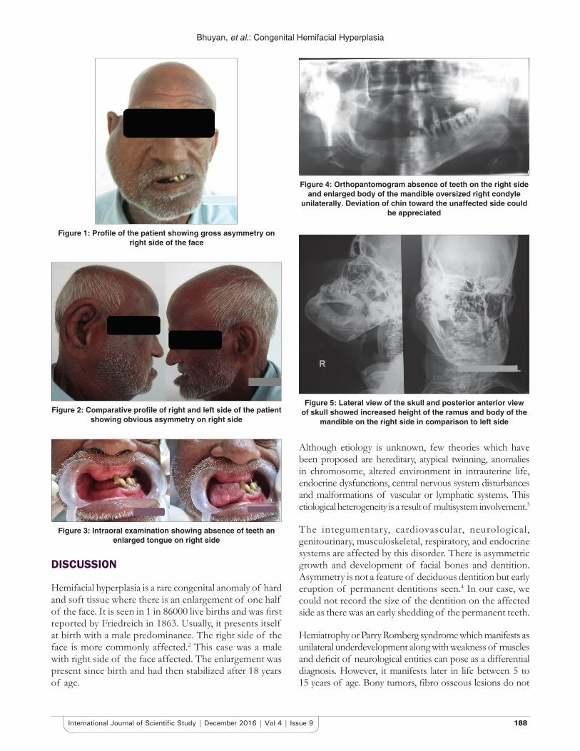
On extra oral examination, a gross facial asymmetry was seen on the right side of the face. The soft tissue on maxillary and mandibular region along with ear pinna and ala of nose was enlarged as compared to the left side of the face. Chin was deviated toward the left side of the face. The mass was soft and nontender on palpation (Figures 1 and 2).
On intraoral examination, enlargement of the maxillary and mandibular alveolar ridge, tongue and gingiva were seen on the affected side. Reduced mouth opening with intermaxillary distance of 1.5 cm was present. The absence of teeth was noticed on the affected side which had exfoliated 2 years back (Figure 3).
Orthopantomogram, lateral view of the skull and posterior anterior view of skull revealed the absence of teeth on the right side and enlarged body of the mandible unilaterally. There was an increased height of the ramus and body of the mandible on the right side in comparison to left side. An oversized right condyle and deviation of chin toward the unaffected side could be appreciated (Figures 4 and 5). Based on these clinical and radiologic examinations, a diagnosis of congenital hemifacial hyperplasia was concluded.
INTRODUCTION
A minor asymmetry is acceptable characteristic of morphogenesis. However, asymmetry which is easily noticeable can affect esthetic, normal functioning and quality of life. A congenital developmental disturbances causing unilateral overdevelopment of tissues of face both hard and soft is seen in this rare disorder called hemifacial hypertrophy. In 1982, Hemihyperplasia was fi rst described by Meckel.1 Rowe in 1962 classifi ed it as complex involving entire half of the body, simple involving one or both limbs, and hemifacial involving half of the face. The term hyperplasia is preferably used since an increase in number of cells is seen rather than increase in size of cells.2 The aim of this report is to present a case of congenital hemifacial hyperplasia and to update the existing clinical knowledge.
CASE REPORT
A 65-year-old male complained of inability to open his mouth since many years. He gave a history of enlarged right
Case Report
Abstract
Hemifacial hypertrophy is a developmental disorder which is characterized by facial asymmetry unilaterally. It is a congenital malformation usually seen at birth and progressively increases with age. Although etiology is unknown, various theories such as chromosomal abnormalities, heredity, atypical twinning, endocrine dysfunction, and altered intrauterine environment have also been proposed. A few cases have been reported in the literature. We herein report a case of true hemifacial hyperplasia in 65 years old male.
Key words: Congenital hemifacial hyperplasia, Facial asymmetry, Hemifacial hypertrophy
Access this article online
www.ijss-sn.com
Month of Submission : 10-2016Month of Peer Review : 11-2016Month of Acceptance : 11-2016Month of Publishing : 12-2016
Corresponding Author: Lipsa Bhuyan, Department of Oral and Maxillofacial Pathology, Kalinga Institute of Dental Sciences, Campus 5, KIIT University, Bhubaneswar - 751 024, Odisha, India. Phone: +91 9439892654. E-mail: [email protected]
Print ISSN: 2321-6379Online ISSN: 2321-595X
DOI: 10.17354/ijss/2016/643
Bhuyan, et al.: Congenital Hemifacial Hyperplasia
188188International Journal of Scientifi c Study | December 2016 | Vol 4 | Issue 9
DISCUSSION
Hemifacial hyperplasia is a rare congenital anomaly of hard and soft tissue where there is an enlargement of one half of the face. It is seen in 1 in 86000 live births and was fi rst reported by Friedreich in 1863. Usually, it presents itself at birth with a male predominance. The right side of the face is more commonly affected.2 This case was a male with right side of the face affected. The enlargement was present since birth and had then stabilized after 18 years of age.
Although etiology is unknown, few theories which have been proposed are hereditary, atypical twinning, anomalies in chromosome, altered environment in intrauterine life, endocrine dysfunctions, central nervous system disturbances and malformations of vascular or lymphatic systems. This etiological heterogeneity is a result of multisystem involvement.3
The integumentary, cardiovascular, neurological, genitourinary, musculoskeletal, respiratory, and endocrine systems are affected by this disorder. There is asymmetric growth and development of facial bones and dentition. Asymmetry is not a feature of deciduous dentition but early eruption of permanent dentitions seen.4 In our case, we could not record the size of the dentition on the affected side as there was an early shedding of the permanent teeth.
Hemiatrophy or Parry Romberg syndrome which manifests as unilateral underdevelopment along with weakness of muscles and defi cit of neurological entities can pose as a differential diagnosis. However, it manifests later in life between 5 to 15 years of age. Bony tumors, fi bro osseous lesions do not
Figure 1: Profi le of the patient showing gross asymmetry on right side of the face
Figure 2: Comparative profi le of right and left side of the patient showing obvious asymmetry on right side
Figure 3: Intraoral examination showing absence of teeth an enlarged tongue on right side
Figure 4: Orthopantomogram absence of teeth on the right side and enlarged body of the mandible oversized right condyle
unilaterally. Deviation of chin toward the unaffected side could be appreciated
Figure 5: Lateral view of the skull and posterior anterior view of skull showed increased height of the ramus and body of the
mandible on the right side in comparison to left side
Bhuyan, et al.: Congenital Hemifacial Hyperplasia
189189 International Journal of Scientifi c Study | December 2016 | Vol 4 | Issue 9
involve the soft tissues and dentition. Cutaneous lesions and vascular malformations are usually bilateral. All these lesions with an exception of hemiatrophy can be diagnosed histopathologically. Proteus syndrome, hyperpitutarism, epidermal nevus syndrome, Maffucci syndrome, Ollier’s syndrome, Klippel-Trenaunay-Weber syndrome, Langer-Giedion syndrome, Russell Silver syndrome, McCune-Albright syndrome, and multiple exostosis syndrome can mimic hemifacial hypertrophy. Unilateral allocation of dental abnormalities and coinciding unilateral tongue enlargement are the striking features of hemifacial hypertrophy.2,4
CONCLUSION
Hemifacial hypertrophy creates a diagnostic dilemma and needs a thorough clinical and radiological evaluation. It
requires a multidisciplinary approach of management because of its multisystem involvement. Unless there is a necessity for cosmetic correction, treatment is not indicated.
REFERENCES
1. Deshingkar SA, Barpande SR, Bhavthankar JD. Congenital hemifacial hyperplasia. Contemp Clin Dent 2011;2:261-4.
2. Bhuta BA, Yadav A, Desai RS, Bansal SP, Chemburkar VV, Dev PV. Clinical and imaging fi ndings of true hemifacial hyperplasia. Case Rep Dent 2013;2013:152528.
3. Nayak R, Baliga MS. Crossed hemifacial hyperplasia: A diagnostic dilemma. J Indian Soc Pedod Prev Dent 2007;25:39-42.
4. Agarwal A, Gupta ND, Agarwal A. Congenital isolated hemifacial hypertrophy: Clinical and 3-dimensional radio graphical scanning features. Br J Med Health Res 2016;3:21-30.
How to cite this article: Bhuyan L, Sambit S, Suryakanti N, Sarat N. Congenital Hemifacial Hyperplasia: Report of a Rare Case with Review of Literature. Int J Sci Stud 2016;4(9):187-189.
Source of Support: Nil, Confl ict of Interest: None declared.
Related Documents