Congenital Heart Disease Dr Vanessa Holme Consultant Paediatrician

Welcome message from author
This document is posted to help you gain knowledge. Please leave a comment to let me know what you think about it! Share it to your friends and learn new things together.
Transcript

Congenital Heart Disease
Dr Vanessa HolmeConsultant Paediatrician

Aims
� Review Cardiac Anatomy� Fetal Circulation� Common defects� Presentations� Investigations� Management

Normal Heart Anatomy

Congenital Heart Defects
� 6-9/100 live births� 8 defects make up 80% CHD� Most Isolated, some assoc with Syndromes
� Acyanotic (Pink)� Isolated left to right shunt
� Cyanotic (Blue)� Right to left shunt

Pink or Blue?
Ventricular Septal Defect (VSD)Persistent Arterial Duct (PDA)Atrial Septal Defect (ASD)Pulmonary StenosisAortic StenosisCoarctation of the AortaTetralogy of FallotTransposition of Great Arteries

Case Presentation 1
� Baby boy� Full Term NVD
� Mum – Gravida 1� Previous child fit and well� Discharged after a normal 6hr check

Presents to A&E at 24hrs old
� Initial assessment:� A – crying� B – RR = 50/min, no recession
cyanosis to tongue and lipssaturations 78% in air
� C – cap refill 2 secs, HR = 140/minno murmur
� D – alert, PERL� E – no rash, slightly mottled

What do you do?
� Give oxygen� sats still 80% in 10 litres
� IV access� bloods and IV antibiotics
� CXR, ECG ? Echo� Treatment
� Prostin� Transfer to specialist centre

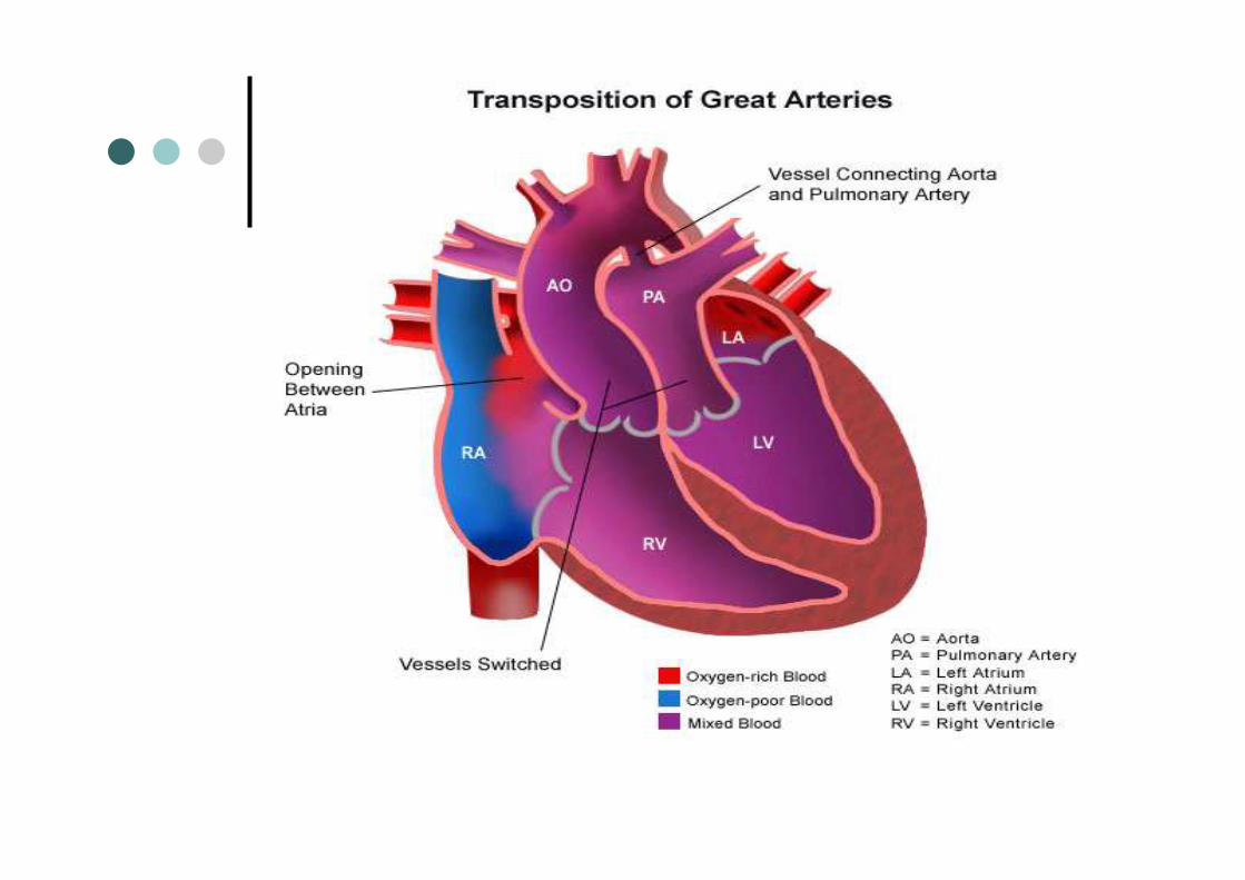
Transposition of Great Arteries
� Commonest cyanotic lesion to present at birth
� Aorta & Pulmonary Arteries arise from incorrect ventricles� Survival dependent on connection b/w
2 circuits� Cyanosis from first few hrs of life
� Gradually worsening over next few days

Fetal Circulation

Management
� Prostin� Balloon Septostomy� “Arterial Switch” Procedure� If Isolated defect then do very well
post-op

Case Presentation 2
� 3 month old baby girl
� Parents first child� Normal antenatal scans� Normal neonatal check

GP 6 week check
� Murmur 3/6
� Femoral pulses present� Child growing along 25th centile
� Bottle feeding well
� Referred to paediatrics

Attended A&E at 3/12
� Initial Assessment� A – crying at times� B – RR = 70/min, mild recession,
sats 93% in air� C – HR 200/min; cap refill < 2 secs
� D – Alert and interactive

Initial Management?
1. Oxygen via face mask2. Call for Help3. Take history from parents & examine
baby4. IV access

Further History
� For last month slower to feed� Occasionally getting SOB and sweaty
when feeding� Last 24 to 48 hrs
� Coughing� Breathing faster� Feeding half normal amounts

Examination
� Baby looks pale and sweaty� HS 2/6 systolic murmur
Active praecordium� Pulses all palpable� BS: bilateral crackles� Liver 3 cm below costal margin

Investigations
� CXR� ECG� Bloods� Echocardiogram?

Chest X-ray

ECG

Treatment
� IV frusemide� Discuss with specialist centre� Eventually will need surgery
� Dependent on growth� Degree of failure� Risk of pulmonary hypertension

Ventricular Septal Defect
� Commonest Congenital Heart Defect
� Very variable in size� Small often asymptomatic with loud
murmur� Large present in failure but often with
softer murmurs

VSD

Progress
� Small VSD often reduce in size leading to spontaneous closure
� Larger ones with failure will require surgical closure
� Failure of closure can lead to Eisenmengers Syndrome.

Eisenmengers Syndrome

Case Presentation 3
� 8 month old boy� Murmur noted at 6/52 check.� Clinically felt to be small VSD
� Thriving� Feeding well� No signs of heart failure
� Awaiting Cardiology Review

Presented to A&E
� Episodes of distress first thing in the morning
� Parents feel he goes blue & sweaty� Settles to sleep on cuddling� Wakes up and appears normal

Examination
� Weight 8.5kg� Slightly cyanosed� Early finger clubbing� RR = 40/min, sats 85% in air� HR = 140/min. normal pulses� Murmur high pitched systolic murmur� No thrill

Rest of examination
� No palpable liver� No resp distress
� No signs of cardiac failure
� Investigations?

CXR

ECG

Tetralogy of Fallot
� VSD� Pulmonary Stenosis� Right Ventricular Hypertrophy� Overriding Aorta
� Important are VSD & pulm stenosis


Clinical Features
� Cyanosis appears late in infancy� Systolic murmur along LLSE, pulm
area and radiates thro to back� Hypoxic Spells
� Marked pallor or cyanois with dyspnoea and distress
� Reduced exercise tolerance with age

Management
� Manage spells with beta blockers
� Definitive repair if possible� May need a temporary shunt between
aorta and pulm artery

Case Presentation 4
� 3 year old boy� Cough and cold for 3 days� Febrile above 39oC� Worsening resp distress
� GP has noticed a murmur and referred to COAU for assessment.

Assessment
� A – audible wheezetalking in short sentences
� B – RR = 45/min, sc recession,tracheal tug, sats 92% in air
� C – HR 130/min, cap refill < 2 secsBP 100/45HS soft systolic murmur
� D- Alert,

Management
� Given salbutamol inhaler via spacer� Improved RR =30/min� Better AE and reduced wheeze
� On discharge recommended see GP for review of murmur

Referred by GP to OPD
� Well grown child� Soft mid systolic murmur 2/6� Left sternal edge & apex� Murmur softer on standing� Louder on lying flat� Normal BP, pulses

Management
� Reassured� Innocent flow murmur� No need for further investigation� Explain murmur is not a medical
problem

Innocent Murmurs
� Can be heard in up to 50% of school age children
� 4 types of innocent murmur� Stills Murmur� Pulmonary Flow Murmur� Carotid Bruit� Venous Hum

Assessment
� Concerning Features� Unwell child� Diastolic
� Grade 3 or louder� Loudest over pulmonary area
� Heart sounds not separate� Any concerns about child

Other Common Defects
� Other Shunt lesions

Persistent Ductus Arteriosus
� Failure of Duct to Close� Commonest in Prematurity
� Premature babies can close after weeks/months
� Symptomatic may need treatment

PDA
� Clinical� May be asymptomatic� Continuous murmur at upper left
sternal border
� Larger• Collapsing pulses• Signs of failure

PDA
� Treatment� Drug – Indomethacin/Ibuprofen
� Surgical – coil or other devices

PDA Closure


Atrial Septal Defect
� Usually in region of foramen ovale� Small ASD can go undetected
� Clinically� Ejection systolic murmur� Second Heart sound split
� Soft diastolic murmur

ASD
� Investigations� CXR - increase in cardiac diameter &
pulm plethora� ECG – Partial RBBB, Rt axis deviation
� Treatment� Closure if significant shunt or when
older

ECG in ASD

AVSD
� 3% of all defects� Commonly seen in Trisomy 21
� Similar to large VSD� Symptomatic within first few months of
life

Obstructive Lesions
� Pulmonary Stenosis� Usually involves valve itself� Asymptomatic in Infancy & Childhood
� Ejection systolic murmur heard in pulm area and radiating to back
� Severe can lead to angina and heart failure with exercise intolerance

Aortic Stenosis
� Abnormal valve� Symptom free in Childhood� Ejection Systolic Murmur
� Right of sternum� Radiates to carotids
� Progressive� Dizziness and Syncope on exertion� Angina, Effort Intolerance, Sudden Death
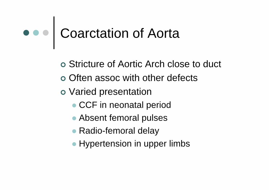
Coarctation of Aorta
� Stricture of Aortic Arch close to duct� Often assoc with other defects� Varied presentation
� CCF in neonatal period� Absent femoral pulses� Radio-femoral delay
� Hypertension in upper limbs


CXR


Treatment
� Newborn� Prostin� Treat Heart Failure
� Early Repair
� Older children� If hypertensive or in second decade

Case Presentation 5
� 4 year old girl� Previously fit and well� Occasionally looks pale and sits down
for few mins
� Otherwise well and active

Brought to A&E
� Looked pale and sweaty� c/o heart feels funny� Mum noticed pulse in neck� Examination
� Alert, RR = 25/min, no recession� HR = 240/min, BP = 95/50
� Cap refill < 2 secs

ECG

Supraventricular Tachycardia
� Commonest paediatric arrythmia� Sudden onset of Tachycardia
� Infants look pale poor feeding
� Older children will c/o funny sensation
� Treatment?� Valsalva manouvre etc
� Adenosine

Delta wave

Long QT syndrome
� Rare cause of collapse and sudden death� Often familial
� Any collapse should calculate QTc� Normal < 0.44
� If concerning features then exercise ECG/cardiology referral

Conclusions
� Congenital Heart Disease significant morbidity and mortality
� Variable presentation� Newborn period often need urgent
treatment� Other defects develop over time

Conclusions
� Heart murmurs often benign� Important to know when to investigate
� Commonest arrythmia is SVT

Any Questions
Related Documents