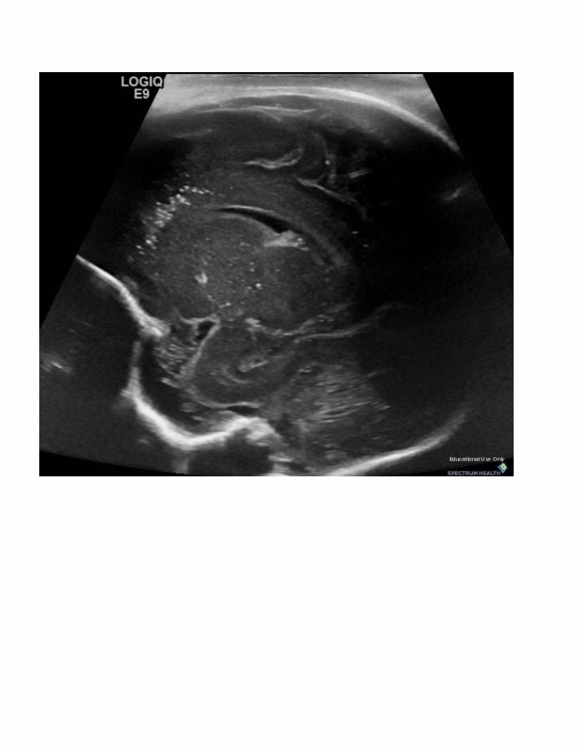
Congenital Cytomegalovirus Infection Joseph Junewick, MD FACR 07/28/2011 History Newborn with trisomy 21 and pancytopenia. Diagnosis Congenital Cytomegalovirus Infection Discussion Congenital CMV infection is one of the most common congenital viral infections. Fetal infection results from transmission of the virus across the placenta. Fetal effects are most severe if when the mother experiences a primary infection and if the infection occurs early in development. Manifestation of CMV infection include microcephaly,loss, intracranial calcifications, hearing loss, hepatosplenomegaly, low birth weight, pneumonitis, and hematologic abnormalities. Imaging findings of congenital CMV infection include intracranial calcification, ventriculomegaly, white matter disease, neuronal migrational disorders, and microcephaly. Not all of these findings are seen in every patient, and abnormalities may range from mild to severe. Intracranial calcification is the most frequently reported imaging finding of congenital CMV infection, occurring in 34%–70% of patients. Calcification occurs most commonly in the periventricular regions but also within the basal ganglia and brain parenchyma. Absence of calcification should not exclude a diagnosis of congenital CMV infection. The presence of intracranial calcification in patients with congenital CMV infection is associated with developmental delays. Findings US-Oblique coronal and sagittal transfontanel images demonstrate perilateral ventricular and thalamic calcifications. Reference Fink KR, Thapa MM, Ishak GE et al. Neuroimaging of Pediatric Central Nervous System Cytomegalovirus Infection. RadioGraphics (2010); 30: 1779-1796.

Welcome message from author
This document is posted to help you gain knowledge. Please leave a comment to let me know what you think about it! Share it to your friends and learn new things together.
Transcript

Congenital Cytomegalovirus InfectionJoseph Junewick, MD FACR
07/28/2011
HistoryNewborn with trisomy 21 and pancytopenia.
DiagnosisCongenital Cytomegalovirus Infection
DiscussionCongenital CMV infection is one of the most common congenital viral infections. Fetal infection resultsfrom transmission of the virus across the placenta. Fetal effects are most severe if when the motherexperiences a primary infection and if the infection occurs early in development. Manifestation of CMVinfection include microcephaly,loss, intracranial calcifications, hearing loss, hepatosplenomegaly, lowbirth weight, pneumonitis, and hematologic abnormalities. Imaging findings of congenital CMVinfection include intracranial calcification, ventriculomegaly, white matter disease, neuronal migrationaldisorders, and microcephaly. Not all of these findings are seen in every patient, and abnormalitiesmay range from mild to severe. Intracranial calcification is the most frequently reported imagingfinding of congenital CMV infection, occurring in 34%–70% of patients. Calcification occurs mostcommonly in the periventricular regions but also within the basal ganglia and brain parenchyma.Absence of calcification should not exclude a diagnosis of congenital CMV infection. The presence ofintracranial calcification in patients with congenital CMV infection is associated with developmentaldelays.
FindingsUS-Oblique coronal and sagittal transfontanel images demonstrate perilateral ventricular and thalamiccalcifications.
ReferenceFink KR, Thapa MM, Ishak GE et al. Neuroimaging of Pediatric Central Nervous SystemCytomegalovirus Infection. RadioGraphics (2010); 30: 1779-1796.


Sponsored By
DisclaimerThis teaching site is partially funded by an educational grant from GE Healthcare and Advanced Radiology Services, PC. The material on this site isindependently controlled by Advanced Radiology Services, PC, and GE Healthcare and Spectrum Health have no influence over the content of this siteContent Download AgreementThe cases and images on this website are owned by Spectrum Health. Permission is granted (for nonprofit educational purposes) to download and printmaterials to distribute for the purpose of facilitating the education of health professionals. The authors retain all rights to the material and users arerequested to acknowledge the source of the material. Site DisclaimerThis site is developed to reach healthcare professionals and medical students. Nothing this site should be considered medical advice.Only your own doctor can help you make decisions about your medical care. If you have a specific medical question or are seeking medical care, pleasecontact your physician.The information in this website is provided for general medical education purposes only and is not meant to substitute for the independent medicaljudgment of a physician relative to diagnostic and treatment options of a specific medical condition.The viewpoints expressed in these cases are those of the authors. They do not represent an endorsement. In no event will Advanced RadiologyAssociates, PC, Spectrum Health Hospitals (Helen Devos Children's Hospital) or GE Healthcare be liable for any decision made or action taken inreliance upon the information provided through this website.
Related Documents