Introduction Vascular rings represent a rare subset of congenital cardiac anomalies that occur during the early development of the aorta. They are subdivided into two categories: complete rings that fully encircle the trachea and esophagus and incomplete rings that do not. Symptoms from vascular rings are generally related to compression of the airway and esophagus. 1 Although the majority of patients with vascular rings are symptomatic in infancy and early childhood, it is not unusual for symptoms to manifest until later in life or not at all. 2 In symptomatic patients, prompt surgical treatment is of importance due to serious complications, such as major tracheal or bronchial damage that occurs or sudden death. 3 Many different forms of vascular rings exist due to the complex development of the aorta. However, two forms of complete vascular rings, the double aortic arch and the right aortic arch with an aberrant left subclavian artery (Figure 1), account for almost 90% of all cases. 4 In this case, we present an asymptomatic pediatric patient with an extremely rare form of a complete vascular ring, a right aortic arch with normal subclavian architecture and ligamentum arteriosum completing the ring. We believe CT 3D reconstruction with inclusion of the esophagus provided detailed anatomical definition, which was crucial for accurate diagnosis and optimal treatment of the patient. Case Report A 4-year-old female patient, with history of recurrent emesis and reflux in infancy, presented with a chief complaint of fever. The initial diagnostic work-up, which included chest x-ray, revealed an incidental finding suspicious for a right aortic arch. An initial CT chest angiogram with 3D rendering, which did not include the esophagus, was obtained for further evaluation. The Vascular Ring with Unique Anatomical Variant Elucidated Using 3D Technology June 2016; Volume 14; Issue 6 International Edition IN THIS ISSUE Vascular Ring with Unique Anatomical Variant Elucidated Using 3D Technology By Michael Hafertepe, BS, MS; Nhu-Nguyen Le, BS; Randy Richardson, MD; Randal Fortuna, MD ~Page 1 Specialty Review in Pediatric Cardiology Celebrating its 40 th Anniversary in September 2016 By Maria Serratto, MD ~Page 5 Highlights from the Third Bangkok International Fetal Echocardiography Symposium: January 18 th -20 th , 2016 By Mark Sklansky, MD ~Page 6 Medical News, Products & Information ~Page 10 CONGENITAL CARDIOLOGY TODAY Editorial and Subscription Offices 16 Cove Rd, Ste. 200 Westerly, RI 02891 USA www.CongenitalCardiologyToday.com Official publication of the CHiP Network © 2016 by Congenital Cardiology Today Published monthly. All rights reserved. CONGENITAL CARDIOLOGY TODAY Timely News and Information for BC/BE Congenital/Structural Cardiologists and Surgeons Upcoming Medical Meetings CSI FOCUS IMAGING Jun. 22, 2016; Boston, MA, USA www.csi-congress.org/focus- imaging.php 2016 Pediatric and Adult Congenital Cardiology Review Course Aug. 21-26, 2016; Dana Point, CA USA ce.mayo.edu/cardiovascular-diseases/ content/pediatric-and-adult-congenital- cardiology-review-course-2016- general-sesson Specialty Review in Pediatric Cardiology Course Sep. 19-23, 2016; Chicago, IL USA www.pediatriccardiology2016.com/ By Michael Hafertepe, BS, MS; Nhu-Nguyen Le, BS; Randy Richardson, MD; Randal Fortuna, MD Figure 1. Right aortic arch with aberrant left subclavian artery. CONGENITAL CARDIOLOGY TODAY CALL FOR CASES AND OTHER ORIGINAL ARTICLES Do you have interesting research results, observations, human interest stories, reports of meetings, etc. to share? Submit your manuscript to: [email protected]

Welcome message from author
This document is posted to help you gain knowledge. Please leave a comment to let me know what you think about it! Share it to your friends and learn new things together.
Transcript

Introduction
Vascular rings represent a rare subset of congenital cardiac anomalies that occur during the early development of the aorta. They are subdivided into two categories: complete rings that fully encircle the trachea and esophagus and incomplete rings that do not. Symptoms from vascular rings are generally related to compression of the airway and esophagus.1
Although the majority of patients with vascular rings are symptomatic in infancy and early childhood, it is not unusual for symptoms to manifest until later in life or not at all.2 In symptomatic patients, prompt surgical treatment is of importance due to serious complications, such as major tracheal or bronchial damage that occurs or sudden death. 3
Many different forms of vascular rings exist due to the complex development of the aorta. However, two forms of complete vascular rings, the double aortic arch and the right aortic arch with an aberrant left subclavian artery (Figure 1), account for almost 90% of all cases.4
In this case, we present an asymptomatic pediatric patient with an extremely rare form of a complete vascular ring, a right aortic arch with normal subclavian architecture and ligamentum arteriosum completing the ring. We believe CT 3D reconstruction with inclusion of the esophagus provided detailed anatomical
definition, which was crucial for accurate diagnosis and optimal treatment of the patient.
Case Report
A 4-year-old female patient, with history of recurrent emesis and reflux in infancy, presented with a chief complaint of fever. The initial diagnostic work-up, which included chest x-ray, revealed an incidental finding suspicious for a right aortic arch.
An initial CT chest angiogram with 3D rendering, which did not include the esophagus, was obtained for further evaluation. The
Vascular Ring with Unique Anatomical Variant Elucidated Using 3D Technology
June 2016; Volume 14; Issue 6International Edition
IN THIS ISSUE
Vascular Ring with Unique Anatomical Variant Elucidated Using 3D TechnologyBy Michael Hafertepe, BS, MS; Nhu-Nguyen Le, BS; Randy Richardson, MD; Randal Fortuna, MD~Page 1
Specialty Review in Pediatric Cardiology Celebrating its 40th Anniversary in September 2016 By Maria Serratto, MD~Page 5
Highlights from the Third Bangkok International Fetal Echocardiography Symposium: January 18th-20th, 2016By Mark Sklansky, MD~Page 6
Medical News, Products & Information ~Page 10
CONGENITAL CARDIOLOGY TODAYEditorial and Subscription Offices16 Cove Rd, Ste. 200Westerly, RI 02891 USAwww.CongenitalCardiologyToday.com
Official publication of the CHiP Network
© 2016 by Congenital Cardiology Today Published monthly. All rights reserved.
CONGENITAL CARDIOLOGY TODAYTimely News and Information for BC/BE Congenital/Structural Cardiologists and Surgeons
Upcoming Medical Meetings
CSI FOCUS IMAGINGJun. 22, 2016; Boston, MA, USA
www.csi-congress.org/focus-imaging.php
2016 Pediatric and Adult Congenital Cardiology Review Course
Aug. 21-26, 2016; Dana Point, CA USAce.mayo.edu/cardiovascular-diseases/content/pediatric-and-adult-congenital-
cardiology-review-course-2016-general-sesson
Specialty Review in Pediatric Cardiology Course
Sep. 19-23, 2016; Chicago, IL USAwww.pediatriccardiology2016.com/
By Michael Hafertepe, BS, MS; Nhu-Nguyen Le, BS; Randy Richardson, MD; Randal Fortuna, MD
Figure 1. Right aortic arch with aberrant left subclavian artery.
CONGENITAL CARDIOLOGY TODAY
CALL FOR CASES AND OTHER ORIGINAL ARTICLES
Do you have interesting research results, observations, human interest stories, reports of meetings, etc. to share? Submit your manuscript to: [email protected]

We are committed to the lifetime management of congenital heart disease.
Transcatheter and Surgical Heart Valves
RVOT Conduits
Ablation Technologies
ICDs
Oxygenators and Filters
Cannulae
Pacemakers
Pulse Oximetry Monitoring for CCHD Screening
3rd Generation PFO, ASD, and PDA Occluders*
Cerebral/Somatic Monitoring
*These products are not available in the US.
InnovatIve TeCHnOlOGIeS. EvEry StEp of the Way.
Medtronic | Minneapolis, Mn 55432-5604 Toll-free: 1 (800) 328-2518
Melody-tPV.com
UC201601683 en ©2015 Medtronic. All rights reserved. 08/2015
MecttronicFurther,Together

images depicted a right aortic arch with mirror image branching and a Kommerell diverticulum with no aberrant left subclavian artery. The anatomy was not suggestive of a vascular ring and a diagnosis was not made at that time.
Due to clinical suspicion of a vascular ring, a repeat 3D CT reconstruction was obtained with inclusion of the esophagus and revealed an indentation on the esophagus (Figure 3). Together, these findings were consistent for diagnosis of a vascular ring encircling the esophagus completed by the ligamentum arteriosum (Figure 4). Maternal concern for long-term health effects of a complete vascular ring led to surgical correction of the anomaly.
Surgery confirmed that the ligamentum arteriosum connected from the thoracic aorta to the left pulmonary artery forming a complete vascular ring. The ligamentum arteriosum was divided. The patient tolerated the procedure well.
Discussion
The type of vascular ring, as well as the degree of compression of the trachea and esophagus, determines the onset and severity of symptoms. Symptoms of airway compression are most common in all age ranges, and include: stridor, wheezing, apnea, cyanosis, and respiratory distress. Gastrointestinal symptoms, such as dysphagia, tend to present in older children. Infants, however, just show non-specific symptoms, such as slow feeding and frequent regurgitation, a possibility for our patient.1
Initial evaluation for vascular rings typically involves a chest x-ray and barium esophagography, with the latter considered to be the only diagnostic tool most frequently needed. However, choice and order of imaging studies vary widely between institutions and providers. Echocardiogram and color-flow Doppler, MRI, and digital subtraction angiography (DSA), and CT are other possible imaging studies, but
CONGENITAL CARDIOLOGY TODAY t www.CongenitalCardiologyToday.com t June 2016 3
Figure 2. 3D image of right aortic arch with mirror image branching with Kommerell diverticulum and no aberrant left subclavian artery.
Figure 3. 3D image of right aortic arch with mirror image branching with the inclusion of 3D esophagus, which displays indentation.
Figure 4. 3-D Right aortic arch with mirror image branching displaying position of ligamentum arteriosum.

are less frequently used due to cost and exposure of pediatric patients to radiation.5
Nevertheless, optimal imaging modalities are crucial in cases where complicated vascular ring anatomy needs to be visualized for operative management.5 For example, the strength of the CT lies in its ability to delineate vascular, tracheobronchial, and esophageal structures and their relative positions. In our case, initial 3D CT imaging demonstrated right aortic arch with mirror-image branching with no aberrant left subclavian artery that was not suspicious for a vascular ring. Only when the esophagus was included in CT imaging was the indentation on the esophagus seen and an accurate diagnosis of a vascular ring made.
This case depicts a rare type of complete vascular ring. Although asymptomatic at the time of presentation, this pediatric patient would likely have developed serious complications later on in life given her medical history of recurrent emesis and reflux as an infant. Three-dimensional technology illustrated optimal anatomical detail, which aided clinical and surgical management.
References
1. Licari A, Manca E, Rispoli GA, Mannarino S, Pelizzo G, Marseglia GL. Congenital vascular rings: a clinical challenge for the pediatrician. Pediatric Pulmonology. 2015;50(5):511.
2. Turner A, Gavel G, Coutts J. Vascular rings--presentation, investigation and outcome. Eur J Pediatr. 2005;164(5):266.
3. Shah RK, Mora BN, Bacha E, Sena LM, Buonomo C, Del Nido P, Rahbar R. The presentation and management of
vascular rings: an otolaryngology p e r s p e c t i v e . I n t J P e d i a t r Otorhinolaryngol. 2007;71(1):57.
4. Backer CL, Mavroudis C, Rigsby CK, Holinger LD. Trends in vascular ring surgery. J Thorac Cardiovasc Surg. 2005;129(6):1339.
5. Humphrey C, Duncan K, Fletcher S. Decade of experience with vascular rings at a single institution. Pediatrics. 2006 June. 117(5):e903-8.
CCT
CONGENITAL CARDIOLOGY TODAY t www.CongenitalCardiologyToday.com t June 2016 4
Archiving Working GroupInternational Society for Nomenclature of Paediatric and Congenital Heart Disease
ipccc-awg.net
Michael Hafertepe is currently a third year medical student at Creighton University School of Medicine - Phoenix Regional Campus. He received his bachelor degree in natural science from Xavier University. He then received a Master’s degree from Tulane University in Cell and Molecular Biology. He plans on pursuing a residency in Diagnostic Radiology.
Nhu-Nguyen Le, BSSt. Joseph’s Hospital and Medical Center and Creighton University School of Medicine350 West Thomas Rd. Phoenix, AZ 85013 USA
Randy Richardson, MD St. Joseph’s Hospital and Medical Center and Creighton University School of MedicineDepartment of Radiology350 West Thomas Rd.Phoenix, AZ 85013 USA
Principal Author
Michael Hafertepe, BS, MSSt. Joseph’s Hospital and Medical Center and Creighton University School of Medicine350 West Thomas Rd. Phoenix, AZ 85013 USAPhone: [email protected]
Randall Fortuna, MDBanner Children's SpecialistsDepartment Pediatric Cardiothoracic Surgery1432 South Dobson Rd, Suite 512Mesa AZ 85202 USA
“We believe CT 3D reconstruction with inclusion of the esophagus provided detailed anatomical definition, which was crucial for accurate diagnosis and optimal treatment for the patient.”
LIVE CASE DEMONSTRATIONS • ABSTRACT
SESSIONS • “MY NIGHTMARE CASE IN THE
CATH LAB” • HOT DEBATES • WORKSHOPS
• SMALLER BREAKOUT SESSIONS •
SAVE THE DATEJAN. 16-19, 2O17LOEWS MIAMI BEACH HOTEL
W W W. P I C S Y M P O S I U M . C O M
MIAMI
With the 2016 offering of Specialty Review in Pediatric Cardiology board review course approaching, I find myself thinking back to its beginnings. Even though pediatric cardiology in the 1970’s was rapidly expanding in diagnostic and surgical techniques, candidates for board examination had to rely on long and repeated visits to the medical library to update their knowledge and refresh the experience gained during their training. A preparatory curriculum was clearly needed.
The first Specialty Review in Pediatric Cardiology was offered in 1976 under the auspices of the Cook County School of Graduate Medicine of Chicago, with faculty drawn from local universities. That year, the program was two days in length and attended by approximately 30 registrants from throughout the United States.
How things have changed! Over the years, the course expanded to its current five-day format, attracting attendees from throughout the U.S., as
well from abroad, with a d i s t i n g u i s h e d n a t i o n a l planning committee and faculty. Perhaps the most important testament to the history, quality, and continuing relevance of the course came in 2010, when sponsorship w a s a s s u m e d b y t h e A m e r i c a n A c a d e m y o f P e d i a t r i c s S e c t i o n o n Cardiology and Cardiac Surgery, in collaboration with the Society of Pediatric Cardiology Training Program Directors.
Technological advances have likewise contributed to course enhancements. An online syllabus now makes it possible for presenter slide materials to be viewed in full-color full-page format. Practice exam Q&A exercises following each presentation are supported by audience response system technology. Presentations are filmed to produce a “watch-it-again” option, available shortly after the course, and a CME-accredited DVD version of the course is released in the spring following each live offering. An online option for those wishing to earn Maintenance of Certification (MOC) credit was introduced in 2014.
Continuing advances in our specialty and expanding needs of our constituency have certainly contributed to course growth as well, especially in recent years as those who are already board certified prepare to meet recertification requirements and practitioners strive to remain current in our ever-expanding field.
The past forty years have been a rewarding journey for me and the many others who have contributed to the success of the course, as we have had the honor to come to know hundreds of fine specialists and interact with them over the course of their careers. Whether you are new to the Specialty Review in Pediatric Cardiology family, or a member of our distinguished alumni, we look forward to having you with us in Chicago this coming September 19th-23rd. For details on the upcoming 2016 offering, please visit the course website: http://PediatricCardiology2016.com.
CCT
CONGENITAL CARDIOLOGY TODAY t www.CongenitalCardiologyToday.com t June 2016 5
Specialty Review in Pediatric Cardiology Celebrating its 40th Anniversary in September 2016By Maria Serratto, MD
24th Parma International Echo Meeting - From Fetus to Young Adult Universita’ di Parma | Associazione Medical Care - Development – Peace Parma, Italy | June 27-28, 2016Centro S.Elisabetta | University Campus | Parma, ItalyFor more information, contact: Professor Umberto Squarcia, MD, FACC - [email protected] or Professor Donald J Hagler, MD - [email protected]
Maria Serratto, MD, FACC, FAAP, FCCPCourse Founder & DirectorProfessor of Pediatrics-CardiologyChildren's Hospital of theUniversity of Illinois at ChicagoChicago, IL [email protected]
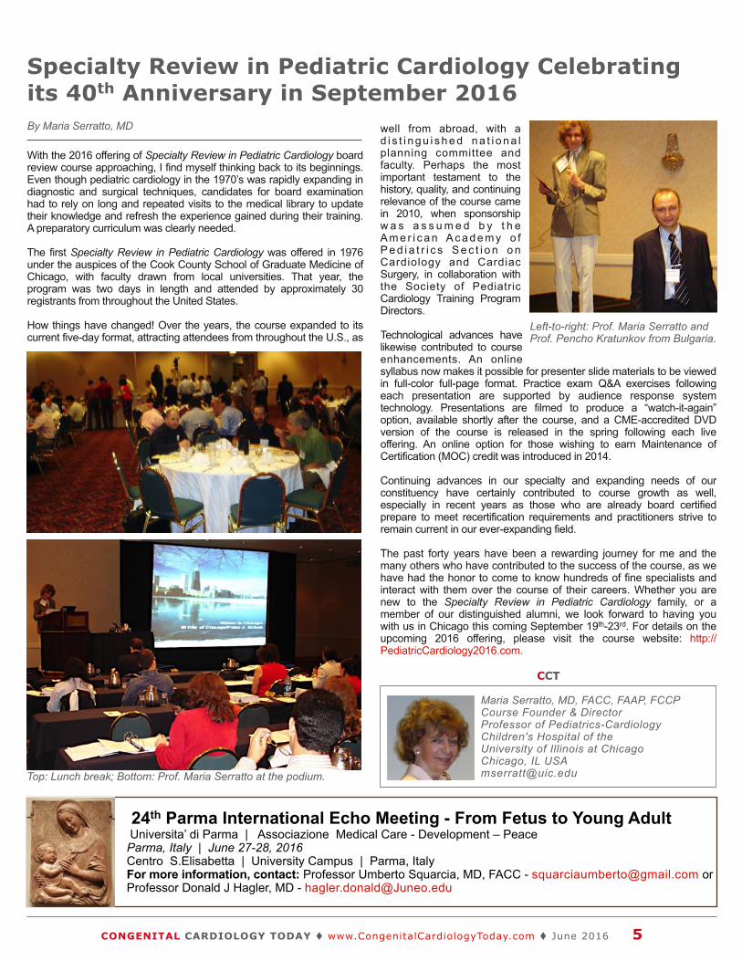
Left-to-right: Prof. Maria Serratto and Prof. Pencho Kratunkov from Bulgaria.
Top: Lunch break; Bottom: Prof. Maria Serratto at the podium.

The Third Bangkok International Fetal Echocardiography Symposium, held in the majestic Dusit Thani Bangkok Hotel in the heart of Bangkok (January 18th-20th, 2016), brought together well over a hundred subspecialists from around the world for three days of state-of-the-art presentations and discussions, live hands-on scanning, and mingling/networking with colleagues, new and old. The symposium provided a clinically oriented series of presentations and lively discussions of topics related to fetal cardiac imaging, diagnostics and management. This third year of the symposium attracted pediatric cardiologists, obstetricians, maternal-fetal medicine and other affiliated subspecialists from throughout Southeast Asia and beyond, including over 35 attendees from: Malaysia, Indonesia, the Philippines, India, the Maldives, Vietnam, Cambodia, Nepal, Hong Kong, Myanmar, Japan, Taiwan, the U.K. and the United States, as well as from throughout Thailand. New this year was the addition of a separate and extremely successful and interactive two-day symposium on Adult Congenital Heart Disease (ACHD) immediately following the Fetal Echocardiography Symposium, also held at the Dusit Thani Bangkok.
Directed by Alisa Limsuwan, MD, and co-directed by Poomiporn Katunyuwong, MD, both of Ramathibodi Hospital, Mahidol Universi ty, Bangkok, the fetal symposium’s organizing committee was led by Boonsri Chanrachakul, MD, and Pattama Promsonthi, MD, both of the Division of Maternal-Fetal Medicine, Bumrungrad International Hospital, Bangkok, along with Suthep Wanitkun, MD, Chief of Pediatric Cardiology, Ramathibodi Hospital , Mahidol Universi ty, and Theera Tongsong, MD, Professor of Obstetrics & Gynecology, Chiang Mai University, Thailand.
This year ’s faculty included an esteemed, international community of experts in the field, including: Satoshi Yasukochi, MD, Director of the Heart Center at Nagano Children’s Hospital, and President of the Japanese Society of Pediatric Cardiology and Cardiac Surgery; Kai-Sheng Hsieh, MD, Chairman of Pediatrics at the Veterans General Hospital, Kaohsiung, Taiwan; Gurleen Sharland, MD, Children’s Hospital St. Thomas Hospital, London; and Mark Sklansky, MD, Chief of Pediatric Cardiology, Mattel Children’s Hospital, David Geffen School of Medicine at UCLA.
The first of the symposium’s three days began with a series of talks on the current approach to fetal cardiac screening, including an update on guidelines and approach to the segmental diagnosis of Congenital Heart Disease (CHD). An expert panel then discussed various approaches to prenatal
CONGENITAL CARDIOLOGY TODAY t www.CongenitalCardiologyToday.com t June 2016 6
Highlights from the Third Bangkok International Fetal Echocardiography Symposium: January 18th-20th, 2016By Mark Sklansky, MD
Faculty, left-to-right: Poomiporn Katanyuwong, MD; Kai-Sheng Hsieh, MD; Gurleen Sharland, MD; Mark Sklansky, MD; Teera Tongsong, MD; Patama Promsonthi, MD; Alisa Limsuwan, MD and Boonsri Chanrachakul, MD.
Faculty and guests.
“The symposium provided a clinically oriented series of presentations and lively discussions of topics related to fetal cardiac imaging, diagnostics and management. This third year of the symposium attracted pediatric cardiologists, obstetricians, maternal-fetal medicine and other affiliated subspecialists from throughout Southeast Asia and beyond....”

!"#"$%&'($)*$+!,$*#-./
!"#$%&'!%"(#")*&+%,'!"#$%&'()%"*)+)(,
)-./012''34567859:-9-4'!;.</6541'$5.=-'3:-45>1
)-./01'#123./456'"431.4735/478'9.7254:'!;.8'"/;9-'0;')/..?5;'@A'+57-'>/795.-'BC'+DEAA@A'3/./8:-65F'GH?9F-4.560'3-.I'''CA,JA,BKJ,LKKK'(5MI''CA,JA,BKJ,LNKK
%5<1%541'=7.25/*7>6-87.'916?45678'!-00/.3'3-.I'''OBLLP'QJRELBNK'%E<5?.I'47,879-8:7;>>/49S<-094/6?8,8/<
#18/2@A9$*,6/B
(/4'5'.?79?6T'/U'?60?859?/67V'8/6945?60?859?/67V'>4-85;9?/67V'H546?6T7V'560'>/9-69?5.'50=-47-'-=-697V'>.-57-'4-U-4'9/'9:-'W6794;89?/67'U/4'X7-,
)-./01'?7'5'4-T?79-4-0'9450-<54Y'/U')-094/6?8,
X+JKARKAQJQ5'%%''ZJKAQ')-094/6?8','*..'4?T:97'4-7-4=-0''KB[JKAQ
)%\#]^'3!$']%\*^G'+#&]XW3'"%!\*+%)%&3
CDE!59?-697'59'J'1-547
!59?-697'59'Q'1-547CFE!"#"$%&'($)*$+!,$*#-./

screening around the world. A series of talks on abnormalities of the outflow tracts and of pulmonary venous return was presented after lunch, followed by live-scanning by Dr. Sharland and a hands-on scanning session for attendees. Day Two began with presentations on critical forms of congenital heart disease (Hypoplastic Left/Right Heart Syndromes), as well as heterotaxy and common abnormalities of the foramen ovale, ductus arteriosus and ductus venosus. Afternoon talks focused on perinatal interventions and counseling, followed by a live-scanning by Dr. Sklansky and additional hands-on scanning by registrants. The third and final day included presentations on fetal arrhythmias and cardiac function, followed by panel discussions on how to set up a fetal heart program and how to minimize medical-legal problems. The symposium ended with an overview on pitfalls and pearls of fetal cardiac imaging by Dr. Sklansky.
CONGENITAL CARDIOLOGY TODAY t www.CongenitalCardiologyToday.com t June 2016 8
Faculty panel, left-to-right: Mark Sklansky, MD; Gurleen Sharland, MD; Kai-Sheng Hsieh, MD; and Satoshi Yasukochi, MD.
Live scanning by Satoshi Yasukochi, MD; Pattama Promsonthi, MD at far left.
PediatricCardiologySPECIALTY REVIEW IN
American Academy of Pediatrics Section on Cardiology & Cardiac Surgeryin collaboration with the Society of Pediatric Cardiology Training Program Directors
September 19-23, 2016 | Chicago pediatriccardiology2016.com
Congenital Cardiology Today Can Help You Recruit:
• Pediatric Cardiologists• pediatric Interventional Cardiologist• Adult Cardiologist focused on CHD• Congenital/Structural Heart Surgeons• Echocardiographers, EPs• Pediatric Transplant Cardiologist
Reach over 6,000 BC/BE Cardiologists focused on CHD worldwide:
• Recruitment ads include color!• Issues’s email blast will include your recruitment ad!• We can create the advertisement for you at no extra
charge!
Contact: Tony Carlson
+1.301.279.2005 or [email protected]

Given the glowing reviews from this third international fetal symposium and first A d u l t C o n g e n i t a l D e a r t D i s e a s e symposium, plans are already underway for next year’s exciting and innovative symposia, currently planned for the week of January 9th-13th, 2017, with the first two days dedicated to fetal cardiology, the third day addressing the fetal-adult continuum of CHD, and the last two days dedicated to adults with congenital heart disease.
For more details, please contact Dr. Limsuwan ([email protected]) or go to the websites at bkkfetalecho.com or adultcongenitalcardiology.com.
CCT
CONGENITAL CARDIOLOGY TODAY t www.CongenitalCardiologyToday.com t June 2016 9
Mark S. Sklansky, MDChief, Division of Pediatric CardiologyJames H. Nicholson Professor of Clinical PediatricsDavid Geffen School of Medicine at UCLAMedical Director, Children’s Heart CenterCo-Director, Fetal Cardiology ProgramMattel Children's Hospital UCLAUCLA Children’s Heart Center200 Medical Plaza, Suite 330Los Angeles, CA 90095 USA
Letters to the EditorCongenital Cardiology Today w e l c o m e s a n d e n c o u r a g e s Letters to the Editor. If you have comments or topics you would like to address, please send an email to: [email protected], and let us know if you would like your comment published or not.
Get involved with CHiP (Congenital Heart International
Professionals Network)
We need your help:• Finding news stories.• Creating journal watch.• Keeping track of upcoming meetings.• Building our presence on Linkedin,
Facebook, and Twitter.• Creating more value for our readers/
subscribers.• Engaging our partner organizations.• Fundraising to support our activities.
Step up! Here's how to contact us:
www.chipnetwork.org/Contact
We'd like to know WHO you are, WHERE you are, and WHAT you do. Please go to www.chipnetwork.org and let us know more about you. It only takes two minutes. Then we'll be able to send you messages targeted to your interests.
I hope you will consider joining the CHiP Network and help foster a strong congenital heart care community.
Sincerely,
Gary Webb, MDCHiP [email protected]
The CHIP Network, the Congenital Heart Professionals Network, is
designed to provide a single global list of all CHD-interested professionals.
C O N G E N I T A L CARDIOLOGY TODAY
CALL FOR CASES AND OTHER ORIGINAL
ARTICLES
Do you have interesting research results, observations, human interest stories, reports of meetings, etc. to share?
Submit your manuscript to: [email protected]
• Title page should contain a brief title and full names of all authors, their professional degrees, and their institutional affiliations. The principal author should be identified as the first author. Contact information for the principal author including phone number, fax number, email address, and mailing address should be included.
• Optionally, a picture of the author(s) June be submitted.
• No abstract should be submitted.• The main text of the article should be
written in informal style using correct English. The final manuscript June be between 400-4,000 words, and contain pictures, graphs, charts and tables. Accepted manuscripts will be published within 1-3 months of receipt. Abbreviations which are commonplace in pediatric cardiology or in the lay literature June be used.
• Comprehensive references are not required. We recommend that you provide only the most important and relevant references using the standard format.
• Figures should be submitted separately as individual separate electronic files. Numbered figure captions should be included in the main Word file after the references. Captions should be brief.
• Only articles that have not been published previously will be considered for publication.
• Published articles become the property of the Congenital Cardiology Today and June not be published, copied or reproduced elsewhere without permission from Congenital Cardiology Today.

SCAI Publishes Updated Guidelines on Best Practices in the Cardiac Catheterization Laboratory
Washington, D.C. – On May 2nd, 2016 the Society for Cardiovascular Angiography and Interventions (SCAI) published an update to its first-of-its-kind 2012 paper outlining best practices in the cardiac catheterization laboratory (CCL), or cath lab. The paper, “SCAI Expert Consensus Statement: 2016 Best Practices in the Cardiac Catheterization Laboratory,” was published online in Catheterization and Cardiovascular Interventions, SCAI’s official journal.
The document provides cath labs around the country with a set of agreed-upon recommended guidelines developed by expert practicing interventional cardiologists to ensure the highest quality of care and ultimately better patient outcomes, as well as improved patient and physician satisfaction. The paper includes pre-, intra-, and post-procedure recommendations.
“SCAI created the Best Practices Guidelines in 2012 because there had been no process standardization in cath labs and labs had been working under only local regulation and policy,” said Srihari S. Naidu, MD, FACC, FSCAI, FAHA, Associate Professor of Medicine at the SUNY Stony Brook School of Medicine, Director of the Cardiac Catheterization Laboratory at Winthrop University Hospital and the paper’s lead author. “There is a tremendous appetite among interventional cardiologists for a comprehensive document outlining the details on how a cardiac cath lab should operate.”
The document includes:• An updated pre-procedure checklist for cardiac catheterization
that provides a nurse, technician, APP, physician extender, or physician with a set of questions to review with the patient.
• Language on cath lab governance and the responsibilities of a cath lab director. Techniques are provided on how CCLs can improve function through effective governance and management strategies.
• Information on cost consideration. Specific strategies CCLs can employ to provide the highest value of care are outlined.
• Information on radial access. There is recent support for radial access procedures (procedures done through the wrist), which were not covered in the 2012 paper. Best practices on when radial access is appropriate and considerations CCLs should take is included.
• New evidence on medications. Recent data includes medications brought to market after the 2012 paper and when to use certain drugs and dosage amounts.
• Recommendations on maintaining appropriate industry relationships, for example:• Industry’s role in individual CCLs should be consistent with
policies set by the hospital and/or director.• Industry should not have “hands-on” equipment in the CCL,
except for defined educational purposes or device preparation.
• Industry should always provide information and advice that is in the best interest of the patient, regardless of other considerations.
• Techniques for enhancing patient satisfaction in the CCL including pre-, intra-, and post-procedure recommendations for improving patient satisfaction.
The paper, which cross-references other SCAI reports and documents, clearly reflects the direction in which the profession of interventional cardiology is heading.
“SCAI is a valuable organization for cardiologists to join as it lends them a voice and the opportunity to impact and influence the future of interventional cardiology,” said Sunil V. Rao, MD, FSCAI, Chair of SCAI’s Quality Improvement Committee, Associate Professor of Medicine at the Duke University Medical Center and Section Cchief of Cardiology at Durham VA Medical Center.
“The guidelines reflect SCAI’s commitment and continued efforts to improve quality of care and the patient experience,” said SCAI President James C. Blankenship, MD, MHCM, FSCAI, MACC, practicing interventional cardiologist at Geisinger Medical Center in Danville, PA, and Director of the Geisinger cardiology department and catheterization laboratories.
Study Details Molecular Mechanism that Regulates How the Heart Pumps Blood
Newswise — In a finding that could lead to new drugs to treat heart failure, researchers have uncovered the molecular mechanism that regulates how the heart pumps blood.
The key molecular player in this mechanism is a giant protein called titin, according to a study led by senior author Pieter de Tombe, PhD of Loyola University Chicago Stritch School of Medicine. The study was published Feb. 8th, 2016 in Proceedings of the National Academy of Sciences.
De Tombe is interim Vice Dean for Research and Chair of the Department of Cell and Molecular Physiology at Loyola University Chicago Stritch School of Medicine.
A healthy heart regulates itself so that with each beat, it pumps out as much blood as it receives. When blood enters the heart, it stretches the wall of the pumping chamber, triggering muscle to contract and pump blood out. This regulatory-control mechanism is known as the Frank-Starling Law, named after physiologists Otto Frank and Ernest Starling.
In heart failure patients, the Frank-Starling Law breaks down. Heart muscle becomes too weak to pump out of the heart the same amount of blood that flows into the heart. To compensate, the heart enlarges, develops more muscle mass and beats faster. But eventually these compensatory measures fall short. The heart cannot pump enough blood to meet the body’s needs for blood and oxygen, leading to shortness of breath, fatigue, weakness, swelling in legs, fluid retention, and other symptoms.
CONGENITAL CARDIOLOGY TODAY t www.CongenitalCardiologyToday.com t June 2016 10
Compiled and Reviewed by Tony Carlson, Senior Editor
Medical News, Products & Information

The study by de Tombe and colleagues found that the titin protein is key to understanding the Frank-Starling mechanism and, therefore, how much blood the heart is able to pump out with each beat. Titin is an essential component of muscle. It’s the largest protein in the body, weighing about 15 times as much as an average protein. In the heart, it acts like a spring, affecting the heart’s ability to contract and relax. Normally when people age, the titin protein gets shorter. But in heart failure patients, the protein grows longer and becomes less effective.
“Our findings provide insights into the molecular basis of the Frank-Starling regulatory mechanism,” de Tombe said. “This will help clarify the field and could point the way to new medications to more effectively treat heart failure. We were able to eliminate a number of other competing theories, including some of our own.”
About 5.1 million people in the United States have heart failure, and heart failure is listed as a contributing cause in one out of nine deaths, according to the Centers for Disease Control and Prevention. About half of heart failure patients die within five years of diagnosis. Heart failure costs the nation about $32 billion each year, including healthcare costs and missed days of work.
The study is titled, “Titin strain contributes to the Frank-Starling Law of the heart by structural rearrangements of both thin-and thick-filament,” and was funded, in part, by grants from the National Institutes of Health (NIH).
De Tombe’s co-authors are: Younss Ait-Mou, Karen Hsu, Gerrie Farman and Mohit Kumar of Loyola University Chicago Stritch School of Medicine; Thomas Irving of the Illinois Institute of Technology; and Marion Greaser of the University of Wisconsin.
Scientists Elucidate Genetic Underpinnings of Congenital Heart Disease
Newswise — Congenital Heart Disease (CHD) is the most common birth defect and the leading cause of all infant deaths in the United States. Mutations in the gene TBX5 have been shown to cause both rare and more prevalent forms of CHD, yet the underlying mechanisms have remained unclear.
A team led by researchers at the University of North Carolina at Chapel Hill has now found evidence pointing to a culprit. The scientists discovered that the TBX5 mutations allow other genes normally involved in cancer and the nervous system to be inappropriately “turned on” or expressed in the developing heart. This gene expression could play a major role in Congenital Heart Disease.
The finding, published in the journal Developmental Cell, provides insight into how patients develop heart disease and a road map for future studies on other genetic defects that lead to a malfunctioning heart.
“Our lab and others have had a long-standing interest in the TBX5 gene because it is essential for heart development and it appears to play a critical role in human disease,” said senior study author Frank L. Conlon, PhD, Professor of Genetics in the UNC School of Medicine and professor of biology in the UNC College of Arts and Sciences. “Yet we never would have guessed that these mutations would generate this effect on other genes. It demonstrates just how much more we have to learn about the origins of heart disease.”
Heart disease is the leading cause of death and disability in the Western world. Approximately 1.4 million children and adults in the U.S. States are currently living with a congenital heart defect. One of the most common defects is a hole in the septum – the wall that divides the right side of the heart from the left. As a result, blood flows to the wrong place or in the wrong direction, and the body’s tissues stop receiving the oxygen they need to function properly.
“The health problem is compounded by the fact that most individuals are asymptomatic,” said Conlon, who is a member of the UNC McAllister Heart Institute. “These kids can be living what seems like a perfectly healthy life, running around outside and stressing their heart, putting themselves into a state of oxygen deprivation, and no one knows until they fall down dead. It is absolutely devastating.”
Mutations in the TBX5 gene cause the rare Holt-Oram Syndrome and the more prevalent Tetralogy of Fallot, two conditions marked by septal defects. Over the last two decades, mounting evidence has indicated that this gene acts as a transcription factor, a kind of master switch that turns on other genes during development. But no one had been able to figure out which genes TBX5 controls in the developing heart, and how.
In this study, Conlon decided to apply the latest proteomic, genetic, and biochemistry tools to determine how defects in TBX5 could lead to heart disease. The project took five years and the efforts of over a dozen researchers at the Conlon lab at UNC, the Ian Davis lab at UNC Lineberger Comprehensive Cancer Center, the Ileana Cristea lab at Princeton University, and the Ivan Moskowitz lab at University of Chicago.
First, the researchers stuck a tag on the TBX5 protein so they could extract it from heart tissue along with all its other associated proteins. They pulled down approximately 100 proteins, including some members of the “NuRD repressor complex” that is known for tightly winding up sections of DNA in a way that turns off a variety of different genes. Using molecular modeling and traditional genetic cross-breeding in mice, the researchers showed that TBX5 binds to DNA and recruits this NuRD complex, which then represses genes normally activated in cancer and in the nervous system.
Finally, the group engineered tissue culture cells to carry the same mutations that cause heart disease in patients and showed that the mutations disrupted the interaction between TBX5 and NuRD, leading to the inappropriate activation of cancer and neural genes in the heart.
“We believe that these cancer genes could fuel the incorrect growth of the heart, and the neural genes could trigger cardiac conduction abnormalities, both of which are commonly found in Congenital Heart Disease,” said Conlon.
He and his colleagues believe that their proteomics-based approach, coupled with molecular modeling, can provide a powerful strategy for predicting which mutations are likely to be responsible for disease in patients and which are more likely to be harmless. In the future, the researchers plan to repeat their experiments with other proteins to further define the molecular mechanisms underpinning CHD.
The research was funded by the National Institutes of Health. Frank Conlon, PhD, is also a member of the UNC Lineberger Comprehensive Cancer Center.
CONGENITAL CARDIOLOGY TODAY t www.CongenitalCardiologyToday.com t June 2016 11
Volunteer / Get Involved www.chimsupport.com
HOW WE OPERATE
The team involved at C.H.I.M.S. is largely a volunteer group of physicians nurses and technicians who are involved in caring for children with congenital heart disease.
The concept is straightforward. We are asking all interested catheter laboratories to register and donate surplus inventory which we will ship to help support CHD mission trips to developing countries.

Post-Traumatic Stress Disorder Seen in Many Adults Living with Congenital Heart Disease
Adults living with Congenital Heart Disease (CHD) may have a significantly higher risk of post-traumatic stress disorder (PTSD) than people in the general population.
A single-center study from The Children's Hospital of Philadelphia (CHOP) found that as many as one in five adult patients had PTSD symptoms, with about one in 10 patients having symptoms directly related to their heart condition. The researchers suggest that clinicians and caregivers need to be aware of possible PTSD symptoms, such as anxiety and depression, in their patients.
"Although the life expectancy of adults living with CHD has improved, ongoing care may include multiple surgeries and procedures," said the study's senior author, Yuli Kim, MD, a cardiologist at CHOP. "These patients remain at risk for both cardiac and non-cardiac effects of their chronic condition, and face unique life stressors that may place them at elevated risk for psychological stress."
Kim is the director of the Philadelphia Adult Congenital Heart Center, a joint project of CHOP and the Hospital of the University of Pennsylvania. Her research team's study appeared in the March issue of the American Journal of Cardiology. It was the first analysis of PTSD in an adult CHD population.
Due to surgical and medical advances, there are now more American adults living with congenital heart defects than the annual number of children being born with them, even though heart defects are the most common birth defect in the U.S.
The researchers enrolled 134 patients with congenital heart defects, and used two validated mental health scales with questions related to anxiety, depression and PTSD. Of 134 patients who completed one scale, 27 (21%) met criteria for global PTSD symptoms. Of the 127 patients who completed another scale, 14 patients (11%) had PTSD symptoms specifically related to their CHD or treatment.
The high prevalence of PTSD in this patient cohort--11% to 21%--is several times higher than the 3.5% rate observed in the general population. The authors noted that the prevalence is comparable to that found in
children with CHD and in adults with acquired heart disease.
The researchers also found two factors most strongly linked to PTSD in their patients: elevated depressive symptoms and the patient's most recent cardiac surgery. Patients who had undergone cardiac surgery at an earlier year were more likely to have PTSD. This finding may reflect recent medical and surgical advances that lessen traumatic impacts, or alternatively, a "residual stress" explanation--that traumatic stress produces chronic, lasting effects.
The study team also noted that non-medical traumatic events may have contributed to PTSD in some patients. In addition, said the authors, the self-report measurements used in the study may not be as accurate as a clinical interview.
Overall, the new study may reveal important unmet needs in a growing population of patients. "The high prevalence of PTSD detected in these adult CHD patients has important cl inical implications," said corresponding author Lisa X. Deng, of CHOP's Division of Cardiology. She noted that less than half of the study patients who showed PTSD symptoms were being treated for PTSD, and added that, "We need to conduct more research to identify measures along the lifespan to support our patients and ensure that they have a good quality of life."
A grant from Big Hearts to Little Hearts supported this research.
Lisa X. Deng, et al, "Prevalence and Correlates of Post-Traumatic Stress Disorder in Adults with Congenital Heart Disease," American Journal of Cardiology, March 2016. http://doi.org/10.1016/j.amjcard.2015.11.065
Co-authors with Kim and Deng were David Drajpuch, MSN, CRNP; Ayesha Qadeer, BS; Stephanie Fuller, MD, MS; Christopher E. Mascio, MD; Jonathan Ludmir, MD, and Sara L. Partington, MD; all of CHOP; Abigail May Khan, MD, and Lynda Tobin, MSN, CRNP, of the Hospital of the University of Pennsylvania; and Adrienne H. Kovacs, PhD, of University Health Network, Toronto.
The Children's Hospital of Philadelphia was founded in 1855 as the nation's first pediatric hosp i ta l . Through i ts long-s tanding commitment to providing exceptional patient care, training new generations of pediatric healthcare professionals and pioneering major
research initiatives, Children's Hospital has fostered many discoveries that have benefited children worldwide. Its pediatric research program is among the largest in the country. In addition, its unique family-centered care and public service programs have brought the 535-bed hospital recognition as a leading advocate for children and adolescents. For more information, visit www.chop.edu.
CONGENITAL CARDIOLOGY TODAY t www.CongenitalCardiologyToday.com t June 2016 12
Course Chair: Mark Sklansky, MDOctober 15, 2016UCLA Meyer & Renee Luskin Conference Center; Los Angeles, CAPartnering with Hopeful Hearts, ACC (California Chapter), CME Office of Continuing Education - David Geffen School of Medicine of UCLA
https://www.cme.ucla.edu/courses
CONGENITAL CARDIOLOGY TODAY© 2016 by Congenital Cardiology Today (ISSN 1554-7787-print; ISSN 1554-0499-online). Published monthly. All rights reserved. www.CongenitalCardiologyToday.com
8100 Leaward Way, PO Box 444, Manzanita, OR 97130 USATel: +1.301.279.2005; Fax: +1.240.465.0692
Publishing Management:• Tony Carlson, Founder, President & Sr.
Editor - [email protected]• Richard Koulbanis, Group Publisher &
Editor-in-Chief - [email protected]• John W. Moore, MD, MPH, Group Medical
Editor - [email protected] • Allan Berthe, Contributing Editor-Special
Projects
Editorial Board: Teiji Akagi, MD; Zohair Al Halees, MD; Mazeni Alwi, MD; Felix Berger, MD; Fadi Bitar, MD; Jacek Bialkowski, MD; Mario Carminati, MD; Anthony C. Chang, MD, MBA; John P. Cheatham, MD; Bharat Dalvi, MD, MBBS, DM; Horacio Faella, MD; Yun-Ching Fu, MD; Felipe Heusser, MD; Ziyad M. Hijazi, MD, MPH; Ralf Holzer, MD; Marshall Jacobs, MD; R. Krishna Kumar, MD, DM, MBBS; John Lamberti, MD; Gerald Ross Marx, MD; Tarek S. Momenah, MBBS, DCH; Toshio Nakanishi, MD, PhD; Carlos A. C. Pedra, MD; Daniel Penny, MD, PhD; James C. Perry, MD; P. Syamasundar Rao, MD; Shakeel A. Qureshi, MD; Andrew Redington, MD; Carlos E. Ruiz, MD, PhD; Girish S. Shirali, MD; Horst Sievert, MD; Hideshi Tomita, MD; Gil Wernovsky, MD; Zhuoming Xu, MD, PhD; William C. L. Yip, MD; Carlos Zabal, MD
Free Subscription to Qualified Professionals: Send your name, title(s), hospital or practice name, work address and url, phone, fax and email to: [email protected].
Statements or opinions expressed in Congenital Cardiology Today reflect the views of the authors and sponsors, and are not necessarily the views of Congenital Cardiology Today.

Related Documents