Congenital Airway Anomalies Lakshmi Uppaluri, MD Faculty Pediatric Pulmonary medicine UMDNJ/RWJMS

Welcome message from author
This document is posted to help you gain knowledge. Please leave a comment to let me know what you think about it! Share it to your friends and learn new things together.
Transcript

Congenital Airway Anomalies
Lakshmi Uppaluri, MD
Faculty Pediatric Pulmonary medicine
UMDNJ/RWJMS

Choanal Atresia
• Most common congenital anomaly of nose with incidence of 1:7000 live births
• Etiology: Failed oronasal membrane ruptureAbnormal migration of neural crest cells into nasal vault
• Clinical manifestations vary depend on involvement(unilateral vs bilateral)

Choanal Atresia‐Clinical manifestations
• Bilateral:Respiratory distress at birth
Presents with cyanosis that worsens during feeds and improves with crying
Could be suspected by inability to pass 6 French catheter through the nose
• Unilateral: Presents later in life
Persistent nasal discharge and recurrent URI

Choanal Atresia‐Diagnosis
• CT scan Confirms the diagnosis Shows narrowing of posterior nasal cavity at level of pterygoid plate
• Endoscopy : Can be performed by flexible or rigidShows obliteration of “plate” or aperture
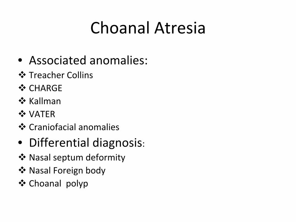
Choanal Atresia
• Associated anomalies:Treacher CollinsCHARGEKallmanVATER Craniofacial anomalies
• Differential diagnosis:Nasal septum deformityNasal Foreign bodyChoanal polyp

Choanal Atresia‐Management
• Bilateral , symptomatic in delivery room. Place oral airway and intubate
• Support growth with gavage feeds
• Repair:Transnasal puncture and stentingEndoscopic resection of posterior nasal septumHigh risk of recurrent stenosis even after successful surgery

Normal larynx
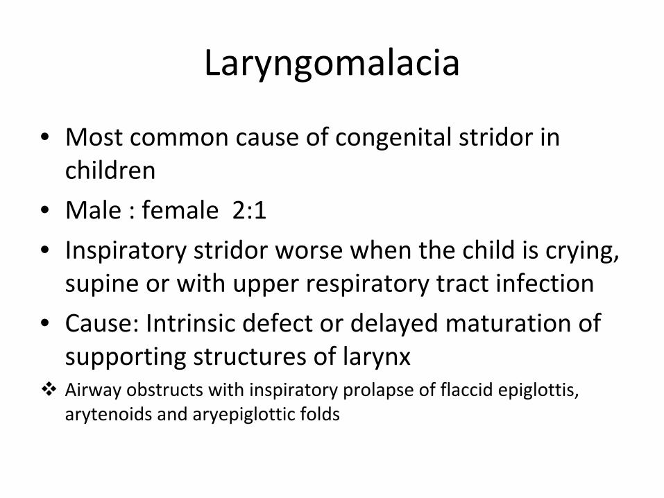
Laryngomalacia
• Most common cause of congenital stridor in children
• Male : female 2:1
• Inspiratory stridor worse when the child is crying, supine or with upper respiratory tract infection
• Cause: Intrinsic defect or delayed maturation of supporting structures of larynx
Airway obstructs with inspiratory prolapse of flaccid epiglottis, arytenoids and aryepiglottic folds

Endoscopic view

Laryngomalacia‐Diagnosis
Fiberoptic laryngoscopy while awake , with omega shaped epiglottis and taught aryepiglottic folds
Direct laryngoscopy with bronchoscopy
Laryngomalacia‐Management:
• Worsens in the first 8 months, plateaus at 9‐12 months and resolves spontaneously by 18‐24
months of age in most patients
• Symptomatic relief of stridor can be sometimes achieving by lying the infant on side
• Severe cases require surgical treatment with supraglotoplasty where excessive arytenoid folds,
lateral epiglottis and supra‐arytenoid folds are resected
• Rarely tracheotomy or Bipap may be required in very severe cases

Tracheomalacia • Abnormal tracheal collapse secondary to
inadequate cartilaginous and myoelastic components supporting the trachea
• Tracheal narrowing occurs during expiration and causes stridor
• Clinical manifestations: Intra thoracic: barky croup like coughExtra thoracic: inspiratory stridor Both are associated with respiratory distress

Tracheomalacia‐Diagnosis
• Barium swallow : extrinsic intrathoracic
compression of trachea secondary to vascular
ring
• Bronchoscopy during spontaneous
respiration
• CT/ MRI can further define the location

Tracheomalacia‐Management
• Spontaneous resolution at 6‐12 months
• With severe airway compromise, recurrent infection, respiratory failure
Tracheotomy or PPV
Tracheal reconstruction
Tracheopexy or Aortopexy
Tracheal stents , rarely due to high failure rate

Vignette
• A 3kg female infant is born to a 23 yr old G1P0 at 36 weeks GA. Her APGARS are 5 and 5. She
was noted to be apneic in the delivery room and cyanotic. Her PE was significant for
decreased breath sounds in chest and a scaphoid abdomen.


Congenital diaphragmatic hernia
• Definition: It is a defect of the diaphragm which allows herniation of abdominal contents into the thorax
• Incidence: 1 in 2000 to 4000 live births • Etiology: It is believed to result from incomplete fusion of the pleuroperitoneal membrane and passage of abdominal contents into the chestThe small bowel, stomach, spleen, and colon are the most frequently herniated organs. Also seen infrequently are pancreas, liver, adrenal glands, and kidneys

Congenital diaphragmatic hernia
• Clinical features: At birth abdomen is scaphoid , chest is funnel shaped Trachea and mediastinum are pushed to contralateral side Clinical presentation can be of frank respiratory failure to apnea and episodes of choking90% are left sided50% are associated with other congenital malformations ( esp. heart, kidneys, gastrointestinal tract, abdominal wall and central nervous system ) Associated with pulmonary hypoplasia and pulmonary hypertensionCan be associated with chromosomal disorders
Congenital diaphragmatic hernia
• Diagnosis: • Prenatal ultrasound: Shows polyhydramnios as well as fluid filled bowel with peristalsis at the level of heartRight sided hernias are usually missed due to echogenicity of liver and lungDifferentials: Congenital cystic adenomatoid malformation, bronchogenic cysts, mediastinal cystic teratoma and neurogenic tumors

Chest X‐ray
Copyright ©1999 American Academy of Pediatrics
Skarsgard, E. D. et al. Pediatrics in Review 1999;20:71-78e
Postnatal chest radiograph in an infant who has a large left CDH

Congenital diaphragmatic hernia
• Management:
• Prenatal: FETO• Respiratory stabilization in the delivery room
• AVOID BAGGGING THE INFANT IN DELIVERY ROOM
• May need Positive pressure ventilation, inhaled nitric oxide, surfactant therapy, ECMO
• Surgical correction after respiratory stabilization

Congenital diaphragmatic hernia
• Prognosis:Has high mortality rate
Is poor if diagnosed in utero
Inability to tolerate enteral nutrition, GERD
Depends also on pulmonary hypoplasia, pulmonary hypertension
Can develop scoliosis eventually

Vignette
• A 3kg male child born to G3 P2L2 @ 40 wks GA. In the delivery room he had increased oral
secretions which were removed with bulb suctioning. Prenatal history is significant for
polyhydramnios. Was admitted to the nursery with vital signs of Temp: 37C, RR: 40, O2sat:
98% on RA. He was attempted to be fed with significant non‐bilious emesis.

X‐ray

Tracheoesophageal Fistula
• Incidence is 1 in 3000 to 5000 live births• Embryology: It is due to incomplete
mesoderm separation of the primitive foregut, resulting in a fistula between esophagus and trachea
• Aspiration is the primary cause of lung damage
• Very good prognosis
Tracheo‐esophageal Fistula
• Types:Esophageal atresia with associated distal tracheoesophageal fistula‐ 80‐90%
Esophageal atresia without a tracheoesophageal fistula‐ 10%
H ‐ type tracheoesophageal fistula‐ 3%

TEF‐EA Esophageal
AtresiaH‐type TE fistula
Rudolph Pediatrics 21st
edition

TEF‐EA
Blind
pouch
Distal TEF
Tracheo‐esophageal fistula with Esophageal atresia
• Is the most common of TE fistulas constituting 85‐90%
• Incidence is 1 in 4000 live births• Frequent component of VATER• Embryology: Interruption of events responsible
for elongation of esophageal and tracheal tubes during 4th
week of development
• Prenatal Ultrasound is characterized by polyhydramnios

Tracheo‐esophageal fistula with Esophageal atresia
• Presentation: Excessive amount of saliva
Intolerance of oral feeds
Non‐ bilious emesis
Gastric distension, not scaphoid abdomen
Tachypnea
Cyanosis

Tracheo‐esophageal fistula with Esophageal Atresia
• Diagnosis:Difficulty to pass a NG tube which ends in the blind pouch KUB/ baby gram shows dilated esophageal pouch or a catheter ending in blind pouch and air through out the GI tract
• Management : Sump catheter into upper pouch Elevate head end to 45 degreesIV fluids and antibiotics

TEF‐EA CXR
Tracheo‐esophageal fistula with Esophageal atresia
• Management: Surgical repair of fistula can be undertaken in infant as little as 1200 gDetermined by associated anomalies and clinical status
• Esophageal Atresia: Double lumen orogastric tube to suctionGastrostomy tube Progressive esophageal dilatationEsophageal substitution procedures like colonic interposition, illeal interposition or reverse gastric tube

Esophageal Atresia without a TE fistula
• Presents with increased oral secretions and drooling• FLAT AND GASLESS ABDOMEN• Inability to tolerate enteral feeding• Esophageal pouches have large gap thus making
anastomosis difficult• Diagnosis: By KUB or passing a catheter that ends in the
blind pouch • Management:
Orogastric tube to suctionG‐tube insertion Progressive dilation of pouches and subsequent insertion of gastric tube, colon or piece of small intestine

H‐Type Tracheoesophageal Fistula
• Present with increased choking in the new born period or chronic cough and recurrent pneumonia in a older child
• Diagnosis:Could be subtle so difficult to diagnoseUpper GI with esophagogramFlexible bronchoscopy with esophagoscopy
• Management: Surgical division by the cervical approach

Tracheo‐esophageal fistula with Esophageal atresia
• Prognosis: Excellent and depends upon associated anomalies
Narrowing or stricture at anastomotic site is very common, requiring progressive dilatation
Esophageal dysmotility at the stricture site, esp. in colonic interposition or illeal interposition

Congenital Cystic adenomatoid malformation
• Most common pulmonary anomaly• It is dysplastic development of terminal
bronchiolar structures resulting in hamartomous lung
• Embryologic insult is around 7th week of gestation
• Communicates with tracheobronchial tree• Symptomatic in new born period or early infancy • Divided into 4 types depending upon its
constituents
Types of CCAM
• Type 1: Cysts are multiloculated and larger than 2 cm. Rare transformation to bronchoalveolar carcinoma
• Type 2: Multiple small cysts. Associated with renal agenesis, CV anomalies, diaphragmatic hernia and Syringomelia
• Type 3: Uncommon. Involve whole lobe. Macroscopically‐solid appearing and microscopically‐
resemeble late fetal lung• Type 4: Very rare. Multiloculated thin walled cysts. Can
have malignant transformation to pleuropulmonary blastoma

Congenital Cystic adenomatoid malformation
• Diagnosis: Prenatal UltrasoundChest X‐ray CT scan
• Management: By resection
• Complications : • Respiratory compromise• Recurrent infection• Could be premalignant

Congenital Cystic adenomatoid malformation

Vignette
• A 10 yr old male presents to ER for sore throat and cough, low grade fever. Physical Exam in ER, Temp: 98.6, RR: 28/ minute, O2sat: 98% on RA. General: Alert,
no tachypnea, HEENT: Within normal limits, Lungs: Decreased breath sounds in right anterior lung fields,
left lung is clear to auscultation, no wheezing, no crackles. Chest: PMI is displaced to the right chest,
RRR, S1S2: normal , no murmur, Abdomen: Soft, non tender, non distended, no hepatosplenomegaly, bowel
sounds: normal, Extremities: No clubbing, no cyanosis. Work up included: rapid streptococcal antigen test:
negative. Chest X‐ray which lead to an incidental diagnosis


Congenital lobar emphysema
• It is progressive over inflation / over distension of the lung
• Etiology: Bronchial atresia/stenosisAbsent or dysplastic bronchial cartilage( bronchomalacia)External compression by vessels
• Clinical findings: Before 6 months : mild tachypnea, wheeze to severe dyspnea and cyanosisPoor feeding and failure to thrive Older child: Incidental finding on chest X‐ray or recurrent pneumonia

Congenital lobar emphysema
• Pathology: Decreased alveoli and bronchial wall cartilage
• Affected lobes remain the same after first year of age and therefore become smaller in relation to
normal lobes• CXR findings: Hyperlucency with scant pulmonary markingsHyper inflated lobe with collapse of the contralateral lobe Mediastinal shift with flattened diaphragm


Congenital lobar emphysema
• Differential diagnosis:Foreign bodyBronchogenic cystCCAM
• Diagnosis:Chest X‐rayFlexible bronchoscopy CT scan is diagnostic

Congenital lobar emphysema
• Complications: Pneumothorax with positive pressure ventilation Compromise respiratory reserve by compressing uninvolved lobes
• Therapy: Surgical excision, indications: respiratory reserve is compromised or in the setting of recurrent infections Rule out congenital heart disease before surgery
Vignette
• 13 month old male presents to the ED with cough, high
fever and increased work of breathing. Vitals in the ER :
Temp: 39 C, RR: 32, O2sat: 96% RA. PE: General: Sub
coastal retractions, HEENT: Rhinnorhea, Lungs: decreased
breath sounds at left base, no crackles , no wheezing and
good air entry, rest of physical exam is unremarkable.
Chest X‐ray showed left lower lobe infiltrate • Past Medical history: Born at 39 wks GA by NSVD. Was
found to be tachypneic in nursery. CXR in nursery, showed
left lower lobe infiltrate suspected to be congenital
pneumonia and was treated with IV antibiotics. Was seen
by pediatrician 5‐6 times with recurrent upper respiratory
tract infections and persistent tachypnea

X‐ray in nursery X‐ray on admission

Pulmonary Sequestration
• Pulmonary tissue that is isolated from normal functioning lung and is nourished by systemic arteries. It is dysplastic and non functioning.
Has no connection to tracheobronchial tree• Embryology: Interruption of orderly lung
development and persistence of perfusion of sequestered lung tissue from systemic
circulation • Rarest of all congenital lung malformations
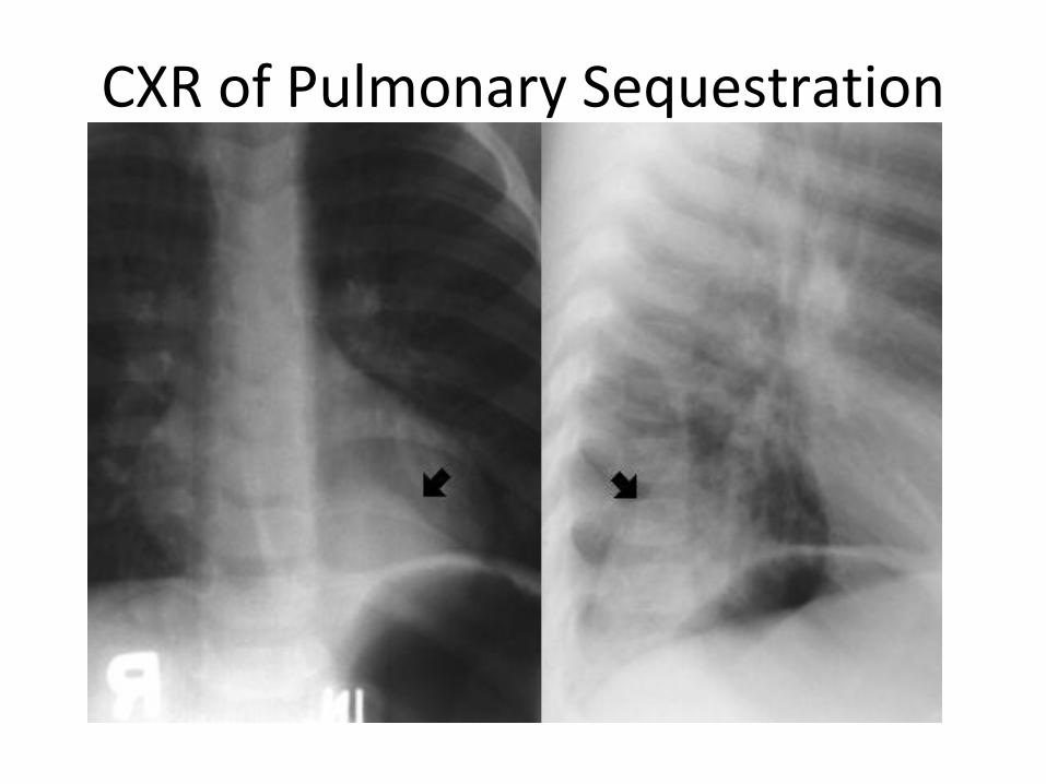
CXR of Pulmonary Sequestration

Pulmonary Sequestration
• Divided into intra lobar and extra lobar types depending upon blood supply and if it is contiguous
with the lung• Intralobar sequestration:3‐6 times more common Pleural covering is contiguous with the lungUsually left sided Arterial supply is by systemic artery, venous drainage is by pulmonary veinsRarely associated with anomaliesPresents in childhood with recurrent pneumonia
Pulmonary Sequestration
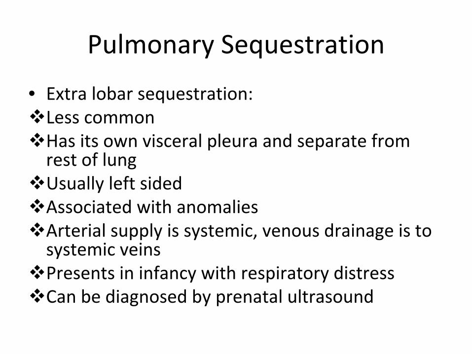
• Extra lobar sequestration:Less commonHas its own visceral pleura and separate from rest of lungUsually left sided Associated with anomaliesArterial supply is systemic, venous drainage is to systemic veins Presents in infancy with respiratory distressCan be diagnosed by prenatal ultrasound

Pulmonary Sequestration
• Diagnosis:Chest X‐ray
CT scan with contrast
MRI/ MRA
• Management:
IV antibiotics
Resection when primary infection is cleared

Arteriogram of sequestration

Congenital anomalies by age of Presentation
At Birth Children
Congenital diaphragmatic hernia H‐type TE fistula
Tracheo
esophageal fistula with
esophageal Atresia
Laryngomalacia
Choanal
Atresia‐
bilateral Tracheomalacia
Extra lobar sequestration Intralobar
sequestration
Congenital cystic adenomatoid
malformation
Congenital lobar emphysema
Congenital lobar Emphysema

Congenital anomalies by presentation Respiratory distress / noisy breathing Recurrent Pneumonia
Congenital diaphragmatic hernia H‐type TE fistula
Choanal
Atresia‐
bilateral Congenital lobar emphysema
Laryngomalacia Congenital cystic adenomatoid
malformation
Tracheoesophageal
fistula and esophageal
Atresia
Intralobar
sequestration
Congenital lobar Emphysema Congenital lobar Emphysema
Tracheomalacia
Congenital cystic adenomatoid
malformation
Related Documents