ORIGINAL RESEARCH published: 11 February 2020 doi: 10.3389/fimmu.2020.00179 Frontiers in Immunology | www.frontiersin.org 1 February 2020 | Volume 11 | Article 179 Edited by: Laura Santambrogio, Cornell University, United States Reviewed by: Alessandra Franco, University of California, San Diego, United States David H. Margulies, National Institute of Allergy and Infectious Diseases (NIAID), United States *Correspondence: Bernhard Loll [email protected] Andreas Ziegler [email protected] † Present address: Christine Rückert, CureVac AG, Tübingen, Germany Specialty section: This article was submitted to Antigen Presenting Cell Biology, a section of the journal Frontiers in Immunology Received: 20 November 2019 Accepted: 23 January 2020 Published: 11 February 2020 Citation: Loll B, Rückert C, Uchanska-Ziegler B and Ziegler A (2020) Conformational Plasticity of HLA-B27 Molecules Correlates Inversely With Efficiency of Negative T Cell Selection. Front. Immunol. 11:179. doi: 10.3389/fimmu.2020.00179 Conformational Plasticity of HLA-B27 Molecules Correlates Inversely With Efficiency of Negative T Cell Selection Bernhard Loll 1 *, Christine Rückert 2† , Barbara Uchanska-Ziegler 2,3 and Andreas Ziegler 3 * 1 Institut für Chemie und Biochemie, Abteilung Strukturbiochemie, Freie Universität Berlin, Berlin, Germany, 2 Institut für Immungenetik, Charité - Universitätsmedizin Berlin, Freie Universität Berlin, Berlin, Germany, 3 Ziegler Biosolutions, Waldshut-Tiengen, Germany The development of autoimmune disorders is incompletely understood. Inefficient thymic T cell selection against self-peptides presented by major histocompatibility antigens (HLA in humans) may contribute to the emergence of auto-reactive effector cells, and molecular mimicry between foreign and self-peptides could promote T cell cross-reactivity. A pair of class I subtypes, HLA-B2705 and HLA-B2709, have previously been intensely studied, because they are distinguished from each other only by a single amino acid exchange at the floor of the peptide-binding groove, yet are differentially associated with the autoinflammatory disorder ankylosing spondylitis. Using X-ray crystallography in combination with ensemble refinement, we find that the non-disease-associated subtype HLA-B2709, when presenting the self-peptide pGR (RRRWHRWRL), exhibits elevated conformational dynamics, and the complex can also be recognized by T cells. Both features are not observed in case of the sequence-related self-peptide pVIPR (RRKWRRWHL) in complex with this subtype, and T cell cross-reactivity between pGR, pVIPR, and the viral peptide pLMP2 (RRRWRRLTV) is only rarely observed. The disease-associated subtype HLA-B2705, however, exhibits extensive conformational flexibility in case of the three complexes, all of which are also recognized by frequently occurring cross-reactive T cells. A comparison of the structural and dynamic properties of the six HLA-B27 complexes, together with their individual ability to interact with T cells, permits us to correlate the flexibility of HLA-B27 complexes with effector cell reactivity. The results suggest the existence of an inverse relationship between conformational plasticity of peptide-HLA-B27 complexes and the efficiency of negative selection of self-reactive cells within the thymus. Keywords: HLA-B27, X-ray structure, peptide binding modes, conformational flexibility, T cell selection, central tolerance, molecular mimicry, ankylosing spondylitis INTRODUCTION Although the vast majority of autoimmune diseases is associated with HLA class II alleles (1), the first association detected and still one of the strongest found so far is that between the class I gene HLA-B ∗ 27 and the autoinflammatory rheumatic disorder ankylosing spondylitis (AS) (2, 3). Rats and mice transgenic for HLA-B ∗ 27 demonstrate a direct involvement of

Welcome message from author
This document is posted to help you gain knowledge. Please leave a comment to let me know what you think about it! Share it to your friends and learn new things together.
Transcript
ORIGINAL RESEARCHpublished: 11 February 2020
doi: 10.3389/fimmu.2020.00179
Frontiers in Immunology | www.frontiersin.org 1 February 2020 | Volume 11 | Article 179
Edited by:
Laura Santambrogio,Cornell University, United States
Reviewed by:
Alessandra Franco,University of California, San Diego,
United StatesDavid H. Margulies,
National Institute of Allergy andInfectious Diseases (NIAID),
United States
*Correspondence:
Bernhard [email protected]
Andreas [email protected]
†Present address:
Christine Rückert,CureVac AG, Tübingen, Germany
Specialty section:
This article was submitted toAntigen Presenting Cell Biology,
a section of the journalFrontiers in Immunology
Received: 20 November 2019Accepted: 23 January 2020
Published: 11 February 2020
Citation:
Loll B, Rückert C, Uchanska-Ziegler Band Ziegler A (2020) Conformational
Plasticity of HLA-B27 MoleculesCorrelates Inversely With Efficiency of
Negative T Cell Selection.Front. Immunol. 11:179.
doi: 10.3389/fimmu.2020.00179
Conformational Plasticity of HLA-B27Molecules Correlates Inversely WithEfficiency of Negative T CellSelectionBernhard Loll 1*, Christine Rückert 2†, Barbara Uchanska-Ziegler 2,3 and Andreas Ziegler 3*
1 Institut für Chemie und Biochemie, Abteilung Strukturbiochemie, Freie Universität Berlin, Berlin, Germany, 2 Institut fürImmungenetik, Charité - Universitätsmedizin Berlin, Freie Universität Berlin, Berlin, Germany, 3 Ziegler Biosolutions,Waldshut-Tiengen, Germany
The development of autoimmune disorders is incompletely understood. Inefficientthymic T cell selection against self-peptides presented by major histocompatibilityantigens (HLA in humans) may contribute to the emergence of auto-reactive effectorcells, and molecular mimicry between foreign and self-peptides could promote Tcell cross-reactivity. A pair of class I subtypes, HLA-B2705 and HLA-B2709, havepreviously been intensely studied, because they are distinguished from each otheronly by a single amino acid exchange at the floor of the peptide-binding groove, yetare differentially associated with the autoinflammatory disorder ankylosing spondylitis.Using X-ray crystallography in combination with ensemble refinement, we find thatthe non-disease-associated subtype HLA-B2709, when presenting the self-peptidepGR (RRRWHRWRL), exhibits elevated conformational dynamics, and the complexcan also be recognized by T cells. Both features are not observed in case of thesequence-related self-peptide pVIPR (RRKWRRWHL) in complex with this subtype, andT cell cross-reactivity between pGR, pVIPR, and the viral peptide pLMP2 (RRRWRRLTV)is only rarely observed. The disease-associated subtype HLA-B2705, however, exhibitsextensive conformational flexibility in case of the three complexes, all of which are alsorecognized by frequently occurring cross-reactive T cells. A comparison of the structuraland dynamic properties of the six HLA-B27 complexes, together with their individualability to interact with T cells, permits us to correlate the flexibility of HLA-B27 complexeswith effector cell reactivity. The results suggest the existence of an inverse relationshipbetween conformational plasticity of peptide-HLA-B27 complexes and the efficiency ofnegative selection of self-reactive cells within the thymus.
Keywords: HLA-B27, X-ray structure, peptide binding modes, conformational flexibility, T cell selection, central
tolerance, molecular mimicry, ankylosing spondylitis
INTRODUCTION
Although the vast majority of autoimmune diseases is associated with HLA class II alleles (1),the first association detected and still one of the strongest found so far is that between theclass I gene HLA-B∗27 and the autoinflammatory rheumatic disorder ankylosing spondylitis(AS) (2, 3). Rats and mice transgenic for HLA-B∗27 demonstrate a direct involvement of
Loll et al. Conformational Plasticity and T Cell Selection
the HLA-B27 protein in the development of diseases in theanimals that share many features with AS (4–8). In humans, notall of the >170 alleles (“subtypes”) of HLA-B∗27 are associatedwith AS: while the prototypical HLA-B∗27:05 subtype (B∗27:05in short) is AS-associated, HLA-B∗27:09 (in short, B∗27:09) isnot (9). Both alleles encode a heavy chain (HC) that associatesnon-covalently with a light chain, β2-microglobulin (β2m), anda self- or foreign peptide. The B2705 and B2709 complexesare distinguished only by an Asp116His exchange within theirHC, located on the floor of the peptide binding groove withinthe F-pocket. Comparative studies of these two subtypes canthus be regarded as promising to understand various aspects ofAS pathogenesis.
Functional, X-ray crystallographic, spectroscopic, andcalorimetric experiments as well as molecular dynamics(MD) simulations have already shown that the Asp116Hismicropolymorphism of the HC, combined with differentialpeptide binding, leads to an overall structural polymorphism ofthe subtypes that is responsible for several distinct features ofB2705 and B2709 [reviewed by (10–12)]. For example, cytotoxicT lymphocytes (CTL) directed against the self-peptides pVIPR[RRKWRRWHL, derived from vasoactive intestinal peptidetype 1 receptor (residues 400–408)] and pGR [RRRWHRWRL,derived from glucagon receptor (residues 412–420)] have beenfound in individuals with B∗27:05 (13, 14). About one sixthof these T cells cross-reacts with the viral pLMP2 peptide[RRRWRRLTV, derived from latent membrane protein 2(residues 236–244)] of Epstein-Barr virus (EBV) (15), suggestingthe existence of B2705-restricted molecular mimicry between thethree peptides (14). In contrast, CTL cross-reactivity betweenthe pVIPR and pLMP2 peptides has only rarely been observedwhen these ligands are displayed by B2709, suggesting lack ofmolecular mimicry (16). X-ray crystallographic studies supportthese conclusions (15) and indicate that structural similarity doesnot only form the basis for the observed CTL cross-reactivity inB∗27:05+ individuals, but argues also in favor of a relationshipwith the pathogenesis of AS. Although crystallographic studieshad thus implicated structural features of complexes with thepGR, pVIPR, and pLMP2 peptides displayed by B2705 as thebasis for CTL recognition and suggested a connection withthe initiation of AS, results from infrared (IR) spectroscopydemonstrated that an increase in conformational flexibilitymight in fact be more important than structural peculiarities inexplaining the association of certainHLA-B∗27 subtypes (besidesB∗27:05 also B∗27:04) with disease (17).
In the present work, we extend the structural analysesof complexes with pGR and related peptides to the B2709subtype, taking functional studies with CTL from B∗27:09+
individuals into account (16). We address three issues inparticular: (i) Is the extent of peptide dynamics a consequenceof display by a particular HLA-B27 subtype? (ii) Does theoverall conformational plasticity of a given peptide-HLA-B27complex allow to correlate this property with CTL reactivity?and (iii) Do the complexes of B2709 displaying pGR, pVIPR,or pLMP2 exhibit a lack of comprehensive molecular mimicry?Together with previously published studies (13–16, 18) thehigh resolution structure of pGR-B2709 that we describe here
permits to shed light on these questions and indicates that theoccurrence of peripheral self-reactive CTL is connected with theconformational plasticity of peptide-HLA-B27 complexes.
MATERIALS AND METHODS
Protein Preparation, Crystallization andData CollectionThe peptide pGR (RRRWHRWRL) was purchased from AltaBioscience (Birmingham, UK). B2709 HC, and β2m wereexpressed separately as inclusion bodies in Escherichia coli,dissolved in 50% urea and pGR-B2709 complexes werereconstituted using a protocol described previously (13, 19). Thecomplexes were isolated by size exclusion chromatography andused for crystallization at concentrations of 13–15 mg/ml in20mM Tris/HCl (pH 7.5), 150mM NaCl, 0.01% sodium azide.Crystals were obtained from drops mixed of 1.5 µl protein and1.5 µl precipitant solution [12–16% (w/v) polyethylene glycol(PEG) 8000, 100mM Tris/HCl, pH 7.5 or 8.0] in a hanging-drop vapor diffusion setup applying streak seeding techniques.Synchrotron X-ray diffraction data to 1.12 Å resolution werecollected at European Synchrotron Radiation Facility (ESRF,Grenoble, France) at beamline ID 14-2 from cryo-cooledcrystals with 10% (w/v) glycerol and 20% (w/v) PEG 8000 ascryoprotectant at 100K. The data were processed and scaledwith the HKL package (20) (Table 1). The raw diffractionimages are made public through www.proteindiffraction.org(doi: 10.18430/m33czf).
Structure Determination, Refinement, andAnalysisThe structure of pGR-B2709 was solved by molecularreplacement with the program EPMR (21) using water-and peptide-depleted m9-B2709 structures as search model(PDB entry 1K5N) (22). The crystallographic asymmetric unitcontains one pGR-B2709 complex with a Matthews coefficientof 2.8 Å3 Da−1 corresponding to a solvent content of 55%(23). For calculation of a free R-factor, a randomly generatedset of 2.5% of the reflections from the diffraction data set wasused and excluded from refinement. Restrained maximum-likelihood refinement was performed using REFMAC5 (24)including anisotropic B-factor refinement followed by iterativemanual model building with the program COOT (25). Watermolecules were positioned with ARP/wARP (26) and manuallyinspected. For data collection and refinement statistics seeTable 1. Intermediate and final structures were evaluated withMOLPROBITY (27) and PROCHECK (28). Figures wereprepared with PYMOL (29). The atomic coordinates andstructure amplitudes have been deposited in the Protein DataBank (PDB entry 3CZF).
Ensemble RefinementThe starting structures for ensemble refinements as implementedin PHENIX (30) were prepared as described (18, 31). Apartfrom pGR-B2709 (this study), the following complexes wereanalyzed: pGR-B2705 [PDB entry 2A83 (14)], pVIPR-B2709
Frontiers in Immunology | www.frontiersin.org 2 February 2020 | Volume 11 | Article 179

Loll et al. Conformational Plasticity and T Cell Selection
TABLE 1 | Data collection and refinement statistics.
Data collection pGR-B2709
PDB entry 3CZF
Wavelength (Å) 0.933
Space group P21
Unit cell [a,b,c (Å); β (◦)] 51.0, 81.9, 65.4; 108.9
Resolution (Å)a 30.0–1.1 (1.12–1.10)
Unique reflectionsa 196,702 (9,285)
Completeness (%)a 95.8 (90.4)
<I/(σ)>a 20.8 (2.7)
Ra,bsym 4.3 (28.8)
CCa1/2 99.9 (80.7)
Wilson B factor (Å2) 14.7
Refinement
Resolution (Å) 15.0–1.20
Number of reflections 149,186
Non-hydrogen atoms 4,243
Ra,ccryst 12.9 (13.8)
Ra,dfree 14.9 (15.9)
Heavy chain, no. of atoms/average B factor (Å2) 2,439/12.5
β2m, no. of atoms/average B factor (Å2) 913/14.2
Peptide, no. of atoms/average B factor (Å2) 149/12.2
Water, no. of molecules/average B factor (Å2) 712/27.1
Glycerol, no. of atoms/average B factor (Å2) 30/23.4
rmsde from ideal geometry, bond length (Å) 0.011
bond angles (◦) 1.427
Ramachandran outliers (%) 0.3
Ramachandran favored (%) 98.1
aValues in parentheses refer to the highest resolution shell.bRsym = ΣhΣi | Ih,I-<Ih >| / ΣhΣi Ih,i .cRcryst = Σh | Fo-Fc | / Σ Fo (working set, no σ cut-off applied).dRfree is the same as Rcryst, but calculated on 2.5% of the data excluded from refinement.eRoot-mean-square deviation (rmsd) from target geometries.
(PDB entry 5IB5), pVIPR-B2705 [PDB entry 5IB2 (18)], pLMP2-B2709 [PDB entry 1UXW (15)], and pLMP2-B2705 [PDB entry1UXS (15)]. Briefly, alternate conformations of amino acid sidechains were removed from the deposited structures in the PDBand the occupancies adjusted to 1. Prior to refinement, explicithydrogen atoms were generated with phenix.ready_set. Theensemble refinements were executed with the standard settings.For glycerol molecules, harmonic restraints were set to avoidstochastic displacement during simulations.
RESULTS
Structural Characteristics of thepGR-B2709 ComplexpGR-B2709 crystallized in space group P21 like B2705 boundto pGR (14), or both subtypes in complex with the sequence-related pVIPR (18) and pLMP2 peptides (15). The structure ofthe pGR-B2709 complex exhibited very high resolution of 1.2Å and was refined to R/Rfree of 0.129/0.149. For detailed X-raydiffraction and refinement statistics see Table 1. As expected, the
FIGURE 1 | General structural properties of the bound pGR peptide displayedby B2709. For sake of clarity, water molecules are omitted in allrepresentations. In (A–C), the view is from the side of the α2-helix, which is notdisplayed. (A) Final 2Fo-Fc electron density maps (blue mesh) contoured at 1.3σ, with pGR in A-conformation (cyan) and in B-conformation (orange) shown instick representation. The subtype-specific residue His116 of the HC is shownas well. (B) Color scheme depicting the anisotropic B-factor distribution inboth pGR conformations. (C) Superimposition of both pGR conformations,viewed as in (A). (D) Superimposition of both pGR conformations, 90◦ rotatedtoward the viewer in comparison to (C).
Frontiers in Immunology | www.frontiersin.org 3 February 2020 | Volume 11 | Article 179
Loll et al. Conformational Plasticity and T Cell Selection
single polypeptide chains, HC and β2m, show the protein foldcharacteristic for HLA class I molecules (32, 33). Differences toother HLA-B27 complexes are restricted to polymorphic residuessuch as Asp116His in the B2705/B2709 pair, to conformationsof solvent-exposed amino acid side-chains and peptide residues,as well as to the location of water molecules. Notably, none ofthe peptide residues in pGR-B2705 or pGR-B2709 is involved incrystal contacts.
As the calculated electron density map is of excellent quality,two conformations of the pGR Cα-backbone, designated A andB, could readily be assigned, between the two arginine residuesat position 6 (p6) and p8 (Figure 1A), although the overalldifference exhibited by both peptide Cα-traces is minimal [rootmean square deviation (r.m.s.d.) of 0.2 Å]. From the electrondensity, the ratio of conformations A to B was estimated tobe about 70–30%. The B-factors of the bound peptides in theA and the B conformation are virtually identical (Figure 1B).In addition to the difference between the Cα-backbones ofthe peptide, double conformations are also observed for theside chains of four peptide residues, pTrp4, pArg6, pTrp7, andpArg8 (Figures 1C,D).
Comparative Structural Features ofpGR-HLA-B27 ComplexesWhen the nona-peptides are neglected, the structures of pGR-B2709 and pGR-B2705 superimpose with an r.m.s.d. of 0.3Å for the Cα-atoms and must thus be considered virtuallyidentical. Both pGR peptide conformations assume the “p6α”binding mode (i.e., main-chain φ/ψ torsion angles in α-helicalconformation at p6) in the B2709 subtype. This unconventional(non-canonical, NC) conformation had been detected so far incase of B2705 in complex with the peptides pVIPR (18), pLMP2(15), pGR (14), and in the pVIPR-U5-B2709 [modified pVIPRwith citrulline at p5 (34)] as well as in the pVIPR-B2706 (17)complex. Whereas, easily distinguishable differences between theCα-conformations of pArg3 to pArg8 were observed in pGR-B2705 (r.m.s.d. of 0.5 Å), the differences in pGR-B2709 arevery small and limited to the region from pArg6 to pArg8. Acomparison of the different conformations of the pGR peptidein B2709 with those in B2705 reveals that they are slightly moresimilar to pGR-B2705-B (r.m.s.d. for Cα positions of 0.2 Å) thanto pGR-B2705-A (r.m.s.d. for Cα positions of 0.4 Å).
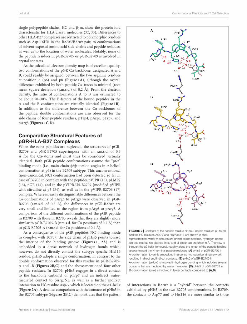
As a consequence of the pGR peptide’s NC binding modein complex with B2709, the side chain of pHis5 points towardthe interior of the binding groove (Figures 1, 2A) and isembedded in a dense network of hydrogen bonds which,however, do not directly contact the subtype-specific His116residue. pHis5 adopts a single conformation, in contrast to thedouble conformation observed for this residue in pGR-B2705-A and -B (Figures 2B,C) and the above-mentioned four otherpeptide residues. In B2709, pHis5 engages in a direct contactto the backbone carbonyl of pTrp7 and an indirect water-mediated contact to pArg3NE as well as a further indirectinteraction to HC residue Asp77 which is located on the α1-helix(Figure 2A). A detailed comparison with the contacts of pHis5 inthe B2705 subtype (Figures 2B,C) demonstrates that the pattern
FIGURE 2 | Contacts of the peptide residue pHis5. Peptide residues p3 to p9and the HC residues Asp77 and His/Asp116 are shown in stickrepresentation, water molecules are drawn as red spheres, hydrogen bondsare depicted as red dashed lines, and all distances are given in Å. The view isthrough the α2-helix (removed), roughly along the length of the peptide bindinggroove toward the N-terminal peptide residues. (A) pHis5 of pGR-B2709 inA-conformation (cyan) is embedded in a dense hydrogen bonding networkresulting in direct and indirect contacts. (B) pHis5 of pGR-B2705 inA-conformation (yellow) is involved in hydrogen bonding which includes severalcontacts that are mediated by water molecules. (C) pHis5 of pGR-B2705 inB-conformation (pink) is involved in fewer contacts compared to (A,B).
of interactions in B2709 is a “hybrid” between the contactsexhibited by pHis5 in the two B2705 conformations. In B2709,the contacts to Asp77 and to His116 are more similar to those
Frontiers in Immunology | www.frontiersin.org 4 February 2020 | Volume 11 | Article 179

Loll et al. Conformational Plasticity and T Cell Selection
found in pGR-B2705-A (Figure 2B), while the interaction withpArg3 resembles that observed in pGR-B2705-B (Figure 2C).Table 2 provides a detailed account of the contacts of the pGRpeptide in complex with B2705 and B2709. Of note, the vander Waals contact between pTrp4 and the HC residue Ile66is present in the complexes of both subtypes. In contrast, thiscontact is restricted to B2705 presenting pVIPR or pLMP2 (13,15, 18), suggesting that the NC peptide binding mode could be aprerequisite for this interaction.
The “hybrid” nature of structural features within the pGR-B2709 complex extends to properties of the side chains of peptideresidues that exhibit conformational dimorphism. A comparisonwith the respective pGR-B2705-A and -B conformations(Figure 3) shows that pTrp4, pArg6, and pArg8 of pGR-B2709-A exhibit a nearly identical conformation to that found inB2705. In contrast, pTrp7 of pGR-B2709-A is very similar tothe B conformation, which this residue adopts in pGR-B2705.In case of the B conformation in B2709, pTrp4 is identicallyand pArg6 similarly bound as in pGR-B2705-B. Conversely,pTrp7 and pArg8 in pGR-B2709-B are nearly identical to the Aconformation, which is observed in B2705 (14).
Despite these differences, the results demonstrate that thereare several features which are shared by the two pGR-HLA-B27 complexes. Irrespective of the subtype or the A orB conformations, the pGR residues pArg1 and pArg2 areidentically anchored in the characteristic binding mode that hasbeen observed before (13–15, 34–36), pHis5 is always nearlyidentically located, and the terminal pLeu9 is embedded withinthe F-pocket as in case of the peptides pVIPR (13), TIS (36), andpVIPR-U5 (34).
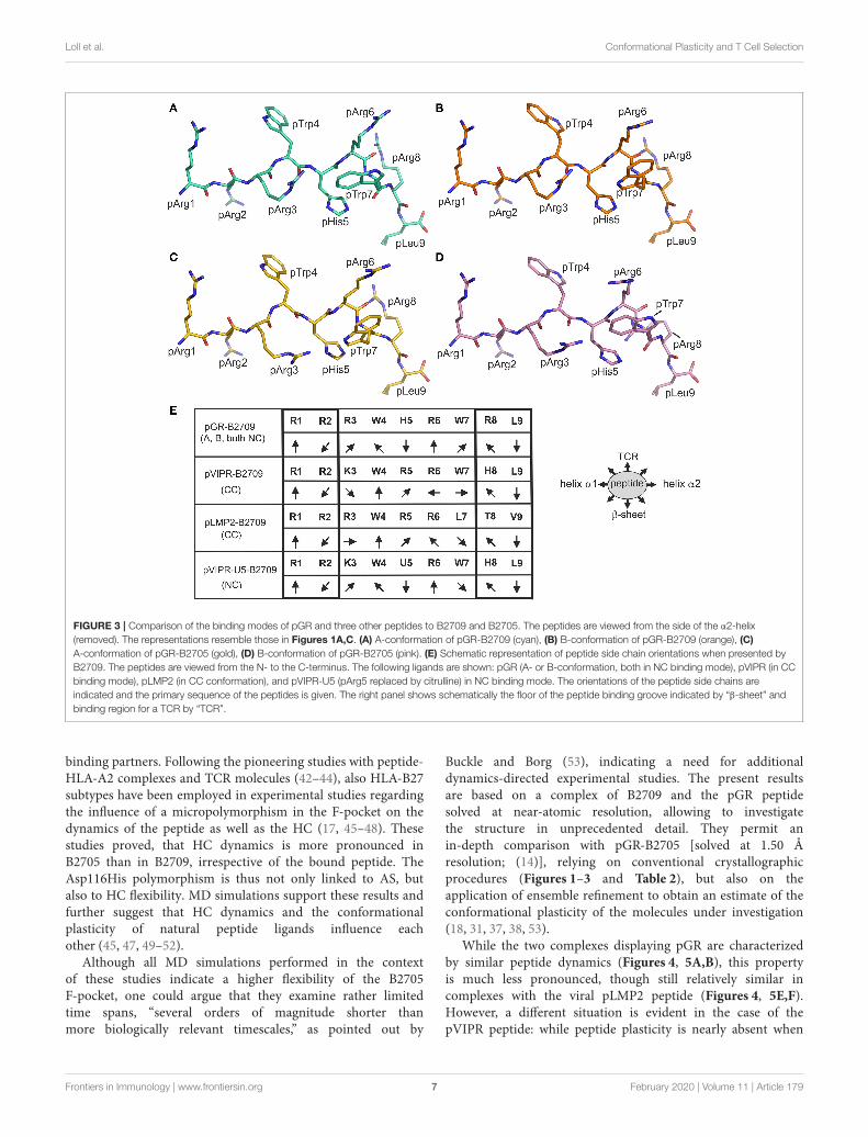
Structural Similarities and DifferencesBetween the pGR, pVIPR, and pLMP2Peptides in Complex With HLA-B27SubtypesThe isomorphous crystallization modes of the HLA-B27complexes with the peptides pVIPR (18), pLMP2 (15), andpGR [(14) and this study], permit a detailed comparison oftheir structures. Previous work had already shown that thestructures of these three peptides are very similar, at least intheir N-terminal two thirds, when presented by the B2705subtype, thereby furthering molecular mimicry (14). This is dueto the NC binding mode of the displayed peptide which isexhibited by all of the complexes. In contrast, despite sharing thecanonical conformation (CC), pVIPR and pLMP2 are differentlypresented by the B∗2709 subtype, primarily because the sidechains at p3 (Lys or Arg) and pArg6 point to distinct directions(Figure 3E) (15).
The comparison of the newly determined pGR-B2709structure with those of pVIPR and pLMP2 reveals that pGR ispresented by this subtype in a further distinct manner, which isdictated by the presence of the NC conformation (Figures 1–3).Despite the expected close similarity around the p1 and p2 aswell as around the p8 and p9 peptide residues, the three peptidesdeviate considerably from p3 to p7 (Figure 3E). The mostdivergent conformations are exhibited by pTrp4 and pArg6 and
the residue (either a tryptophan or a leucine) at p7 (Figure 3C).On the basis of these results, the presence of molecular mimicrybetween pGR, pVIPR, and pLMP2 when displayed by B2709must be regarded as highly unlikely.
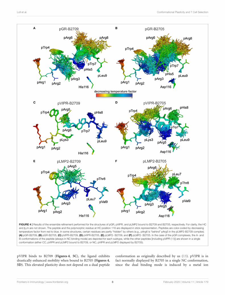
This conclusion is further supported by analyses of peptideplasticity using ensemble refinements (18, 31, 37, 38). Thisapproach permits to gain information on dynamic propertiesof a protein whose structure had been solved by X-raycrystallography, by employing short, steered molecular dynamics(MD) simulations during the refinement process. The results(Figures 4, 5A,B) demonstrate that pArg2 and pLeu9 arevirtually immobile, as expected for residues anchoring thepGR peptide within the binding groove. In contrast, notonly the residues exhibiting double conformations, but allresidues from pLys3 to pArg8 are characterized by extensiveconformational plasticity in the complexes of both HLA-B27subtypes presenting this peptide. The graphic presentation ofensemble-refined structures poses a problem not encounteredwhen B-factors (temperature or Debye-Waller factors) calculatedfor conventionally refined structures (compare Figure 1B) aredepicted. Since ensemble refinement allows to visualize B-factors independent from the atomic fluctuations that can berepresented by multiple structures within the ensemble, high(low) B-factors do not necessarily reflect elevated (decreased)plasticity of residues within a molecule. This is due to thewell-known fact that B-factors do not only report on atomicfluctuations, but are also influenced by factors such as crystallattice defects, rigid body motions, occupancy levels, radiationdamage, or refinement artifacts (39, 40). Consequently, regionswith a greater conformational flexibility, as revealed by ensemblerefinement, could have lower B-factors, since the ensemble ismore precisely reflecting the electron density (41), as seen e.g.,in the case of pArg3 in pLMP2-B2709 (Figure 4E) and pArg5in pLMP2-B2705 (Figure 4F). On the other hand, in case ofpArg1 the conformational flexibility is restricted due to π-π interactions with the side chains of Arg62 and Trp167 ofthe heavy chain (Figures 4, 5), that are both solvent exposed.The protein environment could thus reduce the number ofpossible side chain conformations. In contrast, the anchoringresidue pArg2 displays lower B-factors (Figures 4, 5), since itis not pointing toward the solvent but into the interior of theprotein and as described for pArg1, the number of side chainconformations is limited due to interactions with residues of theheavy chain.
As previously described by us in detail (18), the pVIPR peptideis considerably more mobile in B2705 than in B2709. Only pArg5shows a moderate degree of plasticity in B2709 (Figures 4, 5C).In the case of the pLMP2 peptide, the difference between thesubtypes is not as pronounced as for pVIPR, but the peptideis definitely more mobile in its middle (residues p3–p7) whenbound to B2705 (compare Figures 4, 5E,F).
With regard to molecular mimicry, the ensemble refinementspermit to conclude that the probability that a TCR wouldencounter a similar collection of structures among the threepeptides is far greater in the B2705 subtype than in the caseof B2709. For example, when comparing pVIPR-B2709 andpLMP2-B2709, only the area “above” pArg1, pTrp4, and pArg5
Frontiers in Immunology | www.frontiersin.org 5 February 2020 | Volume 11 | Article 179

Loll et al. Conformational Plasticity and T Cell Selection
TABLE 2 | Comparison of pGR conformations in the B2705 and B2709 subtypes.
pGR-B2705 conformations A and B pGR-B2709 conformations A and B
Peptide residue Contact residue Distance Interaction Peptide residue Contact residue Distance Interaction
p1 & p2 Contacts formed by Arg1 and Arg2 are very similar in both complexes; the side chain of Arg1 is solvent-exposed, whereas Arg2 is buried
p3 pArg3N Tyr99OH 3.09 HB pArg3N Tyr99OH 3.1 HB
pArg3 Tyr99, Leu156, Tyr159 3.6–4.0 vdW pArg3 Tyr99, Leu156, Tyr159 3.6–4.0 vdW
pArg3NH2(A) pHis5ND1(A) 2.9 HB pArg3NE pTrp4O 2.8 HB
p4 The side chain of this residue is solvent-exposed in all complexes
pTrp4 Gln65, Arg62, Ile66 3.6-4.0 vdW pTrp4 Gln65, Arg62, Ile66 3.6-4.0 vdW
p5 The side chain of this residue is buried in all complexes
pHis5ND1(A) pArg3NH2(A) 2.9 HB pHis5NE2 Lys70 3.5 HB
pHis5NE2(B) pTrp7O(B) 3.1 HB pHis5NE2 pTrp7O(A) 3.1 HB
pHis5O (B) pArg8NH2 (B) 3.5 HB pHis5NE2 pTrp7O(B) 3.7 HB
p6 The side chain of this residue is solvent-exposed in all complexes
pArg6O(A) pArg8NH1 (A) 2.8 HB pArg6O(B) pArg8NH1 (A) 2.8 HB
pArg6NH2 (B) Gln155OE1 2.8 HB pArg6O(B) pArg8NH1 (B) 2.8 HB
p7 pTrp7O(A) pHis5NE2 (A) 3.3 HB pTrp7 Val152, Trp147 3.6-4.0 vdW
pTrp7O(B) pHis5NE2 (B) 3.1 HB
pTrp7 Ala150, Val152 ∼3.6 vdW
p8 The side chain of this residue is solvent-exposed in all complexes
pArg8NE (A) Glu76OE1 3.0 SB Arg8NE (A) Glu76OE2 (A) 3.0 SB
pArg8NH2 (A) Thr73OG1 3.5 HB Arg8NE (B) Glu76OE2 (A) 2.5 SB
pArg8NH2 (B) Asp77OD2 3.4 SB Arg8O Trp147NE1 2.9 HB
pArg8NE (B) Asp77OD1 3.2 SB
pArg8O Trp147NE1 2.9 HB
p9 The side chain of this residue is buried in both complexes .
Distances are given in (Å). Only direct intra-peptide contacts and contacts between the peptides and HC residues [distance cut-off 3.5 Å; hydrogen bond (HB), salt bridge (SB)] areincluded; solvent-mediated interactions are omitted, and van der Waals (vdW) contacts are not given explicitly. Double conformations of amino acid residues are indicated by (A) or (B).
might evoke a response by a cross-reactive CTL, and an extensionof this cross-reactivity to pGR-B2709 is difficult to imagine(compare Figures 4, 5A,C,E). This is very different in the case ofthe disease-associated subtype (compare Figures 4, 5B,D,F). Theensemble of structures exhibited by peptides bound to B2705 thatis available for CTL recognition is much larger than in case of theB2709 subtype.
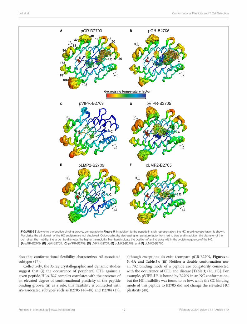
Distinct Dynamic Properties of the PeptideBinding Grooves of the B2705 and B2709SubtypesThe fact that the conformational plasticity of the threepeptides depends on the HLA-B27 subtype by which they aredisplayed, leads to the question how the hidden Asp116Hismicropolymorphism in the F pocket can influence the flexibilityof peptide residues that may be separated by more than 20Å as in the case of His116 and the guanidinium group ofpArg1 (compare e.g., Figures 4, 5C,D). Therefore, we analyzedalso the overall appearance of the binding grooves by ensemblerefinement. Figure 6 depicts the six complexes recognizable by aTCR. It is immediately obvious that pVIPR-B2709 (Figure 6C)exhibits a greatly diminished degree of mobility, both ofthe peptide and of large parts of the binding groove. Thisproperty is not exhibited by any of the other complexes. In
particular, the F pocket, which accommodates the polymorphicresidue 116 (His in B2709), the peptide’s C-terminus, and itssurroundings show very little flexibility. A comparison withthe other five complexes reveals that large parts of the α1-helix and the loops “below” it (HC residues 13–18 and 86–90, Figure 6) as well as the beginning of the α2-helix exhibitvery little dynamics. Only that section of the binding grooveaccommodating the N-terminal third of pVIPR as well asthe loop comprising residues 106–108 show a limited degreeof flexibility. Consequently, the three B2709 complexes shareneither a pronounced structural nor a dynamic similarity,in contrast to the three B2705 complexes (Figures 6B,D,F).Although the ensemble refinements of the peptide-HLA-B27complexes provide a fairly good idea of the differential dynamicswhich characterizes each of the binding grooves, the results donot give a hint for the reasons underlying F-pocket effects onthe flexibility of distant peptide residues (see the interactionbetween the guanidinium moiety of pArg1 and Asp/His116mentioned above).
DISCUSSION
Conformational plasticity is a prerequisite for the successfulinteraction of MHC molecules with extra- and intracellular
Frontiers in Immunology | www.frontiersin.org 6 February 2020 | Volume 11 | Article 179
Loll et al. Conformational Plasticity and T Cell Selection
FIGURE 3 | Comparison of the binding modes of pGR and three other peptides to B2709 and B2705. The peptides are viewed from the side of the α2-helix(removed). The representations resemble those in Figures 1A,C. (A) A-conformation of pGR-B2709 (cyan), (B) B-conformation of pGR-B2709 (orange), (C)A-conformation of pGR-B2705 (gold), (D) B-conformation of pGR-B2705 (pink). (E) Schematic representation of peptide side chain orientations when presented byB2709. The peptides are viewed from the N- to the C-terminus. The following ligands are shown: pGR (A- or B-conformation, both in NC binding mode), pVIPR (in CCbinding mode), pLMP2 (in CC conformation), and pVIPR-U5 (pArg5 replaced by citrulline) in NC binding mode. The orientations of the peptide side chains areindicated and the primary sequence of the peptides is given. The right panel shows schematically the floor of the peptide binding groove indicated by “β-sheet” andbinding region for a TCR by “TCR”.
binding partners. Following the pioneering studies with peptide-HLA-A2 complexes and TCR molecules (42–44), also HLA-B27subtypes have been employed in experimental studies regardingthe influence of a micropolymorphism in the F-pocket on thedynamics of the peptide as well as the HC (17, 45–48). Thesestudies proved, that HC dynamics is more pronounced inB2705 than in B2709, irrespective of the bound peptide. TheAsp116His polymorphism is thus not only linked to AS, butalso to HC flexibility. MD simulations support these results andfurther suggest that HC dynamics and the conformationalplasticity of natural peptide ligands influence eachother (45, 47, 49–52).
Although all MD simulations performed in the contextof these studies indicate a higher flexibility of the B2705F-pocket, one could argue that they examine rather limitedtime spans, “several orders of magnitude shorter thanmore biologically relevant timescales,” as pointed out by
Buckle and Borg (53), indicating a need for additionaldynamics-directed experimental studies. The present resultsare based on a complex of B2709 and the pGR peptidesolved at near-atomic resolution, allowing to investigatethe structure in unprecedented detail. They permit anin-depth comparison with pGR-B2705 [solved at 1.50 Åresolution; (14)], relying on conventional crystallographicprocedures (Figures 1–3 and Table 2), but also on theapplication of ensemble refinement to obtain an estimate of theconformational plasticity of the molecules under investigation(18, 31, 37, 38, 53).
While the two complexes displaying pGR are characterizedby similar peptide dynamics (Figures 4, 5A,B), this propertyis much less pronounced, though still relatively similar incomplexes with the viral pLMP2 peptide (Figures 4, 5E,F).However, a different situation is evident in the case of thepVIPR peptide: while peptide plasticity is nearly absent when
Frontiers in Immunology | www.frontiersin.org 7 February 2020 | Volume 11 | Article 179
Loll et al. Conformational Plasticity and T Cell Selection
FIGURE 4 | Results of the ensemble refinement performed for the structures of pGR, pVIPR, and pLMP2 bound to B2709 and B2705, respectively. For clarity, the HCand β2m are not shown. The peptide and the polymorphic residue at HC position 116 are displayed in stick representation. Peptides are color-coded by decreasingtemperature factor from red to blue. In some structures, certain residues are partly “hidden” by others (e.g., pArg6 is “behind” pArg5 in the pLMP2-B2709 complex).(A) pGR-B2709, (B) pGR-B2705, (C) pVIPR-B2709, (D) pVIPR-B2705, (E) pLMP2- B2709, and (F) pLMP2- B2705. In the case of the pGR complexes, the A- andB-conformations of the peptide (always in NC binding mode) are depicted for each subtype, while the other peptides [including pVIPR (18)] are shown in a singleconformation (either CC, pVIPR and pLMP2 bound to B2709, or NC, pVIPR and pLMP2 displayed by B2705).
pVIPR binds to B2709 (Figures 4, 5C), the ligand exhibitsdrastically enhanced mobility when bound to B2705 (Figures 4,5D). This elevated plasticity does not depend on a dual peptide
conformation as originally described by us (13). pVIPR is infact normally displayed by B2705 in a single NC conformation,since the dual binding mode is induced by a metal ion
Frontiers in Immunology | www.frontiersin.org 8 February 2020 | Volume 11 | Article 179

Loll et al. Conformational Plasticity and T Cell Selection
FIGURE 5 | View as in Figure 4, but rotated by 90◦. Peptides are oriented such as an approaching TCR would “see” them. They are color-coded by decreasingtemperature factor from red to blue. (A) pGR-B2709, (B) pGR-B2705, (C) pVIPR-B2709, (D) pVIPR-B2705, (E) pLMP2-B2709, and (F) pLMP2-B2705. In somestructures, certain residues are partly “hidden” by others (e.g., pArg2 is “behind” pTrp4 in the pGR-B2709 complex). For the conformations and binding modes of thepeptides, please refer to the legend of Figure 4.
bound to the peptide (18). In addition, ensemble refinementanalyses show that enhanced flexibility is not only observedfor peptides bound to the complexes, but characterizes alsothe HC (Figure 6). This HC flexibility is exhibited by residuesbelonging to the beginning of the α1- and the end of theα2-helices and the HC loops “beneath” the binding groove(Figure 6A). The pVIPR-B2709 complex, however, shows adifferent picture: while the regions comprising residues 54–58and 175–181 (at the “left” end of the binding groove, opposite toeach other), as well as 106–108 reveal conformational flexibility,the “right” part of the binding groove, “above” the F-pocket
and within a distance of about 15 Å from His116, exhibitsa nearly complete lack of mobility (Figure 6C). These resultsare in complete agreement with the IR spectroscopic studies ofFabian et al. which demonstrated that the B2705 HC is moreflexible than that of B2709, irrespective of the bound peptide(47). All data are listed in Table 3, together with experimentalresults obtained previously with two further peptides, pVIPR-U5 (a citrullinated version of pVIPR, RRKWURWHL, U =
citrulline) (34) and TIS (RRLPIFSRL), a proven self-peptide forboth B2709 and B2705 (36). Experiments with another pair ofdifferentially AS-associated subtypes, B2704 and B2706, show
Frontiers in Immunology | www.frontiersin.org 9 February 2020 | Volume 11 | Article 179
Loll et al. Conformational Plasticity and T Cell Selection
FIGURE 6 | View onto the peptide binding groove, comparable to Figure 5. In addition to the peptide in stick representation, the HC in coil representation is shown.For clarity, the α3 domain of the HC and β2m are not displayed. Color-coding by decreasing temperature factor from red to blue and in addition the diameter of thecoil reflect the mobility: the larger the diameter, the higher the mobility. Numbers indicate the position of amino acids within the protein sequence of the HC.(A) pGR-B2709, (B) pGR-B2705, (C) pVIPR-B2709, (D) pVIPR-B2705, (E) pLMP2-B2709, and (F) pLMP2-B2705.
also that conformational flexibility characterizes AS-associatedsubtypes (17).
Collectively, the X-ray crystallographic and dynamic studiessuggest that (i) the occurrence of peripheral CTL against agiven peptide-HLA-B27 complex correlates with the presence ofan elevated degree of conformational plasticity of the peptidebinding groove; (ii) as a rule, this flexibility is connected withAS-associated subtypes such as B2705 (46–48) and B2704 (17),
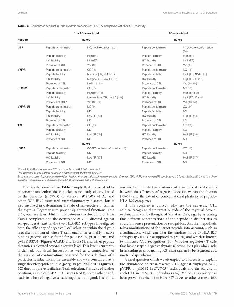
although exceptions do exist (compare pGR-B2709, Figures 4,5, 6A and Table 3); (iii) Neither a double conformation noran NC binding mode of a peptide are obligatorily connectedwith the occurrence of CTL and disease [Table 3; (16, 17)]. Forexample, pVIPR-U5 is bound by B2709 in an NC conformation,but the HC flexibility was found to be low, while the CC bindingmode of this peptide to B2705 did not change the elevated HCplasticity (48).
Frontiers in Immunology | www.frontiersin.org 10 February 2020 | Volume 11 | Article 179
Loll et al. Conformational Plasticity and T Cell Selection
TABLE 3 | Comparison of structural and dynamic properties of HLA-B27 complexes with their CTL-reactivity.
Non AS-associated AS-associated
Peptide B2709 B2705
pGR Peptide conformation NC, double conformation Peptide conformation NC, double conformation(14)
Peptide flexibility High (ER) Peptide flexibility High (ER)
HC flexibility High (ER) HC flexibility High (ER)
Presence of CTL Yes (16) Presence of CTL Yes (14)
pVIPR Peptide conformation CC (18) Peptide conformation NC (18)
Peptide flexibility Marginal [ER, NMR (18)] Peptide flexibility High [ER, NMR (18)]
HC flexibility Marginal {ER, low [IR (47)]} HC flexibility High [ER, IR (47)]
Presence of CTL No& (16, 54) Presence of CTL Yes (16, 54)
pLMP2 Peptide conformation CC (15) Peptide conformation NC (15)
Peptide flexibility High [ER (18)] Peptide flexibility High [ER (18)]
HC flexibility Intermediate {ER, low [IR (48)]} HC flexibility High [ER, IR (46)]
Presence of CTL* Yes (16, 54) Presence of CTL Yes (16, 54)
pVIPR-U5 Peptide conformation NC (34) Peptide conformation CC (34)
Peptide flexibility ND Peptide flexibility ND
HC flexibility Low [IR (48)] HC flexibility High [IR (48)]
Presence of CTL ND Presence of CTL ND
TIS Peptide conformation CC (36) Peptide conformation CC (36)
Peptide flexibility ND Peptide flexibility ND
HC flexibility Low [IR (46)] HC flexibility High [IR (46)]
Presence of CTL ND Presence of CTL ND
B2706 B2704
pVIPR Peptide conformation CC/NC double conformation (17) Peptide conformation CC (17)
Peptide flexibility ND Peptide flexibility ND
HC flexibility Low [IR (17)] HC flexibility High [IR (17)]
Presence of CTL ND Presence of CTL ND
&pLMP2/pVIPR-cross-reactive CTL are rarely found in B*27:09+ individuals.*The presence of CTL against pLMP2 is a consequence of infection with EBV.Structural and dynamic properties were determined by X-ray crystallography with ensemble refinement (ER), NMR, and infrared (IR) spectroscopy. CTL-reactivity is attributed to a givencomplex in individuals with the respective HLA-B*27 subtype (ND, not determined).
The results presented in Table 3 imply that the Asp116Hispolymorphism within the F pocket is not only closely linkedto the presence (B∗27:05) or absence (B∗27:09) of AS andother HLA-B∗27-associated autoinflammatory diseases, but isalso involved in determining the fate of self-reactive T cells inthe thymus. Together with previously obtained functional data(16), our results establish a link between the flexibility of HLAclass I complexes and the occurrence of CTL directed againstself-peptidesat least in the two HLA-B27 subtypes investigatedhere: the efficiency of negative T cell selection within the thymicmedulla is impaired when T cells encounter a highly flexiblebinding groove, such as found for pGR-B2709, pGR-B2705, andpVIPR-B2705 (Figures 6A,B,D and Table 3), and when peptidedynamics is elevated beyond a certain level. This level is currentlyill-defined, but visual inspection as well as a comparison ofthe number of conformations observed for the side chain of aparticular residue within an ensemble allow to conclude that asingle flexible peptide residue (pArg5 in pVIPR-B2709, Figures 4,5C) does not prevent efficient T cell selection. Plasticity of furtherpositions, as in pVIPR-B2705 (Figures 4, 5D), on the other hand,leads to failure of negative selection against this ligand. Therefore,
our results indicate the existence of a reciprocal relationshipbetween the efficiency of negative selection within the thymus(55–57) and the extent of conformational plasticity of peptide-HLA-B27 complexes.
If this scenario is correct, why are the surviving CTLable to recognize their target outside of the thymus? Severalexplanations can be thought of Yin et al. (58), e.g., by assumingthat different concentrations of the peptide in distinct tissuescould influence presentation or recognition. Another hypothesistakes modifications of the target peptide into account, such ascitrullination, which can alter the binding mode to HLA-B27subtypes (pVIPR-U5 as opposed to pVIPR) and which is knownto influence CTL recognition (34). Whether regulatory T cellsthat have escaped negative thymic selection (59) play also a rolein initiating or propagating AS, must currently be regarded as amatter of speculation.
A final question which we attempted to address is to explainthe abundance of cross-reactive CTL against displayed pGR,pVIPR, or pLMP2 in B∗27:05+ individuals and the scarcity ofsuch CTL in B∗27:09+ individuals (16). Molecular mimicry hasbeen proven to exist in the HLA-B27 as well as other contexts (60,
Frontiers in Immunology | www.frontiersin.org 11 February 2020 | Volume 11 | Article 179

Loll et al. Conformational Plasticity and T Cell Selection
61) and may contribute also to the emergence of cross-reactiveCTL in certain subtypes. It remains still unknown whetherthese cells do really play a decisive role in the developmentof HLA-B∗27-associated diseases (62). We have already pointedout that comprehensive molecular mimicry is a hallmark ofpresentation by B2705 (14), and we show now that there isneither a structural nor a dynamic basis for molecular mimicrywhen the B2709 subtype displays the peptides pGR, pVIPR, andpLMP2 (Figures 4–6).
The primary aim of our contribution was to investigate therelationship of peptide- and binding groove-plasticity of HLA-B27 subtypes to the efficiency of negative T cell selection. Dueto their distinct, peptide-independent conformational flexibility(17, 46–48), however, peptide-devoid HC/β2m complexes withina cell possess already the potential to differentially influencepeptide-loading processes (63–67). This could be relevant for thesubtype-dependent initiation of HLA-B∗27-associated diseases.A deeper insight into the dynamics of these molecules is thuscrucial for a more detailed understanding of their function andinteraction with other proteins as well as their involvement in ASand other disorders (53).
DATA AVAILABILITY STATEMENT
The raw data supporting the conclusions of thisarticle are available through www.proteindiffraction.org
(doi: 10.18430/m33czf). The atomic coordinates and structureamplitudes have been deposited in the Protein Data Bank (PDBentry 3CZF).
AUTHOR CONTRIBUTIONS
CR performed protein purification and crystallization. BLperformed all crystallographic procedures, prepared figures, andwrote the manuscript. BU-Z and AZ conceived the study andwrote the manuscript.
FUNDING
The authors are grateful to the DeutscheForschungsgemeinschaft for support in the context of Sfb449 projects B6, Z3 (BU-Z and AZ), the Volkswagen StiftungI/79 989 (AZ), the Fondazione Ceschina (Lugano, Switzerland)(AZ), and to Fonds der Chemischen Industrie (BL).
ACKNOWLEDGMENTS
Generous beam time allocation and support at ESRF (Grenoble,France) are gratefully acknowledged. We acknowledgesupport by the Open Access Publication Initiative of FreieUniversität Berlin.
REFERENCES
1. Horton R, Wilming L, Rand V, Lovering RC, Bruford EA, Khodiyar VK, etal. Gene map of the extended human MHC. Nat Rev Genet. (2004) 5:889–99.doi: 10.1038/nrg1489
2. Brewerton DA, Hart FD, Nicholls A, Caffrey M, James DC, SturrockRD. Ankylosing spondylitis and HL-A 27. Lancet. (1973) 1:904–7.doi: 10.1016/S0140-6736(73)91360-3
3. Schlosstein L, Terasaki PI, Bluestone R, Pearson CM. High association ofan HL-A antigen, W27, with ankylosing spondylitis. N Engl J Med. (1973)288:704–6. doi: 10.1056/NEJM197304052881403
4. Taurog JD. The mystery of HLA-B27: if it isn’t one thing, it’s another. ArthritisRheum. (2007) 56:2478–81. doi: 10.1002/art.22807
5. Bowness P. HLA-B27. Annu Rev Immunol. (2015) 33:29–48.doi: 10.1146/annurev-immunol-032414-112110
6. Colbert RA, Navid F, Gill T. The role of HLA-B∗27 in spondyloarthritis. BestPract Res Clin Rheumatol. (2017) 31:797–815. doi: 10.1016/j.berh.2018.07.012
7. Busch R, Kollnberger S, Mellins ED. HLA associations in inflammatoryarthritis: emerging mechanisms and clinical implications.Nat Rev Rheumatol.
(2019) 15:364–81. doi: 10.1038/s41584-019-0219-58. Hammer RE, Maika SD, Richardson JA, Tang JP, Taurog JD. Spontaneous
inflammatory disease in transgenic rats expressing HLA-B27 and humanβ2m: an animal model of HLA-B27-associated human disorders. Cell. (1990)63:1099–112. doi: 10.1016/0092-8674(90)90512-D
9. D’Amato M, Fiorillo MT, Carcassi C, Mathieu A, Zuccarelli A, BittiPP, et al. Relevance of residue 116 of HLA-B27 in determiningsusceptibility to ankylosing spondylitis. Eur J Immunol. (1995) 25:3199–201.doi: 10.1002/eji.1830251133
10. Fiorillo MT, Sorrentino R. T-cell responses against viral and self-epitopes andHLA-B27 subtypes differentially associated with ankylosing spondylitis. AdvExp Med Biol. (2009) 649:255–62. doi: 10.1007/978-1-4419-0298-6_19
11. Ziegler A, Loll B, Misselwitz R, Uchanska-Ziegler B. Implications of structuraland thermodynamic studies of HLA-B27 subtypes exhibiting differential
association with ankylosing spondylitis.Adv ExpMed Biol. (2009) 649:177–95.doi: 10.1007/978-1-4419-0298-6_13
12. Uchanska-Ziegler B, Loll B, Fabian H, Hee CS, Saenger W, Ziegler A.HLA class I-associated diseases with a suspected autoimmune etiology:HLA-B27 subtypes as a model system. Eur J Cell Biol. (2012) 91:274–86.doi: 10.1016/j.ejcb.2011.03.003
13. Hülsmeyer M, Fiorillo MT, Bettosini F, Sorrentino R, Saenger W, Ziegler A, etal. Dual, HLA-B27 subtype-dependent conformation of a self-peptide. J ExpMed. (2004) 199:271–81. doi: 10.1084/jem.20031690
14. Rückert C, Fiorillo MT, Loll B, Moretti R, Biesiadka J, Saenger W, etal. Conformational dimorphism of self-peptides and molecular mimicry ina disease-associated HLA-B27 subtype. J Biol Chem. (2006) 281:2306–16.doi: 10.1074/jbc.M508528200
15. Fiorillo MT, Rückert C, Hülsmeyer M, Sorrentino R, Saenger W, ZieglerA, et al. Allele-dependent similarity between viral and self-peptidepresentation by HLA-B27 subtypes. J Biol Chem. (2005) 280:2962–71.doi: 10.1074/jbc.M410807200
16. Nurzia E, Panimolle F, Cauli A, Mathieu A, Magnacca A, Paladini F,et al. CD8+ T-cell mediated self-reactivity in HLA-B27 context as aconsequence of dual peptide conformation.Clin Immunol. (2010) 135:476–82.doi: 10.1016/j.clim.2010.01.009
17. Loll B, Fabian H, Huser H, Hee CS, Ziegler A, Uchanska-Ziegler B, etal. Increased conformational flexibility of HLA-B∗27 subtypes associatedwith ankylosing spondylitis. Arthritis Rheumatol. (2016) 68:1172–82.doi: 10.1002/art.39567
18. Driller R, Ballaschk M, Schmieder P, Uchanska-Ziegler B, Ziegler A, LollB. Metal-triggered conformational reorientation of a self-peptide bound toa disease-associated HLA-B∗27 subtype. J Biol Chem. (2019) 294:13269–79.doi: 10.1074/jbc.RA119.008937
19. Garboczi DN, Hung DT, Wiley DC. HLA-A2-peptide complexes - refoldingand crystallization of molecules expressed in Escherichia coli and complexedwith single antigenic peptides. Proc Natl Acad Sci USA. (1992) 89:3429–33.doi: 10.1073/pnas.89.8.3429
Frontiers in Immunology | www.frontiersin.org 12 February 2020 | Volume 11 | Article 179

Loll et al. Conformational Plasticity and T Cell Selection
20. Otwinowski Z, Minor W. Processing of X-Ray Diffraction Data Collected in
Oscillation Mode. New York, NY: Academic Press (1997).21. Kissinger CR, Gehlhaar DK, Fogel DB. Rapid automated molecular
replacement by evolutionary search.Acta Crystallogr D Biol Crystallogr. (1999)55:484–91. doi: 10.1107/S0907444998012517
22. HülsmeyerM, Hillig RC, Volz A, RühlM, SchröderW, SaengerW, et al. HLA-B27 subtypes differentially associated with disease exhibit subtle structuralalterations. J Biol Chem. (2002) 277:47844–53. doi: 10.1074/jbc.M206392200
23. Matthews BW. Solvent content of protein crystals. J Mol Biol. (1968) 33:491–7.doi: 10.1016/0022-2836(68)90205-2
24. Murshudov GN, Vagin AA, Dodson EJ. Refinement of macromolecularstructures by the maximum-likelihood method. Acta Crystallogr D Biol
Crystallogr. (1997) 53:240–55. doi: 10.1107/S090744499601225525. Emsley P, Lohkamp B, Scott WG, Cowtan K. Features and development
of Coot. Acta Crystallogr D Biol Crystallogr. (2010) 66:486–501.doi: 10.1107/S0907444910007493
26. Perrakis A, Morris R, Lamzin VS. Automated protein model buildingcombinedwith iterative structure refinement.Nat Struct Biol. (1999) 6:458–63.doi: 10.1038/8263
27. Williams CJ, Headd JJ, Moriarty NW, Prisant MG, Videau LL, Deis LN, et al.MolProbity: more and better reference data for improved all-atom structurevalidation. Protein Sci. (2018) 27:293–315. doi: 10.1002/pro.3330
28. Laskowski RA, Macarthur MW, Moss DS, Thornton JM. Procheck - aprogram to check the stereochemical quality of protein structures. J ApplCrystallogr. (1993) 26:283–91. doi: 10.1107/S0021889892009944
29. DeLano WL. The PyMOL Molecular Graphics System on World Wide Web.
(2002). Available online at: http://www.pymol.org (accessed January 31, 2020).30. Adams PD, Afonine PV, Bunkoczi G, Chen VB, Davis IW, Echols
N, et al. PHENIX: a comprehensive Python-based system formacromolecular structure solution. Acta Crystallogr Sect D. (2010) 66:213–21.doi: 10.1107/S0907444909052925
31. Burnley BT, Afonine PV, Adams PD, Gros P. Modelling dynamics inprotein crystal structures by ensemble refinement. Elife. (2012) 1:e00311.doi: 10.7554/eLife.00311.029
32. Madden DR. The three-dimensional structure of peptide-MHC complexes. Annu Rev Immunol. (1995) 13:587–622.doi: 10.1146/annurev.iy.13.040195.003103
33. Bjorkman PJ, Saper MA, Samraoui B, Bennett WS, Strominger JL, Wiley DC.Structure of the human class I histocompatibility antigen, HLA-A2. Nature.(1987) 329:506–12. doi: 10.1038/329506a0
34. Beltrami A, Rossmann M, Fiorillo MT, Paladini F, Sorrentino R, SaengerW, et al. Citrullination-dependent differential presentation of a self-peptide by HLA-B27 subtypes. J Biol Chem. (2008) 283:27189–99.doi: 10.1074/jbc.M802818200
35. Hillig RC, Hülsmeyer M, Saenger W, Welfle K, Misselwitz R, WelfleH, et al. Thermodynamic and structural analysis of peptide- and allele-dependent properties of two HLA-B27 subtypes exhibiting differential diseaseassociation. J Biol Chem. (2004) 279:652–63. doi: 10.1074/jbc.M307457200
36. Hülsmeyer M, Welfle K, Pöhlmann T, Misselwitz R, Alexiev U, WelfleH, et al. Thermodynamic and structural equivalence of two HLA-B27subtypes complexed with a self-peptide. J Mol Biol. (2005) 346:1367–79.doi: 10.1016/j.jmb.2004.12.047
37. Gros P, van Gunsteren WF, Hol WG. Inclusion of thermal motion incrystallographic structures by restrained molecular dynamics. Science. (1990)249:1149–52. doi: 10.1126/science.2396108
38. Fodor J, Riley BT, Borg NA, Buckle AM. Previously hidden dynamics atthe TCR-peptide-MHC interface revealed. J Immunol. (2018) 200:4134–45.doi: 10.4049/jimmunol.1800315
39. Kuriyan J, Weis WI. Rigid protein motion as a model for crystallographictemperature factors. Proc Natl Acad Sci USA. (1991) 88:2773–7.doi: 10.1073/pnas.88.7.2773
40. Kuzmanic A, Pannu NS, Zagrovic B. X-ray refinement significantlyunderestimates the level of microscopic heterogeneity in biomolecularcrystals. Nat Commun. (2014) 5:3220. doi: 10.1038/ncomms4220
41. Levin EJ, Kondrashov DA, Wesenberg GE, Phillips GN Jr. Ensemblerefinement of protein crystal structures: validation and application. Structure.(2007) 15:1040–52. doi: 10.1016/j.str.2007.06.019
42. Garboczi DN, Ghosh P, Utz U, Fan QR, Biddison WE, Wiley DC. Structureof the complex between human T-cell receptor, viral peptide and HLA-A2.Nature. (1996) 384:134–41. doi: 10.1038/384134a0
43. Garcia KC, Degano M, Pease LR, Huang M, Peterson PA, TeytonL, et al. Structural basis of plasticity in T cell receptor recognitionof a self peptide-MHC antigen. Science. (1998) 279:1166–72.doi: 10.1126/science.279.5354.1166
44. Armstrong KM, Piepenbrink KH, Baker BM. Conformational changes andflexibility in T-cell receptor recognition of peptide-MHC complexes. BiochemJ. (2008) 415:183–96. doi: 10.1042/BJ20080850
45. Pöhlmann T, Bockmann RA, Grubmüller H, Uchanska-Ziegler B,Ziegler A, Alexiev U. Differential peptide dynamics is linked tomajor histocompatibility complex polymorphism. J Biol Chem. (2004)279:28197–201. doi: 10.1074/jbc.C400128200
46. Fabian H, Huser H, Loll B, Ziegler A, Naumann D, Uchanska-ZieglerB. HLA-B27 heavy chains distinguished by a micropolymorphism exhibitdifferential flexibility. Arthritis Rheum. (2010) 62:978–87. doi: 10.1002/art.27316
47. Fabian H, Huser H, Narzi D, Misselwitz R, Loll B, Ziegler A, etal. HLA-B27 subtypes differentially associated with disease exhibitconformational differences in solution. J Mol Biol. (2008) 376:798–810.doi: 10.1016/j.jmb.2007.12.009
48. Fabian H, Loll B, Huser H, Naumann D, Uchanska-Ziegler B, Ziegler A.Influence of inflammation-related changes on conformational characteristicsof HLA-B27 subtypes as detected by IR spectroscopy. FEBS J. (2011)278:1713–27. doi: 10.1111/j.1742-4658.2011.08097.x
49. Narzi D, Becker CM, Fiorillo MT, Uchanska-Ziegler B, Ziegler A, BockmannRA. Dynamical characterization of two differentially disease associated MHCclass I proteins in complex with viral and self-peptides. J Mol Biol. (2012)415:429–42. doi: 10.1016/j.jmb.2011.11.021
50. Nurzia E, Narzi D, Cauli A, Mathieu A, Tedeschi V, Caristi S, et al. Interactionpattern of Arg 62 in the A-pocket of differentially disease-associated HLA-B27subtypes suggests distinct TCR binding modes. PLoS ONE. (2012) 7:e32865.doi: 10.1371/journal.pone.0032865
51. Abualrous ET, Fritzsche S, Hein Z, Al-Balushi MS, Reinink P, Boyle LH, etal. F pocket flexibility influences the tapasin dependence of two differentiallydisease-associated MHC Class I proteins. Eur J Immunol. (2015) 45:1248–57.doi: 10.1002/eji.201445307
52. Sercinoglu O, Ozbek P. Computational characterization of residuecouplings and micropolymorphism-induced changes in the dynamicsof two differentially disease-associated human MHC class-I alleles. J
Biomol Struct Dyn. (2018) 36:724–40. doi: 10.1080/07391102.2017.1295884
53. Buckle AM, Borg NA. Integrating experiment and theory tounderstand TCR-pMHC dynamics. Front Immunol. (2018) 9:2898.doi: 10.3389/fimmu.2018.02898
54. Fiorillo MT, Maragno M, Butler R, Dupuis ML, Sorrentino R. CD8(+)T-cell autoreactivity to an HLA-B27-restricted self-epitope correlates withankylosing spondylitis. J Clin Invest. (2000) 106:47–53. doi: 10.1172/JCI9295
55. Takaba H, Takayanagi H. The mechanisms of T cell selection in the thymus.Trends Immunol. (2017) 38:805–16. doi: 10.1016/j.it.2017.07.010
56. Klein L, Kyewski B, Allen PM, Hogquist KA. Positive and negative selection ofthe T cell repertoire: what thymocytes see (and don’t see). Nat Rev Immunol.
(2014) 14:377–91. doi: 10.1038/nri366757. Passos GA, Speck-Hernandez CA, Assis AF, Mendes-da-Cruz DA. Update
on Aire and thymic negative selection. Immunology. (2018) 153:10–20.doi: 10.1111/imm.12831
58. Yin L, Dai S, Clayton G, Gao W, Wang Y, Kappler J, et al. Recognition of selfand altered self by T cells in autoimmunity and allergy. Protein Cell. (2013)4:8–16. doi: 10.1007/s13238-012-2077-7
59. Inglesfield S, Cosway EJ, Jenkinson WE, Anderson G. Rethinkingthymic tolerance: lessons from mice. Trends Immunol. (2019) 40:279–91.doi: 10.1016/j.it.2019.01.011
60. Alvarez-Navarro C, Cragnolini JJ, Dos Santos HG, Barnea E, Admon A,Morreale A, et al. Novel HLA-B27-restricted epitopes from Chlamydia
trachomatis generated upon endogenous processing of bacterial proteins
Frontiers in Immunology | www.frontiersin.org 13 February 2020 | Volume 11 | Article 179

Loll et al. Conformational Plasticity and T Cell Selection
suggest a role of molecular mimicry in reactive arthritis. J Biol Chem. (2013)288:25810–25. doi: 10.1074/jbc.M113.493247
61. Luo G, Ambati A, Lin L, Bonvalet M, Partinen M, Ji X, et al. Autoimmunityto hypocretin and molecular mimicry to flu in type 1 narcolepsy.Proc Natl Acad Sci USA. (2018) 115:E12323–32. doi: 10.1073/pnas.1818150116
62. Gracey E, Yao Y, Qaiyum Z, Lim M, Tang M, Inman RD. Altered cytotoxicityprofile of CD8+ T cells in ankylosing spondylitis.Arthritis Rheumatol. (2019).doi: 10.1002/art.41129. [Epub ahead of print].
63. McShan AC, Natarajan K, Kumirov VK, Flores-Solis D, Jiang J, BadstubnerM, et al. Peptide exchange on MHC-I by TAPBPR is driven bya negative allostery release cycle. Nat Chem Biol. (2018) 14:811–20.doi: 10.1038/s41589-018-0096-2
64. Hafstrand I, Sayitoglu EC, Apavaloaei A, Josey BJ, Sun R, Han X, et al.Successive crystal structure snapshots suggest the basis for MHC class Ipeptide loading and editing by tapasin. Proc Natl Acad Sci USA. (2019)116:5055–60. doi: 10.1073/pnas.1807656116
65. Natarajan K, Jiang J, Margulies DH. Structural aspects of chaperone-mediated peptide loading in the MHC-I antigen presentation pathway. CritRev Biochem Mol Biol. (2019) 54:164–73. doi: 10.1080/10409238.2019.1610352
66. Natarajan K, Jiang J, May NA, Mage MG, Boyd LF, McShan AC, et al. The roleof molecular flexibility in antigen presentation and T cell receptor-mediatedsignaling. Front Immunol. (2018) 9:1657. doi: 10.3389/fimmu.2018.01657
67. Kurimoto E, Kuroki K, Yamaguchi Y, Yagi-Utsumi M, Igaki T, IguchiT, et al. Structural and functional mosaic nature of MHC class Imolecules in their peptide-free form. Mol Immunol. (2013) 55:393–9.doi: 10.1016/j.molimm.2013.03.014
Conflict of Interest: AZ is the owner of Ziegler Biosolutions.
The remaining authors declare that the research was conducted in the absence ofany commercial or financial relationships that could be construed as a potentialconflict of interest.
Copyright © 2020 Loll, Rückert, Uchanska-Ziegler and Ziegler. This is an open-access
article distributed under the terms of the Creative Commons Attribution License (CC
BY). The use, distribution or reproduction in other forums is permitted, provided
the original author(s) and the copyright owner(s) are credited and that the original
publication in this journal is cited, in accordance with accepted academic practice.
No use, distribution or reproduction is permitted which does not comply with these
terms.
Frontiers in Immunology | www.frontiersin.org 14 February 2020 | Volume 11 | Article 179
Related Documents