COMPREHENSIVE INVITED REVIEW Conformational Plasticity and Structure=Function Relationships in Cytochromes P450 Thomas C. Pochapsky, 1–3 Sophia Kazanis, 1 and Marina Dang 1 Abstract The cytochrome P450s are a superfamily of enzymes that are found in all kingdoms of living organisms, and typically catalyze the oxidative addition of atomic oxygen to an unactivated C-C or C-H bond. Over 8000 nonredundant sequences of putative and confirmed P450 enzymes have been identified, but three-dimensional structures have been determined for only a small fraction of these. While all P450 enzymes for which structures have been determined share a common global fold, the flexibility and modularity of structure around the active site account for the ability of P450 enzymes to accommodate a vast number of structurally dissimilar substrates and support a wide range of selective oxidations. In this review, known P450 structures are compared, and some structural criteria for prediction of substrate selectivity and reaction type are suggested. The importance of dynamic processes such as redox-dependent and effector-induced conformational changes in determining cat- alytic competence and regio- and stereoselectivity is discussed, and noncrystallographic methods for charac- terizing P450 structures and dynamics, in particular, mass spectrometry and nuclear magnetic resonance spectroscopy are reviewed. Antioxid. Redox Signal. 13, 1273–1296. I. Introduction 1273 II. Crystallographic Structures of Prokaryotic Cytochromes P450 1274 A. CYP101 1274 B. CYP102 and CYP108 1275 C. Macrolide biosynthetic P450s 1276 D. Cytochrome P450 nor (CYP55A1) and CYP105 (MoxA) 1277 E. Coupling reactions- CYP158, CYP121, and P450 OxyB 1277 III. Crystallographic Structures of Eukaryotic Cytochromes P450 1278 A. Cytochromes P450 exported to the endoplasmic reticulum 1278 B. CYP2B4 1279 C. Liver microsomal enzymes CYP3A4 and CYP1A2 1280 D. Prostaglandin biosynthesis: prostacyclin I2 synthase 1280 E. Steroid biosynthetic and catabolic enzymes 1281 F. Computational modeling of P450 structures 1281 IV. Nuclear Magnetic Resonance as a Probe of Cytochrome P450 Structure and Dynamics 1281 V. Structural and Dynamic Insights from Mass Spectrometry 1282 VI. Can We Identify Structural Motifs in P450 Structures That Correlate with Activity? 1283 VII. Conclusions 1289 I. Introduction I t is now over 50 years since the tertiary structure of a protein was first established by X-ray crystallography (50), and 45 years have passed since the first enzyme structure, that of hen egg lysozyme, was described (9). A trickle of protein structures in the 1970s has turned into a flood: 65 unique structures (<90% sequence identity) were added to the PDB database between 1970 and 1980, whereas over 2300 struc- tures were released in the first 9 months of 2009 alone. The Reviewing Editors: Sean C. Gay, Gideon Grogan, David Leys, Kirsty McLean, and Mark A. White Departments of 1 Chemistry and 2 Biochemistry, and 3 the Rosenstiel Basic Medical Science Research Institute, Brandeis University, Waltham, Massachusetts. ANTIOXIDANTS & REDOX SIGNALING Volume 13, Number 8, 2010 ª Mary Ann Liebert, Inc. DOI: 10.1089=ars.2010.3109 1273

Welcome message from author
This document is posted to help you gain knowledge. Please leave a comment to let me know what you think about it! Share it to your friends and learn new things together.
Transcript

COMPREHENSIVE INVITED REVIEW
Conformational Plasticity and Structure=FunctionRelationships in Cytochromes P450
Thomas C. Pochapsky,1–3 Sophia Kazanis,1 and Marina Dang1
Abstract
The cytochrome P450s are a superfamily of enzymes that are found in all kingdoms of living organisms, andtypically catalyze the oxidative addition of atomic oxygen to an unactivated C-C or C-H bond. Over 8000nonredundant sequences of putative and confirmed P450 enzymes have been identified, but three-dimensionalstructures have been determined for only a small fraction of these. While all P450 enzymes for which structureshave been determined share a common global fold, the flexibility and modularity of structure around the activesite account for the ability of P450 enzymes to accommodate a vast number of structurally dissimilar substratesand support a wide range of selective oxidations. In this review, known P450 structures are compared, and somestructural criteria for prediction of substrate selectivity and reaction type are suggested. The importance ofdynamic processes such as redox-dependent and effector-induced conformational changes in determining cat-alytic competence and regio- and stereoselectivity is discussed, and noncrystallographic methods for charac-terizing P450 structures and dynamics, in particular, mass spectrometry and nuclear magnetic resonancespectroscopy are reviewed. Antioxid. Redox Signal. 13, 1273–1296.
I. Introduction 1273II. Crystallographic Structures of Prokaryotic Cytochromes P450 1274
A. CYP101 1274B. CYP102 and CYP108 1275C. Macrolide biosynthetic P450s 1276D. Cytochrome P450nor (CYP55A1) and CYP105 (MoxA) 1277E. Coupling reactions- CYP158, CYP121, and P450 OxyB 1277
III. Crystallographic Structures of Eukaryotic Cytochromes P450 1278A. Cytochromes P450 exported to the endoplasmic reticulum 1278B. CYP2B4 1279C. Liver microsomal enzymes CYP3A4 and CYP1A2 1280D. Prostaglandin biosynthesis: prostacyclin I2 synthase 1280E. Steroid biosynthetic and catabolic enzymes 1281F. Computational modeling of P450 structures 1281
IV. Nuclear Magnetic Resonance as a Probe of Cytochrome P450 Structure and Dynamics 1281V. Structural and Dynamic Insights from Mass Spectrometry 1282
VI. Can We Identify Structural Motifs in P450 Structures That Correlate with Activity? 1283VII. Conclusions 1289
I. Introduction
It is now over 50 years since the tertiary structure of aprotein was first established by X-ray crystallography (50),
and 45 years have passed since the first enzyme structure, that
of hen egg lysozyme, was described (9). A trickle of proteinstructures in the 1970s has turned into a flood: 65 uniquestructures (<90% sequence identity) were added to the PDBdatabase between 1970 and 1980, whereas over 2300 struc-tures were released in the first 9 months of 2009 alone. The
Reviewing Editors: Sean C. Gay, Gideon Grogan, David Leys, Kirsty McLean, and Mark A. White
Departments of 1Chemistry and 2Biochemistry, and 3the Rosenstiel Basic Medical Science Research Institute, Brandeis University,Waltham, Massachusetts.
ANTIOXIDANTS & REDOX SIGNALINGVolume 13, Number 8, 2010ª Mary Ann Liebert, Inc.DOI: 10.1089=ars.2010.3109
1273

importance of structure determination to our understanding ofenzyme function cannot be overstated: active sites are readilyidentified, critical residues are often obvious, and catalyticmechanisms can be proposed and tested based on the informa-tion that these structures provide. Yet a crystallographically-determined enzyme structure is not a panacea. Often, a newlydetermined enzyme structure raises more questions than itanswers. Why does the substrate (or substrate analog) seem tobe in the wrong orientation for the observed chemistry? Are weseeing all of the binding sites, or are there secondary (allosteric)sites that are unoccupied? Is the structure that we see biologi-cally relevant? Sometimes the answers to these questions areobvious, but not always. For example, crystal packing can forceorder upon regions of the polypeptide that in solution havemultiple functionally important conformations. It can alsoselect for a single conformation in regions where conforma-tional changes are critical to the enzyme activity. It is thereforeimportant to keep in mind that a crystallographic enzymestructure presents a snapshot of a dynamic system. It is notalways easy to tell where on a reaction pathway the observedconformation of an enzyme lies, or even if the observed con-former is functionally important.
Nowhere are these considerations more important than whenthinking about structure=function relationships in cytochromesP450. The P450 enzymes are heme-containing monooxygenasesthat catalyze the oxidation of organic species by molecular oxy-gen, often by insertion of an oxygen atom into an unactivatedC-H or C-C bond. Members of the P450 superfamily are foundin every kingdom and phylum of living organism, from archaeato chordates. They catalyze a wide variety of oxidative trans-formations that are essential to primary and secondary meta-bolic processes. In humans, P450 enzymes play important rolesin drug metabolism and activation, carcinogen activation, ste-roid and prostaglandin biosyntheses, and as such provide awide variety of therapeutic targets (77). In other organisms,P450s are critical for the biosynthesis of antibiotics and anti-neoplastics, and are the focus of research aimed at tailoring andmodifying their activity so as to produce novel pharmaceuticals.
While most bacterial and archaeal P450s are water-solublemonomeric enzymes, P450s found in higher organisms areusually membrane-bound or membrane-associated, andpresent considerable challenges to the structural biologist. Anadditional factor that must be considered when examining thestructures of redox-active enzymes such as P450s is that bothstructure and dynamics can be affected by the oxidation stateof the redox-active functionality (heme, in the case of P450)(73, 98). Unless precautions are taken, reduction of metalcenters by photoelectrons produced by the incident X-raybeam can render the oxidation state of the redox-active centerambiguous in crystallographically-derived structures.
In the past, P450 enzymes were often classified according tothe type of electron transfer protein that supplies the reducingequivalents required for turnover. For example, many P450sare reduced directly by NAD(P)H-dependent flavoproteins,whereas others use ferredoxins as intermediate electronshuttles. However, it has become evident that the electrontransport chains supporting P450-catalyzed oxidations areoften more complicated than previously suspected (36), andthat electron transfer partners often serve dual roles, func-tioning as effectors as well as reducing agents (70, 169).
As will be seen in this review, the P450 superfamilycombines a conservative architecture with a remarkable
adaptability for substrate recognition and regio- and stereo-selectivity in the chemistry that is catalyzed. This is possibledue to the modular nature of secondary structural featuressurrounding the P450 active site. Not only do these featuresdiffer between enzymes, but they change even between dif-ferent states of the individual enzyme. In this review, we havechosen examples that we hope will demonstrate this modu-larity, while attempting to find common themes, where pos-sible, in comparing P450 structures. Nevertheless, it isbecoming clear that P450 enzyme structures must be consid-ered, not just in three dimensions, but in four, as flexible anddynamic arrangements that change depending upon thepresence of substrate, cofactor, and oxidation state.
II. Crystallographic Structuresof Prokaryotic Cytochromes P450
A. CYP101
The first crystallographic structures of P450 were deter-mined for a soluble monomeric bacterial enzyme, cytochromeP450cam (CYP101) a camphor hydroxylase from the soil bac-terium Pseudomonas putida (108–110, 113–118). Being a bacterialenzyme, CYP101 is relatively easy to express, handle, andpurify, and much of what we currently know or suspect aboutthe P450 enzyme superfamily has been learned using this en-zyme. The CYP101 structure is a roughly triangular prism, withsecondary structural features being conveniently described interms of their location relative to the plane of the heme por-phyrin macrocycle. As in all P450s, the heme iron is axiallyligated by a cysteine thiolate that defines the proximal side of theheme. The active site is on the opposite (distal) face of the heme,and, depending upon the oxidation state of the enzyme, thedistal Fe axial ligand might be O2, water, hydroxide, or otherLewis base. In the presence of substrate, the distal axial ligationsite can be vacant, yielding the 5-coordinate geometry expectedfor high-spin Fe (III). The secondary structure nomenclaturescheme initially adopted by Poulos et al. for CYP101 (109) hasbecome standard for describing secondary structural featuresin all P450s, and is summarized in Figures 1 and 2. Viewed fromthe distal side of the protein in the orientation shown in Figure1, the active site of CYP101 is bound to the "north" by helix I, tothe "west" by the B’ helix and B’–C loop, "south" by residues ofthe b3 sheet, and "east" by the b5 sheet. The floor of the activesite is provided by the heme porphyrin, while the active site iseffectively closed off from solvent by a cap formed by the F andG helices and the F-G loop. The orientation of substrate in theCYP101 active site readily explains both the regio- and ste-reoselectivity of the observed hydroxylation at the 5-exo posi-tion of camphor.
Viewed from the north as defined in Figure 1, proximalfeatures that can be seen in Figure 2 include the B, C, J, K, andL helices, as well as a region of irregular structure precedingthe axial ligand Cys known as the "b-meander", the b2 andportions of the b4 sheets. The D helix is approximately bi-sected by the plane of the heme. On the distal side, the I helix isthe most prominent structural feature, providing a backbonearound which the heme and the remainder of the polypeptideare arranged. As in almost all P450 structures, the I helix inCYP101 is not completely straight, but is "kinked" near theheme iron due to an interruption in the regular i, iþ 4 NH—O¼C hydrogen bonding pattern of a regular a-helix. Thisinterruption is due to a hydrogen bond from the OH of a
1274 POCHAPSKY ET AL.

strongly conserved threonine (Thr 252 in CYP101) to thecarbonyl of the i-4 residue (Gly 238), and has been proposed toaccommodate the bound dioxygen appropriately for the ob-served chemistry (107). Most of the regular b-sheet structuresof CYP101 are located on the distal side, as are the A, B’, E, F,G, and H helices. An additional short region of 3–10 helix isalso present on the distal side, providing the apex of a roughpyramidal arrangement of helices A, B and K.
B. CYP102 and CYP108
For some years (1986–1993), the CYP101 structure was theonly P450 structure available, and so was used extensively formodeling other P450 enzymes (3, 13, 32, 42, 46, 106, 179).There was initially concern about the validity of such modelsin the absence of other experimentally determined P450structures, as the CYP101 fold was unique among knownglobular protein structures, and sequence homology betweenCYP101 and most eukaryotic P450s is low (106). However,the structure determination of the heme-binding domain ofcytochrome P450BM3 (CYP102) (Fig. 3), a fatty acid o-2-hydroxylase from Bacillus megaterium, showed that the P450fold is indeed conserved (119). Despite low overall sequencehomology between the two enzymes (16% sequence identity),it was found that both enzymes have identical topology andthat most of the secondary structural features of CYP102 havehomologues in CYP101. For convenience, in the course of thisreview, we will use the CYP101 nomenclature to identify
homologous sequence=structural motifs in other P450s, evenif the secondary structure is not identical: that is, regions se-quentially and structurally homologous with the b5 sheet inCYP101 will be identified as b5, although this region is not awell-defined b-sheet in many P450 structures.
The CYP102 structure provided the first indications of theplasticity of the P450 substrate binding regions. The pub-lished CYP101 structures showed relatively modest differ-ences in active site structure, depending on the absence orpresence or type of substrate. The most prominent differ-ences observed in the active site were the number of watermolecules present and their mobility (109, 110, 115–118).Even with a bulky inhibitor bound, the primary changesobserved were to the orientations of side chains near thesubstrate binding site (113). On the other hand, the originalCYP102 asymmetric unit contained two molecules, with onesubstrate binding site being considerably more open thanthe other (119). Not surprisingly, the contributions from sidechains of residues 14–47, the b1, b3, and b5 sheets, as well asthe B’ and F helices makes the substrate binding domain ofCYP102 more extensive than that of CYP101. The resulting22 A hydrophobic channel resembles the binding domains of
FIG. 2. Structure of CYP101 (cytochrome P450cam) fromPDB entry 2CPP, as viewed from the ‘‘north’’ face (top ofstructure in Fig. 1). Distal and proximal features refer toposition with respect to the plane of the heme. Secondarystructural features are color-coded from N-terminal (blue) toC-terminal (red). (For interpretation of the references to colorin this figure legend, the reader is referred to the web versionof this article at www.liebertonline.com=ars).
FIG. 1. Structure of camphor hydroxylase CYP101 (cyto-chrome P450cam) from PDB entry 2CPP, as viewed from thedistal face. Secondary structural features are labeled ac-cording to the scheme of Poulos (109). Secondary structuralfeatures are color-coded from N-terminal (blue) to C-terminal(red). The directional arrow marked ‘‘N’’ indicates the ‘‘north’’described in the text. (For interpretation of the references tocolor in this figure legend, the reader is referred to the webversion of this article at www.liebertonline.com=ars). Exceptwhere noted, all figures were generated using PyMOL (20).(For interpretation of the references to color in this figurelegend, the reader is referred to the web version of this articleat www.liebertonline.com=ars).
CONFORMATIONAL PLASTICITY IN CYTOCHROMES P450 1275

other enzymes that interact with fatty acids and long-chainlipophilic molecules. Comparison of the two molecules inthe original CYP102 asymmetric unit shows significant dis-placements (rms deviations of 1.8 A) of residues 1–49, the B’helix, the F–G loop, and portions of the b5 sheet; in short,residues involved in substrate binding and orientation (119).
The structure of CYP108 (P450terp), another bacterial en-zyme from Pseudomonas, also showed remarkable variabilityin the substrate binding region. The structure was solved inthe absence of substrate (a-terpineol), and the F–G loop ex-pected to cap the active site was disordered in the crystal.This disorder leaves the active site essentially exposed tosolvent in the crystal (Fig. 4). In addition, the B’ helix thatabuts the active site in CYP101 is significantly farther fromthe heme in CYP108. While the N-terminal of the B’ helix inCYP101 is slightly farther from the Fe atom in CYP101 thanCYP108 (20.5 A vs. 19.0 A), the midpoint of the B’ helix isalmost 7 A farther from the Fe atom in CYP108 than inCYP101. In spite of this, characterization of heme that wascovalently modified by phenylhydrazine in the CYP108 ac-tive site suggests that it is more restricted than the active sitesof either CYP101 or CYP102 (27). Clearly, a significant con-formational change is required in CYP108 in order to ac-complish this restriction.
C. Macrolide biosynthetic P450s
Another early focus of P450 structural biology was the classof enzymes involved in macrocyclic antibiotic biosynthesis byoxidative modification. These enzymes are of obvious interestfor their potential in bioengineering of novel antibiotics, andstructures for a number of enzymes of this type have beencharacterized. CYP107 (P450 EryF) from Saccharopolysporaerythreae stereospecifically hydroxylates the macrolide ring inone step of erythromycin biosynthesis, and was the firststructure in this class to be determined (18). This structure wasobtained with a macrolide (6-deoxyerythronolide B) bound,and the B’ helix adjacent to the substrate binding site is seg-mented, with a small helical region preceding the B’ helix, thelong axis of which is, as in CYP108, tilted away from the activesite, increasing the size of the active site relative to that ofCYP101. This is not particularly surprising, given that themacrolide substrate is considerably larger in CYP107 thancamphor in CYP101. Also, the F and G helices are both longerthan in CYP101 by a single helical turn, making the F–G looprelatively short, and likely less flexible than that in CYP101.The macrolide carbon that is hydroxylated is located close tothe heme iron (4.6 A), but so is a substrate methyl group that isnot oxidized. As such, it is unclear whether further rearrange-ment of the enzyme–substrate complex is necessary in order toreach the correct conformation for the observed chemistry.
A related series of structures, those for CYP113A1 (EryK),which catalyzes the 12-hydroxylation of erythromycin Din S. erythreae, have recently been published (127). Thishydroxylation is the penultimate step in erythromycin A
FIG. 3. Structure of fatty acid x-2 hydroxylase CYP102(cytochrome P450BM3) from PDB entry 1BVY (133), asviewed from the distal face. Secondary structural featuresare labeled in analogy to structure of CYP101 (Fig. 1). Sec-ondary structural features are color-coded from N-terminal(blue) to C-terminal (red). Approximate position of the sub-strate binding channel as reported by Ravichandran et al. isindicated by a dotted line (119). (For interpretation of the ref-erences to color in this figure legend, the reader is referred tothe web version of this article at www.liebertonline.com=ars).
FIG. 4. CYP108 (cytochrome P450terp), from PDB entry1CPT (37), viewed from distal face in the same orientationas Figures 1 and 3 (see directional arrow). Note the absenceof the F–G loop, which is disordered in the crystal. (For in-terpretation of the references to color in this figure legend,the reader is referred to the web version of this article atwww.liebertonline.com=ars).
1276 POCHAPSKY ET AL.

biosynthesis, and the substrate differs from that of CYP107 inthat the macrolide is substituted with two sugar moieties, andas such is considerably larger than the macrolide substrate forCYP107. The sequences of CYP113A1 and CYP107 are fairlysimilar (34% identity). However, the B’ helix of CYP107 isabsent in CYP113A1, and is replaced by a short turn-loop-turnmotif joining the B and C helices. On the other hand, the F–Gloop (short in CYP107) is longer in CYP113A1, and the sugarmoieties of the substrate interact primarily with the F–G loopand G helix. The structures of CYP113A1 with and withoutsubstrate bound show considerable differences in the posi-tions of both the B–C and F–G loops, indicating that re-arrangement of these loops are an integral part of substratebinding. Interestingly, recently described structures forCYP107L1 (PikC) (66, 135) from Streptomyces venezuelae ap-pear to combine features of both CYP107 and CYP113A1: ashort F–G loop (as in CYP107) and relatively complex turn-loop arrangement bridging the B and C helices (as inCYP113A1). This enzyme is broader spectrum, productivelybinding a number of different desosaminosugar-substitutedmacrolides. Li and coworkers have parlayed the presence of adesosamine binding site in PikC into an interesting approachfor producing novel oxidations by introducing the deso-samine group into potential substrates. The desosaminegroup binding site in PikC is fixed with contacts on the b3 andb5 (south and east) edges of the active site. The position ofoxidation of the attached substrate is then determined bywhere the desosamine is attached (66, 67).
Another variation on the theme of macrolide biosyntheticP450s is found with P450 EpoK, from the myxobacteriumSorangium cellulosum, which catalyzes the epoxidation ofepothilone C and D to epothilone A and B, respectively. Thesecompounds are under investigation as potential antineo-plastics. Unlike the antibiotic macrolides, these compoundsare substituted with a pendant thiazole moiety instead ofsugars or desosamino sugars. In the structures of Nagano et al.(87), the thiazole interacts with residues on the F and I helices,as well as residues in the B–C turn loops. Unlike other P450structures characterized to date, the F and G helices arecrossed in these structures, with the long F–G loop allowingthe G helix to sit above the F helix as viewed from the distalface (Fig. 5). The B–C loop includes three helical sections inP450 EpoK (labeled B’1, B’2, and B’3 in Fig. 5). By sequencealignment, the last helical segment, B’3, is best aligned withthe canonical B’ helix in CYP101. Structurally, however, helixB’2 appears to play the same structural role as B’ in CYP101.The double bond in the bound epothilone that is epoxidized is*5 A from the heme iron, a considerably longer distance thanthe 5-exo hydrogen that is abstracted from camphor in CYP101(*3.5 A). As with CYP107, it is likely that further rearrange-ment of the P450 EpoK structure is required to reach the cat-alytically competent conformation of the enzyme. However,examination of Figure 5 suggests that moving the B’2 helix intowards the active site would force the F helix in the samedirection and would uncross the F and G helices while mov-ing the double bond to be epoxidized closer to the heme iron.
D. Cytochrome P450nor (CYP55A1)and CYP105 (MoxA)
P450nor (for nitric oxide reductase) is unusual among theP450s characterized to date in that it does not catalyze oxi-
dative addition, but instead converts two NO molecules toN2O using reducing equivalents from NADH. However,P450nor maintains the topology of the P450 superfamily andthe structure is remarkably similar to that of bacterial P450s.The required NADH cofactor is bound with the adenine ringin a cleft between the B’, F, and G helices and the pyridoxalgroup located close to the heme iron, as determined from astructure with an NADH analog bound (91). CYP105 fromthe actinomycete Nonomuraea recticatena is structurally similarto CYP55A1, but unlike that enzyme, is a typical broad-spectrum P450, with roles in not only antibiotic biosynthesisbut degradation of xenobiotics (165).
Recently, the engineering of another related enzyme,CYP105A1, was described. The wild-type enzyme from S.griseolus shows some ability to oxidize vitamin D3 to thephysiologically active form, 1a,25-dihydroxy-vitamin D3.Hayashi et al. described a series of mutations in the B–Cloop that generated forms of the enzyme that preferen-tially oxidized the position that was not oxidized (i.e.,25-hydroxylation in the 1a-hydroxy product and 1a hydrox-ylation in the 25-hydroxylated) relative to the wild-type en-zyme (38, 141). Interestingly, the crystal structure of themutant enzyme with the dihydroxy vitamin D3 bound (3CV9)shows the vitamin molecule aligned with the B’ helix, andwell removed from the heme iron, with the 1a-carbon bound11.9 A and the C25 13.5 A from the heme iron.
E. Coupling reactions: CYP158, CYP121,and P450 OxyB
Given their ability to activate C-H and C-C bonds, it isnot surprising that P450 enzymes play a role in mediating
FIG. 5. Cytochrome P450 EpoK (PDB entry 1Q5D, (87))viewed from distal face in the same orientation as CYP101in Figure 1. Substrate (epothilone B, labeled Epo) (For in-terpretation of the references to color in this figure legend,the reader is referred to the web version of this article atwww.liebertonline.com=ars).
CONFORMATIONAL PLASTICITY IN CYTOCHROMES P450 1277
oxidative coupling reactions. A series of structures by Wa-terman et al. illustrates an interesting case where two flaviolinmolecules are bound in the CYP158A2 active site, where theyare induced to undergo a coupling to generate a flavinoid(171). One flaviolin is in close proximity to the heme, while theother is stacked on top of the first, off-set and held in place bycontacts with the B–C loop. In the CYP158A1 structure, twoflaviolins are also bound, but instead of the close interactionobserved in the CYP158A2 structure, one remains co-planarwith the heme, while the other is *9 A away, bound betweenthe B–C loop and the G helix (173). The two isoforms give riseto different ratios of bi- and tri-flaviolin products, and theauthors discuss the roles of particular amino acid substitu-tions in determining the binding modes of the substrates.
CYP121 from M. tuberculosis was recently identified ascatalyzing the oxidative coupling of tyrosine phenol rings inbis-tyrosyl diketopiperazine (7). This enzyme is of interestbecause it has been shown to be required for M. tuberculosisviability, but the role of the product of the reaction (C-Clinkage between carbon atoms ortho to the phenol OH of thetwo tyrosine rings) is as yet unknown. In the structure de-termined with substrate bound (3G5H), the tyrosine rings thatare coupled are quite remote from the heme iron (*6 A fromthe carbon to be activated) as well as remote from each other(6.7 A between the carbons to be coupled). The substrate in-teracts almost exclusively with the B’, F, and G helices, sothese secondary structures must undergo considerable re-arrangement in order to reach a catalytically competent formprior to reaction.
Perhaps the most atypical P450 structure determined todate is that of P450 OxyB (167). While the enzyme is expectedto catalyze the tyrosine ring coupling involved in vancomycinsynthesis, the reconstituted enzyme system does not give riseto product, suggesting that an additional co-factor or effectoris required for activity in vivo. Unusually, the OxyB active siteresidues on the B–C loop and the b3 and b5 strands expectedto contact substrate are prolines. In addition, nonrestrictiveresidues are present on the I helix. This suggests a fairlyopen but rigid active site involved in initial substrate binding(Fig. 6).
III. Crystallographic Structuresof Eukaryotic Cytochromes P450
A. Cytochromes P450 exportedto the endoplasmic reticulum
This class of P450 enzymes, first identified in mammalianliver microsomal extracts, is of great interest in that they areresponsible for oxidizing xenobiotics, including most com-pounds of pharmaceutical importance, to more soluble andexcretable forms. They are for the most part reduced directlyby NADPH-dependent flavoproteins, but it has been foundthat cytochrome b5 acts as an effector for some P450s in thisclass, increasing the rate and efficiency of substrate oxidation(168).
These enzymes are typically membrane-bound, and as suchhave proven to be a challenge for structural biologists. Ingeneral, their structures have been determined using enzymesthat have been modified to remove trans-membrane do-mains and (in a few cases) hydrophobic surface features thatare likely involved in membrane association. The loss of N-terminal membrane binding domains will likely have little
effect on the overall structures of these enzymes. However,many of these enzymes catalyze oxidations of compoundsthat probably enter the active site via the membrane withwhich the enzyme is associated. In such cases, the active siteentrance is likely buried within the membrane, and the con-formation of important structural features will be at least inpart determined by interaction with the lipid environment. Aswill be seen, in the absence of a lipid bilayer, the conforma-tions of the regions around the active sites of these enzymesshow considerable variability depending on the pres-ence=absence of substrate, substrate=inhibitor size, and othervariables of the crystallization process.
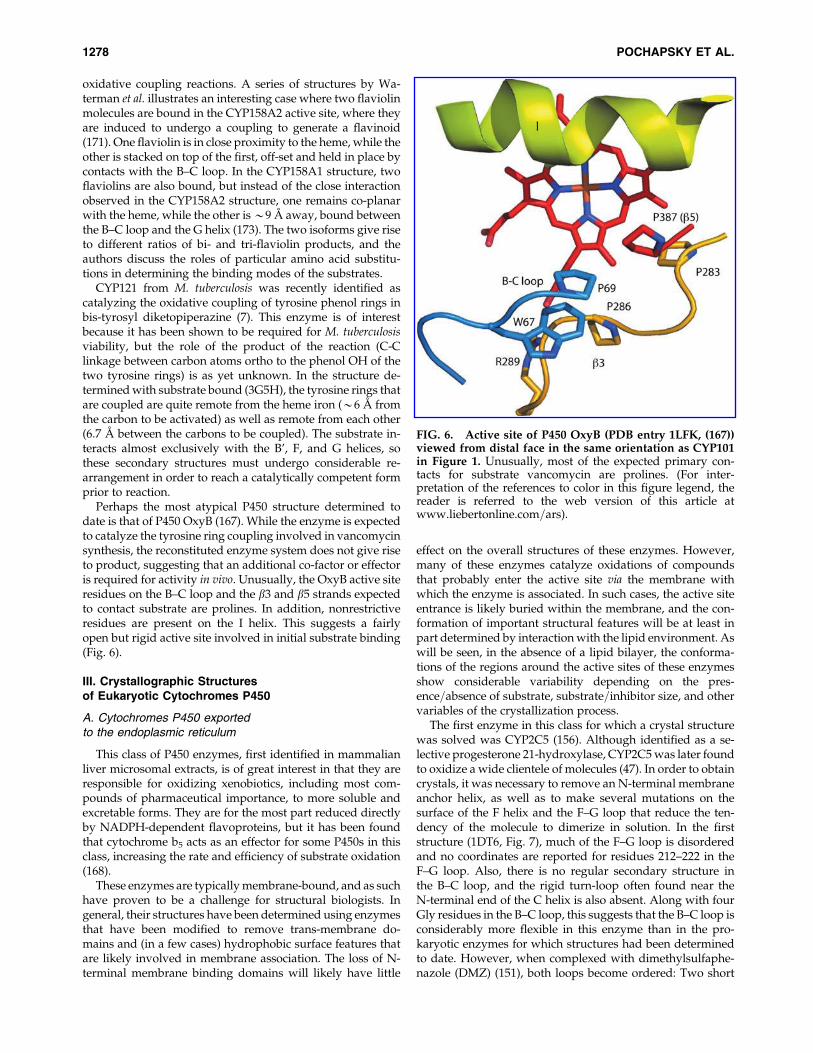
The first enzyme in this class for which a crystal structurewas solved was CYP2C5 (156). Although identified as a se-lective progesterone 21-hydroxylase, CYP2C5 was later foundto oxidize a wide clientele of molecules (47). In order to obtaincrystals, it was necessary to remove an N-terminal membraneanchor helix, as well as to make several mutations on thesurface of the F helix and the F–G loop that reduce the ten-dency of the molecule to dimerize in solution. In the firststructure (1DT6, Fig. 7), much of the F–G loop is disorderedand no coordinates are reported for residues 212–222 in theF–G loop. Also, there is no regular secondary structure inthe B–C loop, and the rigid turn-loop often found near theN-terminal end of the C helix is also absent. Along with fourGly residues in the B–C loop, this suggests that the B–C loop isconsiderably more flexible in this enzyme than in the pro-karyotic enzymes for which structures had been determinedto date. However, when complexed with dimethylsulfaphe-nazole (DMZ) (151), both loops become ordered: Two short
FIG. 6. Active site of P450 OxyB (PDB entry 1LFK, (167))viewed from distal face in the same orientation as CYP101in Figure 1. Unusually, most of the expected primary con-tacts for substrate vancomycin are prolines. (For inter-pretation of the references to color in this figure legend, thereader is referred to the web version of this article atwww.liebertonline.com=ars).
1278 POCHAPSKY ET AL.
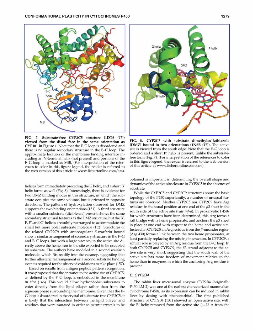
helices form immediately preceding the G helix, and a short B’helix forms as well (Fig. 8). Interestingly, there is evidence fortwo DMZ binding modes in this structure, in which the sub-strate occupies the same volume, but is oriented in oppositedirections. The pattern of hydroxylation observed for DMZsupports the two binding orientations (151). A third structurewith a smaller substrate (diclofenac) present shows the samesecondary structural features as the DMZ structure, but the B’,F, F’, and G’ helices are subtly rearranged to accommodate thesmall but more polar substrate molecule (152). Structures ofthe related CYP2C9 with anticoagulant S-warfarin boundshow a similar arrangement of secondary structure in the F–Gand B–C loops, but with a large vacancy in the active site di-rectly above the heme iron in the site expected to be occupiedby substrate. The authors have modeled in a second warfarinmolecule, which fits readily into the vacancy, suggesting thatfurther allosteric rearrangement or a second substrate bindingevent is required for the observed oxidations to take place (157).
Based on results from antigen peptide pattern recognition,it was proposed that the entrance to the active site of CYP2C5,as defined by the F–G loop, is embedded in the membranein vivo (146). This would allow hydrophobic substrates toenter directly from the lipid bilayer rather than from theaqueous phase surrounding the membrane. Given that the F–G loop is disordered in the crystal of substrate-free CYP2C5, itis likely that the interaction between the lipid bilayer andresidues that were mutated in order to permit crystals to be
obtained is important in determining the overall shape anddynamics of the active site closure in CYP2C5 in the absence ofsubstrate.
While the CYP2C5 and CYP2C9 structures show the basictopology of the P450 superfamily, a number of unusual fea-tures are observed. Neither CYP2C5 nor CYP2C9 have Argresidues in the usual position at one end of the b3 sheet in thesouth side of the active site (vide infra). In prokaryotic P450sfor which structures have been determined, this Arg forms asalt bridge with a heme propionate, and anchors the b3 sheetin place at one end with respect to the heme and active site.Instead, in CYP2C5 an Arg residue from the b-meander region(Arg 430) forms a link between the two heme propionates, atleast partially replacing the missing interaction. In CYP2C9, asimilar role is played by an Arg residue from the B–C loop. Inboth CYP2C5 and CYP2C9, the b3 strand adjacent to the ac-tive site is very short, suggesting that the south wall of theactive site has more freedom of movement relative to theheme than in enzymes in which the anchoring Arg residue ispresent.
B. CYP2B4
The rabbit liver microsomal enzyme CYP2B4 (originallyP450 LM-2) was one of the earliest characterized mammaliancytochrome P450s, as its expression can be induced in rabbitliver by dosing with phenobarbital. The first publishedstructure of CYP2B4 (131) showed an open active site, withthe B’ helix removed from the active site (*22 A from the
FIG. 7. Substrate-free CYP2C5 structure (1DT6 (47))viewed from the distal face in the same orientation asCYP101 in Figure 1. Note that the F–G loop is disordered andthere is no regular secondary structure in the B–C loop. Theapproximate location of the membrane binding interface in-cluding an N-terminal helix (not present) and portions of theF–G loop is marked as MBI. (For interpretation of the refer-ences to color in this figure legend, the reader is referred tothe web version of this article at www.liebertonline.com=ars).
FIG. 8. CYP2C5 with substrate dimethylsulfathiazole(DMZ) bound in two orientations (1N6B (47)). The activesite is viewed from the south edge. Note that the F–G loop isordered and a short B’ helix is present, unlike the substrate-free form (Fig. 7). (For interpretation of the references to colorin this figure legend, the reader is referred to the web versionof this article at www.liebertonline.com=ars).
CONFORMATIONAL PLASTICITY IN CYTOCHROMES P450 1279

midpoint of the helix to the heme Fe) and a distorted helix (F’)serving as the F–G connector. The F’ helix is also remote fromthe heme iron, leaving a very large open active site partiallyoccupied by water molecules. As with CYP2C5, the b3 strandis very short, and the Arg anchor is replaced by a histidine,suggesting that this interaction could be modulated by pH.A structure determined with 4-(4-chlorophenyl)imidazolebound shows a more compact active site, with residues fromthe B–C loop in contact with the bound substrate and the B’helix close-packed (132). Recently, a structure with anotherinhibitor, 1-biphenyl-4-methyl-1H-imidazole, bound, re-sulted in an intimate dimer in which the F’ helices of eachmonomer occupies the active site of the dimer partner, inter-acting with the I helix of the partner, while a portion of theF–F’ loop forms a b-sheet with its counterpart in the othermonomer (28). This indicates that structural features sur-rounding the active site of CYP2B4 are capable of very largedisplacements in response to environment.
C. Liver microsomal enzymes CYP3A4and CYP1A2
The human liver microsomal enzyme CYP3A4 is of par-ticular interest in that it is involved in the metabolism of manyof the approved drugs currently marketed, and inhibition orunintentional overinduction (e.g., by another drug or xeno-biotic) can markedly affect the persistence, effectiveness, andtoxicity of a particular drug (177). The client substrates ofCYP3A4 vary significantly in size, and there is evidence formultiple substrates being bound simultaneously in the largeactive site (111). In one published structure (1TQN, (163))without bound substrate in the active site, the B–C loopcontains only a short distorted helical region, the F helix isconsiderably shorter than is typical, and the F-G loop containstwo helical segments, the F’ and G’ helices (Fig. 9). Both ofthese helices expose hydrophobic residues expected to inter-act with the lipid bilayer of the membrane. The structure de-termined with erythromycin bound (2JOD, (25)) showssurprisingly little change in the overall structure of the activesite: There is more disorder in the F–F’ region, but the samegeneral arrangement of active site features are present. Theerythromycin molecule binds slightly differently in theCYP3A4 active site than in P450 EpoK. The two sugar moie-ties of the substrate, instead of packing near the G helix andF–G loop as in EpoK, interact primarily with the F–F’ loop, theF’ helix, and portions of the A helix. The remainder of thesubstrate molecule interacts with the same structural featuresas seen with many other P450s, including the B–C loop, the Ihelix, and portions of the b3 and b5 sheets. With a smallersubstrate (ketoconazole) bound so that the imidazole ring ofketoconazole provides an axial ligand to the heme iron, thereis some ordering of the B–C loop where it interacts withsubstrate near the N-terminal end of the C helix (PDB entry2V0M). However, despite the differences between erythro-mycin and ketoconazole structures, many of the same resi-dues of CYP3A4 interact with both substrates, including theN-terminal end of the A helix.
The structure of another important drug-metabolizingP450, CYP1A2, has been determined with substrate naph-thoflavone bound (2HI4, (126)). This enzyme, while broadspectrum, appears to target polycyclic substrates withsignificant planar groups (vide infra) (Fig. 10). As with
CYP3A4, the F helix is short and somewhat irregular, whilethe F–G loop contains two short helical segments with ex-posed hydrophobic residues that likely interact with themembrane.
D. Prostaglandin biosynthesis:prostacyclin I2 synthase
The rearrangement of cyclic peroxide PGH2 to prostacyclinI2 is catalyzed by a P450 homologue, CYP8A1. While not aclassical oxygen insertion, it seems likely that there is directinteraction between the peroxide oxygen and the heme, as onestructure with a substrate analog bound has one nitrogen ofthe imide analog of the peroxy oxygen bound to the heme iron(3B99, (68)). There are three published structures of CYP8A1from zebrafish and human. All three structures, including thatwithout substrate analog or inhibitor bound, show a well-defined B’ helix, as well as a short F’ helix connecting the F andG helices. The b5 sheet is poorly defined near the active site,and one b5 strand is interrupted by a short helix. The substrateanalog in 3B99 is contacted by strongly constraining residues(Trp 272 and Val 273 on the I helix, Phe 465 from the b5 loop,and Tyr 97 from the B’ helix). As might be expected, there issome resemblance of the arrangement of structures near theactive site to the structure of CYP102, which also binds fattyacid derivatives; however, this may be coincidental.
FIG. 9. Structure of CYP3A4 determined with inhibitorketoconazole bound (2V0M (25)) viewed from the distalface in the same orientation as CYP101 in Figure 1. Notethat the extended F–G loop with two helical regions F’ andG’. (For interpretation of the references to color in this figurelegend, the reader is referred to the web version of this articleat www.liebertonline.com=ars).
1280 POCHAPSKY ET AL.
E. Steroid biosynthetic and catabolic enzymes
Until recently, there were no structures determined forP450s involved in steroid biosynthesis, even though mem-bers of this class were among the earliest identified andcharacterized in humans. This has changed with the publi-cation of structures for CYP46A1 (cholesterol 24-hydroxylase)(79) and human aromatase (CYP19A1) (29). CYP46A1 wascharacterized both with and without substrate (cholesterolsulfate) bound, and shows increased order in the F–G loopand inward movement of the B’ helix in the substrate-boundform. Aromatase catalyzes the oxidative C-19 demethyla-tion and aromatization of the steroid A ring, convertingandrostene dione to estrone, testosterone to estradiol, andhydroxytestosterone to estratriol. Aromatase inhibitors areprimary therapies for estrogen-dependent breast cancers,making this structure of particular interest for rational drugdesign. The aromatase structure was the first to be determinedusing protein derived from mammalian tissue, as opposed toheterologously expressed enzyme. The enzyme was purifiedfrom human placenta via immunoaffinity chromatography,and presumably contains the intact membrane-binding N-terminal residues, although diffraction was not observed forthe first 45 residues in the enzyme. There is evidence thatCYP19A1 is glycosylated near the N-terminus in vivo, and theglycosylation may act as an anchor on the opposite (lumen)side of the membrane from the protein (29). Three distinctreactions are catalyzed by aromatase, two of which (the oxi-dation of C-19 methyl to primary alcohol to aldehyde) can berationalized by a standard P450 mechanism involving a high-valent Fe-oxo species. To rationalize the final step in the re-action, the authors invoke proton abstraction from the A ringby a Feþ3-bound peroxide intermediate that facilitates loss ofthe 19-C aldehyde via deformylation.
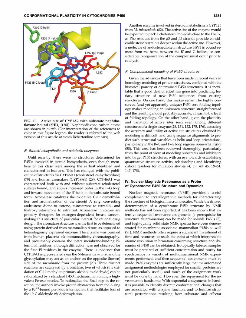
Another enzyme involved in steroid metabolism is CYP125from M. tuberculosis (82). The active site of the enzyme wouldbe expected to pack a cholesterol molecule close to the I helix,as Phe residues from the b3 and b5 strands provide consid-erable steric restraints deeper within the active site. However,a molecule of andostenedione in structure 3IW1 is bound re-mote from the heme between the B’ and G helices, so con-siderable reorganization of the complex must occur prior tocatalysis.
F. Computational modeling of P450 structures
Given the advances that have been made in recent years inhomology modeling of protein structures, combined with thehistorical paucity of determined P450 structures, it is inevi-table that a good deal of effort has gone into predicting ter-tiary structure of new P450 sequences from existingstructures. On one hand, this makes sense: The highly con-served (and yet apparently unique) P450 core folding topol-ogy makes modeling an unknown structure straightforwardand the resulting model probably accurate, at least to the levelof folding topology. On the other hand, given the plasticityand variation of active sites seen even among differentstructures of a single isozyme (28, 131, 132, 175, 176), assessingthe accuracy and utility of active site structures obtained bymodeling is difficult, and using sequence alignments to pre-dict such structural variables as helix and loop orientations,particularly in the B–C and F–G loop regions, somewhat risky(58). This area has been reviewed thoroughly, particularlyfrom the point of view of modeling substrates and inhibitorsinto target P450 structures, with an eye towards establishingquantitative structure–activity relationships and identifyingcritical residues for mutation studies (4, 19, 40, 45, 59–61,147, 178).
IV. Nuclear Magnetic Resonance as a Probeof Cytochrome P450 Structure and Dynamics
Nuclear magnetic resonance (NMR) provides a usefulcomplement to crystallographic methods for characterizingthe structure of biological macromolecules. While the de novodetermination of a cytochrome P450 structure by NMRmethods has not been reported, it has been shown that ex-tensive sequential resonance assignments (a prerequisite forstructure determination) can be made for soluble P450s (5),and high-quality solid state NMR spectra have been demon-strated for membrane-associated mammalian P450s as well(51). NMR methods often require a significant investment oftime and resources to reach the point at which interpretableatomic resolution information concerning structure and dy-namics of P450 can be obtained: Isotopically labeled samplesmust be prepared of sufficient concentration and purity forspectroscopy, a variety of multidimensional NMR experi-ments performed, and then sequential assignments must bemade. P450 enzymes are sufficiently large that the automatedassignment methodologies employed for smaller proteins arenot particularly useful, and much of the assignment workmust be done by hand. However, the repayment for the in-vestment is handsome: With sequential assignments in hand,it is possible to identify discrete conformational changes thatare associated with enzyme function, and to localize struc-tural perturbations resulting from substrate and effector
FIG. 10. Active site of CYP1A2 with substrate naphtho-flavone bound (2HI4, (126)). Naphthoflavone carbon atomsare shown in purple. (For interpretation of the references tocolor in this figure legend, the reader is referred to the webversion of this article at www.liebertonline.com=ars).
CONFORMATIONAL PLASTICITY IN CYTOCHROMES P450 1281

binding and changes in oxidation state (95). Equally impor-tant, protein dynamics on a wide range of time scales becomesaccessible via NMR methods (98).
One of the earliest NMR studies to relate structure andfunction in cytochromes P450 was published by the Robertsgroup (84). Based on the paramagnetically-induced spin re-laxation of water and substrate (lauric acid) in the CYP102active site, this group was able to identify a 6 A relocation ofthe substrate upon reduction of the enzyme, and used thisinformation combined with the published crystallographicstructure of CYP102 to generate models for the binding oflauric acid in oxidized and reduced CYP102. The same grouphas applied this methodology successfully to other P450-substrate complexes as well (83, 120–122). Note that this workdid not require sequential assignments of the protein reso-nances; only the substrate 1H resonances were assigned, andparamagnetic relaxation of specific resonances in substratewere sufficient (in combination with the crystal structure ofCYP102) to model the active site structure. Similar efforts haveused relaxation to examine substrate and ligand-heme inter-actions with other P450s, including CYP1A1, CYP2B1, (85)CYP3A4 (10), CYP2C9 (44), and CYP2B4 (80).
Our group has used NMR to investigate the solution con-formation of the camphor monooxygenase CYP101 as afunction of oxidation state, effector binding, and substrate. Ithas long been known that the Cys4Fe2S2 ferredoxin putidar-edoxin (Pdx) is a required component of the reconstitutedcamphor hydroxylase enzyme system: In the presence of Pdx,reduced O2- and camphor-bound CYP101 rapidly turns overto yield 5-exo-hydroxycamphor (70). In the absence of Pdx, thesame complex slowly decomposes to yield superoxide anionand resting state enzyme. In light of this, the publishedstructures of camphor-bound CYP101 presented an interest-ing puzzle. These structures give an obvious rationale for theobserved regio- and stereochemistry of camphor hydroxyl-ation by CYP101. C5 of camphor, where the hydroxylationoccurs, is the closest substrate carbon to the heme iron, and soalso to the presumed reactive intermediate, the high-valentiron-oxo species Fe(IV)¼O. A time-resolved series of struc-tures in which the hydroxylation reaction was photochemi-cally induced clearly demonstrated the position of thesubstrate with respect to the metal center during the reactionsequence (128). Based on these structures, the lack of turnoverin the absence of Pdx was puzzling. Furthermore, the questionof how the substrate gets into the active site and how productis expelled presented difficulties. The B’ helix and F–G loopprovide effective closure to the CYP101 active site in allCYP101 structures. A series of dynamic simulations of sub-strate expulsion suggested a number of possible paths foractive site ingress=egress, but all of them clearly require sig-nificant displacements of distal structural features in order topermit access to the active site from solvent (71, 72). Now,many of these issues can be resolved by the results frommultidimensional solution NMR experiments. Preliminarytitrations of camphor bound CYP101 with Pdx showed that,despite the proposed Pdx binding site being on the proximalface of the molecule (100), many distal features, including theB’, F, G, and I helices are perturbed upon Pdx addition (99).We proposed these observations to be the result of a Pdx-enforced selection of a closed conformation of the enzyme thatprevents substrate=intermediate loss during the final steps ofoxygen activation and hydrogen abstraction from substrate.
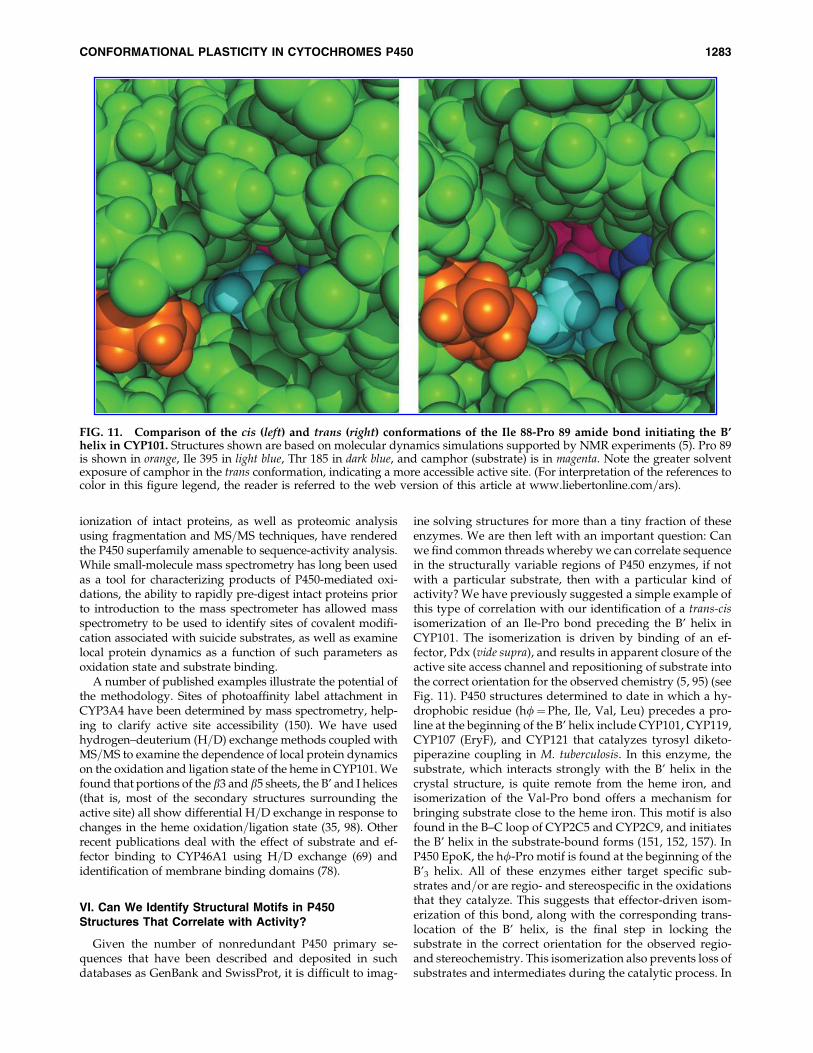
Repeating the titration with perdeuterated CYP101 and Pdx(which renders both proteins invisible to 1H NMR) allowed adetailed characterization of substrate orientation in the activesite, and demonstrated that the orientation of camphor sub-strate in the CYP101 active site differs between solution andthe orientation observed in crystal structures (149). Further-more, a slow conformational change driven by Pdx binding(*180 s�1 at half-saturation) was detected that coincided withsubstrate reorientation, suggesting that a coordinated confor-mational switch occurs upon Pdx binding. Spectroscopic andmutagenic evidence identified the hinge of the conformationalchange as a trans-cis isomerization of the peptide bond betweenIle 88 and Pro 89, at the N-terminus of the B’ helix (95). Thisbond is in the cis conformation in all CYP101 crystal structures,but adopts a trans or distorted trans conformation in solution.Dynamic simulations show a wider gap between Phe 87 andPro 89 on the B–C loop, Thr 185 on the F–G loop, Phe 193 on theG helix, and Ile 395 on the b5 strand at the entrance to the activesite. The trans structure shows greater solvent exposure ofsubstrate, as well as reorientation of substrate relative to thecrystal structure, in agreement with earlier NMR results (5)(Fig. 11). Taken together, these results suggest that upon crys-tallization, CYP101 packs most efficiently in a compact, closed(cis) conformation, but in solution adopts a more open set oftrans conformers that permit relatively easy substrate accessand product egress from the active site. Upon effector binding,the cis conformer predominates, yielding the closed, catalyti-cally competent form of the enzyme.
Other publications describing the use of solution stateNMR methods to characterize P450 enzymes have also ap-peared. Yao et al. have proposed a second camphor bindingsite on CYP101 based on 1H NMR relaxation evidence (164).Jain and co-workers have proposed a model for Pdx-CYP101interactions based on spin label-induced relaxation and re-sidual dipolar couplings (170). Roberts and co-workers usedselective 15N labeling of phenylalanine residues to examinesubstrate binding modes in P450 EryF (120), and Morishima etal. have conducted extensive combined NMR and mutagen-esis experiments to understand the role of Pdx binding indriving conformational changes in CYP101 (89, 142, 143).
As most eukaryotic P450 enzymes are membrane-bound,solution state NMR methods are at present limited in appli-cability to this large and interesting class of targets. However,combined with selective and uniform isotopic labeling, recentdevelopments in solid-state NMR methods promise to makeeukaryotic P450s more accessible (51, 124). Using solid stateNMR, the McDermott group has shown that substrate bind-ing and spin state in CYP102 is temperature dependent,suggesting substrate reorientation as a function of tempera-ture in that enzyme (48, 49). Rienstra and co-workers havedemonstrated that good quality solid-state 13C correlationspectra can be obtained for native CYP3A4 bound to size-constrained phospholipid bilayer structures known as ‘‘na-nodiscs’’ (51).
V. Structural and Dynamic Insightsfrom Mass Spectrometry
Although not precisely a structural tool, mass spectrometry(MS) offers another alternative for examining structural anddynamic relationships in cytochromes P450 that shows con-siderable promise. Recent developments in desorption and
1282 POCHAPSKY ET AL.
ionization of intact proteins, as well as proteomic analysisusing fragmentation and MS=MS techniques, have renderedthe P450 superfamily amenable to sequence-activity analysis.While small-molecule mass spectrometry has long been usedas a tool for characterizing products of P450-mediated oxi-dations, the ability to rapidly pre-digest intact proteins priorto introduction to the mass spectrometer has allowed massspectrometry to be used to identify sites of covalent modifi-cation associated with suicide substrates, as well as examinelocal protein dynamics as a function of such parameters asoxidation state and substrate binding.
A number of published examples illustrate the potential ofthe methodology. Sites of photoaffinity label attachment inCYP3A4 have been determined by mass spectrometry, help-ing to clarify active site accessibility (150). We have usedhydrogen–deuterium (H=D) exchange methods coupled withMS=MS to examine the dependence of local protein dynamicson the oxidation and ligation state of the heme in CYP101. Wefound that portions of the b3 and b5 sheets, the B’ and I helices(that is, most of the secondary structures surrounding theactive site) all show differential H=D exchange in response tochanges in the heme oxidation=ligation state (35, 98). Otherrecent publications deal with the effect of substrate and ef-fector binding to CYP46A1 using H=D exchange (69) andidentification of membrane binding domains (78).
VI. Can We Identify Structural Motifs in P450Structures That Correlate with Activity?
Given the number of nonredundant P450 primary se-quences that have been described and deposited in suchdatabases as GenBank and SwissProt, it is difficult to imag-
ine solving structures for more than a tiny fraction of theseenzymes. We are then left with an important question: Canwe find common threads whereby we can correlate sequencein the structurally variable regions of P450 enzymes, if notwith a particular substrate, then with a particular kind ofactivity? We have previously suggested a simple example ofthis type of correlation with our identification of a trans-cisisomerization of an Ile-Pro bond preceding the B’ helix inCYP101. The isomerization is driven by binding of an ef-fector, Pdx (vide supra), and results in apparent closure of theactive site access channel and repositioning of substrate intothe correct orientation for the observed chemistry (5, 95) (seeFig. 11). P450 structures determined to date in which a hy-drophobic residue (hf¼Phe, Ile, Val, Leu) precedes a pro-line at the beginning of the B’ helix include CYP101, CYP119,CYP107 (EryF), and CYP121 that catalyzes tyrosyl diketo-piperazine coupling in M. tuberculosis. In this enzyme, thesubstrate, which interacts strongly with the B’ helix in thecrystal structure, is quite remote from the heme iron, andisomerization of the Val-Pro bond offers a mechanism forbringing substrate close to the heme iron. This motif is alsofound in the B–C loop of CYP2C5 and CYP2C9, and initiatesthe B’ helix in the substrate-bound forms (151, 152, 157). InP450 EpoK, the hf-Pro motif is found at the beginning of theB’3 helix. All of these enzymes either target specific sub-strates and=or are regio- and stereospecific in the oxidationsthat they catalyze. This suggests that effector-driven isom-erization of this bond, along with the corresponding trans-location of the B’ helix, is the final step in locking thesubstrate in the correct orientation for the observed regio-and stereochemistry. This isomerization also prevents loss ofsubstrates and intermediates during the catalytic process. In
FIG. 11. Comparison of the cis (left) and trans (right) conformations of the Ile 88-Pro 89 amide bond initiating the B’helix in CYP101. Structures shown are based on molecular dynamics simulations supported by NMR experiments (5). Pro 89is shown in orange, Ile 395 in light blue, Thr 185 in dark blue, and camphor (substrate) is in magenta. Note the greater solventexposure of camphor in the trans conformation, indicating a more accessible active site. (For interpretation of the references tocolor in this figure legend, the reader is referred to the web version of this article at www.liebertonline.com=ars).
CONFORMATIONAL PLASTICITY IN CYTOCHROMES P450 1283

turn, this simple structural motif may provide a marker foridentifying P450 enzymes that are specific for particularsubstrate=product combinations. Other enzymes, whilelacking this motif in the B–C loop, have Ile-Pro initiating theF’ helix in the F–G loop. In the structure of lanosterol 14-ademethylase CYP51 (PDB entry 3GW9) from T. brucei, inwhich an inhibitor is bound to the heme iron, the active site isquite exposed to solvent. However, modeling suggests thatisomerization of the Ile 209-Pro 210 amide bond at the N-terminal of the F’ helix from trans to cis would close the activesite in a manner similar to the CYP101 case. A similar ar-rangement of Leu-Pro is seen initiating the F’ helix in alleneoxide synthase from A. thaliana (55, 65), and Phe-Pro ispresent in the F–G loop of PikC (67). There are, however,enzymes that do not have an hf-Pro motif but still targetspecific substrates: CYP108, that oxidizes a-terpineol, is oneexample, so clearly this is not the only means by which ste-reo- and regiospecificity can be enforced.
Regardless of the precise mechanism, it is reasonable toassume, based on comparisons of multiple P450 structures,that the B–C loop, the F and G helices, and F–G loop mustrearrange in order to generate a closed conformation oncesubstrate is bound. This implies that substrate contacts fromthese secondary structural features are less important forinitial substrate recognition than for determining the final‘‘correct’’ orientation of the substrate in the competent enzyme
complex. A similar conclusion was reached by Lepesheva et al.based on a series of mutations made in the B–C and F–Gregions of CYP51, a sterol demethylase found in a wide rangeof organisms (58).
In CYP101, residues Phe 87, Tyr 96, and Phe 98, whichcontact substrate in the closed conformation from the B–C loopand B’ helix, would interact with substrate after binding, whilesubstrate contacts from the I helix (Leu 244 and Val 247) andthe b3 and b5 sheets (Val 295, Ile 395, and Val 396) are morelikely involved in initial substrate binding and recognition. Inlight of this, it is worth considering how the arrangement ofresidues in these features might control initial substrate rec-ognition. In the promiscuous CYP3A4, residues protrudinginto the active site are not bulky or constraining: Close contacts(within 5 A) of ketoconazole in the CYP3A4 active site arealanines on the I helix and at the N-terminal of the b3 sheet thatborders the active site to the south, as viewed in Figures 1 and9. The same two I helix residues are also alanines in the EpoKstructure (Ala 250 and Ala 254), allowing the bulky macrolidesubstrate to pack close to the I helix. However, the side chainsprojecting from the b3 and b5 strands in EpoK are bulky and
FIG. 12. Active site structure of prostacyclin synthase,CYP8A1 (PDB ID 3B99, (68)) with substrate analog 9,11-azoprosta-5(Z),13(E)-dien-1-oic acid bound (labeled U51 infigure). (For interpretation of the references to color in thisfigure legend, the reader is referred to the web version of thisarticle at www.liebertonline.com=ars).
FIG. 13. Anchoring of b3 strand at the south edge of theactive site by a salt bridge between the heme propionateand conserved Arg residues in (a) CYP101 (2CPP, (109)),and (b) P450 epoK (1Q5D, (87)). Substrate contact residuesin register with the conserved Arg are Val 295 in CYP101 andThr 305 in P450 EpoK. Substrates are camphor in CYP101and epothilone B (epoB) in EpoK. See text for complete dis-cussion. In (c) CYP2C5 (1NR6, (156)), the Arg is not presentin the b3 strand, but the salt bridge is conserved via Arg 430from the proximal side of the enzyme. (For interpretation ofthe references to color in this figure legend, the readeris referred to the web version of this article at www.liebertonline.com=ars).
1284 POCHAPSKY ET AL.
restrictive, unlike CYP3A4, reducing the size of the active sitecavity and the freedom of movement of substrate. In CYP1A2,another broad-spectrum xenobiotic metabolizing enzyme,the I helix residues are still nonrestrictive (Gly and Asp), butbulkier side chains project from the b3 and b5 strands tocontact the substrate (a-naphthoflavone) and hold the poly-cyclic planar substrate perpendicular to the heme and parallelto the I helix (Fig. 10). Aromatic residues on the B’, F, and Ghelices also interact with the substrate, and fix its orientationquite precisely (126). A similar arrangement of interactionsinvolving substrate, the I helix and the b3 and b5 strands isfound in CYP2A6 (161). CYP108, which selectively oxidizesa-terpineol, also has a nonrestrictive alanine on the I helix thatlikely contacts substrate, while likely primary contacts on theb3 and N-terminal extension include phenylalanine and va-line, and a phenylalanine on the b5 strand (37). In CYP8A1,which catalyzes the rearrangement of the cyclic peroxidePGH2 to prostacyclin PGI1, close contacts with substrate an-alog are provided by Trp 272 and Val 273 on the the I helix andPhe 463 on the b5 strand (68) (Fig. 12).Again, sterically re-strictive b-branched and aromatic residues provide the initialcontacts for substrate.
If contacts from the b3 sheet are important in the initialbinding and recognition of substrate, this provides anotherpotential correlation between active site structure andfunction in P450s. In most prokaryotic and many eukaryoticP450s, the b3 strand that borders the active site ends with anArg residue that provides a salt bridge to one heme propi-onate group. In CYP101, this is Arg 299. This salt bridgeessentially fixes one end of the b3 sheet with respect to theheme and active site via a flexible hinge. Interestingly, if thisArg residue is present, the other end of the b3 strand oftenhas a residue in register with the Arg side chain that makes
contact with substrate. In CYP101, this is Val 295, the sidechain of which makes direct contact with the camphorgeminal methyl groups (Fig. 13a). In P450 EpoK, a threonine(Thr 305) fills this role, with Arg 307 providing the anchor(Fig. 13b). In the recently published aromatase structure withbound androstenedione, Arg 375 and Val 373 are seen athomologous positions (29). For enzymes with larger sub-strates, the Arg anchor and contact residues from the b3strand are often two residues apart, while in CYP101, with asmall substrate, they are four residues apart. This suggeststhat the larger spacing between the Arg and contact residuemoves the substrate deeper into the active site; that is, thelength of the strand depends upon the size of the substrate,and that this contact is important for determining where thesubstrate sits in the active site relative to activated Fe¼Ospecies. In cases where the spacing is short, (two residuesinstead of four), the N-terminal continuation of the poly-peptide that creates the edge of the b3 sheet often provides aportion of the east wall of the active site. The inward facingresidue of this loop then becomes a key contact for substrateas well.
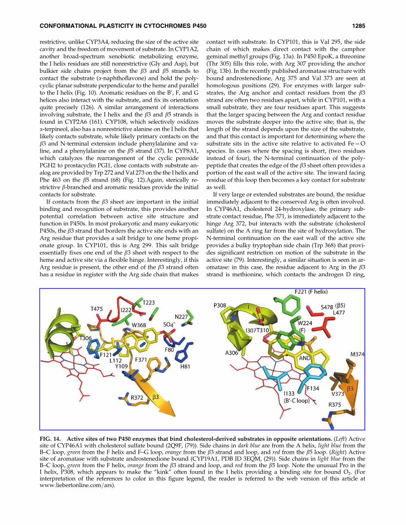
If very large or extended substrates are bound, the residueimmediately adjacent to the conserved Arg is often involved.In CYP46A1, cholesterol 24-hydroxylase, the primary sub-strate contact residue, Phe 371, is immediately adjacent to thehinge Arg 372, but interacts with the substrate (cholesterolsulfate) on the A ring far from the site of hydroxylation. TheN-terminal continuation on the east wall of the active siteprovides a bulky tryptophan side chain (Trp 368) that provi-des significant restriction on motion of the substrate in theactive site (79). Interestingly, a similar situation is seen in ar-omatase: in this case, the residue adjacent to Arg in the b3strand is methionine, which contacts the androgen D ring,
FIG. 14. Active sites of two P450 enzymes that bind cholesterol-derived substrates in opposite orientations. (Left) Activesite of CYP46A1 with cholesterol sulfate bound (2Q9F, (79)). Side chains in dark blue are from the A helix, light blue from theB–C loop, green from the F helix and F–G loop, orange from the b3 strand and loop, and red from the b5 loop. (Right) Activesite of aromatase with substrate androstenedione bound (CYP19A1, PDB ID 3EQM, (29)). Side chains in light blue from theB–C loop, green from the F helix, orange from the b3 strand and loop, and red from the b5 loop. Note the unusual Pro in theI helix, P308, which appears to make the ‘‘kink’’ often found in the I helix providing a binding site for bound O2. (Forinterpretation of the references to color in this figure legend, the reader is referred to the web version of this article atwww.liebertonline.com=ars).
CONFORMATIONAL PLASTICITY IN CYTOCHROMES P450 1285
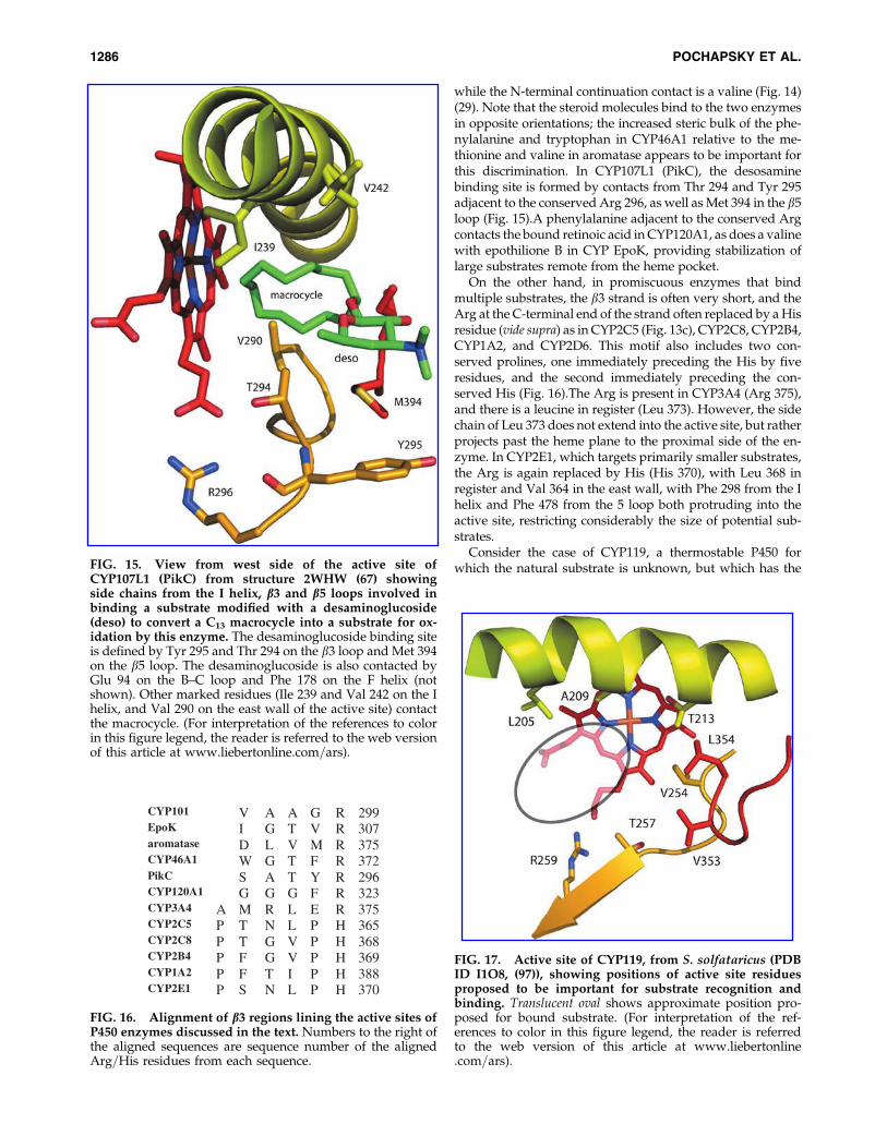
while the N-terminal continuation contact is a valine (Fig. 14)(29). Note that the steroid molecules bind to the two enzymesin opposite orientations; the increased steric bulk of the phe-nylalanine and tryptophan in CYP46A1 relative to the me-thionine and valine in aromatase appears to be important forthis discrimination. In CYP107L1 (PikC), the desosaminebinding site is formed by contacts from Thr 294 and Tyr 295adjacent to the conserved Arg 296, as well as Met 394 in the b5loop (Fig. 15).A phenylalanine adjacent to the conserved Argcontacts the bound retinoic acid in CYP120A1, as does a valinewith epothilione B in CYP EpoK, providing stabilization oflarge substrates remote from the heme pocket.
On the other hand, in promiscuous enzymes that bindmultiple substrates, the b3 strand is often very short, and theArg at the C-terminal end of the strand often replaced by a Hisresidue (vide supra) as in CYP2C5 (Fig. 13c), CYP2C8, CYP2B4,CYP1A2, and CYP2D6. This motif also includes two con-served prolines, one immediately preceding the His by fiveresidues, and the second immediately preceding the con-served His (Fig. 16).The Arg is present in CYP3A4 (Arg 375),and there is a leucine in register (Leu 373). However, the sidechain of Leu 373 does not extend into the active site, but ratherprojects past the heme plane to the proximal side of the en-zyme. In CYP2E1, which targets primarily smaller substrates,the Arg is again replaced by His (His 370), with Leu 368 inregister and Val 364 in the east wall, with Phe 298 from the Ihelix and Phe 478 from the 5 loop both protruding into theactive site, restricting considerably the size of potential sub-strates.
Consider the case of CYP119, a thermostable P450 forwhich the natural substrate is unknown, but which has theFIG. 15. View from west side of the active site of
CYP107L1 (PikC) from structure 2WHW (67) showingside chains from the I helix, b3 and b5 loops involved inbinding a substrate modified with a desaminoglucoside(deso) to convert a C13 macrocycle into a substrate for ox-idation by this enzyme. The desaminoglucoside binding siteis defined by Tyr 295 and Thr 294 on the b3 loop and Met 394on the b5 loop. The desaminoglucoside is also contacted byGlu 94 on the B–C loop and Phe 178 on the F helix (notshown). Other marked residues (Ile 239 and Val 242 on the Ihelix, and Val 290 on the east wall of the active site) contactthe macrocycle. (For interpretation of the references to colorin this figure legend, the reader is referred to the web versionof this article at www.liebertonline.com=ars).
CYP101 V A A G R 299EpoK I G T V R 307aromatase D L V M R 375CYP46A1 W G T F R 372PikC S A T Y R 296CYP120A1 G G G F R 323CYP3A4 A M R L E R 375CYP2C5 P T N L P H 365CYP2C8 P T G V P H 368CYP2B4 P F G V P H 369CYP1A2 P F T I P H 388CYP2E1 P S N L P H 370
FIG. 16. Alignment of b3 regions lining the active sites ofP450 enzymes discussed in the text. Numbers to the right ofthe aligned sequences are sequence number of the alignedArg=His residues from each sequence.
FIG. 17. Active site of CYP119, from S. solfataricus (PDBID I1O8, (97)), showing positions of active site residuesproposed to be important for substrate recognition andbinding. Translucent oval shows approximate position pro-posed for bound substrate. (For interpretation of the ref-erences to color in this figure legend, the reader is referredto the web version of this article at www.liebertonline.com=ars).
1286 POCHAPSKY ET AL.
ability to catalyze the o-1, �2 and �3 hydroxylation of lauricacid (52) and the peroxide-supported oxidation of styrene,phenanthrene, and biphenylene (52). The B’ helix in CYP119 isinitiated by an Ile-Pro motif, suggesting that the enzymecatalyzes a regio- and=or stereoselective oxidation in vivo. Theb3 strand is shorter than in CYP101, with a spacing of tworesidues between the Arg anchor (Arg 259) and Thr 257, theputative substrate contact. This suggests that the substrate isnot bound deeply in the active site, so that oxidation occurs ateither one end of the molecule or at a bend or oblate edge(Fig. 17). The absence of a hydrophobic residue immediatelyadjacent to the conserved Arg indicates that the natural sub-strate is not steroidal nor does it have bulky pendant groupsremote from the site of oxidation. Bulky groups (Leu 205 and
Leu 206) on the I helix as well as valines on the N-terminalloop of the b3 strand and the b5 bend suggest a relativelynarrow binding site, one that might accommodate an aro-matic ring or bent hydrocarbon chain. A phenylalanine fromthe F helix (Phe 153) and Leu 69 from the B’ helix would beexpected to contact substrate after active site closure, furtherrestricting the size of the molecule to be bound. All of theseobservations are consistent with the observed substrate se-lectivity of CYP119. Unfortunately, the context of CYP119 inthe Sulfolobus genome is relatively uninformative concerningthe natural substrate, as most of the neighboring open readingframes are not yet annotated (12).
An arrangement of active site residues similar to those inCYP119 is also found in CYPBioI, which oxidizes fatty acids at
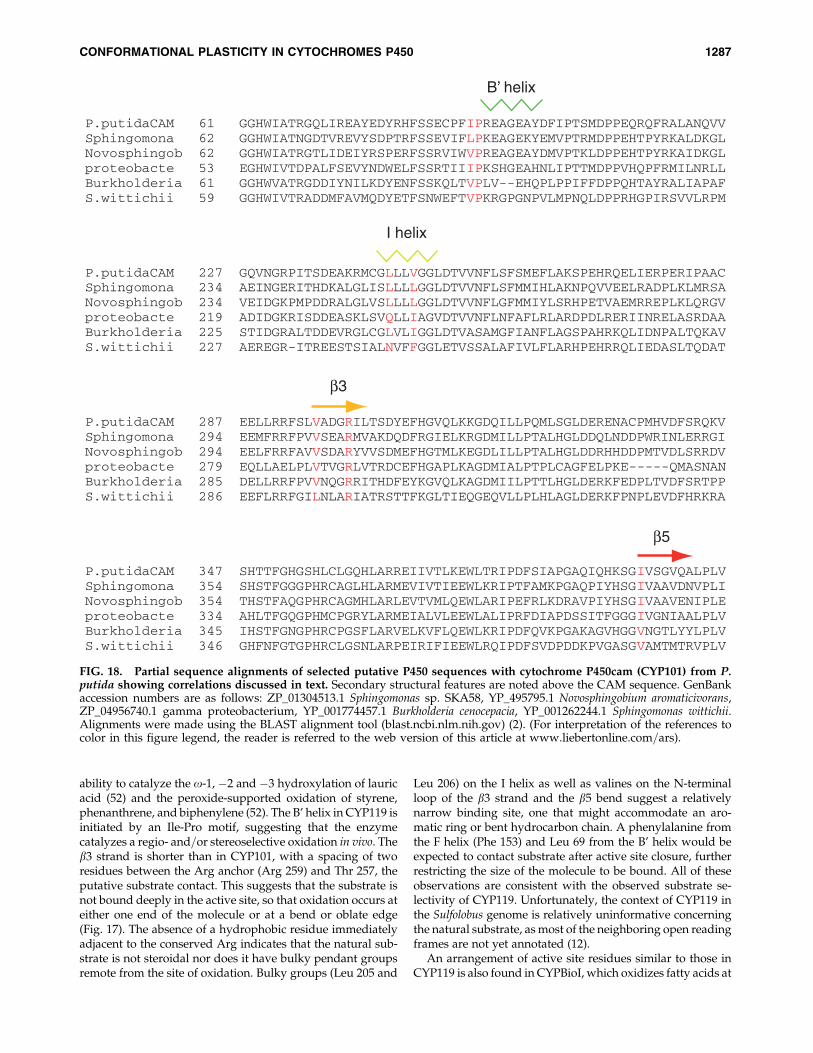
P.putidaCAM 61 GGHWIATRGQLIREAYEDYRHFSSECPFIPREAGEAYDFIPTSMDPPEQRQFRALANQVVSphingomona 62 GGHWIATNGDTVREVYSDPTRFSSEVIFLPKEAGEKYEMVPTRMDPPEHTPYRKALDKGLNovosphingob 62 GGHWIATRGTLIDEIYRSPERFSSRVIWVPREAGEAYDMVPTKLDPPEHTPYRKAIDKGLproteobacte 53 EGHWIVTDPALFSEVYNDWELFSSRTIIIPKSHGEAHNLIPTTMDPPVHQPFRMILNRLLBurkholderia 61 GGHWVATRGDDIYNILKDYENFSSKQLTVPLV--EHQPLPPIFFDPPQHTAYRALIAPAFS.wittichii 59 GGHWIVTRADDMFAVMQDYETFSNWEFTVPKRGPGNPVLMPNQLDPPRHGPIRSVVLRPM
P.putidaCAM 227 GQVNGRPITSDEAKRMCGLLLVGGLDTVVNFLSFSMEFLAKSPEHRQELIERPERIPAACSphingomona 234 AEINGERITHDKALGLISLLLLGGLDTVVNFLSFMMIHLAKNPQVVEELRADPLKLMRSANovosphingob 234 VEIDGKPMPDDRALGLVSLLLLGGLDTVVNFLGFMMIYLSRHPETVAEMRREPLKLQRGVproteobacte 219 ADIDGKRISDDEASKLSVQLLIAGVDTVVNFLNFAFLRLARDPDLRERIINRELASRDAABurkholderia 225 STIDGRALTDDEVRGLCGLVLIGGLDTVASAMGFIANFLAGSPAHRKQLIDNPALTQKAVS.wittichii 227 AEREGR-ITREESTSIALNVFFGGLETVSSALAFIVLFLARHPEHRRQLIEDASLTQDAT
P.putidaCAM 287 EELLRRFSLVADGRILTSDYEFHGVQLKKGDQILLPQMLSGLDERENACPMHVDFSRQKVSphingomona 294 EEMFRRFPVVSEARMVAKDQDFRGIELKRGDMILLPTALHGLDDQLNDDPWRINLERRGINovosphingob 294 EELFRRFAVVSDARYVVSDMEFHGTMLKEGDLILLPTALHGLDDRHHDDPMTVDLSRRDVproteobacte 279 EQLLAELPLVTVGRLVTRDCEFHGAPLKAGDMIALPTPLCAGFELPKE-----QMASNANBurkholderia 285 DELLRRFPVVNQGRRITHDFEYKGVQLKAGDMIILPTTLHGLDERKFEDPLTVDFSRTPPS.wittichii 286 EEFLRRFGILNLARIATRSTTFKGLTIEQGEQVLLPLHLAGLDERKFPNPLEVDFHRKRA
P.putidaCAM 347 SHTTFGHGSHLCLGQHLARREIIVTLKEWLTRIPDFSIAPGAQIQHKSGIVSGVQALPLVSphingomona 354 SHSTFGGGPHRCAGLHLARMEVIVTIEEWLKRIPTFAMKPGAQPIYHSGIVAAVDNVPLINovosphingob 354 THSTFAQGPHRCAGMHLARLEVTVMLQEWLARIPEFRLKDRAVPIYHSGIVAAVENIPLEproteobacte 334 AHLTFGQGPHMCPGRYLARMEIALVLEEWLALIPRFDIAPDSSITFGGGIVGNIAALPLVBurkholderia 345 IHSTFGNGPHRCPGSFLARVELKVFLQEWLKRIPDFQVKPGAKAGVHGGVNGTLYYLPLVS.wittichii 346 GHFNFGTGPHRCLGSNLARPEIRIFIEEWLRQIPDFSVDPDDKPVGASGVAMTMTRVPLV
B’ helix
I helix
b3
b5
FIG. 18. Partial sequence alignments of selected putative P450 sequences with cytochrome P450cam (CYP101) from P.putida showing correlations discussed in text. Secondary structural features are noted above the CAM sequence. GenBankaccession numbers are as follows: ZP_01304513.1 Sphingomonas sp. SKA58, YP_495795.1 Novosphingobium aromaticivorans,ZP_04956740.1 gamma proteobacterium, YP_001774457.1 Burkholderia cenocepacia, YP_001262244.1 Sphingomonas wittichii.Alignments were made using the BLAST alignment tool (blast.ncbi.nlm.nih.gov) (2). (For interpretation of the references tocolor in this figure legend, the reader is referred to the web version of this article at www.liebertonline.com=ars).
CONFORMATIONAL PLASTICITY IN CYTOCHROMES P450 1287

Table 1. Cytochrome P450 Structures Accessible in the RCSB PDB Database
Enzyme Protein Data Base Entries OrganismSubstrate=Product
(if known) References
CYP101 (cam) (2-8)CPP, 3FWF-J, 2QBL-O,2GQX, 2GR6, 2FRZ,2H7Q-S, 2FE6, 2FER,2FEU, 2A1M-O, 1UYU,1T85-8, 1O76, 1LWL, 1K2O,1GJM, 1DZ4, 1DZ8-9,1DZ6, 5CP4, 6CP4, 1AKD,1FAG-H, 1OXA, 1PHA-G,(1-4)CP4
P. putida Camphor=5-exo-hydroxycamphor
1, 22, 23, 26, 75, 76, 88,108-110, 113-118, 128,129, 144, 145
CYP102 (BM3) 3HF2, 3DGI, 3EKB, 3EKD,3EKF, 3BEN, 3CBD, 2UWH,2J1M, 2J4S, 2IJ2-5, 2IJ7,1Z04, 1Z09,1Z0A, 1YQO-P,1SMI-J, 1P0V-X, 1JME,1JPZ, 2BMH, 2HPD
B. megaterium Fatty acid o-2-hydroxylase
14, 30, 31, 33, 34, 39,43, 63, 64, 92, 93,119, 154, 166
CYP105(MoxA)
2Z36 Actinomycetes Broad specificity 165
CYP105A1 3CV8, 3CV9, 2ZBX-Z S. griseolus Vitamin D 38, 141CYP105P1 3E5J-L S. avermitilis Filipin 158CYP107 (EryF) 1Z8O-Q,1JIN-P, 1EUP S. erythreae 6-Deoxyerythronolide B
hydroxylase=Erythromycin D
16–18, 86
CYP107L1(PikC)
2VZ7, 2VZM, 2CD8, 2CA0,2BVJ, 2C6H, 2C7X,2WHW, 2WI9
S. venezuelae Pikromycin,narbomycin
67, 135
CYP108 (terp) 1CPT P. putida a-Terpineol 37CYP113A1
(EryK)2WIO, 2JJN-P, 2VRV, 1EGY S. erythreae Erythromycin D 127
CYP119 1IO7-9, 1F4T-U S. solfataricus Unknown 97, 162CYP120A1 2VE3-4 Synechosystis Retinoic acid 54CYP121 3G5F, 3G5H, 3CXV-Z,
3CY0,3CY1, 1N40, 1N4GM. tuberculosis Tyrosine diketopipera-
zine coupling7, 62, 81, 134
CYP124 2WM4, 2WM5 M. tuberculosis Fatty acido-hydroxylase
Unpublished
CYP125 3IW0-2 M. tuberculosis Cholesterol hydroxylase 82CYP130 2WH8, 2WHF,2WGY,
2UUQ, 2UVNM. tuberculosis Ketoconazole inhibited 94
CYP154A1 1ODO S. coelicolor Unknown 101CYP154C1 1GWI S. coelicolor Macrolide 102CYP158A1 2NZ5, 2NZA, 2DKK S. coelicolor Flaviolin coupling 173CYP158A2 2D09, 2D0E, 1SE6, 1T93,
1S1FS. coelicolor Flaviolin coupling 172
CYP170A1 3DBG S. coelicolor Albaflavinone 174CYP175A1 1N97 T. thermophilus 159CYP199A2 2FR7 Rhodopseudomonas p-Subst. benzoic acid 8CYP231A2 2RFB-C Picrophilus (archeon) 41CYP245A1
(StaP)3A1L, 2Z3T-U Streptomyces Chromopyrrolic acid 74, 148
CYP46A1 2Q9F, 2Q9G H. sapiens Cholesterol24-hydroxylase
79
CYP51 2WO9, 2WOA, 2WOB.1X8V, 1U13, 1EA1, 1E9X
M. tuberculosis Sterol demethylase 11
CYP51 3G1Q, 2VKU, 2CI0, 2CIB,2BZ9, 1H5Z
M. tuberculosis Sterol 14-a demethylase 24, 103, 104
CYP51 3G1Q, 3GW9 T. brucei Sterol 14-a demethylase 57CYP51 3I3K H. sapiens Lanosterol 14-a
demethylase139
CYP55A1(P450nor)
1XQD, 1ULW, 1JFB-C,1GEJ-K, 1GEI, 1GEM,1GED. 1CL6, 1CMJ,1ROM,2ROM,1CMN
F. oxysporum Nitric oxide reductase 53, 96, 136, 137
(continued)
1288 POCHAPSKY ET AL.

a bend in the chain in order to generate precursors for biotinsynthesis in B. subtilis (15). While CYPBioI does not containthe hf-Pro motif at the N-terminal of the B’ helix, the enzymewas crystallized with acyl carrier protein (ACP) bound so asto deliver the bound fatty acid to the active site. The ACP isbound adjacent to the F–G loop and B helix, and is appro-priately positioned to force closure of the active site.
In Figure 18, residues involved in the proposed correlationsare highlighted in a series of P450 sequences that align withCYP101, but are otherwise uncharacterized. Based on theclose alignment, it is expected that, like CYP101, these en-zymes catalyze the oxidation of small terpenoids. (CYP101catalyzes the 5-exo hydroxylation of camphor, a small bicyclicterpene). In all of these sequences, the hj-Pro motif is presentat the likely N-terminal of the B’ helix, suggesting regio- andstereospecificity in the chemistry catalyzed. On the I helix,bulky residues are present corresponding to Leu 244 and Val247 in CYP101, although the Gln and Asn residues in Proteo-bacteria and S. wittichii aligned with Leu 244 in the CYP101sequence may well be removed from the active site. All of thesequences have bulky hydrophobic residues aligned with Val
295 on the b3 strand in CYP101, a primary contact for boundcamphor, indicating binding of substrate deep in the activesite, these residues being four away from the conserved Arg(Arg 299 in CYP101). Finally, all five sequences have a b-branched hydrophobic side chain (either Val or Leu) alignedwith Ile 395, which provides the upper east wall of the activesite in CYP101. Of the five sequences, the S. wittichii enzymelikely has the least-branched substrate, as the Asn-Phe com-bination on the I helix could leave an opening adjacent to the Ihelix to accommodate parts of substrate remote from the siteof activity that is not present in the other sequences, while thelarger but less constrained Leu side chain on the b3 strandhomologous with Val 295 would discourage bulkier groupsfrom binding deep in the active site.
VII. Conclusions
As the number of available P450 structures grows, alongwith improved dynamic information from NMR and MSmethods, it becomes increasingly important to be able to in-terpret this sea of information without getting lost in it. In
Table 1. (Continued)
Enzyme Protein Data Base Entries OrganismSubstrate=Product
(if known) References
CYP74A 2RCH, 2RCL, 2RCM, 3CLI,3DSI-K
A. thaliana Allene oxide synthase 55, 65
CYP74A2 3DAN,3DBM Rubber plant Allene oxide synthase 65CYP1A2 2HI4 H. sapiens Multiple substs. 126CYP19A1 3I3K H. sapiens Androgen (aromatase) 29CYP2A6 3EBS, 2PG5-7, 2FDU-W,
2FDY, 1Z10-1H. sapiens Multiple substs. 21, 160, 161
CYP2A13 2P85 H. sapiens Multiple substs. 138CYP2B4 3G5N, 3G93, 2Q6N, 2BDM,
1SUO, 1PO5Rabbit Multiple substs. 28, 131, 175, 176
CYP2C5 1NR6, 1N6B, 1DT6 Rabbit Multiple substs. 47, 151, 152, 155CYP2C8 2VN0, 2NNH-J, 1PQ2 H. sapiens Multiple substs. 130CYP2C9 1R90, 1OG2, 1OG5, H. sapiens Multiple substs. 153CYP2D6 2F9Q H. sapiens Multiple substs. 123CYP2E1 3E6I, 3E4E H. sapiens Multiple substs. 105CYP2R1 3CZH, 3DL9, 3C6G H. sapiens Vitamin D 140CYP3A4 2V0M, 2J0D, 1TQN,
1W0E-GH. sapiens Multiple substs. 25
CYP7A1 3DAX H. sapiens Cholesterol 7-a-hydroxylase
Unpublished
CYP8A1 3B6H, 2IAG H. sapiens Prostaglandin I2(prostacyclinsynthase)
68
CYP8A1 3B98, 3B99 Zebrafish Prostaglandin I2(prostacyclinsynthase)
68
XplA 2WIV, 2WIY Rhodococcus Reductive degradationof RDX
125
P450 EpoK 1Q5D-E Sorangiumcellulosum
Epothilone C=A 87
P450 OxyB 1LFK, 1LG9, 1LGF Amycolatopsis Vancomycinbiosynthesis
167
P450 OxyC 1UED Amycolatopsis Vancomycinbiosynthesis
112
P450 BSb 1IZO B. subtilis Fatty acid oxidation 56CYPBioI 3EJD, 3EJE3 E. coli Biotin synthesis 15Thermophilic P450 1UE8 S. tokodaii Unknown 90
CONFORMATIONAL PLASTICITY IN CYTOCHROMES P450 1289

particular, as ‘‘personal genomes’’ become a tool for diag-nostics and prescription, the ability to quickly interpret se-quence data in terms of expected P450 activity will be criticalfor assessing the risks and benefits of a particular medicationor chemotherapy for an individual patient (6) (Table 1).
In this review, we have attempted to make use of structuraland dynamic information obtained by a variety of experi-mental methods to make some generalizations about how cy-tochrome P450 enzymes recognize and bind their substrates. Inparticular, we asked what common features could be used tolimit the substrate search space for orphan P450s and toidentify the isozyme most likely to metabolize a given smallmolecule substrate such as a drug or environmental xenobiotic.We conclude, based on comparisons of known P450 structuresand dynamic information gleaned from NMR and massspectrometry, that features of the I helix, b3 and b5 strandsadjacent to the active site (with occasional rarer contributionsfrom the A helix for large substrates) are critical for deter-mining which types of substrates will bind. On the other hand,features of the B–C loop, including the B’ helix, and F–G loopare important for determining the final orientation of thebound substrate in the active site, and must reorganize prior tothe enzyme reaching a catalytically competent conformation,but are less important for initial substrate binding.
Acknowledgments
This work was supported in part by a grant from theUSPHS, R01-GM44191 (TCP). The authors thank ProfessorSusan Sondej Pochapsky for her critical reading of the man-uscript, and all of the reviewers for helpful suggestions.
References
1. Aldag C, Gromov IA, Garcia–Rubio I, von Koenig K,Schlichting I, Jaun B, and Hilvert D. Probing the role of theproximal heme ligand in cytochrome P450cam by recom-binant incorporation of selenocysteine. Proc Natl Acad SciUSA 106: 5481–5486, 2009.
2. Altschul SF, Madden TL, Schaffer AA, Zhang JH, Zhang Z,Miller W, and Lipman DJ. Gapped BLAST and PSI-BLAST:A new generation of protein database search programs.Nucleic Acids Res 25: 3389–3402, 1997.
3. Andersen JF, Tatsuta K, Gunji H, Ishiyama T, and Hutch-inson CR. Substrate-apecificity of 6-Deoxyerythronolide-Bhydroxylase, a bacterial cytochrome P450 of erythromycin-a biosynthesis. Biochemistry 32: 1905–1913, 1993.
4. Arimoto R. Computational models for predicting interac-tions with cytochrome P450 enzyme. Current Topics MedChem 6: 1609–1618, 2006.
5. Asciutto EK, Madura JD, Pochapsky SS, OuYang B, andPochapsky TC. Structural and dynamic implications of aneffector-induced backbone amide cis-trans isomerization incytochrome P450(cam). J Mol Biol 388: 801–814, 2009.
6. Bai JPF. Ongoing challenges in drug interaction safety:From exposure to pharmacogenomics. Drug Metab Phar-macokin 25: 62–71, 2010.
7. Belin P, Le Du MH, Fielding A, Lequin O, Jacquet M,Charbonnier JB, Lecoq A, Thai R, Courcon M, Masson C,Dugave C, Genet R, Pernodet JL, and Gondry M. Identifi-cation and structural basis of the reaction catalyzed byCYP121, an essential cytochrome P450 in Mycobacteriumtuberculosis. Proc Natl Acad Sci USA 106: 7426–7431, 2009.
8. Bell SG, Xu F, Forward I, Bartlam M, Rao Z, and Wong LL.Crystal structure of CYP199A2, a para-substituted benzoicacid oxidizing cytochrome P450 from Rhodopseudomonaspalustris. J Mol Biol 383: 561–574, 2008.
9. Blake CCF, Koenig DF, Mair GA, North ACT, Phillips DC,and Sarma VR. Structure of hen egg-white lysozyme: A 3-dimensional fourier Ssynthesis at 2 A resolution. Nature206: 757, 1965.
10. Cameron MD, Wen B, Allen KE, Roberts AG, Schuman JT,Campbell AP, Kunze KL, and Nelson SD. Cooperativebinding of midazolam with testosterone and alpha-naph-thoflavone within the CYP3A4 active site: A NMR T-1paramagnetic relaxation study. Biochemistry 44: 14143–14151, 2005.
11. Chen CK, Doyle PS, Yermalitskaya LV, Mackey ZB, AngKK, McKerrow JH, and Podust LM. Trypanosoma cruziCYP51 inhibitor derived from a Mycobacterium tuberculosisscreen hit. PLoS Negl Trop Dis 3: e372, 2009.
12. Chen LM, Brugger K, Skovgaard M, Redder P, She QX,Torarinsson E, Greve B, Awayez M, Zibat A, Klenk HP,and Garrett RA. The genome of Sulfolobus acidocaldarius, amodel organism of the Crenarchaeota. J Bacteriol 187: 4992–4999, 2005.
13. Chen S and Zhou DJ. Functional domains of aromatasecytochrome-P450 inferred from comparative analyses ofamino acid sequences and substantiated by site-directedmutagenesis Eexperiments. J Biol Chem 267: 22587–22594,1992.
14. Clark JP, Miles CS, Mowat CG, Walkinshaw MD, Reid GA,Daff SN, and Chapman SK. The role of Thr268 and Phe393in cytochrome P450 BM3. J Inorg Biochem 100: 1075–1090,2006.
15. Cryle MJ and Schlichting I. Structural insights from a P450Carrier Protein complex reveal how specificity is achievedin the P450(BioI) ACP complex. Proc Natl Acad Sci USA 105:15696–15701, 2008.
16. Cupp–Vickery J, Anderson R, and Hatziris Z. Crystalstructures of ligand complexes of P450eryF exhibitinghomotropic cooperativity. Proc Natl Acad Sci USA 97: 3050–3055, 2000.
17. Cupp–Vickery JR, Garcia C, Hofacre A, and McGee–Estrada K. Ketoconazole-induced conformational changesin the active site of cytochrome P450eryF. J Mol Biol 311:101–110, 2001.
18. Cupp–Vickery JR and Poulos TL. Structure of cytochromeP450eryF involved in erythromycin biosynthesis. Nat StructBiol 2: 144–153, 1995.
19. de Graaf C, Oostenbrink C, Keizers PHJ, van Vugt-Lussenburg BMA, van Waterschoot RAB, Tschirret-GuthRA, Commandeur JNM, and Vermeulen NPE. Molecularmodeling-guided site-directed mutagenesis of cytochromeP450 2D6. Current Drug Metabolism 8: 59–77, 2007.
20. DeLano WL. The PyMOL Molecular Graphics System. 2008.21. DeVore NM, Smith BD, Urban MJ, and Scott EE. Key
residues controlling phenacetin metabolism by humancytochrome P450 2A enzymes. Drug Metab Dispos 36: 2582–2590, 2008.
22. Dmochowski IJ, Crane BR, Wilker JJ, Winkler JR, and GrayHB. Optical detection of cytochrome P450 by sensitizer-linkedsubstrates. Proc Natl Acad Sci USA 96: 12987–12990, 1999.
23. Dunn AR, Dmochowski IJ, Bilwes AM, Gray HB, andCrane BR. Probing the open state of cytochrome P450camwith ruthenium-linker substrates. Proc Natl Acad Sci USA98: 12420–12425, 2001.
1290 POCHAPSKY ET AL.

24. Eddine AN, von Kries JP, Podust MV, Warrier T, Kauf-mann SH, and Podust LM. X-ray structure of 4,4’-dihy-droxybenzophenone mimicking sterol substrate in theactive site of sterol 14alpha-demethylase (CYP51). J BiolChem 283: 15152–15159, 2008.
25. Ekroos M and Sjogren T. Structural basis for ligand pro-miscuity in cytochrome P450 3A4. Proc Natl Acad Sci USA103: 13682–13687, 2006.
26. Fedorov R, Ghosh DK, and Schlichting I. Crystal structuresof cyanide complexes of P450cam and the oxygenase do-main of inducible nitric oxide synthase-structural models ofthe short-lived oxygen complexes. Arch Biochem Biophys409: 25–31, 2003.
27. Fruetel JA, Mackman RL, Peterson JA, and Ortiz de Mon-tellano PR. Relationship of active site topology to substratespecificity for cytochrome P450terp (CYP108). J Biol Chem269: 28815–28821, 1994.
28. Gay SC, Sun L, Maekawa K, Halpert JR, and Stout CD.Crystal structures of cytochrome P450 2B4 in complex withthe inhibitor 1-biphenyl-4-methyl-1H-imidazole: Ligand-induced structural response through alpha-helical re-positioning. Biochemistry 48: 4762–4771, 2009.
29. Ghosh D, Griswold J, Erman M, and Pangborn W. Struc-tural basis for androgen specificity and oestrogen synthesisin human aromatase. Nature 457: 219–U119, 2009.
30. Girvan HM, Seward HE, Toogood HS, Cheesman MR, LeysD, and Munro AW. Structural and spectroscopic charac-terization of P450 BM3 mutants with unprecedented P450heme iron ligand sets. New heme ligation states influenceconformational equilibria in P450 BM3. J Biol Chem 282:564–572, 2007.
31. Girvan HM, Toogood HS, Littleford RE, Seward HE, SmithWE, Ekanem IS, Leys D, Cheesman MR, and Munro AW.Novel haem co-ordination variants of flavocytochromeP450BM3. Biochem J 417: 65–76, 2009.
32. Gotoh O. Substrate recognition sites in cytochrome-P450family-2 (CYP2) proteins inferred from comparative ana-lyses of amino acid and coding nucleotide sequences. J BiolChem 267: 83–90, 1992.
33. Haines DC, Chen B, Tomchick DR, Bondlela M, Hegde A,Machius M, and Peterson JA. Crystal structure of inhibitor-bound P450BM-3 reveals open conformation of substrateaccess channel. Biochemistry 47: 3662–3670, 2008.
34. Haines DC, Tomchick DR, Machius M, and Peterson JA.Pivotal role of water in the mechanism of P450BM-3. Bio-chemistry 40: 13456–13465, 2001.
35. Hamuro Y, Molnar KS, Coales SJ, OuYang B, SimorellisAK, and Pochapsky TC. Hydrogen-deuterium exchangemass spectrometry for investigation of backbone dynamicsof oxidized and reduced cytochrome P450(cam). J InorgBiochem 102: 364–370, 2008.
36. Hannemann F, Bichet A, Ewen KM, and Bernhardt R. Cy-tochrome P450 systems. Biological variations of electrontransport chains. Biochim Biophys Acta-Gen Subj 1770: 330–344, 2007.
37. Hasemann CA, Ravichandran KG, Peterson JA, andDeisenhofer J. Crystal structure and refinement of cyto-chrome P450terp at 2.3 A resolution. J Mol Biol 236: 1169–1185, 1994.
38. Hayashi K, Sugimoto H, Shinkyo R, Yamada M, Ikeda S,Ikushiro S, Kamakura M, Shiro Y, and Sakaki T. Structure-based design of a highly active vitamin D hydroxylase fromStreptomyces griseolus CYP105A1. Biochemistry 47: 11964–11972, 2008.
39. Hegde A, Haines DC, Bondlela M, Chen B, Schaffer N,Tomchick DR, Machius M, Nguyen H, Chowdhary PK,Stewart L, Lopez C, and Peterson JA. Interactions of sub-strates at the surface of P450s can greatly enhance substratepotency. Biochemistry 46: 14010–14017, 2007.
40. Hlavica P. Functional interaction of nitrogenous organicbases with cytochrome P450: A critical assessment andupdate of substrate features and predicted key active-siteelements steering the access, binding, and orientation ofamines. Biochim Biophys Acta-Proteins Proteomics 1764: 645–670, 2006.
41. Ho WW, Li H, Nishida CR, de Montellano PR, and PoulosTL. Crystal structure and properties of CYP231A2 from thethermoacidophilic archaeon Picrophilus torridus. Biochem-istry 47: 2071–2079, 2008.
42. Hsu MH, Griffin KJ, Wang Y, Kemper B, and Johnson EF.A single amino acid substitution confers progesterone 6-beta-hydroxylase activity to rabbit cytochrome P450 2C3.J Biol Chem 268: 6939–6944, 1993.
43. Huang WC, Westlake AC, Marechal JD, Joyce MG, MoodyPC, and Roberts GC. Filling a hole in cytochrome P450 BM3improves substrate binding and catalytic efficiency. J MolBiol 373: 633–651, 2007.
44. Hummel MA, Gannett PM, Aguilar J, and Tracy TS. Sub-strate proton to heme distances in CYP2C9 allelic variantsand alterations by the heterotropic activator, dapsone. ArchBiochem Biophys 475: 175–183, 2008.
45. Hutter MC. In silico prediction of drug properties. CurrMed Chem16: 189–202, 2009.
46. Iwasaki M, Darden TA, Pedersen LG, Davis DG, JuvonenRO, Sueyoshi T, and Negishi M. Engineering mouseP450coh to a novel corticosterone 15-alpha-hydroxylaseand modeling steroid-binding orientation in the substratepocket. J Biol Chem 268: 759–762, 1993.
47. Johnson EF, Wester MR, and Stout CD. The structure ofmicrosomal cytochrome P450 2C5: A steroid and drugmetabolizing enzyme. Endocrine Res 28: 435–441, 2002.
48. Jovanovic T, Harris M, and McDermott AE. CytochromeP450BM-3 in complex with its substrate: Temperature-dependent spin state equilibria in the oxidized and reducedstates. Applied Magnetic Resonance 31: 411–429, 2007.
49. Jovanovic T and McDermott AE. Observation of ligandbinding to cytochrome P450 BM-3 by means of solid-stateNMR spectroscopy. J Am Chem Soc 127: 13816–13821, 2005.
50. Kendrew JC, Bodo G, Dintzis HM, Parrish RG, Wyckoff H,and Phillips DC. 3-dimensional model of the myoglobinmolecule obtained by X-ray analysis. Nature 181: 662–666,1958.
51. Kijac AZ, Li Y, Sligar SG, and Rienstra CM. Magic-anglespinning solid-state NMR Spectroscopy of nanodisc-embedded human CYP3A4. Biochemistry 46: 13696–13703,2007.
52. Koo LS, Immoos CE, Cohen MS, Farmer PJ, and de Mon-tellano PRO. Enhanced electron transfer and lauric acidhydroxylation by site-directed mutagenesis of CYP119.J Am Chem Soc 124: 5684–5691, 2002.
53. Kudo T, Takaya N, Park SY, Shiro Y, and Shoun H. Apositively charged cluster formed in the heme-distal pocketof cytochrome P450nor is essential for interaction withNADH. J Biol Chem 276: 5020–5026, 2001.
54. Kuhnel K, Ke N, Cryle MJ, Sligar SG, Schuler MA, andSchlichting I. Crystal structures of substrate-free andretinoic acid-bound cyanobacterial cytochrome P450CYP120A1. Biochemistry 31: 31, 2008.
CONFORMATIONAL PLASTICITY IN CYTOCHROMES P450 1291
55. Lee DS, Nioche P, Hamberg M, and Raman CS. Structuralinsights into the evolutionary paths of oxylipin biosyntheticenzymes. Nature 455: 363–368, 2008.
56. Lee DS, Yamada A, Sugimoto H, Matsunaga I, Ogura H,Ichihara K, Adachi S, Park SY, and Shiro Y. Substraterecognition and molecular mechanism of fatty acid hy-droxylation by cytochrome P450 from Bacillus subtilis.Crystallographic, spectroscopic, and mutational studies.J Biol Chem 278: 9761–9767, 2003.
57. Lepesheva GI, Park HW, Hargrove TY, Vanhollebeke B,Wawrzak Z, Chaudhuri M, Villalta F, and WatermanMR. Crystal structures of Trypanosoma brucei sterol 14alpha-demethylase and implications for selective treatmentof human infections J Biol Chem 285: 1773–1780, 2010.
58. Lepesheva GI, Virus C, and Waterman MR. Conservationin the CYP51 family. Role of the B ’ helix=BC loop andhelices F and G in enzymatic function. Biochemistry 42:9091–9101, 2003.
59. Lewis DFV and Ito Y. Human P450s involved in drugmetabolism and the use of structural modelling for un-derstanding substrate selectivity and binding affinity. Xe-nobiotica 39: 625–635, 2009.
60. Lewis DFV, Ito Y, and Goldfarb PS. Investigating humanP450s involved in drug metabolism via homology withhigh-resolution P450 crystal structures of the CYP2C sub-family. Curr Drug Metab 7: 589–598, 2006.
61. Lewis DFV, Ito Y, and Goldfarb PS. Structural modelling ofthe human drug-metabolizing cytochromes P450. Curr MedChem 13: 2645–2652, 2006.
62. Leys D, Mowat CG, McLean KJ, Richmond A, ChapmanSK, Walkinshaw MD, and Munro AW. Atomic structure ofMycobacterium tuberculosis CYP121 to 1.06 A reveals novelfeatures of cytochrome P450. J Biol Chem 278: 5141–5147,2003.
63. Li H and Poulos TL. Modeling protein-substrate interac-tions in the heme domain of cytochrome P450(BM-3). ActaCrystallogr D Biol Crystallogr 51: 21–32, 1995.
64. Li H and Poulos TL. The structure of the cytochromeP450BM-3 haem domain complexed with the fatty acidsubstrate, palmitoleic acid. Nat Struct Biol 4: 140–146, 1997.
65. Li L, Chang Z, Pan Z, Fu ZQ, and Wang X. Modes of hemebinding and substrate access for cytochrome P450 CYP74Arevealed by crystal structures of allene oxide synthase. ProcNatl Acad Sci USA 105: 13883–13888, 2008.
66. Li S, Ouellet H, Sherman DH, and Podust LM. Analysis oftransient and catalytic desosamine-binding pockets in cy-tochrome P450 PikC from Streptomyces venezuelae. J BiolChem 284: 5723–5730, 2009.
67. Li SY, Chaulagain MR, Knauff AR, Podust LM,Montgomery J, and Sherman DH. Selective oxidation ofcarbolide C-H bonds by an engineered macrolide P450mono-oxygenase. Proc Natl Acad Sci USA 106: 18469–18474,2009.
68. Li YC, Chiang CW, Yeh HC, Hsu PY, Whitby FG, WangLH, and Chan NL. Structures of prostacyclin synthase andits complexes with substrate analog and inhibitor reveal aligand-specific heme conformation change. J Biol Chem 283:2917–2926, 2008.
69. Liao WL, Dodder NG, Mast N, Pikuleva IA, and Turko IV.Steroid and protein ligand binding to cytochrome P45046A1 as assessed by hydrogen-deuterium exchange andmass spectrometry. Biochemistry 48: 4150–4158, 2009.
70. Lipscomb JD, Sligar SG, Namtvedt MJ, and Gunsalus IC.Autooxidation and Hhydroxylation reactions of oxygen-
ated cytochrome P450cam. J Biol Chem 251: 1116–1124,1976.
71. Ludemann SK, Lounnas V, and Wade RC. How do sub-strates enter and products exit the buried active site ofcytochrome P450cam? 1. Random expulsion moleculardynamics investigation of ligand access channels andmechanisms. J Mol Biol 303: 797–811, 2000.
72. Ludemann SK, Lounnas V, and Wade RC. How do sub-strates enter and products exit the buried active site ofcytochrome P450cam? 2. Steered molecular dynamics andadiabatic mapping of substrate pathways. J Mol Biol 303:813–830, 2000.
73. Lyons TA, Ratnaswamy G, and Pochapsky TC. Redox-dependent dynamics of putidaredoxin characterized byamide proton exchange. Protein Science 5: 627–639, 1996.
74. Makino M, Sugimoto H, Shiro Y, Asamizu S, Onaka H, andNagano S. Crystal structures and catalytic mechanism ofcytochrome P450 StaP that produces the indolocarbazoleskeleton. Proc Natl Acad Sci USA 104: 11591–11596, 2007.
75. Makris TM, von Koenig K, Schlichting I, and Sligar SG.Alteration of P450 distal pocket solvent leads to impairedproton delivery and changes in heme geometry. Biochem-istry 46: 14129–14140, 2007.
76. Makris TM, von Koenig K, Schlichting I, and Sligar SG. Thestatus of high-valent metal oxo complexes in the P450 cy-tochromes. J Inorg Biochem 100: 507–518, 2006.
77. Mansuy D. Biocatalysis and substrate chemodiversity:Adaptation of aerobic living organisms to their chemicalenvironment. Catalysis Today 138: 2–8, 2008.
78. Mast N, Liao WL, Pikuleva IA, and Turko IV. Combineduse of mass spectrometry and heterologous expression foridentification of membrane-interacting peptides in cyto-chrome P450 46A1 and NADPH-cytochrome P450 oxido-reductase. Arch Biochem Biophys 483: 81–89, 2009.
79. Mast N, White MA, Bjorkhem I, Johnson EF, Stout CD, andPikuleva IA. Crystal structures of substrate-bound andsubstrate-free cytochrome P450 46A1, the principal cho-lesterol hydroxylase in the brain. Proc Natl Acad Sci USA105: 9546–9551, 2008.
80. McCullough CR, Pullela PK, Im SC, Waskell L, and SemDS. C-13-methyl isocyanide as an NMR probe for cyto-chrome P450 active sites. J Biomol NMR 43: 171–178, 2009.
81. McLean KJ, Carroll P, Lewis DG, Dunford AJ, Seward HE,Neeli R, Cheesman MR, Marsollier L, Douglas P, SmithWE, Rosenkrands I, Cole ST, Leys D, Parish T, and MunroAW. Characterization of active site structure in CYP121. Acytochrome P450 essential for viability of Mycobacteriumtuberculosis H37Rv. J Biol Chem 283: 33406–33416, 2008.
82. McLean KJ, Lafite P, Levy C, Cheesman MR, Mast N,Pikuleva IA, Leys D, and Munro AW. The structure ofMycobacterium tuberculosis CYP125: Molecular basis forcholesterol binding in a P450 needed for host infection.J Biol Chem 284: 35524–35533, 2009.
83. Modi S, Paine MJ, Sutcliffe MJ, Lian LY, Primrose WU,Wolf CR, and Roberts GCK. A model for human cytochromeP450 2D6 based on homology modeling and NMR studiesof substrate binding. Biochemistry 35: 4540–4550, 1996.
84. Modi S, Sutcliffe MJ, Primrose WU, Lian LY, and RobertsGCK. The catalytic mechanism of cytochrome P450 BM3involves a 6 angstrom movement of the bound substrate onreduction. Nature Structural Biology 3: 414–417, 1996.
85. Myers TG, Thummel KE, Kalhorn TF, and Nelson SD. Pre-ferred orientations in the binding of 4’-hydroxyacetanilide(acetaminophen) to cytochrome P450 1A1 and 2B1 isoforms
1292 POCHAPSKY ET AL.

as determined by C13 NMR and N15 NMR relaxation studies.J Med Chem 37: 860–867, 1994.
86. Nagano S, Cupp–Vickery JR, and Poulos TL. Crystalstructures of the ferrous dioxygen complex of wild-typecytochrome P450eryF and its mutants, A245S and A245T:investigation of the proton transfer system in P450eryF.J Biol Chem 280: 22102–22107, 2005.
87. Nagano S, Li H, Shimizu H, Nishida C, Ogura H, Ortiz deMontellano PR, and Poulos TL. Crystal structures of epothi-lone D-bound, epothilone B-bound, and substrate-free formsof cytochrome P450epoK. J Biol Chem 278: 44886–44893, 2003.
88. Nagano S and Poulos TL. Crystallographic study on thedioxygen complex of wild-type and mutant cytochromeP450cam. Implications for the dioxygen activation mecha-nism. J Biol Chem 280: 31659–31663, 2005.
89. Nagano S, Tosha T, Ishimori K, Morishima I, and Poulos TL.Crystal structure of the cytochrome P450cam mutant thatexhibits the same spectral perturbations induced by puti-daredoxin binding. J Biol Chem 279: 42844–42849, 2004.
90. Oku Y, Ohtaki A, Kamitori S, Nakamura N, Yohda M,Ohno H, and Kawarabayasi Y. Structure and direct elec-trochemistry of cytochrome P450 from the thermo-acidophilic crenarchaeon, Sulfolobus tokodaii strain 7. J InorgBiochem 98: 1194–1199, 2004.
91. Oshima R, Fushinobu S, Su F, Zhang L, Takaya N, andShoun H. Structural evidence for direct hydride transferfrom NADH to cytochrome P450nor. J Mol Biol 342: 207–217, 2004.
92. Ost TW, Clark J, Mowat CG, Miles CS, Walkinshaw MD,Reid GA, Chapman SK, and Daff S. Oxygen activation andelectron transfer in flavocytochrome P450 BM3. J Am ChemSoc 125: 15010–15020, 2003.
93. Ost TW, Munro AW, Mowat CG, Taylor PR, Pesseguiero A,Fulco AJ, Cho AK, Cheesman MA, Walkinshaw MD, andChapman SK. Structural and spectroscopic analysis of theF393H mutant of flavocytochrome P450 BM3. Biochemistry40: 13430–13438, 2001.
94. Ouellet H, Podust LM, and de Montellano PR. Myco-bacterium tuberculosis CYP130: crystal structure, biophysicalcharacterization, and interactions with antifungal azoledrugs. J Biol Chem 283: 5069–5080, 2008.
95. OuYang B, Pochapsky SS, Dang M, and Pochapsky TC. Afunctional proline switch in cytochrome P450(cam). Struc-ture 16: 916–923, 2008.
96. Park SY, Shimizu H, Adachi S, Nakagawa A, Tanaka I,Nakahara K, Shoun H, Obayashi E, Nakamura H, Iizuka T,and Shiro Y. Crystal structure of nitric oxide reductase fromdenitrifying fungus Fusarium oxysporum. Nat Struct Biol 4:827–832, 1997.
97. Park SY, Yamane K, Adachi S, Shiro Y, Weiss KE, MavesSA, and Sligar SG. Thermophilic cytochrome P450 (CYP119)from Sulfolobus solfataricus: high resolution structure andfunctional properties. J Inorg Biochem 91: 491–501, 2002.
98. Pochapsky SS, Dang M, OuYang B, Simorellis AK, andPochapsky TC. Redox-dependent dynamics in cytochromeP450(cam). Biochemistry 48: 4254-4261, 2009.
99. Pochapsky SS, Pochapsky TC, and Wei JW. A model foreffector activity in a highly specific biological electrontransfer complex: The cytochrome P450(cam)-putidar-edoxin couple. Biochemistry 42: 5649–5656, 2003.
100. Pochapsky TC, Lyons TA, Kazanis S, Arakaki T, and Rat-naswamy G. A structure-based model for cytochromeP450(cam)-putidaredoxin interactions. Biochimie 78: 723–733, 1996.
101. Podust LM, Bach H, Kim Y, Lamb DC, Arase M, ShermanDH, Kelly SL, Waterman MR, Schoch GA, Yano JK, WesterMR, Griffin KJ, Stout CD, and Johnson EF. Comparison of the1.85 A structure of CYP154A1 from Streptomyces coelicolorA3(2) with the closely related CYP154C1 and CYPs from an-tibiotic biosynthetic pathways. Protein Sci 13: 255–268, 2004.
102. Podust LM, Kim Y, Arase M, Neely BA, Beck BJ, Bach H,Sherman DH, Lamb DC, Kelly SL, and Waterman MR. The1.92-A structure of Streptomyces coelicolor A3(2) CYP154C1.A new monooxygenase that functionalizes macrolide ringsystems. J Biol Chem 278: 12214–12221, 2003.
103. Podust LM, Poulos TL, and Waterman MR. Crystal struc-ture of cytochrome P450 14-alpha-sterol demethylase(CYP51) from Mycobacterium tuberculosis in complex withazole inhibitors. Proc Natl Acad Sci USA 98: 3068–3073, 2001.
104. Podust LM, von Kries JP, Eddine AN, Kim Y, Yerma-litskaya LV, Kuehne R, Ouellet H, Warrier T, Altekoster M,Lee JS, Rademann J, Oschkinat H, Kaufmann SH, andWaterman MR. Small-molecule scaffolds for CYP51 inhib-itors identified by high-throughput screening and definedby X-ray crystallography. Antimicrob Agents Chemother 51:3915–3923, 2007.
105. Porubsky PR, Meneely KM, and Scott EE. Structures ofhuman cytochrome P450 2E1. Insights into the binding ofinhibitors and both small molecular weight and fatty acidsubstrates. J Biol Chem 283: 33698–33707, 2008.
106. Poulos TL. Modeling of mammalian P450s on basis ofP450cam X-ray structure. Methods Enzymol 206: 11–30, 1991.
107. Poulos TL. Structural biology of P450-oxy complexes. DrugMetab Rev 39: 557–566, 2007.
108. Poulos TL, Finzel BC, and Howard AJ. Crystal structure ofsubstrate-free Pseudomonas putida cytochrome P450. Bio-chemistry 25: 5314–5322, 1986.
109. Poulos TL, Finzel BC, and Howard AJ. High-resolutioncrystal structure of cytochrome P450cam. J Mol Biol 195:687–700, 1987.
110. Poulos TL and Howard AJ. Crystal structures of metyr-apone- and phenylimidazole-inhibited complexes of cyto-chrome P450cam. Biochemistry 26: 8165–8174, 1987.
111. Prusis P and Afzelius L. Improvement of site of metabolismpredictions for CYP3A4 by using discriminant analysis ofcompound preference of CYP3A4 X-ray structural con-formers and subsequent docking. QSAR Comb Sci 28: 865–868, 2009.
112. Pylypenko O, Vitali F, Zerbe K, Robinson JA, and SchlichtingI. Crystal structure of OxyC, a cytochrome P450 implicated inan oxidative C-C coupling reaction during vancomycin bio-synthesis. J Biol Chem 278: 46727–46733, 2003.
113. Raag R, Li H, Jones BC, and Poulos TL. Inhibitor-inducedconformational change in cytochrome P450CAM. Biochem-istry 32: 4571–4578, 1993.
114. Raag R, Martinis SA, Sligar SG, and Poulos TL. Crystalstructure of the cytochrome P450cam active site mutantThr252Ala. Biochemistry 30: 11420–11429, 1991.
115. Raag R and Poulos TL. Crystal structure of the carbonmonoxide-substrate-cytochrome P450CAM ternary com-plex. Biochemistry 28: 7586–7592, 1989.
116. Raag R and Poulos TL. Crystal structures of cytochromeP450cam complexed with camphane, thiocamphor, andadamantane: Factors controlling P450 substrate hydroxyl-ation. Biochemistry 30: 2674–2684, 1991.
117. Raag R and Poulos TL. The structural basis for substrate-induced changes in redox potential and spin equilibrium incytochrome P450CAM. Biochemistry 28: 917–922, 1989.
CONFORMATIONAL PLASTICITY IN CYTOCHROMES P450 1293

118. Raag R, Swanson BA, Poulos TL, and Ortiz de MontellanoPR. Formation, crystal structure, and rearrangement of acytochrome P450cam iron–phenyl complex. Biochemistry 29:8119–8126, 1990.
119. Ravichandran KG, Boddupalli SS, Hasermann CA, PetersonJA, and Deisenhofer J. Crystal structure of hemoproteindomain of P450BM-3, a prototype for microsomal P450’s.Science 261: 731–736, 1993.
120. Roberts AG, Diaz MD, Lampe JN, Shireman LM, GrinsteadJS, Dabrowski MJ, Pearson JT, Bowman MK, Atkins WM,and Campbell AP. NMR studies of ligand binding toP450(ery)F provides insight into the mechanism of co-operativity. Biochemistry 45: 1673–1684, 2006.
121. Roberts ES, Ballou DP, Hopkins NE, Alworth WL, andHollenberg PF. Mechanistic Studies of 9-Ethynylphenan-threne-Inactivated Cytochrome-P450 2b1. Arch BiochemBiophys 323: 303–312, 1995.
122. Roberts GCK, Sutcliffe MJ, Modi S, Lian LY, Primrose WU,and Wolf CR. Construction of models of P450-substratecomplexes by a combination of NMR and homologymodelling. FASEB J 11: A778–A778, 1997.
123. Rowland P, Blaney FE, Smyth MG, Jones JJ, Leydon VR,Oxbrow AK, Lewis CJ, Tennant MG, Modi S, Eggleston DS,Chenery RJ, and Bridges AM. Crystal structure of humancytochrome P450 2D6. J Biol Chem 281: 7614–7622, 2006.
124. Rupasinghe SG, Duan H, Schmidt HLF, Berthold DA,Rienstra CM, and Schuler MA. High-yield expression andpurification of isotopically labeled cytochrome P450monooxygenases for solid-state NMR spectroscopy. Bio-chim Biophys Acta-Biomembranes 1768: 3061–3070, 2007.
125. Sabbadin F, Jackson R, Haider K, Tampi G, Turkenburg JP,Hart S, Bruce NC, and Grogan G. The 1.5 A structure ofXplA-heme, an unusual cytochrome-P450 heme domainthat catalyses reductive biotransformation of royal demo-lition explosive (RDX). J Biol Chem 284: 28467–28475, 2009.
126. Sansen S, Yano JK, Reynald RL, Schoch GA, Griffin KJ,Stout CD, and Johnson EF. Adaptations for the oxidation ofpolycyclic aromatic hydrocarbons exhibited by the structureof human P450 1A2. J Biol Chem 282: 14348–14355, 2007.
127. Savino C, Montemiglio LC, Sciara G, Miele AE, KendrewSG, Jemth P, Gianni S, and Vallone B. Investigating thestructural plasticity of a cytochrome P450: Three dimen-sional structures of P450 EryK and binding to its physio-logical substrate. J Biol Chem 284: 291700–29179, 2009.
128. Schlichting I, Berendzen J, Chu K, Stock AM, Maves SA,Benson DE, Sweet RM, Ringe D, Petsko GA, and Sligar SG.The catalytic pathway of cytochrome P450cam at atomicresolution. Science 287: 1615–1622, 2000.
129. Schlichting I, Jung C, and Schulze H. Crystal structure ofcytochrome P450cam complexed with the (1S)-camphorenantiomer. FEBS Lett 415: 253–257, 1997.
130. Schoch GA, Yano JK, Sansen S, Dansette PM, Stout CD, andJohnson EF. Determinants of cytochrome P450 2C8 sub-strate binding: Structures of complexes with montelukast,troglitazone, felodipine, and 9-cis-retinoic acid. J Biol Chem283: 17227–17237, 2008.
131. Scott EE, He YA, Wester MR, White MA, Chin CC, HalpertJR, Johnson EF, and Stout CD. An open conformation ofmammalian cytochrome P450 2B4 at 1.6-A resolution. ProcNatl Acad Sci USA 100: 13196–13201, 2003.
132. Scott EE, White MA, He YA, Johnson EF, Stout CD, andHalpert JR. Structure of mammalian cytochrome P4502B4complexed with 4-(4-chlorophenyl) imidazole at 1.9-ang-strom resolution. Insight into the range of P450 conforma-
tions and the coordination of redox partner binding. J BiolChem 279: 27294–27301, 2004.
133. Sevrioukova IF, Li H, Zhang H, Peterson JA, and Poulos TL.Structure of a cytochrome P450-redox partner electron-transfer complex. Proc Natl Acad Sci USA 96: 1863–1868, 1999.
134. Seward HE, Roujeinikova A, McLean KJ, Munro AW, andLeys D. Crystal structure of the Mycobacterium tuberculosisP450 CYP121-fluconazole complex reveals new azole drug-P450 binding mode. J Biol Chem 281: 39437–39443, 2006.
135. Sherman DH, Li S, Yermalitskaya LV, Kim Y, Smith JA,Waterman MR, and Podust LM. The structural basis forsubstrate anchoring, active site selectivity, and productformation by P450 PikC from Streptomyces venezuelae. J BiolChem 281: 26289–26297, 2006.
136. Shimizu H, Obayashi E, Gomi Y, Arakawa H, Park SY,Nakamura H, Adachi S, Shoun H, and Shiro Y. Protondelivery in NO reduction by fungal nitric-oxide reductase.Cryogenic crystallography, spectroscopy, and kinetics offerric-NO complexes of wild-type and mutant enzymes.J Biol Chem 275: 4816–4826, 2000.
137. Shimizu H, Park SY, Shiro Y, and Adachi S. X-ray structureof nitric oxide reductase (cytochrome P450nor) at atomicresolution. Acta Crystallogr D Biol Crystallogr 58: 81–89, 2002.
138. Smith BD, Sanders JL, Porubsky PR, Lushington GH, StoutCD, and Scott EE. Structure of the human lung cytochromeP450 2A13. J Biol Chem 282: 17306–17313, 2007.
139. Strushkevich N, Usanov SA, and Park H–W. Structuralbasis of human CYP51 inhibition by antifungal azoles.J Mol Biol 397: 1067–1078, 2010.
140. Strushkevich N, Usanov SA, Plotnikov AN, Jones G, andPark HW. Structural analysis of CYP2R1 in complex withvitamin D3. J Mol Biol 380: 95–106, 2008.
141. Sugimoto H, Shinkyo R, Hayashi K, Yoneda S, Yamada M,Kamakura M, Ikushiro S, Shiro Y, and Sakaki T. Crystalstructure of CYP105A1 (P450SU-1) in complex with 1alpha,25-dihydroxyvitamin D3. Biochemistry 47: 4017–4027, 2008.
142. Tosha T, Yoshioka S, Ishimori K, and Morishima I. L358Pmutation on cytochrome P450cam simulates structuralchanges upon putidaredoxin binding. The structuralchanges trigger electron transfer to oxy-P450cam fromelectron donors. J Biol Chem 279: 42836–42843, 2004.
143. Tosha T, Yoshioka S, Takahashi S, Ishimori K, Shimada H,and Morishima I. NMR study on the structural changes ofcytochrome P450cam upon the complex formation withputidaredoxin. Functional significance of the putidaredox-in-induced structural changes. J Biol Chem 278: 39809–39821, 2003.
144. Verras A, Alian A, and de Montellano PR. CytochromeP450 active site plasticity: attenuation of imidazole bindingin cytochrome P450(cam) by an L244A mutation. ProteinEng Des Sel 19: 491–496, 2006.
145. Vidakovic M, Sligar SG, Li H, and Poulos TL. Under-standing the role of the essential Asp251 in cytochromeP450cam using site-directed mutagenesis, crystallography,and kinetic solvent isotope effect. Biochemistry 37: 9211–9219, 1998.
146. vonWachenfeldt C, Richardson TH, Cosme J, and JohnsonEF. Microsomal P450 2C3 is expressed as a soluble dimer inEscherichia coli following modifications of its N-terminus.Arch Biochem Biophys 339: 107–114, 1997.
147. Wang B and Zhou SF. Synthetic and natural compoundsthat interact with human cytochrome P450 1A2 and im-plications in drug development. Curr Med Chem 16: 4066–4218, 2009.
1294 POCHAPSKY ET AL.
148. Wang Y, Chen H, Makino M, Shiro Y, Nagano S, Asamizu S,Onaka H, and Shaik S. Theoretical and experimental studiesof the conversion of chromopyrrolic acid to an antitumorderivative by cytochrome P450 StaP: The catalytic role ofwater molecules. J Am Chem Soc 131: 6748–6762, 2009.
149. Wei JY, Pochapsky TC, and Pochapsky SS. Detection of ahigh-barrier conformational change in the active site ofcytochrome P450(cam) upon binding of putidaredoxin.J Am Chem Soc 127: 6974–6976, 2005.
150. Wen B, Doneanu CE, Gartner CA, Roberts AG, AtkinsWM, and Nelson SD. Fluorescent photoaffinity labeling ofcytochrome P450 3A4 by lapachenole: Identification ofmodification sites by mass spectrometry. Biochemistry 44:1833–1845, 2005.
151. Wester MR, Johnson EF, Marques-Soares C, Dansette PM,Mansuy D, and Stout CD. Structure of a substrate complexof mammalian cytochrome P450 2C5 at 2.3 A resolution:Evidence for multiple substrate binding modes. Biochem-istry 42: 6370–6379, 2003.
152. Wester MR, Johnson EF, Marques-Soares C, Dijols S, Dan-sette PM, Mansuy D, and Stout CD. Structure of mamma-lian cytochrome P450 2C5 complexed with diclofenac at2.1 A resolution: Evidence for an induced fit model ofsubstrate binding. Biochemistry 42: 9335–9345, 2003.
153. Wester MR, Yano JK, Schoch GA, Yang C, Griffin KJ, StoutCD, and Johnson EF. The structure of human cytochromeP450 2C9 complexed with flurbiprofen at 2.0-A resolution.J Biol Chem 279: 35630-35637, 2004.
154. Whitehouse CJ, Bell SG, Yang W, Yorke JA, Blanford CF,Strong AJ, Morse EJ, Bartlam M, Rao Z, and Wong LL. Ahighly active single-mutation variant of P450BM3(CYP102A1). Chembiochem 10: 1654–1656, 2009.
155. Williams PA, Cosme J, Sridhar V, Johnson EF, and McReeDE. Mammalian microsomal cytochrome P450 mono-oxygenase: Structural adaptations for membrane bindingand functional diversity. Mol Cell 5: 121–131, 2000.
156. Williams PA, Cosme J, Sridhar V, Johnson EF, and McReeDE. Microsomal cytochrome P4502C5: comparison to mi-crobial P450s and unique features. J Inorg Biochem 81: 183–190, 2000.
157. Williams PA, Cosme J, Ward A, Angove HC, Matak VinkovicD, and Jhoti H. Crystal structure of human cytochrome P4502C9 with bound warfarin. Nature 424: 464–468, 2003.
158. Xu LH, Fushinobu S, Ikeda H, Wakagi T, and Shoun H.Crystal structures of cytochrome P450 105P1 from Strepto-myces avermitilis: Conformational flexibility and histidineligation state. J Bacteriol 191: 1211–1219, 2009.
159. Yano JK, Blasco F, Li H, Schmid RD, Henne A, and PoulosTL. Preliminary characterization and crystal structure of athermostable cytochrome P450 from Thermus thermophilus.J Biol Chem 278: 608–616, 2003.
160. Yano JK, Denton TT, Cerny MA, Zhang X, Johnson EF, andCashman JR. Synthetic inhibitors of cytochrome P450 2A6:Inhibitory activity, difference spectra, mechanism of inhi-bition, and protein cocrystallization. J Med Chem 49: 6987–7001, 2006.
161. Yano JK, Hsu MH, Griffin KJ, Stout CD, and Johnson EF.Structures of human microsomal cytochrome P450 2A6complexed with coumarin and methoxsalen. Nat Struct MolBiol 12: 822–823, 2005.
162. Yano JK, Koo LS, Schuller DJ, Li H, Ortiz de MontellanoPR, and Poulos TL. Crystal structure of a thermophiliccytochrome P450 from the archaeon Sulfolobus solfataricus.J Biol Chem 275: 31086–31092, 2000.
163. Yano JK, Wester MR, Schoch GA, Griffin KJ, Stout CD,and Johnson EF. The structure of human microsomalcytochrome P450 3A4 determined by X-ray crystallogra-phy to 2.05-A resolution. J Biol Chem 279: 38091–38094, 2004.
164. Yao H, McCullough CR, Costache AD, Pullela PK, and SemDS. Structural evidence for a functionally relevant secondcamphor binding site in P450cam: Model for substrate en-try into a P450 active site. Proteins-Structure FunctionBioinform 69: 125–138, 2007.
165. Yasutake Y, Imoto N, Fujii Y, Fujii T, Arisawa A, and Ta-mura T. Crystal structure of cytochrome P450 MoxA fromNonomuraea recticatena (CYP105). Biochem Biophys ResCommun 361: 876–882, 2007.
166. Yeom H, Sligar SG, Li H, Poulos TL, and Fulco AJ. The roleof Thr268 in oxygen activation of cytochrome P450BM-3.Biochemistry 34: 14733–14740, 1995.
167. Zerbe K, Pylypenko O, Vitali F, Zhang W, Rouset S, HeckM, Vrijbloed JW, Bischoff D, Bister B, Sussmuth RD, PelzerS, Wohlleben W, Robinson JA, and Schlichting I. Crystalstructure of OxyB, a cytochrome P450 implicated in anoxidative phenol coupling reaction during vancomycinbiosynthesis. J Biol Chem 277: 47476–47485, 2002.
168. Zhang H, Im SC, and Waskell L. Cytochrome b5 increasesthe rate of catalysis by cytochrome P4502B4. Acta Pharma-cologica Sinica 27: 213–213, 2006.
169. Zhang HM, Myshkin E, and Waskell L. Role of cytochromeb(5) in catalysis by cytochrome P4502B4. Biochem BiophysRes Commun 338: 499–506, 2005.
170. Zhang W, Pochapsky SS, Pochapsky TC, and Jain NU.Solution NMR structure of putidaredoxin-cytochromeP450cam complex via a combined residual dipolar cou-pling-spin labeling approach suggests a role for Trp106 ofputidaredoxin in complex formation. J Mol Biol 384: 349–363, 2008.
171. Zhao B, Guengerich FP, Bellamine A, Lamb DC, Izumika-wa M, Lei L, Podust LM, Sundaramoorthy M, Kalaitzis JA,Reddy LM, Kelly SL, Moore BS, Stec D, Voehler M, FalckJR, Shimada T, and Waterman MR. Binding of two flaviolinsubstrate molecules, oxidative coupling, and crystal struc-ture of Streptomyces coelicolor A3(2) cytochrome P450158A2. J Biol Chem 280: 11599–11607, 2005.
172. Zhao B, Guengerich FP, Voehler M, and Waterman MR.Role of active site water molecules and substrate hydroxylgroups in oxygen activation by cytochrome P450 158A2: Anew mechanism of proton transfer. J Biol Chem 280: 42188–42197, 2005.
173. Zhao B, Lamb DC, Lei L, Kelly SL, Yuan H, Hachey DL,and Waterman MR. Different binding modes of two fla-violin substrate molecules in cytochrome P450 158A1(CYP158A1) compared to CYP158A2. Biochemistry 46:8725–8733, 2007.
174. Zhao B, Lei L, Vassylyev DG, Lin X, Cane DE, Kelly SL,Yuan H, Lamb DC, and Waterman MR. Crystal structure ofalbaflavenone monooxygenase containing a moonlightingterpene synthase active site. J Biol Chem, 2009.
175. Zhao Y, Sun L, Muralidhara BK, Kumar S, White MA, StoutCD, and Halpert JR. Structural and thermodynamic con-sequences of 1-(4-chlorophenyl)imidazole binding to cyto-chrome P450 2B4. Biochemistry 46: 11559–11567, 2007.
176. Zhao YH, White MA, Muralidhara BK, Sun L, Halpert JR,and Stout CD. Structure of microsomal cytochromeP4502B4 complexed with the antifungal drug bifonazole.Insight into P450 conformational plasticity and membraneinteraction. J Biol Chem 281: 5973–5981, 2006.
CONFORMATIONAL PLASTICITY IN CYTOCHROMES P450 1295
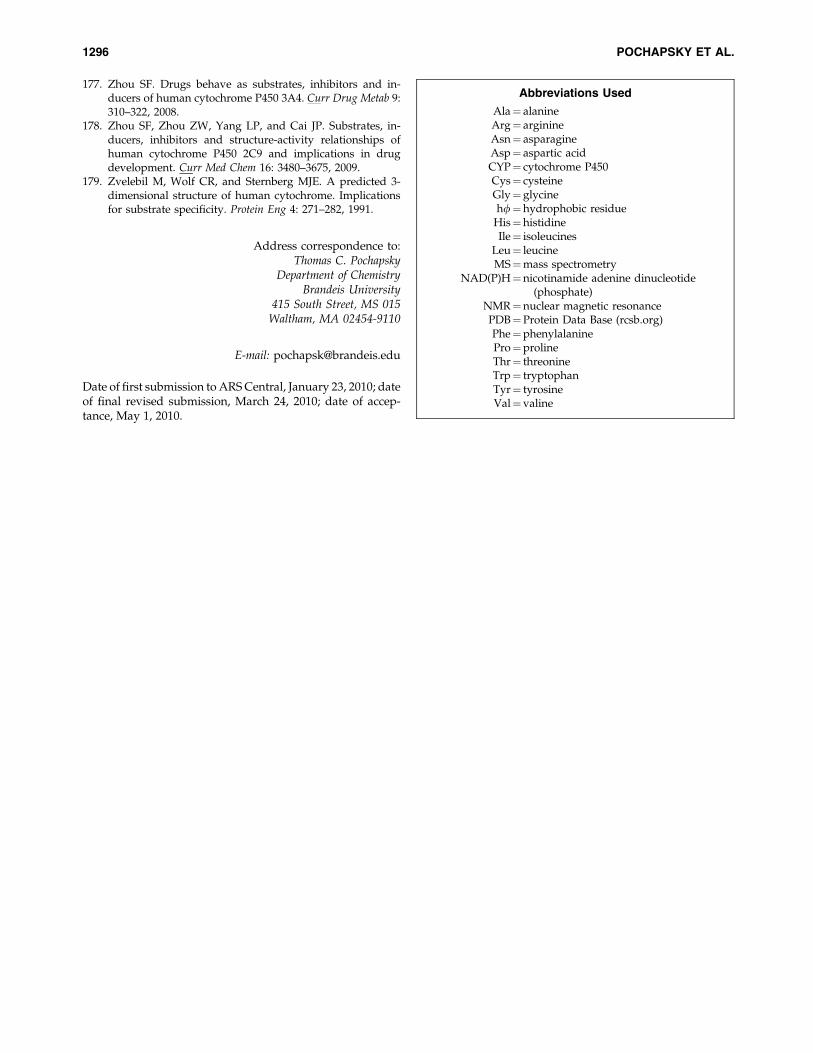
177. Zhou SF. Drugs behave as substrates, inhibitors and in-ducers of human cytochrome P450 3A4. Curr Drug Metab 9:310–322, 2008.
178. Zhou SF, Zhou ZW, Yang LP, and Cai JP. Substrates, in-ducers, inhibitors and structure-activity relationships ofhuman cytochrome P450 2C9 and implications in drugdevelopment. Curr Med Chem 16: 3480–3675, 2009.
179. Zvelebil M, Wolf CR, and Sternberg MJE. A predicted 3-dimensional structure of human cytochrome. Implicationsfor substrate specificity. Protein Eng 4: 271–282, 1991.
Address correspondence to:Thomas C. Pochapsky
Department of ChemistryBrandeis University
415 South Street, MS 015Waltham, MA 02454-9110
E-mail: [email protected]
Date of first submission to ARS Central, January 23, 2010; dateof final revised submission, March 24, 2010; date of accep-tance, May 1, 2010.
Abbreviations Used
Ala¼ alanineArg¼ arginineAsn¼ asparagineAsp¼ aspartic acidCYP¼ cytochrome P450Cys¼ cysteineGly¼ glycinehf¼hydrophobic residue
His¼histidineIle¼ isoleucines
Leu¼ leucineMS¼mass spectrometry
NAD(P)H¼nicotinamide adenine dinucleotide(phosphate)
NMR¼nuclear magnetic resonancePDB¼Protein Data Base (rcsb.org)Phe¼phenylalaninePro¼prolineThr¼ threonineTrp¼ tryptophanTyr¼ tyrosineVal¼valine
1296 POCHAPSKY ET AL.
Related Documents