doi: 10.1098/rspb.2008.0248 , 1491-1499 275 2008 Proc. R. Soc. B Jill A Cowing, Catherine A Arrese, Wayne L Davies, Lyn D Beazley and David M Hunt ) Tarsipes rostratus ) and the honey possum ( Sminthopsis crassicaudata dunnart ( Cone visual pigments in two marsupial species: the fat-tailed References rls http://rspb.royalsocietypublishing.org/content/275/1642/1491.full.html#related-u Article cited in: http://rspb.royalsocietypublishing.org/content/275/1642/1491.full.html#ref-list-1 This article cites 37 articles, 13 of which can be accessed free Subject collections (502 articles) evolution (107 articles) neuroscience (81 articles) molecular biology Articles on similar topics can be found in the following collections Email alerting service here right-hand corner of the article or click Receive free email alerts when new articles cite this article - sign up in the box at the top http://rspb.royalsocietypublishing.org/subscriptions go to: Proc. R. Soc. B To subscribe to This journal is © 2008 The Royal Society on 17 March 2009 rspb.royalsocietypublishing.org Downloaded from

Welcome message from author
This document is posted to help you gain knowledge. Please leave a comment to let me know what you think about it! Share it to your friends and learn new things together.
Transcript
doi: 10.1098/rspb.2008.0248, 1491-1499275 2008 Proc. R. Soc. B
Jill A Cowing, Catherine A Arrese, Wayne L Davies, Lyn D Beazley and David M Hunt
)Tarsipes rostratus) and the honey possum (Sminthopsis crassicaudatadunnart (
Cone visual pigments in two marsupial species: the fat-tailed
References
rlshttp://rspb.royalsocietypublishing.org/content/275/1642/1491.full.html#related-uArticle cited in: http://rspb.royalsocietypublishing.org/content/275/1642/1491.full.html#ref-list-1
This article cites 37 articles, 13 of which can be accessed free
Subject collections
(502 articles)evolution � (107 articles)neuroscience �
(81 articles)molecular biology � Articles on similar topics can be found in the following collections
Email alerting service hereright-hand corner of the article or click
Receive free email alerts when new articles cite this article - sign up in the box at the top
http://rspb.royalsocietypublishing.org/subscriptions go to: Proc. R. Soc. BTo subscribe to
This journal is © 2008 The Royal Society
on 17 March 2009rspb.royalsocietypublishing.orgDownloaded from
Proc. R. Soc. B (2008) 275, 1491–1499
doi:10.1098/rspb.2008.0248
on 17 March 2009rspb.royalsocietypublishing.orgDownloaded from
Cone visual pigments in twomarsupial species: thefat-tailed dunnart (Sminthopsis crassicaudata)
and the honey possum (Tarsipes rostratus)Jill A. Cowing1, Catherine A. Arrese2, Wayne L. Davies2, Lyn D. Beazley2
and David M. Hunt1,*1UCL Institute of Ophthalmology, 11-43 Bath Street, London EC1V 9EL, UK
2School of Animal Biology, University of Western Australia, Crawley, WA 6009, Australia
Published online 15 April 2008
*Autho
ReceivedAccepted
Uniquely for non-primate mammals, three classes of cone photoreceptors have been previously identified
by microspectrophotometry in two marsupial species: the polyprotodont fat-tailed dunnart (Sminthopsis
crassicaudata) and the diprotodont honey possum (Tarsipes rostratus). This report focuses on the genetic basis
for these three pigments. Two cone pigments were amplified from retinal cDNA of both species and
identified by phylogenetics as members of the short wavelength-sensitive 1 (SWS1) and long wavelength-
sensitive (LWS) opsin classes. In vitro expression of the two sequences from the fat-tailed dunnart confirmed
the peak absorbances at 363 nm in the UV for the SWS1 pigment and 533 nm for the LWS pigment. No
additional expressed cone opsin sequences that could account for the middle wavelength cones could be
amplified. However, amplification from the fat-tailed dunnart genomic DNA with RH1 (rod) opsin primer
pairs identified two genes with identical coding regions but sequence differences in introns 2 and 3. Uniquely
therefore for a mammal, the fat-tailed dunnart has two copies of an RH1 opsin gene. This raises the
possibility that the middle wavelength cones express a rod rather than a cone pigment.
Keywords: visual pigments; trichromacy in marsupials; evolution of vision
1. INTRODUCTIONVisual pigments belong to the large family of G-protein-
coupled receptors that share a common structure of seven
a-helical transmembrane regions joined by cytoplasmic and
luminal loops. They form a group of closely related proteins
(opsins) that bind retinal, a derivative of vitamin A. Five
classes of pigments are present in vertebrates, a rod class
and four different cone classes distinguished on the basis of
spectral sensitivity and amino acid sequence of their
respective opsins: long wavelength-sensitive (LWS) with
lmax 500–570 nm; middle wavelength-sensitive (MWS)
with lmax 480–530 nm; and two short wavelength-sensitive
classes, short wavelength-sensitive opsin 2 (SWS2) with
lmax 400–470 nm and short wavelength-sensitive opsin 1
(SWS1) with lmax 355–445 nm. In monotreme and
placental mammals, this complement is reduced to a rod
and two cone classes: SWS2 and LWS in the former
(Davies et al. 2007) and SWS1 and LWS in the latter
(Yokoyama 2000). This loss of cone classes is believed to
have resulted from a nocturnal lifestyle that mammals went
through during their early evolution. As a result, most
mammals are dichromats. In anthropoid or simian
primates, however, trichromacy is generally present, and
this is achieved by different genetic mechanisms in the two
major primate lineages. In the Old World (catarrhine)
primates from Africa and Asia, a duplication of the LWS
gene is present with the two copies encoding a ‘red’ and
‘green’ variant with the lmax values at approximately 565
and 530 nm, respectively (Nathans et al. 1986; Dulai et al.
r for correspondence ([email protected]).
19 February 200827 March 2008
1491
1999), whereas in most New World (platyrrhine) primates
from Central and South America, the trichromacy is based
on the polymorphism of the LWS gene, with different
alleles encoding spectrally distinct red and green pigments
(Mollon et al. 1984). In both cases, the major driving force
behind the evolution of trichromacy, with its improved
colour discrimination in the red/green region of the
spectrum, is argued to be the detection and evaluation of
ripe fruits (Mollon 1989; Osorio & Vorobyev 1996;
Sumner & Mollon 2000; Regan et al. 2001) or young
nutritious leaves (Dominy & Lucas 2001) against the green
foliage of the rainforest.
A recent study of the spectral characteristics of
photoreceptors in two marsupial species representative
of the major marsupial taxonomic divisions, the arrhyth-
mic and insectivorous polyprotodont fat-tailed dunnart
(Sminthopsis crassicaudata) and the crepuscular and
nectivorous diprotodont honey possum (Tarsipes rostratus),
has identified in both species three classes of cone
photoreceptors, with absorbance peaks as determined
by microspectrophotometry (MSP) in the UV for the
short wavelength-sensitive (SWS) class, at 509 and
505 nm, respectively, for the MWS class, and at 535
and 557 nm, respectively, for the LWS class (Arrese et al.
2002). The LWS and MWS visual pigments were found in
both single and double cones, but not as LWS–MWS
pairings in the latter.
Behavioural studies have indicated that trichromacy
is present in these species (Arrese et al. 2006). The
objective of the present study was to identify and
characterize the opsin genes expressed in the different
cone classes.
This journal is q 2008 The Royal Society
Table 1. Oligonucleotide primers used for the amplification of SWS1 and LWS opsin sequences. (Numbers refer to position incoding sequence for gene-specific primers, C denotes a forward primer and K denotes a reverse primer.)
primer position sequence (5 0–3 0)
oligo d(T) anchor GACCACGCGTATCGATGTCGACTTTTTTTTTTTTTTTTVPCR anchor GACCACGCGTATCGATGTCGACopsin AC ACCACCCAGAAGGCAGAGAAGopsin BK GACATAGATGATGGGGTTGTASWS1 AC 158C GTGCTGGTGGCCACACTGCSWS1 BC 637C CCTCATCTGCTTCTCCTACSWS1 EC 894C CCCATCATCTACTGCTTCATGSWS1 FC 886C TCTACAAYCCCATCATCTACTGSWS1 GK 908K GCAGTAGATGATGGGRTTGTAGSWS1 HK 993K CTGAGAGCTGGTGGTTTCAGSWS1 IK 317K CACAGACATGGCGGCCAAAGSWS1 JC 56C TGGGATGGGCCTCAGTACCACLWS AC 938C CCACTATCTACAACCCCATCLWS BK 700K TGATGATGCTGAGAGGAAGGLWS CK 646K GAACGCCAGGGTCGGAGCTGLWS DC 547C ACTGCACCACCCATCTTTGGLWS EK 737K CGGATGGCCAGCCACACTTGLWS FC 763C TCWGARTCYACCCAGAAGGCLWS GC 776C AGAAGGCHGARAAGGAAGTGLWS HK 954K GGGGTTGTAGATRGTGGCACLWS IK 1028K CCATCRTCMACCTTCTTCCCLWS JK 1071K GACGGAAGAGACCTCTGTCC
1492 J. A. Cowing et al. Cone visual pigments
on 17 March 2009rspb.royalsocietypublishing.orgDownloaded from
2. MATERIAL AND METHODS(a) Animals
Fat-tailed dunnarts (nZ3) were obtained from a breeding
colony established at the University of Adelaide (Animal
Service Division), South Australia. Honey possums
(nZ3) were collected in the Mt Lesueur Nature Reserve,
using pit traps, under licence from the Department of
Conservation and Land Management. Animals were term-
inally anaesthetized with saffan (alphaxalone and alphado-
lone acetate, 0.1 ml/10 g body weight, i.p.). The study was
approved by the Animal Ethics and Experimentation
Committee of the University of Western Australia.
(b) Genomic DNA, retinal RNA and cDNA
preparation
Genomic DNA was isolated from liver tissue using a
standard phenol–chloroform method. Dissected retinae
were collected in RNAlater (Ambion, Austin, Tex). Retinal
mRNA was prepared from total RNA using either the
EpiCentre MasterPure RNA Purification Kit followed by
Qiagen Oligotex mRNA Purification Kit or the Quickprep
Micro mRNA Purification Kit (Amersham Biosciences).
Single-stranded cDNA was synthesized using an oligo-
d(T) anchor primer and Superscript III reverse tran-
scriptase (Invitrogen) or transcriptor reverse transcriptase
(Roche Diagnostics). 5 0 and 3 0 RACE was carried out
using a 5 0–3 0 RACE Kit second generation (Roche
Diagnostics), following the manufacturer’s instructions.
(c) PCR amplification and cloning
Primers used in the amplification of opsin gene fragments
are listed in tables 1 and 2. PCR products were visualized
by agarose gel electrophoresis and cloned into pST-Blue-1
(Novagen) or pGEM-T easy cloning vector (Promega).
After colony PCR screening, the inserts from positive
colonies were sequenced using either vector- or opsin
sequence-specific primers. Sequencing was carried out
Proc. R. Soc. B (2008)
using a BIGDYE TERMINATOR v. 3.1 Cycle Sequencing kit on
either an ABI 3730 or ABI 3100 PRISM Genetic Analyser.
(d) Phylogenetic analysis
Neighbour joining (Saitou & Nei 1987) was used to
construct phylogenetic trees from opsin nucleotide
sequences after alignment with CLUSTALW (Higgins et al.
1996). The degree of support for internal branching was
assessed by bootstrapping with 1000 replicates using the
MEGA2 computer package (Kumar et al. 2001).
(e) Expression of recombinant opsins
The entire coding sequences for the fat-tailed dunnart
SWS1 and LWS opsins were amplified from retinal cDNA
with Pfu DNA polymerase, using primer pairs
SWS1F/SWS1R and LWSF/LWSR (table 3) containing
EcoRI and SalI restriction sites. The resulting products
were then cloned via these restriction sites into the
expression vector pMT3 that contains the sequence
for the 1D4 epitope from bovine rod opsin downstream
of and in-frame with the SalI site (Franke et al. 1988). The
opsin sequences were checked using vector-specific
sequencing primers.
The pMT3 vector containing either the SWS1 or
LWS coding sequences was transfected into HEK-293T
cells with GeneJuice (Invitrogen) according to the
manufacturer’s instructions. Thirty 90 mm plates were
used per transfection; the cells were harvested 48 hours
post-transfection and washed with 1! PBS. The
visual pigments were regenerated in 1! PBS with
40 mM 11-cis-retinal in the dark. Dodecyl maltoside (1%
(w/v)) and PMSF (20 mg mlK1) were then added before
passage over a CNBr-activated sepharose-binding column
coupled to an anti-1D4 monoclonal antibody (Molday &
MacKenzie 1983).
Absorption spectra were recorded in the dark using
a dual path spectrophotometer (Spectronic Unicam,
Table 3. Oligonucleotide primers used for the production of expression constructs. (Coding sequences are shown in bold.Eco R1 and SalI restriction enzyme sites are italicized.)
primer sequence
SWS1F 5 0-GCGCGAATTCCACCATGTCAGGGGATGAGGAGTTCK3 0
SWS1R 3 0-CGGCGTCGACGCACTAGGCCCCACTTGGCTGGAG-5 0
LWSF 5 0-GCGCGAATTCCACCATGACACAGGCATGGGACCCK3 0
LWSR 3 0-CGGCGTCGACGCGGCAGGCGCCACAGAGGAGAC-5 0
Table 2. Oligonucleotide primers used for the amplification of RH1 exons and introns. (Numbers and C, K symbols are asgiven in the table legend 1.)
primer position sequence (5 0–3 0)
rod AK 157K GGAAGCCCAGGACGATCAGCrod AC 342C CTTCTTCGCCACCACAGGAGrod BK 394K CCAAAACCACCAAGGCCCAGrod BC 361C GGTGAAGTAGCCCTCTGGGrod CK 563K CCACACGAACATTGCATTCCrod CC 678C GGTCTTCACAGTCAAAGAGrod DK 714K TTGCTGCTGGGCTGCGGCrod EK 958K TGGTGATCATGCAGTTCCGGrod FC 973C AAGAATCCATTGGGTGATGArod GC 939C CCGGAACTGCATGATCACCACrod HK 69K GGGCTCCGGACCACCCCACrod IK 33K GACGTAAAAGTTGGGTCCCrod INT2 AC intron 2 TGCCCCATCGCCAAAAGTTGrod INT2 BC intron 2 TGCCCCATCGCCAAAAGTGGrod INT2 CC intron 2 CCATCGCCAAAAGTTGAGACrod INT2 DC intron 2 CCATCGCCAAAAGTGGAGGCrod INT2 EC intron 2 TTCCCTCCTATTTACCTCCrod INT2 FC intron 2 CCTTGGAAATCTATTTACCTCC
Cone visual pigments J. A. Cowing et al. 1493
on 17 March 2009rspb.royalsocietypublishing.orgDownloaded from
Cambridge, UK). The pigments were either bleached by
exposure to light for 15 min (LWS) or acid denatured
(SWS1) by the addition of 10.8 ml of 1 N HCl. The lmax
value for each pigment was determined by subtracting the
bleached or acid-denatured spectrum from the dark
absorption spectrum to produce a difference spectrum.
This was then fitted to a standard Govardovskii rhodopsin
A1 template (Govardovskii et al. 2000) using an EXCEL
spreadsheet to determine the lmax.
(f ) In situ hybridization
Eye cups were fixed in 4% (w/v) paraformaldehyde in
1! PBS overnight, washed briefly with 1! PBS and cryo-
protected by incubation overnight in 25% (w/v) sucrose.
Ten micrometre thick sections were cut on a cryostat.
Riboprobes were generated from cloned coding
sequences of RH1 and LWS opsin of the fat-tailed
dunnart, RH2 opsin of the black bream (Acanthopagrus
butcheri, a perciform fish from the family of Sparidae) and
SWS2 of the platypus (Ornithorhynchus anatinus) by
restriction enzyme digestion and ligation into pGEM-T
easy. The probes were of 580, 568, 1100 and 1100 bp in
length, respectively. Digoxigenin (DIG)-labelled antisense
and sense RNA probes were then synthesized with SP6
and T7 primers using a DIG RNA labelling kit (Roche).
Retinal sections were prepared for hybridization as
follows: incubation in 1! PBS for 5 min at room
temperature; 4% paraformaldehyde in 1! PBS for
15 min; 1! PBS for 5 min; 1! PBT (1!PBSC0.1% Tween 20)Cproteinase K (5 mg mlK1) for 5 min;
1! PBTCglycine (2 mg mlK1) for 10 min; 1! PBT
Proc. R. Soc. B (2008)
for 1 min; and hybridization solution (50% formamide,
5! SSC, 50 mg mlK1 tRNA, 1% SDS, 50 mg mlK1
heparin) for 15 min at 658C. Denatured probe in hybrid-
ization solution was then added and the slides incubated
overnight at 658C. The slides were washed three times
for 15 min at 658C with wash solution I (50% formamide,
5! SSC, 1% SDS) followed by washing three times for
15 min with wash solution II (50% formamide, 2! SSC)
and three times 10 min at room temperature in 1! TBST
(140 mM NaCl, 2.7 mM KCl, 25 mM Tris–HCl
(pH 7.5), 1% Tween 20). The sections were blocked with
1! TBSTC10% sheep serum for 30 min at room
temperature. Anti-DIG antibody solution (anti-DIG
antibody 1 : 2000 in 1% sheep serum in 1! TBST) was
added and the slides were incubated at room temperature
for 2 hours followed by overnight incubation at 48C. The
slides were then washed four times at room temperature
for 15 min with 1! TBSTand three times for 10 min with
NTMT (100 mM NaCl, 100 mM Tris–HCl (pH 9.5),
50 mM MgCl2, 1% Tween 20). The colour was developed
by incubating the slides in 1!NTMTCNBT (4.5 ml mlK1)
and BCIP (3.5 ml mlK1) for up to 7 days, replenishing
the solution every 24 hours. The sections were fixed by
washing the slides two times with 1! NTMT for 10 min,
with PBT (pH 5.5) for 10 min, two times with 1! PBS
for 10 min, with 4% paraformaldehyde in PBS for 30 min
and with two times with 1! PBS for 10 min. The sections
were finally mounted in 90% glycerol and sealed with
a cover-slip for microscopy. The slides were viewed
under a light microscope and images taken with a Nikon
digital camera.
fat-tailed dunnart
fat-tailed dunnart
honey possum
honey possum
Tamar wallaby
Tamar wallaby
mouse
mouse
fat-tailed dunnarthoney possumTamar wallabymouse
fat-tailed dunnarthoney possumTamar wallabymouse
100
200
300
346
dunnart SWS1
honey possum SWS1
wallaby SWS1
human SWS1
mouse SWS1
chicken SWS1
chicken SWS2
chicken MWS
chicken LWS
Drosophila Rh1
100
100
100100
100
93
0.05
97
300
0.04
0.08
0.12
350 400 450wavelength (nm)
500 550 600
350 400 450 500 550
–0.02
–0.01
0
0.01
0.02
0.03
abso
rban
ce d
iffe
renc
e
max = 363 nm
abso
rban
ce
(a)
(b) (c)
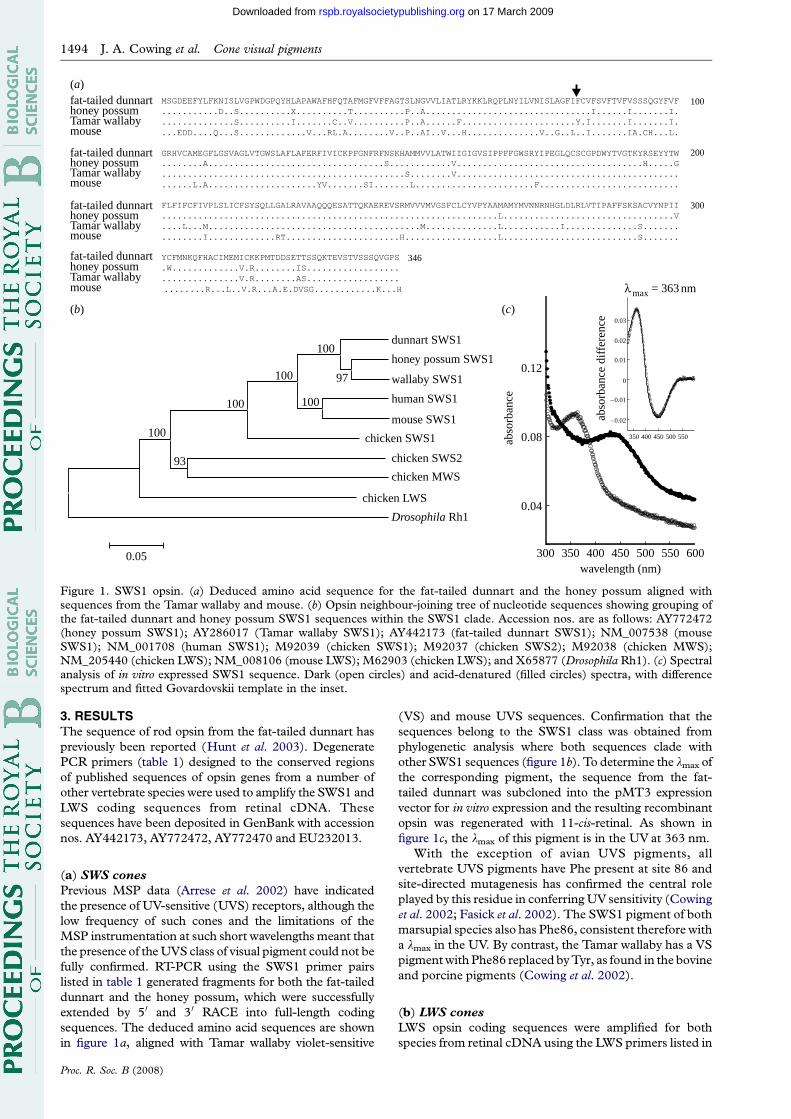
Figure 1. SWS1 opsin. (a) Deduced amino acid sequence for the fat-tailed dunnart and the honey possum aligned withsequences from the Tamar wallaby and mouse. (b) Opsin neighbour-joining tree of nucleotide sequences showing grouping ofthe fat-tailed dunnart and honey possum SWS1 sequences within the SWS1 clade. Accession nos. are as follows: AY772472(honey possum SWS1); AY286017 (Tamar wallaby SWS1); AY442173 (fat-tailed dunnart SWS1); NM_007538 (mouseSWS1); NM_001708 (human SWS1); M92039 (chicken SWS1); M92037 (chicken SWS2); M92038 (chicken MWS);NM_205440 (chicken LWS); NM_008106 (mouse LWS); M62903 (chicken LWS); and X65877 (Drosophila Rh1). (c) Spectralanalysis of in vitro expressed SWS1 sequence. Dark (open circles) and acid-denatured (filled circles) spectra, with differencespectrum and fitted Govardovskii template in the inset.
1494 J. A. Cowing et al. Cone visual pigments
on 17 March 2009rspb.royalsocietypublishing.orgDownloaded from
3. RESULTSThe sequence of rod opsin from the fat-tailed dunnart has
previously been reported (Hunt et al. 2003). Degenerate
PCR primers (table 1) designed to the conserved regions
of published sequences of opsin genes from a number of
other vertebrate species were used to amplify the SWS1 and
LWS coding sequences from retinal cDNA. These
sequences have been deposited in GenBank with accession
nos. AY442173, AY772472, AY772470 and EU232013.
(a) SWS cones
Previous MSP data (Arrese et al. 2002) have indicated
the presence of UV-sensitive (UVS) receptors, although the
low frequency of such cones and the limitations of the
MSP instrumentation at such short wavelengths meant that
the presence of the UVS class of visual pigment could not be
fully confirmed. RT-PCR using the SWS1 primer pairs
listed in table 1 generated fragments for both the fat-tailed
dunnart and the honey possum, which were successfully
extended by 50 and 30 RACE into full-length coding
sequences. The deduced amino acid sequences are shown
in figure 1a, aligned with Tamar wallaby violet-sensitive
Proc. R. Soc. B (2008)
(VS) and mouse UVS sequences. Confirmation that the
sequences belong to the SWS1 class was obtained from
phylogenetic analysis where both sequences clade with
other SWS1 sequences (figure 1b). To determine the lmax of
the corresponding pigment, the sequence from the fat-
tailed dunnart was subcloned into the pMT3 expression
vector for in vitro expression and the resulting recombinant
opsin was regenerated with 11-cis-retinal. As shown in
figure 1c, the lmax of this pigment is in the UV at 363 nm.
With the exception of avian UVS pigments, all
vertebrate UVS pigments have Phe present at site 86 and
site-directed mutagenesis has confirmed the central role
played by this residue in conferring UV sensitivity (Cowing
et al. 2002; Fasick et al. 2002). The SWS1 pigment of both
marsupial species also has Phe86, consistent therefore with
a lmax in the UV. By contrast, the Tamar wallaby has a VS
pigment with Phe86 replaced by Tyr, as found in the bovine
and porcine pigments (Cowing et al. 2002).
(b) LWS cones
LWS opsin coding sequences were amplified for both
species from retinal cDNA using the LWS primers listed in
fat-tailed dunnarthoney possumTamar wallabyhuman greenhuman red
fat-tailed dunnarthoney possumTamar wallabyhuman greenhuman red
fat-tailed dunnarthoney possumTamar wallabyhuman greenhuman red
fat-tailed dunnarthoney possumTamar wallabyhuman greenhuman red
100
200
300
364
dunnart LWShoney possum LWS
wallaby LWS
platypus LWSmouse LWS
chicken LWS
chicken MWSchicken SWS2
chicken SWS1
human SWS1
human green LWS
human red LWS
mouse SWS1
Drosophila Rh1
100
100100
100100
100100
100
64
100
96
0.05
300
0.10
0.06
0.14
0.18
350 400 450wavelength (nm)
500 550 600
350 400 450 500 550
–0.02
–0.01
0
0.01
0.02
abso
rban
ce d
iffe
renc
e
max = 533 nm
abso
rban
ce
(a)
(b) (c)
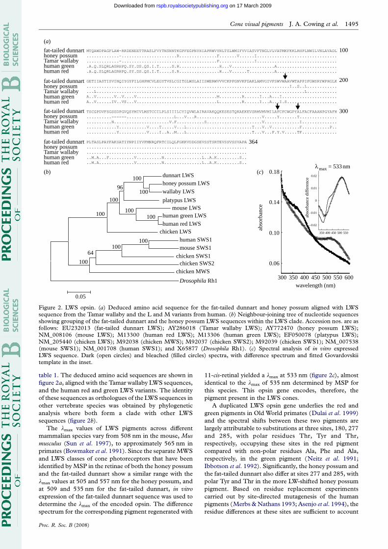
Figure 2. LWS opsin. (a) Deduced amino acid sequence for the fat-tailed dunnart and honey possum aligned with LWSsequence from the Tamar wallaby and the L and M variants from human. (b) Neighbour-joining tree of nucleotide sequencesshowing grouping of the fat-tailed dunnart and the honey possum LWS sequences within the LWS clade. Accession nos. are asfollows: EU232013 (fat-tailed dunnart LWS); AY286018 (Tamar wallaby LWS); AY772470 (honey possum LWS);NM_008106 (mouse LWS); M13300 (human red LWS); M13306 (human green LWS); EF050078 (platypus LWS);NM_205440 (chicken LWS); M92038 (chicken MWS); M92037 (chicken SWS2); M92039 (chicken SWS1); NM_007538(mouse SWS1); NM_001708 (human SWS1); and X65877 (Drosophila Rh1). (c) Spectral analysis of in vitro expressedLWS sequence. Dark (open circles) and bleached (filled circles) spectra, with difference spectrum and fitted Govardovskiitemplate in the inset.
Cone visual pigments J. A. Cowing et al. 1495
on 17 March 2009rspb.royalsocietypublishing.orgDownloaded from
table 1. The deduced amino acid sequences are shown in
figure 2a, aligned with the Tamar wallaby LWS sequences,
and the human red and green LWS variants. The identity
of these sequences as orthologues of the LWS sequences in
other vertebrate species was obtained by phylogenetic
analysis where both form a clade with other LWS
sequences (figure 2b).
The lmax values of LWS pigments across different
mammalian species vary from 508 nm in the mouse, Mus
musculus (Sun et al. 1997), to approximately 565 nm in
primates (Bowmaker et al. 1991). Since the separate MWS
and LWS classes of cone photoreceptors that have been
identified by MSP in the retinae of both the honey possum
and the fat-tailed dunnart show a similar range with the
lmax values at 505 and 557 nm for the honey possum, and
at 509 and 535 nm for the fat-tailed dunnart, in vitro
expression of the fat-tailed dunnart sequence was used to
determine the lmax of the encoded opsin. The difference
spectrum for the corresponding pigment regenerated with
Proc. R. Soc. B (2008)
11-cis-retinal yielded a lmax at 533 nm (figure 2c), almost
identical to the lmax of 535 nm determined by MSP for
this species. This opsin gene encodes, therefore, the
pigment present in the LWS cones.
A duplicated LWS opsin gene underlies the red and
green pigments in Old World primates (Dulai et al. 1999)
and the spectral shifts between these two pigments are
largely attributable to substitutions at three sites, 180, 277
and 285, with polar residues Thr, Tyr and Thr,
respectively, occupying these sites in the red pigment
compared with non-polar residues Ala, Phe and Ala,
respectively, in the green pigment (Neitz et al. 1991;
Ibbotson et al. 1992). Significantly, the honey possum and
the fat-tailed dunnart also differ at sites 277 and 285, with
polar Tyr and Thr in the more LW-shifted honey possum
pigment. Based on residue replacement experiments
carried out by site-directed mutagenesis of the human
pigments (Merbs & Nathans 1993; Asenjo et al. 1994), the
residue differences at these sites are sufficient to account
intron 2 sequences
intron 3 sequences
260
420 430 440 450 460 470 490 500 510 520 530 540
270 280 290 300
(a)
(b)
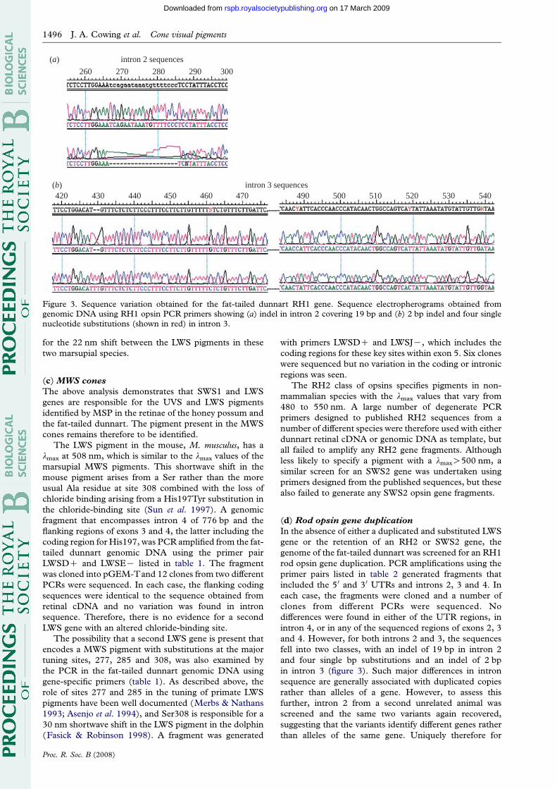
Figure 3. Sequence variation obtained for the fat-tailed dunnart RH1 gene. Sequence electropherograms obtained fromgenomic DNA using RH1 opsin PCR primers showing (a) indel in intron 2 covering 19 bp and (b) 2 bp indel and four singlenucleotide substitutions (shown in red) in intron 3.
1496 J. A. Cowing et al. Cone visual pigments
on 17 March 2009rspb.royalsocietypublishing.orgDownloaded from
for the 22 nm shift between the LWS pigments in these
two marsupial species.
(c) MWS cones
The above analysis demonstrates that SWS1 and LWS
genes are responsible for the UVS and LWS pigments
identified by MSP in the retinae of the honey possum and
the fat-tailed dunnart. The pigment present in the MWS
cones remains therefore to be identified.
The LWS pigment in the mouse, M. musculus, has a
lmax at 508 nm, which is similar to the lmax values of the
marsupial MWS pigments. This shortwave shift in the
mouse pigment arises from a Ser rather than the more
usual Ala residue at site 308 combined with the loss of
chloride binding arising from a His197Tyr substitution in
the chloride-binding site (Sun et al. 1997). A genomic
fragment that encompasses intron 4 of 776 bp and the
flanking regions of exons 3 and 4, the latter including the
coding region for His197, was PCR amplified from the fat-
tailed dunnart genomic DNA using the primer pair
LWSDC and LWSEK listed in table 1. The fragment
was cloned into pGEM-Tand 12 clones from two different
PCRs were sequenced. In each case, the flanking coding
sequences were identical to the sequence obtained from
retinal cDNA and no variation was found in intron
sequence. Therefore, there is no evidence for a second
LWS gene with an altered chloride-binding site.
The possibility that a second LWS gene is present that
encodes a MWS pigment with substitutions at the major
tuning sites, 277, 285 and 308, was also examined by
the PCR in the fat-tailed dunnart genomic DNA using
gene-specific primers (table 1). As described above, the
role of sites 277 and 285 in the tuning of primate LWS
pigments have been well documented (Merbs & Nathans
1993; Asenjo et al. 1994), and Ser308 is responsible for a
30 nm shortwave shift in the LWS pigment in the dolphin
(Fasick & Robinson 1998). A fragment was generated
Proc. R. Soc. B (2008)
with primers LWSDC and LWSJK, which includes the
coding regions for these key sites within exon 5. Six clones
were sequenced but no variation in the coding or intronic
regions was seen.
The RH2 class of opsins specifies pigments in non-
mammalian species with the lmax values that vary from
480 to 550 nm. A large number of degenerate PCR
primers designed to published RH2 sequences from a
number of different species were therefore used with either
dunnart retinal cDNA or genomic DNA as template, but
all failed to amplify any RH2 gene fragments. Although
less likely to specify a pigment with a lmaxO500 nm, a
similar screen for an SWS2 gene was undertaken using
primers designed from the published sequences, but these
also failed to generate any SWS2 opsin gene fragments.
(d) Rod opsin gene duplication
In the absence of either a duplicated and substituted LWS
gene or the retention of an RH2 or SWS2 gene, the
genome of the fat-tailed dunnart was screened for an RH1
rod opsin gene duplication. PCR amplifications using the
primer pairs listed in table 2 generated fragments that
included the 5 0 and 3 0 UTRs and introns 2, 3 and 4. In
each case, the fragments were cloned and a number of
clones from different PCRs were sequenced. No
differences were found in either of the UTR regions, in
intron 4, or in any of the sequenced regions of exons 2, 3
and 4. However, for both introns 2 and 3, the sequences
fell into two classes, with an indel of 19 bp in intron 2
and four single bp substitutions and an indel of 2 bp
in intron 3 (figure 3). Such major differences in intron
sequence are generally associated with duplicated copies
rather than alleles of a gene. However, to assess this
further, intron 2 from a second unrelated animal was
screened and the same two variants again recovered,
suggesting that the variants identify different genes rather
than alleles of the same gene. Uniquely therefore for
(i)(a) (b)
(i)
(ii) (ii)
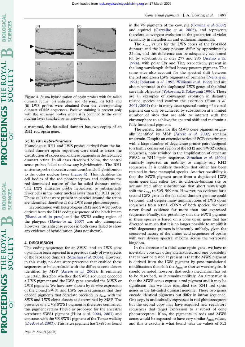
Figure 4. In situ hybridization of opsin probes with fat-taileddunnart retina: (a) antisense and (b) sense. (i) RH1 and(ii) LWS probes were obtained from the correspondingdunnart cDNA sequences. Positive staining is present onlywith the antisense probes where it is confined to the outernuclear layer (marked by an arrowhead).
Cone visual pigments J. A. Cowing et al. 1497
on 17 March 2009rspb.royalsocietypublishing.orgDownloaded from
a mammal, the fat-tailed dunnart has two copies of an
RH1 rod opsin gene.
(e) In situ hybridizations
Homologous RH1 and LWS probes derived from the fat-
tailed dunnart opsin sequences were used to assess the
distribution of expression of these pigments in the fat-tailed
dunnart retina. In all cases described below, the control
sense probes failed to show any hybridization. The RH1
antisense probe showed a continuous band of hybridization
to the outer nuclear layer (figure 4). This identifies the
cell bodies of the rod photoreceptors and confirms the
rod-dominated nature of the fat-tailed dunnart retina.
The LWS antisense probe hybridized to substantially
fewer cells in the outer nuclear layer than the RH1 probe.
These cells that were present in patches around the retina
are identified therefore as the LWS cone photoreceptors.
Hybridization with heterologous RH2 and SWS2 probes
derived from the RH2 coding sequence of the black bream
(Shand et al. in press) and the SWS2 coding region of
the platypus (Davies et al. 2007) was also attempted.
However, the antisense probes in both cases failed to show
any evidence of hybridization (data not shown).
4. DISCUSSIONThe coding sequences for an SWS1 and an LWS cone
opsin have been reported in a previous study of two species
of the fat-tailed dunnart (Strachan et al. 2004). However,
in this study, no data were presented that enabled these
sequences to be correlated with the different cone classes
identified by MSP (Arrese et al. 2002). It remained
uncertain therefore whether the SWS1 sequence encoded
a UVS pigment and the LWS gene encoded the MWS or
LWS pigment. We have now shown by in vitro expression
of the cloned SWS1 and LWS opsin sequences that they
encode pigments that correlate precisely in lmax with the
SWS and LWS clone classes as determined by MSP. The
presence of a UVS SWS1 pigment is therefore confirmed;
this pigment retains Phe86 as proposed for the ancestral
vertebrate SWS1 pigment (Hunt et al. 2004, 2007) and
contrasts with the VS SWS1 pigment of the Tamar wallaby
(Deeb et al. 2003). This latter pigment has Tyr86 as found
Proc. R. Soc. B (2008)
in the VS pigments of the cow, pig (Cowing et al. 2002)
and squirrel (Carvalho et al. 2006), and represents
therefore convergent evolution in the generation of violet
sensitivity in metatherian and eutherian mammals.
The lmax values for the LWS cones of the fat-tailed
dunnart and the honey possum differ by approximately
22 nm, and this difference can be adequately accounted
for by substitution at sites 277 and 285 (Asenjo et al.
1994), with polar Tyr and Thr, respectively, present in
the long-wavelength-shifted honey possum pigment. The
same sites also account for the spectral shift between
the red and green LWS pigments of primates (Neitz et al.
1991; Ibbotson et al. 1992; Williams et al. 1992) and are
also substituted in the duplicated LWS genes of the blind
cave fish, Astyanax (Yokoyama & Yokoyama 1990). These
are all examples of convergent evolution in distantly
related species and confirm the assertion (Hunt et al.
2001, 2004) that in many cases spectral tuning of a visual
pigment can only be achieved by substitution at a limited
number of sites that are able to interact with the
chromophore to achieve the spectral shift and maintain a
fully functional pigment.
The genetic basis for the MWS cone pigment origin-
ally identified by MSP (Arrese et al. 2002) remains
uncertain. Despite an extensive series of PCR experiments
with a large number of degenerate primer pairs designed
to a highly conserved region of the RH2 and SWS2 coding
sequences, none resulted in the amplification of either an
SWS2 or RH2 opsin sequence. Strachan et al. (2004)
similarly reported an inability to amplify any RH2
sequences. It is unlikely therefore that either has been
retained in these marsupial species. Another possibility is
that the MWS pigment arose from a duplicated LWS
opsin gene that either lost its chloride-binding site or
accumulated other substitutions that short wavelength
shift the lmax to 505–509 nm. However, no evidence for a
second LWS gene in the fat-tailed dunnart genome could
be found, and despite many amplifications of LWS opsin
sequences from retinal cDNA of both species, we have
never found evidence for more than one expressed
sequence. Finally, the possibility that the MWS pigment
in these species is based on a cone opsin gene that has
diverged so much that it is no longer possible to amplify it
with degenerate primers is inherently unlikely, given the
conserved nature of the amino acid sequences of opsins
with very diverse spectral maxima across the vertebrate
kingdom.
In the absence of a third cone opsin gene, we have to
inevitably consider other alternatives, and one possibility
that cannot be tested at present is that the MWS pigment
is derived from the LWS pigment by post-translational
modifications that shift the lmax to shorter wavelengths. It
should be noted, however, that such a mechanism has yet
to be described, so it remains unlikely. An alternative is
that the MWS cones express a rod pigment and it may be
significant that we have identified two RH1 rod opsin
genes in the fat-tailed dunnart genome. These two genes
encode identical pigments but differ in intron sequence.
One copy is undoubtedly expressed in rod photoreceptors
but the second copy may have acquired new regulatory
sequences that target expression to a subset of cone
photoreceptors. If so, the pigments in rods and MWS
cones would be expected to have very similar lmax values,
and this is exactly is what found with the values of 512
1498 J. A. Cowing et al. Cone visual pigments
on 17 March 2009rspb.royalsocietypublishing.orgDownloaded from
and 509 nm, respectively, in the fat-tailed dunnart and
502 and 505 nm, respectively, in the honey possum
(Arrese et al. 2002). It would also account for the similar
photochemical properties of the rods and MWS cones
with a post-bleach build-up of photoproduct that
absorbed below 430 nm (Arrese et al. 2002). Unfortu-
nately, in the absence of any differences in the coding
exons, it is not possible to confirm that both gene copies
are expressed or to demonstrate that mRNA from one
copy is present in cones and the other in rods. Such
heterologous expression of visual pigments is not,
however, unknown. The blue sensitive cones and green
rods of the tiger salamander both express the same SWS2
cone pigment (Ma et al. 2001), thereby providing evidence
that cone pigments can function with rod transducin, and
the disparity in flash sensitivity could be attributed to a
higher quantal photon catch by the larger rod outer
segments. The converse situation proposed for the
marsupial MWS cones expressing a rod pigment may be
expected therefore to be fully functional and show a cone-
like sensitivity.
The study was approved by the Animal Ethics andExperimentation Committee of the University of WesternAustralia.
This work was supported by a grant (03/100/256) from theAustralian Research Council. We are grateful to Dr RosalieCrouch for the generous gift of 11-cis-retinal, Dr LyndaErskine for advice on in situ hybridization protocols, Prof.Glen Jeffery for advice and help with the histological analysis,and Ms Alison Oddy for technical assistance.
REFERENCESArrese, C. A., Hart, N. S., Thomas, N., Beazley, L. D. &
Shand, J. 2002 Trichromacy in Australian marsupials.Curr. Biol. 12, 657–660. (doi:10.1016/S0960-9822(02)00772-8)
Arrese, C. A., Beazley, L. D. & Neumeyer, C. 2006Behavioural evidence for marsupial trichromacy. Curr.Biol. 16, R193–R194. (doi:10.1016/j.cub.2006.02.036)
Asenjo, A. B., Rim, J. & Oprian, D. D. 1994 Moleculardeterminants of human red/green color discrimination.Neuron 12, 1131–1138. (doi:10.1016/0896-6273(94)90320-4)
Bowmaker, J. K., Astell, S., Hunt, D. M. & Mollon, J. D.1991 Photosensitive and photostable pigments in theretinae of Old World monkeys. J. Exp. Biol. 156, 1–19.
Carvalho, L. d. S., Cowing, J. A., Wilkie, S. E., Bowmaker,J. K. & Hunt, D. M. 2006 Shortwave visual sensitivity intree and flying squirrels reflects changes in lifestyle. Curr.Biol. 16, R81–R83. (doi:10.1016/j.cub.2006.01.045)
Cowing, J. A., Poopalasundaram, S., Wilkie, S. E., Robinson,P. R., Bowmaker, J. K. & Hunt, D. M. 2002 Themolecular mechanism for the spectral shifts betweenvertebrate ultraviolet- and violet-sensitive cone visualpigments. Biochem. J. 367, 129–135. (doi:10.1042/BJ20020483)
Davies, W. L., Carvalho, L. S., Cowing, J. A., Beazley, L. D.,Hunt, D. M. & Arrese, C. A. 2007 Visual pigments of theplatypus: a novel route to mammalian colour vision. Curr.Biol. 17, R161–R163. (doi:10.1016/j.cub.2007.01.037)
Deeb, S. S., Wakefield, M. J., Tada, T., Marotte, L.,Yokoyama, S. & Marshall Graves, J. A. 2003 The conevisual pigments of an Australian marsupial, the tammarwallaby (Macropus eugenii ): sequence, spectral tuning, andevolution. Mol. Biol. Evol. 20, 1642–1649. (doi:10.1093/molbev/msg181)
Proc. R. Soc. B (2008)
Dominy, N. J. & Lucas, P. W. 2001 Ecological importance oftrichromatic vision to primates. Nature 410, 363–366.(doi:10.1038/35066567)
Dulai, K. S., von Dornum, M., Mollon, J. D. & Hunt, D. M.1999 The evolution of trichromatic color vision by opsingene duplication in New World and Old World primates.Genome Res. 9, 629–638.
Fasick, J. I. & Robinson, P. R. 1998 Mechanism of spectraltuning in the dolphin visual pigments. Biochemistry 37,433–438. (doi:10.1021/bi972500j)
Fasick, J. I., Applebury, M. L. & Oprian, D. D. 2002 Spectraltuning in the mammalian short-wavelength sensitive conepigments. Biochemistry 41, 6860–6865. (doi:10.1021/bi0200413)
Franke, R. R., Sakmar, T. P., Oprian, D. D. & Khorana, H. G.1988 A single amino acid substitution in rhodopsin (lysine248–leucine) prevents activation of transducin. J. Biol.Chem. 263, 2119–2122.
Govardovskii, V. I., Fyhrquist, N., Reuter, T., Kuzmin, D. G.& Donner, K. 2000 In search of the visual pigmenttemplate. Vis. Neurosci. 17, 509–528. (doi:10.1017/S0952523800174036)
Higgins, D. G., Thompson, J. D. & Gibson, T. J. 1996 UsingCLUSTAL for multiple sequence alignments. MethodsEnzymol. 266, 383–402.
Hunt, D. M., Dulai, K. S., Partridge, J. C., Cottrill, P. &Bowmaker, J. K. 2001 The molecular basis for spectraltuning of rod visual pigments in deep-sea fish. J. Exp. Biol.204, 3333–3344.
Hunt, D. M. et al. 2003 The rod opsin pigments from twomarsupial species, the South American bare-tailed woollyopossum and the Australian fat-tailed dunnart. Gene 323,157–162. (doi:10.1016/j.gene.2003.09.016)
Hunt, D. M., Cowing, J. A., Wilkie, S. E., Parry, J.,Poopalasundaram, S. & Bowmaker, J. K. 2004 Divergentmechanisms for the tuning of shortwave sensitive visualpigments in vertebrates. Photochem. Photobiol. Sci. 3,713–720. (doi:10.1039/b314693f)
Hunt, D. M., Carvalho, L. S., Cowing, J. A., Parry, J. W. L.,Wilkie, S. E., Davies, W. L. & Bowmaker, J. K. 2007Spectral tuning of shortwave-sensitive visual pigments invertebrates. Photochem. Photobiol. 83, 303–310.
Ibbotson, R. E., Hunt, D. M., Bowmaker, J. K. & Mollon,J. D. 1992 Sequence divergence and copy number of themiddle- and long-wave photopigment genes in Old Worldmonkeys. Proc. R. Soc. B 247, 145–154. (doi:10.1098/rspb.1992.0021)
Kumar, S., Tamura, K., Jakobsen, I. B. & Nei, M. 2001MEGA2: molecular evolutionary genetics analysis soft-ware. Bioinformatics 17, 1244–1245. (doi:10.1093/bioin-formatics/17.12.1244)
Ma, J.-X. et al. 2001 A visual pigment expressed in both rodand cone photoreceptors. Neuron 32, 451–461. (doi:10.1016/S0896-6273(01)00482-2)
Merbs, S. L. & Nathans, J. 1993 Role of hydroxyl-bearingamino acids in differentially tuning the absorption spectraof the human red and green cone pigments. Photochem.Photobiol. 58, 706–710. (doi:10.1111/j.1751-1097.1993.tb04956.x)
Molday, R. S. & MacKenzie, D. 1983 Monoclonal antibodiesto rhodopsin: characterization, cross-reactivity, and appli-cation as structural probes. Biochemistry 22, 653–660.(doi:10.1021/bi00272a020)
Mollon, J. D. 1989 “Tho’ she kneel’d in that place where theygrew.” The uses and origins of primate colour vision.J. Exp. Biol. 146, 21–38.
Mollon, J. D., Bowmaker, J. K. & Jacobs, G. H. 1984 Variationsof colour vision in a New World primate can be explainedby polymorphism of retinal photopigments. Proc. R. Soc. B222, 373–399. (doi:10.1098/rspb.1984.0071)
Cone visual pigments J. A. Cowing et al. 1499
on 17 March 2009rspb.royalsocietypublishing.orgDownloaded from
Nathans, J., Thomas, D. & Hogness, D. S. 1986 Moleculargenetics of human color vision: the genes encoding blue,green, and red pigments. Science 232, 193–202. (doi:10.1126/science.2937147)
Neitz, M., Neitz, J. & Jacobs, G. H. 1991 Spectral tuning ofpigments underlying red–green color vision. Science 252,971–974. (doi:10.1126/science.1903559)
Osorio, D. & Vorobyev, M. 1996 Colour vision as anadaptation to frugivory in primates. Proc. R. Soc. B 263,593–599. (doi:10.1098/rspb.1996.0089)
Regan, B. C., Julliot, C., Simmen, B., Vienot, F., Charles-Dominique, P. & Mollon, J. D. 2001 Fruits, foliage and theevolution of primate colour vision. Phil. Trans. R. Soc. B356, 229–283. (doi:10.1098/rstb.2000.0773)
Saitou, N. & Nei, M. 1987 The neighbor-joining method: anew method for reconstructing phylogenetic trees. Mol.Biol. Evol. 4, 406–425.
Shand, J. et al. In press. The influence of ontogeny and lightenvironment on the expression of visual pigment opsins inthe retina of the black bream, Acanthopagrus butcheri. J.Exp. Biol.
Strachan, J., Chang, L. Y., Wakefield, M. J., Graves, J. A. &Deeb, S. S. 2004 Cone visual pigments of the Australian
Proc. R. Soc. B (2008)
marsupials, the stripe-faced and fat-tailed dunnarts:
sequence and inferred spectral properties. Vis. Neurosci.
21, 223–229. (doi:10.1017/S0952523804213281)
Sumner, P. & Mollon, J. D. 2000 Catarrhine photopigments
are optimized for detecting targets against a foliage
background. J. Exp. Biol. 203(Pt 13), 1963–1986.
Sun, H., Macke, J. P. & Nathans, J. 1997 Mechanisms of
spectral tuning in the mouse green cone pigment. Proc.
Natl Acad. Sci. USA 94, 8860–8865. (doi:10.1073/pnas.
94.16.8860)
Williams, A. J., Hunt, D. M., Bowmaker, J. K. & Mollon, J. D.
1992 The polymorphic photopigments of the marmoset:
spectral tuning and genetic basis. EMBO J. 11,
2039–2045.
Yokoyama, S. 2000 Molecular evolution of vertebrate visual
pigments. Prog. Retin. Eye Res. 19, 385–419. (doi:10.1016/
S1350-9462(00)00002-1)
Yokoyama, R. & Yokoyama, S. 1990 Convergent
evolution of the red- and green-like visual pigment
genes in fish, Astyanax fasciatus, and human. Proc. Natl
Acad. Sci. USA 87, 9315–9318. (doi:10.1073/pnas.87.
23.9315)
Related Documents


