doi:10.1136/jnnp.2006.106583 2006;6;342-357 Practical Neurology Tim Anderson, Marcus Heitger and A D Macleod Concussion and mild head injury http://pn.bmj.com/cgi/content/full/6/6/342 Updated information and services can be found at: These include: References http://pn.bmj.com/cgi/content/full/6/6/342#BIBL This article cites 48 articles, 17 of which can be accessed free at: Rapid responses http://pn.bmj.com/cgi/eletter-submit/6/6/342 You can respond to this article at: service Email alerting top right corner of the article Receive free email alerts when new articles cite this article - sign up in the box at the Notes http://www.bmjjournals.com/cgi/reprintform To order reprints of this article go to: http://www.bmjjournals.com/subscriptions/ go to: Practical Neurology To subscribe to on 23 November 2006 pn.bmj.com Downloaded from

Welcome message from author
This document is posted to help you gain knowledge. Please leave a comment to let me know what you think about it! Share it to your friends and learn new things together.
Transcript
doi:10.1136/jnnp.2006.106583 2006;6;342-357 Practical Neurology
Tim Anderson, Marcus Heitger and A D Macleod
Concussion and mild head injury
http://pn.bmj.com/cgi/content/full/6/6/342Updated information and services can be found at:
These include:
References
http://pn.bmj.com/cgi/content/full/6/6/342#BIBL
This article cites 48 articles, 17 of which can be accessed free at:
Rapid responses http://pn.bmj.com/cgi/eletter-submit/6/6/342
You can respond to this article at:
serviceEmail alerting
top right corner of the article Receive free email alerts when new articles cite this article - sign up in the box at the
Notes
http://www.bmjjournals.com/cgi/reprintformTo order reprints of this article go to:
http://www.bmjjournals.com/subscriptions/ go to: Practical NeurologyTo subscribe to
on 23 November 2006 pn.bmj.comDownloaded from

Practical Neurology342
Practical Neurology 2006; 6: 342-357
10.1136/jnnp.2006.106583
Concussion and mild head injuryTim Anderson, Marcus Heitger, A D Macleod
A concussion is a physical injury to the head resulting in altered mental function, with expectation of recovery within 2–3 weeks. In a
significant minority of cases the symptoms persist longer, thereby comprising a symptom complex commonly referred to as the “post concussion syndrome”, that is, one or more somatic (for example, headaches, dizziness), cognitive (for example, poor concentration, memory), or behavioural/affective (for example, irritability, mood swings) symptoms. Unfortunately, the referral of a patient with the possibility of post concussion syndrome to a busy neurology outpatient clinic can precipitate an inward sigh of reluctant resignation in even the most diligent neurologist or neurosurgeon. We know we are in for a potentially lengthy consultation—long on symptoms and short on signs. Moreover, the process can be convoluted and meandering, as unrewarding for the patient as it is unsatisfying for the clinician.
It is important to acknowledge at the outset that there is a dearth of evidence-based knowledge of the underlying pathogenesis, and even less of the best management of post concussive symptoms. Thus, much of the
information and advice in this article is empirical and based on expert and personal experience.
DEFINITION OF CONCUSSION AND MILD HEAD INJURYThe terminology surrounding trauma to the head confuses patients, doctors, and lay commentators alike. Terms such as concussion, mild head injury, mild TBI (traumatic braintraumatic brain injury), cerebral concussion, and post concussion cerebral concussion, and post concussion syndrome are often used interchangeably to describe the physical injury itself as well as its immediate and later symptomatic consequences. There is in fact no commonly agreed definition of concussion, or whether it even differs from the term mild TBI. Our own working definition is that concussion is an acute trauma-induced change of mental function which generally lasts less than 24 hours (with or without preceding loss of consciousness) and associated with other symptoms (such as headaches and dizziness) which usually recover within 2–3 weeks. A more complete description is provided by the Concussion in Sports Group (table 1).
The term �concussion� emphasises anhe term �concussion� emphasises an impaired functional status as a result of head trauma, whereas the terms “mild head injury”
T AndersonCas Van Der Veer Chair in Parkinson’s disease and movement disorders, Neurology Department, Christchurch Hospital, Christchurch, New Zealand
M HeitgerPost Doctoral Fellow, Van Der Veer Institute for Parkinson’s and Brain Research, Christchurch, New Zealand
A D MacleodConsultant Psychiatrist, Van Der Veer Institute for Parkinson’s and Brain Research, Christchurch, New Zealand, and Concussion Clinic, Burwood Hospital, Christchurch, New Zealand
Correspondence to: Dr T Anderson, Van Der Veer Institute for Parkinson’s and Brain Research, 16 St Asaph Street, Christchurch, New Zealand; [email protected]
REvIEw
on 23 November 2006 pn.bmj.comDownloaded from
www.practical-neurology.com
343Anderson, Heitger, Macleod
Concussion and mild head injuryTim Anderson, Marcus Heitger, A D Macleod
or “traumatic brain injury” primarily refer to the adverse pathophysiological impact of biomechanical trauma to the head and brain. However, in the context of clinical practice and research, this distinction is rarely made and—regrettably—all these terms are used interchangeably.
The term �head injury� incorporates any injury to the head (for example, scalp and skin abrasions, facial or dental injuries, bone fractures) which may not necessarily be equivalent to brain injury. However, although head injury can occur in the absence of cerebral pathology, most head injuries, including mild head trauma, are likely to cause cerebral injury to some extent.1 This injury of the brain is the most critical consequence of head trauma and the probable cause of any adverse outcome, though it seems unlikely that the mild structural changes seen pathologically or with brain MRI can account for the range and extent of all the symptoms comprising the post concussion syndrome. Structural changes might explain, for instance, vestibular and visual symptoms, but it is likely that functional changes (for example, in biochemical, synaptic, and neuronal membrane processes) are involved in persistent symptom complexes, particularly those with a more psychological flavour.
GRADING OF MILD HEAD INJURYThere are no fewer than 41 different guidelines no fewer than 41 different guidelines for the grading of mild head injury, some of which relate specifically to sports injury, admission policy, imaging, neuropsychological assessment, and management of children.3 Many have been developed and reported in a systematic and methodologically sound manner but they are all plagued by the lack of empirical evidence supporting their use. However, at least a basic consensus about exactly what constitutes mild head injury has emerged: a Glasgow Coma Scale (GCS) score of 13–15 atat first assessment (commonly within the first 30–60 minutes of injury) without falling below 13 at any later assessment, brief disturbance or loss assessment, brief disturbance or loss of consciousness of less than 20 minutes and/or post-traumatic amnesia (PTA) duration of up to 24 hours, with or without other neurological abnormalities (for example, focal signs). There are usually no structural abnormalities on CT head scan (MR seldom being clinically indicated), although about 10% of head injury admissions with a GCS of 15 may have a relevant cerebral lesion on CT.4
USA(Kraus et al, 1988)
Australia(Tate et al, 1998)
South Africa(Nell and Brown, 1991)
Scotland(Jennett, 1996)
TransportationSweden
(Peloso et al, 2004)
Falls
Sport or recreation
Assaults or violence
Other causes
Concussion is defined as a complex pathophysiological process affecting the brain, induced by traumatic biomechanical forces. Several common features that incorporate clinical, pathological, and biomechanical injury constructs that may be used in defining the nature of a concussive head injury include:
• Concussion may be caused by a direct blow to the head, face, neck, or elsewhere on the body with an “impulsive” force transmitted to the head.
• Concussion typically results in the rapid onset of short lived impairment of neurological function that resolves spontaneously.
• Concussion may result in neuropathological changes but the acute clinical symptoms largely reflect a functional disturbance rather than structural injury.
• Concussion results in a graded set of clinical syndromes that may or may not involve loss of consciousness. Resolution of the clinical and cognitive symptoms typically follows a sequential course.
• Concussion is typically associated with grossly normal structural neuroimaging studies.
TABLE 1 The Concussion in Sports Group definition of concussion2
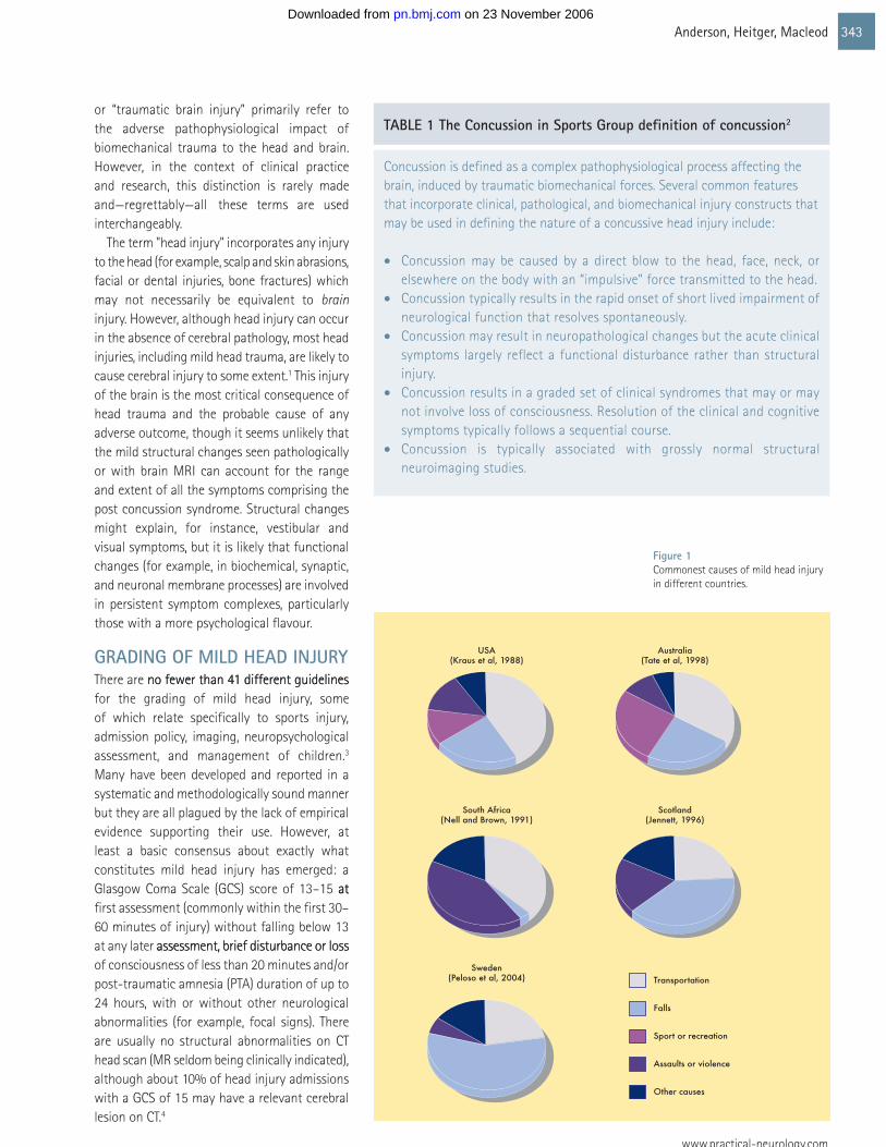
Figure 1 Commonest causes of mild head injury in different countries.
on 23 November 2006 pn.bmj.comDownloaded from
Practical Neurology344
10.1136/jnnp.2006.106583
EPIDEMIOLOGY OF MILD HEAD INJURYApproximately 80% of head injury admissions are categorised as mild, resulting in estimated admission rates of between 100 and 300 cases per year per 100,000 population, depending on country, region, age, and gender.5, 6 Males, in particular those aged between 15 and 35, are up to twice as likely to sustain a mild head injury than females, a difference that attenuates with increasing age.6 The commonest causes are motor vehicle accidents, falls, sports and recreational activities, and assaults, although the proportion
of these causes depends on the population and country, gender and age group (fig 1). (fig 1)..5–9
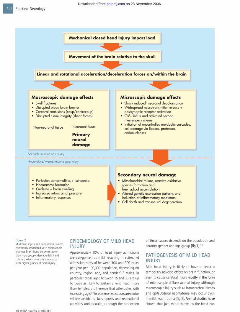
PATHOGENESIS OF MILD HEAD INJURYMild head injury is likely to have at least a temporary adverse effect on brain function, or even to cause cerebral injury mostly in the formmostly in the form of microscopic diffuse axonal injury, although macroscopic injury such as intracerebral bleeds and epi/subdural haematoma may occur even in mild head trauma (fig 2). Animal studies have. Animal studies have shown that just minor blows to the head can
Figure 2 Mild head injury and concussion is most commonly associated with microscopic changes (right hand column) rather than macroscopic damage (left hand column) which is mostly associated with higher grades of head injury.
Mechanical closed head injury impact load
Movement of the brain relative to the skull
Linear and rotational acceleration/deceleration forces on/within the brain
Macroscopic damage effects• Skull fractures• Disrupted blood brain barrier• Cerebral contusions (coup/contrecoup)• Disrupted tissue integrity (shear forces)
Microscopic damage effects• ‘Shock induced’ neuronal depolarisation• Widespread neurotransmitter release +
postsynaptic receptor activation• Ca2+ influx and activated second
messenger systems• Initiation of uncontrolled metabolic cascades,
cell damage via lipases, proteases, endonucleases
Secondary neural damage• Mitochondrial failure, reactive oxidative
species formation and free radical accumulation
• Altered genetic expression patterns and induction of inflammatory mediators
• Cell death and transneural degeneration
Non-neuronal tissue Neuronal tissue
Primary neural damage
Seconds–minutes post injury
Hours–days/weeks/months post injury
• Perfusion abnormalities + ischaemia• Haematoma formation• Oedema + brain swelling• Increased intracranial pressure• Inflammatory responses
on 23 November 2006 pn.bmj.comDownloaded from

www.practical-neurology.com
345Anderson, Heitger, Macleod
result in neural injury.1 Similarly, head trauma related neural damage has been documented in humans even after mild head trauma, both in histological studies in patients who died of other causes and by metabolic abnormalities and hypoperfusion in functional imaging studies. Although brain CT and MRI have shown. Although brain CT and MRI have shown cerebral lesions following mild head trauma in some cases (fig 3), functional imaging with single photon emission computed tomography (SPECT) and functional MRI are much more useful.10 Hyperintensities on advanced structural MRI head scans and hypometabolism/hypoperfusion detected by functional imaging can be observed even beyond three months of the injury.11 These metabolic changes and perfusion abnormalities are most common in prefrontal and temporal brain regions.10
Trauma to the head generates immediate impact forces of high magnitude and relatively short duration (5–200 ms) which translate into acceleration forces on the head and brain. These forces include translational (that is, the head’s centre moves along a straight line) and rotational (that is, the head moves around its centre of gravity) components, the largest injury capacity being assigned to the rotational component. While macroscopic injuries suchWhile macroscopic injuries such as fractures of the skull and face (about 10% of mild head injury cases), vasogenic oedema with subsequent brain swelling, significant intracranial haemorrhage and epi/subdural and subarachnoid haematoma, or cerebral ischaemia/hypoxaemia can occur following mild head injury, their frequency, compared with
moderate-to-severe head injury, is much lower. Indeed, less than 1% of patients with mild head injury need neurosurgical intervention..4
At a later stage (that is, several hours to days/weeks post-injury), microscopic changes are observed which include altered expression of DNA transcription factors and apoptotic proteins, the uncontrolled release
of proteases, lipases, and endonucleases, with the subsequent oxidative stress causing degradation of cell membrane and cytoskeletal proteins, mitochondrial failure, and production of free radicals. Other changes that have been observed include axonal swelling and anterograde and retrograde transneuronal degeneration of otherwise undamaged neurons. These changes may lead to secondary (delayed, non-mechanical) injury and are triggered by a cascade of biochemical events resulting in a variety of harmful physiological stress responses and the disruption of the intracellular balances within the brain, including metabolic changes, increased neurotransmitter release, and initiation of inflammatory processes.1 It has been proposed that the altered neuroplasticity and subsequent changes in cerebral connectivity
Figure 3 Brain MR scan five days post-injury in a 24 year old male with prolonged post-traumatic amnesia from mild–moderate head injury following a motor vehicle accident. The gradient-T2 sequence (B) shows the susceptibility effect (dark punctuate signal changes) indicative of petechial haemorrhages from diffuse axonal injury, particularly at the junction of white and grey matter. These changes are not as well defined in the traditional spin echo T2 sequence (A). (Figure courtesy of Dr Mike Hurrell.)
Most post-concussive symptoms resolve spontaneously within 2–3 weeks but sometimes they can be disabling and require specific investigation and treatment
on 23 November 2006 pn.bmj.comDownloaded from
Practical Neurology346
10.1136/jnnp.2006.106583
may facilitate functional recovery from traumatic brain injury and enable the brain, over time, to compensate for any functional deficits that have resulted from injury related neurophysiological dysfunction. According to this notion, recovery comprises a functional reorganisation in cortical and subcortical areas and may include reafferentation.12
EARLY POST-CONCUSSIvE SYMPTOMSMost post-concussive symptoms resolve spontaneously within 2–3 weeks but sometimes they can be disabling and require specific investigation and treatment.13
EARLY INTERvENTION: EvIDENCE FROM RANDOMISED CONTROLLED TRIALSEarly intervention by a specialist service hasintervention by a specialist service has been shown to reduce social restriction and post-concussion symptoms in two randomised controlled trials14, 15 while a third reported that a single brief educational intervention delivered soon after a mild head injury was as effective as a more intensive regimen of
assessment and education.16 Bed rest for several days immediately after the injury was no more effective than normal activity in preventing post-concussive symptoms in a fourth randomised trial.17 Thus, patients should be offered at least some education about the expected sequelae and coping strategies after their head injury.
HeadacheEarly post-traumatic headaches are common and should be treated with simple analgesics, though these are not always as effective as just the passage of time.18 Paracetamol with or without codeine or non-steroidal anti-inflammatory drugs such as ibuprofen or diclofenac can be helpful and the patient reassured that the headache will likely settle in a few weeks. When headaches persist for longer, traditional headache prophylaxis—
depending on the headache characteristics—may be required (see below and tables 2 and 3). Daily analgesia should be avoided to prevent the emergence of rebound headaches. Persisting focal head pains beyond two weeks may suggest localised traumatic injury (for example, fracture, neuralgia, infection) and demand specific investigation or referral. Post-concussion headaches can often be delayed up to 2–3 weeks after the injury and these will frequently require pharmacological treatment as discussed below. Early post-traumatic global or hemicranial headache which persists and worsens should prompt CT brain scanning just to rule out the outside possibility of subdural haematoma, but delayed onset headache (2 or 3 weeks after injury) rarely demands investigation. Localised head pains may require specific imaging (looking for fracture) or referral to a relevant specialist (such as an ophthalmologist or otolaryngologist, for orbital or facial/ear pain respectively). Early localised neck muscle tenderness on deep palpation (for example, splenius or trapezius) is common when there is associated whiplash and will usually settle spontaneously. Persistent or later onset post-traumatic tension type headache is often associated with similar neck muscle tenderness on palpation and may respond to amitriptyline or nortriptyline (table 3).
Dizziness, nausea, and unsteadiness Dizziness and unsteadiness with associated nausea are frequent early on and usually rather non-specific. Sensations include light headedness, floating, mild swaying, and disequilibrium. The origin of these symptoms is uncertain but they may represent either
peripheral or perhaps more likely central vestibular pathway dysfunction. It is important though to recognise movement induced vertigo, usually rotatory, in the form of benign
Patients should be offered at least some education about the expected sequelae and coping strategies after their head injury
Dizziness and unsteadiness with associated nausea
are frequent early on and usually rather non-specific
on 23 November 2006 pn.bmj.comDownloaded from

www.practical-neurology.com
347Anderson, Heitger, Macleod
Symptom Differential Comments Tips Investigation and therapy
Dizziness and vertigo Non-specific post-
traumatic dizziness
v benign paroxysmal
positional vertigo (BPPV)
Whiplash often causes
dizziness. BPPV occurs
after mild as well as
severe head injury
BPPV is induced by head movement,
particularly lying down, turning, or
sitting up in bed
Dix-Hallpike manoeuvre diagnostic.
Treat with particle repositioning
manoeuvre
Blank spells Concentration lapses (ie
day dreaming) v epilepsy
Post-traumatic epilepsy
is rare after mild head
injury
Concentration lapses occur when tired
or unstimulated, the person can be
“snapped” out of lapses and there is no
post-ictal confusion
Investigation is usually unnecessary.
EEG if high index of suspicion. A
negative EEG is not informative
Temper outbursts Behavioural change v
epilepsy
Post-traumatic epilepsy
is rare after mild head
injury
There is a “trigger”, often trivial, to
behavioural outbursts. They are usually
directed (eg targeted at a partner or
family member). Seizures are not usually
triggered or directed and are stereotyped
Fatigue and tiredness Post-traumatic fatigue v
depression
Post-traumatic fatigue
is very common. Rarely
traumatic hypopituitarism
is the cause
Fatigue associated with depression
tends to be the same throughout the
day whereas post-traumatic fatigue gets
worse through the day
Consider checking pituitary function.
Limit physical activity and mental
demands. Consider psychiatric or
neuropsychological assessment.
Treat with antidepressants when
appropriate
Headaches Post-traumatic migraine
v other headache types
Headaches are a very
common part of the post
concussion symptom
complex. Post-traumatic
migraine can arise in
the absence of any prior
history of migraine
Features suggesting migraine are
unilateral location, throbbing,
photophobia, and vomiting along with
intermittency. Post-traumatic migraine
is frequently bilateral. Post-traumatic
tension headache or chronic daily
headache is bilateral, constant, non-
throbbing and usually daily, worse
through the day. Note that some nausea
and photophobia can be present
Treat migrainous headaches with
usual antimigraine prophylactic
preparations (table 3), and acute
migraine headache with analgesia
and/or a triptan. Treat tension type
headaches with amitriptyline or
valproate prophylaxis and avoid
regular daily analgesics
Sleep disorders Insomnia v excessive
daytime somnolence v
depression
Insomnia and daytime
hypersomnolence
frequently coincide
People with sleep problems are more
likely to also experience fatigue
Nocturnal hypnotics (table
3). Consider stimulants (eg
methylphenidate) for debilitating
daytime somnolence, and
antidepressants where depression
identified
Poor memory and
concentration
Post-traumatic cognitive-traumatic cognitivetraumatic cognitive
impairment v depression,
early dementia, fugue
states, malingering
Depressed people complain of
forgetfulness, but those with dementia
and the organically impaired tend not
to recognise it. Depression amplifies
amnesia. Depressive pseudodementia
usually has a definable onset whereas
dementia does not
TABLE 2 Post-concussion symptoms and differential diagnosis
on 23 November 2006 pn.bmj.comDownloaded from

Practical Neurology348
10.1136/jnnp.2006.106583
Symptom Medication (dose range) Comment/cautions
Headache
Acute Simple analgesics
• Paracetamol (1 g QID)
May induce paradoxical or rebound headache with prolonged use
Longer term
(i) Prophylaxis “vascular” (migrainous)
or tension type (see table 2)
Non-steroidal anti-inflammatory drugs
• Diclofenac (100–150 mg/day)
Gastric adverse effects
Tricyclic antidepressants
• Amitriptyline (10–50 mg nocte)
β-blockers
• Propranolol (20–160 mg /day)
Antiepileptics
• Sodium valoproate (200–1600 mg/day)
• Gabapentin (300–1800 mg/day)
Pizotifen (0.5–2.0 mg/day)
May be poorly tolerated (sedation, psychomotor slowing)
May increase fatigue
Sedative adverse effects
(ii) Attack related Triptans
• Sumatriptan (25–100 mg/attack)
Caution when combined with SSRIs
Analgesics (see acute headache above)Anxiety
Acute Benzodiazepine
• Lorazepam (0.5 mg prn – tds)
• Clonazepam (0.5 mg prn – tds)
Short term use only, may impair recovery
Chronic SSRIs
• Citalopram (10–40 mg mane)
• Fluoxetine (10–40 mg mane)
Serotonin syndrome, gastrointestinal adverse effects
Depression Psychostimulant
• Methylphenidate (10–30 mg nocte)
Tachycardia, insomnia, gastrointestinal adverse effects
SSRIs
• Citalopram (10–40 mg mane)
• Fluoxetine (10–40 mg mane)
Long half-life, risk of interactions
Tricyclic antidepressants
• Nortriptyline (10–50 mg nocte) Better tolerated than amitriptylineInsomnia (initial)
(if not responsive to non-drug
interventions)
Benzodiazepine
• Temazepam (10–20 mg nocte)
Cyclopryrrolone
• Zopiclone (7.5–15 mg nocte)
Tricyclic antidepressants
• Nortriptyline (10–25 mg nocte)
Major tranquillizers
• Quetiapine (25–75 mg nocte)
Short term use only, may impair recovery
May impair recoveryFatigue/cognitive slowing Psychostimulants
• Methylphenidate (10–30 mg mane)
• Modafinil (200–400 mg mane)Dopaminergic agents
• Bromocriptine (1.25–5 mg/day)
• Levodopa (50–600 mg/day)Cholinesterase inhibitors
• Donepezil (5–10 mg nocte)Mania/psychosis Major tranquillizers
• Haloperidol (1–3 mg/day)
• Quetiapine (50–400 mg/day)
• Risperidone (1–4 mg/day)
May retard cognitive recovery
Extrapyramidal adverse effects
Sedative adverse effects
Weight gain
TABLE 3 Medications and mild head in�uryMedications and mild head in�ury
on 23 November 2006 pn.bmj.comDownloaded from

www.practical-neurology.com
349Anderson, Heitger, Macleod
paroxysmal positional vertigo (BPPV). Typically the patient experiences vertigo on lying down, sitting up or turning in bed, turning or extending the head, or looking or bending down. Provocative manoeuvres such as the Dix-Hallpike (fig 4) should be undertaken in patients complaining of post-traumatic giddiness to exclude this eminently treatable—with the particle repositioning manoeuvre—and disabling condition (although traumatic BPPV is more difficult to treat than idiopathic BPPV, and also has a greater tendency to recur19). Of course, head positioning procedures should not be undertaken in the immediate post-head injury period until any possibility of cervical spine injury has been excluded. Medical treatment of dizziness and vertigo is unrewarding but traditional antiemetics such as metoclopramide or prochlorperazine may reduce any associated nausea. Physical exertion will usually aggravate headaches and dizziness and, if so, best avoided until the symptoms have resolved.
Blurred visionMinor visual complaints are common—blurred vision or difficulties with focusing as a result accommodation dysfunction. Diplopia which is non-comitant (that is, it changes with gaze direction) suggests an oculomotor palsy from orbital fracture. Fleeting diplopia is more common and can result from temporary impairment of vergence mechanisms. Intrusive but comitant diplopia may be due to decompensation of congenital strabismus. These complaints almost always recover spontaneously but may require the assistance of an orthoptist or ophthalmologist.
Figure 4 The Dix-Hallpike manoeuvre is useful to confirm a diagnosis of benign paroxysmal positional vertigo. Note that it should not be undertaken when cervical spine injury is suspected.
Head positioning procedures should not be undertaken in the immediate post-head injury period until any possibility of cervical spine injury has been excluded
on 23 November 2006 pn.bmj.comDownloaded from

Practical Neurology350
10.1136/jnnp.2006.106583
Irritability and insomniaIntolerance of light, noise, conversation, and socialising is typical in the early post-concussive period. Treatment is directed at reducing exposure to aggravating stimuli, along with rest and the avoidance of premature return
to full educational or employment activities. Insomnia with paradoxical daytime somnolence is commonly encountered. Standard nocturnal sedation with benzodiazepines or zopiclone can be useful in the short term, but should be avoided in the long term because of tolerance.
POST-CONCUSSION SYNDROMEPersisting post-concussional symptoms 3–6 months following injury may have different causes to the acute symptoms (albeit the initiating event is the same). The symptoms—somatic, cognitive, and affective—are best considered individually because treatment
strategies need to be tailored to the particular symptom. In this respect the syndromal concept of post-concussional syndrome is unhelpful20 although still a convenient label for patients, carers, and employers in giving credence to a collection of vague symptoms which might otherwise be considered entirely non-organic or malingering.
The prevalence of enduring symptoms is difficult to determine. By 3 months 75% of mild traumatic brain injured patients are symptom-free.21 By 12 months 5–15% still remain with symptoms attributable to the injury.21, 22 Over time, the features of the symptomatology tend to change from physiological or somatic (headache, dizziness, fatigue) to psychopathological (anxiety, dysthymia, hypersensitivity). There are a multitude of risk factors for the persistence of symptoms which can be grouped as being organic, psychiatric, and psychological and they may be predisposing, precipitating, or perpetuating (table 4).
One must remember, of course, that symptoms identical to those of the post-concussion syndrome are present in a significant number of the normal population: three or more concussion-like symptoms occur
Predisposing factors Precipitating factors Perpetuating factors
Organic influences Prior head injuryFemaleAge >40Low IQ, dementiaPoor educationAPOE-4 allele
Severity of head injury:- GCS 13–14- PTA >1 hour- intracranial abnormality on imaging- acute headache, dizziness, nausea- anosmia- “double” trauma (intoxication, re-“double” trauma (intoxication, re-double” trauma (intoxication, re-” trauma (intoxication, re- trauma (intoxication, re-injury prior to recovery)- serum S-100B
Psychotropic medicationAnalgesic use Alcohol use Cannabis use
Psychiatric influences Anxiety Major depression
Traumatic memories of the event (traumatic memory)
Anxiety statePost-traumatic stress disorderMajor depression
Psychologicalinfluences
Expectation of disability“Stress”Stress””Inadequate informationIatrogenic secondary to failed interventions (eg, return to work)
Deactivation“Fear” avoidanceFear” avoidance” avoidance avoidanceSecondary gain (personal, social, or financial gain from the impairments)Litigation
TABLE 4 Risk factors for persisting post-concussional symptoms
There are a multitude of risk factors for the persistence of symptoms which can be grouped as being organic, psychiatric, and psychological
on 23 November 2006 pn.bmj.comDownloaded from
www.practical-neurology.com
351Anderson, Heitger, Macleod
in over 80% of non-head injured persons.22 Self-reported memory problems, inability to become interested in things, frequent loss of temper, irritability, fatigue, and impatience are all as prevalent in undergraduate students as head injured patients. On the other hand, anosmia, dizziness, headache, and insomnia are more indicative of a relevant trauma history.
The controversy over an organic or psychogenic aetiology has been brewing for more than a century. Lishman’s seminal paper in 1988 proposed an organic genesis but psychologically-driven persistence of symptoms.23 Jacobson suggested psychological cognitive-behavioural and social factors operate over the entire course of the disorder.24 A double insult to the limbic circuitry of the hippocampus, the first being mechanical, the second a maladaptive neuroendocrine stress response reinforcing disability, is a more recent and elaborate conceptualisation. The analogy between post-concussion syndrome and chronic pain syndromes is obvious.3
AssessmentThe task at the bedside or outpatient consultation is to sift through all the possible influences, sorting out the ongoing symptoms, and therapeutically targeting each influence for each symptom. Here the clinical history is invariably the best source of information, but a competent biopsychosocial assessment is also necessary. The neurologist’s focus should be to identify organic factors, particularly those that are potentially treatable such as migraine and benign paroxysmal positional vertigo. The expertise of occupational therapy, physiotherapy, psychology, and psychiatry may all be required. There are noThere are no systematic guidelines on neuropsychological evaluation; a comprehensive review found little evidence suggesting an optimal role for neuropsychological testing in the diagnosis of acute mild head injury sequelae, but it may be of value in those with persistent complaints. The most relevant cognitive functions to assess are attention, concentration, speed of information processing, and memory.3
Neck injuries and damage to the visual and vestibular systems require different interventions to ongoing cognitive sequelae or injuries of the mind suffered in the accident. The particular features of the injury, pre-injury level of functioning, expectations and anxieties
all influence impairment and illness behaviours. The “self” may be shaken. The characteristics of the person and their head are relevant as is the quality of the recuperative environment enjoyed by the patient. Medications (particularly major tranquillizers and benzodiazepines) have been shown in animal studies to impair recovery.25 Those who “use their brains” for complex and
sophisticated functions are more likely to recognise subtle impairments of function. The rate of spontaneous recovery depends on the individual and on “brain reserve”. Not only is each brain injury unique, so too is the victim.
In addition to the traditional assessment tools of injury severity such as the GCS, PTA, imaging, and neuropsychological testing, new approaches have emerged: biochemical markers in the serum such as S-100,26 the EEG,27 and the presence of functional abnormalities by way of advanced oculomotor screening, but their value has yet to be proven.28
ManagementProper evaluation of the symptoms is the basis of management. Persisting acute symptoms and subtle cognitive impairment need careful consideration. Medication-induced fatigue, headache of migrainous or cervical origin, benign paroxysmal positional vertigo, deteriorated sleep hygiene, alcohol use, and depression may all account for persisting symptoms. Further investigations such as cervical radiology to look for facet syndromes may be indicated.
Post-concussional symptoms can be used as a “temperature gauge” by the patient. Symptoms increase with physical and cognitive exertion, and settle with rest. Though days of bed rest have not been shown to be of benefit, aside from reduced dizziness in the first few weeks,17 relieving the brain of some of its load may diminish symptom intensity in the early stages. Forcing return to normal functioning can cause the explosive resurgence of acute
The task at the bedside or outpatient consultation is to sift through all the possible influences, sorting out the ongoing symptoms, and therapeutically targeting each influence for each symptom
on 23 November 2006 pn.bmj.comDownloaded from

Practical Neurology352
10.1136/jnnp.2006.106583
symptoms (particularly fatigue, irritability, and headache) with adverse psychological consequences. This unpleasant experience sensitises the individual and creates fear and hesitation in future similar situations. Many, without guidance, are likely to return to work too early. A failed return to work undermines subsequent return to work confidence.29 Careful pacing and titrating of stimulation is critically important, with timing being crucial. The involvement of occupational therapy may be helpful in gauging the appropriate grade of the resumption of activity.
Education about the effects of concussion shortens overall symptom duration and severity. Defusing the expectation that severe damage has occurred with reassurance and re-attributing the symptoms to benign causes with education and psychological treatment, even in chronic cases, is warranted.30 Though commonly requested by patients, neuroimaging rarely convincingly reassures, and certainly not to the extent provided by neuropsychological assessment, which is likely to be therapeutic in discounting any cognitive impairment.31 Active coping strategies should be encouraged. Cognitive restructuring techniques such as the replacement of negative biased thoughts,
thought stopping, and anxiety management training, together with relaxation training and activity scheduling, have been shown to be effective in symptoms persisting at 6 months. Cognitive rehabilitation such as memory exercises, language processing, and visual scanning using computerised games and tasks to consolidate cognition skills are promising.32 A comprehensive and integrated biopsychosocial model guiding management principles is finding favour with some head injury rehabilitation practitioners.33
Affective symptomsNervousness and worry are inevitable features of post-concussion syndrome. The anxiety may be reactive, or symptomatic of an underlying anxiety disorder. Generalised anxiety, panic
attacks, travel phobia, and post-traumatic stress states can be induced in as many as 20–30% of mild head injury patients.34 Post-traumatic stress disorder is characterised by the imaginary re-experiencing of a traumatic event, accompanied by heightened arousal, and behavioural attempts to avoid stimuli that may serve as reminders of the trauma. Transient, more minor re-experiencing of symptoms, are to be expected following trauma and are not necessarily pathological, and do not represent post-traumatic stress disorder. ConcussionConcussion and PTA are protective factors against post-traumatic stress states although the post-injury environment, if turbulent, may serve to initiate the anxiety of post-traumatic stress disorder.post-traumatic stress disorder.. Memories of the injury, “pseudomemories” gleaned from second-hand reports, or “islands” of memory, can be traumatic stressors, thus the protection is not complete. Anxiety is also a feature of mood disturbance, as despondency is an aspect of anxiety. Combined cognitive-behavioural psychological interventions, relaxation techniques, and medications may be indicated. Benzodiazepines risk enhancing cognitive deficits, and so selective serotonin reuptake inhibitors (SSRIs) such as citalopram are preferred.
Overt physiological symptoms of depression such as early morning waking, anorexia and motor retardation are less discriminative in the medical ill or injured, including those with mild head injury. The cognitive and affective symptoms of depression (poor concentration, sluggish thoughts, low mood, anhedonia, hopelessness, guilt, negativism, irritability, and suicidal ideation) are clinically more helpful. A grumbling dysthymic state is more typical and might encourage a three month trial of an antidepressant.35 In our experience tricyclic antidepressants are rarely tolerable because the anxious and/or brain injured often have a low threshold to adverse medication effects. In addition, these drugs have pharmacological similarities to the major tranquillisers and may thus impede recovery. SSRIs are therefore our first choice antidepressant. Response rates are not particularly impressive, perhaps 40–50% as compared with 70–80% in a physically well population.
SleepEnsuring sleep is important. Short term use of benzodiazepines is sometimes warranted. Good
Many, without guidance, are likely to return to work too early
The neurologist’s focus should be to identify organic factors, particularly those that are potentially treatable such as migraine and benign paroxysmal positional vertigo
on 23 November 2006 pn.bmj.comDownloaded from

www.practical-neurology.com
353Anderson, Heitger, Macleod
traumatic brain injury.41 Undoubtedly litigation is an aggravating and perpetuating influence on post-concussional symptoms.
Total reliance on neuropsychology as a diagnostic instrument is unwise. Inadequate effort or motivation in the testing process will distort the results.43 The existence of a medico-legal claim is a stronger predictor of invalid performance on neuropsychological testing than measures of head injury severity such as duration of post-traumatic amnesia.44 Thus, results of neuropsychological testing need to be interpreted with care and in context where compensation issues are known to be present. Malingering seems to be rare, whereas amplification of symptom intensity for medico-legal and a multitude of other reasons is common.
OutcomeAssessing outcome is difficult. The resolution of newly acquired motion sickness, the ability to be able to shop in a busy supermarket, and take the escalator without exacerbation of symptoms usually indicates that spontaneous recovery has occurred. Likewise, having the energy to enjoy weekend activities. Of course, some post-concussional syndrome-like
sleep hygiene and low dose atypical neuroleptics such as quetiapine (which has a paucity of neurological adverse effects) can stabilise the sleep pattern. Sleep reversal however tends not to be influenced by medications. Profound fatigue and apathy or hypersomnolence may warrant a trial of a psychostimulant (for example, methylphenidate 5–20 mg daily36); a several week course may be sufficient, though for some maintenance may be necessary.
Compensation One sees individuals whose subjective distress following a mild traumatic brain injury seems out of proportion to the usual severity indicators. The impact of litigation and compensation on post-concussional symptoms has long been contentious. Miller’s often quoted and almost as frequently misinterpreted 1961 paper37 reported 47 patients in a medico-legal practice with “indubitably psychoneurotic complaints” and a history of head injury. The majority showed “symptomatic recovery” after settlement. But Rutherford has reported symptomatic improvement in similar patients independently of medico-legal events.38 An intriguing recent study in Lithuania, where litigation is rare, suggests that post-concussion syndrome is also rare.39 However most other studies have failed to find any significant causal link between compensation or litigation and post-concussive symptoms.20
This is not to suggest that there is no relation between post-concussional symptoms and litigation. Symptomatic patients are more likely to seek compensation.40 Twice as many of those who seek compensation after injury have post-concussional symptoms compared to those who do not.21 Litigation is a consistent indicator of poor prognosis.13 Persisting symptoms following an accident which may be a consequence of human fault may reasonably encourage the seeking of redress. The exacerbation of symptoms around medico-legal proceedings is a frequent clinical observation. It would not be surprising if the mild traumatic brain injured patient is more vulnerable to the stress of adversarial litigation than the non-injured population. Litigation has been demonstrated to modulate and reinforce symptom presentation and severity, and has a negative effect on outcome.41, 42 Financial incentives may account for 20–30% of the abnormal symptoms and signs associated with
Figure 5 Diffusion MRI distribution of microstructural damage in professional boxers. Rendered single brain image from comparison of 81 boxers with 12 controls.48
on 23 November 2006 pn.bmj.comDownloaded from

Practical Neurology354
10.1136/jnnp.2006.106583
symptoms may have antedated the head injury. Those who have to use their cognitive abilities to a high level in their employment may take longer to full recovery but, on the other hand, innate intelligence may confound and disguise the recognition of subtle impairments. If symptoms persist at 12 months they may be permanent and influencing them is very difficult and perhaps not possible. The returning of retrograde memory is to be expected in recovery.
“Organic” and “psychological” factors tend to coalesce. Unravelling them is difficult
When a player shows ANY symptoms or signs of a concussion:1. The player should not be allowed to return to play in the current game or
practice.2. The player should not be left alone; regular monitoring for deterioration is
essential.3. The player should be medically evaluated after the injury.4. Return to play muct follow a medically supervised stepwise process.
TABLE 5 Guidelines for acute management of a sports related concussion2
on 23 November 2006 pn.bmj.comDownloaded from
www.practical-neurology.com
355Anderson, Heitger, Macleod
and demanding. Patients are often resistant to any consideration of non-organic influences. If post-concussive symptoms persist, psychological and psychiatric factors, whether primary or secondary, are clinically relevant. Referral for assistance to occupational therapy, physiotherapy, psychology, and psychiatric colleagues is generally advisable, ideally around 3–6 months after the injury.
SPORTS CONCUSSIONSSports concussions comprise a significant proportion of mild head injuries and a common question in children and young adults is the timing of return to routine activities and the sporting arena. There are a host of different guidelines but not one has been validated. However, a sensible guideline for the acute injury was provided in 2001 by the 1st International Conference on Concussion in Sport (table 5). There is general agreement that the head injured athlete should be symptom free before returning to sports activities.
The 2nd International Conference on Concussion in Sport45 provided pragmatic management guidance of sports concussions incorporating two categories, simple and complex. FollowingFollowing simple concussion, the most common form of sports head injury, there is progressive resolution without complication over 7–10 days. Management is deferral of sports activity until symptom resolution and then a graduated activity programme before return to sport. Complex concussion is associated with persistent post-concussion symptomatology, including ongoing cognitive impairment or unusual consequences (for example, concussive convulsions). Formal neuropsychological testing
and other investigations may be pertinent and more careful rehabilitative planning is usually indicated before contemplating or permitting a return to sport.
REPEATED CONCUSSIONSThere is no definitive evidence that repeated concussions result in cumulative brain damage other than possibly after motor vehicle accidents and in boxers and jockeys (fig 5).46–48
One or two concussions do not generally result in enduring cognitive deficits.49 More than two concussions can be associated with two concussions can be associated with persisting symptoms, some modest deficits on neuropsychological testing,47 and greater vulnerability to subsequent concussive injuries50
but evidence thus far is suggestive rather than conclusive. The presence of the ApoE4 geneThe presence of the ApoE4 gene may be associated with an increased risk of long term encephalopathy in boxers51 but is presently of unknown influence in repeated concussions in other contexts. Given that the
presence of at least one ApoE4 allele confers poorer performance on neuropsychological testing, in comparison to ApoE4-negative individuals, after a single mild head injury,52 it follows that those with ApoE4 alleles will likely be vulnerable to long term cognitive impairment
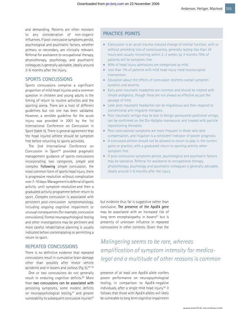
• Concussion is an acute trauma-induced change of mental function, with or without preceding loss of consciousness, generally lasting less than 24 hours and usually recovering within 2–3 weeks; by 3 months 75% of patients will be symptom-free. • 80% of head injury admissions are categorised as mild.• Less than 1% of patients with mild head injury need neurosurgical intervention.• Education about the effects of concussion shortens overall symptom duration and severity.• Early post-traumatic headaches are common and should be treated with simple analgesics, though these are not always as effective as just the passage of time.• Later post-traumatic headaches can be migrainous and then respond to conventional anti-migraine therapies.• Post-traumatic vertigo may be due to benign paroxysmal positional vertigo, can be confirmed on the Dix-Hallpike manoeuvre, and treated with particle repositioning therapies.• Post-concussional symptoms are more frequent in those who seek compensation, and litigation is a consistent indicator of poorer prognosis.• A concussed athlete should not be allowed to return to play in the current game or practice, with a graduated return to sporting activity when symptom free. • If post-concussive symptoms persist, psychological and psychiatric factors may be operative. Referral for assistance to occupational therapy, physiotherapy, psychology and psychiatric colleagues is generally advisable, ideally around 3–6 months after the injury.
PRACTICE POINTS
Malingering seems to be rare, whereas amplification of symptom intensity for medico-legal and a multitude of other reasons is common
on 23 November 2006 pn.bmj.comDownloaded from
Practical Neurology356
10.1136/jnnp.2006.106583
from repeated concussions. However there However there is no association between single or repeated mild head injury and the development of Alzheimer’s disease and other dementias,53,
54 even though repeated mild head trauma in animals accelerates amyloid beta peptide accumulation and cognitive impairment. On the other hand, a single mild closed-head injury is significantly associated with Parkinson’s disease in discordant twin pairs and the association is higher for two head injuries.55 This recent observation supports the notion, controversial until now, that repeated head injury is a risk factor for Parkinson’s disease.
Further research is needed to detect those who might be truly at risk of cognitive impairment or neurodegenerative brain disease from repeated concussions. This has particular pertinence for those involved in sports where there is liability to head injury (for example, rugby, football, horse riding). In the absence of any direct proof that repeated concussions are damaging, with the exception perhaps of boxing, there is no specific reason to advise an athlete against returning to sport once asymptomatic
ACkNOwLEDGEMENTThis article was reviewed by Derick Wade, Oxford, UK.
REFERENCES1. Giza CC, Hovda DA. The neurometabolic cascade of
concussion. J Athl Train 2001;36:228–35.2. Aubry M, Cantu R, Dvorak J, et al. Summary and
agreement statement of the First International Conference on Concussion in Sport, Vienna 2001. Recommendations for the improvement of safety and health of athletes who may suffer concussive injuries. Br J Sports Med 2002;36:6–10.
3. Peloso PM, Carroll LJ, Cassidy JD, et al. Critical evaluation of the existing guidelines on mild traumatic brain injury. J Rehabil Med 2004;43:106–12.
4. Haydel MJ, Preston CA, Mills TJ, et al. Indications for computed tomography in patients with minor head injury. N Engl J Med 2000;343:100–5.
5. Jennett B. Epidemiology of head injury. J Neurol Neurosurg Psychiatry 1996;60:362–9.
6. Kraus JF, Nourjah P. The epidemiology of mild uncomplicated brain injury. J Trauma 1988;28:1637–43.
7. Nell V, Brown DS. Epidemiology of traumatic brain injury in Johannesburg—II. Morbidity, mortality and etiology. Soc Sci Med 1991;33:289–96.
8. Tate RL, McDonald S, Lulham JM. Incidence of hospital-treated traumatic brain injury in an Australian community. Aust N Z J Public Health 1998;22:419–23.
9. Peloso PM, von Holst H, Borg J. Mild traumatic brain injuries presenting to Swedish hospitals in 1987–2000. J Rehabil Med 2004;43:22–7.
10. McAllister TW, Sparling MB, Flashman LA, et al. Neuroimaging findings in mild traumatic brain injury. J Clin Exp Neuropsychol 2001;23:775–91.
11. Hofman PA, Verhey FR, Wilmink JT, et al. Brain lesions in patients visiting a memory clinic with postconcussional sequelae after mild to moderate brain injury. J Neuropsychiatry Clin Neurosci 2002;14:176–84.
12. Levin HS. Neuroplasticity following non-penetrating traumatic brain injury. Brain Injury 2003;17:665–74.
13. Carroll LJ, Cassidy JD, Peloso PM, et al. Prognosis for mild traumatic brain injury: results of the WHO Collaborating Centre Task Force on Mild Traumatic Brain Injury. J Rehabil Med 2004;43:84–105.
14. Wade DT, Crawford S, Wenden FJ, et al. Does routine follow up after head injury help? A randomised controlled trial. J Neurol Neurosurg Psychiatry 1997;62:478–84.
15. Wade DT, King NS, Wenden FJ, et al. Routine follow up after head injury: a second randomised controlled trial. J Neurol Neurosurg Psychiatry 1998;65:177–83.
16. Paniak C, Toller-Lobe G, Reynolds S, et al. A randomized trial of two treatments for mild traumatic brain injury: 1 year follow-up. Brain Inj 2000;14:219–26.
17. De Kruijk JR, Leffers P, Meerhoff S, et al. Effectiveness of bed rest after mild traumatic brain injury: a randomised trial of no versus six days of bed rest. J Neurol Neurosurg Psychiatry 2002;73:167–72.
18. Evans RW. Post-traumatic headaches. Neurol Clin 2004;22:237–49.
19. Gordon CR, Levite R, Joffe V, et al. Is posttraumatic benign paroxysmal positional vertigo different from the idiopathic form? Arch Neurol 2004;61:1590–3.
20. McAllister TW. Mild brain injury and the postconcussion syndrome. In: Silver JM, McAllister TW, Yudofsky SC (eds). Textbook of traumatic brain injury. Washington, DC, US: American Psychiatric Publishing, Inc, 2005: 279–308.
21. King NS. Post-concussion syndrome: Clarity amid the controversy? Br J Psychiatry 2003;183:276–8.
22. Iverson GL. Outcome from mild traumatic brain injury. Curr Opin Psychiatry 2005;18:301–17.
23. Lishman WA. Physiogenesis and psychogenesis in the ‘post-concussional syndrome’. Br J Psychiatry 1988;153:460–9.
24. Jacobson RR. The post-concussional syndrome: physiogenesis, psychogenesis and malingering. An integrative model. J Psychosom Res 1995;39:675–93.
25. Goldstein LB. Prescribing of potentially harmful drugs to patients admitted to hospital after head injury. J Neurol Neurosurg Psychiatry 1995;58:753–5.
26. Ingebrigtsen T, Romner B. Biochemical serum markers for brain damage: a short review with emphasis on clinical utility in mild head injury. Restor Neurol Neurosci 2003;21:171–6.
27. Duff J. The usefulness of quantitative EEG (QEEG) and neurotherapy in the assessment and treatment of post-concussion syndrome. Clin EEG Neurosci 2004;35:198–209.
28. Heitger MH, Anderson TJ, Jones RD, et al. Eye movement and visuomotor arm movement deficits following mild closed head injury. Brain 2004;127:575–90.
on 23 November 2006 pn.bmj.comDownloaded from

www.practical-neurology.com
357Anderson, Heitger, Macleod
29. Mateer CA. Systems of care for post-concussive syndrome. In: Horn L, Zasler N (eds).In: Horn L, Zasler N (eds). Rehabilitation of post-concussive disorders. Philadelphia: Henley & Belfus, 1992:143–60.
30. Mittenberg W, Canyock EM, Condit D, et al. Treatment of post-concussion syndrome following mild head injury. J Clin Exp Neuropsychol 2001;23:829–36.
31. Binder LM, Rohling ML, Larrabee J. A review of mild head trauma. Part I: Meta-analytic review of neuropsychological studies. J Clin Exp Neuropsychol 1997;19:421–31.
32. Cicerone KD, Dahlberg C, Malec JF, et al. Evidence-based cognitive rehabilitation: updated review of the literature from 1998 through 2002. Arch Phys Med Rehabil 2005;86:1681–92.
33. Yeates KO, Taylor HG. Neurobehavioural outcomes of mild head injury in children and adolescents. Pediatr Rehabil 2005;8:5–16.
34. Moore EL, Terryberry-Spohr L, Hope DA. Mild traumatic brain injury and anxiety sequelae: A review of the literature. Brain Inj 2006;20:117–32.
35. Alderfer BS, Arciniegas DB, Silver JM. Treatment of depression following traumatic brain injury. J Head Trauma Rehabil 2005;20:544–62.
36. Lee H, Kim SW, Kim JM, et al. Comparing effects of methylphenidate, sertraline and placebo on neuropsychiatric sequelae in patients with traumatic brain injury. Hum Psychopharmacol 2005;20:97–104.
37. Miller H. Accident neurosis. BMJ 1961;1:919–25, 992–8.
38. Rutherford WH. Postconcussion symptoms: relationship to acute neurological indices, individual differences, and circumstances of injury. In: Levin HS, Eisenberg HM, Benton AL (eds). Mild head injury. New York: Oxford University Press, 1989:217–28.
39. Mickeviciene D, Schrader H, Obelieniene D, et al. A controlled prospective inception cohort study on the post-concussion syndrome outside the medicolegal context. Eur J Neurol 2004;11:411–19.
40. Binder LM. Assessment of malingering after mild head trauma with the Portland Digit Recognition Test. J Clin Exp Neuropsychol 1993;15:170–82.
41. Binder LM, Rohling ML. Money matters: a meta-analytic review of the effects of financial incentives on recovery after closed-head injury. Am J Psychiatry 1996;153:7–10.
42. Feinstein A, Ouchterlony D, Somerville J, et al. The
effects of litigation on symptom expression: a prospective study following mild traumatic brain injury. Med Sci Law 2001;41:116–21.
43. Vickery CD, Berry DTR, Hanlon Inman T, et al. Detection of inadequate effort on neuropsychological testing: A meta-analytic review of selected procedures. Arch Clin Neuropsychol 2001;16:45–73.
44. Moore BA, Donders J. Predictors of invalid neuropsychological test performance after traumatic brain injury. Brain Inj 2004;18:975–84.
45. McCrory P, Johnston K, Meeuwisse W, et al. Summary and agreement statement of the 2nd International Conference on Concussion in Sport, Prague 2004. Br J Sports Med 2005;39:196–204.
46. McCrory P. 2002 Refshauge Lecture. When to retire after concussion?.J Sci Med Sport 2002;5:169–82.
47. Wall SE, Williams WH, Cartwright-Hatton S, et al. Neuropsychological dysfunction following repeat concussions in jockeys. J Neurol Neurosurg Psychiatry 2006;77:518–20.
48. Chappell MH, Ulug AM, Zhang L, et al. Distribution ofDistribution of microstructural damage in the brains of professional boxers: a diffusion MRI study. J Magn Reson Imaging 2006;24:537–42.
49. Iverson GL, Brooks BL, Lovell MR, et al. No cumulative effects for one or two previous concussions. Br J Sports Med 2006;40:72–5.
50. Collins MW, Lovell MR, Iverson GL, et al. Cumulative effects of concussion in high school athletes. Neurosurgery 2002;51:1175–81.
51. Jordan B. Genetic susceptibility to brain injury in sports: a role for genetic testing in athletes? Phys Sportsmed 1998;26:25–6.
52. Sundstrom A, Marklund P, Nilsson LG, et al. APOE influences on neuropsychological function after mild head injury: within-person comparisons. Neurology 2004;62:1963–6.
53. Mehta KM, Ott A, Kalmijn S, et al. Head trauma and risk of dementia and Alzheimer’s disease: The Rotterdam Study. Neurology 1999;53:1959–62.
54. Plassman BL, Havlik RJ, Steffens DC, et al. Documented head injury in early adulthood and risk of Alzheimer’s disease and other dementias. Neurology 2000;55:1158–66.
55. Goldman SM, Tanner CM, Oakes D, et al. Head injury and Parkinson’s disease risk in twins. Ann Neurol 2006;60:65–72.
on 23 November 2006 pn.bmj.comDownloaded from
Related Documents