Concomitant Endoscopic Radiofrequency Ablation and Laparoscopic Reflux Operative Results in More Effective and Efficient Treatment of Barrett Esophagus Trudie A Goers, MD, Pedro Leão, MD, Maria A Cassera, BS, Christy M Dunst, MD, FACS, Lee L Swanström, MD, FACS BACKGROUND: Barrett esophagus (BE) caused by gastroesophageal reflux disease can lead to esophageal cancer. The success of endoscopic treatments with BE eradication depends on esophageal anatomy and post-treatment acid exposure. STUDY DESIGN: Between January 2008 and December 2009, 10 patients were selected for combination treat- ment of BE using laparoscopic anti-reflux surgery and endoscopic radiofrequency ablation. Retrospective review of preoperative, procedural, and postoperative data was performed. RESULTS: Seven study patients had a pathologic diagnosis of nondysplastic BE and 3 patients had a diagnosis of low-grade dysplasia. Average length of BE lesions was 6.4 4.8 cm. Procedure time averaged 154.4 46.4 minutes. At the time of surgery, the mean number of ablations per- formed was 4.39 1.99. Six patients were noted to have major hiatal hernias requiring reduction. Five patients (80%) had 100% resolution of their BE at their first postoperative endoscopy. The remaining 3 patients had a 50% resolution and underwent subsequent endoscopic ablation. Symptomatic results revealed that 4 patients had substantial dysphagia to solids and other symptoms were minimal. Two patients were noted to have complications related to the ablative treatments. One stricture and 1 perforation were observed. CONCLUSIONS: Endoscopic radiofrequency ablation of BE at the time of laparoscopic fundoplication is feasible and can effectively treat BE lesions. A single combined treatment can result in fewer overall procedures performed to obtain BE eradication. ( J Am Coll Surg 2011;213:486–492. © 2011 by the American College of Surgeons) Esophageal cancer has the fastest growing incidence rate of all cancers in the United States and Western Europe, in- creasing 400% during the past 35 years. 1-4 Barrett esopha- gus (BE) represents a marker for the potential development of esophageal adenocarcinoma. This condition develops when gastroesophageal reflux damages the distal esopha- geal squamous mucosa and the resulting injury heals by a metaplastic process. 4,5 BE can progress in a stepwise fashion from intestinal metaplasia to low-grade dysplasia to high- grade dysplasia to carcinoma in situ and, ultimately, inva- sive adenocarcinoma. The mechanisms of this progression are not completely understood. With current understanding of BE’s relationship to gastroesophageal reflux disease and its risk of cancer, we believe that treatment of dysplastic BE should be 2-fold, ie, attention to the dysplastic mucosa and effective treat- ment of chronic gastric reflux exposure that leads to such changes. Radiofrequency ablation (RFA) has been safely and ef- fectively applied to BE with and without dysplastic changes. 6-9 It usually requires 2 to 3 ablation sessions to achieve complete eradication of BE. However, anatomic distortion of the esophagus from large hiatal hernias, stric- tures, esophageal dilation or tortuosity, shortened- esophagus, or, possibly, a previous fundoplication (Fig. 1), can make it more difficult to achieve effective ablation. 10-12 In these scenarios, RFA of BE can require multiple at- tempts or be impossible to accomplish. Disclosure Information: Authors have nothing to disclose. Timothy J Eber- lein, Editor-in-Chief, has nothing to disclose. Abstract presented at Digestive Disease Week, New Orleans, LA, May 2010. Received November 3, 2010; Revised June 15, 2011; Accepted June 15, 2011. From the GI/Minimally Invasive Surgery Division, The Oregon Clinic, Port- land, OR (Goers, Cassera, Dunst, Swanström) and Department of Surgery, Hospital de Braga, Braga, Portugal (Leão). Correspondence address: Lee L Swanström, MD, FACS, GI/Minimally In- vasive Surgery Division, Oregon Health Sciences University, 1040 NW 22 nd Ave, Suite 560, Portland, OR 97210. email: [email protected] 486 © 2011 by the American College of Surgeons ISSN 1072-7515/11/$36.00 Published by Elsevier Inc. doi:10.1016/j.jamcollsurg.2011.06.419

Welcome message from author
This document is posted to help you gain knowledge. Please leave a comment to let me know what you think about it! Share it to your friends and learn new things together.
Transcript

L
f
Concomitant Endoscopic RadiofrequencyAblation and Laparoscopic Reflux OperativeResults in More Effective and Efficient Treatmentof Barrett EsophagusTrudie A Goers, MD, Pedro Leão, MD, Maria A Cassera, BS, Christy M Dunst, MD, FACS,
ee L Swanström, MD, FACS
BACKGROUND: Barrett esophagus (BE) caused by gastroesophageal reflux disease can lead to esophageal cancer.The success of endoscopic treatments with BE eradication depends on esophageal anatomy andpost-treatment acid exposure.
STUDY DESIGN: Between January 2008 and December 2009, 10 patients were selected for combination treat-ment of BE using laparoscopic anti-reflux surgery and endoscopic radiofrequency ablation.Retrospective review of preoperative, procedural, and postoperative data was performed.
RESULTS: Seven study patients had a pathologic diagnosis of nondysplastic BE and 3 patients had adiagnosis of low-grade dysplasia. Average length of BE lesions was 6.4 � 4.8 cm. Procedure timeaveraged 154.4 � 46.4 minutes. At the time of surgery, the mean number of ablations per-formed was 4.39 � 1.99. Six patients were noted to have major hiatal hernias requiringreduction. Five patients (80%) had 100% resolution of their BE at their first postoperativeendoscopy. The remaining 3 patients had a �50% resolution and underwent subsequentendoscopic ablation. Symptomatic results revealed that 4 patients had substantial dysphagia tosolids and other symptoms were minimal. Two patients were noted to have complicationsrelated to the ablative treatments. One stricture and 1 perforation were observed.
CONCLUSIONS: Endoscopic radiofrequency ablation of BE at the time of laparoscopic fundoplication is feasibleand can effectively treat BE lesions. A single combined treatment can result in fewer overallprocedures performed to obtain BE eradication. (J Am Coll Surg 2011;213:486–492. © 2011
by the American College of Surgeons)gsa
gbimc
fc
Esophageal cancer has the fastest growing incidence rate ofall cancers in the United States and Western Europe, in-creasing 400% during the past 35 years.1-4 Barrett esopha-gus (BE) represents a marker for the potential developmentof esophageal adenocarcinoma. This condition developswhen gastroesophageal reflux damages the distal esopha-geal squamous mucosa and the resulting injury heals by ametaplastic process.4,5 BE can progress in a stepwise fashionrom intestinal metaplasia to low-grade dysplasia to high-
Disclosure Information: Authors have nothing to disclose. Timothy J Eber-lein, Editor-in-Chief, has nothing to disclose.Abstract presented at Digestive Disease Week, New Orleans, LA, May 2010.
Received November 3, 2010; Revised June 15, 2011; Accepted June 15, 2011.From the GI/Minimally Invasive Surgery Division, The Oregon Clinic, Port-land, OR (Goers, Cassera, Dunst, Swanström) and Department of Surgery,Hospital de Braga, Braga, Portugal (Leão).Correspondence address: Lee L Swanström, MD, FACS, GI/Minimally In-
nd
vasive Surgery Division, Oregon Health Sciences University, 1040 NW 22Ave, Suite 560, Portland, OR 97210. email: [email protected]486© 2011 by the American College of SurgeonsPublished by Elsevier Inc.
rade dysplasia to carcinoma in situ and, ultimately, inva-ive adenocarcinoma. The mechanisms of this progressionre not completely understood.
With current understanding of BE’s relationship toastroesophageal reflux disease and its risk of cancer, weelieve that treatment of dysplastic BE should be 2-fold,e, attention to the dysplastic mucosa and effective treat-
ent of chronic gastric reflux exposure that leads to suchhanges.
Radiofrequency ablation (RFA) has been safely and ef-ectively applied to BE with and without dysplastichanges.6-9 It usually requires 2 to 3 ablation sessions to
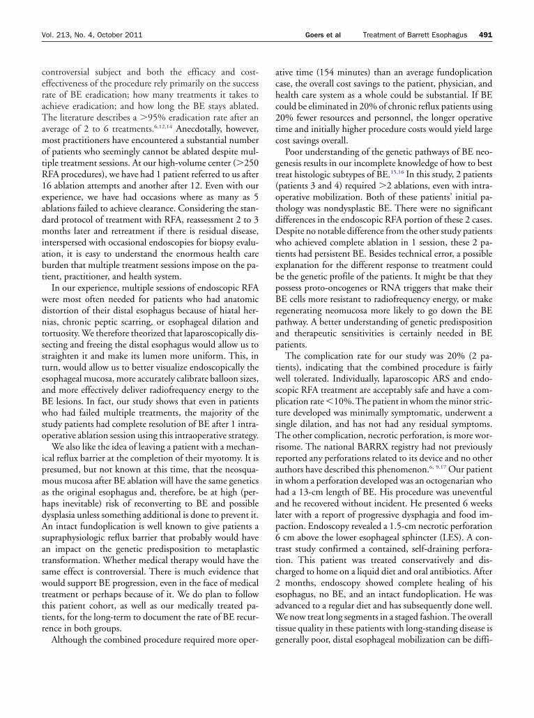
achieve complete eradication of BE. However, anatomicdistortion of the esophagus from large hiatal hernias, stric-tures, esophageal dilation or tortuosity, shortened-esophagus, or, possibly, a previous fundoplication (Fig. 1),can make it more difficult to achieve effective ablation.10-12
In these scenarios, RFA of BE can require multiple at-
tempts or be impossible to accomplish.ISSN 1072-7515/11/$36.00doi:10.1016/j.jamcollsurg.2011.06.419

487Vol. 213, No. 4, October 2011 Goers et al Treatment of Barrett Esophagus
As the patient’s underlying genetic predisposition to de-velop BE remains unchanged, prevention of reflux seemsmandatory to prevent recurrence. Optimal control of re-flux after ablation, however, has not been defined. Currentoptions include lifetime high-dose acid suppression or anti-reflux surgery (ARS). The role of ARS certainly has theo-retical appeal because it provides an absolute barrier to allgastric contents and there is ample evidence that acid aloneis not the causative agent of BE or dysplasia. If one acceptsthe idea of adding an ARS to BE ablation, the question ofthe timing of the 2 procedures arises. For example, al-though ARS before ablation can straighten the esophagusand cure esophagitis and strictures, the fundoplication it-self can interfere with effective RFA by obscuring the land-marks of the gastroesophageal junction or making access tothe distal-most segments of the metaplasia more difficult.Performing ARS after an ablation can also be more difficultbecause of transmural inflammatory changes, or completeablation can never be possible because of the anatomicaldistortion of the esophagus (angulation, dilatation, short-ening) related to hiatal herniation.13
We hypothesize that performing endoscopic RFA of BEat the time of ARS would be a safe and effective methodthat can reduce the number of treatments needed to erad-icate the metaplasia by reducing the hernia and straighten-ing the esophagus, as well as providing the best chance ofpreventing future BE recurrence.
METHODSThis study involved the retrospective review of patientcharts and information that was prospectively collected foran IRB-approved data registry containing patients who hadendoscopic balloon�based esophageal RFA. All proce-dures were performed between January 2008 and Decem-ber 2009.
PatientsTwo groups of patients were included. The first includedpatients who were scheduled for ARS because of eitherfailure or dislike of chronic medical therapy and who hadendoscopic and histologic presence of BE with no or low-grade dysplasia. Patients with high-grade dysplasia were
Abbreviations and Acronyms
ARS � anti-reflux surgeryBE � Barrett esophagusRFA � radiofrequency ablation
excluded from selection in this group, as the presence of a
fundoplication might compromise future oncologic resec-tion and reconstruction if needed.
The second group of patients included those who orig-inally had biopsy-proven BE with either low- or high-gradedysplasia and who were considered failures to ablate (�3attempts) because of anatomic distortion of the esophagusfrom dilation, tortuosity, or hiatal hernias; and those whocurrently had either nondysplastic or low-grade dysplasiaon biopsy. Likewise, patients with persistent high-gradedysplasia were excluded and were instead considered forminimally invasive esophagectomy.
Presenting and procedural dataDemographic data were prospectively collected includingage, sex, and medical history. Histopathology in the ab-sence of esophagitis, 24-hour pH, and esophageal mano-
Figure 1. Contrast study of patient with long-standing gastroesoph-ageal reflux disease, paraesophageal hernia, and chronic esopha-geal mucosal scarring.
metric testing were recorded in an Access-based (Mi-

MdNcftofdd(
cosa
488 Goers et al Treatment of Barrett Esophagus J Am Coll Surg
crosoft) database. Baseline BE measurements were takenfrom the first endoscopy. Endoscopy was performed usingwhite light and narrow-band imaging. The endoscopic bi-opsy protocol included 4 quadrant biopsies taken at 2-cmintervals and all nodular areas were removed with endoscopicsubmucosal resection (EMR). Dysplasia was recorded basedonly on biopsies taken in the absence of acute inflammation.Presence, type, and size of hiatal hernia were also recorded. Atthe time of surgical procedure, information about the lengthof ablation, type of electrode, and details of treatment wererecorded from the operative report.
Surgical procedureAll procedures were done under general anesthesia in thesupine position using 5 laparoscopic ports. Mediastinal dis-sections were performed with the end point of the gastro-esophageal junction lying 3 cm below the hiatus (Fig. 2).After mobilization of the distal esophagus and stomach, theesophagus was sized with a sizing balloon and anappropriate-sized RFA ablation balloon (HALO360; Barrx
edical) was selected by a second surgeon performing en-oscopy. After irrigating the esophagus with 1%-acetylcysteine to remove mucous, the balloon ablation
atheter was inserted over a guidewire and ablation per-ormed as the laparoscopic surgeon applied careful tractiono straighten and align the esophagus to maximize contactf the ablation balloon. Two cycles of ablations were per-ormed at either 10 J (no dysplasia) or 12 J (history ofysplasia). Two patients with more localized and limitedisease had ablation with a endoscope-mounted electrodeHALO90; Barrx Medical) instead of the balloon electrode,
but using the same ablation protocol. Once the ablationwas completed, repair of any hiatal hernia was performed
Figure 2. (A) Extensive type II mediastinal diagus and the gastroesophageal junction reballoon electrode can contact esophageal mu
and a tailored fundoplication was performed.
Postoperative careAll patients were admitted overnight and observed. Pa-tients were placed on proton pump inhibitors and weregiven liquid pain medication as needed. Those who hadconcomitant performance of a paraesophageal hernia werekept nothing per os overnight and underwent esophago-gastric radiographic study with water-soluble contrast onthe first postoperative morning. If no leak was detected,patients were started on a liquid diet. Those with minimalhiatal hernias were started on a liquid diet the night ofsurgery. All patients were instructed to continue the liquiddiet for 2 days, followed by a pureed diet for 2 weeks. Thepatients were instructed to take a single-dose proton pumpinhibitor for the first 3 weeks after surgery to protect thedenuded distal esophagus.
Outcome measurementsA standardized gastrointestinal symptom assessment toolwas administered at each visit. Patients were followed up inclinic at 2 weeks, 3 months, and 6 months and then yearly.A focused physical examination was performed and anycomplications or side effects from the surgery were re-corded. Patients who had any severe symptoms at any visitunderwent appropriate studies and treatment at that visit.
Routine follow-up endoscopy was performed 2 to 3months after the combination procedure to assess com-pleteness of the ablation. Esophageal mucosa was examinedusing white light and narrow-band imaging. Subjectivemapping of residual BE lesions’ length and circumferentialvs. focal nature were recorded in the endoscopic report.Four quadrant biopsies were repeated at 2-cm intervals ofboth normal-appearing mucosa and columnar mucosa us-ing endoscopic jumbo forceps and were sent in formalin forpathology. Percentage of BE resolution was estimated by
ion allows straightening of the distal esoph-ithin the abdomen. (B) The radiofrequency.
ssectsts w
the endoscopist based on comparison with preoperative

ot
c
cnlpatdflpsb
A
F
PCB
F
489Vol. 213, No. 4, October 2011 Goers et al Treatment of Barrett Esophagus
photos and detailed endoscopy notes. Then, at 6 months,the integrity of the fundoplication was determined per ourusual protocol using endoscopy, high-resolution manome-try, and 24-hour pH studies. A competent fundoplicationwas defined as an intact wrap on endoscopic retroflection(Hill grade 1) and/or normal acid exposure by 24-hour pHtests (DeMeester score �14.7). If BE was present at the 3-r 6-month visit, the patient had an additional RFA at thatime.
Data analysisData that was collected was stored in an IRB-approveddatabase that was developed and maintained by the princi-pal investigator. The means of all of the continuous vari-ables were compared using appropriate parametric or non-parametric tests. Statistical analysis was performed usingPredictive Analytics Software (version 18.0; SPSS, Inc).
RESULTSDuring the 24-month data collection period, 78 patientswith BE underwent treatment, of these, 15 patients met theinclusion criteria to undergo concomitant endoscopic RFAand laparoscopic fundoplication for the simultaneoustreatment of BE and gastroesophageal reflux disease. Ofthese, 1 patient requested esophagectomy, 1 patient re-jected the follow-up protocol, and 3 did not want a fundo-plication. Therefore, 10 patients agreed to the combinedprocedure.
Patients varied in age from 23 to 80 years old (Table 1).Similarly, American Society of Anesthesiologists scoreswere all 2, with the exception of a 23-year-old patient who
Table 1. Demographic and Preoperative Data ofStudy PatientsAge, y, mean � SD 58 � 16.6Body mass index, mean � SD 34 � 9Male sex, n (%) 7 (70)ASA, n (%)
1 1 (10)2 9 (90)
Histology, n (%)Nondysplastic 7 (70)Low-grade dysplasia 3 (30)GERD 10 (100)Esophagitis 1 (10)
Preprocedure ablations, n (%)None 3 (30)Multiple 7 (4 –6)
ASA, American Society of Anesthesiologists; GERD, gastroesophageal refluxdisease.
was scored as a 1. The average body mass index of the group
was in the category of obese (34 � 9), and no patient wasonsidered to be normal (interquartile range 26.1�38.0).
At the time of the study, all patients had biopsy-onfirmed BE. Seven study patients had a pathologic diag-osis of nondysplastic BE and 3 patients had a diagnosis of
ow-grade dysplasia. All 10 patients had abnormal 24-hourH testing. All patients had high-resolution manometrynd 1 patient had a profound primary esophageal dysmo-ility. Only 1 patient had active esophagitis at the time ofiagnosis. Seven of the patients were considered to haveailed RFA because of BE persistence despite multiple ab-ation attempts (4 to 7 attempts). The other 3 were de novoatients with long-segment BE (2 with low-grade dyspla-ia, 1 without) who were seeking ARS for symptom controlecause of failure of medical management.
Procedural dataThe combined procedure time averaged 154.4 � 46.4minutes (Table 2). At the time of surgery, 6 patients werenoted to have major hiatal hernias (type III) requiringreduction and crural reconstruction. Nine patients un-derwent 360-degree fundoplication and the patient withpoor esophageal motility had a 270-degree posteriorfundoplication.
The average length of BE lesions was 6.4 � 4.8 cm(Table 2). Eighty percent required the 360-degree balloonelectrode for circumferential disease. The 90-degreeHALO electrode was used in 20% for more focused energyapplication. One patient had both circumferential and fo-cal islands of disease and required the use of both the 360-degree and 90-degree electrodes. Of the 3 patients with
Table 2. Procedural Data of Study PatientsLength of procedure, min, mean � SD 154.4 � 46.41Barrett’s esophagus length, cm, mean � SD 6.4 � 4.78
blations performed during initial procedure, n,mean � SD 4.39 � 1.99
undoplication, n (%)Nissen 9 (90)Toupet 1 (10)
resence of large hiatal hernia, n (%) 6 (60)ircumferential lesions/360 balloon used, n (%) 8 (80)alloon size, mm, n (%)25 5 (50)28 2 (20)31 3 (30)
ocal lesions/HALO90 used, n (%) 3 (30)Energy, J, n (%)
10 6 (60)12 4 (40)
dysplasia, all had energy applied at 12 J/cm2 3 with the

b
bdbs
ul
w
490 Goers et al Treatment of Barrett Esophagus J Am Coll Surg
360-degree device. The diameter of the 360-degree balloonelectrode used varied. Fifty percent of patients required the25-mm balloon. However, other patients had larger esoph-ageal diameters and the 28-mm (n � 2) or 31-mm (n � 3)alloon electrodes were used.There were no surgical or endoscopic complications and
lood loss was �50 mL in all cases. Eight patients wereischarged home on postoperative day 1 and 2 at 48 hoursecause of transportation issues. There were no readmis-ions or acute perioperative problems.
Symptomatic evaluationPatients were seen between 2 and 4 weeks postoperativelyfor acute recovery data, including pain control, diet pro-gression, and other subjective data. Results of the symptomquestionnaire are shown in Figure 3.
BE resolutionLong-term follow-up ranged from 7 months to 28 months(mean 17 months). All patients completed their 6-monthcomprehensive evaluation, 8 completed their 1-year evalu-ation, and 4 completed a 24-month follow-up.
All patients were free of BE at time of last follow-up.One had biopsies with columnar epithelium, but no intes-tinal metaplasia. Six patients (60%) had 100% resolutionof their BE after 1 intraoperative ablative treatment per-formed at the time of their fundoplication (Table 3). Thisincluded 4 patients who had failed previous multiple at-tempts at ablation. None of the remaining BE patients haddysplasia. The remaining 4 patients had a �50% resolu-tion and underwent endoscopic ablation. At their secondfollow-up endoscopy, 3 patients were found to have resid-ual BE, however, their overall disease burden was less. A
Figure 3. Postoperative patient symptomatic evaluation at 2 to 4eeks. White bar, absent; black bar, present.
third ablation succeeded in complete control, although 1 k
patient continued to have columnar epithelium with nointestinal metaplasia at 24-month follow-up.
Two patients had major complications related to theablation treatments. One patient had a soft stricture notedat their first diagnostic endoscopy performed on postoper-ative day 48 for mild solid-food dysphagia. A second pa-tient was evaluated at 6 weeks postoperatively because of areport of a food impaction. This patient was evaluated withupper endoscopy and was found to have a 1.5-cm perfora-tion within the proximal RFA field.
There were no particular postoperative complicationsattributable to the fundoplication, although patients fre-quently noted common side effects, such as early satiety,bloating, and flatulence. At longest follow-up, no patientshad reflux complaints and 1 patient reported heartburn.Three patients were on peptic medications. All fundopli-cations were intact (Hill grade I) on last endoscopy. Eightpatients had postoperative manometry and pH studies andall results were within the normal range, including the 1patient with heartburn and all 3 patients on proton pumpinhibitors postoperatively.
DISCUSSIONTreatment of BE with endoscopic RFA is a relatively newconcept. The technique so far has been reported to be botheffective and well-tolerated.10,11 Exact indications for its
se, however, have yet to be worked out completely and theong-term efficacy of endoscopic ablations remains un-
Table 3. Endoscopic Evaluation Results of Barrett Esopha-gus Resolution Status Post Combined Therapy
Patientno.
Dayspost-
operation%
Resolution ComplicationHill grade
fundoplication
1 205 100 No 12 48 100 Yes, stricture* 13 90 50 No 1
159 75 No 1220 100 No
4 78 85 No 1166 95 No 1249 100 No
5 42 100 Yes, perforation 16 202 100 No 17 186 100 No 18 187 50 No 1
255 100 No 19 190 100 No 1
10 60 80 No 1376 100* No 1
*Columnar epithelium without intestinal metaplasia.
nown. The cost-effectiveness of this treatment is also a
motR1eadmiabt
wdntssteaBwso
ipmahdAsatswtttr
achc2tc
gt
ihalp6ttc2eaWt
491Vol. 213, No. 4, October 2011 Goers et al Treatment of Barrett Esophagus
controversial subject and both the efficacy and cost-effectiveness of the procedure rely primarily on the successrate of BE eradication; how many treatments it takes toachieve eradication; and how long the BE stays ablated.The literature describes a �95% eradication rate after anaverage of 2 to 6 treatments.6,12,14 Anecdotally, however,
ost practitioners have encountered a substantial numberf patients who seemingly cannot be ablated despite mul-iple treatment sessions. At our high-volume center (�250FA procedures), we have had 1 patient referred to us after6 ablation attempts and another after 12. Even with ourxperience, we have had occasions where as many as 5blations failed to achieve clearance. Considering the stan-ard protocol of treatment with RFA, reassessment 2 to 3onths later and retreatment if there is residual disease,
nterspersed with occasional endoscopies for biopsy evalu-tion, it is easy to understand the enormous health careurden that multiple treatment sessions impose on the pa-ient, practitioner, and health system.
In our experience, multiple sessions of endoscopic RFAere most often needed for patients who had anatomicistortion of their distal esophagus because of hiatal her-ias, chronic peptic scarring, or esophageal dilation andortuosity. We therefore theorized that laparoscopically dis-ecting and freeing the distal esophagus would allow us totraighten it and make its lumen more uniform. This, inurn, would allow us to better visualize endoscopically thesophageal mucosa, more accurately calibrate balloon sizes,nd more effectively deliver radiofrequency energy to theE lesions. In fact, our study shows that even in patientsho had failed multiple treatments, the majority of the
tudy patients had complete resolution of BE after 1 intra-perative ablation session using this intraoperative strategy.
We also like the idea of leaving a patient with a mechan-cal reflux barrier at the completion of their myotomy. It isresumed, but not known at this time, that the neosqua-ous mucosa after BE ablation will have the same genetics
s the original esophagus and, therefore, be at high (per-aps inevitable) risk of reconverting to BE and possibleysplasia unless something additional is done to prevent it.n intact fundoplication is well known to give patients a
upraphysiologic reflux barrier that probably would haven impact on the genetic predisposition to metaplasticransformation. Whether medical therapy would have theame effect is controversial. There is much evidence thatould support BE progression, even in the face of medical
reatment or perhaps because of it. We do plan to followhis patient cohort, as well as our medically treated pa-ients, for the long-term to document the rate of BE recur-ence in both groups.
Although the combined procedure required more oper- g
tive time (154 minutes) than an average fundoplicationase, the overall cost savings to the patient, physician, andealth care system as a whole could be substantial. If BEould be eliminated in 20% of chronic reflux patients using0% fewer resources and personnel, the longer operativeime and initially higher procedure costs would yield largeost savings overall.
Poor understanding of the genetic pathways of BE neo-enesis results in our incomplete knowledge of how to bestreat histologic subtypes of BE.15,16 In this study, 2 patients
(patients 3 and 4) required �2 ablations, even with intra-operative mobilization. Both of these patients’ initial pa-thology was nondysplastic BE. There were no significantdifferences in the endoscopic RFA portion of these 2 cases.Despite no notable difference from the other study patientswho achieved complete ablation in 1 session, these 2 pa-tients had persistent BE. Besides technical error, a possibleexplanation for the different response to treatment couldbe the genetic profile of the patients. It might be that theypossess proto-oncogenes or RNA triggers that make theirBE cells more resistant to radiofrequency energy, or makeregenerating neomucosa more likely to go down the BEpathway. A better understanding of genetic predispositionand therapeutic sensitivities is certainly needed in BEpatients.
The complication rate for our study was 20% (2 pa-tients), indicating that the combined procedure is fairlywell tolerated. Individually, laparoscopic ARS and endo-scopic RFA treatment are acceptably safe and have a com-plication rate �10%.The patient in whom the minor stric-ture developed was minimally symptomatic, underwent asingle dilation, and has not had any residual symptoms.The other complication, necrotic perforation, is more wor-risome. The national BARRX registry had not previouslyreported any perforations related to its device and no otherauthors have described this phenomenon.6, 9,17 Our patientn whom a perforation developed was an octogenarian whoad a 13-cm length of BE. His procedure was uneventfulnd he recovered without incident. He presented 6 weeksater with a report of progressive dysphagia and food im-action. Endoscopy revealed a 1.5-cm necrotic perforationcm above the lower esophageal sphincter (LES). A con-
rast study confirmed a contained, self-draining perfora-ion. This patient was treated conservatively and dis-harged to home on a liquid diet and oral antibiotics. After
months, endoscopy showed complete healing of hissophagus, no BE, and an intact fundoplication. He wasdvanced to a regular diet and has subsequently done well.e now treat long segments in a staged fashion. The overall
issue quality in these patients with long-standing disease is
enerally poor, distal esophageal mobilization can be diffi-
492 Goers et al Treatment of Barrett Esophagus J Am Coll Surg
cult and result in tissue damage and thinning of the esoph-ageal wall. With this, radiofrequency energy applicationoverall distance can compromise tissue microvasculatureand result in easier necrosis. Because of these concerns, wecurrently limit our ablations to lesions �5 cm or we will dothem in a planned staged program.
Despite these questions, there is little doubt that RFA forBE has dramatically altered the treatment paradigm fordysplastic BE and has resulted in the sparing of many anesophagus that would have otherwise been removed for thisproblem. The purpose of this study was to use endoscopicablative technology in conjunction with laparoscopic ARSto improve electrode contact, thereby increasing the successrate of complete ablation. We show that this improves over-all BE ablation efficiency, decreases BE recurrence, and willhopefully impact long-term cancer risk.
CONCLUSIONSIntraoperative endoscopic RFA of BE at the time of lapa-roscopic fundoplication is feasible and might be a moreefficient and cost-effective way to treat BE. We show that asingle combined treatment results in the need for feweroverall procedures performed to obtain BE eradication. Al-though the complication rate of this pilot study was notnegligible, patients did well with conservative treatmentand our procedural approach has been subsequently mod-ified with promising results. We believe that prospectivestudy of this combined treatment modality for patientswith BE is warranted.
Author ContributionsStudy conception and design: Goers, Swanström.Acquisition of data: Goers, Leão, Cassera.Analysis and interpretation of data: Goers, Leão, Cassera,
Swanström.Drafting of manuscript: Goers, Cassera, Swanström.Critical revision: Goers, Cassera, Dunst, Swanström.
REFERENCES
1. Sharma P, Dent J, Armstrong D, et al. The development andvalidation of an endoscopic grading system for Barrett’s esoph-agus: the Prague C & M criteria. Gastroenterology 2006;131:
1392–1399.2. Sharma P, McQuaid K, Dent J, et al. A critical review of thediagnosis and management of Barrett’s esophagus: the AGAChicago Workshop. Gastroenterology 2004;127:310–330.
3. Wang KK, Sampliner RE. Updated guidelines 2008 for the di-agnosis, surveillance and therapy of Barrett’s esophagus. Am JGastroenterol 2008;103:788–797.
4. Shaheen NJ, Sharma P, Overholt BF, et al. Radiofrequency ab-lation in Barrett’s esophagus with dysplasia. N Engl J Med 2009;360:2277–2288.
5. Spechler SJ. Clinical practice. Barrett’s esophagus. N Engl J Med2002;346:836–842.
6. Fleischer DE, Overholt BF, Sharma VK, et al. Endoscopic abla-tion of Barrett’s esophagus: a multicenter study with 2.5-yearfollow-up. Gastrointest Endosc 2008;68:867–876.
7. Ganz RA, Utley DS, Stern RA, et al. Complete ablation ofesophageal epithelium with a balloon-based bipolar electrode: aphased evaluation in the porcine and in the human esophagus.Gastrointest Endosc 2004;60:1002–1010.
8. Dunkin BJ, Martinez J, Bejarano PA, et al. Thin-layer ablationof human esophageal epithelium using a bipolar radiofrequencyballoon device. Surg Endosc 2006;20:125–130.
9. Vassiliou MC, von Renteln D, Wiener DC, et al. Treatment ofultralong-segment Barrett’s using focal and balloon-based radio-frequency ablation. Surg Endosc 2010;24:786–791.
10. Hubbard N, Velanovich V. Endoscopic endoluminal radiofre-quency ablation of Barrett’s esophagus in patients with fundo-plications. Surg Endosc 2007;21:625–628.
11. dos Santos RS, Bizekis C, Ebright M, et al. Radiofrequencyablation for Barrett’s esophagus and low-grade dysplasia in com-bination with an antireflux procedure: a new paradigm. JThoracCardiovasc Surg 2010;139:713–716.
12. Smith CD, Bejarano PA, Melvin WS, et al. Endoscopic ablationof intestinal metaplasia containing high-grade dysplasia inesophagectomy patients using a balloon-based ablation system.Surg Endosc 2007;21:560–569.
13. Awad ZT, Mittal SK, Roth TA, et al. Esophageal shorteningduring the era of laparoscopic surgery. World J Surg 2001;25:558–561.
14. Sharma VK, Wang KK, Overholt BF, et al. Balloon-based, cir-cumferential, endoscopic radiofrequency ablation of Barrett’sesophagus: 1-year follow-up of 100 patients. Gastrointest En-dosc 2007;65:185–195.
15. Nicholson A, Jankowski J. Editorial: one small step for metapla-sia, but one giant leap for biomarkers is needed. Am J Gastro-enterol 2009;104:2681–2683.
16. Vallbohmer D, Marjoram P, Kuramochi H, et al. Towards themolecular characterization of disease: comparison of molecularand histological analysis of esophageal epithelia. J GastrointestSurg 2007;11:1095–1104.
17. Csendes A, Braghetto I, Burdiles P, et al. Late results of thesurgical treatment of 125 patients with short-segment Barrett
esophagus. Arch Surg 2009;144:921–927.
Related Documents