Concentric organization of A- and B-type lamins predicts their distinct roles in the spatial organization and stability of the nuclear lamina Bruce Nmezi a,1 , Jianquan Xu b,c,1 , Rao Fu b,c,d,1 , Travis J. Armiger e , Guillermo Rodriguez-Bey a , Juliana S. Powell a , Hongqiang Ma b,c , Mara Sullivan f , Yiping Tu g , Natalie Y. Chen g , Stephen G. Young g , Donna B. Stolz f , Kris Noel Dahl e,2 , Yang Liu b,c,2 , and Quasar S. Padiath a,2 a Department of Human Genetics, University of Pittsburgh, Pittsburgh, PA 15261; b Department of Medicine, University of Pittsburgh, Pittsburgh, PA 15213; c Department of Bioengineering, University of Pittsburgh, Pittsburgh, PA 15213; d College of Chemical Engineering, Northeast Electric Power University, Jilin Province, China 132012; e Department of Biomedical Engineering, Carnegie Mellon University, Pittsburgh, PA 15213; f Department of Cell Biology, University of Pittsburgh, Pittsburgh, PA 15213; and g Department of Medicine, University of California, Los Angeles, CA 90095 Edited by John W. Sedat, University of California, San Francisco, CA, and approved January 15, 2019 (received for review June 15, 2018) The nuclear lamina is an intermediate filament meshwork adjacent to the inner nuclear membrane (INM) that plays a critical role in maintaining nuclear shape and regulating gene expression through chromatin interactions. Studies have demonstrated that A- and B-type lamins, the filamentous proteins that make up the nuclear lamina, form independent but interacting networks. However, whether these lamin subtypes exhibit a distinct spatial organization or whether their organization has any functional consequences is unknown. Using stochastic optical reconstruction microscopy (STORM) our studies reveal that lamin B1 and lamin A/C form concentric but overlapping networks, with lamin B1 forming the outer concentric ring located adjacent to the INM. The more peripheral localization of lamin B1 is mediated by its carboxyl-terminal farnesyl group. Lamin B1 localization is also curvature- and strain-dependent, while the localization of lamin A/C is not. We also show that lamin B1’s outer-facing localization stabilizes nuclear shape by restraining outward protrusions of the lamin A/C network. These two findings, that lamin B1 forms an outer concentric ring and that its localization is energy-dependent, are significant as they suggest a distinct model for the nuclear lamina—one that is able to predict its behavior and clarifies the distinct roles of individual nuclear lamin proteins and the conse- quences of their perturbation. nucleus | lamin | bleb | curvature | meshwork T he nuclear lamina is a meshwork of intermediate filaments that lies beneath the inner nuclear membrane (INM) in all metazoan cells (1). In addition to playing a critical role in reg- ulating the shape and structural integrity of the nuclear envelope, it also has important functions in regulating gene expression through chromatin interactions and integrating cytoskeletal dy- namics within the cell (2, 3). Two types of intermediate filament proteins make up the nuclear lamina: A-type lamins, primarily lamin A and lamin C, are coded for by LMNA, while B-type lamins include lamin B1 and lamin B2 and are coded for by LMNB1 and LMNB2, respectively (4–6). The two lamin subtypes also undergo distinct posttranslational modifications, with the B- type lamins retaining the addition of a farnesyl group, while this is not present in A-type lamins (7). A-type lamin expression is associated with differentiated mesenchymal cells and with stiffer cells and nuclei (8). Mice null for Lmna initially develop normally but succumb to muscular dystrophy and cardiomyopathy after 4–6 wk (9). Alternatively, neurons and glia in the central nervous system have high lamin C expression but little to no lamin A (10). In contrast, B-type lamins are expressed in all cell types throughout development and differentiation and are required for proper organogenesis as well as neuronal migration and patterning during brain devel- opment (11, 12). Mouse embryonic fibroblasts (MEFs) from Lmna-knockout mice demonstrate highly elongated nuclei with the loss of nuclear envelope proteins, including B-type lamins, from one pole (9). In contrast, knockouts of Lmnb1 result in excessive nuclear herniations known as blebs that are highly as- sociated with gene-rich euchromatin (13). Mutations in lamin A result in a wide variety of diseases, collectively termed as lam- inopathies, that include muscular dystrophies, lipodystrophies, and premature aging phenotypes such as Hutchinson–Gilford progeria syndrome (HGPS), while mutations in lamin B1 cause the de- myelinating disorder autosomal dominant leukodystrophy (14). Studies utilizing optical microscopy and cryo-electron tomog- raphy to visualize the structural organization of lamins in mammalian cells revealed the presence of a meshwork structure and indicated that each component formed separate but interacting meshworks (13, 15, 16). However, no distinct spatial organization of the indi- vidual lamin subtypes across the nuclear envelope was reported. Here, we used stochastic optical reconstruction microscopy (STORM) and quantitative image analysis to examine the spatial localization of lamin A/C and lamin B1 at the nuclear periphery. Significance The nuclear lamina is an integral component of all metazoan cells. While the individual constituents of the nuclear lamina, the A- and B-type lamins, have been well studied, whether they exhibit a distinct spatial organization is unclear. Using stochastic optical reconstruction microscopy, we have identi- fied two organizing principles of the nuclear lamina: lamin B1 forms an outer concentric ring, and its localization is curvature-dependent. This suggests that a role of lamin B1 is to stabilize nuclear shape by restraining outward protrusions of the lamin A/C network. These findings provide a model for the nuclear lamina that can predict its behavior and clarify the distinct functional roles for the individual lamina components and the consequences of their perturbation. Author contributions: B.N., J.X., R.F., D.B.S., K.N.D., Y.L., and Q.S.P. designed research; B.N., J.X., R.F., T.J.A., G.R.-B., J.S.P., M.S., D.B.S., and K.N.D. performed research; Y.T., N.Y.C., and S.G.Y. contributed new reagents/analytic tools; B.N., J.X., R.F., T.J.A., G.R.-B., J.S.P., H.M., K.N.D., Y.L., and Q.S.P. analyzed data; and B.N., K.N.D., Y.L., and Q.S.P. wrote the paper. The authors declare no conflict of interest. This article is a PNAS Direct Submission. Published under the PNAS license. 1 B.N., J.X., and R.F. contributed equally to this work. 2 To whom correspondence may be addressed. Email: [email protected], [email protected], or [email protected]. This article contains supporting information online at www.pnas.org/lookup/suppl/doi:10. 1073/pnas.1810070116/-/DCSupplemental. Published online February 14, 2019. www.pnas.org/cgi/doi/10.1073/pnas.1810070116 PNAS | March 5, 2019 | vol. 116 | no. 10 | 4307–4315 CELL BIOLOGY Downloaded by guest on March 9, 2020

Welcome message from author
This document is posted to help you gain knowledge. Please leave a comment to let me know what you think about it! Share it to your friends and learn new things together.
Transcript
Concentric organization of A- and B-type laminspredicts their distinct roles in the spatial organizationand stability of the nuclear laminaBruce Nmezia,1, Jianquan Xub,c,1, Rao Fub,c,d,1, Travis J. Armigere, Guillermo Rodriguez-Beya, Juliana S. Powella,Hongqiang Mab,c, Mara Sullivanf, Yiping Tug, Natalie Y. Cheng, Stephen G. Youngg, Donna B. Stolzf, Kris Noel Dahle,2,Yang Liub,c,2, and Quasar S. Padiatha,2
aDepartment of Human Genetics, University of Pittsburgh, Pittsburgh, PA 15261; bDepartment of Medicine, University of Pittsburgh, Pittsburgh, PA 15213;cDepartment of Bioengineering, University of Pittsburgh, Pittsburgh, PA 15213; dCollege of Chemical Engineering, Northeast Electric Power University, JilinProvince, China 132012; eDepartment of Biomedical Engineering, Carnegie Mellon University, Pittsburgh, PA 15213; fDepartment of Cell Biology, Universityof Pittsburgh, Pittsburgh, PA 15213; and gDepartment of Medicine, University of California, Los Angeles, CA 90095
Edited by John W. Sedat, University of California, San Francisco, CA, and approved January 15, 2019 (received for review June 15, 2018)
The nuclear lamina is an intermediate filament meshwork adjacentto the inner nuclear membrane (INM) that plays a critical role inmaintaining nuclear shape and regulating gene expressionthrough chromatin interactions. Studies have demonstrated thatA- and B-type lamins, the filamentous proteins that make up thenuclear lamina, form independent but interacting networks.However, whether these lamin subtypes exhibit a distinct spatialorganization or whether their organization has any functionalconsequences is unknown. Using stochastic optical reconstructionmicroscopy (STORM) our studies reveal that lamin B1 and laminA/C form concentric but overlapping networks, with lamin B1forming the outer concentric ring located adjacent to the INM.The more peripheral localization of lamin B1 is mediated by itscarboxyl-terminal farnesyl group. Lamin B1 localization is alsocurvature- and strain-dependent, while the localization of laminA/C is not. We also show that lamin B1’s outer-facing localizationstabilizes nuclear shape by restraining outward protrusions ofthe lamin A/C network. These two findings, that lamin B1 forms anouter concentric ring and that its localization is energy-dependent,are significant as they suggest a distinct model for the nuclearlamina—one that is able to predict its behavior and clarifies thedistinct roles of individual nuclear lamin proteins and the conse-quences of their perturbation.
nucleus | lamin | bleb | curvature | meshwork
The nuclear lamina is a meshwork of intermediate filamentsthat lies beneath the inner nuclear membrane (INM) in all
metazoan cells (1). In addition to playing a critical role in reg-ulating the shape and structural integrity of the nuclear envelope,it also has important functions in regulating gene expressionthrough chromatin interactions and integrating cytoskeletal dy-namics within the cell (2, 3). Two types of intermediate filamentproteins make up the nuclear lamina: A-type lamins, primarilylamin A and lamin C, are coded for by LMNA, while B-typelamins include lamin B1 and lamin B2 and are coded for byLMNB1 and LMNB2, respectively (4–6). The two lamin subtypesalso undergo distinct posttranslational modifications, with the B-type lamins retaining the addition of a farnesyl group, while this isnot present in A-type lamins (7).A-type lamin expression is associated with differentiated
mesenchymal cells and with stiffer cells and nuclei (8). Mice nullfor Lmna initially develop normally but succumb to musculardystrophy and cardiomyopathy after 4–6 wk (9). Alternatively,neurons and glia in the central nervous system have high lamin Cexpression but little to no lamin A (10). In contrast, B-typelamins are expressed in all cell types throughout developmentand differentiation and are required for proper organogenesis aswell as neuronal migration and patterning during brain devel-opment (11, 12). Mouse embryonic fibroblasts (MEFs) from
Lmna-knockout mice demonstrate highly elongated nuclei withthe loss of nuclear envelope proteins, including B-type lamins,from one pole (9). In contrast, knockouts of Lmnb1 result inexcessive nuclear herniations known as blebs that are highly as-sociated with gene-rich euchromatin (13). Mutations in lamin Aresult in a wide variety of diseases, collectively termed as lam-inopathies, that include muscular dystrophies, lipodystrophies, andpremature aging phenotypes such as Hutchinson–Gilford progeriasyndrome (HGPS), while mutations in lamin B1 cause the de-myelinating disorder autosomal dominant leukodystrophy (14).Studies utilizing optical microscopy and cryo-electron tomog-
raphy to visualize the structural organization of lamins in mammaliancells revealed the presence of a meshwork structure and indicatedthat each component formed separate but interacting meshworks(13, 15, 16). However, no distinct spatial organization of the indi-vidual lamin subtypes across the nuclear envelope was reported.Here, we used stochastic optical reconstruction microscopy
(STORM) and quantitative image analysis to examine the spatiallocalization of lamin A/C and lamin B1 at the nuclear periphery.
Significance
The nuclear lamina is an integral component of all metazoancells. While the individual constituents of the nuclear lamina,the A- and B-type lamins, have been well studied, whetherthey exhibit a distinct spatial organization is unclear. Usingstochastic optical reconstruction microscopy, we have identi-fied two organizing principles of the nuclear lamina: laminB1 forms an outer concentric ring, and its localization iscurvature-dependent. This suggests that a role of lamin B1 is tostabilize nuclear shape by restraining outward protrusions ofthe lamin A/C network. These findings provide a model for thenuclear lamina that can predict its behavior and clarify the distinctfunctional roles for the individual lamina components and theconsequences of their perturbation.
Author contributions: B.N., J.X., R.F., D.B.S., K.N.D., Y.L., and Q.S.P. designed research;B.N., J.X., R.F., T.J.A., G.R.-B., J.S.P., M.S., D.B.S., and K.N.D. performed research; Y.T.,N.Y.C., and S.G.Y. contributed new reagents/analytic tools; B.N., J.X., R.F., T.J.A., G.R.-B.,J.S.P., H.M., K.N.D., Y.L., and Q.S.P. analyzed data; and B.N., K.N.D., Y.L., and Q.S.P. wrotethe paper.
The authors declare no conflict of interest.
This article is a PNAS Direct Submission.
Published under the PNAS license.1B.N., J.X., and R.F. contributed equally to this work.2To whom correspondence may be addressed. Email: [email protected], [email protected], [email protected].
This article contains supporting information online at www.pnas.org/lookup/suppl/doi:10.1073/pnas.1810070116/-/DCSupplemental.
Published online February 14, 2019.
www.pnas.org/cgi/doi/10.1073/pnas.1810070116 PNAS | March 5, 2019 | vol. 116 | no. 10 | 4307–4315
CELL
BIOLO
GY
Dow
nloa
ded
by g
uest
on
Mar
ch 9
, 202
0

We have found that farnesylated lamin B1 forms an outer rimwithin the nuclear lamina, preferentially localizing closest to theINM, while the lamin A/C meshwork faces the nucleoplasm withsignificant overlap between the two networks. Additionally, A-type lamins form tightly spaced structures juxtaposed by theloosely spaced, deformable lamin B1 filament network. We alsoshow that the localization of lamin B1 is dependent upon thepresence of its farnesyl moiety and is excluded from regions oftight curvature, while lamin A/C is not. This multielementcomposite can deform on different length scales, which is crucialfor maintaining nuclear integrity across various levels of me-chanical strains. These fundamental organizing principles predictthe behavior of the nuclear lamina and help clarify the disparatefunctional and structural consequences of the perturbation ofindividual lamina components.
ResultsDifferential Localization, Membrane Association, and Network Organizationof Lamin B1 Versus A/C. To visualize the spatial relationship betweenlamin A/C and B1, we used two-color STORM imaging in cellsimmunostained with primary antibodies against two nuclear laminaproteins: lamin A/C and lamin B1, and then with secondary antibodiesconjugated with activator–reporter dye pair (17) [Cy2–Alexa Fluor(AF) 647 andAF405-AF647]. The use of the same reporter dye (AlexaFluor 647) eliminates chromatic aberration, which is crucial for theprecise localization of the two types of lamins. We specificallyfocused on the equatorial plane of the cell as the superior reso-lution in the x-y axes would allow us to better appreciate differ-ences in spatial localization between the lamin A/C and B1 speciesacross nuclear periphery. We discovered that lamin B1 preferen-tially localizes closer to the INM, whereas lamin A/C is localizedcloser toward the nucleoplasm; there is also significant spatialoverlap between the A-type and B-type lamina meshworks. Thispattern was consistent across different cell types and species in-cluding primary MEFs (Fig. 1 A and B and SI Appendix, Fig. S1A),HeLa cells (Fig. 1 C and D and SI Appendix, Fig. S1B), and pri-mary human fibroblasts (Fig. 1 E and F and SI Appendix, Fig.S1C). Lamin B1 was localized closer to the INM compared withlamin A/C along the entirety of the nuclear periphery (SI Ap-pendix, Fig. S2). To control against chromatic aberration artifactsassociated with STORM imaging, these results were confirmed byswitching the fluorescent dyes attached to immunolabeled laminB1 and lamin A/C (SI Appendix, Fig. S3 A and B).To independently test association of lamin B1 with the INM,
we subjected isolated MEF nuclei to extraction buffers of in-creasing stringency that sequentially isolate proteins from thelipid-bound fractions. We observed that lamin B1 requires morestringent conditions for its release into solution. In the two leaststringent conditions, 80% of the total lamin A/C is extracted,compared to only 40% of lamin B1 in MEFs (Fig. 1G and SIAppendix, Fig. S3C). Since A-type and B-type lamin proteinshave similar percentages of hydrophobic amino acids (lamin A:32%; lamin C: 33%; lamin B1: 34%), differences in extractionprofiles are most likely due to differences in membrane associ-ation. We replicated these results in HeLa cells (SI Appendix,Fig. S3 C and D), and they are consistent with our STORMimaging data that lamin B1 is more closely associated with theINM compared with lamin A/C. Localization of lamin B1 closestto the INM was also confirmed at the bottom surface of the cellnucleus using 3D STORM imaging in the x-z and y-z planes (SIAppendix, Fig. S4). For further confirmation, we carried outelectron microscopy with immunogold labeling of lamin A/C andB1 proteins. These experiments clearly demonstrated a moreperipheral localization for lamin B1 (SI Appendix, Fig. S5).STORM images of WT MEF nuclei surfaces (Fig. 2 A and B)
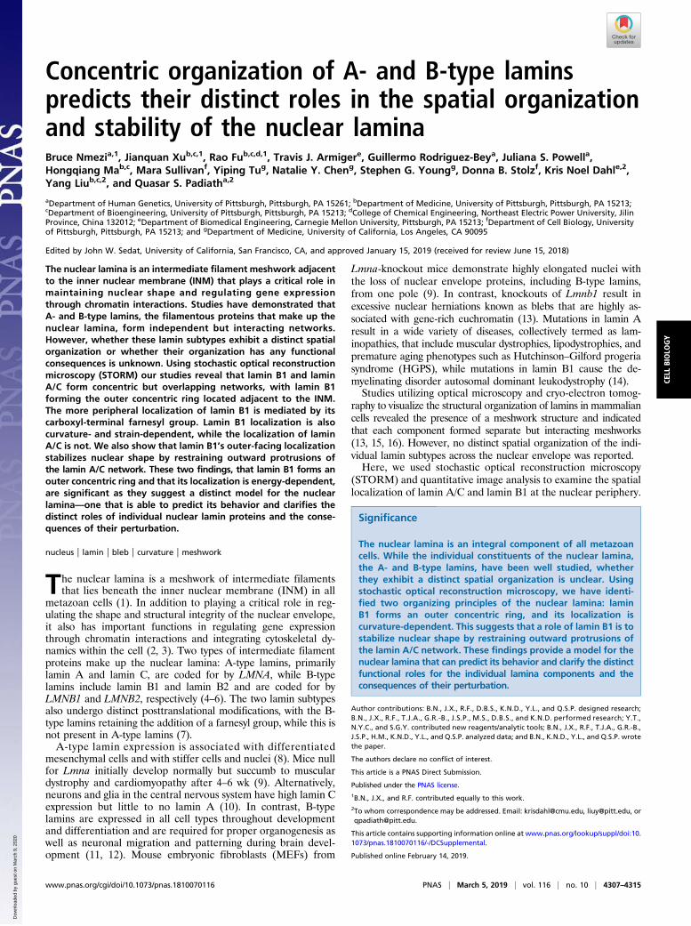
revealed that lamin A/C and lamin B1 networks exhibit differentdensities, with lamin A/C forming denser meshworks, whilelamin B1 forms networks with clusters that are farther apart (Fig.
2 D and E), as indicated by nearest-neighbor distance (NND)measurements. Dual-color imaging (Fig. 2C) revealed mostlyindependent networks, with only 18% colocalization between theA-type and B1-type lamins (Fig. 2F). The meshwork formed bylamin B1 has larger interstitial spaces, as indicated by the higherpercentage of unoccupied area (Fig. 2G). Taken together, theseresults suggest lamin B1 forms a less closely packed network withlarger interstitial spaces, overlaying a denser lamin A/C network(Fig. 2H).
The C-Terminal Farnesyl Group Is Necessary for Proper Lamin B1Localization and Network Organization. Given that lamin B1 re-tains its farnesyl group, we sought to test whether this moietymight be responsible for its association with the INM and
0%
20%
40%
60%
80%
100%
Sonication 1M NaCl 2% TX-100 4M Urea
% o
f Tot
al M
EF L
amin
s Stringency
** LA/CLB1
G
A B
C D
E F
-100 -50 0 50 1000
0.4
1
0.2
0.8
0.6
Inte
nsity
( a.u
.)
Distance (nm)Nuc.
LB1
LA/C
1 8
2 0
Sep
arat
ion
(nm
)
Inte
nsity
( a.u
.)
LB1
LA/C
1 5
1 8
Inte
nsity
(a.u
.)
LB1
LA/C
5
1 0
1 5
2 0
HeLaLA/CLB1
H. fib.LA/CLB1
MEFLA/CLB1
Distance (nm)
Distance (nm)
Mem.
-100 -50 0 50 1000
0.4
1
0.2
0.8
0.6
-100 -50 0 50 1000
0.4
1
0.2
0.8
0.6
Sep
arat
ion
(nm
)S
epar
atio
n (n
m)
Nuc. Mem.
Nuc. Mem.
Fig. 1. Spatially distinct localization and differential membrane binding oflamin B1 and lamin A/C. (A, C, and E) STORM images of immunofluorescentlylabeled lamin B1 (green) and lamin A/C (red) nuclear proteins in MEF (A),HeLa (C), and human fibroblast (E) nuclei at their equatorial planes. (Scalebar: 2 μm.) Rectangles denote Inset zoomed areas. (Inset scale bars: 500 and100 nm.) (B, D, and F) Fluorescence-intensity profile plots across the nuclearenvelopes in STORM images. x axis, distance (nm). Zero distance denotescenter of nuclear lamina y axis: intensity (arbitrary units). (Inset) Box-and-whisker plot of separation (nm) between lamin A/C and B1 fluorescent-intensity peaks (n = 5 nuclei). (G) Quantification of fractional amount ofnuclear lamin proteins solubilized in increasingly stringent sequential ex-tractions in WT MEFs. Graph represents fraction of total lamin B1 and laminA/C signal in each extraction from three independent experiments. Bars,means ± SEM. *P ≤ 0.05 (unpaired two-tailed t test).
4308 | www.pnas.org/cgi/doi/10.1073/pnas.1810070116 Nmezi et al.
Dow
nloa
ded
by g
uest
on
Mar
ch 9
, 202
0
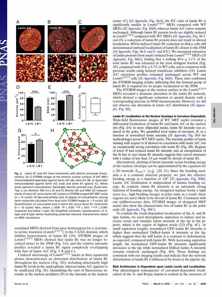
examined MEFs derived from mice homozygous for a cysteine-to-serine mutation (Lmnb1CS/CS) in the CAAX domain, whichinhibits farnesylation of lamin B1 (18). STORM images ofLmnb1CS/CS MEFs showed that lamin B1 was no longer lo-calized closer to the INM (Fig. 3A), and the relative intensityprofiles revealed a lamin B1 signal completely overlappingwith that of lamin A/C (Fig. 3 B and C).Confocal microscopy of Lmnb1CS/CS nuclei at their equatorial
planes demonstrated an abnormal distribution of lamin B1throughout the nucleus (Fig. 3D), including abnormally highlamin B1 levels in the nucleoplasm, whereas lamin A/C appears tobe unaffected (Fig. 3E). Quantifying the ratio of fluorescence in-tensity at the nuclear periphery (P) to the intensity at the nuclear
center (C) (SI Appendix, Fig. S6A), the P/C ratio of lamin B1 issignificantly smaller in Lmnb1CS/CS MEFs compared with WTMEFs (SI Appendix, Fig. S6B), whereas lamin A/C ratios remainsunchanged. Although lamin B1 protein levels are slightly reducedin Lmnb1CS/CS compared with WT MEFs (SI Appendix, Fig. S6 Cand D), a reduction of lamin B1 protein does not result in alteredlocalization. RNAi-induced lamin B1 reduction in HeLa cells stilldemonstrated outward localization of lamin B1 closest to the INM(SI Appendix, Figs. S6 E and F, and S7C). We measured extractionof lamin proteins from nuclei isolated from Lmnb1CS/CS MEFs (SIAppendix, Fig. S6G), finding that a striking 89.4 ± 3.1% of thetotal lamin B1 was extracted in the least stringent fraction (Fig.3F), compared with 29.6 ± 8.7% inWT cells, and is consistent withprevious results using farnesyl transferase inhibitors (19). LaminA/C extraction profiles remained unchanged across WT andLmnb1CS/CS cells (SI Appendix, Fig. S6H). These data confirmedthe STORM imaging results, indicating that the farnesyl group oflamin B1 is required for its proper localization to the INM.The STORM images at the nuclear surface in the Lmnb1CS/CS
MEFs revealed a dramatic alteration in the lamin B1 network,which showed a significant reduction in spatial density with acorresponding increase in NND measurements. However, we didnot observe any alteration in lamin A/C distribution (SI Appen-dix, Fig. S8).
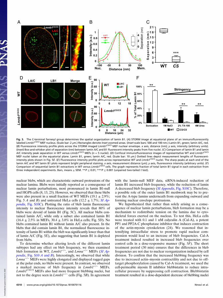
Lamin B1 Localization at the Nuclear Envelope Is Curvature-Dependent.Wide-field fluorescence images of WT MEF nuclei revealed adifferential localization of lamin B1 and lamin A/C at the nuclearrim (Fig. 4A): in more ellipsoidal nuclei, lamin B1 intensity was re-duced at the poles. We quantified local radius of curvature, R, as afunction of normalized lamin intensity (SI Appendix, Fig. S9A formethodology) across WTMEF nuclei. The intensity profiles of laminstaining with respect to R showed no correlation with lamin A/C, butan exceptionally strong correlation with lamin B1 (Fig. 4B). Regionsof lower R had reduced lamin B1 intensity and an extrapolation ofthe fit line to zero lamin B1 intensity suggests that curved structureswith a radius of less than 1.8 μm would be devoid of lamin B1.Alternatively, plotting of lamin intensity versus bending energy
of the nuclear envelope can be approximated by the equation fora 2D network: Ebend =
�12 κ
1R2
�(20, 21). Since the bending mod-
ulus κ is a constant material property, we plot the effectivebending energy as a function of 1/R2 (SI Appendix, Fig. S9B).Lamin A/C localization is independent of lamina bending en-ergy. In contrast, lamin B1 intensity is an extremely strongfunction of bending energy. An elongated nucleus forms a tightcurve (i.e., high bending energy) at polar ends; as a result, theseregions are more likely to be devoid of lamin B1. Consistent withour epifluorescence data, STORM images of elongated MEFnuclei also show the characteristic loss of lamin B1 at the polarends (SI Appendix, Fig. S9C).To confirm the strain-dependent localization of the A- and B-
type lamins, we used micropipette aspiration to induce and in-crease strain and visualize lamin intensity at the tip in the pi-pette, which is the region of highest strain (22) (Fig. 4C). Atsmall aspiration lengths, normalized GFP–lamin B1 intensity ishigher than normalized DsRed–lamin A intensity at the tip,which suggests that the stiff lamin A is resistant to deformation,as expected, compared with lamin B1. With increasing aspirationlength, the normalized GFP–lamin B1 intensity significantlydecreases at the tip, while normalized DsRed–lamin A intensityremains relatively unchanged (Fig. 4D). These findings areconsistent with our imaging results and indicate that the networkdeformation of lamin B1 is influenced by strain at the pipette tip.
Bleb Architecture Is Dependent upon Lamin Levels and Localization.One physiological consequence of curvature-dependent locali-zation of the A- and B-type lamins is evident in the structure of
A
H
CLamin B1 LB1
LA/CLamin A/C
L A /C L B 10
2 0
4 0
6 0
8 0
1 0 0
De
ns
ity
(µm
-2) ***
D E F
L A /C L B 10
2 0
4 0
6 0
8 0
1 0 0
NN
D(n
m)
***
B
G
L A /C L B 10
2 0
4 0
6 0
8 0
%U
no
cc
up
ied
*
1 0
1 5
2 0
Co
loca
liza
tio
n( %
)
Fig. 2. Lamin B1 and A/C have meshworks with distinct structural charac-teristics. (A–C) STORM images at the bottom nuclear surfaces of WT MEFsimmunolabeled separately against lamin A/C (A), lamin B1 (B), or dual-colorimmunolabeled against lamin A/C (red) and lamin B1 (green) (C). Whiteareas represent colocalization. Rectangles denote zoomed area. [Scale bars:Top, 2 μm; Bottom, 500 nm.] (D and E) Density (D) and NND (E) measure-ments of lamin B1 versus lamin A/C clusters in STORM-imagedWT MEF nuclei(n = 15 nuclei). (F) Box-and-whisker plot of degree of colocalization amonglamin molecules calculated from dual-color STORM images (n = 5 nuclei). (G)Quantification of unoccupied area in lamin A/C versus lamin B1 meshworks(n = 15 nuclei). Bars, means ± SEM. *P ≤ 0.05; **P ≤ 0.01; ***P ≤ 0.001(unpaired two-tailed t test). (H) Simplified schematic representation of A-type and B-type lamins illustrating potential network characteristics basedon NND calculations.
Nmezi et al. PNAS | March 5, 2019 | vol. 116 | no. 10 | 4309
CELL
BIOLO
GY
Dow
nloa
ded
by g
uest
on
Mar
ch 9
, 202
0
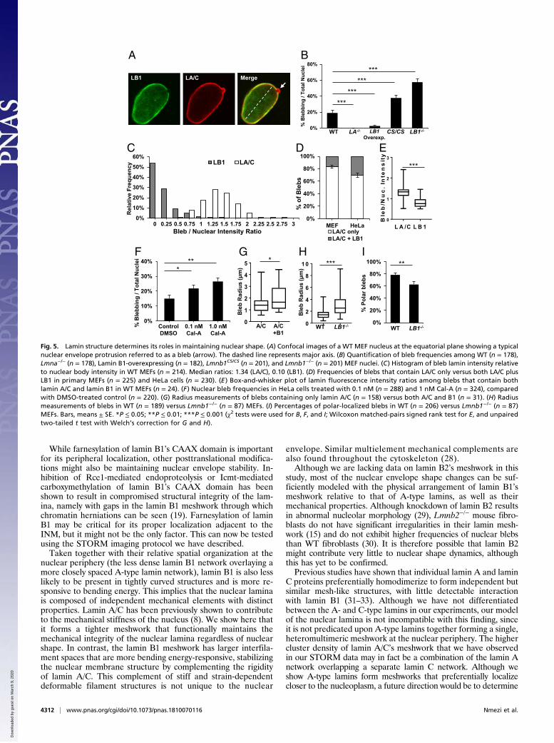
nuclear blebs, which are characteristic outward protrusions of thenuclear lamina. Blebs were initially reported as a consequence ofnuclear lamin perturbations, most pronounced in lamin B1-nulland HGPS cells (8, 13, 23). However, we observed that these blebswere also present in a small fraction of WT MEFs (19.1 ± 2.9%;Fig. 5 A and B) and untreated HeLa cells (12.2 ± 1.7%; SI Ap-pendix, Fig. S10C). Plotting the ratio of bleb lamin fluorescenceintensity to nuclear fluorescence intensity reveals that 80% ofblebs were devoid of lamin B1 (Fig. 5C). All nuclear blebs con-tained lamin A/C, while only a subset also contained lamin B1(16.4 ± 2.5% in MEFs, 30.4 ± 3.0% in HeLa cells; Fig. 5D). Noblebs contained lamin B1 without lamin A/C also present. In theblebs that did contain lamin B1, the normalized fluorescence in-tensity of lamin B1 within the bleb was significantly lower than thatof lamin A/C (Fig. 5E), also suggesting that lamin B1 is depletedfrom blebs.To determine whether altering levels of the different lamin
subtypes had any effect on bleb frequency, we then examinedbleb formation in WT, Lmnb1−/−, and Lmna−/− MEFs (SI Ap-pendix, Fig. S10 A and B). Interestingly, we observed that whileLmna−/− MEFs were highly elongated and displayed ragged gapsat the polar ends, no blebs were present. In contrast, we observeda marked increase of bleb frequency in Lmnb1−/− MEFs.Lmnb1CS/CS MEFs also had more frequent blebbing nuclei, butnot to the degree seen in Lmnb1−/− cells (Fig. 5B). In agreement
with the lamin-null MEF data, siRNA-induced reduction oflamin B1 increased bleb frequency, while the reduction of laminA decreased bleb frequency (SI Appendix, Fig. S10C). Therefore,a possible role of the outer lamin B1 meshwork may be to pre-vent the A-type lamins underneath from expanding outward andforming nuclear envelope protrusions.We hypothesized that rather than solely arising as a conse-
quence of nuclear lamin perturbations, bleb formation may be amechanism to redistribute tension on the lamina due to cyto-skeletal forces exerted on the nucleus. To test this, HeLa cellswere treated with 0.1 and 1 nM calyculin A (Cal-A), a potentPP1 and PP2A-C phosphatase inhibitor that induces contractionof the actin-myosin cytoskeleton (24). We reasoned that in-tensifying intracellular stress to promote rapid nuclear com-pression would lead to an increase of bleb frequencies. Cal-Atreatment indeed resulted in increased bleb frequencies overcontrol cells in a dose-responsive manner (Fig. 5F). The shorttreatment period (30 min) ensures that the differences in blebfrequencies are not due to nuclear reorganization induced by celldivision. To confirm that the increased blebbing frequency wasdue to increased actin–myosin contractility and not due to off-target phosphorylation caused by Cal-A treatment, we used thecell-permeable myosin II inhibitor blebbistatin to reduce intra-cellular pressure by suppressing cell contraction. Blebbistatintreatment resulted in a dose-dependent decrease of blebbing nuclei
Lmnb1CS/CS
Inte
nsity
( a.u
.)
Distance (µm)
0
30
60
90
0 200
30
60
90
0 8 16 24 32
A B C
D E
W T C S /C S0369
1 21 51 82 1
Se
pa
rati
on
(nm
)
***
F
WT
Inte
nsity
(a.u
.)
0
30
60
90
0 6 12 180
30
60
90
0 6 12 18
0%
20%
40%
60%
80%
100%
Sonication 1M NaCl 2% TX-100 4M Urea
% o
f Tot
al L
amin
B1
Stringency
WT CS/CS
**
** **
Lmnb1C
S/CS
WT
LA/C LB1 Merge
-100 -50 0 50 1000
0.4
1
0.2
0.8
0.6
Inte
nsity
( a.u
.)
Distance (nm)Nuc. Mem.
LB1
LA/C
Sep
arat
ion
(nm
)
0
2
4
Lmnb1CS/CSLA/CLB1
WT CS
Fig. 3. The C-terminal farnesyl group determines the spatial organization of lamin B1. (A) STORM image at equatorial plane of an immunofluorescentlylabeled Lmnb1CS/CS MEF nucleus. (Scale bar: 2 μm.) Rectangles denote Inset zoomed areas. (Inset scale bars: 500 and 100 nm.) Lamin B1, green; lamin A/C, red.(B) Fluorescence intensity profile plots across the STORM imaged Lmnb1CS/CS MEF nuclear envelope. x axis, distance (nm); y axis, intensity (arbitrary units).(Inset) Box-and-whisker plot of separation (nm) between lamin A/C and B1 fluorescent-intensity peaks from five nuclei. (C) Comparison of lamin B1 and laminA/C intensity peak separation in WT versus Lmnb1CS/CS MEFs (n = 5 nuclei). (D) Confocal immunofluorescence images of representative WT and Lmnb1CS/CS
MEF nuclei taken at the equatorial plane. Lamin B1, green; lamin A/C, red. (Scale bar: 10 μm.) Dotted lines depict measurement lengths of fluorescent-intensity plots shown in Fig. 5E. (E) Fluorescence intensity profile plots across representative WT and Lmnb1CS/CS nuclei. The sharp peaks at each end of thelamin A/C and WT lamin B1 plots represent bright peripheral staining. x axis, measurement distance (μm); y axis, fluorescence intensity (arbitrary units). (F)Comparison of sequential lamin B1 extractions in WT versus Lmnb1CS/CS cells. The graph represents fraction of total lamin B1 signal in each extraction fromthree independent experiments. Bars, means ± SEM. **P ≤ 0.01; ***P ≤ 0.001 (unpaired two-tailed t test).
4310 | www.pnas.org/cgi/doi/10.1073/pnas.1810070116 Nmezi et al.
Dow
nloa
ded
by g
uest
on
Mar
ch 9
, 202
0

(SI Appendix, Fig. S10D). Notably, it eliminated Cal-A’s ability toinduce bleb formation, indicating that blebs likely arose due to in-creased intracellular pressure and not from nonspecific effects ofcalyculin treatment.The architecture of the blebs allows us to test predictions that
arise out of our model of the nuclear lamina, where the laminB1 network is located toward the outside of lamin A/C, and thelocation of lamin B1 is curvature-dependent. Based on ourfindings, we expect that lamin B1 is likely to be excluded fromtightly curved regions of the nuclear envelope. Consistent withthis prediction, we found that blebs containing lamin B1 arelarger on average than lamin A/C-only blebs (Fig. 5G). In ad-dition, blebs that form in Lmnb1−/− MEFs are significantly largerthan those in WT cells (Fig. 5H). Further proof of the functionalconsequences of our model is seen in the location of blebs. If arole for lamin B1 is to stabilize the outward protrusion of thelamin A/C network, we would expect to see a higher frequency ofblebs in regions depleted of lamin B1. In agreement with ourmodel, we observe most blebs form at the major axis poles,where the radius of curvature is low, resulting in a consequentdepletion of lamin B1. However, in Lmnb1−/− MEFs, where theconstraining effects of lamin B1 are no longer present, blebsassume a more random distribution with a significant reductionin polar blebs (Fig. 5I).To further test our hypothesis that lamin B1 inhibits the lamin
A/C meshwork from protruding outward, we examined lamin B1-overexpressing MEFS (SI Appendix, Fig. S10E). Compared withWT and untreated cells, lamin B1-overexpressing cells had asignificantly lower frequency of blebbing nuclei (Fig. 5B and SIAppendix, Fig. S10F). By increasing lamin B1 expression, wecould almost eliminate the formation of nuclear blebs.
DiscussionSuperresolution localization microscopy is a powerful imagingtool for simultaneous visualization of multiple molecular speciesat nanometer resolutions (17). Here, we have used STORM
combined with quantitative image analysis to identify the spatialorganization of A- and B-type lamins. We find that the laminB1 meshwork is closest to the INM and forms a less dense outerrim around the more tightly spaced lamin A/C meshwork facingthe nucleoplasm, with a spatial separation of ∼15–20 nm. Suchprecise localization of the two lamin species was achieved byseveral key technical attributes of our approach. First, it requiredchromatic aberration-free, two-color superresolution imaging. Acomplete correction of chromatic aberration is often difficult toachieve, especially when the imaging target is farther away fromthe coverslip surface (as in our case of imaging equatorial plane ofthe nuclear periphery). To overcome this hurdle, we used thesame reporter dye in the activator–reporter dye pairs in two-colorSTORM imaging to essentially eliminate any chromatic aberra-tion. Second, by averaging spatial distribution around the nuclearperiphery, we can quantify the separation of different lamintypes at a higher precision than the STORM imaging resolutionof ∼20 nm (25). Third, focusing on the equatorial plane alsomaximizes the spatial resolution by imaging the projection of thecross-sectional profiles of the two lamin types across thenuclear periphery.Our biochemical data confirm the STORM imaging observa-
tions that lamin B1 is more closely associated with INM thanlamin A/C and that this association is dependent upon the far-nesylation of lamin B1. All lamins are initially farnesylated, butonly B-type lamins retain their farnesyl group after prelamin Aundergoes proteolytic cleavage at its C terminus (2). This far-nesyl tail can allow lamin B1 to tightly associate with the INM,similar to the way Ras GTPases anchor to the cell membrane(26, 27), and provides a rationale for the retention of the farnesylgroup by lamin B1. Interestingly, although the lamin B1 farnesylmutant significantly altered the lamin B1 network structure,consistent with earlier reports using conventional fluorescencemicroscopy (18), our results also show that it did not alter laminA/C distribution, suggesting that proper localization of laminB1 is not required for the integrity of the lamin A/C network.
A
C D0
0.5
1
1.5
2
2.5
1 10
Nor
mal
ized
Lam
in In
tens
ity
Radius of curvature, R (µm)
LB1 LA/C
RI=0=1.8 µm R2 = 0.84
BLB1 LA/C Merge
1 .2 1 .5 1 .70 .3
0 .6
0 .9
N o rm a liz e d A s p ira t io n L e n g th (L /D )
No
rma
lize
dT
ipIn
ten
sit
y
D s R e d -L A
G F P -L B 1
*****
****
**
**
Pre-
asp
L/D
= 1
.2L/
D =
1.7
DsRed-LAGFP-LB1 Merge+DNA BF
Fig. 4. Lamin B1 meshwork is curvature and strain-responsive. (A) Wide-field fluorescent microscopy images of WT MEF nuclei displaying differential lo-calization of lamin B1 and lamin A/C across the nucleus. Dashed lines represent major axes. The vertices at the ends of the major axis are defined as thenuclear poles. Arrowheads point to areas of decreased lamin B1 intensity. (Scale bar: 10 μm.) (B) Plot of normalized lamin intensity versus radius of curvature Rfor 30 points across 17 nuclei, with logarithmic trend lines. At the x intercept, R = 1.8 μm. (C) MEFs were doubly transfected with GFP-LMNB1 and DsRed-LMNAplasmids and then aspirated with a micropipette. Normalized aspiration length, L/D, is defined as the aspirated projection length, L, divided by the diameterof the micropipette, D. Arrows point to aspiration tip and base. (Scale bar: 10 μm.) (D) Graph of lamin intensities at the aspiration tip versus L/D (n = 5). Laminintensities at the aspiration tip were normalized to its intensity throughout the rest of the nucleus. Bars, means ± SEM. *P ≤ 0.05; **P ≤ 0.01; ***P ≤ 0.001(two-way ANOVA with Sidak’s multiple comparisons test).
Nmezi et al. PNAS | March 5, 2019 | vol. 116 | no. 10 | 4311
CELL
BIOLO
GY
Dow
nloa
ded
by g
uest
on
Mar
ch 9
, 202
0
While farnesylation of lamin B1’s CAAX domain is importantfor its peripheral localization, other posttranslational modifica-tions might also be maintaining nuclear envelope stability. In-hibition of Rce1-mediated endoproteolysis or Icmt-mediatedcarboxymethylation of lamin B1’s CAAX domain has beenshown to result in compromised structural integrity of the lam-ina, namely with gaps in the lamin B1 meshwork through whichchromatin herniations can be seen (19). Farnesylation of laminB1 may be critical for its proper localization adjacent to theINM, but it might not be the only factor. This can now be testedusing the STORM imaging protocol we have described.Taken together with their relative spatial organization at the
nuclear periphery (the less dense lamin B1 network overlaying amore closely spaced A-type lamin network), lamin B1 is also lesslikely to be present in tightly curved structures and is more re-sponsive to bending energy. This implies that the nuclear laminais composed of independent mechanical elements with distinctproperties. Lamin A/C has been previously shown to contributeto the mechanical stiffness of the nucleus (8). We show here thatit forms a tighter meshwork that functionally maintains themechanical integrity of the nuclear lamina regardless of nuclearshape. In contrast, the lamin B1 meshwork has larger interfila-ment spaces that are more bending energy-responsive, stabilizingthe nuclear membrane structure by complementing the rigidityof lamin A/C. This complement of stiff and strain-dependentdeformable filament structures is not unique to the nuclear
envelope. Similar multielement mechanical complements arealso found throughout the cytoskeleton (28).Although we are lacking data on lamin B2’s meshwork in this
study, most of the nuclear envelope shape changes can be suf-ficiently modeled with the physical arrangement of lamin B1’smeshwork relative to that of A-type lamins, as well as theirmechanical properties. Although knockdown of lamin B2 resultsin abnormal nucleolar morphology (29), Lmnb2−/− mouse fibro-blasts do not have significant irregularities in their lamin mesh-work (15) and do not exhibit higher frequencies of nuclear blebsthan WT fibroblasts (30). It is therefore possible that lamin B2might contribute very little to nuclear shape dynamics, althoughthis has yet to be confirmed.Previous studies have shown that individual lamin A and lamin
C proteins preferentially homodimerize to form independent butsimilar mesh-like structures, with little detectable interactionwith lamin B1 (31–33). Although we have not differentiatedbetween the A- and C-type lamins in our experiments, our modelof the nuclear lamina is not incompatible with this finding, sinceit is not predicated upon A-type lamins together forming a single,heteromultimeric meshwork at the nuclear periphery. The highercluster density of lamin A/C’s meshwork that we have observedin our STORM data may in fact be a combination of the lamin Anetwork overlapping a separate lamin C network. Although weshow A-type lamins form meshworks that preferentially localizecloser to the nucleoplasm, a future direction would be to determine
BA
F G H I
DC E
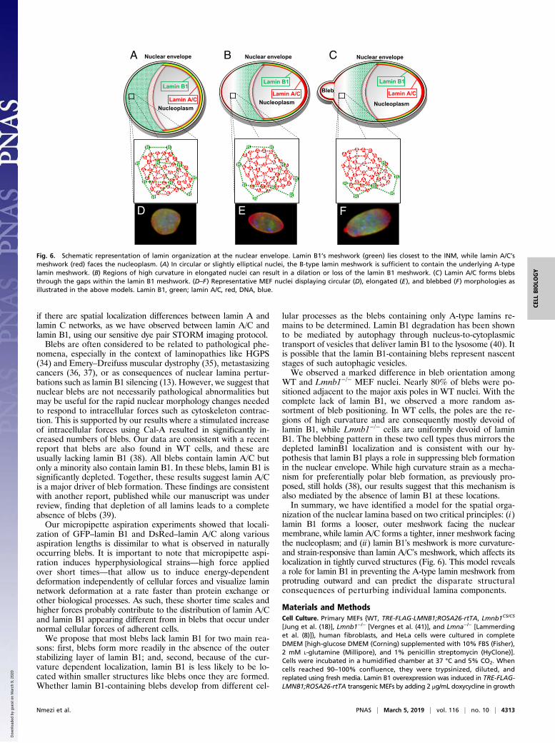
Fig. 5. Lamin structure determines its roles in maintaining nuclear shape. (A) Confocal images of a WT MEF nucleus at the equatorial plane showing a typicalnuclear envelope protrusion referred to as a bleb (arrow). The dashed line represents major axis. (B) Quantification of bleb frequencies among WT (n = 178),Lmna−/− (n = 178), Lamin B1-overexpressing (n = 182), Lmnb1CS/CS (n = 201), and Lmnb1−/− (n = 201) MEF nuclei. (C) Histogram of bleb lamin intensity relativeto nuclear body intensity in WT MEFs (n = 214). Median ratios: 1.34 (LA/C), 0.10 (LB1). (D) Frequencies of blebs that contain LA/C only versus both LA/C plusLB1 in primary MEFs (n = 225) and HeLa cells (n = 230). (E ) Box-and-whisker plot of lamin fluorescence intensity ratios among blebs that contain bothlamin A/C and lamin B1 in WT MEFs (n = 24). (F ) Nuclear bleb frequencies in HeLa cells treated with 0.1 nM (n = 288) and 1 nM Cal-A (n = 324), comparedwith DMSO-treated control (n = 220). (G) Radius measurements of blebs containing only lamin A/C (n = 158) versus both A/C and B1 (n = 31). (H) Radiusmeasurements of blebs in WT (n = 189) versus Lmnb1−/− (n = 87) MEFs. (I) Percentages of polar-localized blebs in WT (n = 206) versus Lmnb1−/− (n = 87)MEFs. Bars, means ± SE. *P ≤ 0.05; **P ≤ 0.01; ***P ≤ 0.001 (χ2 tests were used for B, F, and I; Wilcoxon matched-pairs signed rank test for E, and unpairedtwo-tailed t test with Welch’s correction for G and H).
4312 | www.pnas.org/cgi/doi/10.1073/pnas.1810070116 Nmezi et al.
Dow
nloa
ded
by g
uest
on
Mar
ch 9
, 202
0
if there are spatial localization differences between lamin A andlamin C networks, as we have observed between lamin A/C andlamin B1, using our sensitive dye pair STORM imaging protocol.Blebs are often considered to be related to pathological phe-
nomena, especially in the context of laminopathies like HGPS(34) and Emery–Dreifuss muscular dystrophy (35), metastasizingcancers (36, 37), or as consequences of nuclear lamina pertur-bations such as lamin B1 silencing (13). However, we suggest thatnuclear blebs are not necessarily pathological abnormalities butmay be useful for the rapid nuclear morphology changes neededto respond to intracellular forces such as cytoskeleton contrac-tion. This is supported by our results where a stimulated increaseof intracellular forces using Cal-A resulted in significantly in-creased numbers of blebs. Our data are consistent with a recentreport that blebs are also found in WT cells, and these areusually lacking lamin B1 (38). All blebs contain lamin A/C butonly a minority also contain lamin B1. In these blebs, lamin B1 issignificantly depleted. Together, these results suggest lamin A/Cis a major driver of bleb formation. These findings are consistentwith another report, published while our manuscript was underreview, finding that depletion of all lamins leads to a completeabsence of blebs (39).Our micropipette aspiration experiments showed that locali-
zation of GFP–lamin B1 and DsRed–lamin A/C along variousaspiration lengths is dissimilar to what is observed in naturallyoccurring blebs. It is important to note that micropipette aspi-ration induces hyperphysiological strains—high force appliedover short times—that allow us to induce energy-dependentdeformation independently of cellular forces and visualize laminnetwork deformation at a rate faster than protein exchange orother biological processes. As such, these shorter time scales andhigher forces probably contribute to the distribution of lamin A/Cand lamin B1 appearing different from in blebs that occur undernormal cellular forces of adherent cells.We propose that most blebs lack lamin B1 for two main rea-
sons: first, blebs form more readily in the absence of the outerstabilizing layer of lamin B1; and, second, because of the cur-vature dependent localization, lamin B1 is less likely to be lo-cated within smaller structures like blebs once they are formed.Whether lamin B1-containing blebs develop from different cel-
lular processes as the blebs containing only A-type lamins re-mains to be determined. Lamin B1 degradation has been shownto be mediated by autophagy through nucleus-to-cytoplasmictransport of vesicles that deliver lamin B1 to the lysosome (40). Itis possible that the lamin B1-containing blebs represent nascentstages of such autophagic vesicles.We observed a marked difference in bleb orientation among
WT and Lmnb1−/− MEF nuclei. Nearly 80% of blebs were po-sitioned adjacent to the major axis poles in WT nuclei. With thecomplete lack of lamin B1, we observed a more random as-sortment of bleb positioning. In WT cells, the poles are the re-gions of high curvature and are consequently mostly devoid oflamin B1, while Lmnb1−/− cells are uniformly devoid of laminB1. The blebbing pattern in these two cell types thus mirrors thedepleted laminB1 localization and is consistent with our hy-pothesis that lamin B1 plays a role in suppressing bleb formationin the nuclear envelope. While high curvature strain as a mecha-nism for preferentially polar bleb formation, as previously pro-posed, still holds (38), our results suggest that this mechanism isalso mediated by the absence of lamin B1 at these locations.In summary, we have identified a model for the spatial orga-
nization of the nuclear lamina based on two critical principles: (i)lamin B1 forms a looser, outer meshwork facing the nuclearmembrane, while lamin A/C forms a tighter, inner meshwork facingthe nucleoplasm; and (ii) lamin B1’s meshwork is more curvature-and strain-responsive than lamin A/C’s meshwork, which affects itslocalization in tightly curved structures (Fig. 6). This model revealsa role for lamin B1 in preventing the A-type lamin meshwork fromprotruding outward and can predict the disparate structuralconsequences of perturbing individual lamina components.
Materials and MethodsCell Culture. Primary MEFs {WT, TRE-FLAG-LMNB1;ROSA26-rtTA, Lmnb1CS/CS
[Jung et al. (18)], Lmnb1−/− [Vergnes et al. (41)], and Lmna−/− [Lammerdinget al. (8)]}, human fibroblasts, and HeLa cells were cultured in completeDMEM [high-glucose DMEM (Corning) supplemented with 10% FBS (Fisher),2 mM L-glutamine (Millipore), and 1% penicillin streptomycin (HyClone)].Cells were incubated in a humidified chamber at 37 °C and 5% CO2. Whencells reached 90–100% confluence, they were trypsinized, diluted, andreplated using fresh media. Lamin B1 overexpression was induced in TRE-FLAG-LMNB1;ROSA26-rtTA transgenic MEFs by adding 2 μg/mL doxycycline in growth
A B CNuclear envelope
Nucleoplasm
Lamin B1
Lamin A/C
Nuclear envelope
Nucleoplasm
Lamin B1
Lamin A/C
Nuclear envelope
Nucleoplasm
Lamin B1
Lamin A/CBleb
AA A
AAA
A
A
A A AAA
A AAA AAA
A AA
A
A
AAA AA
A A AAA
AA
AAB
B
B
B
B
BB
B
B
B
D E F
AA A
AAA
A
A
A A AAA
A AAA AAA
A AA
A
A
AAA AA
A A AAA
AA
AA B
B
B
B
BB
B
B
B
AA A
AAA
A
A
A A AAA
A AAA AAA
A AA
A
A
AAA AA
A A AAA
AA
AA
B
BB
B
B
B
Fig. 6. Schematic representation of lamin organization at the nuclear envelope. Lamin B1’s meshwork (green) lies closest to the INM, while lamin A/C’smeshwork (red) faces the nucleoplasm. (A) In circular or slightly elliptical nuclei, the B-type lamin meshwork is sufficient to contain the underlying A-typelamin meshwork. (B) Regions of high curvature in elongated nuclei can result in a dilation or loss of the lamin B1 meshwork. (C) Lamin A/C forms blebsthrough the gaps within the lamin B1 meshwork. (D–F) Representative MEF nuclei displaying circular (D), elongated (E), and blebbed (F) morphologies asillustrated in the above models. Lamin B1, green; lamin A/C, red, DNA, blue.
Nmezi et al. PNAS | March 5, 2019 | vol. 116 | no. 10 | 4313
CELL
BIOLO
GY
Dow
nloa
ded
by g
uest
on
Mar
ch 9
, 202
0
medium for 5 d. For Cal-A and blebbistatin treatments, HeLa cells grown oncoverslips were treated with 0.1 and 1 nM Cal-A (no. 508226; Sigma) for 30 min,or 10, 50, and 100 μM blebbistatin (no. 203389; Sigma) for 2 h at 37 °C and5% CO2. Cells were washed and fixed in 4% paraformaldehyde solution inPBS immediately after treatment.
STORM Imaging and Analysis. Cells for STORM imaging were prepared aspreviously described (42). Rabbit anti-lamin B1 (no. ab16048; Abcam) werediluted 1:600 and/or mouse anti-lamin A/C (no. 4777; Cell Signaling) werediluted 1:300 in PBS plus 3% BSA and incubated on the cells overnight at4 °C. The next morning, cells were washed with PBS three times for 5 mineach at room temperature. Secondary antibodies were diluted in PBS plus3% BSA and incubated with the cells for 2 h at room temperature in thedark. Unconjugated secondary antibodies (nos. 711-005-152 and 715-005-150; Jackson ImmunoResearch) were conjugated with AF647 (no. A20006;Thermo Fisher Scientific) in the laboratory of Y.L. AF647-conjugated sec-ondary antibodies were used for single-color STORM imaging. For two-colorSTORM imaging based on dye pairs, secondary antibodies labeled with ac-tivator–reporter dye pairs (AF405-AF647, Cy2-AF647) were used. AF405-AF647 conjugated donkey anti-mouse secondary antibody was used to la-bel lamin A/C, and Cy2-AF647 conjugated donkey anti-rabbit secondaryantibody was used to label lamin B1. For the dye-switch experiment, AF405-AF647 conjugated donkey anti-rabbit secondary antibody was used to labellamin B1, and Cy2-AF647 conjugated donkey anti-mouse secondary antibodywas used to label lamin A/C. Immediately before imaging, the buffer wasswitched to STORM imaging buffer [10% wt/vol glucose (Sigma-Aldrich),0.56 mg/mL glucose oxidase (Sigma-Aldrich), and 0.17 mg/mL catalase(Sigma-Aldrich)]. For single-color imaging, 0.14 M β-mercaptoethanol(Sigma-Aldrich) was used, and for two-color imaging, 0.1 M mercaptoe-thylamine (Sigma-Aldrich) was used. Single-color STORM imaging was per-formed on a custom-built system using an Olympus IX71 inverted microscopeframe with a 60× oil objective. Fluorescent beads [0.1 μm diameter (no.F8803; Fisher Scientific), excited using 488 nm laser] were used as fiduciarymarkers on the coverslip to correct for 3D system drift every 200 frames.Two-color STORM images using dye pairs were acquired on a commercialimaging system (N-STORM; Nikon Instruments). The samples were periodi-cally activated with a sequence of 405-nm, 488-nm laser pulses and thenimaged with the 647-nm laser. In each switching cycle, the activation laserwas turned on for one frame, followed by three frames of illumination withthe red imaging laser. A total of 40,000 frames, including 10,000 activationframes and 30,000 imaging frames for each channel, were acquired at anexposure time of 20 ms. Imaging frames immediately following an activationpulse were recognized as controlled activation events, and a color wasassigned accordingly. A crosstalk subtraction algorithm was used to subtractthe nonspecific activation signal (17). Nuclei were imaged at the equator formeasuring lamina thickness and relative localization. The bottom surfaces ofnuclei were imaged to minimize curvature artifacts. The reconstruction of asuperresolution image and Gaussian clustering were performed using acustom program written in Matlab 2015 (MathWorks), as previously de-scribed (43). The degree of colocalization was calculated using Clus-DoC al-gorithm (44) and defined as the degree of colocalization in lamin A/C andlamin B1 with respect to the combined lamin A/C and lamin B1. For mea-suring lamina thickness, the nuclear periphery of the superresolution imagewas automatically divided into numerous small segments at a length of∼50 nm. Intensity peaks were measured along the steepest gradient per-pendicular to the nuclear envelope and averaged. The intensity profile wasplotted as distance (x axis) versus normalized lamin intensity (y axis), andthicknesses was defined as the full width at half-maximum.
Isolation of Nuclei. Low-passage cells were seeded in 100-mm tissue culturedishes (Thermo BioLite) and grown to 90% confluence. Nuclei were isolatedas previously described (45), with modifications. The cells were washed oncewith 3 mL of ice-cold PBS and then scraped down with 1 mL of cold PBS into1.5-mL microcentrifuge tubes. Samples were centrifuged at 10,000 × g for15 s at 4 °C, and the supernatants were discarded. Cell pellets were resus-pended in 1 mL of cold PBS+IGEPAL [1× PBS plus 0.1% IGEPAL CA-630(Sigma) plus protease inhibitors], triturated 5× on ice with a 1,000 μL pipettip, and 100 μL was transferred into new microcentrifuge tubes as the wholecell fraction. Remaining volumes were centrifuged at 10,000 × g for 15 s at4 °C. Supernatants were transferred to new microcentrifuge tubes labeled“cytosol.” Pellets were resuspended in 1 mL of PBS+IGEPAL and thencentrifuged at 10,000 × g for 15 s at 4 °C, and the supernatants were dis-carded. Remaining pellets were used for sequential protein extractions.Cytosol samples were centrifuged at 10,000 × g for 1 min at 4 °C, and then300 μL of the supernatant was transferred to new microcentrifuge tubes asthe clean cytosolic fraction.
Sequential Extraction of Nuclear Envelope Proteins. Subfractionation of nu-clear proteins was performed as previously described (46). Nuclear pelletswere resuspended in 300 μL of nuclear isolation buffer [10 mM Hepes (pH7.4), 2 mM MgCl2, 25 mM KCl, 250 mM sucrose, 1 mM DTT, protease in-hibitors]; 50 μL of each sample were transferred to new microcentrifugetubes as the “whole nuclei” fraction. Samples were sonicated on ice withtwo 5-s pulses at 10-μm amplitude and then centrifuged at 20,000 × g for5 min at 4 °C. The supernatants were transferred to new microcentrifugetubes as the “sonication” fraction. Pellets were resuspended in 250 μL ofnuclear extraction buffer [20 mM Hepes (pH 7.4), 1 M NaCl, proteaseinhibitors] and incubated for 20 min at room temperature with end-over-endrotation. After incubation, samples were centrifuged at 20,000 × g for 5 minat 4 °C, and the supernatants were transferred to new microcentrifuge tubesas the “1 M NaCl” fraction. Extractions were repeated on the pellets using250 μL of nuclear extraction buffer with 2% vol/vol Triton X-100, 4 M urea,then 8 M urea in sequential incubations. All protein extracts were storedat −80 °C until ready for immunoblotting.
Statistical Analysis. Two-sided t tests were used to calculate statistical sig-nificance between two groups. For micropipette aspiration comparisons,two-way ANOVAs were used followed by Sidak’s multiple comparisons test.For sequential protein extractions, lamin signal percentages were arcsine-transformed before statistical analysis. χ2 tests were used to assess statisticalsignificance of blebbing ratios. Bleb versus nuclear lamin intensity differ-ences were compared using Wilcoxon matched-pairs signed rank test. AP value ≤ 0.05 was considered statistically significant. The following conventionfor representing P values was followed: *P ≤ 0.05, **P ≤ 0.01, ***P ≤0.001. All error bars represent SE unless otherwise specified. Data weregraphed and analyzed in Microsoft Excel 2016 and GraphPad Prism 7.Additional materials and methods information and SI figures are locatedin SI Appendix.
ACKNOWLEDGMENTS. We thank members of the K.N.D., Y.L., and Q.S.P.labs for helpful discussions; Loren Fong for help with providing the Lmnb1−/−,Lmna−/−, and Lmnb1CS/CS MEFs; the Center for Biological Imaging at the Uni-versity of Pittsburgh for assistance with STORM, EM, and confocal imaging;and the Biomedical Mass Spectrometry Center for helpful discussions. Thiswork was supported by NIH Grant R01NS095884 and National Multiple Scle-rosis Society Research Grant 5045A1 (to Q.S.P.); NSF Civil, Mechanical andManufacturing Innovation Grant 1634888 (to K.N.D.); NIH Grant EB003392(to T.J.A. and K.N.D.); NIH Grants R01EB016657 and R01CA185363 (to Y.L.);and NIH Grants 1S10RR019003-01 and 1S10RR025488-01 (to D.B.S.).
1. Gerace L, Huber MD (2012) Nuclear lamina at the crossroads of the cytoplasm andnucleus. J Struct Biol 177:24–31.
2. Ho CY, Lammerding J (2012) Lamins at a glance. J Cell Sci 125:2087–2093.3. Burke B, Stewart CL (2013) The nuclear lamins: Flexibility in function. Nat Rev Mol Cell
Biol 14:13–24.4. Biamonti G, et al. (1992) The gene for a novel human lamin maps at a highly tran-
scribed locus of chromosome 19 which replicates at the onset of S-phase.Mol Cell Biol12:3499–3506.
5. Lin F, Worman HJ (1995) Structural organization of the human gene (LMNB1) en-coding nuclear lamin B1. Genomics 27:230–236.
6. Lin F, Worman HJ (1993) Structural organization of the human gene encoding nuclearlamin A and nuclear lamin C. J Biol Chem 268:16321–16326.
7. Dechat T, et al. (2008) Nuclear lamins: Major factors in the structural organization andfunction of the nucleus and chromatin. Genes Dev 22:832–853.
8. Lammerding J, et al. (2006) Lamins A and C but not lamin B1 regulate nuclear me-chanics. J Biol Chem 281:25768–25780.
9. Sullivan T, et al. (1999) Loss of A-type lamin expression compromises nuclear envelopeintegrity leading to muscular dystrophy. J Cell Biol 147:913–920.
10. Jung HJ, et al. (2012) Regulation of prelamin A but not lamin C by miR-9, a brain-specific microRNA. Proc Natl Acad Sci USA 109:E423–E431.
11. Kim Y, et al. (2011) Mouse B-type lamins are required for proper organogenesis butnot by embryonic stem cells. Science 334:1706–1710.
12. Coffinier C, et al. (2011) Deficiencies in lamin B1 and lamin B2 cause neurodevelopmentaldefects and distinct nuclear shape abnormalities in neurons. Mol Biol Cell 22:4683–4693.
13. Shimi T, et al. (2008) The A- and B-type nuclear lamin networks: Microdomains in-volved in chromatin organization and transcription. Genes Dev 22:3409–3421.
14. Padiath QS, et al. (2006) Lamin B1 duplications cause autosomal dominant leuko-dystrophy. Nat Genet 38:1114–1123.
15. Shimi T, et al. (2015) Structural organization of nuclear lamins A, C, B1, andB2 revealed by superresolution microscopy. Mol Biol Cell 26:4075–4086.
16. Turgay Y, et al. (2017) The molecular architecture of lamins in somatic cells. Nature543:261–264.
4314 | www.pnas.org/cgi/doi/10.1073/pnas.1810070116 Nmezi et al.
Dow
nloa
ded
by g
uest
on
Mar
ch 9
, 202
0
17. Bates M, Huang B, Dempsey GT, Zhuang X (2007) Multicolor super-resolution imagingwith photo-switchable fluorescent probes. Science 317:1749–1753.
18. Jung HJ, et al. (2013) Farnesylation of lamin B1 is important for retention of nuclearchromatin during neuronal migration. Proc Natl Acad Sci USA 110:E1923–E1932.
19. Maske CP, et al. (2003) A carboxyl-terminal interaction of lamin B1 is dependent onthe CAAX endoprotease Rce1 and carboxymethylation. J Cell Biol 162:1223–1232.
20. Israelachvili JN (2011) Intermolecular and Surface Forces (Academic, Burlington, MA),3rd Ed, pp 382–385.
21. Boal DH (2012) Mechanics of the Cell (Cambridge Univ Press, Cambridge, UK), 2nd Ed,pp 156–171.
22. Discher DE, Boal DH, Boey SK (1998) Simulations of the erythrocyte cytoskeleton atlarge deformation. II. Micropipette aspiration. Biophys J 75:1584–1597.
23. Candelario J, Sudhakar S, Navarro S, Reddy S, Comai L (2008) Perturbation of wild-type lamin A metabolism results in a progeroid phenotype. Aging Cell 7:355–367.
24. Ishihara H, et al. (1989) Calyculin A and okadaic acid: Inhibitors of protein phospha-tase activity. Biochem Biophys Res Commun 159:871–877.
25. Szymborska A, et al. (2013) Nuclear pore scaffold structure analyzed by super-resolution microscopy and particle averaging. Science 341:655–658.
26. Seabra MC (1998) Membrane association and targeting of prenylated Ras-likeGTPases. Cell Signal 10:167–172.
27. Michaelson D, et al. (2005) Postprenylation CAAX processing is required for properlocalization of Ras but not Rho GTPases. Mol Biol Cell 16:1606–1616.
28. Pegoraro AF, Janmey P, Weitz DA (2017) Mechanical properties of the cytoskeletonand cells. Cold Spring Harb Perspect Biol 9:a022038.
29. Sen Gupta A, Sengupta K (2017) Lamin B2 modulates nucleolar morphology, dy-namics, and function. Mol Cell Biol 37:e00274-17.
30. Coffinier C, et al. (2010) Abnormal development of the cerebral cortex and cerebel-lum in the setting of lamin B2 deficiency. Proc Natl Acad Sci USA 107:5076–5081.
31. Burke B (1990) On the cell-free association of lamins A and C with metaphase chro-mosomes. Exp Cell Res 186:169–176.
32. Xie W, et al. (2016) A-type lamins form distinct filamentous networks with differentialnuclear pore complex associations. Curr Biol 26:2651–2658.
33. Kolb T, Maass K, Hergt M, Aebi U, Herrmann H (2011) Lamin A and lamin C formhomodimers and coexist in higher complex forms both in the nucleoplasmic fractionand in the lamina of cultured human cells. Nucleus 2:425–433.
34. Eriksson M, et al. (2003) Recurrent de novo point mutations in lamin A causeHutchinson-Gilford progeria syndrome. Nature 423:293–298.
35. Fidzia�nska A, Toniolo D, Hausmanowa-Petrusewicz I (1998) Ultrastructural abnor-mality of sarcolemmal nuclei in Emery-Dreifuss muscular dystrophy (EDMD). J NeurolSci 159:88–93.
36. Denais CM, et al. (2016) Nuclear envelope rupture and repair during cancer cell mi-gration. Science 352:353–358.
37. Raab M, et al. (2016) ESCRT III repairs nuclear envelope ruptures during cell migrationto limit DNA damage and cell death. Science 352:359–362.
38. Stephens AD, et al. (2018) Chromatin histone modifications and rigidity affect nuclearmorphology independent of lamins. Mol Biol Cell 29:220–233.
39. Chen NY, et al. (2018) Fibroblasts lacking nuclear lamins do not have nuclear blebs orprotrusions but nevertheless have frequent nuclear membrane ruptures. Proc NatlAcad Sci USA 115:10100–10105.
40. Dou Z, et al. (2015) Autophagy mediates degradation of nuclear lamina. Nature 527:105–109.
41. Vergnes L, Péterfy M, Bergo MO, Young SG, Reue K (2004) Lamin B1 is required formouse development and nuclear integrity. Proc Natl Acad Sci USA 101:10428–10433.
42. Xu J, Ma H, Liu Y (2017) Stochastic optical reconstruction microscopy (STORM). CurrProtoc Cytom 81:12.46.1–12.46.27.
43. Ma H, Xu J, Jin J, Huang Y, Liu Y (2017) A simple marker-assisted 3D nanometer driftcorrection method for superresolution microscopy. Biophys J 112:2196–2208.
44. Pageon SV, Nicovich PR, Mollazade M, Tabarin T, Gaus K (2016) Clus-DoC: A combinedcluster detection and colocalization analysis for single-molecule localization micros-copy data. Mol Biol Cell 27:3627–3636.
45. Nabbi A, Riabowol K (2015) Rapid isolation of nuclei from cells in vitro. Cold SpringHarb Protoc 2015:769–772.
46. Otto H, Dreger M, Bengtsson L, Hucho F (2001) Identification of tyrosine-phosphorylatedproteins associated with the nuclear envelope. Eur J Biochem 268:420–428.
Nmezi et al. PNAS | March 5, 2019 | vol. 116 | no. 10 | 4315
CELL
BIOLO
GY
Dow
nloa
ded
by g
uest
on
Mar
ch 9
, 202
0
Related Documents