International Scholarly Research Network ISRN Dentistry Volume 2012, Article ID 312031, 5 pages doi:10.5402/2012/312031 Research Article Computed Tomography Evaluation of Craniomandibular Articulation in Class II Division 1 Malocclusion and Class I Normal Occlusion Subjects in North Indian Population K. C. Prabhat, 1 Sanjeev Kumar Verma, 1 Sandhya Maheshwari, 1 Ibne Ahmad, 2 and Mohd. Tariq 1 1 Department of Orthodontics and Dental Anatomy, Dr. Z. A. Dental College, Aligarh Muslim University, Aligarh 212001, India 2 Department of Radio Diagnosis, Aligarh Muslim University, Aligarh 212001, India Correspondence should be addressed to K. C. Prabhat, [email protected] Received 12 April 2012; Accepted 25 June 2012 Academic Editors: A. J¨ ager and D. Wray Copyright © 2012 K. C. Prabhat et al. This is an open access article distributed under the Creative Commons Attribution License, which permits unrestricted use, distribution, and reproduction in any medium, provided the original work is properly cited. Objective. The purpose of this study is to investigate the Craniomandibular articulation morphology and position of condyle in mandibular fossae in Angle’s class I normal occlusion and Angle’s class II division 1 malocclusion. Materials and Methods. The present study was conducted on 40 subjects with 20 subjects in each group, and the computed tomography images were obtained using spiral computed tomography technique. Each measurement was compared by two-factor analysis of variance (ANOVA) while changes in anterior and posterior joint spaces were done by paired t -test. Results. Statistically significant anterior positioning of condyle (P> 0.05) was observed in class I normal malocclusion, and it was significant only on right side in class II division 1 malocclusion. Conclusions. There was no difference found in the condylar process and joint morphology between right and left sides of both Angle’s Class I normal occlusion and Angle’s class II division 1 malocclusion. Evaluation of the position of the condyles in their respective mandibular fossae showed concentric position with a tendency towards anterior positioning for both right and left sides of the subjects with Angle’s Class I normal occlusion as well as subjects with Angle’s class II division 1 malocclusion. 1. Introduction The Craniomandibular articulation (CMA) is a bicondylar articulation [1], with the mouth closed; the condyle is located in a centric position in glenoid fossae. The influence of occlusion on the joint morphology is still not completely understood. Some investigators have indicated that occlusal factors are related to joint morphology [2, 3] whereas others have failed to demonstrate such a correlation [4, 5]. Actually the morphology and function are intimately related. The loads to which the CMA is subjected vary according to the subjects’ dentofacial morphologies. Therefore it can be suggested that both the condyle and the mandibular fossa differ in morphology in subjects with various malocclusions [6]. In previous research, morphological characteristics of CMA particularly condyle and mandible in association with malocclusion have been studied with various imaging modalities. Diagnostic accuracy with the conventional two dimensional radiography is limited because of difficulties in imaging of the points, the location of condyle within cranial base result in bony superimposition, and structural distortion in film techniques [7]. All such difficulties of Craniomandibular articulation imaging might be eliminated by using computed tomography (CT), which allows pre- cise visualization of anatomic details. Thus reliable data concerning morphology, irregularities, and condyle-fossa relationship can be obtained. Correct diagnosis of a malocclusion is essential for the planning of any orthodontic treatment. A thorough understanding of Craniomandibular articulation morphol- ogy and its spatial position in the different malocclusion and influence of orthodontic treatment on its structure during the stages of human development is still challenging job for orthodontist. If we accept the long held on belief that “function affect form” [8], the articular tissue of the

Welcome message from author
This document is posted to help you gain knowledge. Please leave a comment to let me know what you think about it! Share it to your friends and learn new things together.
Transcript

International Scholarly Research NetworkISRN DentistryVolume 2012, Article ID 312031, 5 pagesdoi:10.5402/2012/312031
Research Article
Computed Tomography Evaluation of CraniomandibularArticulation in Class II Division 1 Malocclusion and Class INormal Occlusion Subjects in North Indian Population
K. C. Prabhat,1 Sanjeev Kumar Verma,1 Sandhya Maheshwari,1
Ibne Ahmad,2 and Mohd. Tariq1
1 Department of Orthodontics and Dental Anatomy, Dr. Z. A. Dental College, Aligarh Muslim University, Aligarh 212001, India2 Department of Radio Diagnosis, Aligarh Muslim University, Aligarh 212001, India
Correspondence should be addressed to K. C. Prabhat, [email protected]
Received 12 April 2012; Accepted 25 June 2012
Academic Editors: A. Jager and D. Wray
Copyright © 2012 K. C. Prabhat et al. This is an open access article distributed under the Creative Commons Attribution License,which permits unrestricted use, distribution, and reproduction in any medium, provided the original work is properly cited.
Objective. The purpose of this study is to investigate the Craniomandibular articulation morphology and position of condyle inmandibular fossae in Angle’s class I normal occlusion and Angle’s class II division 1 malocclusion. Materials and Methods. Thepresent study was conducted on 40 subjects with 20 subjects in each group, and the computed tomography images were obtainedusing spiral computed tomography technique. Each measurement was compared by two-factor analysis of variance (ANOVA)while changes in anterior and posterior joint spaces were done by paired t-test. Results. Statistically significant anterior positioningof condyle (P > 0.05) was observed in class I normal malocclusion, and it was significant only on right side in class II division 1malocclusion. Conclusions. There was no difference found in the condylar process and joint morphology between right and leftsides of both Angle’s Class I normal occlusion and Angle’s class II division 1 malocclusion. Evaluation of the position of the condylesin their respective mandibular fossae showed concentric position with a tendency towards anterior positioning for both right andleft sides of the subjects with Angle’s Class I normal occlusion as well as subjects with Angle’s class II division 1 malocclusion.
1. Introduction
The Craniomandibular articulation (CMA) is a bicondylararticulation [1], with the mouth closed; the condyle is locatedin a centric position in glenoid fossae. The influence ofocclusion on the joint morphology is still not completelyunderstood. Some investigators have indicated that occlusalfactors are related to joint morphology [2, 3] whereas othershave failed to demonstrate such a correlation [4, 5]. Actuallythe morphology and function are intimately related. Theloads to which the CMA is subjected vary according tothe subjects’ dentofacial morphologies. Therefore it can besuggested that both the condyle and the mandibular fossadiffer in morphology in subjects with various malocclusions[6].
In previous research, morphological characteristics ofCMA particularly condyle and mandible in associationwith malocclusion have been studied with various imaging
modalities. Diagnostic accuracy with the conventional twodimensional radiography is limited because of difficultiesin imaging of the points, the location of condyle withincranial base result in bony superimposition, and structuraldistortion in film techniques [7]. All such difficulties ofCraniomandibular articulation imaging might be eliminatedby using computed tomography (CT), which allows pre-cise visualization of anatomic details. Thus reliable dataconcerning morphology, irregularities, and condyle-fossarelationship can be obtained.
Correct diagnosis of a malocclusion is essential forthe planning of any orthodontic treatment. A thoroughunderstanding of Craniomandibular articulation morphol-ogy and its spatial position in the different malocclusionand influence of orthodontic treatment on its structureduring the stages of human development is still challengingjob for orthodontist. If we accept the long held on beliefthat “function affect form” [8], the articular tissue of the

2 ISRN Dentistry
Craniomandibular articulation has considerable potentialfor adaptation to changing functional demands; this shouldbe kept in mind when planning orthodontic treatment [9].The use of tomographic X-ray prior to orthodontic treat-ment, as well as 3-4 months prior to debonding, is helpful inevaluating the presence of irregularities within the structureof the joint and also to evaluate the patient clinical condyleposition. In most cases minor changes can be made duringthe finishing stage [10] of the treatment to allow for the cor-rection but no data, available in Indian population regardingskeletal morphology of the Craniomandibular joint (CMA).
The purpose of this study is to investigatethe Cran-iomandibular articulation morphology and position ofcondyle in mandibular fossae in Angle’s class I normalocclusion and Angle’s class II division 1 malocclusion andto evaluate and compare the quantitative differences injoint morphology of right and left sides in north Indianpopulation.
2. Materials and Methods
The present study was conducted on 40 subjects (19 male and21 female) in the department of Orthodontics and DentalAnatomy (Dr. Z. A. Dental College, AMU, Aligarh, India).Before initiating this research work, the study was approvedby board of studies of our university, and a written informedconsent was obtained from each participant or their parentsbefore inclusion in this study. Total study subjects weredivided into two groups with 20 subjects in each groupon the basis of inclusion criteria, detailed medical anddental history, and clinical examination. Group I includedthe subjects with Angle’s class I normal occlusion whilegroup II included subjects with Angle’s class II division 1malocclusion with the overjet more than 5 mm. Subjects withthe age range of 14 to 25 years and those who had full setof teeth (with the exception of third molars) were includedin this study. Subjects with the history of any congenitaldefect in dentofacial or in head and neck region, history oforthodontic/orthopaedic or surgical treatment in past, anyvisible facial asymmetry or subjects with dual bite tendencywere not included in this study.
The computed tomography (CT) examination was doneby using spiral computed tomography as described byVitral et al. [11]. The computed tomography images wereobtained with the patients in centric occlusion (maximumdental intercuspation), and their heads were positionedso that Frankfort horizontal and midsagittal plane wereperpendicular to floor. The spiral CT (Somatom Balance,Siemens, Germany) was performed at 130 kV and 90 mA.We obtained 1 mm thick tomographic imaging slices spacedat 2 mm intervals, using spiral technique. Because thisprocedure provides images in axial plane (Figure 1), it wasreformatted to produce image sagittally. The measurementswere determined directly on the selected image structures(Figure 2) on the screen by two examiners for all sub-jects. Interexaminer reliability of the reproducibility of themeasurement was assessed twice during the study in sevensubjects by repeating all measurements (k-score for eachmeasurement was never lower than 0.76). Examination of
Figure 1: Axial pilot view of condyles (arrow) with the placementfor unilateral nonorthogonal sagittal image.
Figure 2: Sagittal slice computed tomography image of Cran-iomandibular articulation.
Articular slope External auditory meatus
DMF
APWAT
AJS SJS
PJS
APTC
Articular tubercle Head of condyle
Figure 3: Parameters used for assessing the Craniomandibulararticulation.
Craniomandibular articulation morphology and condyleposition was done using following parameters (Figure 3) forthat on both right and left sides.
(1) Depth of mandibular fossae (DMF): measured fromthe most superior point of the fossae to the planeformed by the most inferior point of the articulartubercle to the most inferior point of auditorymeatus.
ISRN Dentistry 3
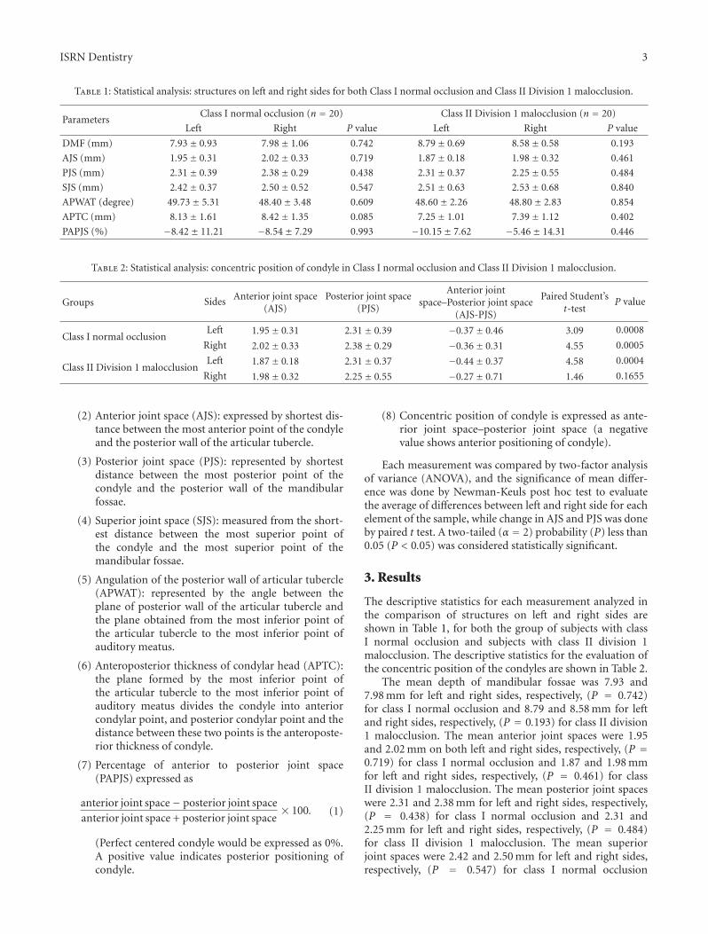
Table 1: Statistical analysis: structures on left and right sides for both Class I normal occlusion and Class II Division 1 malocclusion.
ParametersClass I normal occlusion (n = 20) Class II Division 1 malocclusion (n = 20)
Left Right P value Left Right P value
DMF (mm) 7.93± 0.93 7.98± 1.06 0.742 8.79± 0.69 8.58± 0.58 0.193
AJS (mm) 1.95± 0.31 2.02± 0.33 0.719 1.87± 0.18 1.98± 0.32 0.461
PJS (mm) 2.31± 0.39 2.38± 0.29 0.438 2.31± 0.37 2.25± 0.55 0.484
SJS (mm) 2.42± 0.37 2.50± 0.52 0.547 2.51± 0.63 2.53± 0.68 0.840
APWAT (degree) 49.73± 5.31 48.40± 3.48 0.609 48.60± 2.26 48.80± 2.83 0.854
APTC (mm) 8.13± 1.61 8.42± 1.35 0.085 7.25± 1.01 7.39± 1.12 0.402
PAPJS (%) −8.42± 11.21 −8.54± 7.29 0.993 −10.15± 7.62 −5.46± 14.31 0.446
Table 2: Statistical analysis: concentric position of condyle in Class I normal occlusion and Class II Division 1 malocclusion.
Groups Sides Anterior joint space(AJS)
Posterior joint space(PJS)
Anterior jointspace–Posterior joint space
(AJS-PJS)
Paired Student’st-test
P value
Class I normal occlusionLeft 1.95± 0.31 2.31± 0.39 −0.37± 0.46 3.09 0.0008
Right 2.02± 0.33 2.38± 0.29 −0.36± 0.31 4.55 0.0005
Class II Division 1 malocclusionLeft 1.87± 0.18 2.31± 0.37 −0.44± 0.37 4.58 0.0004
Right 1.98± 0.32 2.25± 0.55 −0.27± 0.71 1.46 0.1655
(2) Anterior joint space (AJS): expressed by shortest dis-tance between the most anterior point of the condyleand the posterior wall of the articular tubercle.
(3) Posterior joint space (PJS): represented by shortestdistance between the most posterior point of thecondyle and the posterior wall of the mandibularfossae.
(4) Superior joint space (SJS): measured from the short-est distance between the most superior point ofthe condyle and the most superior point of themandibular fossae.
(5) Angulation of the posterior wall of articular tubercle(APWAT): represented by the angle between theplane of posterior wall of the articular tubercle andthe plane obtained from the most inferior point ofthe articular tubercle to the most inferior point ofauditory meatus.
(6) Anteroposterior thickness of condylar head (APTC):the plane formed by the most inferior point ofthe articular tubercle to the most inferior point ofauditory meatus divides the condyle into anteriorcondylar point, and posterior condylar point and thedistance between these two points is the anteroposte-rior thickness of condyle.
(7) Percentage of anterior to posterior joint space(PAPJS) expressed as
anterior joint space− posterior joint spaceanterior joint space + posterior joint space
× 100. (1)
(Perfect centered condyle would be expressed as 0%.A positive value indicates posterior positioning ofcondyle.
(8) Concentric position of condyle is expressed as ante-rior joint space–posterior joint space (a negativevalue shows anterior positioning of condyle).
Each measurement was compared by two-factor analysisof variance (ANOVA), and the significance of mean differ-ence was done by Newman-Keuls post hoc test to evaluatethe average of differences between left and right side for eachelement of the sample, while change in AJS and PJS was doneby paired t test. A two-tailed (α = 2) probability (P) less than0.05 (P < 0.05) was considered statistically significant.
3. Results
The descriptive statistics for each measurement analyzed inthe comparison of structures on left and right sides areshown in Table 1, for both the group of subjects with classI normal occlusion and subjects with class II division 1malocclusion. The descriptive statistics for the evaluation ofthe concentric position of the condyles are shown in Table 2.
The mean depth of mandibular fossae was 7.93 and7.98 mm for left and right sides, respectively, (P = 0.742)for class I normal occlusion and 8.79 and 8.58 mm for leftand right sides, respectively, (P = 0.193) for class II division1 malocclusion. The mean anterior joint spaces were 1.95and 2.02 mm on both left and right sides, respectively, (P =0.719) for class I normal occlusion and 1.87 and 1.98 mmfor left and right sides, respectively, (P = 0.461) for classII division 1 malocclusion. The mean posterior joint spaceswere 2.31 and 2.38 mm for left and right sides, respectively,(P = 0.438) for class I normal occlusion and 2.31 and2.25 mm for left and right sides, respectively, (P = 0.484)for class II division 1 malocclusion. The mean superiorjoint spaces were 2.42 and 2.50 mm for left and right sides,respectively, (P = 0.547) for class I normal occlusion

4 ISRN Dentistry
and 2.51 and 2.53 mm for left and right sides, respectively,(P = 0.840) for class II division 1 malocclusion. The meanangulations of the posterior wall of articular tubercle were49.73 and 48.40 degree for left and right sides, respectively,(P = 0.609) for class I normal occlusion and 48.60 and 48.80degree for left and right sides, respectively, (P = 0.854) forclass II division 1 malocclusion. The mean anteroposteriorthickness of condylar head was 8.13 and 8.42 mm for leftand right sides, respectively, (P = 0.085) for class I normalocclusion and 7.25 and 7.39 mm for left and right sides,respectively, (P = 0.402) for class II division 1 malocclusion.The percentage of anterior to posterior joint spaces was−8.42 and −8.54% for left and right sides, respectively,(P = 0.003) for class I normal occlusion and −10.15% and−5.46% for left and right sides, respectively, (P = 0.290) forclass II division 1 malocclusion.
4. Discussion
We assessed Craniomandibular articulation (CMA) mor-phology by computed tomography (CT) as CT has beenshown to be ideal tool for CMA assessment because it allowsvery accurate evaluation of skeletal anatomic details [12, 13]without superimposition of any other structure [14]. CTscanning has following advantages [15] over conventionalradiography as follows It provides three-dimensional infor-mation in the form of thin slices, so internal structurescan be evaluated: this eliminates the superimpositions. CTcan detect density differences in the tissues of less than1% while conventional radiography depicts the tissues thatshow a density differences at least 10%. The sagittal sliceCT is the most appropriate for assessing the condyle-fossaerelationship [15]. In the present study CMA morphology wasstudied in the subjects with the age ranging from 14 to 25years as it is reported in literature [16, 17] that mandibularfossae attain their adult sizes before the age of 8 and did notshow significant change after this age.
We studied CMA morphology in subjects with Angle’sclass I normal occlusion and subjects with Angle’s classII malocclusion because class II malocclusion has beendescribed as most frequent treatment problem in orthodon-tic practice [18], and till the date of the planning of thestudy we did not find any CT study of Craniomandibulararticulation morphology reported in literature on subjectswith Angle’s class I normal occlusion. Hence this finding ofpresent study could be a valuable reference for evaluation andcomparison of TMJ morphology in different malocclusionsin north Indian population.
Our result, for the depth of mandibular fossae both inclass I normal occlusion and class II division 1 malocclusionwere not statistically significant (P > 0.05) when the rightand left sides were compared (Table 1). Nonsignificant (P >0.05) results were also obtained when the condyle-fossaerelationship was assessed for right and left sides through themeasurements of the anterior, superior, posterior joint spacesand anteroposterior thickness of condylar head (Table 1)in both class I normal occlusion and class II division 1malocclusion subjects. The lack of asymmetry in thesemeasurements is similar to those previous studies in which
the same methodology was applied for the different typesof the malocclusion [11, 19, 20]. Since the sagittal evalu-ation showed no significant differences regarding condylardimension and positioning, the asymmetry in the posteriorarticular space can be explained by different dimensions ofmandibular fossae.
Our result for the angulation of the posterior wall ofarticular tubercle (Table 1) is similar to the study conductedby Vitral et al. [11] and they found value of 51.37◦ for classI and 52.40◦ for class II side in class II division 1 subdivisionmalocclusion. Our result for APWAT is not in line with thestudy conducted by Christiansen et al. [21]. However, theirresults cannot be compared with our study because they didnot specify the plane from which the angle was measured.
We assessed the concentric positioning of the condyleby comparing the differences in AJS and PJS (Table 2). Wefound that AJS for both right and left sides was significantly(P < 0.01) smaller than PJS in the subjects with class Inormal occlusion. While the subjects with class II division1 malocclusion AJS were significantly (P < 0.01) smallerthan PJS on the left sides, no significant difference wasfound on right sides. Therefore findings of the present studysuggest anteriorly position of condyle in mandibular fossae.Initial studies conducted for the evaluation of condylarconcentricity showed centralization of condylar process [22–24]. Pullinger et al. [25] also showed that anterior positioningof condyle is the feature of class II division 1 malocclusionsample. Vitral et al. [11] with the same methodology used inour study found a more anterior condylar position bilaterallyin subjects with class II division 1 subdivision malocclusion.The recent studies [19, 20] that used more sophisticateddiagnostics and imaging techniques did not confirm thefindings of older studies, which described the centralizedcondylar positioning in relation to the mandibular fossae.Result of our finding regarding the concentric positioningof condyle is similar to the previous study conducted byCohlmia et al. [26]. They reported that left condyle wasmore anteriorly positioned than the right in a sampleof the patients with malocclusion after comparing theanterior articular space for right and left TMJ. Accordingto these authors this asymmetry could be related to normalcranial base asymmetries and side preference during themastication.
In present study on comparing the mean PAPJS (Table 1)at the left and right sides, did not differ significantly (P >0.05) in both class I normal occlusion and class II division1 malocclusion. This result showed anterior positioning ofcondyle in mandibular fossae. This equation (suggested byPullinger) determined the percentage of anterior or posteriordisplacement of condyle, with concentricity as a reference. Ascore of −12% to +12% of posterior to anterior joint spaceis used to describe concentricity of condyle in mandibularfossae. Hence the overall findings of this study suggest thatboth right and left condyles were centered in mandibularfossae with a tendency towards anterior positioning. Thecondylar-fossae morphology, as it is presented here inour study, was assessed from the sagittal slice computedtomogram as a step towards the objective measurementof the shape of the condyle and fossae by using methods

ISRN Dentistry 5
that are applied in the wider field of biologic sciences.There is no doubt that this methodology is very simple andsophisticated. Further evaluation of the relationship betweenshape and function of the Craniomandibular articulation isneeded in various groups of malocclusions.
5. Conclusion
From the present study, the following conclusion can bedrawn.
(1) There were no differences found in the condylarprocess and joint morphology between right and leftsides of both Angle’s class I normal occlusion andAngle’s class II division 1 malocclusion.
(2) Evaluation of the position of the condyles in theirrespective mandibular fossae showed concentricposition with a tendency towards anterior position-ing for both right and left sides of the subjects withAngle’s Class I normal occlusion as well as subjectswith Angle’s class II division 1 malocclusion.
References
[1] E. Lloyd DuBrul, “The Craniomandibular articulation,” inSicher and DuBrul’ Oral Anatomy, pp. 107–108, Isbiyaku Euro-America, St. Louis, Mo, USA, 8th edition, 1988.
[2] F. Mongini, “Anatomic and clinical evaluation of the relation-ship between the temporomandibular joint and occlusion,”The Journal of Prosthetic Dentistry, vol. 38, no. 5, pp. 539–551,1977.
[3] F. O’Ryan and B. N. Epker, “Temporomandibular jointfunction and morphology: observations on the spectra ofnormalcy,” Oral Surgery Oral Medicine and Oral Pathology, vol.58, no. 3, pp. 272–279, 1984.
[4] M. A. Burley, “An examination of the relation between theradiographic appearance of the temporomandibular joint andsome features of the occlusion,” Brazilian Dental Journal, vol.110, pp. 195–200, 1961.
[5] M. A. Matsumoto and A. M. Bolognese, “Radiographic mor-phology of the temporomandibular joint related to occlusalcharacteristics,” Brazilian Dental Journal, vol. 5, no. 2, pp. 115–120, 1994.
[6] E. G. Katsavrias and D. J. Halazonetis, “Condyle and fossashape in class II and class III skeletal patterns: a morphometrictomographic study,” American Journal of Orthodontics andDentofacial Orthopedics, vol. 128, no. 3, pp. 337–346, 2005.
[7] K. A. Ebner, L. L. Otis, R. Zakhary, and R. A. Danforth, “Axialtemporomandibular joint morphology: a correlative study ofradiographic and gross anatomic findings,” Oral Surgery OralMedicine and Oral Pathology, vol. 69, no. 2, pp. 247–252, 1990.
[8] K. Hiiemae, “Functional aspects of primate jaw morphology,”in Food Acquisition and Processing in Primates, D. J. Hivers,B. A. Wood, and A. Bilsborough, Eds., pp. 257–281, PlenumPress, New York, NY, USA, 1984.
[9] H. J. Blackwood, “Pathology of the temporomandibular joint,”The Journal of the American Dental Association, vol. 79, no. 1,pp. 118–124, 1969.
[10] J. C. Bennett and R. P. McLaughlin, Orthodontic TreatmentMechanics and Preadjusted Appliance, Mosby-Wolfe, London,UK, 1993.
[11] R. W. Vitral, C. d’s Telles, M. R. Fraga, R. S. Oliveira,and O. M. Tanaka, “Computed tomography evaluation oftemporomandibular joint alterations in patients with classII division 1 subdivision malocclusions: condyle-fossa rela-tionship,” American Journal of Orthodontics and DentofacialOrthopedics, vol. 126, no. 1, pp. 48–52, 2004.
[12] R. W. Katzberg, “Temporomandibular joint imaging,” Radiol-ogy, vol. 170, no. 2, pp. 297–307, 1989.
[13] T. A. Larheim and A. Kolbenstvedt, “High-resolution com-puted tomography of the osseous temporomandibular joint:some normal and abnormal appearances,” Acta Radiologica,vol. 25, no. 6, pp. 465–469, 1984.
[14] H. R. Cohen, S. Ross, R. E. Gordon, and A. M. Deutsch,“Computed tomography in TMJ diagnosis,” Journal of ClinicalOrthodontics, vol. 19, no. 9, pp. 659–662, 1985.
[15] K. L. Bontrager, Textbook of Radiographic Positioning andRelated Anatomyed, Mosby, St. Louis, Mo, USA, 6th edition,2005.
[16] D. M. Wright and B. C. Moffett Jr., “The postnatal devel-opment of the human temporomandibular joint,” AmericanJournal of Anatomy, vol. 141, no. 2, pp. 235–249, 1974.
[17] H. J. Scott, “Growth changes in the glenoid fossa,” DentalPractice, vol. 6, pp. 117–120, 1955.
[18] R. S. Nanda, T. C. Dandajena, and R. Nanda, “Biomechanicsstrategies for nonextraction class II malocclusions,” in Biome-chanics and Esthetic Strategies in Clinical Orthodontics, p. 177,Elsevier Saunders, St. Louis, Mo, USA, 1st edition, 2009.
[19] A. F. Rodrigues, M. R. Fraga, and R. W. Vitral, “Computedtomography evaluation of the temporomandibular joint inclass I malocclusion patients: condylar symmetry and condyle-fossa relationship,” American Journal of Orthodontics andDentofacial Orthopedics, vol. 136, no. 2, pp. 192–198, 2009.
[20] A. F. Rodrigues, M. R. Fraga, and R. W. Vitral, “Computedtomography evaluation of the temporomandibular joint inclass II division 1 and class III malocclusion patients: condylarsymmetry and condyle-fossa relationship,” American Journalof Orthodontics and Dentofacial Orthopedics, vol. 136, no. 2,pp. 199–206, 2009.
[21] E. L. Christiansen, T. T. Chan, J. R. Thompson, A. N. Hasso,D. B. Hinshaw Jr., and S. Kopp, “Computed tomography ofthe normal temporomandibular joint,” Scandinavian Journalof Dental Research, vol. 95, no. 6, pp. 499–509, 1987.
[22] B. Madsen, “Normal variations in anatomy, condylar move-ments, and arthrosis frequency of the temporomandibularjoints,” Acta Radiologica, vol. 4, no. 3, pp. 273–288, 1966.
[23] L. A. Weinberg, “Correlation of temporomandibular dysfunc-tion with radiographic findings,” The Journal of ProstheticDentistry, vol. 28, no. 5, pp. 519–539, 1972.
[24] D. D. Blaschke and T. J. Blaschke, “Normal TMJ bonyrelationships in centric occlusion,” Journal of Dental Research,vol. 60, no. 2, pp. 98–104, 1981.
[25] A. G. Pullinger, W. K. Solberg, L. Hollender, and A. Petersson,“Relationship of mandibular condylar position to dentalocclusion factors in an asymptomatic population,” AmericanJournal of Orthodontics and Dentofacial Orthopedics, vol. 91,no. 3, pp. 200–206, 1987.
[26] J. T. Cohlmia, J. Ghosh, P. K. Sinha, R. S. Nanda, and G.F. Currier, “Tomographic assessment of temporomandibularjoints in patients with malocclusion,” The Angle Orthodontist,vol. 66, no. 1, pp. 27–36, 1996.

Submit your manuscripts athttp://www.hindawi.com
Hindawi Publishing Corporationhttp://www.hindawi.com Volume 2014
Oral OncologyJournal of
DentistryInternational Journal of
Hindawi Publishing Corporationhttp://www.hindawi.com Volume 2014
Hindawi Publishing Corporationhttp://www.hindawi.com Volume 2014
International Journal of
Biomaterials
Hindawi Publishing Corporationhttp://www.hindawi.com Volume 2014
BioMed Research International
Hindawi Publishing Corporationhttp://www.hindawi.com Volume 2014
Case Reports in Dentistry
Hindawi Publishing Corporationhttp://www.hindawi.com Volume 2014
Oral ImplantsJournal of
Hindawi Publishing Corporationhttp://www.hindawi.com Volume 2014
Anesthesiology Research and Practice
Hindawi Publishing Corporationhttp://www.hindawi.com Volume 2014
Radiology Research and Practice
Environmental and Public Health
Journal of
Hindawi Publishing Corporationhttp://www.hindawi.com Volume 2014
The Scientific World JournalHindawi Publishing Corporation http://www.hindawi.com Volume 2014
Hindawi Publishing Corporationhttp://www.hindawi.com Volume 2014
Dental SurgeryJournal of
Drug DeliveryJournal of
Hindawi Publishing Corporationhttp://www.hindawi.com Volume 2014
Hindawi Publishing Corporationhttp://www.hindawi.com Volume 2014
Oral DiseasesJournal of
Hindawi Publishing Corporationhttp://www.hindawi.com Volume 2014
Computational and Mathematical Methods in Medicine
ScientificaHindawi Publishing Corporationhttp://www.hindawi.com Volume 2014
PainResearch and TreatmentHindawi Publishing Corporationhttp://www.hindawi.com Volume 2014
Preventive MedicineAdvances in
Hindawi Publishing Corporationhttp://www.hindawi.com Volume 2014
EndocrinologyInternational Journal of
Hindawi Publishing Corporationhttp://www.hindawi.com Volume 2014
Hindawi Publishing Corporationhttp://www.hindawi.com Volume 2014
OrthopedicsAdvances in
Related Documents


![ClassificationofAlzheimer’sDiseaseandMildCognitive ...downloads.hindawi.com/journals/jhe/2020/3743171.pdf · 2020. 11. 5. · the subjects [12]. e margin of the training data is](https://static.cupdf.com/doc/110x72/60fd1faaadda8a43df20cdb4/classificationofalzheimerasdiseaseandmildcognitive-2020-11-5-the-subjects.jpg)


![ProsthodonticRehabilitationofHereditaryEctodermal ...downloads.hindawi.com/journals/crid/2012/489769.pdfin the face [5, 6]. The deviation from normal facial growth of HED subjects](https://static.cupdf.com/doc/110x72/5fa75692681f776ac5661f4b/prosthodonticrehabilitationofhereditaryectodermal-in-the-face-5-6-the-deviation.jpg)

