Computational Protein Design: The Proteus Software and Selected Applications Thomas Simonson,* [a] Thomas Gaillard, [a] David Mignon, [a] Marcel Schmidt am Busch, [a,c] Anne Lopes, [a,d] Najette Amara, [a] Savvas Polydorides, [a,b] Audrey Sedano, [a] Karen Druart, [a] and Georgios Archontis AQ1 [b] We describe an automated procedure for protein design, implemented in a flexible software package, called Proteus. System setup and calculation of an energy matrix are done with the XPLOR modeling program and its sophisticated com- mand language, supporting several force fields and solvent models. A second program provides algorithms to search sequence space. It allows a decomposition of the system into groups, which can be combined in different ways in the energy function, for both positive and negative design. The whole procedure can be controlled by editing 2–4 scripts. Two applications consider the tyrosyl-tRNA synthetase enzyme and its successful redesign to bind both O-methyl-tyrosine and D- tyrosine. For the latter, we present Monte Carlo simulations where the D-tyrosine concentration is gradually increased, dis- placing L-tyrosine from the binding pocket and yielding the binding free energy difference, in good agreement with experiment. Complete redesign of the Crk SH3 domain is pre- sented. The top 10000 sequences are all assigned to the cor- rect fold by the SUPERFAMILY library of Hidden Markov Models. Finally, we report the acid/base behavior of the SNase protein. Sidechain protonation is treated as a form of muta- tion; it is then straightforward to perform constant-pH Monte Carlo simulations, which yield good agreement with experi- ment. Overall, the software can be used for a wide range of application, producing not only native-like sequences but also thermodynamic properties with errors that appear comparable to other current software packages.Copyright V C 2013 Wiley Periodicals, Inc. DOI: 10.1002/jcc.23418 Introduction Computational protein design (CPD) continues to develop as an important tool for biotechnology. [1–8] AQ2 Early applications led to proteins with novel ligand-binding functions, [9,10] novel enzyme activity, [11] and proteins that were completely “redesigned”: around 2/3 of their sequence was mutated, yet their structure and stability were retained. [12] In the last few years, CPD has allowed the creation of new protein folds, [13–15] completely new enzymes, [16–18] and the assembly or deassem- bly of multiprotein complexes. [19–23] CPD methods are mainly characterized by (a) the energy function, (b) the description of the folded protein’s conforma- tional space, (c) the treatment of the unfolded state, and (d) the search method used to explore sequences and conforma- tions. Although the search method is important for efficiency, the accuracy of the results is mainly determined by the first three ingredients, especially the energy function. Energy func- tions from molecular simulations are developed from first prin- ciples [24–28] and have the capability to predict protein structure, stability, and ligand binding with a high accuracy. [29– 33] In a CPD context, however, additional approximations are necessary, so that the energy function is modified, both by the use of an implicit solvent model [34–38] and through additional, empirical, contributions. [2,3,12,13,39–42] Several software implementations have been reported. The Rosetta suite is currently the most successful and widely used, [12,13,15,18,41,43] but others exist and have also been successful. [6–10,44–54] They differ in the characteristics (a–d) listed above, the range of choices offered for each one, the degree of empiricism of the energy function, the applicability to differ- ent classes of molecules, the mode of user interaction, the availability and ease of development of source code, and so on. Here, we describe a software implementation, Proteus 2.0, that significantly extends and improves an earlier one. [54–56] Its three main components are (1) the molecular simulation pro- gram XPLOR, [57] with its capability to describe biomolecular interaction energies; (2) a sophisticated set of scripts, written in the XPLOR scripting language, [57,58] that control the calcula- tion of an energy matrix for the system of interest [59] ; (3) a C program, “Proteus,” for exploring the space of sequences and conformations using various search algorithms, including a mean field and a Monte Carlo method. XPLOR can be [a] T. Simonson, T. Gaillard, D. Mignon, M. S. am Busch, A. Lopes, N. Amara, S. Polydorides, A. Sedano, K. Druart Laboratoire de Biochimie (CNRS UMR7654), Department of Biology, Ecole Polytechnique, Palaiseau 91128, France AQ7 Email: [email protected] [b] S. Polydorides, G. Archontis AQ1 Department of Physics, University of Cyprus, Nicosia, Cyprus [c] M. S. am Busch Institut fuer theoretische Physik, Johannes Kepler Universitaet Linz, Alten- berger Strasse 69, Linz 4040, Austria AQ8 [d] A. Lopes Institut de G en etique Mol eculaire, Universit e de Paris-Sud, Orsay, France V C 2013 Wiley Periodicals, Inc. Journal of Computational Chemistry 2013, DOI: 10.1002/jcc.23418 1 FULL PAPER WWW.C-CHEM.ORG J_ID: JCC Customer A_ID: JCC23418 Cadmus Art: JCC23418 Ed. Ref. No.: 13-0237.R1 Date: 13-August-13 Stage: Page: 1 ID: muralir Time: 21:10 I Path: N:/3b2/JCCT/Vol00000/130172/APPFile/JW-JCCT130172

Welcome message from author
This document is posted to help you gain knowledge. Please leave a comment to let me know what you think about it! Share it to your friends and learn new things together.
Transcript
Computational Protein Design: The Proteus Software andSelected Applications
Thomas Simonson,*[a] Thomas Gaillard,[a] David Mignon,[a] Marcel Schmidt am Busch,[a,c]
Anne Lopes,[a,d] Najette Amara,[a] Savvas Polydorides,[a,b] Audrey Sedano,[a] Karen Druart,[a]
and Georgios ArchontisAQ1 [b]
We describe an automated procedure for protein design,
implemented in a flexible software package, called Proteus.
System setup and calculation of an energy matrix are done
with the XPLOR modeling program and its sophisticated com-
mand language, supporting several force fields and solvent
models. A second program provides algorithms to search
sequence space. It allows a decomposition of the system into
groups, which can be combined in different ways in the
energy function, for both positive and negative design. The
whole procedure can be controlled by editing 2–4 scripts. Two
applications consider the tyrosyl-tRNA synthetase enzyme and
its successful redesign to bind both O-methyl-tyrosine and D-
tyrosine. For the latter, we present Monte Carlo simulations
where the D-tyrosine concentration is gradually increased, dis-
placing L-tyrosine from the binding pocket and yielding the
binding free energy difference, in good agreement with
experiment. Complete redesign of the Crk SH3 domain is pre-
sented. The top 10000 sequences are all assigned to the cor-
rect fold by the SUPERFAMILY library of Hidden Markov
Models. Finally, we report the acid/base behavior of the SNase
protein. Sidechain protonation is treated as a form of muta-
tion; it is then straightforward to perform constant-pH Monte
Carlo simulations, which yield good agreement with experi-
ment. Overall, the software can be used for a wide range of
application, producing not only native-like sequences but also
thermodynamic properties with errors that appear comparable
to other current software packages.Copyright VC 2013 Wiley
Periodicals, Inc.
DOI: 10.1002/jcc.23418
Introduction
Computational protein design (CPD) continues to develop as
an important tool for biotechnology.[1–8]AQ2 Early applications led
to proteins with novel ligand-binding functions,[9,10] novel
enzyme activity,[11] and proteins that were completely
“redesigned”: around 2/3 of their sequence was mutated, yet
their structure and stability were retained.[12] In the last few
years, CPD has allowed the creation of new protein folds,[13–15]
completely new enzymes,[16–18] and the assembly or deassem-
bly of multiprotein complexes.[19–23]
CPD methods are mainly characterized by (a) the energy
function, (b) the description of the folded protein’s conforma-
tional space, (c) the treatment of the unfolded state, and (d)
the search method used to explore sequences and conforma-
tions. Although the search method is important for efficiency,
the accuracy of the results is mainly determined by the first
three ingredients, especially the energy function. Energy func-
tions from molecular simulations are developed from first prin-
ciples[24–28] and have the capability to predict protein
structure, stability, and ligand binding with a high accuracy.[29–
33] In a CPD context, however, additional approximations are
necessary, so that the energy function is modified, both by the
use of an implicit solvent model[34–38] and through additional,
empirical, contributions.[2,3,12,13,39–42]
Several software implementations have been reported. The
Rosetta suite is currently the most successful and widely
used,[12,13,15,18,41,43] but others exist and have also been
successful.[6–10,44–54]They differ in the characteristics (a–d) listed
above, the range of choices offered for each one, the degree
of empiricism of the energy function, the applicability to differ-
ent classes of molecules, the mode of user interaction, the
availability and ease of development of source code, and
so on.
Here, we describe a software implementation, Proteus 2.0,
that significantly extends and improves an earlier one.[54–56] Its
three main components are (1) the molecular simulation pro-
gram XPLOR,[57] with its capability to describe biomolecular
interaction energies; (2) a sophisticated set of scripts, written
in the XPLOR scripting language,[57,58] that control the calcula-
tion of an energy matrix for the system of interest[59]; (3) a C
program, “Proteus,” for exploring the space of sequences and
conformations using various search algorithms, including a
mean field and a Monte Carlo method. XPLOR can be
[a] T. Simonson, T. Gaillard, D. Mignon, M. S. am Busch, A. Lopes, N. Amara,
S. Polydorides, A. Sedano, K. Druart
Laboratoire de Biochimie (CNRS UMR7654), Department of Biology, Ecole
Polytechnique, Palaiseau 91128, France AQ7
Email: [email protected]
[b] S. Polydorides, G. Archontis AQ1
Department of Physics, University of Cyprus, Nicosia, Cyprus
[c] M. S. am Busch
Institut fuer theoretische Physik, Johannes Kepler Universitaet Linz, Alten-
berger Strasse 69, Linz 4040, Austria AQ8
[d] A. Lopes
Institut de G�en�etique Mol�eculaire, Universit�e de Paris-Sud, Orsay, France
VC 2013 Wiley Periodicals, Inc.
Journal of Computational Chemistry 2013, DOI: 10.1002/jcc.23418 1
FULL PAPERWWW.C-CHEM.ORG
J_ID: JCC Customer A_ID: JCC23418 Cadmus Art: JCC23418 Ed. Ref. No.: 13-0237.R1 Date: 13-August-13 Stage: Page: 1
ID: muralir Time: 21:10 I Path: N:/3b2/JCCT/Vol00000/130172/APPFile/JW-JCCT130172
simonson ([email protected])
Sticky Note
names are fine here, but see correction at the bottom of the page.
simonson ([email protected])
Cross-Out
simonson ([email protected])
Sticky Note
Notice the lowercase "p" here: Proteus and proteus are two different things!
simonson ([email protected])
Cross-Out
simonson ([email protected])
Cross-Out
simonson ([email protected])
Cross-Out
simonson ([email protected])
Sticky Note
Division names are already given: "Institut etc" and "Institut etc".
downloaded by academic users from the Yale University web
site, whereas local modifications to XPLOR, our XPLOR scripts,
the Proteus source code, and documentation are available on
request (and will soon be available online). The software is
modular and flexible, allowing the use of four different molec-
ular mechanics force fields, four solvent models, including two
generalized born (GB) variants,[60–62] several rotamer libraries,
and a wide range of fitness functions, using any combination
of protein stability, ligand affinity, and ligand specificity, includ-
ing positive and negative design. The current version does not
allow the most recent methods for flexible backbone
design,[63–65] but calculations can be performed using an
ensemble of predefined backbone conformations.
This and the earlier implementation have been used for sev-
eral applications with good success. One application consisted
in redesigning 95 small proteins from six structural families,
then using the designed sequences to perform homolog
searching.[66,67] For this application, the energy matrix calcula-
tions were ported to a volunteer distributed computing frame-
work, based on the Berkeley Open Infrastructure for Network
Computing,[68] and made available through our Protein-
s@Home project, in which over 20,000 volunteers partici-
pated.[66,67] Where comparison was possible, the quality of the
designed sequences was comparable to several other CPD
implementations. Over 85% of the designed sequences were
assigned to their correct SCOP family by the SUPERFAMILY
library of Hidden Markov Models for fold recognition.[69] We
also tested their capability to retrieve natural homologs from
sequence databases. Using low energy designed sequences,
we could retrieve 60–70% of known SH2, SH3, and PDZ
domains and around 90% of known Kunitz-type inhibitors and
interleukin-8 chemokines.
A second application consisted in redesigning the
asparaginyl-tRNA synthetase enzyme to decrease binding of its
natural substrate asparagine (Asn) and increase binding of the
substrate analog aspartate (Asp).[70,71] The best designed
sequences did not display detectable catalytic activity; never-
theless, MD simulations and Poisson–Boltzmann (PB) free
energy calculations gave good evidence that they did indeed
have a strongly reduced Asn binding and increased Asp bind-
ing, compared to the native enzyme.[71] A third application
(not yet published) led to a successful redesign of the stereo-
specificity of the tyrosyl-tRNA synthetase (TyrRS) enzyme, with
one designed variant having a preference for the substrate
analog D-tyrosine, with respect to the natural substrate L-
tyrosine. A fourth application used the software in a somewhat
different way, to study acid/base changes (mutations) in a test
set of 12 proteins.[31,72] Indeed, the CPD problem maps pre-
cisely onto the problem of computing acid/base constants
(pKa’s) for protein sidechains, as long as one uses an appropri-
ate algorithm (constant-pH Monte Carlo) to sample the Boltz-
mann ensemble of conformations and protonation states.
Calculations with the Amber ff99SB force field and a good GB
variant gave good agreement between computed and experi-
mental pKa’s, with rms errors of about 1.2 pKa units, almost as
good as the widely used PropKa program[73,74] (0.8 pKa units
for the same test). Finally, sidechain reconstruction tests gave
good results earlier,[62] and better results very recently with
the superior, Amber force field: for nine proteins, we obtained
77% of sidechains with correct v1 and v2 angles, compared to
81% with the widely used SCWRL4 program[75] (which uses a
much larger rotamer library); these tests will be published
elsewhere.
Below, we describe the methods used in our current soft-
ware and some illustrative applications. In the “Theoretical
Methods” section, we describe the energy function, including
the solvent and unfolded state models. The energy function
combines a molecular mechanics treatment of the protein(s)
and any ligands with an implicit model of the solvent. When a
GB solvent model is used, we introduce a “Native Environment
(NE)” approximation for each sidechain, described below. It
allows the total energy to be expressed as a sum of one- and
two-residue terms. This makes possible the usual two-stage
CPD procedure, introduced by Mayo and coworkers, where an
energy matrix is precomputed, then used during a second,
sequence exploration stage.[59] The solvent model also
includes terms that depend on the solvent-accessible surface
area of each atom. Here, too, approximations are used to
reduce the surface areas to a sum over one- and two-residue
terms.[62,76] For the unfolded state, we use a simple model,
where the protein is viewed as a collection of independent tri-
peptides.[42,54] The energy of these tripeptides includes an
empirical correction for each amino acid type,[13,39,77] opti-
mized so that the abundancies of each type match some tar-
get values, such as the natural frequencies in a given protein
family.
In the same, theoretical section, we describe the Monte
Carlo exploration of sequence space and the relation between
the sampled distribution of sequences and conformations and
the folding free energy of each sequence. We include the spe-
cial case where “sequence” changes correspond to changes in
the sidechain protonation states. We also describe the mean
field exploration method and a heuristic method.
In the next, Computational Protocols section, we describe
the computational steps and their implementation in a typical
design calculation. These include the setup of the system for
XPLOR, the positioning of sidechain rotamers on the protein
backbone, the precalculation of GB solvation radii, the calcula-
tion of diagonal and off-diagonal energy matrix terms (the
most expensive step), the exploration of sequence and confor-
mation space. Many of the details are in the Supporting
Information.
Finally, we describe some illustrative applications. We focus
on practical aspects and difficulties, and features that are spe-
cific to the present, most recent software implementation. We
first consider the design of two protein:ligand complexes. Both
involve the TyrRS enzyme, with two possible substrate analogs
as its ligand: O-methyl-tyrosine (me-Tyr) and D-tyrosine (D-Tyr).
In particular, we report MC AQ3simulations where L-Tyr and D-Tyr
are both present, but the concentration of D-Tyr is gradually
increased so that it displaces L-Tyr from the binding pocket.
The midpoint concentration can then be interpreted as a bind-
ing free energy difference. Next, we consider whole protein
design, with the c-Crk SH3 domain as an example. Finally, we
J_ID: JCC Customer A_ID: JCC23418 Cadmus Art: JCC23418 Ed. Ref. No.: 13-0237.R1 Date: 13-August-13 Stage: Page: 2
ID: muralir Time: 21:10 I Path: N:/3b2/JCCT/Vol00000/130172/APPFile/JW-JCCT130172
FULL PAPER WWW.C-CHEM.ORG
2 Journal of Computational Chemistry 2013, DOI: 10.1002/jcc.23418 WWW.CHEMISTRYVIEWS.COM

describe the calculation of acid/base constants for the SNase
protein. The calculation of thermodynamic properties in the D-
Tyr and SNase applications helps to illustrate the accuracy of
our free energy function and structural models. Additional
thermodynamic calculations (such as stability changes due to
point mutations) will be reported elsewhere. Full details on
the computational methods used for the present applications
are given in Supporting Information.
Theoretical Methods
Energy function: general form
We use a molecular mechanics energy function along with an
implicit solvent model.[62] The molecular mechanics parameter-
izations that are currently available within Proteus correspond
to the Charmm19 force field[78] and the ff99SB version of the
Amber force field.[25,79] XPLOR also allows a “polar hydrogen”
version of OPLS[80] and the Charmm22 force field,[24] but these
two parameterizations are not yet implemented within the
Proteus CPD scripts. As the energy function includes a contri-
bution from the implicit solvent, it should be viewed as a
potential of mean force.[34,81]
Four main solvent models are available.[62] The two simplest
use a simple dielectric screening function to reduce the Cou-
lombic interactions between protein atoms: either a uniform
screening factor (“CDIE” option in XPLOR) or a distance-
dependent screening factor (“RDIE” option).[62] The other two
solvent models use a more complex, GB screening function.
The GB energy contribution has the form:
EGB5s2
Xij
qiqjgij; (1)
where the sum is over all pairs of protein charges, s5 1ew
2 1ep
,
ep is the protein dielectric constant, ew is the solvent dielectric
constant (80 at room temperature), and gij represents the
interaction (or Green’s function) between a unit charge at the
qi position and the solvent polarization induced by another
unit charge at the qj position. This last quantity is approxi-
mated by
gij5ðr2ij 1bibj exp ½2r2
ij=4bibj�Þ21=2 (2)
where rij is the distance between atoms i; j and bi is the
“solvation radius” of atom i. This radius approximates the dis-
tance from i to the protein surface and is a function of the
coordinates of all the protein atoms. Two GB implementations
are available in XPLOR and Proteus: the “GB/ACE” version of
Schaefer and Karplus[82,83] and the version of Truhlar and
coworkers,[84] which we refer to as “GB/HCT.” They differ by
the method used to compute the bi ; for details, see Moulinier
et al.[61]
All four screening functions above can be combined with a
solvent accessible surface energy term:
Esurf5aX
i
riAi (3)
The sum is over the protein atoms i, Ai is the solvent acces-
sible surface area of atom i, ri is an atomic solvation coeffi-
cient (measured in kcal/mol/A2) that depends on the atom
type, and a is an overall scaling factor for the surface term.
GB energy term: approximation as a sum over atom pairs
Because the solvation radii bi depend on all the protein coor-
dinates,[60,61,82,84] the GB energy contribution EGB cannot be
expressed as a sum over atom or residue pairs, violating the
two-stage, Mayo CPD protocol.[59] To avoid this difficulty, vari-
ous approximations have been proposed.[38,45,85–87] Proteus
uses the following simple method.[71] Consider two different
residues, I and J, with given sidechain types and rotamers. Let
i and j be atoms, belonging to I and J, respectively. When
computing the I; J interaction energy, a contribution s2 qiqjgij
arises from the i; j pair. To compute bi in gij [eq. (2)], we
assume the whole protein except for I is in a fixed, reference
conformation with a fixed, reference sequence; similarly for bj.
The reference sequence and conformation are normally taken
from the experimental native structure (but other choices are
possible). We refer to this as the “native environment,” or NE
approximation. Notice that for each i; j pair, the NE approxima-
tion assumes three different sequences when computing bi, bj ,
and gij. The net effect is that each residue pair I; J experiences
an effective, native-like, dielectric environment. For sidechain:-
backbone interactions, the same method applies, taking J to
represent the backbone (assumed to have a fixed conforma-
tion). In practice, all the bi are precomputed at the same time
as the diagonal I; I terms of the energy matrix (see below). The
quality of the NE approximation is quite good, as illustrated by
the successful applications below and described in detail else-
where (manuscript in preparation).
Accessible surface area energy term: approximation as a
sum over atom pairs
Surface areas are computed using the Lee and Richards algo-
rithm,[88] implemented in the XPLOR program.[57] For reasons
of efficiency, following Street and Mayo,[76] we assume that
Ai can be obtained by summing the contact areas Aij
between atom i and its neighbors j, and subtracting the con-
tact, or solvent-inaccessible area Ci5P
j Aij from the total
area of atom i. This approximation has the enormous advant-
age that the surface energy takes the form of a sum over
pairs of amino acids. However, it leads to a systematic error,
as the contact areas can overlap: a portion of atom i can be
in contact with two atoms j and j0 at a time. Street and
Mayo showed, and we confirmed[62] that the systematic error
can be largely corrected by applying a scaling factor of less
than one to contact areas Aij that involve at least one buried
atom (i or j); for details, see Ref. [62]. In earlier work, we did
extensive testing and comparison of several different sets of
surface parameters, based on sidechain reconstruction, pro-
tein solvation energies, and mutations of over 1000 side-
chains (including buried sidechains).[55,62] Details on our
current implementation and its accuracy will be published
elsewhere.
J_ID: JCC Customer A_ID: JCC23418 Cadmus Art: JCC23418 Ed. Ref. No.: 13-0237.R1 Date: 13-August-13 Stage: Page: 3
ID: muralir Time: 21:10 I Path: N:/3b2/JCCT/Vol00000/130172/APPFile/JW-JCCT130172
FULL PAPERWWW.C-CHEM.ORG
Journal of Computational Chemistry 2013, DOI: 10.1002/jcc.23418 3
simonson ([email protected])
Cross-Out

Unfolded energy: approximation as a sum over residues
In the unfolded state model, following earlier studies,[42,59,89]
the amino acid sidechains do not interact with each other, but
only with nearby backbone and with solvent. One way to
implement this idea is to consider that each amino acid X is
part of a tripeptide with a sequence Ala-X-Ala and a given
backbone geometry. In practice, we and others have found
that this simple model should be supplemented by an empiri-
cal energy correction, eX , optimized so that the overall amino
acid compositions are reasonable when whole proteins are
designed.[13,39,54,77] Notice that if a correction is to be added,
we can simply view the whole contribution EX of each amino
acid as an empirical quantity that depends only on the amino
acid type of X, without referring to tripeptides.
To optimize the EX values, we typically consider a test set of
proteins and determine the experimental amino acid frequen-
cies f expX for a set of experimental homologs. We then proceed
iteratively, with the EX initially set to a plausible starting guess.
At each iteration, several thousand sequences are computed
for each test protein. The corresponding amino acid frequen-
cies, f calcX , averaged over all sequences, proteins, and amino
acid positions, are compared to the experimental frequencies
f expX . The energy EX is then modified according to the
Boltzmann-like relation:
EnewX 5 Eold
X 1 0:5 lnf exp
X
f calcX
: (4)
With this scheme, if a given type X is too abundant in the
designed sequences, eq. (4) leads to an increased stability of
the unfolded state when X is present, so that X will be less
abundant in the next round. After about 10 rounds, the fre-
quencies converge to the target values and the procedure
can be stopped. Illustrative values are shown in FigureF1 1 and
TableT1 1, and compared to the values obtained with the tripep-
tide method. The EX values shown were optimized for the SH3
protein family, using CPD runs with the Amber ff99SB force
field, the GB/HCT solvent model, and a protein dielectric con-
stant of 16. The tripeptide values were computed with the
same energy function, and averaged over all positions in two
proteins (SNase and lysozyme) and over 800 conformations
(rotamer sets) for each protein. The dispersion of the tripep-
tide energies is comparable to the size of the dots in Figure 1
and is not visible. The two sets are very similar, which shows
that only small empirical tuning of EX is needed to reproduce
the experimental amino acid frequencies.
Sequence exploration: heuristic and mean field optimization
methods
Proteus allows three sequence exploration methods. The first
is a mean field approach, which has been described else-
where.[62,90–92] This method calculates iteratively the Boltz-
mann probability Pði; kÞ of each rotamer k of each residue i,
which is related to the mean energy Eði; kÞ of sidechain i:
Eði; kÞ / 2kBT ln Pði; kÞ: (5)
Eði; kÞ is the Boltzmann average of the interaction energy
between sidechain i and its environment; kB is the Boltzmann
constant and T is the temperature. As the protein backbone is
fixed, we can write
Eði; kÞ5EBBði; kÞ1Xj 6¼ i
Xl
Eðik; jlÞPðj; lÞ (6)
Figure 1. Unfolded state energies. The EX values were optimized for the
SH3 protein family, using CPD runs with the Amber ff99SB force field, a GB
solvent model, and a protein dielectric constant of 16. The tripeptide ener-
gies are computed with the same energy function, and averaged over sev-
eral positions in two proteins (SNase and lysozyme) and over 800
conformations (rotamer sets) for each protein. The dispersion of the tripep-
tide energies is comparable to the size of the dots and is not visible.
Table 1. Unfolded state energies EX and tripeptide energies E3.
AA type Number[a] E3 EX
Ala 27 6.21 (0.02)[b] 7.27
Arg 18 217.79 (0.16) 216.27
Asn 17 1.66 (0.08) 2.25
Asp 16 25.94 (0.13) 24.00
Cys 2 6.44 (0.22) 4.06
Gln 10 1.42 (0.23) 2.02
Glu 19 24.93 (0.12) 24.75
HID[c] 1 17.26 (0.02) 15.99
HIE[c] 4 18.59 (0.23) 15.77
HIP[c] 1 17.26 (0.02) 18.06
Ile 15 9.35 (0.07) 10.18
Leu 27 7.10 (0.06) 7.95
Lys 35 22.26 (0.09) 21.44
Met 9 5.90 (0.16) 2.93
Phe 8 4.09 (0.16) 4.42
Ser 9 4.26 (0.09) 3.64
Thr 19 4.53 (0.07) 4.71
Trp 4 1.37 (0.10) 1.22
Tyr 13 22.93 (0.07) 20.21
Val 18 6.91 (0.06) 7.33
The energies are also shown in Figure 1. They are computed with the
ff99SB force field, the GB/HCT solvent model, a protein dielectric con-
stant of 16, and a surface energy term. The atomic surface energy coef-
ficients are as follows (in kcal/mol/A2): alkane atoms 5 20.005; polar
atoms 5 20.08; aromatic atoms 5 20.04; ionic atoms 5 20.10; hydro-
gens 5 0. [a] Positions in SNase and lysozyme with the corresponding
amino acid type. [b] Tripeptide energies are averaged over several posi-
tions in two proteins, and over 800 conformations (rotamer sets) for
each protein (standard deviation in parentheses). [c] Two singly proto-
nated and a doubly protonated isoform of His.
J_ID: JCC Customer A_ID: JCC23418 Cadmus Art: JCC23418 Ed. Ref. No.: 13-0237.R1 Date: 13-August-13 Stage: Page: 4
ID: muralir Time: 21:10 I Path: N:/3b2/JCCT/Vol00000/130172/APPFile/JW-JCCT130172
FULL PAPER WWW.C-CHEM.ORG
4 Journal of Computational Chemistry 2013, DOI: 10.1002/jcc.23418 WWW.CHEMISTRYVIEWS.COM
simonson ([email protected])
Sticky Note
Do not indent!! Not a new paragraph. Please use our indentations! (see our pdf)
simonson ([email protected])
Sticky Note
Do not indent!! This is not a new paragraph! Please respect our text.
where EBB is the interaction energy with the backbone, the
first sum is over protein sidechains j, the second sum is over
the rotamers l of sidechain j, and Eðik; jlÞ is the interaction
energy between sidechains i and j when they occupy rotamers
k and l. We assume the optimal sidechain positions correspond
to the most probable rotamers.
The second exploration method is a heuristic procedure
developed by Wernisch et al.[54,89]AQ4 A “heuristic cycle” proceeds
as follows: an initial amino acid sequence and set of sidechain
rotamers are chosen randomly. These are improved in a step-
wise way. At a given amino acid position i, the best amino
acid type and rotamer are selected, with the rest of the
sequence held fixed. The same is done for the following posi-
tion i 1 1, and so on, performing multiple passes over the
amino acid sequence until the energy no longer improves (or
a set, large number of passes is reached), and the cycle ends.
The method can be viewed as a steepest descent minimiza-
tion, starting from a random sequence, and leading to a
nearby, local, (folding) energy minimum. For design calcula-
tions, we typically perform �500,000 heuristic cycles for each
protein, thus sampling a large number of local minima on the
energy surface.
Sequence exploration: statistical mechanical framework and
Monte Carlo method
The third exploration method in Proteus is a Monte Carlo one.
This method has a considerable advantage, as it leads to a dis-
tribution of sidechain types and rotamers that is rigorously
defined and controlled by the user. Thus, the Metropolis
Monte Carlo algorithm leads to a Boltzmann distribution,
which is thermodynamically correct for ordinary molecular sys-
tems.[93,94] However, the CPD context is unusual, as it involves
a protein whose sidechain types can fluctuate, a situation that
might appear unphysical. In fact, this situation can be modeled
as a large collection of variants of the given protein P, say P1,
P2, � � �. In principle, this collection should include all possible
mutants of P, and each mutant should be present in large
numbers. A Monte Carlo move that mutates one variant Pi
(with a specific set of rotamers), into another, Pj, can then be
viewed as a conformational change, where Pj changes from its
unfolded to a folded state, whereas Pi changes from the
folded to the unfolded state. Thus, the CPD situation maps
onto an ordinary molecular system; the partition function has
the usual form,[95] and the Boltzmann distribution of sidechain
types and conformations has a simple physical interpretation.
If we assume Pi and Pj differ by a single mutation, the ratio of
their Boltzmann probabilities has the form:
Pj
Pi5exp 2b
�ðEj2EiÞ2ðEXj
2EXi�h i: (7)
Here, b51=kBT , Ei and Ej are the energies of the folded Pi
and Pj (with the chosen sets of rotamers), and the energies EXi,
EXjrepresent the contribution of the mutated sidechain to the
unfolded state energy before and after the mutation. The
exponent on the right is the energy change that enters into
the Metropolis Monte Carlo test.
When the “mutations” take the form of sidechain protona-
tion/deprotonation, the problem is the classic one of constant-
pH Monte Carlo.[31,96–100] If we compare two states that differ
by the addition of a proton to a specific titratable sidechain j,
with the protein in a given conformational state, say J, the
ratio of Boltzmann probabilities has the form[31]:
PJðprotÞPJðdeprotÞ5 exp½2bðDEJ2DEj
modelÞ12:303ðpKja;model2pHÞ�:
(8)
Here, the energy changes correspond to protonation of,
respectively, the folded protein (DEJ) and a small model com-
pound (DEjmodel) that is chemically similar to the titrating side-
chain j and whose experimental pKja;model is known. The
energy change associated with the model compound is
DEjmodel12:303 kTðpKj
a;model2pHÞ. This quantity plays the same
role as the unfolded energy change EXj2EXi
in the case of the
sidechain mutation.
Computational Protocols: General Features
The energy matrix: system setup and XPLOR scripts
With a pairwise energy function and a finite conformational
space, the residue:residue interaction energies can be precom-
puted and stored in an energy matrix.[101] Here, the calculation
is done with XPLOR.[57] XPLOR has its own scripting language,
like its ancestor, CHARMM[28] and its descendants, CNS[58,102],
and NIH-XPLOR.[103] The system setup, the calculation of the
diagonal and off-diagonal blocks of the matrix are done using
a sophisticated set of XPLOR scripts (about 4000 lines). Only
three of them are normally edited by the user to specify the
details of the design, including the choice of force field, sol-
vent model, rotamer library, and the position and nature of
the allowed mutations. A few shell scripts automate the whole
procedure.
The system to be designed is first set up with XPLOR in the
usual way for a molecular mechanics study.[57] The chosen
force field is implemented through molecular topology and
force field parameter files. Both the Charmm19 polar hydrogen
and the Amber ff99SB all-hydrogen protein force fields are
fully supported for CPD. The OPLS polar hydrogen and
Charmm22 all-hydrogen force fields are supported by XPLOR
but not fully implemented in our CPD procedure. Two GB var-
iants are supported: GB/ACE (compatible with Charmm19)[82,83]
and GB/HCT (compatible with ff99SB).[61,62,84,104]
As usual in molecular mechanics, the system is divided into
“residues,” which usually include backbone and sidechain moi-
eties. The backbone and possibly some of the sidechains are
held fixed, or “frozen.” The other sidechains can be “active”
(allowed to mutate) or “inactive” (they do not mutate). Ligands
can be present and can be frozen, inactive, or active. For each
nonfrozen ligand, sets of rotamers must be provided by the
user. To allow mutations, the XPLOR setup includes a step
where (a) all possible sidechains are grafted (or patched) onto
each active backbone position. Later, at any given step of the
J_ID: JCC Customer A_ID: JCC23418 Cadmus Art: JCC23418 Ed. Ref. No.: 13-0237.R1 Date: 13-August-13 Stage: Page: 5
ID: muralir Time: 21:10 I Path: N:/3b2/JCCT/Vol00000/130172/APPFile/JW-JCCT130172
FULL PAPERWWW.C-CHEM.ORG
Journal of Computational Chemistry 2013, DOI: 10.1002/jcc.23418 5
simonson ([email protected])
Cross-Out

Monte Carlo exploration, only one or two at a time will inter-
act with the rest of the system, the others behaving as dum-
mies. A similar approach is used for an “active” ligand: all
possible ligand types are added to the system. At this stage,
the list of allowed sidechain types can be manually edited to
impose restrictions at particular sites. For example, one residue
might be allowed to explore only nonpolar types, whereas
another has only acid/base activity (switching between differ-
ent protonation states). Such restrictions can also be imposed
readily at later stages (see below).
With the molecular topology in place, the setup includes
several further steps. First, (b) for each protein residue, a dis-
crete set of possible conformations is drawn from a rotamer
library; the corresponding conformations are constructed
within XPLOR and the sidechain coordinates saved. The result
of (b) is a collection of PDB files, one for each residue (active
or inactive) and each sidechain type and rotamer at that posi-
tion (around 200 files for a single active sidechain). For each
position, type, and rotamer, (c) the Born solvation radii are
computed with XPLOR, with all the other positions occupying
their native type and conformation. These radii are written to
a single file. For each position, type, and rotamer, (d) the intra-
sidechain and sidechain:backbone interaction energies are
computed and stored in a file. These energies correspond to
the diagonal of the energy matrix. The energy is actually com-
puted after a short energy minimization (usually around
Nmin515 steps), with only the current sidechain allowed to
move (see Supporting Information). This minimization is
designed to alleviate the rotamer approximation. Finally, (e)
rotamers that have high backbone:sidechain energies are elim-
inated (by a shell script); for each residue and sidechain type,
a file is produced containing the list of retained rotamers.
Steps (b–d) are performed with a single XPLOR script. Com-
pared to the original XPLOR 3.854 distribution (http://
www.csb.yale.edu), there are very few modifications to the
XPLOR code itself. The main one is the GB implementation.[61]
String arrays have also been added to the script language and
XPLOR string variables can now be assigned directly from the
shell command line. Amber force field files have been created
(including extensions for some minor sidechain protonation
states).
With the setup in place, calculation of the remaining, off-
diagonal matrix blocks is done automatically, with a shell script
either submitting individual pairs of positions to a PBS batch
queue system or running them directly from the shell. Each
pair is computed by a single XPLOR script, which loops over
allowed residue types and rotamers. As for the diagonal matrix
terms, the energy is computed after a short minimization (Nmin
steps), with only the current pair allowed to move. Notice that
there is no further, on-the-fly minimization during the subse-
quent Monte Carlo exploration. In applications, it may be nec-
essary to adjust Nmin empirically. The individual energy terms
are written to a file, with a verbosity level that is set by the
user. With the highest verbosity, the files contain enough infor-
mation to modify the energy function substantially a posteriori
(with a perl script): the solute dielectric constant, the atomic
surface coefficients, and the unfolded energies EX can all be
modified without recomputing the matrix, so that many differ-
ent parameterizations can be tested efficiently. Modifying the
EX can also allow different experimental conditions to be mod-
eled, including different pH values or ligand concentrations
(see below).
Sequence exploration: the Proteus program
The next step is to explore sequence and conformation space.
This is done with a second program, called Proteus, written in
C. The program is controlled by a command file with an XML
format and a simple syntax. Individual commands (with sensi-
ble defaults) are used to control the exploration method
(mean field, heuristic, or Monte Carlo), the number of explora-
tion steps, the temperature, details of the Monte Carlo move
scheme, the starting sequence and conformation, and loca-
tions of input files (the energy matrix) and output files
(sequences, energies). Possible Monte Carlo moves are rotamer
and/or type changes for one or two residues at a time. Back-
bone moves are not currently supported, although multiple
backbone conformations can be present.
Individual commands can also be used to apply restrictions
to the system in a flexible and powerful way. In the matrix cal-
culation, above, individual residues were assigned a list of pos-
sible types and rotamers, defining an exploration space SM.
Here, the system can be restricted to a subset of SM. The sim-
plest example is to make an active amino acid keep its native
type. Another example is to make it occupy just one or a few
rotamers. More complex examples are given below.
Another command allows one to define groups of residues.
These, in turn, can be used to impose additional restrictions
within SM. For example, one group of residues can be made to
behave as a copy of another, occupying the same amino acid
sequence. The two copies might correspond to two particular
backbone conformations that we want to select for or against.
They could also correspond to a protein with or without a
bound ligand; the holo and apo copies would then have the
same sequence but explore different rotamer sets. In the first
example (two backbones), each group would be present as a
distinct physical object in the XPLOR setup and the energy
matrix. In the second example, however (apo vs. holo), only
one physical object is needed in the energy matrix; the sec-
ond, virtual copy is created within Proteus.
Finally, a single Proteus command is used to define one or
more energy functions, which will drive the exploration. By
default, the total energy is used (which includes the contribu-
tion of the unfolded state, through the EX ). However, more
complex functions can be constructed that treat individual
groups differently. For example, arbitrary weights can be
applied to individual groups or group interactions. Weights
can be zero, so that the corresponding interactions are
ignored. They can be positive or negative, so that the interac-
tions are selected for or against, allowing both positive and
negative design. A threshold can also be applied to individual
group interactions; if the energy increases beyond the thresh-
old, its value is replaced by the threshold. This allows a partic-
ular energy to help drive the exploration only when its value
J_ID: JCC Customer A_ID: JCC23418 Cadmus Art: JCC23418 Ed. Ref. No.: 13-0237.R1 Date: 13-August-13 Stage: Page: 6
ID: muralir Time: 21:10 I Path: N:/3b2/JCCT/Vol00000/130172/APPFile/JW-JCCT130172
FULL PAPER WWW.C-CHEM.ORG
6 Journal of Computational Chemistry 2013, DOI: 10.1002/jcc.23418 WWW.CHEMISTRYVIEWS.COM
simonson ([email protected])
Cross-Out
simonson ([email protected])
Sticky Note
Please do not randomly change lower and upper case as used in our manuscript/pdf file! Here, it is proteus, lowercase p.
simonson ([email protected])
Cross-Out
simonson ([email protected])
Cross-Out
simonson ([email protected])
Cross-Out

is in a particular range; for example, the folding free energy
might contribute fully only when it drops below a threshold.
In the context of Monte Carlo exploration, we would typically
combine the individual group or group pair combinations into
a weighted sum. All these mechanisms (groups, space restric-
tions, energy weights and thresholds) can be used easily to
build up complex models involving multiple backbone confor-
mations, multiple ligands, and complex optimization criteria,
involving both positive and negative design.
Selected Applications
The complex between TyrRS and O-methyl-tyrosyl adenylate
TyrRS attaches tyrosine (Tyr) to the appropriate (cognate)
tRNATyr, establishing the link between the amino acid type
and the nucleotide triplet that forms the anticodon within the
tRNA. This and other aminoacyl-tRNA synthetases have been
extensively engineered to modify their preferred amino acid
substrate, replacing the wildtype substrate by a variety of ana-
logs.[105,106] The engineering has been done by experimental
directed evolution, and has allowed the genetic code to be
expanded to include nonnatural amino acids, such as
O-methyl-tyrosine (me-Tyr). We have performed a similar engi-
neering using CPD. We started from a mutant TyrRS, which dif-
fers from the wildtype, archaeal, Methanococcus jannaschii
enzyme at four positions. The mutations were selected experi-
mentally to enhance me-Tyr binding and activity.[105] The crys-
tal structure of the mutant TyrRS, without the amino acid
ligand, is known (PDB code 1U7X)[107]; it has a Ca deviation of
0.7 A with respect to the wildtype apo-enzyme.[107] Three of
the mutations are close to the ligand; one (Glu107Thr) is far
away (12.5 A from the substrate sidechain). The backbone
structural changes are mainly localized near the Leu162Pro
mutation, which shortens a helix by two amino acids.
In our redesign, we kept the backbone fixed in the mutant,
1U7X conformation. We considered the enzyme bound to O-
methyl-tyrosyl adenylate (me-TyrAMP), a stable intermediate
formed from me-Tyr and ATP (in the absence of tRNA). The
ligand’s backbone was fixed in the conformation seen in a
close ortholog (from yeast: PDB code 2DLC).[108] We kept the
1U7X sidechain types at two of the mutated positions: Thr107
(which is distant) and Pro162 (which affects the backbone).
The other two positions that are mutated in 1U7X, Tyr32Gln,
and Asp158Ala, were treated as active, and allowed to mutate
freely (but not to Gly, Cys, or Pro). Other sidechains within 16
A of the me-Tyr sidechain were inactive, so that they can
explore their rotamer space but not mutate. More distant side-
chains were held fixed, or “frozen.” The me-Tyr sidechain had
the same rotamers as a Tyr sidechain, including four possible
orientations of the methyl, relative to the phenyl ring: two in-
plane and two perpendicular. Calculations were done with the
Charmm19 force field, the CASA solvent model with a uniform
dielectric constant of 8, and a surface area energy term, with
the following atomic surface coefficients (in kcal/mol/A2):
alkane atoms 5 20.005; polar atoms 5 20.08; aromatic atoms
5 20.04; ionic atoms 5 20.10; hydrogens 5 0. The dielectric
value is lower than the value used earlier (16) for several pro-
tein:ligand binding tests, including Tyr binding to TyrRS
mutants,[55,70] but gives good results, possibly because the
redesign of TyrRS to bind a methylated ligand is not very sen-
sitive to the electrostatic treatment. We used a rotamer library
of Tuffery et al. from either 1995 or 2003,[109] and the heuristic
exploration method.[89] Additional details, including the
unfolded energy values EX (needed for the two active posi-
tions) are given in Supporting Information.
Results are summarized in Table T22. Six sequences were
obtained, shown with their mean ligand binding (free) ener-
gies. Among the 289 theoretically possible sequences, the
experimental 1U7X sequence (Gln32, Ala158) is correctly pre-
dicted, and has the second highest binding affinity. The Gln32
sidechain found in 1U7X is actually retained in four out of six
mutants, even though it was free to mutate into 16 other
types. The experimental Ala158 mutation is correctly predicted
for the second highest-scoring sequence. With this mutant,
the Gln32 sidechain occupies a rotamer similar to the crystal
structure. The me-Tyr sidechain phenyl has an orientation simi-
lar to the wildtype holoenzyme; its methyl points toward the
Gln32 sidechain. The other five high scoring sequences are QL,
AL, QQ, AA, and QF; all six mutants are within 1.3 kcal/mol of
each other in terms of ligand binding affinity. Performing short
MD simulations (2 ns each) and estimating the binding free
energies with a PB model,[33,71,110] the experimental, QA
mutant is ranked fourth, with a binding free energy within 2.2
kcal/mol of the top mutant. The mutants QL and QQ have
poorer affinities (by about 3 kcal/mol) according to the PB
model. It may be that the other three high-scoring mutants,
AL, AA, and QF also have an experimental activity for me-Tyr
binding and possibly catalysis, even though they were not
selected by the experimental directed evolution.[105]
The complex between TyrRS and D-tyrosine
Another goal we have pursued is to change the TyrRS stereo-
specificity, switching from an L-tyrosine to a D-tyrosine prefer-
ence. A TyrRS with inverted specificity could potentially be
used to help incorporate D-tyrosine into proteins in vivo.
Selected results are shown here, as another illustration of
Table 2. Variants of M. jannaschii TyrRS designed for O-methyl-Tyr
binding.
Rotamer library
Amino acids
32, 158
(kcal/mol)
Affinity[b] [c]PBFE affinity
1995 AL 296.0 3.1
1995 AA 295.5 3.8
2003 QF 294.3 5.1
1995 QA[a] 296.6 5.3
1995 QL 296.8 8.5
2003 QQ 295.6 9.4
[a] This sequence was shown experimentally to be active.[105] [b] Affin-
ity is averaged over the top 1000 conformations obtained from a con-
formation exploration for each sequence. [c] From PB binding free
energy calculations using 400 snapshots from a 2-ns MD trajectory for
each TyrRS variant (with explicit solvent).
J_ID: JCC Customer A_ID: JCC23418 Cadmus Art: JCC23418 Ed. Ref. No.: 13-0237.R1 Date: 13-August-13 Stage: Page: 7
ID: muralir Time: 21:10 I Path: N:/3b2/JCCT/Vol00000/130172/APPFile/JW-JCCT130172
FULL PAPERWWW.C-CHEM.ORG
Journal of Computational Chemistry 2013, DOI: 10.1002/jcc.23418 7
simonson ([email protected])
Cross-Out
simonson ([email protected])
Inserted Text
(Coulomb Accessible Surface Area model, which uses the CDIE screening method)

Proteus capabilities. More details will be published elsewhere.
Experimental work showed recently that a single amino acid
substitution in the Escherichia coli enzyme (Asp81Arg), sug-
gested by the computational design, does indeed lead to an
inverted specificity, with a distinct preference of the mutant
enzyme for D-Tyr (Plateau and Ye-Lehmann, personal commu-
nication). In FigureF2 2, we show the results of MC simulations
of both the wildtype and mutant enzyme, where the amino
acid ligand is allowed to freely adopt either the L- or D-
stereoisomer. In effect, the ligand is treated as active, and
allowed to mutate between two types. The simulations are
performed with the Amber ff99SB force field, the GB/HCT sol-
vent model (with a protein dielectric constant of 8), a surface
area term (as above), and the Tuffery rotamer library. The con-
centration of L-Tyr is held fixed, whereas the D-Tyr concentra-
tion is gradually increased. This is achieved by adding a term
dE5kT log [D-Tyr] to the energy of the unbound D-Tyr [EX in
eq. (7)], a method that is precisely analogous to pH variation
in constant-pH MC simulations.[31,100,111] For the matrix ele-
ments involving the nonnatural ligand, D-Tyr, we explored the
possibility of using a slightly larger number Nmin of minimiza-
tion steps than for the natural ligand and the intraprotein
terms. Good results were obtained using Nmin525 for matrix
elements involving D-Tyr and 15 for the rest of the matrix. We
also tested different protein dielectric constants, obtaining
comparable results with 2, 4, 6, and 8.
Figure 2 shows the titration curves for both the wildtype
and mutant TyrRS as [D-Tyr] is increased, using a protein
dielectric of two. The fraction of bound D-Tyr increases from
zero to one, with midpoints of 1.8 and 1.4 kcal/mol, respec-
tively. Each midpoint value represents the binding free energy
difference between L- and D-Tyr, with the positive sign indicat-
ing a preference for L-Tyr. The computed wildtype preference
is a bit larger than experiment (about 1.3 kcal/mol; Plateau
and Ye-Lehmann, personal communication). The smaller value
for the mutant indicates a reduced preference for L-Tyr. How-
ever, the mutant value is larger than the experimental one,
which is known to be negative, though its precise value could
not be measured. Nevertheless, with reasonable choices
for the two adjustable parameters, eP and the D-Tyr Nmin, the
qualitative behavior and error magnitudes are reasonable. In
addition, the results are rather robust, as with Nmin515 for D-
Tyr, the wildtype/mutant difference is similar, and the different
dielectric constants only change the free energies by about 1
kcal/mol or less (not shown).
The Crk SH3 domain and its peptide ligand
SH3 domains are small, all-beta domains of about 60 residues
that help control protein–protein binding.[112,113] We have
already used several SH3 domains to help parameterize and
test our software.[54,66] Here, we present some recent results
for the complete redesign of the Crk SH3 domain with an
improved force field and solvent model, alone or in complex
with a deca-peptide ligand (sequence: PPPALPPKKR). We start
from the crystal structure of the protein:peptide complex (PDB
code 1CKA).[114] The last, Arg residue of the deca-peptide is
missing from the PDB structure and omitted from our model.
The system was modeled with the Amber ff99SB all-atom force
field and an implicit solvent model that combines the GB/HCT
generalized Born variant with a solvent accessible surface area
contribution.[55,62,84] A large protein dielectric constant of 16
was used, similar to earlier whole protein designs.[55,66,67] We
used the Tuffery rotamers[109] and unfolded energies EX that
were optimized to reproduce the amino acid abundancies in
the SH3 family (Table 1). The entire protein sequence (57 resi-
dues) was allowed to vary, except for four Pro and four Gly
residues. The peptide, when present, was inactive (fixed
sequence, variable rotamers). Sequence exploration was done
both with the Monte Carlo method. Full computational details
are given in Supporting Information. In particular, Figure 1 in
Supporting Information gives a flowchart for the calculations,
with a description of the input and output files. In terms of
CPU and memory use, the Monte Carlo runs required about
0.6 gigabytes of memory and took a few hours on a single
core of a 3-GHz Intel Xeon processor. The energy matrix calcu-
lations take much longer. For a single pair of active residues,
there are around 40,000 possible type/rotamer combinations
and the corresponding block of matrix elements requires
about 8 h on a single core of a similar processor. The overall
matrix for 1CKA involves about 1200 such pairs and can
require several days, depending on the size of the cluster
used. Notice that the matrix can then be edited automatically
to change the dielectric constant, surface energy coefficients,
or reference energies without further computation.
Results are summarized in Figure F33. Designed and experi-
mental sequences are plotted as logos. The designed sequen-
ces correspond to the top 10,000 folding energies obtained
with Monte Carlo exploration for the protein:peptide complex.
Results for the apoprotein are mostly similar (not shown). The
experimental sequences correspond to the SH3 seed align-
ment in the Pfam database (61 proteins).[115] The native Crk
sequence and the top 25 designed sequences (holoprotein)
are also shown as an alignment. The designed amino acid
types are in good agreement with the Pfam types. Eleven posi-
tions that make up the hydrophobic core are very well repro-
duced (red dots between the two sequence logos). Only five
Figure 2. Titrating TyrRS with D-Tyr. Each dot represents an MC simulation
with a specific D-Tyr concentration. Lines are sigmoidal fits. Black: wildtype
E. coli protein; grey: Asp81Arg mutant.
J_ID: JCC Customer A_ID: JCC23418 Cadmus Art: JCC23418 Ed. Ref. No.: 13-0237.R1 Date: 13-August-13 Stage: Page: 8
ID: muralir Time: 21:10 I Path: N:/3b2/JCCT/Vol00000/130172/APPFile/JW-JCCT130172
FULL PAPER WWW.C-CHEM.ORG
8 Journal of Computational Chemistry 2013, DOI: 10.1002/jcc.23418 WWW.CHEMISTRYVIEWS.COM
simonson ([email protected])
Cross-Out
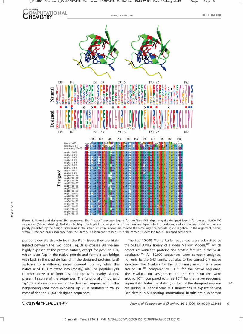
positions deviate strongly from the Pfam types; they are high-
lighted between the two logos (Fig. 3) as crosses. All five are
highly exposed at the protein surface, except for position 150,
which is an Asp in the native protein and forms a salt bridge
with Lys8 in the peptide ligand. In the designed proteins, Lys8
switches to a different, more exposed rotamer, while the
native Asp150 is mutated into (mostly) Ala. The peptide Lys8
rotamer allows it to form a salt bridge with nearby Glu149,
present in some of the sequences. The functionally important
Trp170 is always preserved in the designed sequences, but the
neighboring (and more exposed) Trp171 is mutated to Val in
most of the top 10,000 designed sequences.
The top 10,000 Monte Carlo sequences were submitted to
the SUPERFAMILY library of Hidden Markov Models,[69] which
detect similarities to proteins and protein families in the SCOP
database.[116] All 10,000 sequences were correctly assigned,
not only to the SH3 family, but also to the correct Crk native
structure. The E-values for the SH3 family assignments were
around 10210, compared to 10220 for the native sequence.
The E-values for assignment to the Crk structure were
around 1023, compared to three 1025 for the native sequence.
Figure F44 illustrates the stability of two of the designed sequen-
ces during 20 nanosecond MD simulations in explicit solvent
(see details in Supporting Information). Results are also shown
COL
OR
Figure 3. Natural and designed SH3 sequences. The “natural” sequence logo is for the Pfam SH3 alignment; the designed logo is for the top 10,000 MC
sequences (Crk numbering). Red dots highlight hydrophobic core positions, blue dots are ligand-binding positions, and crosses are positions that are
poorly predicted by the design. Sidechains in the stereo structure, above, are colored the same way; the peptide ligand is yellow. In the alignment, below,
“Pfam” is the consensus sequence from the Pfam SH3 alignment; “consensus” is the consensus over the top 25 designed sequences.
J_ID: JCC Customer A_ID: JCC23418 Cadmus Art: JCC23418 Ed. Ref. No.: 13-0237.R1 Date: 13-August-13 Stage: Page: 9
ID: muralir Time: 21:10 I Path: N:/3b2/JCCT/Vol00000/130172/APPFile/JW-JCCT130172
FULL PAPERWWW.C-CHEM.ORG
Journal of Computational Chemistry 2013, DOI: 10.1002/jcc.23418 9
for the wildtype Crk protein and for a sequence designed ear-
lier with the simpler, CASA solvent model.[66] The older design
was produced experimentally and found to be only partly
structured (Guijarro and Plateau, personal communication).
The newer designs have a significantly improved stability in
the MD simulations and are expected to be structured; experi-
mental tests are underway.
The acid/base behavior of Staphylococcal nuclease
Sidechain acid/base reactions can be treated as “mutations”
and treated with practically the same formalism as protein
design, with only minor changes to the software. The main dif-
ference is the interpretation of the “unfolded” reference state.
Instead of an unfolded protein, the reference state now corre-
sponds to a collection of model compounds in solution. Each
model compound is the analog of a titrating sidechain type,
as usual in pKa calculations for proteins.[31,112,117,118] The refer-
ence energies EX are the energies of each model compound in
its optimal rotamer in solution. Here, as an illustration, we
report the titration behavior of the protein Staphylococcal
nuclease, or SNase. We consider a hyperstable SNase mutant,
known as D1PHS, for which 17 Asp and Glu sidechain pKa’s
are known experimentally.[119] Calculations were done using
the experimental backbone structure (PDB code 3BDC), the
Amber ff99SB force field, the GB/HCT solvent, a surface area
term (same atomic coefficients as for TyrRS above), and a pro-
tein dielectric constant of 4. Results with a dielectric of 8 are
slightly poorer. Monte Carlo simulations were done at pH val-
ues between 0 and 15, every 0.5 pH units, with 26 million MC
steps at each pH value. The last 20 million steps at each pH
value were used for averaging. The full pH scan (0.8 billion MC
steps) takes about 12 h on a recent laptop computer using a
single core.
The titration curves for the first four Asp and Glu sidechains
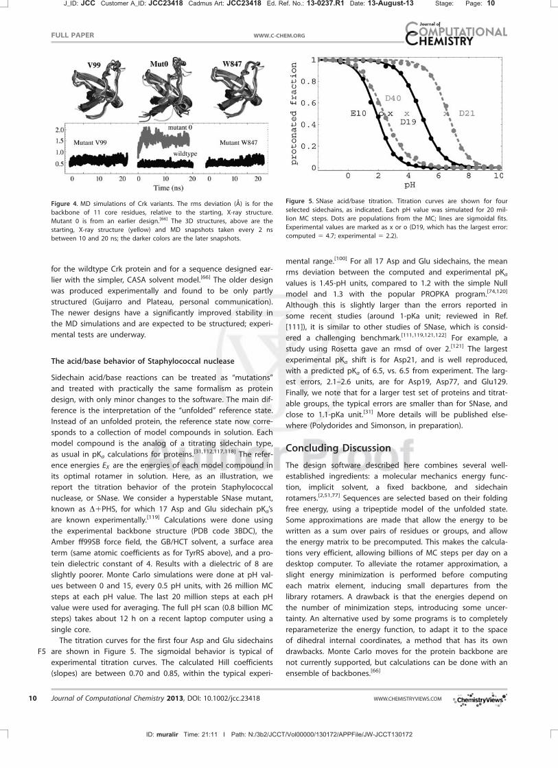
are shown in FigureF5 5. The sigmoidal behavior is typical of
experimental titration curves. The calculated Hill coefficients
(slopes) are between 0.70 and 0.85, within the typical experi-
mental range.[100] For all 17 Asp and Glu sidechains, the mean
rms deviation between the computed and experimental pKa
values is 1.45-pH units, compared to 1.2 with the simple Null
model and 1.3 with the popular PROPKA program.[74,120]
Although this is slightly larger than the errors reported in
some recent studies (around 1-pKa unit; reviewed in Ref.
[111]), it is similar to other studies of SNase, which is consid-
ered a challenging benchmark.[111,119,121,122] For example, a
study using Rosetta gave an rmsd of over 2.[121] The largest
experimental pKa shift is for Asp21, and is well reproduced,
with a predicted pKa of 6.5, vs. 6.5 from experiment. The larg-
est errors, 2.1–2.6 units, are for Asp19, Asp77, and Glu129.
Finally, we note that for a larger test set of proteins and titrat-
able groups, the typical errors are smaller than for SNase, and
close to 1.1-pKa unit.[31] More details will be published else-
where (Polydorides and Simonson, in preparation).
Concluding Discussion
The design software described here combines several well-
established ingredients: a molecular mechanics energy func-
tion, implicit solvent, a fixed backbone, and sidechain
rotamers.[2,51,77] Sequences are selected based on their folding
free energy, using a tripeptide model of the unfolded state.
Some approximations are made that allow the energy to be
written as a sum over pairs of residues or groups, and allow
the energy matrix to be precomputed. This makes the calcula-
tions very efficient, allowing billions of MC steps per day on a
desktop computer. To alleviate the rotamer approximation, a
slight energy minimization is performed before computing
each matrix element, inducing small departures from the
library rotamers. A drawback is that the energies depend on
the number of minimization steps, introducing some uncer-
tainty. An alternative used by some programs is to completely
reparameterize the energy function, to adapt it to the space
of dihedral internal coordinates, a method that has its own
drawbacks. Monte Carlo moves for the protein backbone are
not currently supported, but calculations can be done with an
ensemble of backbones.[66]
Figure 4. MD simulations of Crk variants. The rms deviation (A) is for the
backbone of 11 core residues, relative to the starting, X-ray structure.
Mutant 0 is from an earlier design.[66] The 3D structures, above are the
starting, X-ray structure (yellow) and MD snapshots taken every 2 ns
between 10 and 20 ns; the darker colors are the later snapshots.
Figure 5. SNase acid/base titration. Titration curves are shown for four
selected sidechains, as indicated. Each pH value was simulated for 20 mil-
lion MC steps. Dots are populations from the MC; lines are sigmoidal fits.
Experimental values are marked as x or o (D19, which has the largest error:
computed 5 4.7; experimental 5 2.2).
J_ID: JCC Customer A_ID: JCC23418 Cadmus Art: JCC23418 Ed. Ref. No.: 13-0237.R1 Date: 13-August-13 Stage: Page: 10
ID: muralir Time: 21:11 I Path: N:/3b2/JCCT/Vol00000/130172/APPFile/JW-JCCT130172
FULL PAPER WWW.C-CHEM.ORG
10 Journal of Computational Chemistry 2013, DOI: 10.1002/jcc.23418 WWW.CHEMISTRYVIEWS.COM

The Proteus software is made up of the XPLOR molecular
modeling program (modified locally), a sophisticated collection
of scripts written in the XPLOR command language (totalling
about 4000 lines), a C program, Proteus, for sequence/confor-
mation exploration, and a set of perl and shell scripts. Source
code is freely available to academics or anyone with an XPLOR
source code license. Energy matrix calculations are mainly con-
trolled through three XPLOR scripts and two shell scripts.
Sequence/conformation exploration is controlled by one main
Proteus script (with an XML format). System setup is done
with XPLOR, in a way that is highly automated and similar to
other modeling programs, like Charmm or Amber.
The software is designed to be flexible, allowing several
molecular mechanics force fields and solvent models to be
used, and any rotamer library, including backbone-dependent
ones. The system can be decomposed into groups, which can
be present with multiple copies and contribute to the energy
in various ways. For example, protein sequences can be
selected based on stability, binding affinity for one ligand, and
specificity relative to another ligand.[71] The software has some
unusual features, illustrated in the applications above. Thus,
acid/base activity is fully supported, so that sidechain titration
curves can be obtained easily, and protonation states can vary
when nearby positions mutate. In addition, multiple ligands
can be present and interconvert via Monte Carlo “mutations,”
as shown for TyrRS. Here too, titration curves are easily
obtained, giving estimates of the binding free energy differen-
ces between the ligand species.
The applications above included classic sequence explora-
tion for the small protein Crk and a TyrRS:ligand complex, with
the latter leading to an experimentally active sequence. We
also described calculations of thermodynamic properties,
including TyrRS:L-Tyr/D-Tyr binding free energy differences and
SNase acid/base constants. These applications illustrate the
need to adjust certain parameters empirically for different
applications, especially the protein dielectric constant with GB,
but also the atomic surface coefficients and the unfolded ener-
gies. This is not expensive, thanks to the matrix editing capa-
bility with the more verbose matrix formats. For the acid/base
calculations, accuracy is comparable to several other recent
software tools.[31,111] The ligand binding calculations are sensi-
tive to the number Nmin of minimization steps performed for
each matrix element, and their accuracy was only qualitative
for the TyrRS application. More work is needed for this type of
application to evaluate and improve the Proteus performance.
Its potential for large-scale, competitive ligand binding simula-
tions will be reported elsewhere. We believe that by doing
sequence exploration and thermodynamic calculations with
the same software and energy function, we are more likely to
identify physically meaningful parameterizations of the model,
and successfully design a wide range of new proteins.
Acknowledgments
This work was supported by the Agence Nationale pour la Recher-
che (High Performance Computing program; ProtiCAD project).
Some of the calculations were done using the French national
supercomputer center CINES. The authors thank Alexey Aleksan-
drov, Seydou Traor�e, Nicolas Panel, and Jialin Liu for discussions.
Keywords: molecular recognition � protein engineering � com-
puter simulation � Monte Carlo � aminoacyl-tRNA synthetase
How to cite this article: T. Simonson, T. Gaillard, D. Mignon, M.
S. am Busch, A. Lopes, N. Amara, S. Polydorides, A. Sedano, K.
Druart, G. Archontis. J. Comput. Chem. 2013, DOI: 10.1002/
jcc.23418
] Additional Supporting Information may be found in the
online version of this article
[1] D. Baker, Philos. Trans. R. Soc. London 2006. 361, 459.
[2] G. L. Butterfoss, B. Kuhlman, Annu. Rev. Biophys. Biomol. Struct. 2006,
35, 49.
[3] R. Gu�erois, M. Lopez de la Paz, Eds. Protein Design: Methods and
Applications; Humana Press, 2007. AQ5
[4] S. M. Lippow, B. Tidor, Curr. Opin. Biotechnol. 2007, 18, 305.
[5] J. Pleiss, Curr. Opin. Biotechnol. 2011, 22, 611.
[6] R. J. Pantazes, M. J. Greenwood, C. D. Maranas, Curr. Opin. Struct. Biol.
2011, 21, 467.
[7] J. G. Saven, Curr. Opin. Chem. Biol. 2011, 15, 452.
[8] I. Samish, C. M. MacDermaid, J. M. Perez-Aguilar, J. G. Saven, Annu.
Rev. Phys. Chem. 2011, 62, 129.
[9] L. L. Looger, M. A. Dwyer, J. J. Smith, H. W. Hellinga, Nature 2003, 423,
185.
[10] J. J. Havranek, P. B. Harbury, Nat. Struct. Mol. Biol. 2003, 10, 45.
[11] D. Bolon, S. L. Mayo, Proc. Natl. Acad. Sci. USA 2001, 98, 14274.
[12] G. Dantas, B. Kuhlman, D. Callender, M. Wong, D. Baker, J. |Mol. Biol.
2003, 332, 449.
[13] B. Kuhlman, G. Dantas, G. Ireton, G. Varani, B. Stoddard, D. Baker, Sci-
ence 2003, 302, 1364.
[14] H. Liang, H. Chen, K. Fan, P. Wei, X. Guo, C. Jin, C. Zeng, C. Tang, L. H.
Lai, Angew. Chem. Int. Ed. 2009, 48, 3301.
[15] N. Koga, R. Tatsumi-Koga, G. Liu, R. Xiao, T. B. Acton, G. T. Montelione,
D. Baker, Nature 2012, 491, 222.
[16] D. R€othlisberger, O. Khersonsky, M. Wollacott, L. Jiang, J. DeChancie,
J. Betker, J. L. Gallaher, E. A. Althoff, A. Zanghellini, O. Dym, S. Albeck,
K. N. Houk, D. S. Tawfik, D. Baker, Nature 2008, 453, 190.
[17] L. Jiang, E. A. Althoff, F. R. Clemente, L. Doyle, D. R€othlisberger,
A. Zanghellini, J. L. Gallaher, J. L. Betker, F. Tanaka, C. F. Barbas, III,
D. Hilvert, K. N. Houk, B. L. Stoddard, D. Baker, Science 2008, 319,
1387.
[18] F. Richter, A. Leaver-Kay, S. D. Khare, S. Bjelic, D. Baker, PLoS One 2011,
6, e19230.
[19] J. G. Saven, Curr. Opin. Colloid Interface Sci. 2010, 15, 13.
[20] C. Fortenberry, E. A. Bowman, W. Proffitt, B. Dorr, S. Combs, J. Harp,
L. Mizoue, J. Meiler, J. Am. Chem. Soc. 2011, 133, 18026.
[21] G. Grigoryan, Y. H. Kim, R. Acharya, K. Axelrod, R. M. Jain, L. Willis,
M. Dmdic, J. M. Kikkawa, W. F. DeGrado, Science 2011, 332, 1071.
[22] N. P. King, W. Scheffer, M. R. Sawaya, B. S. Vollmar, J. P. Sumida,
I. Andre, T. Gonen, T. O. Yeates, D. Baker, Science 2012, 336, 1171.
[23] C. J. Lanci, C. M. MacDermaid, S. G. Kang, R. Acharya, B. North, X.
Yang, X. J. Qiu, W. F. DeGrado, J. G. Saven, Proc. Natl. Acad. Sci. USA
2012, 109, 7304.
[24] A. D. Mackerell, D. Bashford, M. Bellott, R. L. Dunbrack, J. Evanseck,
M. J. Field, S. Fischer, J. Gao, H. Guo, S. Ha, D. Joseph, L. Kuchnir,
K. Kuczera, F. T. K. Lau, C. Mattos, S. Michnick, T. Ngo, D. T. Nguyen,
B. Prodhom, W. E. Reiher, B. Roux, J. Smith, R. Stote, J. Straub,
M. Watanabe, J. Wiorkiewicz-Kuczera, D. Yin, M. Karplus, J. Phys. Chem.
B 1998, 102, 3586.
[25] D. A. Case, D. Pearlman, J. Caldwell, T. Cheatham, III, W. Ross,
C. Simmerling, T. Darden, K. Merz, R. Stanton, A. Cheng, J. Vincent,
M. Crowley, V. Tsui, R. Radmer, Y. Duan, J. Pitera, I. Massova, G. Seibel,
J_ID: JCC Customer A_ID: JCC23418 Cadmus Art: JCC23418 Ed. Ref. No.: 13-0237.R1 Date: 13-August-13 Stage: Page: 11
ID: muralir Time: 21:11 I Path: N:/3b2/JCCT/Vol00000/130172/APPFile/JW-JCCT130172
FULL PAPERWWW.C-CHEM.ORG
Journal of Computational Chemistry 2013, DOI: 10.1002/jcc.23418 11
simonson ([email protected])
Cross-Out
simonson ([email protected])
Cross-Out

U. Singh, P. Weiner, P. Kollman, AMBER 6; University of California: San
Francisco, 1999.
[26] J. Ponder, D. A. Case, Adv. Protein Chem. 2003, 66, 27.
[27] W. L. Jorgensen, J. Tirado-Rives, Proc. Natl. Acad. Sci. USA 2005, 102,
6665.
[28] B. Brooks, C. L. Brooks, III, A. D. Mackerell, Jr., L. Nilsson, R. J. Petrella,
B. Roux, Y. Won, G. Archontis, C. Bartels, S. Boresch, A. Caflisch,
L. Caves, Q. Cui, A. R. Dinner, M. Feig, S. Fischer, J. Gao, M. Hodoscek,
W. Im, K. Kuczera, T. Lazaridis, J. Ma, V. Ovchinnikov, E. Paci, R. W.
Pastor, C. B. Post, J. Z. Pu, M. Schaefer, B. Tidor, R. M. Venable, H. L.
Woodcock, X. Wu, W. Yang, D. M. York, M. Karplus, J. Comput. Chem.
2009, 30, 1545.
[29] D. E. Shaw, P. Maragakis, K. Lindorff-Larsen, S. Piana, R. O. Dror, M. P.
Eastwood, J. A. Bank, J. M. Jumper, J. K. Salmon, Y. Shan, W. Wriggers,
Science 2010, 330, 341.
[30] Y. Deng, B. Roux, J. Phys. Chem. B 2009, 113, 2234.
[31] A. Aleksandrov, D. Thompson, T. Simonson, J. Mol. Recognit. 2010, 23,
117.
[32] J. Wereszczynski, J. A. McCammon, Q. Rev. Biophys. 2012, 45, 1.
[33] T. Simonson, G. Archontis, M. Karplus, Acc. Chem. Res. 2002, 35, 430.
[34] B. Roux, T. Simonson, Biophys. Chem. 1999, 78, 1.
[35] T. Simonson, Rep. Prog. Phys. 2003, 66, 737.
[36] N. A. Baker, Methods Enzymol. 2004, 383, 94.
[37] M. Feig, C. L. Brooks, III, Curr. Opin. Struct. Biol. 2004, 14, 217.
[38] C. L. Vizcarra, N. G. Zhang, S. A. Marshall, N. S. Wingreen, C. Zeng, S. L.
Mayo, J. Comput. Chem. 2008, 29, 1153.
[39] S. Liang, N. V. Grishin, Proteins 2004, 54, 271.
[40] R. Gu�erois, J. E. Nielsen, L. Serrano, J. Mol. Biol. 2002, 320, 369.
[41] T. Kortemme, A. Morozov, D. Baker, J. Mol. Biol. 2003, 326, 1239.
[42] N. Pokola, T. M. Handel, J. Mol. Biol. 2005, 347, 203.
[43] J. Smadbeck, M. B. Peterson, G. A. Khoury, M. S. Taylor, C. A. Floudas,
J. Vis. Exp. (in press).AQ6
[44] H. Hellinga, F. Richards, Proc. Natl. Acad. Sci. USA 1994, 91, 5803.
[45] M. S. Wisz, H. Hellinga, Proteins 2003, 51, 360.
[46] P. Koehl, M. Levitt, J. Mol. Biol. 1999, 293, 1161.
[47] J. Zou, J. G. Saven, J. Chem. Phys. 2003, 118, 3843.
[48] S. Ventura, L. Serrano, Proteins 2004, 56, 1.
[49] A. B. Chowdry, K. A. Reynolds, M. S. Hanes, M. Voorhies, N. Pokala,
T. M. Handel, J. Comput. Chem. 2007, 28, 2378.
[50] S. M. Lippow, K. D. Wittrup, B. Tidor, Nat. Biotechnol. 2007, 25,
1171.
[51] A. Jaramillo, L. Wernisch, S. H�ery, S. Wodak, Proc. Natl. Acad. Sci. USA
2002, 99, 13554.
[52] M. Suarez, A. Jaramillo, J. R. Soc. Interface 2009, 6, S477
[53] P. Gainza, K. E. Roberts, I. Georgiev, R. H. Lilien, D. A. Keedy, C. Chen,
F. Reza, A. C. Anderson, D. C. Richardson, J. S. Richardson, B. R.
Donald. Methods Enzymol. 2013, 523, 87–107.
[54] M. Schmidt am Busch, A. Lopes, D. Mignon, T. Simonson, J. Comput.
Chem. 2008, 29, 1092.
[55] M. Schmidt am Busch, A. Lopes, N. Amara, C. Bathelt, T. Simonson,
BMC Bioinformatics 2008, 9, 148.
[56] M. Schmidt am Busch, A. Lopes, D. Mignon, T. Gaillard, T. Simonson, In
Quantum Simulations of Materials and Biological Systems; J. Zeng,
R. Q. Zhang, H. Treutlein, Eds.; Springer Science: Dordrecht, 2012;
pp. 121–140.
[57] A. T. Br€unger, X-Plor Version 3.1, A System for X-ray Crystallography
and NMR; Yale University Press: New Haven, 1992.
[58] A. T. Br€unger, P. D. Adams, W. L. DeLano, P. Gros, R. W. Grosse-Kunstleve,
J. Jiang, N. S. Pannu, R. J. Read, L. M. Rice, T. Simonson, In International
Tables for Crystallography, Vol. F; M. Rossmann, E. Arnold, Eds.; Kluwer
Academic Publishers: Dordrecht, Netherlands, 2001; pp. 710–720.
[59] B. I. Dahiyat, S. L. Mayo, Science 1997, 278, 82.
[60] D. Bashford, D. Case, Annu. Rev. Phys. Chem. 2000, 51, 129.
[61] L. Moulinier, D. A. Case, T. Simonson, Acta Crystallogr. 2003, D 59,
2094.
[62] A. Lopes, A. Aleksandrov, C. Bathelt, G. Archontis, T. Simonson, Proteins
2007, 67, 853.
[63] C. A. Smith, T. Kortemme, J. Mol. Biol. 2008, 380, 742.
[64] D.J. Mandell, T. Kortemme, Curr. Opin. Biotechnol. 2009, 20, 420.
[65] P. S. Huang, Y. E. Ban, F. Richter, I. Andre, R. Vernon, W. R. Schief,
D. Baker, PLoS One 2011, 6,e24109.
[66] M. Schmidt am Busch, D. Mignon, T. Simonson, Proteins 2009, 77, 139.
[67] M. Schmidt am Busch, A. Sedano, T. Simonson, PLoS One 2010, 5,
e10410.
[68] D. P. Anderson, BOINC: A System for Public-Resource Computing and
Storage, In 5th IEEE/ACM International Workshop on Grid Computing;
IEEE Computer Society Press, 2004.
[69] D. Wilson, M. Madera, C. Vogel, C. Chothia, J. Gough, Nucleic Acids Res.
2007, 35, D308.
[70] A. Lopes, M. Schmidt am Busch, T. Simonson, J. Comput. Chem. 2010,
31, 1273.
[71] S. Polydorides, N. Amara, T. Simonson, G. Archontis, Proteins 2011, 79,
3448.
[72] S. Polydorides, T. Simonson, J. Phys. Chem. B (in press).
[73] H. Li, A. D. Robertson, J. H. Jensen, Proteins 2005, 61, 704.
[74] M. H. M. Olsson, C. R. Sondergaard, M. Rostowski, J. H. Jensen,
J. Chem. Theory Comput. 2011, 7, 525.
[75] G. G. Krivov, M. V. Shapalov, R. L. Dunbrack, Proteins 2009, 77, 778.
[76] A. G. Street, S. Mayo, Fold. Des. 1998, 3, 253.
[77] D. Baker, Nature 2000, 405, 39.
[78] B. Brooks, R. Bruccoleri, B. Olafson, D. States, S. Swaminathan,
M. Karplus, J. Comput. Chem. 1983, 4, 187.
[79] W. Cornell, P. Cieplak, C. Bayly, I. Gould, K. Merz, D. Ferguson,
D. Spellmeyer, T. Fox, J. Caldwell, P. Kollman, J. Am. Chem. Soc. 1995,
117, 5179.
[80] W. Jorgensen, J. Tirado-Rives, J. Am. Chem. Soc. 1988, 110, 1657.
[81] T. Simonson, J. Phys. Chem. B 2000, 104, 6509.
[82] M. Schaefer, M. Karplus, J. Phys. Chem. 1996, 100, 1578.
[83] N. Calimet, M. Schaefer, T. Simonson, Proteins 2001, 45, 144.
[84] G. D. Hawkins, C. Cramer, D. Truhlar, Chem. Phys. Lett. 1995, 246, 122.
[85] N. Pokala, T. Handel, Protein Sci. 2004, 13, 925.
[86] G. Archontis, T. Simonson, J. Phys. Chem. B 2005, 109, 22667.
[87] P. Barth, T. Alber, P. B. Harbury, Proc. Natl. Acad. Sci. USA 2007, 104,
4898.
[88] B. Lee, F. Richards, J. Mol. Biol. 1971, 55, 379.
[89] L. Wernisch, S. H�ery, S. Wodak, J. Mol. Biol. 2000, 301, 713.
[90] P. Koehl, M. Delarue, J. Mol. Biol. 1994, 239, 249.
[91] J. G. Saven, P. G. Wolynes, J. Phys. Chem. B 1997, 101, 8375.
[92] B. J. Zou, J. G. Saven, J. Chem. Phys. 2005, 123, 154908.
[93] N. Metropolis, A. W. Rosenbluth, M. N. Rosenbluth, A. H. Teller,
E. Teller, J. Chem. Phys. 1953, 21, 1087.
[94] D. Frenkel, B. Smit, Understanding Molecular Simulation; Academic
Press: New York, 1996.
[95] T. Hill, Introduction to Statistical Thermodynamics; Addison-Wesley:
Reading, MA, 1962.
[96] A. M. Baptista, P. J. Martel, S. B. Petersen, Proteins 1997, 27, 523.
[97] U. B€orjesson, P. H. H€unenberger, J. Chem. Phys. 2001, 114, 9706.
[98] M. S. Lee, F. R. Salsbury, Jr., C. L. Brooks, III, Proteins 2004, 56, 738.
[99] J. Mongan, D. A. Case, J. A. McCammon, J. Comput. Chem. 2004, 25,
2038.
[100] E. R. Georgescu, E. Alexov, M. Gunner, Biophys. J. 2002, 83, 1731.
[101] B. I. Dahiyat, S. L. Mayo, Protein Sci. 1996, 5, 895.
[102] A. Br€unger, P. Adams, G. Clore, W. Delano, P. Gros, R. Grosse-
Kunstleve, J. Jiang, J. Kuszewski, M. Nilges, N. Pannu, R. Read, L. Rice,
T. Simonson, G. Warren, Acta Crystallogr. 1998, D54, 905
[103] C. Schweiters, J. Kuszewski, N. Tjandra, G. Clore, J. Biomol. NMR 2003,
160, 65.
[104] A. Onufriev, D. A. Case, M. Ullmann, Biochemistry 2001, 40, 3413.
[105] L. Wang, A. Brock, B. Herberich, P. G. Schultz, Science 2001, 292, 498.
[106] T. S. |Young, P. G. Schultz, J. Biol. Chem. 2010, 285, 11039.
[107] Y. Zhang, L. |Wang, P. G. Schultz, I. A. Wilson, Protein Sci. 2005, 14,
1340.
[108] M. Tsunoda, Y. Kusakabe, N. Tanaka, S. Ohno, M. Nakamura, T. Senda,
T. Moriguchi, N. Asai, M. Sekine, T. Yokogawa, K. Nishikawa, K. T.
Nakamura, Nucleic Acids Res. 2007, 35, 4289.
[109] P. Tuffery, C. Etchebest, S. Hazout, R. Lavery, J. Biomol. Struct. Dyn.
1991, 8, 1267.
[110] G. Archontis, T. Simonson, M. Karplus, J. Mol. Biol. 2001, 306, 307.
[111] E. Alexov, E. L. Mehler, N. Baker, A. M. Baptista, Y. Huang, F. Milletti,
J. E. Nielsen, D. Farrell, T. Carstensen, M. H. M. Olsson, J. K. Shen,
J. Warwicker, S. Williams, J. M. Word, Proteins 2011, 79, 3260.
[112] T. Pawson, Nature 1995, 373, 573.
J_ID: JCC Customer A_ID: JCC23418 Cadmus Art: JCC23418 Ed. Ref. No.: 13-0237.R1 Date: 13-August-13 Stage: Page: 12
ID: muralir Time: 21:11 I Path: N:/3b2/JCCT/Vol00000/130172/APPFile/JW-JCCT130172
FULL PAPER WWW.C-CHEM.ORG
12 Journal of Computational Chemistry 2013, DOI: 10.1002/jcc.23418 WWW.CHEMISTRYVIEWS.COM
simonson ([email protected])
Cross-Out
simonson ([email protected])
Cross-Out

[113] I. Broutin, A. Ducruix, Med. Sci. 2000, 16, 611.
[114] X. Wu, B. Knudsen, S. M. Feller, J. Zheng, A. Sali, D. Cowburn,
H. Hanafusa, J. Kuriyan, Structure 1995, 3, 215.
[115] R. D. Finn, J. Mistry, B. Schuster-Bckler, S. Griffiths-Jones, V. Hollich,
T. Lassmann, S. Moxon, M. Marshall, A. Khanna, R. Durbin, S. R. Eddy,
E. L. L. Sonnhammer, A. ateman, Nucleic Acids Res. 2006, 34, D247.
[116] A. Andreeva, D. Howorth, S. E. Brenner, J. J. Hubbard, C. Chothia,
A. G. Murzin, Nucleic Acids Res. 2004, 32, D226.
[117] D. Bashford, M. Karplus, Biochemistry 1990, 29, 10219.
[118] T. Simonson, J. Carlsson, D. A. Case, J. Am. Chem. Soc. 2004, 126,
4167.
[119] C. A. Castaneda, C. A. Fitch, A. Majumdar, V. Khangulov, J. L.
Schlessman, B. Garcia-Moreno, Proteins 2009, 77, 570.
[120] D. C. Bas, D. M. Rogers, J. H. Jensen, Proteins 2008, 73, 765.
[121] K. Kilambi, J. J. Gray, Biophys. J. 2012, 103, 587.
[122] M. Gunner, X. Zhu, M. C. Klein, Proteins 2011, 79, 3306.
Received: 13 May 2013Revised: 8 July 2013Accepted: 28 July 2013Published online on 00 Month 2013
J_ID: JCC Customer A_ID: JCC23418 Cadmus Art: JCC23418 Ed. Ref. No.: 13-0237.R1 Date: 13-August-13 Stage: Page: 13
ID: muralir Time: 21:11 I Path: N:/3b2/JCCT/Vol00000/130172/APPFile/JW-JCCT130172
FULL PAPERWWW.C-CHEM.ORG
Journal of Computational Chemistry 2013, DOI: 10.1002/jcc.23418 13

SGML and CITI Use OnlyDO NOT PRINT
Using the Proteus computation design software and Monte Carlo simulations, D-Tyr is titrated into the
active site of a wild type and a redesigned tyrosyl-tRNA synthetase, displacing L-Tyr and obtaining
binding free energy differences similar to experiments.
J_ID: JCC Customer A_ID: JCC23418 Cadmus Art: JCC23418 Ed. Ref. No.: 13-0237.R1 Date: 13-August-13 Stage: Page: 14
ID: muralir Time: 21:11 I Path: N:/3b2/JCCT/Vol00000/130172/APPFile/JW-JCCT130172

AQ1: Please confirm that all author names are OK and are set with first name first, surname last.
AQ2: Please note that Refs. [28] and [102] are identical, hence Ref. 102 has been deleted and renumbering has beendone for subsequent references to make citations sequential.
AQ3: Please spell out MC, OPLS, MD, ACE, HCT, CASA, and SCOP in text.
AQ4: Please note that “Wernisch et al.” does not match with Ref. 54. Please check.
AQ5: Please provide location for Ref. [3] and [68].
AQ6: Please update Ref. [43] and [72].
AQ7: Please provide university for affiliation [a].
AQ8: Please provide department/division name (if any) for affiliations [c] and [d].
J_ID: JCC Customer A_ID: JCC23418 Cadmus Art: JCC23418 Ed. Ref. No.: 13-0237.R1 Date: 13-August-13 Stage: Page: 15
ID: muralir Time: 21:11 I Path: N:/3b2/JCCT/Vol00000/130172/APPFile/JW-JCCT130172
Related Documents