Samant et al., IJPSR, 2015; Vol. 6(4): 1000-09. E-ISSN: 0975-8232; P-ISSN: 2320-5148 International Journal of Pharmaceutical Sciences and Research 1000 IJPSR (2015), Vol. 6, Issue 4 (Research Article) Received on 26 August, 2014; received in revised form, 29 November, 2014; accepted, 27 December, 2014; published 01 April, 2015 COMPUTATIONAL MODELLING AND FUNCTIONAL CHARACTERIZATION OF HDAC-11 L. R. Samant. * 1 , V. C. Sangar 2 , A. Gulamaliwala 3 and A. S. Chowdhary 1, 2 Systems Biomedicine Division 1 , Department of Virology & Immunology 2 , Haffkine Institute for Training, Research & Testing, Acharya Donde Marg, Parel, Mumbai 400012. India. Department of Biotechnology and Bioinformatics 3 , Padmashree Dr. D.Y. Patil University, CBD Belapur, Navi Mumbai-400614, India ABSTRACT: In eukaryotes, DNA is packaged into chromatin structures, whose basic unit is the nucleosome. A principle component of chromatin that plays an important role in the regulation of DNA is the modification of histones. Since histones are post-translationally modified, there inludes a large number of different histone post-translational modifications. These histone modifications create a repressive environment for gene expression, which in case of histone acetylation, are controlled by competing activities of two families of enzymes, histone acetyltransferases (HAT’s) and histone deacetylases (HDACs). HDAC11 is a class IV protein of the HDAC family. The present aim of this study is to develop a model of HDAC11 by using bioinformatics applications. The design of the model is based on thorough evaluation of the HDAC-11 query sequence, Q96DB2, which was retrieved from UniProtKB. The physiochemical and primary analysis were computed using ExPASy Protparam tool. Functional characterization was computed using RaptorX, HMMTOP and Softberry Server’s CYS_REC tool (Cysteine Recognition Server) which predicted the secondary structure composition, presence of transmembrane proteins and the presence of cysteine residues respectively. The molecular model was generated using PHYRE2 server, since it was best suited as it provided higher query sequence coverage and confidence. Model refinement was computed using UCSF Chimera V1.9 and validation was performed using RAMPAGE server which explains the feature of Psi and Phi angle orientation. Verify 3D Structure Evaluation Server was used to determine the 3D-profiling of the residues in the model. The overall quality score of the model was calculated by ProSA Web Server. INTRODUCTION: In eukaryotes, DNA is packaged into chromatin structures, whose basic unit is the nucleosome. Histones are highly conserved basic proteins which associate with DNA to constitute the nucleosome, each nucleosome consists of ∼148 bp DNA, wrapping around a core histone octamer, which contains copies each of H2A, H2B, H3, and H4. Also a H1 linker histone is associated, which binds externally with the nucleosome and helps in further compaction of the chromatin structure. QUICK RESPONSE CODE DOI: 10.13040/IJPSR.0975-8232.6(4).1000-09 Article can be accessed online on: www.ijpsr.com DOI link: http://dx.doi.org/10.13040/IJPSR.0975-8232.6(4).1000-09 Within a nucleosome, these exist as two dimers of (H2A-H2B) and a complex of (H32-H42) ultimately forming an octamer. 1, 2 . A principle component of chromatin that plays an important role in the regulation of DNA is the modification of histones. It is clear from recent studies, that histone modifications play fundamental roles in most biological processes that are involved in the manipulation and expression of DNA. 3 Since histones are post-translationally modified, there involves a large number of different histone post-translational modifications which include Histone acetylation, Histone Methylation, Histone phosphorylation and other modifications, of which Histone Acetylation is the best understood modification. 3 Hypoacetylated chromatin is associated with gene silencing, whereas hyperacetylation correlates with gene activation. Keywords: HDAC-11, HDAC-11 modelbuilding, PHYRE2, ProSA Web Server, Verfy3D, UCSF Chimera V1.9 Correspondence to Author: Lalit R. Samant Systems Biomedicine Division, Haffkine Institute for Training, Research & Testing, Acharya Donde Marg, Parel, Mumbai 400012. E-mail: [email protected]

Welcome message from author
This document is posted to help you gain knowledge. Please leave a comment to let me know what you think about it! Share it to your friends and learn new things together.
Transcript
Samant et al., IJPSR, 2015; Vol. 6(4): 1000-09. E-ISSN: 0975-8232; P-ISSN: 2320-5148
International Journal of Pharmaceutical Sciences and Research 1000
IJPSR (2015), Vol. 6, Issue 4 (Research Article)
Received on 26 August, 2014; received in revised form, 29 November, 2014; accepted, 27 December, 2014; published 01 April, 2015
COMPUTATIONAL MODELLING AND FUNCTIONAL CHARACTERIZATION OF HDAC-11
L. R. Samant. *1, V. C. Sangar
2, A. Gulamaliwala
3 and A. S. Chowdhary
1, 2
Systems Biomedicine Division
1, Department of Virology & Immunology
2, Haffkine Institute for
Training, Research & Testing, Acharya Donde Marg, Parel, Mumbai 400012. India.
Department of Biotechnology and Bioinformatics 3, Padmashree Dr. D.Y. Patil University, CBD Belapur,
Navi Mumbai-400614, India
ABSTRACT: In eukaryotes, DNA is packaged into chromatin structures, whose basic unit is
the nucleosome. A principle component of chromatin that plays an important role in the
regulation of DNA is the modification of histones. Since histones are post-translationally
modified, there inludes a large number of different histone post-translational modifications.
These histone modifications create a repressive environment for gene expression, which in
case of histone acetylation, are controlled by competing activities of two families of enzymes,
histone acetyltransferases (HAT’s) and histone deacetylases (HDACs). HDAC11 is a class IV
protein of the HDAC family. The present aim of this study is to develop a model of HDAC11
by using bioinformatics applications. The design of the model is based on thorough evaluation
of the HDAC-11 query sequence, Q96DB2, which was retrieved from UniProtKB. The
physiochemical and primary analysis were computed using ExPASy Protparam tool.
Functional characterization was computed using RaptorX, HMMTOP and Softberry Server’s
CYS_REC tool (Cysteine Recognition Server) which predicted the secondary structure
composition, presence of transmembrane proteins and the presence of cysteine residues
respectively. The molecular model was generated using PHYRE2 server, since it was best
suited as it provided higher query sequence coverage and confidence. Model refinement was
computed using UCSF Chimera V1.9 and validation was performed using RAMPAGE server
which explains the feature of Psi and Phi angle orientation. Verify 3D Structure Evaluation
Server was used to determine the 3D-profiling of the residues in the model. The overall
quality score of the model was calculated by ProSA Web Server.
INTRODUCTION: In eukaryotes, DNA is
packaged into chromatin structures, whose basic
unit is the nucleosome. Histones are highly
conserved basic proteins which associate with
DNA to constitute the nucleosome, each
nucleosome consists of ∼148 bp DNA, wrapping
around a core histone octamer, which contains
copies each of H2A, H2B, H3, and H4. Also a H1
linker histone is associated, which binds externally
with the nucleosome and helps in further
compaction of the chromatin structure.
QUICK RESPONSE CODE
DOI: 10.13040/IJPSR.0975-8232.6(4).1000-09
Article can be accessed online on: www.ijpsr.com
DOI link: http://dx.doi.org/10.13040/IJPSR.0975-8232.6(4).1000-09
Within a nucleosome, these exist as two dimers of
(H2A-H2B) and a complex of (H32-H42)
ultimately forming an octamer.1, 2
.
A principle component of chromatin that plays an
important role in the regulation of DNA is the
modification of histones. It is clear from recent
studies, that histone modifications play
fundamental roles in most biological processes that
are involved in the manipulation and expression of
DNA.3 Since histones are post-translationally
modified, there involves a large number of different
histone post-translational modifications which
include Histone acetylation, Histone Methylation,
Histone phosphorylation and other modifications,
of which Histone Acetylation is the best understood
modification.3 Hypoacetylated chromatin is
associated with gene silencing, whereas
hyperacetylation correlates with gene activation.
Keywords:
HDAC-11, HDAC-11
modelbuilding, PHYRE2,
ProSA Web Server, Verfy3D,
UCSF Chimera V1.9
Correspondence to Author:
Lalit R. Samant
Systems Biomedicine Division,
Haffkine Institute for Training,
Research & Testing, Acharya Donde
Marg, Parel, Mumbai 400012.
E-mail: [email protected]
Samant et al., IJPSR, 2015; Vol. 6(4): 1000-09. E-ISSN: 0975-8232; P-ISSN: 2320-5148
International Journal of Pharmaceutical Sciences and Research 1001
However, recent studies have shown that histone
deacetylation can also play a significant role in
transcriptional activation 3
These histone modifications create a repressive
environment for gene expression, which in case of
histone acetylation, are controlled by competing
activities of two families of enzymes, histone
acetyltransferases (HAT’s) and histone
deacetylases (HDACs).1
The HATs utilize acetyl CoA as cofactor and
catalyse the transfer of an acetyl group to the
epsilon-amino group of lysine side chains in the
NH2-terminal tails of core histones. In doing so,
they neutralize the lysine's positive charge and this
action has the potential to weaken the interactions
between histones and DNA. The HATs are
classified into two classes: type-A and type-B. The
type-B HATs are predominantly cytoplasmic,
acetylating free histones but not those that are
already deposited into chromatin. The type B-
HAT’s also acetylate newly synthesize histones.
The type-A HATs are a more diverse family of
enzymes than the type-Bs. Nevertheless, they can
be classified into at least three separate groups
depending on amino-acid sequence homology and
conformational structure: GNAT, MYST and
CBP/p300 families 3
HDAC enzymes oppose the effects of HATs and
reverse lysine acetylation, an action that restores
the positive charge of the lysine. This potentially
stabilizes the local chromatin architecture thus
allowing the DNA to wrap more tighty and is
consistent with HDACs being predominantly
transcriptional repressors 3
HDAC’s based on their homology to yeast
orthologues Rpd3, HdaI and Sir2, respectively,
comprise a family of 18 genes, which are grouped
into classes I–IV. The Classes I, II, and IV consist
of 11 family members, which are referred to as
classical HDAC’s, whereas the class II, which
consists of 7 members are called Sirtuins.4
Classes I and II contain enzymes that are most
closely related to yeast scRpd3 and scHda1,
respectively, Class I being closely related to yeast
scRpd3, comprise of HDAC1, HDAC2, HDAC3
and HDAC8. Class II having closely related to
yeast scHda1 and are divided into subclass IIA
(HDAC4, HDAC5, HDAC7 and HDAC9) and
subclass IIB (HDAC6 and HDAC10). 3, 4
Class IV has only a single member, HDAC11,
while class III (sirtuins) are homologous to yeast
scSir2.3 The sirtuins have a catalytic domain,
unique to this family characterized by its
requirement for nicotine adenine dinucleotide
(NAD) as a cofactor.5
Classical HDACs are Zn2+
-dependent enzymes
which harbour a catalytic pocket with a Zn2+
ion at
its base that can be inhibited by Zn2+
chelating
compounds such as hydroxamic acids. In contrast,
these compounds are not active against sirtuins.
Taking into consideration, Classical HDAC’s being
a promising novel class of anti-cancer drug target 4,
also histone modifications like DNA methylation
and histone acetylation play an important role in a
wide range of brain disorders,
From recent research, Histone Deacetylase
Inhibitors are suggested to act as neuroprotectors
by enhancing synaptic plasticity and learning and
memory in a wide range of neurodegenerative and
psychiatric disorders, such as Alzheimer’s disease
and Parkinson’s disease 6. The present study aims
at developing fully modelled structures of
HDAC11 as its 3D structure is currently not
available The development of these structures may
provide information related to functional
mechanisms and also help us in further docking
studies and aid in anticancer and neuroprotector
drugs.
MATERIALS AND METHODS:
Sequence Retrieval:
Since the 3D structure for our interested protein
HDAC11 is not available on UniProtKB database,
its FASTA sequence was retrieved from
UniProtKB (Q96DB2) consisting of 347
aminoacids and was subjected for physiochemical
characterization
Physiochemical Characterization:
The physiochemical characterization of our protein
Q96DB2 consisting of 347AA residues is
computed by ExPASy-ProtParam tool 7. The tool
Samant et al., IJPSR, 2015; Vol. 6(4): 1000-09. E-ISSN: 0975-8232; P-ISSN: 2320-5148
International Journal of Pharmaceutical Sciences and Research 1002
provides sequence fragment analysis also, but here
the entire sequence analysis is computed. This tool
allows the computation of various physical and
chemical parameters for a given protein. The
computed parameters include the molecular weight,
theoretical pI (isoelectric point), amino acid
composition, atomic composition, extinction
coefficient, estimated half-life, instability index,
aliphatic index and grand average of hydropathicity
(GRAVY).
Secondary structure Analysis:
The secondary structure prediction of our protein
Q96DB2 was computed by using various online
softwares, which included RaptorX, HMMTOP and
CYS_REC.
The RaptorX is a protein structure prediction server
which was used to predict secondary structures8,
excelling at predicting 3D structures for protein
sequences without close homologs in the Protein
Data Bank (PDB). The FASTA sequence of our
protein was retrieved and was submitted to the
RaptorX server. Using RaptorX server, number of
secondary structure components such as α-helix, β-
sheets, turns, random coils were predicted.
The presence of Transmembrane Proteins was
predicted by using HMMTOP 9. HMMTOP is an
automated server which predict’s transmembrane
helices and topology of proteins. The submission of
our protein was done by submitting the FASTA
sequence of our protein Q96DB2.
Also the presence of Di-sulphide bonds was
computed by using Softberry server’s CYS_REC
tool 10
, which predicts SS-bonding States of
Cysteines and disulphide bridges in Protein
Sequences. These predictions are computed by
submitting the FASTA sequence. It predicted the
absence of Di-sulphide bonds.
Molecular Modelling:
The molecular modelling of our protein Q96DB2
was carried out using multiple Protein Homology
structure prediction servers. The best results were
found in PHYRE2 with the highest query sequence
coverage and confidence. 11
The best template
which provided the maximum query coverage and
confidence based on the ranking of raw alignment
score was selected. The modelled HDAC11 is
shown in Picture 1. Using RasMol software.
Model Refinement:
The model refinement and energy minimization
was carried out using UCSF Chimera V1.9 12
.
UCSF Chimera is a highly extensible program for
interactive visualization and analysis of molecular
structures and related data, including density maps,
supramolecular assemblies, sequence alignments,
docking results, trajectories, and conformational
ensembles. 13
An initial model built will usually contain errors, In
order to produce an accurate model, it is necessary
to carry out model refinement, which includes the
addition of H-bonds, in the expected regions. Then
is the energy minimization of the extended atomic
model using a combination of physics and
knowledge based force fields. The energy
minimized model is the final refined model.
Model Validation and Verification:
The model validation was carried out using
multiple servers. The model thus generated was
subjected to a series of analysis to determine its
stability and reliability. The Backbone
conformation of the refined model was computed
by the Rampage web server which explains the
feature of Psi and Phi angle orientation 14
. Verify
3D Structure Evaluation Server was used to
determine the 3D-profiling of the residue in the
model 15
. The overall quality score of the model
was calculated by ProSA Web server.16, 17
RESULTS AND DISCUSSIONS:
Sequence Retrieval and Primary Sequence
Analysis:
Initial analysis was to identify the query sequence
of HDAC-11 (Q96DB2) which was retrieved from
UniProtKB (Table 1), which consisted of 347
AminoAcid residues.
The primary analysis of the query was computed
using Expasy proteomics server ProtParam tool and
the physicochemical properties were analyzed. In
Protparam, no additional information was required
about the query protein. The query sequence can
either be specified as Swiss-Prot/TrEMBL
accession number or ID, or in the form of a raw
Samant et al., IJPSR, 2015; Vol. 6(4): 1000-09. E-ISSN: 0975-8232; P-ISSN: 2320-5148
International Journal of Pharmaceutical Sciences and Research 1003
sequence. The header of the sequence was
removed.
The molecular weight of our protein HDAC11
(Q96DB2) was found to be 39183.1, consisting of
347AA residues. Theoretical pI (Isoelctric point)
was found to be 7.17, thus helping out the
purification of the protein by efficient buffer
systems. The Extinction coefficients are in units of
M-1
cm-1
, at 280 nm measured in water provides a
value of 44015. The extinction coefficient indicates
how much light a protein absorbs at a certain
wavelength.
Two values are produced by ProtParam based on
the above equations, both for our protein measured
in water at 280 nm. The first one shows the
computed value based on the assumption that all
cysteine residues appear as half cystines (i.e. all
pairs of Cys residues form cystines), and the
second one assuming that no cysteine appears as
half cystine (i.e. assuming all Cys residues are
reduced). Experience shows that the computation is
quite reliable for proteins containing Trp residues;
however there may be more than 10% error for
proteins without Trp residues. Extinction
Coefficient is calculated on the basis of Trp and
Tyr that help us in the quantitative study of the
protein-protein and protein-ligand interactions in
solution.
Our value here indicates higher concentration of
Trytophan and Tyrosine. The predictive charged
residues (+R, -R) indicate that our interested
protein is neutral in nature with equal number of
charged residues of (Asp + Glu) and (Arg + Lys)
that is 44. The instability index indicates less than
40, i.e. 39.10, which represents that our protein is
stable.
The estimated half life period was derived for the
prediction of the time it takes for half of the amount
of protein in a cell to disappear after its synthesis in
the cell. The aliphatic index (AI), a positive factor
for the increase of thermal stability of globular
proteins was found to be high- 96.05%, indicates
greater amount of aliphatic to aromatic residues.
Thus, both proteins appear to be stable over a wide
range of temperatures.
The High Grand Average hydropathy (GRAVY)
value of the protein was calculated to predict its
solubility and a positive score indicates
hydrophobicity while a negative score indicates
hydrophilicity. The very low GRAVY indices of
both proteins indicate they could interact well with
water. Our computed value is -0.209, which
concludes to be hydrophilic in nature. All the
parameter values are represented in (Table 2).
Detailed amino acid composition of HDAC11
protein is shown (Table 3).
TABLE 1: HDAC11 QUERY SEQUENCE RETREIVED FROM UNIPROTKB.\
>sp|Q96DB2|HDA11_HUMAN Histone deacetylase 11 OS=Homo sapiens GN=HDAC11 PE=1 SV=1
MLHTTQLYQHVPETRWPIVYSPRYNITFMGLEKLHPFDAGKWGKVINFLKEEKLLSDSMLVEAREASEED
LLVVHTRRYLNELKWSFAVATITEIPPVIFLPNFLVQRKVLRPLRTQTGGTIMAGKLAVERGWAINVGGGF
HHCSSDRGGGFCAYADITLAIKFLFERVEGISRATIIDLDAHQGNGHERDFMDDKRVYIMDVYNRHIYPGD
RFAKQAIRRKVELEWGTEDDEYLDKVERNIKKSLQEHLPDVVVYNAGTDILEGDRLGGLSISPAGIVKRDE
LVFRMVRGRRVPILMVTSGGYQKRTARIIADSILNLFGLGLIGPESPSVSAQNSDTPLLPPAVP
TABLE 2: EXPASY PROTPARAM RESULT OF OUR PROTEIN HDAC11
Parameters Values
Lengh (Aminoacid residues) 347
Molecular Weight 39183.1
Theoretical pI (Isoelctric point) 7.17
Positively charged residues ( Asp + Glu) (+R) 44
Negatively charged residues ( Arg + Lys) (- R) 44
Extinction Coefficient (M-1
cm-1
) 44015
Estimated Half life 30 hours (mammalian reticulocytes, in vitro)
>20 hours (yeast, in vivo).
>10 hours (Escherichia coli, in vivo)
Instability Index 39.10
Aliphatic Index (AI) 96.05%
GRAVY -0.209
Samant et al., IJPSR, 2015; Vol. 6(4): 1000-09. E-ISSN: 0975-8232; P-ISSN: 2320-5148
International Journal of Pharmaceutical Sciences and Research 1004
TABLE 3: AMINO ACID COMPOSITION OF HDAC11
ALA (A) 21 6.1%
ARG (R) 27 7.8%
ASN (N) 11 3.2%
ASP (D) 21 6.1%
CYS (C) 2 0.6%
GLN (Q) 9 2.6%
GLU (E) 23 6.6%
GLY (G) 29 8.4%
HIS (H) 10 2.9%
ILE (I) 25 7.2%
LEU (L) 35 10.1%
LYS (K) 17 4.9%
MET (M) 8 2.3%
PHE (F) 14 4.0%
PRO (P) 18 5.2%
SER (S) 17 4.9%
THR (T) 17 4.9%
TRP (W) 5 1.4%
TYR (Y) 11 3.2%
VAL (V) 27 7.8%
PHY (O) 0 0%
SEC (U) 0 0%
(B) 0 0%
(Z) 0 0%
(X) 0 0%
Secondary Structure Analysis and Functional
Characterization:
As percentage of Cysteine (Cys) is very low in our
HDAC11 protein under study (Table 3), also none
of these proteins have disulphide bond linkages, as
indicated by CYS_REC result which indicates the
instability of the protein. (Table 4a, 4b). The
extensive hydrogen bonding may provide stability
to these proteins in absence of disulphide bonds.
Secondary structures of our query protein were
predicted using RaptorX. Secondary structure
prediction is provided in 3 state secondary structure
mode, which are abbreviated as H, E, and C, which
represent helix, beta-sheet and loop, respectively.
(Table 5)
HMMTOP which is an automatic server for
predicting transmembrane helices and topology of
proteins, this tool is used to analyze the number of
transmembrane domain in our given protein. The
orientation of the helices may be present from 87-
106, 119-136, and 149-166. (Table 6)
TABLE 4a: CYS_REC RESULT PROVIDING THE
NUMBER OF CYSTEINE RESIDEUS AND ITS
POSITIONS.
No. Of Cysteines Position of Cysteine
2 144 , 153
TABLE 4b: CYS_REC RESULT PROVIDING THE
SCORE OF CYSTEINE RESIDEUS.
CYS 144 is probably not
SS-bounded
Score: -2.8
CYS 153 is probably not
SS-bounded
Score: -1.8
TABLE 5: PREDICTION OF SECONDARY
STRUCTURES USING RAPTORX
Secondary Structure Percentage of Secondary
Structure
H ( Alpha Helix) 37%
E ( Beta- Sheet) 12%
C (Loop) 50%
TABLE 6: PREDICTION OF TRANSMEMBRANE HELICES USING HMMTOP
Protein Length N-Terminus No. of Transmembrane helices Transmembrane helices
HDAC11 347 Out 3 87-106, 119-136, 149-166.
Molecular Modelling Studies:
Our query protein HDAC11 was subjected for
modelling using PHYRE2 ((Protein
Homology/AnalogY Recognition Engine). Phyre2
is a major update to the original Phyre server with a
range of new features, accuracy is improved, using
the alignment of hidden Markov models via HH
search to significantly improve accuracy of
alignment and detection rate.
PHYRE2 works on the algorithm of PSI-BLAST in
which the target sequence is subjected to PSI-
BLAST iterations which detects the evolutionary
relationships between the homologous sequences.
From the PSI-BLAST results, a HMM (Hidden
Markov Model) is made out of the evolutionary
patterns among the homologous sequences, thus
making an evolutionary fingerprint.
When an unknown sequence is submitted, the
algorithm which has already made HMM of known
structures are compared with our sequence to make
a 3-D model. PHYRE2 provides accurate results
even in >15% sequence identity An HTML link is
provided, which gives the result summary. The top
ranked models generated by Phyre2 are represented
in (Table 7)
Samant et al., IJPSR, 2015; Vol. 6(4): 1000-09. E-ISSN: 0975-8232; P-ISSN: 2320-5148
International Journal of Pharmaceutical Sciences and Research 1005
TABLE 7: TOP 5 RANKED MODELS GENERATED FOR OUR QUERY PROTEIN USING PHYRE2
Template Alignment
Coverage
Confidence Percentage
Identity
Template Information
c1zz0C 93% (15-341
residues of the
sequence aligned)
100 20 Chain: C:
PDB Molecule:histone deacetylase-
like amidohydrolase;
c3maxB 95% (15-347
residues of the
sequence aligned)
100 24 Chain: B:
PDB Molecule:histone deacetylase
2
c4a69A 95% (15-347
residues of the
sequence aligned)
100 23 Chain: A:
PDB Molecule:histone deacetylase
3
d3c10a1 92% (13-335
residues of the
sequence aligned)
100 20 Fold:Arginase/deacetylase
Superfamily:Arginase/deacetylase
Family: Histone deacetylase, HDAC
d1c3pa 90% (15-330
residues of the
sequence aligned)
100 24 Fold:Arginase/deacetylase
Superfamily:Arginase/deacetylase
Family:Histone deacetylase, HDAC
The matches are ranked by a raw alignment score
(not shown) that is based on the number of aligned
residues and the quality of alignment. This in turn
is based on the similarity of residue probability
distributions for each position, secondary structure
similarity and the presence or absence of insertions
and deletions. The Percentage Identity determines
the accuracy of the model. Even with low
Percentage Identity (<15%), the models can be
useful as far as the confidence is high. Confidence
represents the probability (from 0 to 100) that the
match between our protein and the template is
homologous.
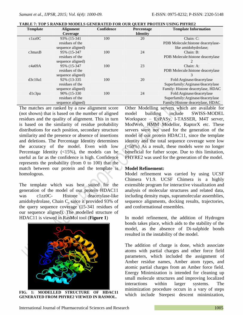
The template which was best suited for the
generation of the model of our protein HDAC11
was c1zz0C- Histone deacetylase-like
amidohydrolase, Chain C, since it provided 93% of
the query sequence coverage (15-341 residues of
our sequence aligned). The modelled structure of
HDAC11 is viewed in RasMol tool (Figure 1)
FIG. 1: MODELLED STRUCTURE OF HDAC11
GENERATED FROM PHYRE2 VIEWED IN RASMOL.
Other Modelling servers which are available for
model building include SWISS-MODEL
Workspace – ExPASy, I-TASSER, M4T server,
ModWeb, HMM Modellor, RaptorX etc. These
servers were not used for the generation of the
model of our protein HDAC11, since the template
identity and the total sequence coverage were low
(<50%) As a result, these models were no longer
beneficial for futher scope. Due to this limitation,
PHYRE2 was used for the generation of the model.
Model Refinement:
Model refinement was carried by using UCSF
Chimera V1.9. UCSF Chimera is a highly
extensible program for interactive visualization and
analysis of molecular structures and related data,
including density maps, supramolecular assemblies,
sequence alignments, docking results, trajectories,
and conformational ensembles.
In model refinement, the addition of Hydrogen
bonds takes place, which aids to the stability of the
model, as the absence of Di-sulphide bonds
resulted in the instability of the model.
The addition of charge is done, which associate
atoms with partial charges and other force field
parameters, which included the assignment of
Amber residue names, Amber atom types, and
atomic partial charges from an Amber force field.
Energy Minimization is intended for cleaning up
small molecule structures and improving localized
interactions within larger systems. The
minimization procedure occurs in a vary of steps
which include Steepest descent minimization,
Samant et al., IJPSR, 2015; Vol. 6(4): 1000-09. E-ISSN: 0975-8232; P-ISSN: 2320-5148
International Journal of Pharmaceutical Sciences and Research 1006
which is performed first to relieve highly
unfavourable clashes, followed by conjugate
gradient minimization, which is much slower but
more effective at reaching an energy minimum
after severe clashes have been relieved. 17
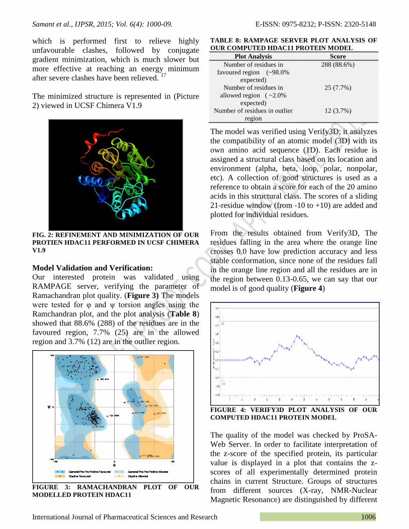
The minimized structure is represented in (Picture
2) viewed in UCSF Chimera V1.9
FIG. 2: REFINEMENT AND MINIMIZATION OF OUR
PROTIEN HDAC11 PERFORMED IN UCSF CHIMERA
V1.9
Model Validation and Verification:
Our interested protein was validated using
RAMPAGE server, verifying the parameter of
Ramachandran plot quality. (Figure 3) The models
were tested for φ and ψ torsion angles using the
Ramchandran plot, and the plot analysis (Table 8)
showed that 88.6% (288) of the residues are in the
favoured region, 7.7% (25) are in the allowed
region and 3.7% (12) are in the outlier region.
FIGURE 3: RAMACHANDRAN PLOT OF OUR
MODELLED PROTEIN HDAC11
TABLE 8: RAMPAGE SERVER PLOT ANALYSIS OF
OUR COMPUTED HDAC11 PROTEIN MODEL
Plot Analysis Score
Number of residues in
favoured region (~98.0%
expected)
288 (88.6%)
Number of residues in
allowed region ( ~2.0%
expected)
Number of residues in outlier
region
25 (7.7%)
12 (3.7%)
The model was verified using Verify3D; it analyzes
the compatibility of an atomic model (3D) with its
own amino acid sequence (1D). Each residue is
assigned a structural class based on its location and
environment (alpha, beta, loop, polar, nonpolar,
etc). A collection of good structures is used as a
reference to obtain a score for each of the 20 amino
acids in this structural class. The scores of a sliding
21-residue window (from -10 to +10) are added and
plotted for individual residues.
From the results obtained from Verify3D, The
residues falling in the area where the orange line
crosses 0.0 have low prediction accuracy and less
stable conformation, since none of the residues fall
in the orange line region and all the residues are in
the region between 0.13-0.65, we can say that our
model is of good quality (Figure 4)
FIGURE 4: VERIFY3D PLOT ANALYSIS OF OUR
COMPUTED HDAC11 PROTEIN MODEL
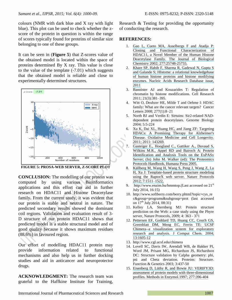
The quality of the model was checked by ProSA-
Web Server. In order to facilitate interpretation of
the z-score of the specified protein, its particular
value is displayed in a plot that contains the z-
scores of all experimentally determined protein
chains in current Structure. Groups of structures
from different sources (X-ray, NMR-Nuclear
Magnetic Resonance) are distinguished by different
Samant et al., IJPSR, 2015; Vol. 6(4): 1000-09. E-ISSN: 0975-8232; P-ISSN: 2320-5148
International Journal of Pharmaceutical Sciences and Research 1007
colours (NMR with dark blue and X ray with light
blue). This plot can be used to check whether the z-
score of the protein in question is within the range
of scores typically found for proteins of similar size
belonging to one of these groups.
It can be seen in (Figure 5) that Z-scores value of
the obtained model is located within the space of
proteins determined By X ray. This value is close
to the value of the template (-7.01) which suggests
that the obtained model is reliable and close to
experimentally determined structures.
FIGURE 5: PROSA-WEB SERVER, Z-SCORE PLOT
CONCLUSION: The modelling of our protein was
computed by using various Bioinformatics
applications and this effort can aid in further
research on HDAC11 and Histone Deacetylase
family. From the current study, it was evident that
our protein is stable and neutral in nature. The
predicted secondary results showed the dominant
coil regions. Validation and evaluation result of 3-
D structure of our protein HDAC11 shows that
predicted model is a stable structural model and of
good quality because it shows maximum residues
(88.6%) in favoured region.
Our effort of modelling HDAC11 protein may
provide information related to functional
mechanisms and also help us in further docking
studies and aid in anticancer and neuroprotector
drugs.
ACKNOWLEDGMENT: The research team was
grateful to the Haffkine Institute for Training,
Research & Testing for providing the opportunity
of conducting the research.
REFERENCES:
1. Gao L, Cueto MA, Asselbergs F and Atadja P:
Cloning and Functional Characterization of
HDAC11, a Novel Member of the Human Histone
Deacetylase Family. The Journal of Biological
Chemistry 2002; 277:25748-25755.
2. Khare SP, Habib F, Sharma R, Gadewal N, Gupta S
and Galande S; HIstome: a relational knowledgebase
of human histone proteins and histone modifying
enzymes. Nucleic Acids Research Database issue,
2011
3. Bannister AJ and Kouzarides T: Regulation of
chromatin by histone modifications. Cell Research
2011; 21(3):381–395.
4. Witt O, Deubzer HE, Milde T and Oehme I: HDAC
family: What are the cancer relevant targets? Cancer
Letters 2008; 277(1):8–21
5. North BJ and Verdin E: Sirtuins: Sir2-related NAD-
dependent protein deacetylases. Genome Biology
2004; 5:5-224
6. Xu K, Dai XL, Huang HC, and Jiang ZF: Targeting
HDACs: A Promising Therapy for Alzheimer's
Disease. Oxidative Medicine and Cell Longevity.
2011; 2011: 143269.
7. Gasteiger E., Hoogland C., Gattiker A., Duvaud S,
Wilkins M.R., Appel RD and Bairoch A: Protein
Identification and Analysis Tools on the ExPASy
Server; (In) John M. Walker (ed): The Proteomics
Protocols Handbook, Humana Press 2005.
8. Källberg M, Wang H, Wang S, Peng J, Wang Z, Lu
H, Xu J: Template-based protein structure modeling
using the RaptorX web server. Nature Protocols
2012; 7:1511–1522,
9. http://www.enzim.hu/hmmtop.(Last accessed on 21st
July 2014, 16:15)
10. http://www.softberry.com/berry.phtml?topic=cys_re
c&group=programs&subgroup=prot (last accessed
on 17th
July 2014, 08:31)
11. Kelley LA, Sternberg MJ: Protein structure
prediction on the Web: a case study using the Phyre
server, Nature Protocols, 2009; 4: 363 - 371
12. Pettersen EF, Goddard TD, Huang CC, Couch GS,
Greenblatt DM, Meng EC, Ferrin TE: UCSF
Chimera--a visualization system for exploratory
research and analysis. J Comput Chem. 2004;
13:1605-12
13. http://www.cgl.ucsf.edu/chimera
14. Lovell SC, Davis IW, Arendall WB, de Bakker PI,
Word JM, Prisant MG, Richardson JS, Richardson
DC: Structure validation by Calpha geometry: phi,
psi and Cbeta deviation. Proteins: Structure.
Function & Genetics 2003; 3:437-50
15. Eisenberg D, Lüthy R, and Bowie JU: VERIFY3D:
assessment of protein models with three-dimensional
profiles. Methods in Enzymol.1997; 277:396-404
Samant et al., IJPSR, 2015; Vol. 6(4): 1000-09. E-ISSN: 0975-8232; P-ISSN: 2320-5148
International Journal of Pharmaceutical Sciences and Research 1008
16. Wiederstein, M. & Sippl M.J: ProSA-web:
interactive web service for the recognition of errors
in three-dimensional structures of proteins. Nucleic
Acids Research.2007; 35: W407–W410
17. Sippl, M.J: Recognition of Errors in Three-
Dimensional Structures of Proteins. Proteins Proteins
1993;17:355-362
18. https://www.cgl.ucsf.edu/chimera/docs/ContributedS
oftware/minimize/minimize.html
All © 2013 are reserved by International Journal of Pharmaceutical Sciences and Research. This Journal licensed under a Creative Commons Attribution-NonCommercial-ShareAlike 3.0 Unported License.
This article can be downloaded to ANDROID OS based mobile. Scan QR Code using Code/Bar Scanner from your mobile. (Scanners are
available on Google Playstore)
Reviewer’s recommendations:
1. Specify designation and current full address of corresponding author.
2. Check for spelling, grammar and punctuation error(s).
How to cite this article:
Samant LR, Sangar VC, Gulamaliwala A and Chowdhary AS: Computational Modelling and Functional Characterization of
HDAC-11. Int J Pharm Sci Res 2015; 6(4): 1000-09.doi: 10.13040/IJPSR.0975-8232.6(4).1000-09.
Related Documents