Nucleic Acids and Molecular Biology,Vol.15 Janusz M. Bujnicki (Ed.) Practical Bioinformatics © Springer-Verlag Berlin Heidelberg 2004 Computational Methods for Protein Structure Prediction and Fold Recognition I. Cymerman, M. Feder, M. PawŁowski, M.A. Kurowski, J.M. Bujnicki 1 Primary Structure Analysis Amino acid sequence analysis provides important insight into the structure of proteins, which in turn greatly facilitates the understanding of its biochem- ical and cellular function. Efforts to use computational methods in predicting protein structure based only on sequence information started 30 years ago (Nagano 1973; Chou and Fasman 1974). However, only during the last decade, has the introduction of new computational techniques such as protein fold recognition and the growth of sequence and structure databases due to mod- ern high-throughput technologies led to an increase in the success rate of pre- diction methods, so that they can be used by the molecular biologist or bio- chemist as an aid in the experimental investigations. 1.1 Database Searches Sequence similarity searching is a crucial step in analyzing newly determined (hereafter called “target”) protein sequences. Typically, large sequence data- bases such as the non-redundant (nr) database at the NCBI (synthesis of Gen- Bank,EMBL and DDBJ databases) or genome sequences are scanned for DNA or amino acid sequences that are similar to a target sequence. Alignments of the target sequence are constructed for each database entry, typically using dynamic programming algorithms (Needleman and Wunsch 1970; Smith and Waterman 1981), scores derived from these alignments are used to identify statistically significant matches. Matches which have a low probability of occurrence by chance are interpreted as likely to indicate homology, i.e. that I. Cymerman, M. Feder, M. Pawłowski, M.A. Kurowski, J.M. Bujnicki Bioinformatics Laboratory, International Institute of Molecular and Cell Biology in Warsaw, Trojdena 4, 02-109 Warsaw, Poland

Welcome message from author
This document is posted to help you gain knowledge. Please leave a comment to let me know what you think about it! Share it to your friends and learn new things together.
Transcript
Nucleic Acids and Molecular Biology,Vol. 15Janusz M. Bujnicki (Ed.)Practical Bioinformatics© Springer-Verlag Berlin Heidelberg 2004
Computational Methods for Protein StructurePrediction and Fold Recognition
I. Cymerman, M. Feder, M. PawŁowski, M.A. Kurowski,J.M. Bujnicki
1 Primary Structure Analysis
Amino acid sequence analysis provides important insight into the structureof proteins, which in turn greatly facilitates the understanding of its biochem-ical and cellular function. Efforts to use computational methods in predictingprotein structure based only on sequence information started 30 years ago(Nagano 1973; Chou and Fasman 1974). However, only during the last decade,has the introduction of new computational techniques such as protein foldrecognition and the growth of sequence and structure databases due to mod-ern high-throughput technologies led to an increase in the success rate of pre-diction methods, so that they can be used by the molecular biologist or bio-chemist as an aid in the experimental investigations.
1.1 Database Searches
Sequence similarity searching is a crucial step in analyzing newly determined(hereafter called “target”) protein sequences. Typically, large sequence data-bases such as the non-redundant (nr) database at the NCBI (synthesis of Gen-Bank, EMBL and DDBJ databases) or genome sequences are scanned for DNAor amino acid sequences that are similar to a target sequence. Alignments ofthe target sequence are constructed for each database entry, typically usingdynamic programming algorithms (Needleman and Wunsch 1970; Smith andWaterman 1981), scores derived from these alignments are used to identifystatistically significant matches. Matches which have a low probability ofoccurrence by chance are interpreted as likely to indicate homology, i.e. that
I. Cymerman, M. Feder, M. Pawłowski, M.A. Kurowski, J.M. BujnickiBioinformatics Laboratory, International Institute of Molecular and Cell Biology in Warsaw, Trojdena 4, 02-109 Warsaw, Poland
the target protein and the matched protein share a common ancestor andtheir sequences have diverged by accumulating a number of substitutions.However, pairwise similarities (especially if confined to very short regions)can also reflect convergent evolution or simply coincidental resemblance.Hence, percent identity or percent similarity should not be used as a primarycriterion for homology. Modern methods for database searches usuallyemploy extreme value distributions to estimate the distribution of the scoresbetween the target and the database entries and a probability of a randommatch (Pearson 1998; Pagni and Jongeneel 2001) For the search for homo-logues to be effective and the score to be accurately estimated, the databasemust contain many unrelated sequences.
Traditionally, searches were carried out using programs for pairwisesequence comparisons like FASTA (Pearson and Lipman 1988) or BLAST(Altschul et al. 1990). However, sequences of homologous proteins can divergebeyond the point where their relationship can be recognized by pairwisesequence comparisons. The most sensitive methods available today use theinitial search for homologues to construct a multiple sequence alignment(MSA), which provide insight into the positional constraints of the aminoacid composition, and allow the identification of conserved and variableregions in the family, comprising the target and its presumed homologues.The MSA is then converted to a position-specific score matrix (PSSM) andused as a target to search the database for more distant homologues that sharesimilarity not only with the initial target, but with the whole family of relatedsequences in the MSA. The MSA can be updated with new sequences andsearches can be carried out in an iterative fashion until no new sequences arereported with the score above the threshold of statistical significance; PSI-BLAST (Altschul et al. 1997; Aravind and Koonin 1999; Schaffer et al. 2001) iswell-optimized and currently the most popular tool in which the PSSM-basedsearch strategy has been implemented. Alternatively to PSSMs, the MSA canbe used to create a Hidden Markov Model (HMM), which also can be itera-tively compared with the database to identify new statistically significantmatches (Karplus et al. 1998).
A related “intermediate sequence search” (ISS) strategy (Park et al. 1997,1998) employs a series of database scans initiated with the target and thencontinued with its homologues. Saturated BLAST is a freely available softwarepackage that performs ISS with BLAST in an automated manner (Li et al.2000). This strategy is computationally more demanding than iterative MSA-based searches (all homologues should be used as search targets), but it cansometimes identify links to remotely related outliers, which may be missed byPSI-BLAST or HMM, which preferentially detect sequences most similar tothe average of the family. However, MSA-based searches can be used to searchfor new sequences that are compatible with very subtle trends of sequenceconservation in the target family, which may be undetectable in any pairwisecomparisons. Recently, it was suggested that an increased number of target
I. Cymerman et al.2
homologues can be found by a combination of various pairwise alignmentmethods for database searches (Webber and Barton 2003). The recommendedstrategy in database searches (as well as in other bioinformatic tasks) is to usemultiple methods and take the agreement between methods as confirmation.
1.2 Protein Domain Identification
Most proteins are composed from a finite number of evolutionarily conservedmodules or domains. Protein domains are distinct units of three-dimensionalprotein structures, which often carry a discrete molecular function, such asthe binding of a specific type of molecule or catalysis (reviews: (Thornton etal. 1999; Aravind et al. 2002)). Proteins can be composed of single or multipledomains. If this information is available, it can be used to make a detailed pre-diction about the protein function (for instance a protein composed of aphosphodiesterase domain and a DNA-binding domain can be speculated tobe a deoxyribonuclease), but if the domain structure is obscure, it can lead toerroneous conclusions about the output of software for sequence analysis.
A common problem in sequence searches is homology of various parts ofthe target to different protein families, which is often the case in multidomainproteins. Naïve exhaustive ISS searches that detect and use multidomain pro-teins can result in an erroneous inference of homology between unrelatedproteins, which happen to be related to different domains fused together inone of the sequences extracted from a database. Hence, domain identificationshould be an essential step in analyzing protein sequences, preferably preced-ing or concurrent to sequence database searches.
A few thousand conserved domains, which cover more than two thirds ofknown protein sequences have been identified and described in literature.Several searchable databases have been created, which store annotated MSAs(sometimes in the form of PSSMs or HMMs) of protein domains, which canbe used to identify conserved modules in the target sequence (Table 1). PFAMand SMART databases are the largest collections of the manually curettedprotein domains of information. Each deposited domain family is extensivelyannotated in the form of textual descriptions, as well as cross-links to otherresources and literature references. Both resources contain friendly but pow-erful web-based interfaces, which provide several types of database searchand exploration. The database can be queried using a protein sequence or anaccession number to examine its domain organization. Alternatively, thedomains can be searched by keywords or browsed via an alphabetical index.Apart from PFAM and SMART there are a number of other databases thatclassify the domains according to their mutual similarity or inferred evolu-tionary relationships (Table 1). They differ from each other either through thetechnical aspects or by concentrating on a specific group of domains. TheMSA deposited in these databases as well as their annotations (e.g. in the form
Computational Methods for Protein Structure Prediction and Fold Recognition 3
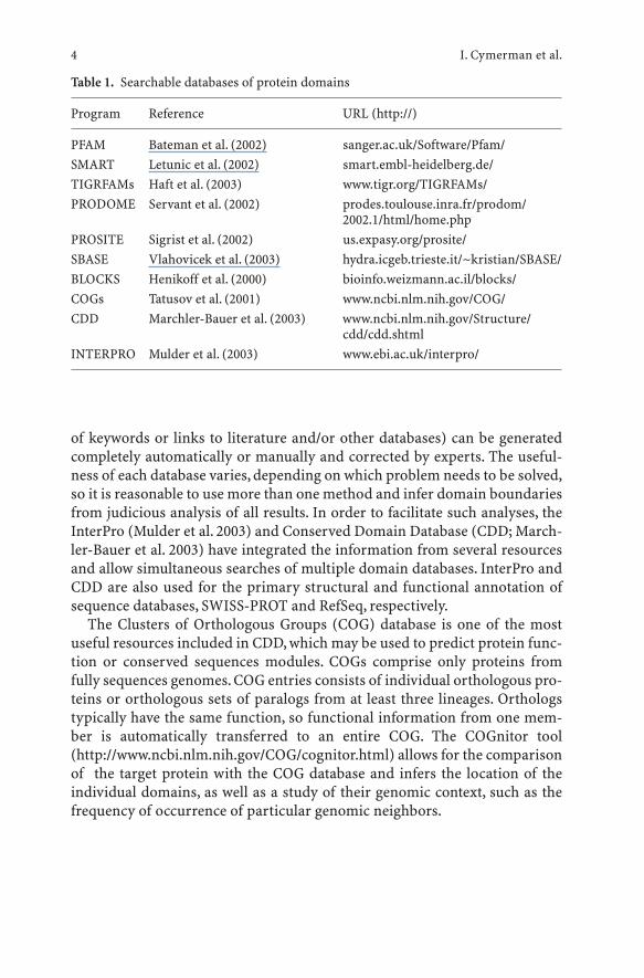
of keywords or links to literature and/or other databases) can be generatedcompletely automatically or manually and corrected by experts. The useful-ness of each database varies, depending on which problem needs to be solved,so it is reasonable to use more than one method and infer domain boundariesfrom judicious analysis of all results. In order to facilitate such analyses, theInterPro (Mulder et al. 2003) and Conserved Domain Database (CDD; March-ler-Bauer et al. 2003) have integrated the information from several resourcesand allow simultaneous searches of multiple domain databases. InterPro andCDD are also used for the primary structural and functional annotation ofsequence databases, SWISS-PROT and RefSeq, respectively.
The Clusters of Orthologous Groups (COG) database is one of the mostuseful resources included in CDD, which may be used to predict protein func-tion or conserved sequences modules. COGs comprise only proteins fromfully sequences genomes. COG entries consists of individual orthologous pro-teins or orthologous sets of paralogs from at least three lineages. Orthologstypically have the same function, so functional information from one mem-ber is automatically transferred to an entire COG. The COGnitor tool(http://www.ncbi.nlm.nih.gov/COG/cognitor.html) allows for the comparisonof the target protein with the COG database and infers the location of theindividual domains, as well as a study of their genomic context, such as thefrequency of occurrence of particular genomic neighbors.
I. Cymerman et al.4
Table 1. Searchable databases of protein domains
Program Reference URL (http://)
PFAM Bateman et al. (2002) sanger.ac.uk/Software/Pfam/SMART Letunic et al. (2002) smart.embl-heidelberg.de/TIGRFAMs Haft et al. (2003) www.tigr.org/TIGRFAMs/PRODOME Servant et al. (2002) prodes.toulouse.inra.fr/prodom/
2002.1/html/home.phpPROSITE Sigrist et al. (2002) us.expasy.org/prosite/SBASE Vlahovicek et al. (2003) hydra.icgeb.trieste.it/~kristian/SBASE/BLOCKS Henikoff et al. (2000) bioinfo.weizmann.ac.il/blocks/COGs Tatusov et al. (2001) www.ncbi.nlm.nih.gov/COG/CDD Marchler-Bauer et al. (2003) www.ncbi.nlm.nih.gov/Structure/
cdd/cdd.shtmlINTERPRO Mulder et al. (2003) www.ebi.ac.uk/interpro/
1.3 Prediction of Disordered Regions
Recently, it has been suggested that the classical protein structure-functionparadigm should be extended to proteins and protein fragments whose nativeand functional state is unstructured or disordered (Wright and Dyson 1999).Many protein domains, especially in eukaryotic proteins appear to lack afolded structure and display a random coil-like conformation under physio-logical conditions (reviews: Liu et al. 2002; Tompa 2002).A significant fractionof the intrinsically unstructured sequences exhibits low complexity, i.e. a non-random compositional bias (Wootton 1994).
On the one hand, low-complexity sequences create a serious problem fordatabase searches, as they are not encompassed by the random model used bythese methods to evaluate alignment statistics.For instance running a databasesearch with a target sequence including a compositionally biased fragment maylead to erroneous identification of a large number of matches with spuriouslyhigh similarity scores. Algorithms such as SEG (Wootton and Federhen 1996)may be used to mask the low-complexity segments for database searches.
On the other hand, identification of disordered, non-globular regions mayhelp to delineate domains. Independently folded globular structures can beseparated from each other if a flexible linker that connects them is identified.Alternatively, if a protein with many low-complexity regions is known to com-prise only a single domain, its rigid core can be identified by masking off flexi-ble insertions. The latter case is typical for many proteins from humanpathogens such as Plasmodium or Trypanosomes, which use the large flexibleloops as hypervariable immunodominant epitopes that contribute to a smoke-screen strategy enacted by the parasite against the host immunogenic response(Pizzi and Frontali 2001).In any case,dissection of the target sequence into a setof relatively rigid, independently folded domains may greatly facilitate tertiarystructure prediction, especially by fold-recognition methods (see below). Thefreely available on-line servers for prediction of disordered loopy regions inproteins are: NORSP (http://cubic.bioc.columbia.edu/services/NORSp/) andGLOBPLOT (http://globplot.embl.de/). The state-of-the art commercial pro-gram PONDR is available from Molecular Kinetics (http://www.pondr.com/);at the time of writing the company promised to introduce a free academiclicense in the near future.
2 Secondary Structure Prediction
2.1 Helices and Strands and Otherwise
Globular protein domains are typically composed of the two basic secondarystructure types, the a-helix and the b-strand, which are easily distinguish-able because of their regular (periodic) character. Other types of secondary
Computational Methods for Protein Structure Prediction and Fold Recognition 5
structures such as different turns, bends, bridges, and non-a helices (such as3/10 and p) are less frequent and more difficult to observe and classify for anon-expert. The non-a, non-b structures are often referred to as coil or loopand the majority of secondary structure prediction methods are aimed atpredicting only these three classes of local structure. Given the observed dis-tribution of the three states in globular proteins (about 30 % a-helix, 20 % b-strand and 50 % coil), random prediction should yield about 40 % accuracyper residue. The accuracy of the secondary structure prediction methodsdevised earlier, such as Chou-Fasman (1974) or GOR (Garnier et al. 1978) isin the range of 50–55 %. The best modern secondary structure predictionmethods (Table 2) have reached a sustained level of 76 % accuracy for thelast 2 years, with a-helices predicted with ca. 10 % higher accuracy than b-strands (Koh et al. 2003). Hence, it is quite surprising that the early mediocremethods are still used in good faith by many researchers; maybe even moresurprising that they are sometimes recommended in contemporary reviewsof bioinformatic software or built in as a default method in new versions ofcommercial software packages for protein sequence analysis and structuremodeling.
Modern secondary structure prediction methods typically perform analy-ses not for the single target sequences, but rather utilize the evolutionaryinformation derived from MSA provided by the user or generated by an inter-nal routine for database searches and alignment (Levin et al. 1993). The infor-mation from the MSA provides a better insight into the positional conser-vation of physico-chemical features such as hydrophobicity and hints at aposition of loops in the regions of insertions and deletions (indels) corre-sponding to gaps in the alignment. It is also recommended to combine differ-ent methods for secondary structure prediction; the ways of combing predic-tions may include the calculation of a simple consensus or more advancedapproaches, including machine learning, such as voting, linear discrimina-tion, neural networks and decision trees (King et al. 2000). JPRED (Cuff et al.1998) is an example of a consensus meta-server that returns predictions fromseveral secondary structure prediction methods (mostly third-party algo-rithms) and infers a consensus using a neural network, thereby improving theaverage accuracy of prediction. In addition, JPRED predicts the relative sol-vent accessibility of each residue in the target sequence, which is very usefulfor identification of solvent-exposed and buried faces of amphipathic helices.
In general, the most effective secondary structure prediction strategies fol-low these rules: (1) if an experimentally determined three-dimensional struc-ture of a closely related protein is known, copy the secondary structureassignment from the known structure rather than attempt to predict it denovo. (2) If no related structures are known, use multiple sequence informa-tion. If your target sequence shows similarity to only a few (or none) otherproteins with sequence identity <90 %, try different databases (for examplepreliminary data from unfinished genomes) to build an MSA comprising a
I. Cymerman et al.6
Computational Methods for Protein Structure Prediction and Fold Recognition 7
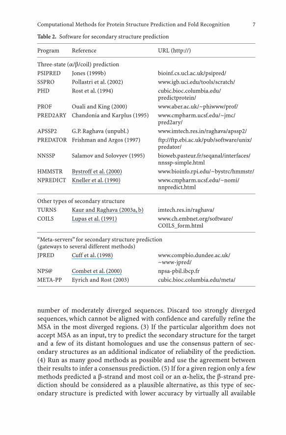
Table 2. Software for secondary structure prediction
Program Reference URL (http://)
Three-state (a/b/coil) predictionPSIPRED Jones (1999b) bioinf.cs.ucl.ac.uk/psipred/SSPRO Pollastri et al. (2002) www.igb.uci.edu/tools/scratch/PHD Rost et al. (1994) cubic.bioc.columbia.edu/
predictprotein/PROF Ouali and King (2000) www.aber.ac.uk/~phiwww/prof/PRED2ARY Chandonia and Karplus (1995) www.cmpharm.ucsf.edu/~jmc/
pred2ary/APSSP2 G.P. Raghava (unpubl.) www.imtech.res.in/raghava/apssp2/PREDATOR Frishman and Argos (1997) ftp://ftp.ebi.ac.uk/pub/software/unix/
predator/NNSSP Salamov and Solovyev (1995) bioweb.pasteur.fr/seqanal/interfaces/
nnssp-simple.htmlHMMSTR Bystroff et al. (2000) www.bioinfo.rpi.edu/~bystrc/hmmstr/NPREDICT Kneller et al. (1990) www.cmpharm.ucsf.edu/~nomi/
nnpredict.html
Other types of secondary structureTURNS Kaur and Raghava (2003a, b) imtech.res.in/raghava/COILS Lupas et al. (1991) www.ch.embnet.org/software/
COILS_form.html
“Meta-servers” for secondary structure prediction (gateways to several different methods)JPRED Cuff et al. (1998) www.compbio.dundee.ac.uk/
~www-jpred/NPS@ Combet et al. (2000) npsa-pbil.ibcp.frMETA-PP Eyrich and Rost (2003) cubic.bioc.columbia.edu/meta/
number of moderately diverged sequences. Discard too strongly divergedsequences, which cannot be aligned with confidence and carefully refine theMSA in the most diverged regions. (3) If the particular algorithm does notaccept MSA as an input, try to predict the secondary structure for the targetand a few of its distant homologues and use the consensus pattern of sec-ondary structures as an additional indicator of reliability of the prediction.(4) Run as many good methods as possible and use the agreement betweentheir results to infer a consensus prediction. (5) If for a given region only a fewmethods predicted a b-strand and most coil or an a-helix, the b-strand pre-diction should be considered as a plausible alternative, as this type of sec-ondary structure is predicted with lower accuracy by virtually all available
methods. (6) Reconfirm the prediction of loops by correlating their presencewith regions of indels in the MSA.
In our own hands, the application of these rules in a semi-automated man-ner (i.e. human post-processing of prediction generated by various individualmethods) led to a very high accuracy of 83 % per residue (better than any sin-gle server or any other human predictor) according to the recent evaluationwithin the CASP-5 experiment (http://predictioncenter.llnl.gov/casp5/).
2.2 Transmembrane Helices
Membrane proteins are an abundant and functionally relevant subset of pro-teins predicted to include up to 30 % of proteins in the fully sequencedgenomes. Membrane proteins are associated with the cell membrane andcomprise one or more transmembrane segments. Because of the hydrophobicenvironment within the cell membrane, the transmembrane segments aregenerally hydrophobic too. On the one hand, typical cytoplasmic membraneproteins comprise hydrophobic a-helical regions separated by hydrophilicloops. On the other hand, bacterial and organellar outer membrane proteinsexhibit a characteristic b-barrel structure comprising different even numbersof b-strands. Specialized structure predictors have been designed for bothtypes of membrane proteins. Because both sides of the lipid bilayer are non-equivalent, structure prediction methods for transmembrane proteins oftenattempt to identify not only the secondary structure elements (a-helices or b-strands), but also the topology of the protein, i.e. the orientation of the ele-ments with respect to both surfaces (which side of transmembrane protein isintra- or extracellular). For instance, the “positive inside rule” (von Heijne1986, 1992) indicates that the positively charged residues have a preference forthe inside of internal membrane proteins.
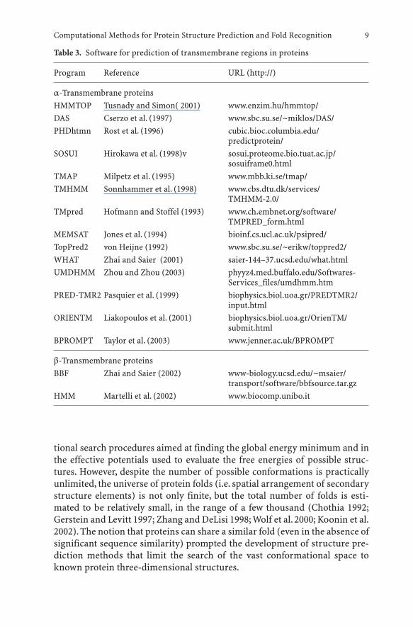
As with orthodox secondary structure prediction methods, the recom-mended strategy for identification of transmembrane segments and predic-tion of their distribution and topology in protein sequences is to use manydifferent methods and refer to the consensus as the most robust structuralmodel (Ikeda et al. 2002). Table 3 lists available programs for prediction oftransmembrane segments and topology.A meta-server BPROMPT for predic-tion of transmembrane helices has been recently developed that combines theresults of other prediction methods, providing a more accurate consensusprediction (Taylor et al. 2003).
3 Protein Fold-Recognition
The success of the prediction of protein tertiary (three-dimensional) struc-ture from its amino acid sequence is limited by deficiencies in the conforma-
I. Cymerman et al.8
tional search procedures aimed at finding the global energy minimum and inthe effective potentials used to evaluate the free energies of possible struc-tures. However, despite the number of possible conformations is practicallyunlimited, the universe of protein folds (i.e. spatial arrangement of secondarystructure elements) is not only finite, but the total number of folds is esti-mated to be relatively small, in the range of a few thousand (Chothia 1992;Gerstein and Levitt 1997; Zhang and DeLisi 1998; Wolf et al. 2000; Koonin et al.2002). The notion that proteins can share a similar fold (even in the absence ofsignificant sequence similarity) prompted the development of structure pre-diction methods that limit the search of the vast conformational space toknown protein three-dimensional structures.
Computational Methods for Protein Structure Prediction and Fold Recognition 9
Table 3. Software for prediction of transmembrane regions in proteins
Program Reference URL (http://)
a-Transmembrane proteinsHMMTOP Tusnady and Simon( 2001) www.enzim.hu/hmmtop/DAS Cserzo et al. (1997) www.sbc.su.se/~miklos/DAS/PHDhtmn Rost et al. (1996) cubic.bioc.columbia.edu/
predictprotein/SOSUI Hirokawa et al. (1998)v sosui.proteome.bio.tuat.ac.jp/
sosuiframe0.htmlTMAP Milpetz et al. (1995) www.mbb.ki.se/tmap/TMHMM Sonnhammer et al. (1998) www.cbs.dtu.dk/services/
TMHMM-2.0/TMpred Hofmann and Stoffel (1993) www.ch.embnet.org/software/
TMPRED_form.htmlMEMSAT Jones et al. (1994) bioinf.cs.ucl.ac.uk/psipred/TopPred2 von Heijne (1992) www.sbc.su.se/~erikw/toppred2/WHAT Zhai and Saier (2001) saier-144–37.ucsd.edu/what.htmlUMDHMM Zhou and Zhou (2003) phyyz4.med.buffalo.edu/Softwares-
Services_files/umdhmm.htmPRED-TMR2 Pasquier et al. (1999) biophysics.biol.uoa.gr/PREDTMR2/
input.htmlORIENTM Liakopoulos et al. (2001) biophysics.biol.uoa.gr/OrienTM/
submit.htmlBPROMPT Taylor et al. (2003) www.jenner.ac.uk/BPROMPT
b-Transmembrane proteinsBBF Zhai and Saier (2002) www-biology.ucsd.edu/~msaier/
transport/software/bbfsource.tar.gzHMM Martelli et al. (2002) www.biocomp.unibo.it
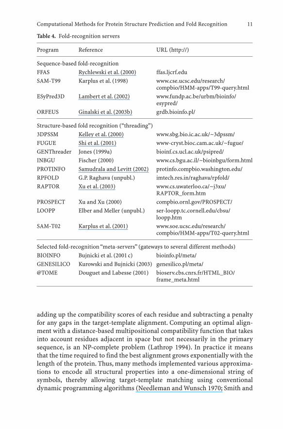
The protein fold-recognition approach to structure prediction aims toidentify the known structural framework (i.e. the backbone of an experimen-tally determined protein structure) that accommodates the target proteinsequence in the best way. Typically, a fold-recognition program comprisesfour components: (1) the representation of the template structures (usuallycorresponding to proteins from the Protein Data Bank database), (2) the eval-uation of the compatibility between the target sequence and a template fold,(3) the algorithm to compute the optimal alignment between the targetsequence and the template structure, and (4) the way the ranking is computedand the statistical significance is estimated (Fischer et al. 1996).
Two main types of fold-recognition algorithms may be defined: those thatdetect sequence similarity (without utilizing structural information from thetemplate) and those that detect structure similarity (Table 4).
Sequence-based fold recognition methods do not utilize explicitly thestructural information from the templates. The simplest sequence-only fold-recognition operation is to use BLAST or PSI-BLAST to search the ProteinData Bank for structurally characterized proteins that exhibit significantsequence similarity to the target protein. However, the principal task of pro-tein fold-recognition methods is to identify sequence similarities that mostbiologists wouldn’t easily call evident and that cannot be identified in trivialdatabase searches. The evolutionary information used to detect remote rela-tionships is usually compiled in the form of a profile, or a HMM. However, themost sensitive sequence-based fold-recognition methods available today aremore advanced than sequence-profile comparisons implemented in methodssuch as PSI-BLAST, IMPALA or HMMs and utilize the evolutionary informa-tion available both for the target and the template by performing profile-pro-file alignment and the evaluation of the likelihood that two protein familiesare related to each other; examples include FFAS (Rychlewski et al. 2000) andthe prof_sim algorithm (Yona and Levitt 2002). A recently developed methodORFeus uses sequence profiles and disregards the experimental structuralinformation from the template, and attempts to predict the structure de novoboth for the target and the template families (Ginalski et al. 2003b).
Structure-based fold-recognition, often referred to as threading, utilizesthe experimentally determined structural information from the template. Thetarget sequence can be enhanced by including sequence-derived (predicted)structural features of the target. The two typically used structural features arethe patterns of secondary structure elements and local environment classes(combination of solvent accessibility, polarity of the side chain environmentand local backbone conformation). The target-template compatibility func-tions of the early threading methods were based mainly on physicochemicalproperties and evaluation of pseudo-energy of interactions and utilizedeither distance-based (Godzik et al. 1992; Jones et al. 1992; Sippl and Weitckus1992; Bryant and Lawrence 1993) or profile-based scoring-functions (Bowieet al. 1991; Ouzounis et al. 1993). The compatibility score is computed by
I. Cymerman et al.10
adding up the compatibility scores of each residue and subtracting a penaltyfor any gaps in the target-template alignment. Computing an optimal align-ment with a distance-based multipositional compatibility function that takesinto account residues adjacent in space but not necessarily in the primarysequence, is an NP-complete problem (Lathrop 1994). In practice it meansthat the time required to find the best alignment grows exponentially with thelength of the protein. Thus, many methods implemented various approxima-tions to encode all structural properties into a one-dimensional string ofsymbols, thereby allowing target-template matching using conventionaldynamic programming algorithms (Needleman and Wunsch 1970; Smith and
Computational Methods for Protein Structure Prediction and Fold Recognition 11
Table 4. Fold-recognition servers
Program Reference URL (http://)
Sequence-based fold-recognitionFFAS Rychlewski et al. (2000) ffas.ljcrf.eduSAM-T99 Karplus et al. (1998) www.cse.ucsc.edu/research/
compbio/HMM-apps/T99-query.htmlESyPred3D Lambert et al. (2002) www.fundp.ac.be/urbm/bioinfo/
esypred/ORFEUS Ginalski et al. (2003b) grdb.bioinfo.pl/
Structure-based fold recognition (“threading”)3DPSSM Kelley et al. (2000) www.sbg.bio.ic.ac.uk/~3dpssm/FUGUE Shi et al. (2001) www-cryst.bioc.cam.ac.uk/~fugue/GENThreader Jones (1999a) bioinf.cs.ucl.ac.uk/psipred/INBGU Fischer (2000) www.cs.bgu.ac.il/~bioinbgu/form.htmlPROTINFO Samudrala and Levitt (2002) protinfo.compbio.washington.edu/RPFOLD G.P. Raghava (unpubl.) imtech.res.in/raghava/rpfold/RAPTOR Xu et al. (2003) www.cs.uwaterloo.ca/~j3xu/
RAPTOR_form.htmPROSPECT Xu and Xu (2000) compbio.ornl.gov/PROSPECT/LOOPP Elber and Meller (unpubl.) ser-loopp.tc.cornell.edu/cbsu/
loopp.htmSAM-T02 Karplus et al. (2001) www.soe.ucsc.edu/research/
compbio/HMM-apps/T02-query.html
Selected fold-recognition “meta-servers” (gateways to several different methods)BIOINFO Bujnicki et al. (2001 c) bioinfo.pl/meta/GENESILICO Kurowski and Bujnicki (2003) genesilico.pl/meta/@TOME Douguet and Labesse (2001) bioserv.cbs.cnrs.fr/HTML_BIO/
frame_meta.html
Waterman 1981), as in sequence-based methods. The early threaders werequite successful in identification of the correct fold, however the quality of thereported target-template alignments was often poor. Apparently, correct fold-recognition could be achieved, despite poor alignment quality, by a generallyunspecific maximization of the hydrophobic interactions, and a reasonablygood prediction of the local secondary structure (Lemer et al. 1995).
Modern fold-recognition methods utilize both the structural information(experimentally determined for the potential templates and predicted for thetarget) and the evolutionary information inferred from the MSA available forthe target and the templates. According to the recent evaluations (Bujnicki etal. 2001a, b), best fold-recognition algorithms are able to make up to 40 % ofcorrect structural predictions for targets, which exhibit no significant simi-larity to any of the potential templates (i.e. similarities that cannot be detectedby BLAST or PSI-BLAST searches run with default parameters). One of themost significant unsolved problems is the lack of an accurate scoring functionfor discrimination between correct and incorrect fold-recognition align-ments. It is quite often the case that the correct template is reported amongthe best ten results returned by a fold-recognition server, but its score is verysimilar to scores for nine false positives or it is below the threshold of statisti-cal significance. In other words, the sensitivity and specificity of fold-recogni-tion methods are insufficient to confidently identify the correct template, if itexists in the Protein Data Bank. Recently, consensus meta-servers have beendeveloped which greatly increase the sensitivity and specificity of fold-recog-nition (Douguet and Labesse 2001; Bujnicki et al. 2001 c; Lundstrom et al.2001; Kurowski and Bujnicki 2003; Ginalski et al. 2003a). Most of them com-bine not only fold-recognition methods, but integrate many different kinds ofprotein structure prediction methods described in this article, from identifi-cation of domains, to secondary structure prediction, to modeling of the tar-get based on the best-scoring template structures (for detailed description oftwo examples see the following section and a separate review by Cohen et al.(this Vol.); a separate discussion on various aspects of meta prediction is pro-vided in a review by Bujnicki and Fischer).
4 Predicting all-in-one-go
The GeneSilico meta-server (http://genesilico.pl/meta/; Kurowski andBujnicki 2003) will serve here as an example of a freely available on-line ser-vice for integrated prediction of different aspects of protein structure. Asmentioned earlier, the recommended strategy is to predict the target proteinstructure using not only the single sequence information, but to enhance itwith aligned homologous sequences. The GeneSilico meta-server allows sub-mission of single sequences or user-defined multiple alignments (MSA). Asingle sequence is processed further by individual methods, which often gen-
I. Cymerman et al.12
erate their own alignments, typically using PSI-BLAST (Altschul et al. 1997)with different parameters. Automatically generated sequence alignments areusually sufficient, but sometimes the target sequence has an unusual aminoacid composition or atypical insertions, which may cause the default iterateddatabase search to produce erroneous alignments that will degrade the evolu-tionary signal instead of enhancing it. Moreover, some sequences have only afew homologues in the traditionally used databases such as NRDB or Swiss-Prot and in order to build a useful alignment, additional searches of otherdatabases are necessary. Therefore, it is strongly recommended for experi-enced predictors to submit their own MSA, in addition to the single-sequencequeries. The GeneSilico meta-server will forward the MSA to those serversthat allow such input, while for the others, which accept only single-sequencequeries, a single consensus sequence will be calculated from the MSA usingone of many different options selected by the user (from majority-rule toscoring derived from different substitution matrices). Furthermore, the userwill have an option to delete or retain loopy regions corresponding to gaps inthe sequence alignment – this option causes a limitation on the fold-recogni-tion analysis to regions most likely to correspond to the true globular core ofthe target protein.
As mentioned earlier, the crucial step in protein structure prediction is toidentify protein domains in the target sequence. This task is accomplished bythe HMMPFAM tool, which scans the PFAM database of known proteindomains (Bateman et al. 2002) with the HMMER method (Eddy 1996). If theresults obtained from the HMMPFAM search suggest the presence of morethan one domain in the target sequence, it is strongly recommended to splitthe target into the respective fragments (possibly retaining some regions ofoverlap, 10–50 aa, depending on the confidence of the domain prediction)and resubmit the individual domains as separate prediction queries.
Secondary structure is predicted in three states (a, b, and coil) by PSI-PRED (Jones 1999b), PROF (Ouali and King 2000), and SAM-T02 (Karplus etal. 2001). Identification of potential transmembrane helices is attempted usingTMPRED (Hofmann and Stoffel 1993) MEMSAT (Jones et al. 1994), andTMHMM (Sonnhammer et al. 1998). If all methods predict a transmembranesegment or a long region with no a or b structure in the target sequence, it isagain strongly recommended to remove such regions, as they are unlikely toform any globular domain identifiable by fold-recognition methods, and toresubmit the remaining part of the target as a new prediction query.
The GeneSilico metaserver serves as a gateway for a number of third-partyfold-recognition methods, both sequence-dependent, and structure-depen-dent, including FUGUE (Shi et al. 2001), 3DPSSM (Kelley et al. 2000), SAM-T02(Karplus et al. 2001), GENTHREADER (Jones 1999a), FFAS (Rychlewski et al.2000), INBGU (Fischer 2000), and RAPTOR (Xu et al. 2003). However, beforethe extensive fold-recognition calculations are carried out, the PDB databaseis searched with the PSI-BLAST method to identify trivial similarities of the
Computational Methods for Protein Structure Prediction and Fold Recognition 13
target to proteins of known structure (three iterations against the NRDB data-base are carried out with the target sequence to generate a MSA, which is sub-sequently used to search the PDB database for significant similarities). If thetarget exhibits significant similarity to a known structure, the fold-recogni-tion analysis is halted and the user is notified; otherwise (or if the userdecides to resume the analysis) the query (i.e. the single sequence or the MSA)is sent to the above-mentioned fold-recognition servers. Typically, the collec-tion of results from all servers (up to ten target-template alignments perserver) requires about 24 h, however some sequence-based servers returntheir predictions within a few minutes. The meta-server presents all target-template alignments and the corresponding confidence scores assigned by theindividual methods according to their internal criteria. These scores aremutually incompatible and further analysis is required to provide a commonranking of results returned by different fold-recognition servers. Hence, whenall results are available, they are further processed by the consensus serverPCONS (two different versions, 2 and 5; Lundstrom et al. 2001; Wallner andElofsson 2003), which does not produce any new predictions, but selects theten potentially best target-template alignments from those reported by theoriginal methods and assigns its own confidentiality scores. It has beenshown that PCONS is more sensitive (i.e. able to identify correct templates)and specific (i.e. able to generate significant scores) than any individualmethod incorporated as a slave in the prediction pipeline.
Finally, the user of the GeneSilico server has an opportunity to generatepreliminary three-dimensional models of the target structure based on thealignments proposed by all servers. These models may be incomplete andcontain significant errors even if they are based on correct templates, but usu-ally serve as a useful starting point for further refinement. The preliminaryevaluation is carried out using the VERIFY3D method, whose score tells howmuch the characteristics of the model resemble the features of high-resolu-tion crystal structures i.e. how much the theoretical model is protein-like orprotein-unlike, compared to the known structures.
5 Pitfalls of Fold Recognition
As soon as the sequence of the target protein is optimally mounted on the pre-sumably best template structure, the corresponding sequence-structurealignment can be used to initiate reconstruction of a complete full-atommodel of the target protein by various comparative modeling techniques(reviewed by Cohen et al. in this volume; see also the following references:(Sanchez and Sali 2000; Krieger et al. 2003)). The comparative modelingapproach assumes that the target and the template share the polypeptidebackbone and the differences are limited to the solvent-exposed loops and theconformation of the side chains, according to the notion that protein spatial
I. Cymerman et al.14
structures are more conserved in evolution than amino acid sequences(Chothia and Lesk 1986). This assumption is certainly valid in many cases,especially if the sequence identity between the target and the template is veryhigh (>50 %). However, the recent sequence and structure analyses led to theaccumulation of examples of homologous proteins with globally distinctstructures. It has been found that even in proteins with significant sequencesimilarity, insertions, deletions and mutual conversions of a-helices and b-strands can occur both at the periphery and in the core of the fold; moreover,the global topology of the fold can be changed by circular permutations, andrearrangements in the order of strands in b-sheets (reviews: Murzin 1998;Grishin 2001a). Such structural changes are usually undetectable by computa-tional methods that operate on the level of protein sequence similarities andeven for structure-based threading methods it is extremely hard to predictdifferences between the three-dimensional folds of the target and the tem-plate other than the deletion or insertion of secondary structure elements.
It also becomes clear that domains are not the only units of homology.Some protein superfamilies have been reported to contain segments ofhomology often limited to a few elements of secondary structure unable tofold independently, such as the bba-Me finger in many nucleases, embeddedinto non-homologous regions acquired independently between proteins(Kuhlmann et al. 1999; Grishin 2001b). In contrast, unrelated segmentsacquired independently could be embedded into the regions of homology. Insuch cases, detection of a strong local homology by fold-recognition pro-grams can be erroneously extended to the entire length of the target and thetemplate. Currently, no fully automated methods exist for prediction of foldirregularities. However, recent progress in the ab initio protein structure pre-diction field, especially the development of methods that use confident pre-dictions of the protein core made by fold-recognition methods to initiateextensive folding simulation to assemble the peripheral elements (Simons etal. 1997; Kihara et al. 2001) suggest that in the near future these limitations ofthe current fold-recognition methods may be overcome.
Presently, the best strategy, however, is to validate the computational pre-diction of the protein fold by experimental analyses which on its own wouldnot be sufficient to solve protein structure, but when combined with bioinfor-matics, may serve to identify one reasonable structural model and then guideits refinement. Such experimental investigations may include generation ofboth specific and non-specific distance constraints by intramolecular cross-linking, chemical modification, or simple NMR analyses, identification of sol-vent-exposed loops by proteolysis, identification of important residues bymutagenesis etc. Several examples of combination of computational andexperimental analyses are discussed elsewhere in this volume (see chapters byLinge and Nilges; Alber et al; and Friedhoff). Clearly, the development of aconvenient computational method for automated combination of heterolo-gous experimental data and low-resolution structure prediction by fold-
Computational Methods for Protein Structure Prediction and Fold Recognition 15
recognition and ab initio bioinformatic methods would greatly facilitatestructural analyses of proteins and bring protein modeling closer to the work-bench of a biochemist or a molecular biologist.
Acknowledgements. The authors’ research on various aspects of combination of compu-tational and experimental methods for protein structure analysis is supported by KBN(grants 6P04B00519, 3P04A01124, and 3P05A02024). J.M.B. is an EMBO and HowardHughes Medical Institute Young Investigator and a Fellow of the Foundation for PolishScience.
References
Altschul SF, Gish W, Miller W, Myers EW, Lipman DJ (1990) Basic local alignment searchtool. J Mol Biol 215:403–410
Altschul SF, Madden TL, Schaffer AA, Zhang J, Zhang Z, Miller W, Lipman DJ (1997)Gapped BLAST and PSI-BLAST: a new generation of protein database search pro-grams. Nucleic Acids Res 25:3389–3402
Aravind L, Koonin EV (1999) Gleaning non-trivial structural, functional and evolution-ary information about proteins by iterative database searches. J Mol Biol287:1023–1040
Aravind L, Mazumder R, Vasudevan S, Koonin EV (2002) Trends in protein evolutioninferred from sequence and structure analysis. Curr Opin Struct Biol 12:392–399
Bateman A, Birney E, Cerruti L, Durbin R, Etwiller L, Eddy SR, Griffiths-Jones S, HoweKL, Marshall M, Sonnhammer EL (2002) The Pfam protein families database. NucleicAcids Res 30:276–280
Bowie JU, Luthy R, Eisenberg D (1991) A method to identify protein sequences that foldinto a known three-dimensional structure. Science 253:164–170
Bryant SH, Lawrence CE (1993) An empirical energy function for threading proteinsequence through the folding motif. Proteins 16:92–112
Bujnicki JM, Elofsson A, Fischer D, Rychlewski L (2001a) LiveBench-1: continuousbenchmarking of protein structure prediction servers. Protein Sci 10:352–361
Bujnicki JM, Elofsson A, Fischer D, Rychlewski L (2001b) LiveBench-2: Large-scale auto-mated evaluation of protein structure prediction servers. Proteins 45:184–191
Bujnicki JM, Elofsson A, Fischer D, Rychlewski L (2001 c) Structure prediction MetaServer. Bioinformatics 17:750–751
Bystroff C, Thorsson V, Baker D (2000) HMMSTR: a hidden Markov model for localsequence-structure correlations in proteins J Mol Biol 301:173–190
Chandonia JM, Karplus M (1995) Neural networks for secondary structure and struc-tural class predictions. Protein Sci 4:275–285
Chothia C (1992) Proteins. One thousand families for the molecular biologist. Nature357:543–544
Chothia C, Lesk AM (1986) The relation between the divergence of sequence and struc-ture in proteins. EMBO J 5:823–826
Chou PY, Fasman GD (1974) Prediction of protein conformation. Biochemistry13:222–245
Combet C, Blanchet C, Geourjon C, Deleage G (2000) NPS@: network protein sequenceanalysis. Trends Biochem Sci 25:147–150
I. Cymerman et al.16
Cserzo M,Wallin ESimon Ivon Heijne G, Elofsson A (1997) Prediction of transmembranealpha-helices in prokaryotic membrane proteins: the dense alignment surfacemethod. Protein Eng 10:673–676
Cuff JA, Clamp ME, Siddiqui AS, Finlay M, Barton GJ (1998) JPred: a consensus sec-ondary structure prediction server. Bioinformatics 14:892–893
Douguet D, Labesse G (2001) Easier threading through web-based comparisons andcross-validations. Bioinformatics 17:752–753
Eddy SR (1996) Hidden Markov models. Curr Opin Struct Biol 6:361–365Eyrich VA, Rost B (2003) META-PP: single interface to crucial prediction servers. Nucleic
Acids Res 31:3308–3310Fischer D (2000) Hybrid fold recognition: combining sequence derived properties with
evolutionary information. Pac Symp Biocomput , pp 119–130Fischer D, Elofsson A, Rice D, Eisenberg D (1996) Assessing the performance of fold
recognition methods by means of a comprehensive benchmark. Pac Symp Biocomput, pp 300–318
Frishman D, Argos P (1997) Seventy-five percent accuracy in protein secondary struc-ture prediction. Proteins 27:329–335
Garnier J, Osguthorpe DJ, Robson B (1978) Analysis of the accuracy and implications ofsimple methods for predicting the secondary structure of globular proteins. J MolBiol 120:97–120
Gerstein M, Levitt M (1997) A structural census of the current population of proteinsequences. Proc Natl Acad Sci USA 94:11911–11916
Ginalski K, Elofsson A, Fischer D, Rychlewski L (2003a) 3D-Jury: a simple approach toimprove protein structure predictions. Bioinformatics 19:1015–1018
Ginalski K, Pas J, Wyrwicz LS, von Grotthuss M, Bujnicki JM, Rychlewski L (2003b)ORFeus: detection of distant homology using sequence profiles and predicted sec-ondary structure. Nucleic Acids Res 31:3804–3807Godzik A, Kolinski A, Skolnick J(1992) Topology fingerprint approach to the inverse protein folding problem. J MolBiol 227:227–238
Grishin NV (2001a) Fold change in evolution of protein structures. J Struct Biol134:167–185
Grishin NV (2001b) Treble clef finger–a functionally diverse zinc-binding structuralmotif. Nucleic Acids Res 29:1703–1714
Haft DH, Selengut JD, White O (2003) The TIGRFAMs database of protein families.Nucleic Acids Res 31: 371–373
Henikoff JG, Greene EA, Pietrokovski S, Henikoff S (2000) Increased coverage of proteinfamilies with the blocks database servers. Nucleic Acids Res 28:228–230
Hirokawa T, Boon-Chieng S, Mitaku S(1998) SOSUI: classification and secondary struc-ture prediction system for membrane proteins. Bioinformatics 14:378–379
Hofmann K, Stoffel W (1993) TMbase – a database of membrane spanning proteins seg-ments. Biol Chem 374:166
Ikeda M, Arai M, Lao DM, Shimizu T (2002) Transmembrane topology prediction meth-ods: a re-assessment and improvement by a consensus method using a dataset ofexperimentally-characterized transmembrane topologies. In Silico Biol 2:19–33
Jones DT (1999a) GenTHREADER: an efficient and reliable protein fold recognitionmethod for genomic sequences. J Mol Biol 287:797–815
Jones DT (1999b) Protein secondary structure prediction based on position-specificscoring matrices. J Mol Biol 292:195–202
Jones DT, Taylor WR, Thornton JM (1992) A new approach to protein fold recognition.Nature 358:86–89
Computational Methods for Protein Structure Prediction and Fold Recognition 17
Jones DT, Taylor WR, Thornton JM (1994) A model recognition approach to the predic-tion of all-helical membrane protein structure and topology. Biochemistry 33:3038–3049
Karplus K, Barrett C, Hughey R (1998) Hidden Markov models for detecting remote pro-tein homologies. Bioinformatics 14:846–856
Karplus K, Karchin R, Barrett C, Tu S, Cline M, Diekhans M, Grate L, Casper J, Hughey R(2001) What is the value added by human intervention in protein structure predic-tion? Proteins 45(Suppl 5):86–91
Kaur H, Raghava GP (2003a) A neural-network based method for prediction of gamma-turns in proteins from multiple sequence alignment. Protein Sci 12:923–929
Kaur H, Raghava GP (2003b) Prediction of beta-turns in proteins from multiple align-ment using neural network. Protein Sci 12:627–634
Kelley LA, McCallum CM, Sternberg MJ (2000) Enhanced genome annotation usingstructural profiles in the program 3D-PSSM. J Mol Biol 299:501–522
Kihara D, Lu H, Kolinski A, Skolnick J (2001) TOUCHSTONE: an ab initio protein struc-ture prediction method that uses threading-based tertiary restraints. Proc Natl AcadSci USA 98:10125–10130
King RD, Ouali M, Strong AT,Aly A, Elmaghraby A, Kantardzic M, Page D (2000) Is it bet-ter to combine predictions? Protein Eng 13:15–19
Kneller DG, Cohen FE, Langridge R (1990) Improvements in protein secondary struc-ture prediction by an enhanced neural network. J Mol Biol 214:171–182
Koh IY, Eyrich VA, Marti-Renom MA, Przybylski D, Madhusudhan MS, Eswar N, GranaO, Pazos F, Valencia A, Sali A, Rost B (2003) EVA: evaluation of protein structure pre-diction servers. Nucleic Acids Res 31:3311–3315
Koonin EV, Wolf YI, Karev GP (2002) The structure of the protein universe and genomeevolution. Nature 420:218–223
Krieger E, Nabuurs SB, Vriend G (2003) Homology modeling. Methods Biochem Anal44:509–523
Kuhlmann UC, Moore GR, James R, Kleanthous C, Hemmings AM (1999) Structural par-simony in endonuclease active sites: should the number of homing endonucleasefamilies be redefined? FEBS Lett 463:1–2
Kurowski MA, Bujnicki JM (2003) GeneSilico protein structure prediction meta-server.Nucleic Acids Res 31:3305–3307
Lambert C, Leonard N, De B, X, Depiereux E (2002) ESyPred3D: Prediction of proteins3D structures. Bioinformatics 18:1250–1256
Lathrop RH (1994) The protein threading problem with sequence amino acid interac-tion preferences is NP-complete. Protein Eng 7:1059–1068
Lemer CM, Rooman MJ, Wodak SJ (1995). Protein structure prediction by threadingmethods: evaluation of current techniques. Proteins 23:337–355
Letunic I, Goodstadt L, Dickens NJ, Doerks T, Schultz J, Mott R, Ciccarelli F, Copley RR,Ponting CP, Bork P (2002) Recent improvements to the SMART domain-basedsequence annotation resource. Nucleic Acids Res 30:242–244
Levin JM, Pascarella S, Argos P, Garnier J (1993) Quantification of secondary structureprediction improvement using multiple alignments. Protein Eng 6:849–854
Li W, Pio F, Pawlowski K, Godzik A (2000) Saturated BLAST: an automated multiple inter-mediate sequence search used to detect distant homology. Bioinformatics 16:1105–1110
Liakopoulos TD, Pasquier C, Hamodrakas SJ (2001) A novel tool for the prediction oftransmembrane protein topology based on a statistical analysis of the SwissProt data-base: the OrienTM algorithm. Protein Eng 14:387–390
Liu J, Tan H, Rost B (2002) Loopy proteins appear conserved in evolution. J Mol Biol322:53–64
I. Cymerman et al.18
Lundstrom J, Rychlewski L, Bujnicki JM, Elofsson A (2001) Pcons: a neural-network-based consensus predictor that improves fold recognition. Protein Sci 10:2354–2362
Lupas A,Van Dyke M, Stock J (1991) Predicting coiled coils from protein sequences. Sci-ence 252:1162–1164
Marchler-Bauer A, Anderson JB, DeWeese-Scott C, Fedorova ND, Geer LY, He S, HurwitzDI., Jackson JD, Jacobs AR, Lanczycki CJ, Liebert CA, Liu C, Madej T, Marchler GH,Mazumder R, Nikolskaya AN, Panchenko AR, Rao BS, Shoemaker BA, Simonyan V,Song JS, Thiessen PA, Vasudevan S, Wang Y, Yamashita RA, Yin JJ, Bryant SH (2003)CDD: a curated Entrez database of conserved domain alignments. Nucleic Acids Res31:383–387
Martelli PL, Fariselli P, Krogh A, Casadio R (2002) A sequence-profile-based HMM forpredicting and discriminating beta barrel membrane proteins. Bioinformatics18(Suppl 1):S46-S53
Milpetz F, Argos P, Persson B (1995) TMAP: a new email and WWW service for mem-brane-protein structural predictions. Trends Biochem Sci 20:204–205
Mulder NJ,Apweiler R,Attwood TK, Bairoch A, Barrell D, Bateman A, Binns D, Biswas M,Bradley P, Bork P, Bucher P, Copley RR, Courcelle E, Das U, Durbin R, Falquet L, Fleis-chmann W, Griffiths-Jones S, Haft D, Harte N, Hulo N, Kahn D, Kanapin A, Krestyani-nova M, Lopez R, Letunic I, Lonsdale D, Silventoinen V, Orchard SE, Pagni M, PeyrucD, Ponting CP, Selengut JD, Servant F, Sigrist CJ, Vaughan R, Zdobnov EM (2003) TheInterPro Database, 2003 brings increased coverage and new features. Nucleic AcidsRes 31:315–318
Murzin AG (1998) How far divergent evolution goes in proteins. Curr Opin Struct Biol 8380–387
Nagano K (1973) Logical analysis of the mechanism of protein folding. I. Predictions ofhelices, loops and beta-structures from primary structure. J Mol Biol 75:401–420
Needleman SB, Wunsch CD (1970) A general method applicable to the search for simi-larities in the amino acid sequence of two proteins. J Mol Biol 48:443–453
Ouali M, King RD (2000) Cascaded multiple classifiers for secondary structure predic-tion. Protein Sci 9:1162–1176
Ouzounis C, Sander C, Scharf M, Schneider R (1993) Prediction of protein structure byevaluation of sequence-structure fitness. Aligning sequences to contact profilesderived from three- dimensional structures. J Mol Biol 232:805–825
Pagni M, Jongeneel CV (2001) Making sense of score statistics for sequence alignments.Brief Bioinform 2:51–67
Park J, Karplus K, Barrett C, Hughey R, Haussler D, Hubbard T, Chothia C (1998)Sequence comparisons using multiple sequences detect three times as many remotehomologues as pairwise methods. J Mol Biol 284:1201–1210
Park J, Teichmann SA, Hubbard T, Chothia C (1997). Intermediate sequences increase thedetection of homology between sequences. J Mol Biol 273:349–354
Pasquier C, Promponas VJ, Palaios GA, Hamodrakas JS, Hamodrakas SJ (1999) A novelmethod for predicting transmembrane segments in proteins based on a statisticalanalysis of the SwissProt database: the PRED-TMR algorithm. Protein Eng 12:381–385
Pearson WR (1998) Empirical statistical estimates for sequence similarity searches. JMol. Biol 276:71–84
Pearson WR, Lipman DJ (1988) Improved tools for biological sequence comparison. ProcNatl Acad Sci U. S. A. 85:2444–2448
Pizzi E, Frontali C.(2001) Low-complexity regions in Plasmodium falciparum proteins.Genome Res 11:218–229
Pollastri G, Przybylski D, Rost B, Baldi P (2002) Improving the prediction of protein sec-ondary structure in three and eight classes using recurrent neural networks and pro-files. Proteins 47:228–235
Computational Methods for Protein Structure Prediction and Fold Recognition 19
Rost B, Fariselli P, and Casadio R (1996) Topology prediction for helical transmembraneproteins at 86 % accuracy. Protein Sci 5:1704–1718
Rost B, Sander C, Schneider R (1994) PHD–an automatic mail server for protein sec-ondary structure prediction. Comput Appl Biosci 10:53–60
Rychlewski L, Jaroszewski L, Li W, Godzik A (2000) Comparison of sequence profiles.Strategies for structural predictions using sequence information. Protein Sci9:232–241
Salamov AA, Solovyev VV (1995) Prediction of protein secondary structure by combin-ing nearest-neighbor algorithms and multiple sequence alignments. J Mol Biol 247:11–15
Samudrala R, Levitt M (2002) A comprehensive analysis of 40 blind protein structurepredictions. BMC Struct Biol 2:3
Sanchez R, Sali A (2000) Comparative protein structure modeling. Introduction andpractical examples with modeller. Methods Mol Biol 143:97–129
Schaffer AA, Aravind L, Madden TL, Shavirin S, Spouge JL, Wolf YI, Koonin EV, AltschulSF (2001) Improving the accuracy of PSI-BLAST protein database searches with com-position-based statistics and other refinements. Nucleic Acids Res 29:2994–3005
Servant F, Bru C, Carrere S, Courcelle E, Gouzy J, Peyruc D, Kahn D (2002) ProDom: auto-mated clustering of homologous domains. Brief Bioinform 3(3):246–251
Shi J, Blundell TL, Mizuguchi K (2001) Fugue: sequence-structure homology recognitionusing environment-specific substitution tables and structure-dependent gap penal-ties. J Mol Biol 310:243–257
Sigrist CJ, Cerutti L, Hulo N, Gattiker A, Falquet L, Pagni M, Bairoch A, Bucher P (2002)PROSITE: a documented database using patterns and profiles as motif descriptors.Brief Bioinform 3:265–274
Simons KT, Kooperberg C, Huang E, Baker D (1997) Assembly of protein tertiary struc-tures from fragments with similar local sequences using simulated annealing andBayesian scoring functions. J Mol Biol 268:209–225
Sippl MJ, Weitckus S (1992) Detection of native-like models for amino acid sequences ofunknown three-dimensional structure in a data base of known protein conforma-tions. Proteins 13:258–271
Smith TF, Waterman MS (1981) Identification of common molecular subsequences. JMol Biol 147:195–197
Sonnhammer EL, von Heijne G, Krogh A (1998) A hidden Markov model for predictingtransmembrane helices in protein sequences. Proc Int Conf Intell Syst Mol Biol6:175–182
Tatusov RL, Natale DA, Garkavtsev IV, Tatusova TA, Shankavaram UT, Rao BS, KiryutinB, Galperin MY, Fedorova ND, Koonin EV (2001) The COG database: new develop-ments in phylogenetic classification of proteins from complete genomes. NucleicAcids Res 29:22–28
Taylor PD, Attwood TK, Flower DR (2003) BPROMPT: a consensus server for membraneprotein prediction. Nucleic Acids Res 31:3698–3700
Thornton JM, Orengo CA, Todd AE, Pearl FM (1999) Protein folds, functions and evolu-tion. J Mol Biol 293:333–342
Tompa P (2002) Intrinsically unstructured proteins. Trends Biochem Sci 27:527–533Tusnady GE, Simon I (2001) The HMMTOP transmembrane topology prediction server.
Bioinformatics 17:849–850Vlahovicek K, Kajan L, Murvai J, Hegedus Z, Pongor S (2003) The SBASE domain
sequence library, release 10: domain architecture prediction. Nucleic Acids Res31:403–405
I. Cymerman et al.20

von Heijne G (1986) The distribution of positively charged residues in bacterial innermembrane proteins correlates with the trans-membrane topology. EMBO J 5:3021–3027
von Heijne G (1992) Membrane protein structure prediction. Hydrophobicity analysisand the positive-inside rule. J Mol. Biol 225:487–494
Wallner B, Elofsson A (2003) Can correct protein models be identified? Protein Sci12:1073–1086
Webber C, Barton GJ (2003) Increased coverage obtained by combination of methods forprotein sequence database searching. Bioinformatics 19:1397–1403
Wolf YI, Grishin NV, Koonin EV (2000) Estimating the number of protein folds and fam-ilies from complete genome data. J Mol Biol 299:897–905
Wootton JC (1994) Sequences with “unusual” amino acid composition. Curr Opin StructBiol 4:413–421
Wootton JC, Federhen S (1996) Analysis of compositionally biased regions in sequencedatabases. Methods Enzymol 266:554–571
Wright PE, Dyson HJ (1999) Intrinsically unstructured proteins: re-assessing the proteinstructure-function paradigm. J Mol Biol 293:321–331
Xu J, Li M, Lin G, Kim D, Xu Y (2003) Protein structure prediction by linear program-ming. Pac Symp Biocomput 264:75
Xu Y, Xu D (2000) Protein threading using PROSPECT: design and evaluation. Proteins40 (3):343–354
Yona G, Levitt M (2002) Within the twilight zone: a sensitive profile-profile comparisontool based on information theory. J Mol Biol 315:1257–1275
Zhai Y, Saier MH Jr (2001) A web-based program (WHAT) for the simultaneous predic-tion of hydropathy, amphipathicity, secondary structure and transmembrane topol-ogy for a single protein sequence. J Mol Microbiol Biotechnol 3:501–502
Zhai Y, Saier MH Jr (2002) The beta-barrel finder (BBF) program, allowing identificationof outer membrane beta-barrel proteins encoded within prokaryotic genomes. Pro-tein Sci 11:2196–2207
Zhang C, DeLisi C (1998) Estimating the number of protein folds. J Mol. Biol284:1301–1305
Zhou H, Zhou Y (2003) Predicting the topology of transmembrane helical proteins usingmean burial propensity and a hidden-Markov-model-based method. Protein Sci12:1547–1555
Computational Methods for Protein Structure Prediction and Fold Recognition 21

‘Meta’ Approaches to Protein Structure Prediction
J.M. Bujnicki, D. Fischer
1 Introduction
The computational assignment of three-dimensional structures to newlydetermined protein sequences is becoming an increasingly important ele-ment in experimental structure determination and in structural genomics(Fischer et al. 2001a). In particular, fold-recognition methods aim to predictapproximate three-dimensional (3D) models for proteins bearing no evidentsequence similarity to any protein of known structure (see the review byCymerman et al., this Vol.). The assignment is carried out by searching alibrary of known structures (usually obtained from the Protein Data Bank)for a compatible fold. A variety of fold-recognition methods has been pub-lished, both structure-dependent (i.e.threading) (Sippl and Weitckus 1992;Godzik et al. 1992; Jones et al. 1992; Ouzounis et al. 1993; Bryant and Lawrence1993; Rost 1995; Alexandrov et al. 1996; Di Francesco et al. 1997; Fischer 2000;Kelley et al. 2000; Shi et al. 2001) and sequence-only dependent (Karplus et al.1998; Rychlewski et al. 2000). The state-of-the-art in the field of fold recogni-tion is currently to combine the evolutionary information available from mul-tiple sequence alignments for the target and the template (to detect remotehomology between protein families) and the structural information from thetemplate (to detect similarities of folds of compared proteins regardless oftheir evolutionary relationship, i.e. analogs and homologues as well).
Nucleic Acids and Molecular Biology,Vol. 15Janusz M. Bujnicki (Ed.)Practical Bioinformatics© Springer-Verlag Berlin Heidelberg 2004
J.M. BujnickiBioinformatics Laboratory, International Institute of Molecular and Cell Biology inWarsaw, Trojdena 4, 02-109 Warsaw, PolandD. FischerBioinformatics, Dept. Computer Science, Ben Gurion University, Beer-Sheva 84015,Israel
2 The Utility of Servers as Standard Tools for ProteinStructure Prediction
Automatic structure prediction has witnessed significant progress during thelast few years. A large number of fully automated servers, covering variousaspects of structure prediction, are currently available to the scientific com-munity. In addition to the biannual Critical Assessment of Structure Predic-tion (CASP) experiment, which evaluates the state-of-the-art in the method-ology and the skills of modeling teams and individual modelers (Moult et al.1995, 1997, 1999, 2001), ta number of evaluation experiments exist that areaimed at assessing the capabilities and limitations of the servers. These exper-iments assess the reliability of the programs when applied to specific predic-tion targets and provide predictors with valuable information that can helpthem in choosing which programs to use and thereby make best use of theautomated tools. One of these experiments is CAFASP (Fischer et al. 1999,2001b), where the evaluation is carried out over the set of the CASP predictiontargets by fully automatic web servers that submit the predictions without anyhuman-expert intervention. CAFASP servers cover various aspects of proteinstructure prediction, such as secondary structure, inter-residue contacts, andtertiary structure. Another experiment is LiveBench, which differs fromCAFASP in that it is run continuously and on a much larger set of targets. Thetargets are selected from protein structures newly submitted to the ProteinData Bank, if their sequences show no trivial similarity to any of the previ-ously available structures (Bujnicki et al. 2001a, b).
However, despite significant progress, protein structure prediction meth-ods still have a number of limitations . Fully automated fold-recognitionmethods can currently produce reliable sequence-structure assignments foronly a fraction of target sequences with no significant sequence similarity toproteins of known structure (Bujnicki et al. 2001b). In the case of remotestructural similarities, the sequence alignments between the target and thetemplate reported by fold recognition often contain large errors (shifts).Needless to say, fold-recognition methods perform poorly when the targetprotein exhibits only partial structural similarity (i.e. not the same, but arelated fold) to proteins in the database or when the sought fold is completelynovel and cannot be recognized among the known structures. Another limi-tation of fold-recognition methods is the uncertainty as to the identity of thebest model among the top candidates. Quite often, the correct fold is reportedwithin the best ten predictions, but with a non-significant confidence score,buried among false positives.
J. M. Bujnicki and D. Fischer24
2.1 Consensus ‘Meta-Predictors’: Is the Whole Greater Than the Sum ofthe Parts?
The use of a number of models and methods to produce better predictionshas already proven useful in a number of areas, including artificial intelli-gence and computer vision (Marr 1982). Not surprisingly, this approachworks well also in protein structure prediction. It has been observed in pro-tein secondary structure prediction (consensus of various methods (Cuff etal. 1998; Selbig et al. 1999; Cuff and Barton 2000)), in homology modeling(multiple-parent structures; (Marti-Renom et al. 2000)) and in ab initio pro-tein folding methods (clustering models and deriving recurring constraintsfrom various models (Simons et al. 1999; Kihara et al. 2001; Kolinski et al.2001).
The most vigorous development of meta approaches has been recently inthe field of protein fold recognition. From the series of CASP experiments, ithas become clear that often a correct protein fold prediction can be obtainedby one server but not by the others. It has also been observed that no servercan reliably distinguish between weak hits (predictions with below-thresholdscores) and wrong hits, and that often a correct model is found among the tophits of the server, but scoring below a number of incorrect models. From such,and other, observations, many human expert predictors have realized that inorder to produce better predictions, the results from a number of indepen-dent methods need to be analyzed.
CASP has shown that the combined use of human expertise and automatedmethods can often result in successful predictions. This, however, requiresextensive human intervention, because a human predictor has to improve themodel manually, has to determine whether the rank-1 model obtained is cor-rect, whether there is a lower ranking model that corresponds to a correct pre-diction, or whether the results of the method indicate that no prediction at allcan be obtained. To this end, human expert predictors have developed a num-ber of semi-automated strategies. One such strategy has been the applicationof a number of independent methods to extract a prediction from the topranking predictions. This has proven useful because for some prediction tar-gets, one method may succeed in producing a correct prediction while othersfail, yet for other targets, this same method may fail while the others succeed.Because it is impossible to determine a priori for which targets a givenmethod will succeed, human expert predictors attempt to extract any usefulinformation from results obtained with different methods.
To study whether it was possible to obtain a better prediction using a verysimple consensus method that utilized the information from several servers,in CASP4, a group of four human predictors, Leszek Rychlewski, Arne Elofs-son and both authors of this chapter, pioneered the consensus idea by sub-mitting to CASP manually selected consensus predictions under the group-name CAFASP-CONSENSUS. The consensus predictions were obtained by
‘Meta’ Approaches to Protein Structure Prediction 25
analyzing the predictions of the fold-recognition servers that participated inthe parallel CAFASP2 experiment. This group performed better than any ofthe CAFASP servers and ranked seventh among all other human predictors ofCASP (Fischer et al. 2001b). This finding illustrated the utility of the servers’results when taken as a whole. Since then, meta-prediction has become themost successful approach, and has been applied by a large number of humanpredictors, including some of the best CASP5 performers.
For example, in the comparative modeling section of CASP5, three groupsexcelled (Tramontano 2003), including the GeneSilico group (Janusz Bujnickiand colleagues). This group applied a new semi-automated multi-step meta-protocol named Frankenstein’s Monster, which uses the results of diverse fold-recognition methods to generate initial target-template alignments (Kosinskiet al. 2003; Kurowski and Bujnicki 2003). Full-atom models were built by aseries of steps aimed at assembling hybrid models using the most conservedand most reliable fragments from the various models. Because this procedurerequired extensive human intervention (over 24 h/model), it is clear thathuman-meta-predicting is a difficult task requiring extensive expertise, andthat automated procedures are sorely needed.
2.2 Automated Meta-Predictors
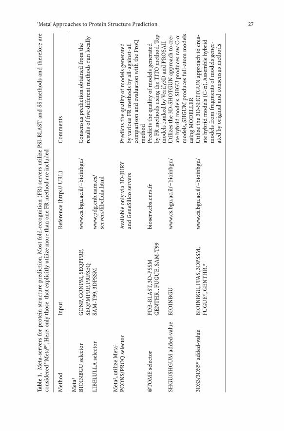
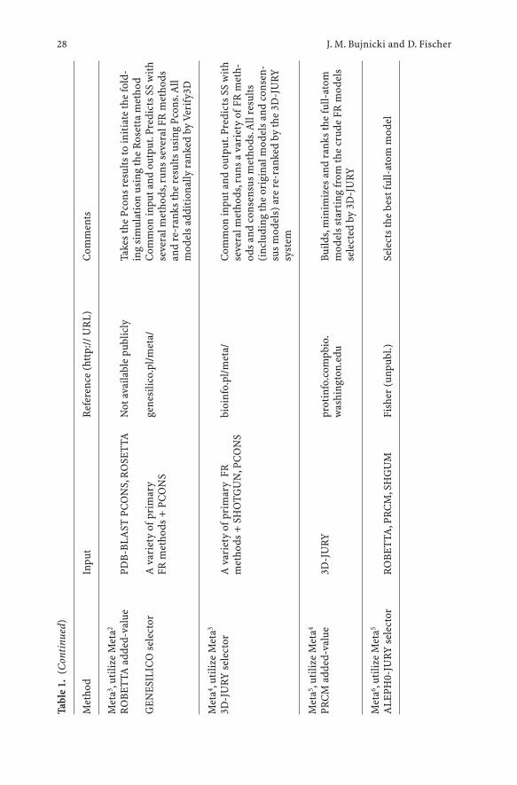
Following the proven success of manual meta-predictors, several groups havealready implemented fully-automated versions of the meta-approach(Table 1).Automated meta-predictors can be divided into two types: (1) selec-tors, which simply select models from the input and (2) added-value meta-predictors, which use the input models to generate new models.
One of the earliest meta-predictors was developed by Arne Elofsson byimplementing the CAFASP-CONSENSUS ideas from CASP4 into the auto-mated program Pcons (Lundstrom et al. 2001). Pcons receives,as input; the topmodels produced by different fold-recognition servers and selects the modelsthat are evaluated to be more likely to be correct, based on the structural sim-ilarities among the input models. That is, it does not produce any new models,only re-ranks the existing ones, based on their mutual similarity and the orig-inal scores assigned by the individual servers. Pcons corroborated thestrength of the consensus idea in the subsequent LiveBench experiments(Bujnicki et al. 2001b). It was demonstrated that PCONS2 (version trainedspecifically for a few original, i.e. non-meta servers) combined the sensitivityof the most sensitive original method (3D-PSSM; Kelley et al. 2000) with avery high specificity (higher than any individual server). The most importantfeature contributing to the improved performance of an early version ofPCONS was its scoring system, which allowed to confidently identify the cor-rect models, although it was not always able to identify the absolutely bestmodel among similar top solutions. The newest version of PCONS, reinforced
J. M. Bujnicki and D. Fischer26
‘Meta’ Approaches to Protein Structure Prediction 27
Tabl
e1.
Met
a-se
rver
s fo
r pr
otei
n st
ruct
ure
pred
icti
on.M
ost f
old-
reco
gnit
ion
(FR
) se
rver
s ut
ilize
PSI
-BLA
ST a
nd S
S m
etho
ds a
nd th
eref
ore
are
cons
ider
ed “
Met
a0 ”.H
ere,
only
thos
e th
at e
xplic
itly
uti
lize
mor
e th
an o
ne F
R m
etho
d ar
e in
clud
ed
Met
hod
Inpu
tR
efer
ence
(ht
tp://
UR
L)C
omm
ents
Met
a1
BIO
INBG
U s
elec
tor
GO
NP,
GO
NPM
,SEQ
PPR
F,w
ww
.cs.
bgu.
ac.il
/~bi
oinb
gu/
Con
sens
us p
redi
ctio
n ob
tain
ed fr
om th
eSE
QPM
PRF,
PRFS
EQre
sults
off
ive
diff
eren
t met
hods
run
loca
llyLI
BELU
LLA
sel
ecto
rSA
M-T
99,3
DPS
SMw
ww
.pdg
.cnb
.uam
.es/
serv
ers/
libel
lula
.htm
l
Met
a2 ,ut
ilize
Met
a1
PCO
NS/
PRO
Q s
elec
tor
Avai
labl
e on
ly v
ia 3
D-J
URY
Pr
edic
ts th
e qu
alit
y of
mod
els
gene
rate
d an
d G
eneS
ilico
ser
vers
by v
ario
us F
R m
etho
ds b
y al
l-ag
ains
t-al
lco
mpa
riso
n an
d ev
alua
tion
wit
h th
e Pr
oQm
etho
d@
TOM
E se
lect
orPD
B-BL
AST
,3D
-PSS
M
bios
erv.
cbs.
cnrs
.frPr
edic
ts th
e qu
alit
y of
mod
els
gene
rate
dG
ENT
HR
.,FU
GU
E,SA
M-T
99by
FR
met
hods
usi
ng th
e T
ITO
met
hod.
Top
mod
els
rank
ed b
y Ve
rify
3D a
nd P
RO
SAII
SHG
U/S
HG
UM
add
ed-v
alue
BIO
INBG
Uw
ww
.cs.
bgu.
ac.il
/~bi
oinb
gu/
Uti
lizes
the
3D-S
HO
TGU
N a
ppro
ach
to c
re-
ate
hybr
id m
odel
s.SH
GU
pro
duce
s ra
w C
-am
odel
s,SH
GU
M p
rodu
ces
full-
atom
mod
els
usin
g M
OD
ELLE
R3D
S3/3
DS5
* ad
ded-
valu
eBI
OIN
BGU
,FFA
S,3D
PSSM
,w
ww
.cs.
bgu.
ac.il
/~bi
oinb
gu/
Uti
lize
the
3D-S
HO
TGU
N a
ppro
ach
to c
rea-
FUG
UE*
,GEN
TH
R.*
ate
hybr
id m
odel
s (C
-a).
Ass
embl
e hy
brid
mod
els
from
frag
men
ts o
fmod
els
gene
r-at
ed b
y or
igin
al a
nd c
onse
nsus
met
hods
J. M. Bujnicki and D. Fischer28
Tabl
e1.
(Con
tinu
ed)
Met
hod
Inpu
tR
efer
ence
(ht
tp://
UR
L)C
omm
ents
Met
a3 ,ut
ilize
Met
a2
RO
BET
TA a
dded
-val
uePD
B-BL
AST
PC
ON
S,R
OSE
TTA
Not
ava
ilabl
e pu
blic
lyTa
kes
the
Pcon
s re
sults
to in
itia
te th
e fo
ld-
ing
sim
ulat
ion
usin
g th
e R
oset
ta m
etho
dG
ENES
ILIC
O s
elec
tor
A v
arie
ty o
fpri
mar
y ge
nesi
lico.
pl/m
eta/
Com
mon
inpu
t and
out
put.
Pred
icts
SS
wit
hFR
met
hods
+ P
CO
NS
seve
ral m
etho
ds,r
uns
seve
ral F
R m
etho
dsan
d re
-ran
ks th
e re
sults
usi
ng P
cons
.All
mod
els
addi
tion
ally
ran
ked
by V
erif
y3D
Met
a4 ,ut
ilize
Met
a3
3D-J
URY
sel
ecto
rA
var
iety
ofp
rim
ary
FR
bioi
nfo.
pl/m
eta/
Com
mon
inpu
t and
out
put.
Pred
icts
SS
wit
hm
etho
ds +
SH
OTG
UN
,PC
ON
Sse
vera
l met
hods
,run
s a
vari
ety
ofFR
met
h-od
s an
d co
nsen
sus
met
hods
.All
resu
lts(i
nclu
ding
the
orig
inal
mod
els
and
cons
en-
sus
mod
els)
are
re-
rank
ed b
y th
e 3D
-JU
RYsy
stem
Met
a5 ,ut
ilize
Met
a4
PRC
M a
dded
-val
ue3D
-JU
RYpr
otin
fo.c
ompb
io.
Build
s,m
inim
izes
and
ran
ks th
e fu
ll-at
omw
ashi
ngto
n.ed
um
odel
s st
arti
ng fr
om th
e cr
ude
FR m
odel
sse
lect
ed b
y 3D
-JU
RY
Met
a6 ,ut
ilize
Met
a5
ALE
PH0-
JURY
sel
ecto
rR
OBE
TTA
,PR
CM
,SH
GU
MFi
sher
(un
publ
.)Se
lect
s th
e be
st fu
ll-at
om m
odel
by the PROQ method for protein model evaluation (Wallner and Elofsson2003), exhibits even higher specificity; moreover, it is able to use the set of anyexternal (original or meta) methods as model generators.
LIBELULLA (Juan et al. 2003) is a system of neural networks trained toselect correct folds from among the results of two primary fold-recognitionmethods implemented as web servers, SAM-T99 (Karplus et al. 1999) and3DPSSM (Kelley et al. 2000). It uses a set of associated characteristics such asthe quality of the sequence-structure alignment, distribution of sequence fea-tures (sequence-conserved positions and apolar residues), and compactnessof the resulting models.
Another fully-automated meta-predictor that simply selects models fromthose produced by other servers is 3D-JURY (Ginalski et al. 2003). It takes asinput any set of models, structurally compares all against all using MaxSub(Siew et al. 2000), and selects one that appears to contain the largest recurrentsubset of common coordinates. It does not use any special characteristics ofthe models or of the servers. 3D-JURY is coupled to the BioInfo.PL Meta-server and, thus, can use any model including selection of the most commonmodel from a user-defined subset.
2.3 Hybrid Methods: Going Beyond the “Simple Selection” of Models
Some automated meta-predictors go beyond the simple selection. PMOD usesMODELLER (Sali and Blundell 1993) to generate full-atom models based onthe selection of fold-recognition results reported by PCONS, amended by sec-ondary structure predicted by PSI-PRED (Jones 1999). These models are eval-uated using the PROQ method (Wallner and Elofsson 2003). ROBETTA (D.Baker, unpubl.) builds full-atom models using the ROSETTA fragment inser-tion method (Simons et al. 1997), starting from structures detected by PDB-BLAST or PCONS and aligned by the K*SYNC alignment method. PRCMtakes as input the top models selected by 3D-JURY and builds full-atom mod-els, which are minimized and evaluated using energy functions. ALEPH0-JURY (D. Fischer, unpubl.) selects a model from those of ROBETTA, PRCM,and SHGUM using a combination of the 3D-SHOTGUN technology (seebelow) and evaluation using knowledge-based potentials.
Another successful practice observed in previous CASPs was to buildhybrid models from fragments (e.g. Bujnicki and GeneSilico; see above).Automated meta-predictors using this approach have also been developed.Conceptually, the first method to use the fragment-splicing approach (whichnevertheless should not be considered a meta-server) was David Baker’sROSETTA protein folding simulation algorithm that uses the fragment inser-tion Monte Carlo approach (Simons et al. 1997). The general premise of thismethod is that the protein conformation is reasonably well approximated bythe distribution of local structures adopted by known, not necessarily homol-
‘Meta’ Approaches to Protein Structure Prediction 29
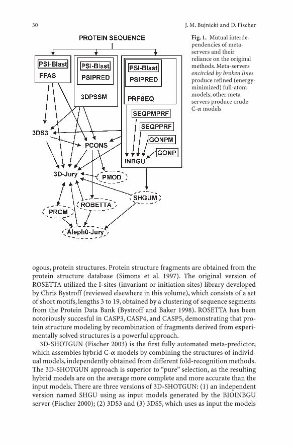
ogous, protein structures. Protein structure fragments are obtained from theprotein structure database (Simons et al. 1997). The original version ofROSETTA utilized the I-sites (invariant or initiation sites) library developedby Chris Bystroff (reviewed elsewhere in this volume), which consists of a setof short motifs, lengths 3 to 19, obtained by a clustering of sequence segmentsfrom the Protein Data Bank (Bystroff and Baker 1998). ROSETTA has beennotoriously succesful in CASP3, CASP4, and CASP5, demonstrating that pro-tein structure modeling by recombination of fragments derived from experi-mentally solved structures is a powerful approach.
3D-SHOTGUN (Fischer 2003) is the first fully automated meta-predictor,which assembles hybrid C-a models by combining the structures of individ-ual models, independently obtained from different fold-recognition methods.The 3D-SHOTGUN approach is superior to “pure” selection, as the resultinghybrid models are on the average more complete and more accurate than theinput models. There are three versions of 3D-SHOTGUN: (1) an independentversion named SHGU using as input models generated by the BIOINBGUserver (Fischer 2000); (2) 3DS3 and (3) 3DS5, which uses as input the models
J. M. Bujnicki and D. Fischer30
Fig. 1. Mutual interde-pendencies of meta-servers and theirreliance on the originalmethods. Meta-serversencircled by broken linesproduce refined (energy-minimized) full-atommodels, other meta-servers produce crude C-a models
from three or five different independent fold-recognition servers, respec-tively. A new automated version of the SHOTGUN series, which was very suc-cessful in CASP is SHGUM, which generates full-atom, refined models, with-out the collisions and gaps seen in some of the raw spliced models. SHGUM isan independent server using the same input as SHGU (i.e. the results ofBIOINBGU).
Figure 1 shows the diagram of mutual interdependencies of “metaN-servers” and their reliance on the input from original servers and other“metaN-1-servers”.
3 Future Prospects
The recent CASP5/CAFASP3 evaluation has clearly shown that the meta-servers, on average, perform much better than these primary servers and thehigher the N in the metaN, the more the meta-server is likely to succeed. Thisworks because no program is suitable for all cases, and each program has itsstrengths and weaknesses, and with each layer of “meta”- analysis thestrengths can be amplified. The idea of meta-servers, or more precisely – thepost-CASP5 proliferation of meta-servers – has met, however, with ambigu-ous reactions of the community of developers of bioinformatic methods. Onthe one hand, it is much easier to develop meta-servers (especially a relativelysimple selector) than to develop a new, original fold-recognition method. Onthe other hand, because of the out-sourcing, the existing meta-servers are veryslow: always slower than the slowest of the external servers used to generateprimary predictions.
The idea of “meta-prediction” is well known in areas such as artificial intel-ligence or the stock market,where independent agents are used to obtain a con-sensus prediction that will be on average more accurate than any of the indi-vidual agents. Initially, you consult with various external brokers, but if youhave the money, you just hire them to sit at your location. Thus, the richest willbe the winner.Likewise,in order to obtain a fast protein fold-recognition meta-server-type method applicable at genomic scales, meta-predictors will runeach of the “lower-rank”components locally,without the dependence on otherexternal servers. Many of the existing fold-recognition methods have imple-mented a local version of PSI-BLAST (Altschul et al. 1997) for databasesearches and generation of multiple sequence alignment,and PSI-PRED (Jones1999) (which itself utilizes PSI-BLAST) for prediction of secondary structure.It has been envisaged that the future meta-servers would utilize local imple-mentations of original fold-recognition methods and lower-rank meta-proto-cols. Hence, some of the criticism attributed to the first generation of meta-servers may not be justified and will certainly fade away in the future, whenfast,powerful independent meta-predictors will challenge the best human pre-dictors.Whether this will happen at CASP6 or later, remains to be seen.
‘Meta’ Approaches to Protein Structure Prediction 31
References
Alexandrov NN, Nussinov R, Zimmer RM (1996) Fast protein fold recognition viasequence to structure alignment and contact capacity potentials. Pac Symp Biocom-put, pp 53–72
Altschul SF, Madden TL, Schaffer AA, Zhang J, Zhang Z, Miller W, Lipman DJ (1997)Gapped BLAST and PSI-BLAST: a new generation of protein database search pro-grams. Nucleic Acids Res 25:3389–3402
Bryant SH, Lawrence CE (1993) An empirical energy function for threading proteinsequence through the folding motif. Proteins 16:92–112
Bujnicki JM, Elofsson A, Fischer D, Rychlewski L (2001a) LiveBench-1: continuousbenchmarking of protein structure prediction servers. Protein Sci 10:352–361
Bujnicki JM, Elofsson A, Fischer D, Rychlewski L (2001b) LiveBench-2: Large-scale auto-mated evaluation of protein structure prediction servers. Proteins 45:184–191
Bystroff C, Baker D (1998) Prediction of local structure in proteins using a library ofsequence- structure motifs. J Mol Biol 281:565–577
Cuff JA, Barton GJ (2000) Application of multiple sequence alignment profiles toimprove protein secondary structure prediction. Proteins 40:502–511
Cuff JA, Clamp ME, Siddiqui AS, Finlay M, Barton GJ (1998) JPred: a consensus sec-ondary structure prediction server. Bioinformatics 14:892–893
Di Francesco V, Geetha V, Garnier J, Munson PJ (1997) Fold recognition using predictedsecondary structure sequences and hidden Markov models of protein folds. Proteins(Suppl 1):123–128
Fischer D (2000) Hybrid fold recognition: combining sequence derived properties withevolutionary information. Pac Symp Biocomput, pp 119–130
Fischer D (2003) 3D-SHOTGUN: a novel, cooperative, fold-recognition meta-predictor.Proteins (in press)
Fischer D, Baker D, Moult J (2001a) We need both computer models and experiments.Nature 409:558
Fischer D, Barrett C, Bryson K, Elofsson A, Godzik A, Jones D, Karplus KJ, Kelley LA, Mac-Callum RM, Pawlowski K, Rost B, Rychlewski L, Sternberg M (1999) CAFASP-1: criti-cal assessment of fully automated structure prediction methods. Proteins (Suppl3):209–217
Fischer D, Elofsson A, Rychlewski L, Pazos F, Valencia A, Rost B, Ortiz AR, Dunbrack RLJr (2001b) CAFASP2: The second critical assessment of fully automated structure pre-diction methods. Proteins 45 (Suppl 5):171–183
Ginalski K, Elofsson A, Fischer D, Rychlewski L (2003) 3D-Jury: a simple approach toimprove protein structure predictions. Bioinformatics 19:1015–1018
Godzik A, Kolinski A, Skolnick J (1992) Topology fingerprint approach to the inverseprotein folding problem. J Mol Biol 227:227–238
Jones DT (1999) Protein secondary structure prediction based on position-specific scor-ing matrices. J Mol Biol 292:195–202
Jones DT, Taylor WR, Thornton JM (1992) A new approach to protein fold recognition.Nature 358:86–89
Juan D, Grana O, Pazos F, Fariselli P, Casadio R, Valencia A (2003) A neural networkapproach to evaluate fold recognition results. Proteins 50:600–608
Karplus K, Barrett C, Cline M, Diekhans M, Grate L, Hughey R (1999) Predicting proteinstructure using only sequence information. Proteins (Suppl 3):121–125
Karplus K, Barrett C, Hughey R (1998) Hidden Markov models for detecting remote pro-tein homologies. Bioinformatics 14:846–856
Kelley LA, McCallum CM, Sternberg MJ (2000) Enhanced genome annotation usingstructural profiles in the program 3D-PSSM. J Mol Biol 299:501–522
J. M. Bujnicki and D. Fischer32
Kihara D, Lu H, Kolinski A, Skolnick J (2001) TOUCHSTONE: an ab initio protein struc-ture prediction method that uses threading-based tertiary restraints. Proc Natl AcadSci USA 98:10125–10130
Kolinski A, Betancourt MR, Kihara D, Rotkiewicz P, Skolnick J (2001) Generalized com-parative modeling (GENECOMP): a combination of sequence comparison, threading,and lattice modeling for protein structure prediction and refinement. Proteins44:133–149
Kosinski J, Cymerman IA, Feder M, Kurowski MA, Sasin JM, Bujnicki JM (2003) A‘Frankenstein’s monster’ approach to comparative modeling: merging the finest frag-ments of fold-recognition models and iterative model refinement aided by 3D struc-ture evaluation. Proteins (in press)
Kurowski MA, Bujnicki JM (2003) GeneSilico protein structure prediction meta-server.Nucleic Acids Res 31:3305–3307
Lundstrom J, Rychlewski L, Bujnicki JM, Elofsson A (2001) Pcons: A neural-network-based consensus predictor that improves fold recognition. Protein Sci 10:2354–2362
Marr D (1982) Vision. Freeman, New YorkMarti-Renom MA, Stuart AC, Fiser A, Sanchez R, Melo F, Sali A (2000) Comparative pro-
tein structure modeling of genes and genomes. Annu Rev Biophys Biomol Struct29:291–325
Moult J, Fidelis K, Zemla A, Hubbard T (2001) Critical assessment of methods of proteinstructure prediction (CASP): round IV. Proteins (Suppl 5):2–7
Moult J, Hubbard T, Bryant SH, Fidelis K, Pedersen JT (1997) Critical assessment of meth-ods of protein structure prediction (CASP): round II. Proteins (Suppl 1):2–6
Moult J, Hubbard T, Fidelis K, Pedersen JT (1999) Critical assessment of methods of pro-tein structure prediction (CASP): round III. Proteins (Suppl 3):2–6
Moult J, Pedersen JT, Judson R, Fidelis K (1995) A large-scale experiment to assess pro-tein structure prediction methods. Proteins 23:ii–iv
Ouzounis C, Sander C, Scharf M, Schneider R (1993) Prediction of protein structure byevaluation of sequence-structure fitness. Aligning sequences to contact profilesderived from three-dimensional structures. J Mol Biol 232:805–825
Rost B(1995) TOPITS: threading one-dimensional predictions into three-dimensionalstructures. ISMB 3:314–321
Rychlewski L, Jaroszewski L, Li W, Godzik A (2000) Comparison of sequence profiles.Strategies for structural predictions using sequence information. Protein Sci9:232–241
Sali A, Blundell TL (1993) Comparative protein modelling by satisfaction of spatialrestraints. J Mol Biol 234:779–815
Selbig J, Mevissen T, Lengauer T (1999) Decision tree-based formation of consensus pro-tein secondary structure prediction. Bioinformatics 15:1039–1046
Shi J, Blundell TL, Mizuguchi K (2001) Fugue: sequence-structure homology recognitionusing environment-specific substitution tables and structure-dependent gap penal-ties. J Mol Biol 310:243–257
Siew N, Elofsson A, Rychlewski L, Fischer D (2000) MaxSub: an automated measure forthe assessment of protein structure prediction quality. Bioinformatics 16:776–785
Simons KT, Kooperberg C, Huang E, Baker D (1997) Assembly of protein tertiary struc-tures from fragments with similar local sequences using simulated annealing andBayesian scoring functions. J Mol Biol 268:209–225
Simons KT, Ruczinski I, Kooperberg C, Fox BA, Bystroff C, Baker D.(1999) Improvedrecognition of native-like protein structures using a combination of sequence-depen-dent and sequence-independent features of proteins. Proteins 34:82–95
Sippl MJ, Weitckus S (1992) Detection of native-like models for amino acid sequences ofunknown three-dimensional structure in a data base of known protein conforma-tions. Proteins 13:258–271
‘Meta’ Approaches to Protein Structure Prediction 33

Tramontano A (2003) Of men and machines. Nat Struct Biol 10:87–90Wallner B, Elofsson A (2003) Can correct protein models be identified? Protein Sci
12:1073–1086
J. M. Bujnicki and D. Fischer34
Related Documents