Fungal Genetics and Biology 43 (2006) 343–356 www.elsevier.com/locate/yfgbi 1087-1845/$ - see front matter Published by Elsevier Inc. doi:10.1016/j.fgb.2006.01.003 Computational analysis of the Phanerochaete chrysosporium v2.0 genome database and mass spectrometry identiWcation of peptides in ligninolytic cultures reveal complex mixtures of secreted proteins Amber Vanden Wymelenberg a , Patrick Minges a , Grzegorz Sabat b , Diego Martinez c , Andrea Aerts d , Asaf Salamov d , Igor Grigoriev d , Harris Shapiro d , Nik Putnam d , Paula Belinky e , Carlos Dosoretz f , Jill Gaskell g , Phil Kersten g , Dan Cullen g,¤ a Department of Bacteriology, University of Wisconsin, Madison, WI 53706, USA b Genetics and Biotechnology Center, University of Wisconsin, Madison, WI 53706, USA c Joint Genome Institute, Los Alamos National Laboratories, Los Alamos, NM 87545, USA d Joint Genome Institute, Walnut Creek, CA 94598, USA e Environmental Biotechnology Laboratory, Migal-Galilee Technology Center, P.O. Box 831 Kiryat-Shmona, Israel f Civil and Environmental Engineering, Technion-Israel Institute of Technology, Haifa 32000, Israel g USDA Forest Products Laboratory, One GiVord Pinchot Dr., Madison, WI 53726, USA Received 21 November 2005; accepted 11 January 2006 Available online 9 March 2006 Abstract The white-rot basidiomycete Phanerochaete chrysosporium employs extracellular enzymes to completely degrade the major polymers of wood: cellulose, hemicellulose, and lignin. Analysis of a total of 10,048 v2.1 gene models predicts 769 secreted proteins, a substantial increase over the 268 models identiWed in the earlier database (v1.0). Within the v2.1 ‘computational secretome,’ 43% showed no signiW- cant similarity to known proteins, but were structurally related to other hypothetical protein sequences. In contrast, 53% showed signiW- cant similarity to known protein sequences including 87 models assigned to 33 glycoside hydrolase families and 52 sequences distributed among 13 peptidase families. When grown under standard ligninolytic conditions, peptides corresponding to 11 peptidase genes were identiWed in culture Wltrates by mass spectrometry (LS–MS/MS). Five peptidases were members of a large family of aspartyl proteases, many of which were localized to gene clusters. Consistent with a role in dephosphorylation of lignin peroxidase, a mannose-6-phospha- tase (M6Pase) was also identiWed in carbon-starved cultures. Beyond proteases and M6Pase, 28 speciWc gene products were identiWed including several representatives of gene families. These included 4 lignin peroxidases, 3 lipases, 2 carboxylesterases, and 8 glycosyl hydro- lases. The results underscore the rich genetic diversity and complexity of P. chrysosporium’s extracellular enzyme systems. Published by Elsevier Inc. Keywords: Phanerochaete chrysosporium; Secretion; Secretome; Proteome; Gene cluster 1. Introduction Lignin, the component of plant cell walls that gives strength to wood, is the second most abundant natural polymer on earth. This amorphous and insoluble aro- matic material lacks stereoregularity, and unlike hemicel- lulose and cellulose is not susceptible to hydrolytic attack. A relatively small group of microbes, collectively referred to as ‘white-rot’ fungi, are uniquely able to completely degrade lignin to gain access to the carbohydrate poly- mers of plant cell walls, which they use as carbon and energy sources. The white-rot basidiomycete Phanerocha- ete chrysosporium has become the model system for study- ing the physiology and genetics of lignin degradation (for * Corresponding author. Fax: +1 608 231 9262. E-mail address: dcullen@facstaV.wisc.edu (D. Cullen).

Welcome message from author
This document is posted to help you gain knowledge. Please leave a comment to let me know what you think about it! Share it to your friends and learn new things together.
Transcript
Fungal Genetics and Biology 43 (2006) 343–356
www.elsevier.com/locate/yfgbi
Computational analysis of the Phanerochaete chrysosporium v2.0 genome database and mass spectrometry identiWcation of peptides in
ligninolytic cultures reveal complex mixtures of secreted proteins
Amber Vanden Wymelenberg a, Patrick Minges a, Grzegorz Sabat b, Diego Martinez c, Andrea Aerts d, Asaf Salamov d, Igor Grigoriev d, Harris Shapiro d, Nik Putnam d, Paula Belinky e,
Carlos Dosoretz f, Jill Gaskell g, Phil Kersten g, Dan Cullen g,¤
a Department of Bacteriology, University of Wisconsin, Madison, WI 53706, USAb Genetics and Biotechnology Center, University of Wisconsin, Madison, WI 53706, USA
c Joint Genome Institute, Los Alamos National Laboratories, Los Alamos, NM 87545, USAd Joint Genome Institute, Walnut Creek, CA 94598, USA
e Environmental Biotechnology Laboratory, Migal-Galilee Technology Center, P.O. Box 831 Kiryat-Shmona, Israelf Civil and Environmental Engineering, Technion-Israel Institute of Technology, Haifa 32000, Israel
g USDA Forest Products Laboratory, One GiVord Pinchot Dr., Madison, WI 53726, USA
Received 21 November 2005; accepted 11 January 2006Available online 9 March 2006
Abstract
The white-rot basidiomycete Phanerochaete chrysosporium employs extracellular enzymes to completely degrade the major polymersof wood: cellulose, hemicellulose, and lignin. Analysis of a total of 10,048 v2.1 gene models predicts 769 secreted proteins, a substantialincrease over the 268 models identiWed in the earlier database (v1.0). Within the v2.1 ‘computational secretome,’ 43% showed no signiW-cant similarity to known proteins, but were structurally related to other hypothetical protein sequences. In contrast, 53% showed signiW-cant similarity to known protein sequences including 87 models assigned to 33 glycoside hydrolase families and 52 sequences distributedamong 13 peptidase families. When grown under standard ligninolytic conditions, peptides corresponding to 11 peptidase genes wereidentiWed in culture Wltrates by mass spectrometry (LS–MS/MS). Five peptidases were members of a large family of aspartyl proteases,many of which were localized to gene clusters. Consistent with a role in dephosphorylation of lignin peroxidase, a mannose-6-phospha-tase (M6Pase) was also identiWed in carbon-starved cultures. Beyond proteases and M6Pase, 28 speciWc gene products were identiWedincluding several representatives of gene families. These included 4 lignin peroxidases, 3 lipases, 2 carboxylesterases, and 8 glycosyl hydro-lases. The results underscore the rich genetic diversity and complexity of P. chrysosporium’s extracellular enzyme systems.Published by Elsevier Inc.
Keywords: Phanerochaete chrysosporium; Secretion; Secretome; Proteome; Gene cluster
1. Introduction
Lignin, the component of plant cell walls that givesstrength to wood, is the second most abundant naturalpolymer on earth. This amorphous and insoluble aro-
* Corresponding author. Fax: +1 608 231 9262.E-mail address: [email protected] (D. Cullen).
1087-1845/$ - see front matter Published by Elsevier Inc.doi:10.1016/j.fgb.2006.01.003
matic material lacks stereoregularity, and unlike hemicel-lulose and cellulose is not susceptible to hydrolytic attack.A relatively small group of microbes, collectively referredto as ‘white-rot’ fungi, are uniquely able to completelydegrade lignin to gain access to the carbohydrate poly-mers of plant cell walls, which they use as carbon andenergy sources. The white-rot basidiomycete Phanerocha-ete chrysosporium has become the model system for study-ing the physiology and genetics of lignin degradation (for

344 A.Vanden Wymelenberg et al. / Fungal Genetics and Biology 43 (2006) 343–356
review, see Cullen and Kersten, 2004). Initially released in2002 (Martinez et al., 2004), the P. chrysosporium genomeassembly and gene models were recently updated(www.jgi.doe.gov/whiterot).
Major components of the P. chrysosporium lignin depoly-merization systems include lignin peroxidase (LiP), manga-nese peroxidase (MnP), and a peroxide-generating enzyme,glyoxal oxidase (GLOX). DeWned media with limitingamounts of carbon or nitrogen are routinely employed forenzyme production, and under these conditions P. chrysospo-rium secretes multiple LiP and MnP isozymes. Ligninperoxidase gene lipD encodes the dominant isozyme in car-bon-limited medium, and evidence suggests this protein isdephosphorylated by a mannose-6-phosphatase (M6Pase)(Kuan and Tien, 1989; Rothschild et al., 1997; Rothschildet al., 1999). The LiPs and MnPs are encoded by families of10 and 5 structurally related genes, respectively. Eight lips areclosely linked within a 100kb region (Stewart and Cullen,1999), and mnp1 lies 5.7 kb from mnp4 (Martinez et al., 2004).The lips and mnps exhibit dramatic diVerential regulation inresponse to media composition, but no clear relationship hasbeen observed between transcriptional regulation and geno-mic organization (reviewed in Cullen and Kersten, 2004).
Beyond the oxidative enzymes and M6Pase, relatively littleis known of extracellular proteins present in ligninolytic cul-tures. Several proteases have been partially characterized fromsubmerged cultures, but it remains uncertain whether extra-cellular peroxidases are substantially degraded under lignino-lytic conditions (Bonnarme et al., 1993; Dass et al., 1995;Dosoretz et al., 1990a,b). The relationship between the prote-ases produced under such nutrient limitation and those pro-duced in colonized wood pulp (Datta, 1992) or in cellulolyticcultures (Eriksson and Pettersson, 1982) is also unclear. Thelatter enzymes have been implicated in the activation of cellu-lase activity (Eriksson and Pettersson, 1982) and in the cleav-age of cellobiose dehydrogenase functional domains (Eggertet al., 1996; Habu et al., 1993). With regard to P. chrysospo-rium protease genetics, a cDNA encoding a serine proteasewas recently isolated from non-ligninolytic cultures (Faracoet al., 2005), and a family of clustered glutamic proteases hasbeen observed within the genome (Sims et al., 2004). LC–MS/MS peptide identiWcation demonstrated the expression ofthree aspartyl protease genes in a medium containing celluloseas sole carbon source (Vanden Wymelenberg et al., 2005).
Herein, we describe the computational identiWcation andanalysis of v2.1 protein models with predicted secretion sig-nals. Shotgun LC–MS/MS on Wltrates from carbon- andnitrogen-limited cultures identiWed the expected peroxi-dases and glyoxal oxidases in addition to an impressivearray of previously unknown extracellular proteins.
2. Methods
2.1. Fungal strains and culture conditions
P. chrysosporium strain RP78 ((Stewart et al., 2000)FGSC strain 9002), a homokaryotic derivative of BKM-F-
1767 (Center for Forest Mycology Research, Forest Prod-ucts Laboratory, Madison, WI), was used throughout.Standard B3 salts media with limiting carbon or nitrogenwere grown statically at 39 °C as previously described(Brown et al., 1988; Kirk et al., 1978) and harvested on days4 and 5, respectively. LiP activities as measured by veratrylalcohol oxidation (Tien and Kirk, 1984) were 8.41 and18.6 nmol min¡1 ml¡1 in C-limited and N-limited cultures,respectively.
2.2. Protein analysis
Carbon- and nitrogen-limited cultures were harvested byWltration through Miracloth (Calbiochem, La Jolla, CA)and the Wltrates were stored at ¡20 °C. One hundred sev-enty-Wve milliliters of each Wltrate was concentrated 100-fold in an Amicon 8400 stirred ultraWltration cell with a5000 MWCO polyethersulfone membrane (MilliporeCorp., Bedford, MA). Five hundred microliters Amicon-concentrated protein was further concentrated in a 10,000MWCO Nanosep centrifugal device (Pall Life Sciences,Ann Arbor, MI) to a Wnal volume of 50 �l. Twenty-Wvemicroliters were mixed with 20 �l Laemmli buVer (Bio-RadLaboratories, Inc, Hercules, CA) and loaded onto a 12.5%Criterion Tris–HCL Ready Gel (Bio-Rad Laboratories) forSDS–PAGE. Electrophoresis was performed in a Bio-RadCriterion Cell, 200 V, 50 min, 23 °C. Gels were stained withCoomassie Blue R-250 (Bio-Rad Laboratories) to estimateprotein abundance and MW distribution. Total proteinresolved on the gel was manually fractioned with a surgicalblade into 10, 2 mm long and 5 mm wide strips. These gelstrips were further cut into »1 mm pieces and placed inindividual siliconized 1.5 ml microcentrifuge tubes (FisherScientiWc) for subsequent enzymatic digestion.
‘In Gel’ digestion and mass spectrometric analysis wereperformed as described (www.biotech.wisc.edu/ServicesRe-search/MassSpec/ingel.htm.) In short, gel pieces were de-stained completely in MeOH/H2O/NH4HCO3 (50%:50%:100mM), dehydrated for 10min in acetonitrile/H2O/NH4HCO3(50%:50%:25 mM) and then once more for 1min in 100%acetonitrile. The samples were dried in a Speed-Vac for5 min, reduced in 25mM DTT (dithiothreitol in 25 mMNH4HCO3) for 30min at 56 °C, alkylated with 55 mM IAA(iodoacetamide in 25 mM NH4HCO3) in darkness at roomtemperature for 30 min, washed in H2O for 20min, equili-brated in 25 mM NH4HCO3 for 10 min, dehydrated for10 min in acetonitrile/H2O/NH4HCO3 (50%: 50%:25 mM)and then once more for 1 min in 100% acetonitrile. Followingdrying, samples were rehydrated with 25�l of trypsinsolution (20 ng/�l trypsin (Sequence Grade ModiWed,Promega Inc., Madison, WI) in 25 mM NH4HCO3) or with25�l Asp-N solution (8ng/�l endoproteinase Asp-N, RocheBiochemicals) in 50 mM Na2HPO4, 5 mM Tris–HCl, pH 8.0.Additional buVer overlay (»15�l) was provided to keep gelfragments immersed. The digestions were conducted over-night (18 h) at 37°C, then terminated by acidiWcation with0.1% TFA (triXuoroacetic acid). Peptides generated from
A.Vanden Wymelenberg et al. / Fungal Genetics and Biology 43 (2006) 343–356 345
digestions were extracted in two subsequent steps, Wrst withan equal volume of 0.1% TFA (»50�l) and vigorous vor-texing for 15min, then with the same volume of acetonitrile/H2O/TFA (70%:25%:5%) and vortexing. The collected pep-tide solution was dried completely in a Speed-Vac, re-sus-pended in 50�l of 0.1% TFA, and solid phase extracted (C18SPEC-PLUS™-PT pipette tips Varian, Inc., Lake Forest,CA). Peptides were eluted oV the C18 column with acetoni-trile/H2O/TFA (70%:25%:0.2%), dried in a Speed-Vac andWnally reconstituted with 45�l of 0.1% formic acid.
Peptide fractions were individually analyzed bynanoLC–MS/MS using 1100 series LC/MSD Trap SL spec-trometer (Agilent, Palo Alto, CA). Chromatography ofpeptides prior to mass spectral analysis was accomplishedusing C18 reverse phase HPLC trap column (Zorbax300SB-C18, 5 �M, 5£0.3 mm, Agilent) and separation col-umn (Zorbax 300SB-C18, 3.5 �m, 0.075£150 mm, Agilent)onto which 40 �l of each extracted peptide fraction wasautomatically loaded. An Agilent 1100 series HPLC deliv-ered solvents A: 0.1% (v/v) formic acid in water, and B: 95%(v/v) acetonitrile, 0.1% (v/v) formic acid, at either 10 �l/minto load sample, or at 0.28 �l/min to elute peptides directlyinto the nano-electrospray. The elution was for 80 min in agradient from 20 to 60% (v/v) solvent B. Peptides elutingfrom the HPLC column/electrospray source were trappedin an ion cell and sequential MS/MS spectra spanning from300 to 2200 m/z were generated for the four most abundantions present at each switching event. Redundancy was lim-ited by dynamic exclusion. MS/MS data were converted tomatrix generic format (mfg) Wles using Data Analysis Soft-ware (Agilent). Spectrum Mill MS Proteomics Workbench(Agilent) and an in-house licensed Mascot search engine(Matrix Science, London, UK) were used to identify pep-tides using a dataset of 10,048 gene models described below.Throughout, protein similarity scores are based on theSmith–Waterman algorithm (Smith and Waterman, 1981)using the BLOSUM62 matrix.
2.3. Genome assembly and automated annotation
The v.2.0 assembly and v2.1 gene models are consider-ably improved relative to earlier versions (Table 1). Thev1.0 assembly was based on a 9.75-fold redundant wholegenome shotgun (WGS) dataset in paired-end reads fromthree 3.1§0.2 Kb genomic DNA libraries (Martinez et al.,2004). To supplement this data set and improve the assem-bly, an additional 116 Mb of high quality shotgun sequencewas generated in the form of paired-end sequences from6.3 Kb plasmid and 35 Kb fosmid clones. All sequence readsare available from the NCBI trace archive (http://www.ncbi.nih.gov/Traces/). The WGS reads were assembledwith version 1.0.3 of the JAZZ Assembler. The assemblycontains a total of 32.5 Mb of sequence (excluding gaps)and 1252 contigs. Half of this assembled contig sequence(N50) is contained in the largest 44 contigs, the smallest ofwhich is 228 kb in length. There are a total of 232 scaVoldsin the assembly, and 95% of the assembled sequence is con-
tained in the longest 24 scaVolds, which all have a netlength (excluding internal gaps) of at least 210 Kb.
A total of 10,048 gene models were predicted and anno-tated in the v2.1 P. chrysosporium genome assembly usingJGI Annotation pipeline. Predicted genes, supporting evi-dence, annotations, and analyses are available throughinteractive visualization and analysis tools from JGIGenome Portal (www.jgi.doe.gov/whiterot).
Gene prediction methods used for annotation of the newassembly include ab initio methods Fgenesh (Salamov andSolovyev, 2000), homology-based methods, Fgenesh+(www.softberry.com) and Genewise (Birney and Durbin,2000). Fgenesh was trained on a set of available mRNAs,ESTs, and reliable homology gene models and showed78.3% sensitivity (fraction of correctly detected true exons)and 77.6% speciWcity (fraction of true exons among all pre-dicted exons). GeneWise models were extended when possi-ble to include start and stop codons. When multiple modelswere predicted at the same locus, the model with besthomology, including coverage in both model and hitsequences, was selected for the non-redundant set of genescalled ‘BestModels, v2.1.’ This set includes 66.5% Fge-nesh(+) and 33.5% Genewise gene models. Only 6% aresupported by both methods.
Approximately 75% of genes in the non-redundant sethave known functional domains or show homology toknown proteins in other genomes. Genes have beenannotated and classiWed according to GO (gene ontologyconsortium http://www.geneontology.org/), eukaryoticorthologous groups (KOGs (Koonin et al., 2004)), andKEGG metabolic pathways (Kanehisa et al., 2004). Fol-lowing KEGG annotation, E.C. numbers have beenassigned to 2252 genes. 7220 and 4923 genes have KOGand GO assignments, respectively.
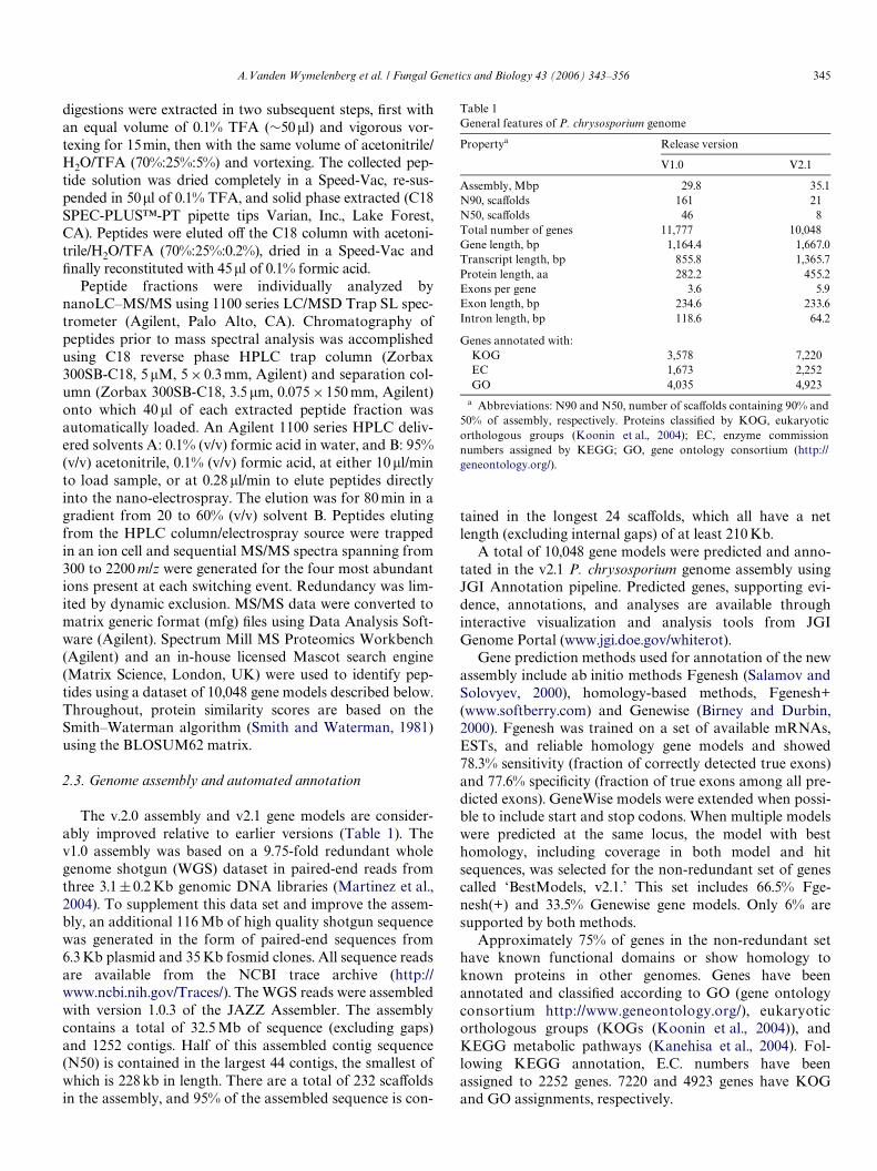
Table 1General features of P. chrysosporium genome
a Abbreviations: N90 and N50, number of scaVolds containing 90% and50% of assembly, respectively. Proteins classiWed by KOG, eukaryoticorthologous groups (Koonin et al., 2004); EC, enzyme commissionnumbers assigned by KEGG; GO, gene ontology consortium (http://geneontology.org/).
Propertya Release version
V1.0 V2.1
Assembly, Mbp 29.8 35.1N90, scaVolds 161 21N50, scaVolds 46 8Total number of genes 11,777 10,048Gene length, bp 1,164.4 1,667.0Transcript length, bp 855.8 1,365.7Protein length, aa 282.2 455.2Exons per gene 3.6 5.9Exon length, bp 234.6 233.6Intron length, bp 118.6 64.2
Genes annotated with:KOG 3,578 7,220EC 1,673 2,252GO 4,035 4,923
346 A.Vanden Wymelenberg et al. / Fungal Genetics and Biology 43 (2006) 343–356
Comparison with 11,777 gene models from the earlierannotation release v1.0 (Martinez et al., 2004), where Gene-wise (Birney and Durbin, 2000) and GrailEXP (Xu andUberbacher, 1997) were used for gene prediction, showsconsiderable improvement. Contaminants and transposonshave been removed; gene fragments were combined intomore complete and dense models. Gene structure statisticssummarized in Table 1 indicate smaller introns, highernumber of exons, and larger transcripts and proteins. Man-ual validation of the models also supports these observa-tions.
V1.0 gene models were mapped on the new assembly(displayed as ‘BestModels v1.0’ track) and compared withv2.1 model set. Seventy-seven percent of gene loci of v1.0are present in v2.1. While the majority of release-speciWcgene models are ab initio predictions, 8% of missed modelsfrom the old set contain PFAM domain and half of themare related to repeats. Fourteen percent of the new modelscontain additional functional domains.
Based on analysis of domain composition in both genesets, the repertoire of molecular functions of the new releasebecame richer. According to analysis of PFAM domains,out of 1637 PFAM domains, 589 occur more frequently innew set with 309 not present in the earlier release. One hun-dred and eighty-nine lost domains from the previous releaseinclude large number of transposon-related domains likeintegrase and reverse transcriptase that were removed fromthe current set of genes.
3. Results
3.1. Secretome computational analysis
A secretome dataset of P. chrysosporium was generatedusing the most recent assembly (www.jgi.doe.gov/whiterot).The 10,048 v2.1 protein models were submitted toPHOBIUS (http://phobius.cgb.ki.se/index.html), predictivesoftware with improved discrimination between transmem-brane helices and signal peptides (Kall et al., 2004). A totalof 874 potentially secreted proteins were predicted, andthen further reduced to 769 by Wltering out proteinswith putative mitochondrial targeting signals (http://www.cbs.dtu.dk/services/TargetP/). Thus, at least 7.6% ofthe P. chrysosporium gene models are predicted to encodesecreted proteins. For comparison, 4.5% of the 6165 ORFsof Candida albicans were predicted using a similar compu-tational approach (Lee et al., 2003b). It is important to notethat some of these proteins are likely not extracellular,including those that may be cell wall bound, residing withinvacuoles, or ER-related.
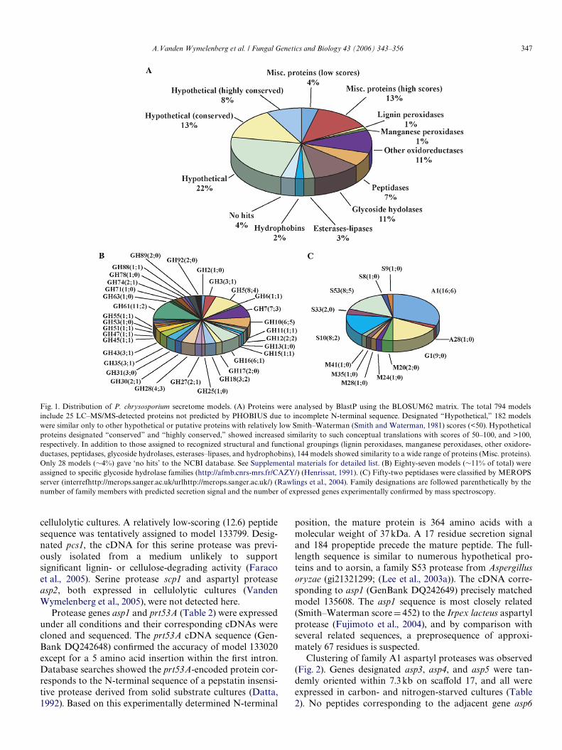
BlastP analysis against the NCBI non-redundant data-base categorized all but 38 sequences by some similarity (>50Smith–Waterman score) to current accessions. Nearly halfwere similar only to hypothetical proteins, many of whichwere conceptual translations from other fungal genome pro-jects (Fig. 1). ClustalW analysis of the 359 hypothetical pro-teins revealed few closely related sequences, i.e., a total of Wve
pairs with >80% amino acid identity. Hypothetical proteinssharing >35% sequence identity tended to cluster within thegenome. More speciWcally, we observed 15 separate clusterseach containing 2–7 members. Five pairs of structurallyrelated hypothetical genes showed no apparent linkage (Sup-plemental material). A genome-wide examination of all genesencoding secreted proteins revealed a decidedly non-randomdistribution. Among the 12 longest scaVolds, the percentageof genes encoding extracellular enzymes ranged from 9.5(scaVold 1) to 1.6 (scaVold 6). Closer inspection of scaVoldsshowed clusters unevenly distributed along their length, as isclear from the examples shown in Fig. 2.
The 407 models with similarity to known proteins werebroadly categorized as glycoside hydrolases (87), oxidore-ductases (84), peptidases (52), and esterases–lipases (21),while others could be assigned to more speciWc groupingsuch as hydrophobins (14), lignin peroxidases (10), andmanganese peroxidases (5). The 134 models not easilyassigned to large families or broad functional categories(Fig. 1A, Misc. proteins), included 103 proteins with signiW-cant similarity (Smith–Waterman scores >100) to knownproteins. Glycosyl hydrolases and peptidases were allocatedto speciWc families or clans (Figs. 1B and C) accordingto systems of Coutinho and Henrissat (http://afmb.cnrs-mrs.fr/CAZY/; (Henrissat, 1991)) and Rawlingset al. (http://merops.sanger.ac.uk/; (Rawlings et al., 2004)),respectively.
3.2. Protein identiWcations in ligninolytic media
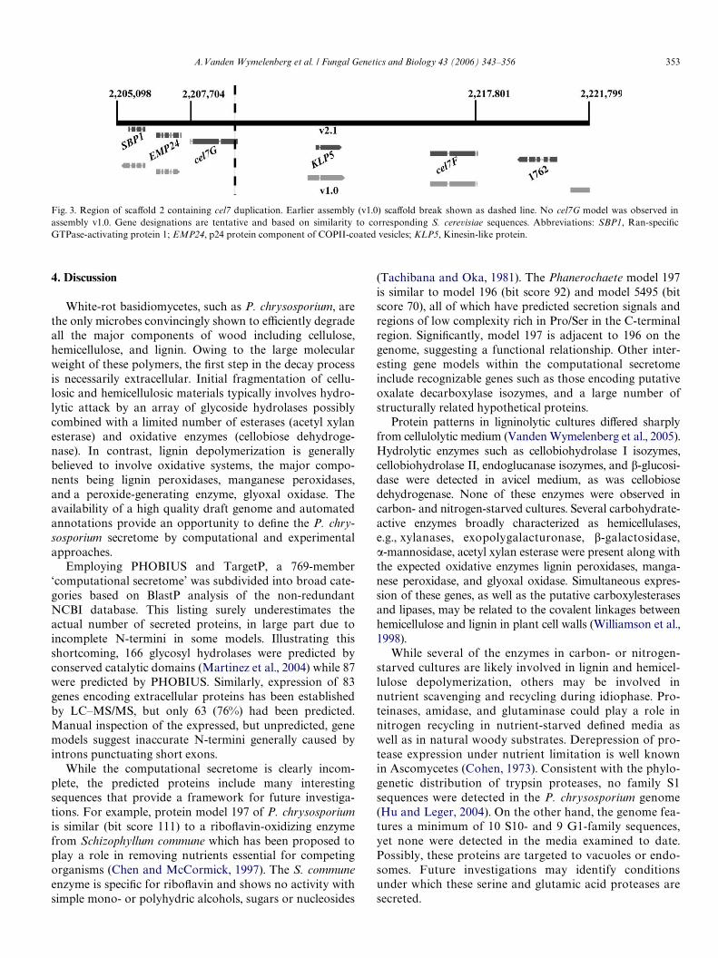
Total soluble protein from carbon- and nitrogen-limitedcultures was concentrated by ultraWltration, size fraction-ated by SDS–PAGE, and subjected to LC–MS/MS analy-sis. Searches of the 10,048 v2.1 protein database usingconservative cut-oV scores (>13, SpectrumMill; >40, Mas-cot), allowed unambiguous assignment of 77 unique pep-tide sequences to 40 speciWc gene models (Tables 2 and 3).Expression of 11 of these genes was previously observed incellulolytic cultures (Vanden Wymelenberg et al., 2005). Ofthe 29 new extracellular proteins, 15 were detected only incarbon-limited Wltrates, while 5 were expressed in both car-bon and nitrogen cultures. Analysis of previously acquiredspectra using the v2.1 model database demonstrated theexpression of 5 additional genes in cellulolytic medium(Table 4). A complete listing of the computational secre-tome, all peptide sequences, scores, and previously pub-lished results are available online (Supplemental material).Results for particular protein families follow.
3.2.1. PeptidasesA total of 31 peptidase sequences were detected in car-
bon- and nitrogen-limited cultures. Twenty uniquesequences were assigned to 11 speciWc gene models, repre-senting MEROPS peptidase families A1, S10, and S53. Norepresentatives of other gene families, including the largefamily of glutamic proteases (Fig. 1C; (Sims et al., 2004)),were detected in these studies or in earlier investigations of
A.Vanden Wymelenberg et al. / Fungal Genetics and Biology 43 (2006) 343–356 347
cellulolytic cultures. A relatively low-scoring (12.6) peptidesequence was tentatively assigned to model 133799. Desig-nated pcs1, the cDNA for this serine protease was previ-ously isolated from a medium unlikely to supportsigniWcant lignin- or cellulose-degrading activity (Faracoet al., 2005). Serine protease scp1 and aspartyl proteaseasp2, both expressed in cellulolytic cultures (VandenWymelenberg et al., 2005), were not detected here.
Protease genes asp1 and prt53A (Table 2) were expressedunder all conditions and their corresponding cDNAs werecloned and sequenced. The prt53A cDNA sequence (Gen-Bank DQ242648) conWrmed the accuracy of model 133020except for a 5 amino acid insertion within the Wrst intron.Database searches showed the prt53A-encoded protein cor-responds to the N-terminal sequence of a pepstatin insensi-tive protease derived from solid substrate cultures (Datta,1992). Based on this experimentally determined N-terminal
position, the mature protein is 364 amino acids with amolecular weight of 37 kDa. A 17 residue secretion signaland 184 propeptide precede the mature peptide. The full-length sequence is similar to numerous hypothetical pro-teins and to aorsin, a family S53 protease from Aspergillusoryzae (gi21321299; (Lee et al., 2003a)). The cDNA corre-sponding to asp1 (GenBank DQ242649) precisely matchedmodel 135608. The asp1 sequence is most closely related(Smith–Waterman scoreD 452) to the Irpex lacteus aspartylprotease (Fujimoto et al., 2004), and by comparison withseveral related sequences, a preprosequence of approxi-mately 67 residues is suspected.
Clustering of family A1 aspartyl proteases was observed(Fig. 2). Genes designated asp3, asp4, and asp5 were tan-demly oriented within 7.3 kb on scaVold 17, and all wereexpressed in carbon- and nitrogen-starved cultures (Table2). No peptides corresponding to the adjacent gene asp6
Fig. 1. Distribution of P. chrysosporium secretome models. (A) Proteins were analysed by BlastP using the BLOSUM62 matrix. The total 794 modelsinclude 25 LC–MS/MS-detected proteins not predicted by PHOBIUS due to incomplete N-terminal sequence. Designated “Hypothetical,” 182 modelswere similar only to other hypothetical or putative proteins with relatively low Smith–Waterman (Smith and Waterman, 1981) scores (<50). Hypotheticalproteins designated “conserved” and “highly conserved,” showed increased similarity to such conceptual translations with scores of 50–100, and >100,respectively. In addition to those assigned to recognized structural and functional groupings (lignin peroxidases, manganese peroxidases, other oxidore-ductases, peptidases, glycoside hydrolases, esterases–lipases, and hydrophobins), 144 models showed similarity to a wide range of proteins (Misc. proteins).Only 28 models (»4%) gave ‘no hits’ to the NCBI database. See Supplemental materials for detailed list. (B) Eighty-seven models (»11% of total) wereassigned to speciWc glycoside hydrolase families (http://afmb.cnrs-mrs.fr/CAZY/) (Henrissat, 1991). (C) Fifty-two peptidases were classiWed by MEROPSserver (interrefhttp://merops.sanger.ac.uk/urlhttp://merops.sanger.ac.uk/) (Rawlings et al., 2004). Family designations are followed parenthetically by thenumber of family members with predicted secretion signal and the number of expressed genes experimentally conWrmed by mass spectroscopy.
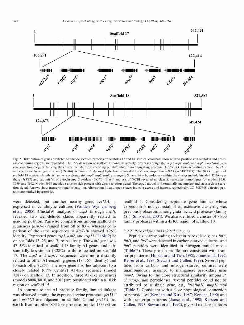
348 A.Vanden Wymelenberg et al. / Fungal Genetics and Biology 43 (2006) 343–356
were detected, but another nearby gene, cel12A, isexpressed in cellulolytic cultures (Vanden Wymelenberget al., 2005). ClustalW analysis of asp3 through asp10revealed two well-deWned clades apparently related togenome position. Pairwise comparisons among scaVold 17sequences (asp3-6) ranged from 50 to 83%, whereas com-parison of the same sequences to asp7-10 showed <25%identity. Expressed genes asp1, asp2, and asp11 (Table 2) lieon scaVolds 13, 25, and 7, respectively. The asp1 gene was47–58% identical to scaVold 18 family A1 genes, and sub-stantially less similar (<28%) to those located on scaVold17. The asp2 and asp11 sequences were more distantlyrelated to other A1-encoding genes (18–36% identity) andto each other (28%). The asp1 gene also lies adjacent to aclosely related (65% identity) A1-like sequence (model7287) on scaVold 13. In addition, three A1-like sequences(models 8008, 8010, and 8011) are positioned within a 10 kbregion on scaVold 15.
In contrast to the A1 protease family, limited linkagewas observed among the expressed serine proteases. prt53Cand prt53D are adjacent on scaVold 2, and prt53A lies6.6 kb from another S53-like protease (model 133398) on
scaVold 1. Considering peptidase gene families whoseexpression is not yet established, extensive clustering waspreviously observed among glutamic acid proteases (familyG1) (Sims et al., 2004). We also identiWed a cluster of 7 S33family proteases within a 45 Kb region of scaVold 10.
3.2.2. Peroxidases and related enzymesPeptides corresponding to lignin peroxidase genes lipA,
lipD, and lipE were detected in carbon-starved cultures, andlipC peptides were identiWed in nitrogen-limited media(Table 3). These protein proWles are consistent with tran-script patterns (Holzbaur and Tien, 1988; James et al., 1992;Reiser et al., 1993; Stewart and Cullen, 1999). Several pep-tides from carbon- and nitrogen-starved cultures wereunambiguously assigned to manganese peroxidase genemnp2. Owing to the close structural similarity among P.chrysosporium peroxidases, several peptides could not beattributed to a single gene, e.g., lipA/lipH, mnp1/mnp4(Table 3). Consistent with a close physiological connectionto peroxidases (Kersten and Kirk, 1987; Kersten, 1990) andwith transcript patterns (Janse et al., 1998; Kersten andCullen, 1993; Stewart et al., 1992), glyoxal oxidase peptides
Fig. 2. Distribution of genes predicted to encode secreted proteins on scaVolds 17 and 18. Vertical crossbars show relative positions on scaVolds and prote-ase-containing regions are expanded. The 16.5 kb region of scaVold 17 contains aspartyl proteases designated asp3, asp4, asp5, and asp6. Saccharomycescerevisiae homologues Xanking the cluster include those encoding putative ubiquitin-conjugating protease (UBC1), GTPase-activating protein (GLO3),and coproporphyrinogen oxidase (HEM6). A family 12 glycosyl hydrolase is encoded by P. chrysosporium cel12A (gi 51872339). The 20.8 kb region ofscaVold 18 contains family A1 sequences designated asp7, asp8, asp9, and asp10. S. cerevisiae homologues within the cluster include histidyl tRNA syn-thase (HTS1) and subunit VI of cytochrome C oxidase (COX6). BlastP analysis of NCBI revealed no clear S. cerevisiae homologues for models 8658,8659, and 8662. Model 8658 encodes a glycine-rich protein with clear secretion signal. The asp10 model is N-terminally incomplete and lacks a clear secre-tion signal. Arrows show transcriptional orientation. Alternating Wll and open spaces indicate exons and introns, respectively. LC–MS/MS-detected pro-teins are marked by asterisks.
A.Vanden Wymelenberg et al. / Fungal Genetics and Biology 43 (2006) 343–356 349
were detected in carbon- and nitrogen-starved media. UsingMALDI-MS Wngerprinting, glyoxal oxidase was recentlyidentiWed in similar media supplemented with vanillin (Shi-mizu et al., 2005). Clustering of lignin peroxidase genes hasbeen reported (Stewart and Cullen, 1999), and lipA, lipC,and lipE are located within a 36 kb region on scaVold 19.The lipD gene is unlinked to all other peroxidases (Gaskellet al., 1994; Stewart et al., 1992).
Several peptides in carbon-starved cultures were matchedto a model (3383) with similarity to the endonuclease/exonu-clease/phosphatase family of proteins (pfam03372). A full-length cDNA clone was obtained (GenBank DQ242647),revealing several relatively minor errors related to intron–exon boundaries in the model. Subsequent analysis showed aprecise match with the experimentally derived N-terminalsequence of an extracellular mannose-6-phosphatase (Roths-child et al., 1999). Designated mpa1, the gene encodes a 22amino acid secretion signal, followed by a 5 residue propep-tide, and a mature peptide of 327 residues. The predictedmolecular weight of 35 kDa and pI of 5.2 are in good agree-ment with experimentally determined properties of themonomer. The enzyme has been shown to dephosphorylatelignin peroxidase isozyme H2, the product of lipD (Roths-child et al., 1999).
3.2.3. Glycosyl hydrolasesEighteen unique peptide sequences were assigned to
eight speciWc glycosyl hydrolase (GH) genes (Table 3), mostof which have been implicated in degradation of hemicellu-lose or pectin. Expression of putative xylanase xyn10D andexoglucanase exg55A were previously observed in sub-merged cultures with ground wood (Abbas et al., 2004) orwith avicel (Vanden Wymelenberg et al., 2005) as sole car-bon sources. xyn10A- and xyn10C-encoded peptides werealso detected in avicel medium (Vanden Wymelenberget al., 2005), and a genomic clone of the former was success-fully expressed in Aspergillus niger (Decelle et al., 2004).
Peptides corresponding to three previously unidentiWedGHs were detected in carbon-limited cultures. A GH family28 sequence showed substantial sequence similarity (Smith–Waterman scores »200) to several known exopolygalactu-ronases and was designated epg28B. The P. chrysosporiumgenome contains a minimum of Wve GH28-like sequences(Martinez et al., 2004), and the deduced epg28B sequence is20 and 25% identical to epg28A and rhg28, respectively. Thelatter gene encodes a putative rhamogalacturonase (Van-den Wymelenberg et al., 2005). In contrast to the GH28family, no representative GH35 or GH47 proteins had beenpreviously observed in P. chrysosporium cultures.
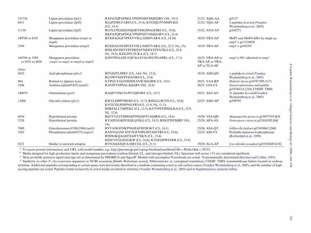
Table 2Phanerochaete chrysosporium protease peptides detected in deWned media
a To access protein information, end URL with model number, e.g., http://genome.jgi-psf.org/cgi-bin/dispGeneModel?db D Phchr1&id D 8470.b Peptidase families identiWed by MEROPS (http://merops.sanger.ac.uk/) as described (Rawlings et al., 2004).c Peptide sequences, media, and Spectrum Mill scores for P. chrysosporium strain RP78 cultivated in media designed for high production of lignin perox-
idases (carbon-limited, CL; and nitrogen-limited, NL) as described in text.d Most probable secretion signal cleavage site as determined by PHOBIUS and SignalP. Although predicted as secreted, the precise cleavage site could
not be determined for the asp2-encoded protein. Models with incomplete N-terminals are noted.e Peptides corresponding to four proteases were previously detected in medium containing avicel as sole carbon source (Vanden Wymelenberg et al.,
2005).
Modela Familyb Peptide sequencec (medium, high score) Probable cleavaged Commentse
8470 A1 (asp5) KIFQTGQSSTAVDQHKT (CL, 16.8; NL, 16.7); RAQDAIVDTGTTLLIVDPTSATAIHRQ (NL, 21.2); RNLYTEFDFGGERV (NL, 19.3)
19/20: AVA-SP
126189 A1 (asp11) KNDGEITFGGLDESKF (CL, 13.7) Incomplete8469 A1 (asp4) KTFQTGSSSTAVDQRK (CL, 17.0; NL, 17.7);
RVGFAPVVLK (CL, 10.1; NL, 12.4)18/19: LAA-AS
8468 A1 (asp3) RTFNTGASSTAVDQKQ (CL, 17.3; NL, 18.8); RGSLAFTPVSIRN (NL, 14.9)
21/22: ASP-AP
135608 A1 (asp1) KATGATLDNNTGLLRL (CL, 17.2; NL, 13.8) 19/20: VAA-TP 7 peptides in avicel(Vanden Wymelenberg et al., 2005)
40125 A1 (asp2) Uncertain 4 peptides in avicel (Vanden Wymelenberg et al., 2005)
1914 S10 (scp1) 22/23: AHA-RM 1 peptide in avicel(Vanden Wymelenberg et al., 2005)
133799 S10 (pcs1) RLAFGTPLLRA (CL, 12.6) 20/21: ALA-AK AJ748587 (Faraco et al., 2005)130748 S53 (prt53B) KGVSVLFSSGDGGVGGSQSTRC (CL, 18.5; NL, 19.8);
RLGLATTPFTTATTN (CL, 12.4)Incomplete
1483 S53 (prt53C) KQLNAVGYTPSAKS (CL, 12.7; NL, 15.0) 18/19: VAA-AP129261 S53 (prt53D) RAYPDVSAQADNFRI (CL, 17.8) 18/19: AVA-VP26825 S53 (prt53E) RFQPNFPASCPFVTTVGATTRV (CL, 17.4; NL, 19.0);
RGSSIMFSSGDDGVGAGNCLTNDGKN (NL, 18)Incomplete
133020 S53 (prt53A) KATQSSNTLGVSGFIDQFANQADLTTFLNRF (CL, 16.5); RGTSILFASGDGGVSGGQSQSCTKF (CL, 19.5; NL, 16.2); KGWDPVTGLGTPNFAALKA (CL, 19.9; NL, 15.3);RSLANNLCNAYAQLGARG (CL, 14.5; NL, 13.1)
17/18: AFA-KP 3 peptides in avicel (Vanden Wymelenberg et al., 2005). Corresponds to wood-derived protease (Datta, 1992).
350A
.Vanden W
ymelenberg et al. / F
ungal Genetics and B
iology 43 (2006) 343–356
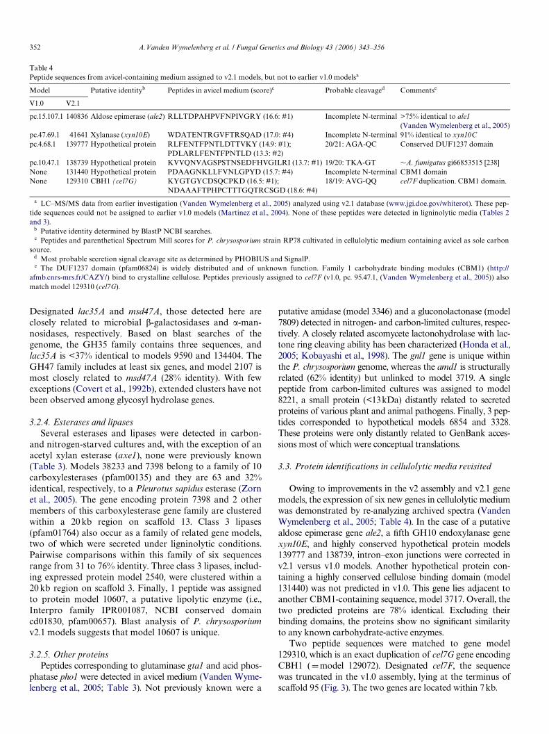
Table 3Peptides identiWed in P. chrysospo
Protein modela Putative id Commentsd
Esterases–lipases38233 Carboxyle P. sapidus gi55466915 [583]7398 Carboxyle 35.8% identical to protein 38233
2540 Lipase (pfa Ustilagu maydis ct gi71008942 [151]8996 Lipase (pfa 49% identical to protein model 2540
10607 GDSL-like Aspergillus fumigatus ct gi66846850 [343]126075 axe1 (acety 4 peptides in avicel (Vanden
Wymelenberg et al., 2005)
Glycosyl hydrolases10763 GH10 xyla 5 peptides in avicel (Vanden
Wymelenberg et al., 2005)30981 GH10 xyla 1 peptide in avicel (Vanden
Wymelenberg et al., 2005)
138345 GH10 xyla 2 peptides in avicel (Vanden Wymelenberg et al., 2005)
4449 GH28 exop Aspergillus tubingensis gi1483221 [199]9466 GH35 �-ga Penicillium emersonii gi44844271 [499]
4550 GH47 �-m Aspergillus saitoi gi1171477 [273]8072 Exo-1,3-�- 12 peptides in avicel (Vanden
Wymelenberg et al., 2005)
41123 GH61 end 1 peptide in avicel
Peroxidases10957 Lignin per gi12628510957 or 121806 Lignin per gi126285 or gi5669882
rium culture Wltrates
entity Peptides detected (medium, high score)b Probable cleavagec
sterase (pfam00135) RTGCSGSADTLQCLRQ (CL, 18.0) Incompletesterase (pfam00135) RAAIFDSSTGPFKT (CL, 19.1); KAVGCTSGPGSFECLQRV
(CL, 16.7; NL, 11.7); KTAPPASTYDEADKPFALLTKA(CL, 16.2)
23/24: AKA-GS
m01764) RINNKEDPIPIVPGRF (CL, 12.5) 19/20: ALA-APm01764) RINNESDPIPIVPGRF (CL, 15.0; NL, 13.2);
RVGNPDFAALFDGEVSDFERI (CL, 15.3)19/20: AHA-AA
lipase (pfam00657) RVLADGLGPNALGRI (CL, 14.8) Incompletel xylan esterase) FAISNWGVDPNRV (CL, 13.0) 24/25: SQC-LP
nase (xyn10C) KLYWGTAADQNRF (CL, 14.9) Incomplete
nase (xyn10D) RGVFTFANADTIANLARN (CL, 23); KLYINDFNIEGTFAKS (CL, 19.4); RMTLPSTPALLQAQKA (CL, 16.2; NL, 16.1); KSTAMQNLVRS (CL, 14.1)
Incomplete
nase (xyn10A) RMTLPSTPALLAQQKT (CL, 16.1) 19/20: VQA-QS
olygalacturonase (epg28B) KVFGGNPSPTSTAGGGTGFVKN (CL, 17.1) Incompletelactosidase (lac35A) RFPVPVGILNPNGKK (CL, 18.4); RPNDTGAQFIIVRQ
(CL, 13.7); RTLPGVATFAGVKL (CL, 13.9); KVILTDYTFGNPANANKL (CL, 19.6)
22/23: ANS-AV
annosidase (msd47A) KEFAFGHDDLEPVSKS (CL, 14.1) 21/22: VAA-GQglucanase (exg55A) KGDGNTDDTAAIQAAINAGGRC (CL, 20.9; NL, 14.1);
KSHPQYTGYAPSDFVSVRS (CL, 21.4); RSNNPNGFADTITAWTRN (CL, 20.9); KVSSPLVVLYQTQLIGDAKN (CL, 20.2); RWSGASSGHLQGSLVLNNIQLTNVPVAVGVKG (CL, 19.5)
26/27: ASG-LG
oglucanase (cel61C) RVPPNNNPVTDVTSKD (CL, 14.8) Incomplete
oxidase (lipA) RAPATQPAPDGLVPEPFHTVDQIINRV (CL, 19.3) 21/22: ANA-AAoxidase (lipA or lipH) RGTAFPGSGGNQGEVESPLPGEIRI (CL, 21.5; NL, 19.4) 21/22: ANA-AA or
VQG-AA
A.V
anden Wym
elenberg et al. / Fungal G
enetics and Biology 43 (2006) 343–356
351
?db D Phchr1&id D 38233.L). Spectrum mill scores >13 are considered signiWcant.rminals are noted. ¤Experimentally determined (Kersten and Cullen, 1993).nceptual translation; COOH� TMH, transmembrane helices located at carboxy
s sole carbon source (Vanden Wymelenberg et al., 2005), and the number of high-l., 2005) and in Supplementary material online.
131738 Lignin peroxidase (lipC) RAPATQPAPDGLVPEPFHSVDQIIDRV (NL, 19.5) 21/22: AQG-AA gi31376811 Lignin peroxidase (lipD) RLQTDHLFARD (CL, 15.4); KTGIQGTVMSPLKG 21/22: TQA-AP 2 peptides in avicel (Vanden
Wymelenberg et al., 2005) )
21/22: ANA-AV gi169271
18/19: VRA-AP MnP1 and MnP4 diVer by single aa. mnp1 D gi13124450
L, 23); 22.4;
18/19: TRA-AP mnp2 D gi169292
17.3) 18/19: VRA-AP or TRA-AP or VRA-AP or TLA-AP
mnp5 is 90% identical to mnp1
18/19: AHS-QN 1 peptide in avicel (Vanden Wymelenberg et al., 2005)
20/21: VAA-RP Haliotis discus gi34787299 (127)20/21: LSA-CL Stenotrophomonas maltophilia
gi19744118 (234). COOH� TMH20/21: ASA-AV 21 peptides in avicel(Vanden
Wymelenberg et al., 2005) 10.4);
A (CL, 12.9;
22/23: ASD-AP¤ gi399595
19/20: VSA-QD Magnaporthe grisea ct gi39977919 [65]QRF (NL, 25/26: APA-AS Neurospora crassa ct gi32416302 [64]
25/26: ASA-QT Gibberella fujikuri gi52430041 [248]);
(CL, 15.9)
22/23: ASS-VV Probable mannose-6-phosphatase (Rothschild et al., 1999)
19/20: ALA-AP Coccidioides posadasii gi25528649 [116]
a To access protein information, end URL with model number, e.g., http://genome.jgi-psf.org/cgi-bin/dispGeneModelb Media designed for high production lignin and manganese peroxidases (carbon-limited, CL; and nitrogen-limited, Nc Most probable secretion signal cleavage site as determined by PHOBIUS and SignalP. Models with incomplete N-ted Similarity to other P. chrysosporium sequences or NCBI accessions [Smith–Waterman scores]. Abbreviations: ct, coterminus. Additional peptides corresponding to certain genes were previously identiWed in a medium containing avicel ascoring peptides are noted. Peptides found exclusively in avicel media are listed in reference (Vanden Wymelenberg et a
(CL, 16.5)11110 Lignin peroxidase (lipE) RGTLFPGSGGNQGEVESGMAGEIRI (CL, 19.4);
RKPATQPAPDGLVPEPFHTVDQIIARV (CL, 21.4140708 or 8191 Manganese peroxidase (mnp1 or
mnp4)RFEDAGGFTPFEVVSLLASHSVARA (CL, 18.24)
3589 Manganese peroxidase (mnp2) RFEDAGNFSPFEVVSLLASHTVARA (CL, 22.5; NRSSLIDCSDVVPVPKPAVNKPATFPATKG (CL, NL, 19.3); KDLDTLTCKA (CL, 14.7)
140708 or 3589 or 8191 or 4636
Manganese peroxidase (mnp1 or mnp2 or mnp4 or mnp5)
KHNTISAADLVQFAGAVALSNCPGAPRL (CL,
Other5655 Acid phosphatase (pho1) RFGIQTLSPKF (CL, 14.6; NL, 15.2);
RLNWVNSFPVDAVRF(CL, 15.4)964 Related to alginate lyase KVPGLYGGNSDDEAVSCSGGRR (CL, 19.6)3346 Amidase (pfam01425) (amd1) RAVIETNPSALAQARV (NL, 18.4)
140079 Glutaminase (gta1) RAQFVNSGTLPNTQDTRF (CL, 14.7)
11068 Glyoxal oxidase (glx1) RIETLDPPFMFRS (CL, 15.7); RISGLLSCFD (CL,KNTETILPDIPNGVRV(CL, 11.9; NL, 11.3); RSRPALLTMPEKL (CL, 12.5); KVTVPITIPSDLKNL, 12.4)
6854 Hypothetical protein REFVVATVDPDAPTPQNPTVAQIRH (CL, 14.6)3328 Hypothetical protein KVAIFGGKPGEQLQYKG (CL, 14.5); RISGTDFS
14);7809 Glucolactonase (COG3386) (gnl1) RVVADGFDKPNGIIAFSEDGKT (CL, 16.1)3383 Phosphatase (pfam03372) (mpa1) RANVGGNFATFTGFNSPGDTASFTRI (CL, 19.0
RDDGKQAGEFSAIFYNKN (CL, 15.4); RIDFVFGGSNGKW (CL, 16.8); KTGEQPWSTRR
8221 Similar to secreted antigens RVNAQAEQVAASECGL (CL, 21.5)
352 A.Vanden Wymelenberg et al. / Fungal Genetics and Biology 43 (2006) 343–356
Designated lac35A and msd47A, those detected here areclosely related to microbial �-galactosidases and �-man-nosidases, respectively. Based on blast searches of thegenome, the GH35 family contains three sequences, andlac35A is <37% identical to models 9590 and 134404. TheGH47 family includes at least six genes, and model 2107 ismost closely related to msd47A (28% identity). With fewexceptions (Covert et al., 1992b), extended clusters have notbeen observed among glycosyl hydrolase genes.
3.2.4. Esterases and lipasesSeveral esterases and lipases were detected in carbon-
and nitrogen-starved cultures and, with the exception of anacetyl xylan esterase (axe1), none were previously known(Table 3). Models 38233 and 7398 belong to a family of 10carboxylesterases (pfam00135) and they are 63 and 32%identical, respectively, to a Pleurotus sapidus esterase (Zornet al., 2005). The gene encoding protein 7398 and 2 othermembers of this carboxylesterase gene family are clusteredwithin a 20 kb region on scaVold 13. Class 3 lipases(pfam01764) also occur as a family of related gene models,two of which were secreted under ligninolytic conditions.Pairwise comparisons within this family of six sequencesrange from 31 to 76% identity. Three class 3 lipases, includ-ing expressed protein model 2540, were clustered within a20 kb region on scaVold 3. Finally, 1 peptide was assignedto protein model 10607, a putative lipolytic enzyme (i.e.,Interpro family IPR001087, NCBI conserved domaincd01830, pfam00657). Blast analysis of P. chrysosporiumv2.1 models suggests that model 10607 is unique.
3.2.5. Other proteinsPeptides corresponding to glutaminase gta1 and acid phos-
phatase pho1 were detected in avicel medium (Vanden Wyme-lenberg et al., 2005; Table 3). Not previously known were a
putative amidase (model 3346) and a gluconolactonase (model7809) detected in nitrogen- and carbon-limited cultures, respec-tively. A closely related ascomycete lactonohydrolase with lac-tone ring cleaving ability has been characterized (Honda et al.,2005; Kobayashi et al., 1998). The gnl1 gene is unique withinthe P. chrysosporium genome, whereas the amd1 is structurallyrelated (62% identity) but unlinked to model 3719. A singlepeptide from carbon-limited cultures was assigned to model8221, a small protein (<13kDa) distantly related to secretedproteins of various plant and animal pathogens. Finally, 3 pep-tides corresponded to hypothetical models 6854 and 3328.These proteins were only distantly related to GenBank acces-sions most of which were conceptual translations.
3.3. Protein identiWcations in cellulolytic media revisited
Owing to improvements in the v2 assembly and v2.1 genemodels, the expression of six new genes in cellulolytic mediumwas demonstrated by re-analyzing archived spectra (VandenWymelenberg et al., 2005; Table 4). In the case of a putativealdose epimerase gene ale2, a Wfth GH10 endoxylanase genexyn10E, and highly conserved hypothetical protein models139777 and 138739, intron–exon junctions were corrected inv2.1 versus v1.0 models. Another hypothetical protein con-taining a highly conserved cellulose binding domain (model131440) was not predicted in v1.0. This gene lies adjacent toanother CBM1-containing sequence, model 3717. Overall, thetwo predicted proteins are 78% identical. Excluding theirbinding domains, the proteins show no signiWcant similarityto any known carbohydrate-active enzymes.
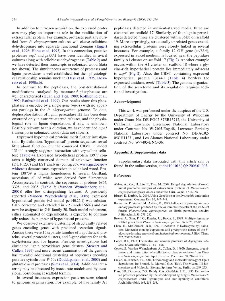
Two peptide sequences were matched to gene model129310, which is an exact duplication of cel7G gene encodingCBH1 (Dmodel 129072). Designated cel7F, the sequencewas truncated in the v1.0 assembly, lying at the terminus ofscaVold 95 (Fig. 3). The two genes are located within 7 kb.
Table 4Peptide sequences from avicel-containing medium assigned to v2.1 models, but not to earlier v1.0 modelsa
a LC–MS/MS data from earlier investigation (Vanden Wymelenberg et al., 2005) analyzed using v2.1 database (www.jgi.doe.gov/whiterot). These pep-tide sequences could not be assigned to earlier v1.0 models (Martinez et al., 2004). None of these peptides were detected in ligninolytic media (Tables 2and 3).
b Putative identity determined by BlastP NCBI searches.c Peptides and parenthetical Spectrum Mill scores for P. chrysosporium strain RP78 cultivated in cellulolytic medium containing avicel as sole carbon
source.d Most probable secretion signal cleavage site as determined by PHOBIUS and SignalP.e The DUF1237 domain (pfam06824) is widely distributed and of unknown function. Family 1 carbohydrate binding modules (CBM1) (http://
afmb.cnrs-mrs.fr/CAZY/) bind to crystalline cellulose. Peptides previously assigned to cel7F (v1.0, pc. 95.47.1, (Vanden Wymelenberg et al., 2005)) alsomatch model 129310 (cel7G).
Model Putative identityb Peptides in avicel medium (score)c Probable cleavaged Commentse
V1.0 V2.1
pc.15.107.1 140836 Aldose epimerase (ale2) RLLTDPAHPVFNPIVGRY (16.6: #1) Incomplete N-terminal >75% identical to ale1(Vanden Wymelenberg et al., 2005)
pc.47.69.1 41641 Xylanase (xyn10E) WDATENTRGVFTRSQAD (17.0: #4) Incomplete N-terminal 91% identical to xyn10Cpc.4.68.1 139777 Hypothetical protein RLFENTFPNTLDTTVKY (14.9: #1);
PDLARLFENTFPNTLD (13.3: #2)20/21: AGA-QC Conserved DUF1237 domain
pc.10.47.1 138739 Hypothetical protein KVVQNVAGSPSTNSEDFHVGILRI (13.7: #1) 19/20: TKA-GT »A. fumigatus gi66853515 [238]None 131440 Hypothetical protein PDAAGNKLLFVNLGPYD (15.7: #4) Incomplete N-terminal CBM1 domainNone 129310 CBH1 (cel7G) KYGTGYCDSQCPKD (16.5: #1);
NDAAAFTPHPCTTTGQTRCSGD (18.6: #4)18/19: AVG-QQ cel7F duplication. CBM1 domain.
A.Vanden Wymelenberg et al. / Fungal Genetics and Biology 43 (2006) 343–356 353
4. Discussion
White-rot basidiomycetes, such as P. chrysosporium, arethe only microbes convincingly shown to eYciently degradeall the major components of wood including cellulose,hemicellulose, and lignin. Owing to the large molecularweight of these polymers, the Wrst step in the decay processis necessarily extracellular. Initial fragmentation of cellu-losic and hemicellulosic materials typically involves hydro-lytic attack by an array of glycoside hydrolases possiblycombined with a limited number of esterases (acetyl xylanesterase) and oxidative enzymes (cellobiose dehydroge-nase). In contrast, lignin depolymerization is generallybelieved to involve oxidative systems, the major compo-nents being lignin peroxidases, manganese peroxidases,and a peroxide-generating enzyme, glyoxal oxidase. Theavailability of a high quality draft genome and automatedannotations provide an opportunity to deWne the P. chry-sosporium secretome by computational and experimentalapproaches.
Employing PHOBIUS and TargetP, a 769-member‘computational secretome’ was subdivided into broad cate-gories based on BlastP analysis of the non-redundantNCBI database. This listing surely underestimates theactual number of secreted proteins, in large part due toincomplete N-termini in some models. Illustrating thisshortcoming, 166 glycosyl hydrolases were predicted byconserved catalytic domains (Martinez et al., 2004) while 87were predicted by PHOBIUS. Similarly, expression of 83genes encoding extracellular proteins has been establishedby LC–MS/MS, but only 63 (76%) had been predicted.Manual inspection of the expressed, but unpredicted, genemodels suggest inaccurate N-termini generally caused byintrons punctuating short exons.
While the computational secretome is clearly incom-plete, the predicted proteins include many interestingsequences that provide a framework for future investiga-tions. For example, protein model 197 of P. chrysosporiumis similar (bit score 111) to a riboXavin-oxidizing enzymefrom Schizophyllum commune which has been proposed toplay a role in removing nutrients essential for competingorganisms (Chen and McCormick, 1997). The S. communeenzyme is speciWc for riboXavin and shows no activity withsimple mono- or polyhydric alcohols, sugars or nucleosides
(Tachibana and Oka, 1981). The Phanerochaete model 197is similar to model 196 (bit score 92) and model 5495 (bitscore 70), all of which have predicted secretion signals andregions of low complexity rich in Pro/Ser in the C-terminalregion. SigniWcantly, model 197 is adjacent to 196 on thegenome, suggesting a functional relationship. Other inter-esting gene models within the computational secretomeinclude recognizable genes such as those encoding putativeoxalate decarboxylase isozymes, and a large number ofstructurally related hypothetical proteins.
Protein patterns in ligninolytic cultures diVered sharplyfrom cellulolytic medium (Vanden Wymelenberg et al., 2005).Hydrolytic enzymes such as cellobiohydrolase I isozymes,cellobiohydrolase II, endoglucanase isozymes, and �-glucosi-dase were detected in avicel medium, as was cellobiosedehydrogenase. None of these enzymes were observed incarbon- and nitrogen-starved cultures. Several carbohydrate-active enzymes broadly characterized as hemicellulases,e.g., xylanases, exopolygalacturonase, �-galactosidase,�-mannosidase, acetyl xylan esterase were present along withthe expected oxidative enzymes lignin peroxidases, manga-nese peroxidase, and glyoxal oxidase. Simultaneous expres-sion of these genes, as well as the putative carboxylesterasesand lipases, may be related to the covalent linkages betweenhemicellulose and lignin in plant cell walls (Williamson et al.,1998).
While several of the enzymes in carbon- or nitrogen-starved cultures are likely involved in lignin and hemicel-lulose depolymerization, others may be involved innutrient scavenging and recycling during idiophase. Pro-teinases, amidase, and glutaminase could play a role innitrogen recycling in nutrient-starved deWned media aswell as in natural woody substrates. Derepression of pro-tease expression under nutrient limitation is well knownin Ascomycetes (Cohen, 1973). Consistent with the phylo-genetic distribution of trypsin proteases, no family S1sequences were detected in the P. chrysosporium genome(Hu and Leger, 2004). On the other hand, the genome fea-tures a minimum of 10 S10- and 9 G1-family sequences,yet none were detected in the media examined to date.Possibly, these proteins are targeted to vacuoles or endo-somes. Future investigations may identify conditionsunder which these serine and glutamic acid proteases aresecreted.
Fig. 3. Region of scaVold 2 containing cel7 duplication. Earlier assembly (v1.0) scaVold break shown as dashed line. No cel7G model was observed inassembly v1.0. Gene designations are tentative and based on similarity to corresponding S. cerevisiae sequences. Abbreviations: SBP1, Ran-speciWcGTPase-activating protein 1; EMP24, p24 protein component of COPII-coated vesicles; KLP5, Kinesin-like protein.
354 A.Vanden Wymelenberg et al. / Fungal Genetics and Biology 43 (2006) 343–356
In addition to nitrogen acquisition, the expressed prote-ases may play an important role in the modiWcation ofextracellular protein. For example, proteases partially puri-Wed from P. chrysosporium cultures will cleave cellobiosedehydrogenase into separate functional domains (Eggertet al., 1996; Habu et al., 1993). In this connection, putativeproteases asp1 and prt53A have been identiWed in avicelcultures along with cellobiose dehydrogenase (Table 2) andwe have detected their transcripts in colonized wood (datanot shown). The simultaneous occurrence of proteases andlignin peroxidases is well established, but their physiologi-cal relationship remains unclear (Dass et al., 1995; Doso-retz et al., 1990a,b).
In contrast to the peptidases, the post-translationalmodiWcations catalyzed by mannose-6-phosphatase arewell characterized (Kuan and Tien, 1989; Rothschild et al.,1997; Rothschild et al., 1999). Our results show this phos-phatase is encoded by a single gene (mpa1) with no appar-ent paralogs in the P. chrysosporium genome. To date,dephosphorylation of lignin peroxidase H2 has been dem-onstrated only in nutrient-starved cultures, and the physio-logical role in lignin degradation, if any, is unknown.Possibly relevant to this question, we have identiWed mpa1transcripts in colonized wood (data not shown).
Expressed hypothetical proteins merit further investiga-tion. By deWnition, ‘hypothetical’ protein sequences reveallittle about function, but the conserved CBM1 in model131440 strongly suggests interaction with crystalline cellu-lose (Table 4). Expressed hypothetical protein 13977 con-tains a highly conserved domain of unknown function(DUF1237) and EST analysis (contig 267, www.jgi.doe.gov/whiterot) demonstrates expression in colonized wood. Pro-tein 138739 is highly homologous to several GenBankaccessions, all of which were derived from WlamentousAscomycetes. In contrast, the sequences of proteins 6854,3328, and 2035 (Table 3; (Vanden Wymelenberg et al.,2005)) oVer few distinguishing features. A previouslyreported (Vanden Wymelenberg et al., 2005) expressedhypothetical protein (v.1 model pc.140.25.1) was substan-tially corrected and extended in v.2 (model 5607) and cannow be assigned to GH family 30. Such model reWnement,either automated or experimental, is expected to continu-ally reduce the number of hypothetical proteins.
We observed extensive clustering of structurally relatedgenes encoding genes with predicted secretion signals.Among these were 15 separate families of hypothetical pro-teins, several protease clusters, and 3-gene clusters for carb-oxylesterase and for lipases. Previous investigations hadelucidated lignin peroxidases gene clusters (Stewart andCullen, 1999) and more recent examination of the genomehas revealed additional clustering of sequences encodingputative cytochrome P450s (Doddapaneni et al., 2005) andglutamic acid proteases (Sims et al., 2004). Additional clus-tering may be obscured by inaccurate models and by occa-sional positioning at scaVold termini.
In several instances, expression patterns seem relatedto genomic organization. For example, of Wve family A1
peptidases detected in nutrient-starved media, three areclustered on scaVold 17. Similarly, of four lignin peroxi-dases detected, three are clustered within 36 kb on scaVold19. More surprisingly, structurally unrelated genes encod-ing extracellular proteins were closely linked in severalinstances. For example, a family 12 GH gene (cel12A),expressed in avicel medium, is located near the peptidasefamily A1 cluster on scaVold 17 (Fig. 2). Another exampleoccurs within the A1 cluster on scaVold 18 where a gly-cine-rich hypothetical protein lies immediately adjacentto asp8 (Fig. 2). Also, the CBM1 containing expressedhypothetical protein 131440 (Table 4) borders theexpressed amidase, amd1 (Table 3). The genome organiza-tion of the secretome and its regulation requires addi-tional investigation.
Acknowledgment
This work was performed under the auspices of the U.S.Department of Energy by the University of Wisconsinunder Grant No. DE-FG02-87ER13712, the University ofCalifornia, Lawrence Livermore National Laboratoryunder Contract No. W-7405-Eng-48, Lawrence BerkeleyNational Laboratory under contract No. DE-AC02-05CH11231, and Los Alamos National Laboratory undercontract No. W-7405-ENG-36.
Appendix A. Supplementary data
Supplementary data associated with this article can befound, in the online version, at doi:10.1016/j.fgb.2006.01.003.
References
Abbas, A., Koc, H., Liu, F., Tien, M., 2004. Fungal degradation of wood:initial proteomic analysis of extracellular proteins of Phanerochaetechrysosporium grown on oak substrate. Curr. Genet. 47, 49–56.
Birney, E., Durbin, R., 2000. Using GeneWise in the Drosophila annotationexperiment. Genome Res. 10, 547–548.
Bonnarme, P., Asther, M., Asther, M., 1993. InXuence of primary and sec-ondary proteases produced by free or immobilized cells of the white rotfungus Phanerochaete chrysosporium on lignin peroxidase activity.J. Biotechnol. 30, 271–282.
Brown, A., Sims, P.F.G., Raeder, U., Broda, P., 1988. Multiple ligninase-related genes from Phanerochaete chrysosporium. Gene 73, 77–85.
Chen, H., McCormick, D.B., 1997. RiboXavin 5�-hydroxymethyl oxida-tion. Molecular cloning, expression, and glycoprotein nature of the 5�-aldehyde-forming enzyme from Schizophyllum commune. J. Biol. Chem.272, 20077–20081.
Cohen, B.L., 1973. The neutral and alkaline proteases of Aspergillus nidu-lans. J. Gen. Microbiol. 77, 521–528.
Covert, S., Vanden Wymelenberg, A., Cullen, D., 1992b. Structure, organi-zation and transcription of a cellobiohydrolase gene cluster from Phan-erochaete chrysosporium. Appl. Environ. Microbiol. 58, 2168–2175.
Cullen, D., Kersten, P.J., 2004. Enzymology and molecular biology of lignindegradation. In: Brambl, R., Marzulf, G.A. (Eds.), The Mycota III Bio-chemistry and Molecular Biology. Springer-Verlag, Berlin, pp. 249–273.
Dass, S.B., Dosoretz, C.G., Reddy, C.A., Grethlein, H.E., 1995. Extracellu-lar proteases produced by the wood-degrading fungus Phanerochaetechrysosporium under ligninolytic and non-ligninolytic conditions.Arch. Microbiol. 163, 254–258.
A.Vanden Wymelenberg et al. / Fungal Genetics and Biology 43 (2006) 343–356 355
Datta, A., 1992. PuriWcation and characterization of a novel protease fromsolid substrate cultures of Phanerochaete chrysosporium. J. Biol. Chem.267, 728–732.
Decelle, B., Tsang, A., Storm, R., 2004. Cloning, functional expression andcharacterization of three Phanerochaete chrysosporium endo-1,4-b-xylanases. Curr. Genet. 46, 166–175.
Doddapaneni, H., Chakraborty, R., Yadav, J.S., 2005. Genome-wide structuraland evolutionary analysis of the P450 monooxygenase genes (P450ome) inthe white rot fungus Phanerochaete chrysosporium: evidence for gene dupli-cations and extensive gene clustering. BMC Genomics 6, 92.
Dosoretz, C., Dass, B., Reddy, C.A., Grethlein, H., 1990a. Protease-medi-ated degradation of lignin peroxidase in liquid cultures of Phanerocha-ete chrysosporium. Appl. Environ. Microbiol. 56, 3429–3434.
Dosoretz, C.D., Chen, H.-C., Grethlein, H.E., 1990b. EVect of environmen-tal conditions on extracellular protease activity in lignolytic cultures ofPhanerochaete chrysosporium. Appl. Environ. Microbiol. 56, 395–400.
Eggert, C., Habu, N., Temp, U., Eriksson, K.-E.L., 1996. Cleavage of Phan-erochaete chrysosporium cellobiose dehydrogenase (CDH) by threeendogenous proteases. In: Srebotnik, E., Messner, K. (Eds.), Biotech-nology in the Pulp and Paper Industry. Fakultas-Universitatsverlag,Vienna, pp. 551–554.
Eriksson, K.-E., Pettersson, B., 1982. PuriWcation and partial characteriza-tion of two acidic proteases from the white rot fungus Sporotrichiumpulverulentum. Eur. J. Biochem. 124, 635–642.
Faraco, V., Palmieri, G., Festa, G., Monti, M., Sannia, G., Giardina, P.,2005. A new subfamily of fungal subtilases: structural and functionalanalysis of a Pleurotus ostreatus member. Microbiology 151, 457–466.
Fujimoto, Z., Fujii, Y., Kaneko, S., Kobayashi, H., Mizuno, H., 2004. Crys-tal structure of aspartic proteinase from Irpex lacteus in complex withinhibitor pepstatin. J. Mol. Biol. 341, 1227–1235.
Gaskell, J., Stewart, P., Kersten, P., Covert, S., Reiser, J., Cullen, D., 1994.Establishment of genetic linkage by allele-speciWc polymerase chainreaction: application to the lignin peroxidase gene family of Phanero-chaete chrysosporium. Bio/technology 12, 1372–1375.
Habu, N., Samejima, M., Dean, J.F.D., Eriksson, K.-E., 1993. Release of theFAD domain from cellobiose oxidase by proteases from cellulolytic cul-tures of Phanerochaete chrysosporium. FEBS Lett. 327, 101–106.
Henrissat, B., 1991. A classiWcation of glycosyl hydrolases based on aminoacid sequence similarities. Biochem. J. 280 (Pt. 2), 309–316.
Holzbaur, E., Tien, M., 1988. Structure and regulation of a lignin peroxi-dase gene from Phanerochaete chrysosporium. Biochem. Biophys. Res.Commun. 155, 626–633.
Honda, K., Tsuboi, H., Minetoki, T., Nose, H., Sakamoto, K., Kataoka,M., Shimizu, S., 2005. Expression of the Fusarium oxysporum lactonasegene in Aspergillus oryzae: molecular properties of the recombinantenzyme and its application. Appl. Microbiol. Biotechnol. 66, 520–526.
Hu, G., Leger, R.J., 2004. A phylogenomic approach to reconstructing thediversiWcation of serine proteases in fungi. J. Evol. Biol. 17, 1204–1214.
James, C.M., Felipe, M.S.S., Sims, P.F.G., Broda, P., 1992. Expression of asingle lignin peroxidase-encoding gene in Phanerochaete chrysosporiumstrain ME446. Gene 114, 217–222.
Janse, B.J.H., Gaskell, J., Akhtar, M., Cullen, D., 1998. Expression of Phan-erochaete chrysosporium genes encoding lignin peroxidases, manganeseperoxidases, and glyoxal oxidase in wood. Appl. Environ. Microbiol.64, 3536–3538.
Kall, L., Krogh, A., Sonnhammer, E.L., 2004. A combined transmembranetopology and signal peptide prediction method. J. Mol. Biol. 338,1027–1036.
Kanehisa, M., Goto, S., Kawashima, S., Okuno, Y., Hattori, M., 2004. TheKEGG resource for deciphering the genome. Nucleic Acids Res. 32,D277–D280.
Kersten, P.J., 1990. Glyoxal oxidase of Phanerochaete chrysosporium; itscharacterization and activation by lignin peroxidase. Proc. Natl. Acad.Sci. USA 87, 2936–2940.
Kersten, P., Cullen, D., 1993. Cloning and characterization of a cDNAencoding glyoxal oxidase, a peroxide-producing enzyme from the lig-nin-degrading basidiomycete Phanerochaete chrysosporium. Proc. Natl.Acad. Sci. USA 90, 7411–7413.
Kersten, P.J., Kirk, T.K., 1987. Involvement of a new enzyme, glyoxal oxi-dase, in extracellular H2O2 production by Phanerochaete chrysospo-rium. J. Bacteriol. 169, 2195–2201.
Kirk, T.K., Schultz, E., Conners, W.J., Lorentz, L.F., Zeikus, J.G., 1978.InXuence of culture parameters on lignin metabolism by Phanerochaetechrysosporium. Arch. Microbiol. 117, 277–285.
Kobayashi, M., Shinohara, M., Sakoh, C., Kataoka, M., Shimizu, S., 1998.Lactone-ring-cleaving enzyme: genetic analysis, novel RNA editing,and evolutionary implications. Proc. Natl. Acad. Sci. USA 95, 12787–12792.
Koonin, E.V., Fedorova, N.D., Jackson, J.D., Jacobs, A.R., Krylov, D.M.,Makarova, K.S., Mazumder, R., Mekhedov, S.L., Nikolskaya, A.N.,Rao, B.S., Rogozin, I.B., Smirnov, S., Sorokin, A.V., Sverdlov, A.V.,Vasudevan, S., Wolf, Y.I., Yin, J.J., Natale, D.A., 2004. A comprehen-sive evolutionary classiWcation of proteins encoded in completeeukaryotic genomes. Genome Biol. 5, R7.
Kuan, I.C., Tien, M., 1989. Phosphorylation of lignin peroxidase fromPhanerochaete chrysosporium. J. Biol. Chem. 264, 20350–20355.
Lee, B.R., Furukawa, M., Yamashita, K., Kanasugi, Y., Kawabata, C., Hir-ano, K., Ando, K., Ichishima, E., 2003a. Aorsin, a novel serine protein-ase with trypsin-like speciWcity at acidic pH. Biochem. J. 371, 541–548.
Lee, S.A., Wormsley, S., Kamoun, S., Lee, A.F., Joiner, K., Wong, B.,2003b. An analysis of the Candida albicans genome database for solu-ble secreted proteins using computer-based prediction algorithms.Yeast 20, 595–610.
Martinez, D., Larrondo, L.F., Putnam, N., Sollewijn Gelpke, M.D.,Huang, K., Chapman, J., Helfenbein, K.G., Ramaiya, P., Detter,J.C., Larimer, F., Coutinho, P.M., Henrissat, B., Berka, R., Cullen,D., Rokhsar, D., 2004. Genome sequence of the lignocellulosedegrading fungus Phanerochaete chrysosporium strain RP78. Nat.Biotechnol. 22, 695–700.
Rawlings, N.D., Tolle, D.P., Barrett, A.J., 2004. MEROPS: the peptidasedatabase. Nucleic Acids Res. 32, D160–D164.
Reiser, J., Walther, I., Fraefel, C., Fiechter, A., 1993. Methods to investi-gate the expression of lignin peroxidase genes by the white-rot fun-gus Phanerochaete chrysosporium. Appl. Environ. Microbiol. 59,2897–2903.
Rothschild, N., Hadar, Y., Dosoretz, C., 1997. Lignin peroxidase isozymesfrom Phanerochaete chrysosporium can be enzymatically dephospho-rylated. Appl. Environ. Microbiol. 63, 857–861.
Rothschild, N., Levkowitz, A., Hadar, Y., Dosoretz, C., 1999. Extracellularmannose-6-phosphatase of Phanerochaete chrysosporium: a lignin per-oxidase-modifying enzyme. Arch. Biochem. Biophys. 372, 107–111.
Salamov, A.A., Solovyev, V.V., 2000. Ab initio gene Wnding in Drosophilagenomic DNA. Genome Res. 10, 516–522.
Shimizu, M., Yuda, N., Nakamura, T., Tanaka, H., Wariishi, H., 2005.Metabolic regulation at the tricarboxylic acid and glyoxylate cycles ofthe lignin-degrading basidiomycete Phanerochaete chrysosporiumagainst exogenous addition of vanillin. Proteomics 5, 3919–3931.
Sims, A.H., Dunn-Coleman, N.S., Robson, G.D., Oliver, S.G., 2004. Glu-tamic protease distribution is limited to Wlamentous fungi. FEMSMicrobiol. Lett. 239, 95–101.
Smith, T.F., Waterman, M.S., 1981. IdentiWcation of common molecularsubsequences. J. Mol. Biol. 147, 195–197.
Stewart, P., Cullen, D., 1999. Organization and diVerential regulation of acluster of lignin peroxidase genes of Phanerochaete chrysosporium. J.Bacteriol. 181, 3427–3432.
Stewart, P., Gaskell, J., Cullen, D., 2000. A homokaryotic derivative of aPhanerochaete chrysosporium strain and its use in genomic analysis ofrepetitive elements. Appl. Environ. Microbiol. 66, 1629–1633.
Stewart, P., Kersten, P., Vanden Wymelenberg, A., Gaskell, J., Cullen, D.,1992. The lignin peroxidase gene family of Phanerochaete chrysosporium:complex regulation by carbon and nitrogen limitation, and the identiWca-tion of a second dimorphic chromosome. J. Bacteriol. 174, 5036–5042.
Tachibana, S., Oka, M., 1981. Occurrence of vitamin B-2 aldehyde formingenzyme in Schizophyllum commune. J. Biol. Chem. 256, 6682–6685.
Tien, M., Kirk, T.K., 1984. Lignin-degrading enzyme from Phanerochaetechrysosporium: puriWcation, characterization, and catalytic properties

356 A.Vanden Wymelenberg et al. / Fungal Genetics and Biology 43 (2006) 343–356
of a unique H2O2-requiring oxygenase. Proc. Natl. Acad. Sci. USA 81,2280–2284.
Williamson, G., Kroon, P.A., Faulds, C.B., 1998. Hairy plant polysaccharides:a close shave with microbial esterases. Microbiology 144 (Pt. 8), 2011–2023.
Vanden Wymelenberg, A.V., Sabat, G., Martinez, D., Rajangam, A.S.,Teeri, T.T., Gaskell, J., Kersten, P.J., Cullen, D., 2005. The Phanerocha-ete chrysosporium secretome: database predictions and initial mass
spectrometry peptide identiWcations in cellulose-grown medium. J. Bio-technol. 118, 17–34.
Xu, Y., Uberbacher, E.C., 1997. J. Comput. Biol. 4, 325–338.Zorn, H., Bouws, H., Takenberg, M., Nimtz, M., GetzlaV, R., Breithaupt,
D.E., Berger, R.G., 2005. An extracellular carboxylesterase from thebasidiomycete Pleurotus sapidus hydrolyses xanthophyll esters. Biol.Chem. 386, 435–440.
Related Documents