Comprehensive presurgical functional MRI language evaluation in adult patients with epilepsy Jerzy P. Szaflarski a,b,c,* , Scott K. Holland d , Lisa M. Jacola e , Christopher Lindsell f , Michael D. Privitera a,c , and Magdalena Szaflarski g,h a Department of Neurology, University of Cincinnati, Cincinnati, OH, USA b Center for Imaging Research, Cincinnati, OH, USA c Cincinnati Epilepsy Center, Cincinnati, OH, USA d Imaging Research Center, Cincinnati, OH, USA e Division of Neurology, Cincinnati Children's Hospital Medical Center, Cincinnati, OH, USA f Department of Emergency Medicine, University of Cincinnati Medical Center, Cincinnati, OH, USA g Institute for the Study of Health, Cincinnati, OH, USA h Department of Family Medicine, University of Cincinnati Medical Center, Cincinnati, OH, USA Abstract Functional magnetic resonance imaging (fMRI) has the potential to replace the intracarotid amobarbital procedure (IAP) in presurgical evaluation of patients with epilepsy patients. In this study, we compared fMRI verb generation (VG) and semantic decision/tone decision (SDTD) tasks and the IAP in their ability to localize language functions in patients with epilepsy undergoing presurgical evaluation. We enrolled 50 healthy controls to establish normal language activation patterns for VG and SDTD tasks at 3 or 4 T, and to design language regions of interest (ROIs) that were later applied to 38 patients with epilepsy (28 of 38 also underwent the IAP). We calculated laterality indices (LIs) for each task for each subject based on the ROIs, and we used general linear modeling to analyze the fMRI data. All healthy and epileptic subjects activated language areas with both fMRI tasks. We found significant correlations in language lateralization between the fMRI tasks (r = 0.495, P ≤ 0.001) and between VG and IAP (r = 0.652, P < 0.001) and SDTD and IAP (r = 0.735, P < 0.001). The differences in LIs between SDTD and VG tasks were small and not affected by age, gender, epilepsy status, handedness, or performance. SDTD and VG tasks combined explained approximately 58.4% in the variability of the IAP/language. In the general linear modeling, only the SDTD task significantly contributed to the determination of language lateralization in patients with epilepsy undergoing presurgical evaluation. Results indicate a moderate convergent validity between both fMRI language tasks and between IAP and fMRI tasks. The results of this study indicate that either of these fMRI tasks can be used for language lateralization in patients with epilepsy undergoing presurgical evaluation, but that the SDTD task is likely to provide more information regarding language lateralization than the VG task. © 2007 Elsevier Inc. All rights reserved. * Corresponding author. Address: University of Cincinnati Academic Health Center; Department of Neurology; Stetson Building, Room 2350, ML 0525, 260 Stetson Street, Cincinnati, OH 45267−0525, USA. Fax: +1 513 558 4305. E-mail address: [email protected] (J.P. Szaflarski).. NIH Public Access Author Manuscript Epilepsy Behav. Author manuscript; available in PMC 2009 October 19. Published in final edited form as: Epilepsy Behav. 2008 January ; 12(1): 74–83. doi:10.1016/j.yebeh.2007.07.015. NIH-PA Author Manuscript NIH-PA Author Manuscript NIH-PA Author Manuscript

Welcome message from author
This document is posted to help you gain knowledge. Please leave a comment to let me know what you think about it! Share it to your friends and learn new things together.
Transcript

Comprehensive presurgical functional MRI language evaluation inadult patients with epilepsy
Jerzy P. Szaflarskia,b,c,*, Scott K. Hollandd, Lisa M. Jacolae, Christopher Lindsellf, MichaelD. Priviteraa,c, and Magdalena Szaflarskig,ha Department of Neurology, University of Cincinnati, Cincinnati, OH, USAb Center for Imaging Research, Cincinnati, OH, USAc Cincinnati Epilepsy Center, Cincinnati, OH, USAd Imaging Research Center, Cincinnati, OH, USAe Division of Neurology, Cincinnati Children's Hospital Medical Center, Cincinnati, OH, USAf Department of Emergency Medicine, University of Cincinnati Medical Center, Cincinnati, OH, USAg Institute for the Study of Health, Cincinnati, OH, USAh Department of Family Medicine, University of Cincinnati Medical Center, Cincinnati, OH, USA
AbstractFunctional magnetic resonance imaging (fMRI) has the potential to replace the intracarotidamobarbital procedure (IAP) in presurgical evaluation of patients with epilepsy patients. In this study,we compared fMRI verb generation (VG) and semantic decision/tone decision (SDTD) tasks and theIAP in their ability to localize language functions in patients with epilepsy undergoing presurgicalevaluation. We enrolled 50 healthy controls to establish normal language activation patterns for VGand SDTD tasks at 3 or 4 T, and to design language regions of interest (ROIs) that were later appliedto 38 patients with epilepsy (28 of 38 also underwent the IAP). We calculated laterality indices (LIs)for each task for each subject based on the ROIs, and we used general linear modeling to analyze thefMRI data. All healthy and epileptic subjects activated language areas with both fMRI tasks. Wefound significant correlations in language lateralization between the fMRI tasks (r = 0.495, P ≤ 0.001)and between VG and IAP (r = 0.652, P < 0.001) and SDTD and IAP (r = 0.735, P < 0.001). Thedifferences in LIs between SDTD and VG tasks were small and not affected by age, gender, epilepsystatus, handedness, or performance. SDTD and VG tasks combined explained approximately 58.4%in the variability of the IAP/language. In the general linear modeling, only the SDTD tasksignificantly contributed to the determination of language lateralization in patients with epilepsyundergoing presurgical evaluation. Results indicate a moderate convergent validity between bothfMRI language tasks and between IAP and fMRI tasks. The results of this study indicate that eitherof these fMRI tasks can be used for language lateralization in patients with epilepsy undergoingpresurgical evaluation, but that the SDTD task is likely to provide more information regardinglanguage lateralization than the VG task.
© 2007 Elsevier Inc. All rights reserved.* Corresponding author. Address: University of Cincinnati Academic Health Center; Department of Neurology; Stetson Building, Room2350, ML 0525, 260 Stetson Street, Cincinnati, OH 45267−0525, USA. Fax: +1 513 558 4305. E-mail address: [email protected](J.P. Szaflarski)..
NIH Public AccessAuthor ManuscriptEpilepsy Behav. Author manuscript; available in PMC 2009 October 19.
Published in final edited form as:Epilepsy Behav. 2008 January ; 12(1): 74–83. doi:10.1016/j.yebeh.2007.07.015.
NIH
-PA Author Manuscript
NIH
-PA Author Manuscript
NIH
-PA Author Manuscript
KeywordsEpilepsy; Temporal lobe epilepsy; Surgery; Functional magnetic resonance imaging; Intracarotidamobarbital procedure; Language
1. IntroductionApproximately 30−40% of patients with epilepsy fail medical treatment and are consideredfor epilepsy surgery [1–3]. Such decisions are made on the basis of ictal EEG recordings, withthe results of other studies (imaging, neuropsychological testing, and intracarotid amobarbitalprocedure) providing outcome and neurological risk data to the patient before she or he decideswhether to undergo the resective procedure. Reliable language evaluation is therefore a crucialpart of the presurgical evaluation. It is currently performed by means of the intracarotidamobarbital procedure (IAP), which provides information about language lateralization.Although the IAP has remained the “gold standard” for language lateralization, it has severalrisks and disadvantages [4–8]. It may be falsely lateralizing [9,10]; the precise pattern of sodiumamobarbital perfusion rate is unknown even with angiography showing the intracranial vessels[11]. Performing the procedure may not be possible in patients with complicated vascularanatomy or a known allergy to iodine-based contrast agents or barbiturates. Finally, the IAPdoes not provide information about the localization of the language functions. Because of therisks associated with the IAP and the availability of functional MRI (fMRI) as an alternative,it is necessary to fully explore the effectiveness of fMRI for presurgical language assessment.
In recent years, several studies have compared fMRI and the IAP with respect to languagelateralization, or evaluated the ability of fMRI to localize language functions in children andadults with epilepsy [12–24]. These and other authors used various language fMRI tasksincluding semantic decision [14,16,21,25–27], verb generation/verbal fluency [13,19,20,23,28–30], word generation [15,18,22,24], and sentence reading [17,31]. Some studies have alsoreported the use of more than one fMRI paradigm for language localization and lateralization[10,12,13,29,32,33], but the results of these studies have been mixed, with some tasks showinggood and some poor correlation with the results of IAP. It is still not clear whether performingmore than one fMRI task will contribute to the presurgical evaluation of patients with epilepsyand whether different tasks (e.g., semantic decision and word production) are of similarimportance in predicting language lateralization.
The main goal of this study was to establish an effective fMRI language evaluation protocolfor presurgical evaluation of patients with epilepsy and to evaluate the concordance betweenthe two most widely used fMRI language tasks—a semantic decision/tone decision task(SDTD) and a verb generation task (VG)—and the IAP in patients with medication-resistantepilepsy undergoing presurgical staging. Our hypothesis was that the fMRI language tasksused in this study should be similar in their lateralization patterns in healthy controls and thosewith epilepsy, and that the results of language lateralization with fMRI would be concordantwith the results of the IAP.
2. Material and methods2.1. Subjects
Fifty healthy controls were recruited by word of mouth; 38 epilepsy subjects were recruitedfrom the Epilepsy Monitoring Unit at the University Hospital in Cincinnati, OH, USA. In allsubjects with epilepsy, the diagnosis of epilepsy was confirmed by prolonged video/EEGmonitoring (PVEM) in conjunction with the neuropsychological testing, PET, and MRI data.All subjects signed an informed consent approved by the institutional review board of the
Szaflarski et al. Page 2
Epilepsy Behav. Author manuscript; available in PMC 2009 October 19.
NIH
-PA Author Manuscript
NIH
-PA Author Manuscript
NIH
-PA Author Manuscript
University of Cincinnati and completed the Edinburgh Handedness Inventory (EHI) [34] priorto the scanning procedure; one subject less than 18 years of age signed assent and her parentsigned informed consent approved by the Cincinnati Children's Hospital Medical Center.
2.2. Language tasks2.2.1. Verb generation task (VG)—The design of this blocked language fMRI paradigmwas based on the description by Petersen et al., later used by many others including our groupto study language development and localization in healthy subjects and those who had a strokeor epilepsy [30,35–38]. Briefly, in the active condition, a noun is presented binaurally every 5seconds, and the subject is required to silently generate verbs that are associated with eachnoun. For example, if the noun stove is presented, the subject might generate the verbs cook,bake, and clean. The subject is instructed to generate verbs without saying them, to minimizethe motion artifact associated with speech. Five periods of active condition lasting 30 secondseach are separated by control periods, each also lasting 30 seconds. During the control periods,the subject is instructed to perform sequential, bilateral finger taps starting with the thumb/fifthdigit opposition in response to each frequency-modulated tone centered on 400 Hz presentedevery 5 seconds. The rate of the sequential tapping was self-paced, and the subjects wereinstructed to stop after touching each finger once. The finger tapping part of this paradigm wasdesigned to control for the auditory prompt used in VG and to distract the subjects fromperforming verb generation, as this type of activity is relatively incompatible with continuedengagement in verbal processing; finger tapping is known to decrease the ability to generatefluent speech in adults [39,40]. Monitoring finger tapping via closed-circuit TV also provideda gross assessment of task performance by the subjects. This word fluency fMRI task is knownto activate areas involved in lexical processing including the inferior frontal gyrus, dorsolateralprefrontal cortex, superior and middle temporal gyri, and anterior cingulated gyrus [41]. Thetotal duration of this task is 5 minutes 30 seconds. As previously [42], to evaluate subjects’engagement in the verb generation part of the task, a noun recall was administered to all subjectsscanned at 4 T (N = 49: 25 healthy controls and 24 patients with epilepsy) immediately onleaving the scanner.
2.2.2. Semantic decision/tone decision task (SDTD)—This language fMRI paradigmconsists of two intervening (block design) conditions: the control condition (tone recognition,performed eight times) and the active condition (semantic recognition, performed seven times)[14,43]. Presentation of each condition lasted 30 seconds (15 seconds for the first tonerecognition), with stimuli presented every 3.75 seconds. In the tone condition, subjects heardeight brief sequences of four to seven 500- and 750-Hz tones. They responded with anondominant hand button press for any sequence containing either two 750-Hz tones (“1”) orother than two 750-Hz tones (“2”). In the active condition, subjects heard eight spoken Englishnouns designating animals and responded “1” with a nondominant hand button press to stimulithat met two criteria: “native to the United States” and “commonly used by humans.” In allother cases, they responded with button press “2.” This task is known to activate numerousbrain areas involved in language processing, including the prefrontal cortex of the inferior,middle, and superior frontal gyri, posterior cingulated gyrus and retrosplenial cortex, anterior/superior temporal sulcus and middle temporal gyrus, posterior/inferior temporal gyrus,fusiform and anterior parahippocampal gyri, anterior hippocampus, angular gyrus, andposterior cerebellum [44]. The total duration of this task is 7 minutes 15 seconds.
Prior to entering the scanner, all subjects learned the fMRI tasks. Their understanding of thelanguage tasks was tested by performing a mock run that included, for the SDTD task, asequence of five sets of tones followed by a sequence of five nouns designating animals. Forthe VG task, the subjects listened to a sequence of six nouns presented every 5 seconds. Subjectswere allowed to proceed to the scanner only if they responded correctly to all 10 SDTD items
Szaflarski et al. Page 3
Epilepsy Behav. Author manuscript; available in PMC 2009 October 19.
NIH
-PA Author Manuscript
NIH
-PA Author Manuscript
NIH
-PA Author Manuscript

and if they were able to generate at least one verb associated with each of the presented nounswithin the allotted time.
2.3. Functional MRIOn completion of the standard MRI screening procedures, all subjects underwent fMRI usingVG and SDTD tasks with either a 3-T Bruker Bio-spec 30/60 (Bruker Medizintechnik,Karlsruhe, Germany, in the Imaging Research Center at the Cincinnati Children's HospitalMedical Center (IRC)) or a 4-T Varian (Oxford Magnet Technology, Oxford, UK, in the Centerfor Imaging Research at the University of Cincinnati (CIR)) MRI scanner. A detaileddescription of the differences between the procedures at 3 and 4 T is provided elsewhere[37]. Here, we provide only a brief description of the MRI procedure for the Bruker 3-T MRIscanner, followed by a description of the differences between the Bruker and Varianprocedures.
The 3-T Bruker MRI scanner is equipped with an audiovisual system for presentation of taskstimuli (SV 4120; Avotech Systems Inc., Jensen Beach, FL, USA). Foam padding and a headrestraint were used to control head movement. The subjects were given a button box forgenerating measurable responses, as well as to alert the MRI technologist to a problem ifnecessary. Echo planar imaging (EPI) was performed in thirty-two 5-mm-thick planes suffcientto cover areas extending superiorly from below the inferior aspect of the cerebellum to theapex of the cerebrum in an adult brain. The specific protocol was a T2*-weighted gradient-echo EPI pulse sequence (TR/TE = 3000/38 ms, FOV = 25.6 × 25.6 cm, matrix 64 × 64 pixels,slice thickness = 5 mm, flip angle = 90°). Finally, a high-resolution T1-weighted three-dimensional anatomical scan was obtained using a modified driven equilibrium Fouriertransform (MDEFT) protocol (TR = 15 ms, TI = 550 ms, TE = 4.3 ms, FOV = 25.6 × 19.2 ×16.2, flip angle = 20°, spatial resolution of 1 × 1 × 1.5 mm) to provide images for anatomicallocalization of the activation maps. For the scans obtained at 4 T, 30 axial planes to be imagedin the fMRI procedures were identified from initial scout images. The specific protocol for theEPI scans was TR/TE = 3000/25 ms, FOV 25.6 × 25.6 cm, matrix 64 × 64 pixels, slice thickness= 4 mm, flip angle array: 85/180/180/90; and that for the anatomical scans was TR = 13 ms,TE = 6 ms, FOV = 25.6 × 19.2 × 15.0, flip angle array of 3: 22/90/180 with the voxel size of1 × 1 × 1 mm.
For all data, the fMRI image postprocessing was performed using software developed in theIRC in the IDL software environment (IDL 6.3; Research Systems Inc., Boulder, CO, USA).Geometric distortion due to B0 field inhomogeneity was corrected for during reconstructionusing a multi-echo reference scan [45]. Next, data were first co-registered and motion correctedusing a pyramid iterative algorithm [46]. Finally, affne spatial transformation was used withthe brain rotated into AC–PC coordinate frame and then linearly scaled into the Talairachreference frame prior to statistical analysis [47].
Individual participant data for each task were analyzed using a general linear model to identifyvoxels with a time course similar to the time course of stimulus presentation. Signal drift andrespiratory and cardiac signals were accounted for by using a set of cosine basis functions ascovariates. Z-score maps were computed from the results of this analysis. Individual participantmaps for each task were concatenated, and a random-effects analysis was performed todetermine regions of significant group activation. All composite activation maps weregenerated using a threshold Z score of 2.58. This nominal Z-score value, combined with acluster size of at least 10, resulted in a corrected P value of 0.001 for all images as determinedvia Monte-Carlo simulation.
To calculate the laterality indices (LIs), we designated specific functional regions of interest(ROI) based on the activation maps from healthy subjects’ fMRI data defined from the global
Szaflarski et al. Page 4
Epilepsy Behav. Author manuscript; available in PMC 2009 October 19.
NIH
-PA Author Manuscript
NIH
-PA Author Manuscript
NIH
-PA Author Manuscript

composite map separately for each task (Fig. 1). These primary ROIs include the lateral frontalregion (corresponds to Broca's area) and the lateral/posterior temporal region (corresponds toWernicke's area) in the left hemisphere and corresponding homologs in the right hemisphere.Further, we combined the frontal and temporal ROIs into one “global language ROI.” ROIsdefined by active regions on the left and right were mirrored onto the contralateral side, andthe Talairach coordinates of all active pixels encompassed within these regions were stored.ROIs designed this way correspond to the classically recognized areas responsible forlanguage. Only voxels with Z scores ≥ 2.58 within an ROI were used in the calculation of LIs.Pixels were counted, and a LI was defined as the difference in the number of activated voxels,summed independently for the left and right regions of interest, divided by the total of activevoxels in the left and right regions of interest. This approach yielded LIs ranging from −1(complete right lateralization) to 1 (complete left lateralization). Laterality indices for thepatients with epilepsy were calculated using the ROIs generated based on the healthy subjects’fMRI data. On the basis of previous studies, we assumed VG to be symmetric if the LI rangedbetween −0.1 and 0.1, to be left hemisphere dominant if the LI was >0.1, and to be righthemisphere dominant if the LI was less than −0.1; for the SDTD, the cutoff points were −0.2and 0.2 [27,41].
2.4. Intracarotid amobarbital procedure (IAP)On injection of 100−150 mg of amobarbital into the hemisphere opposite the seizure focus asdocumented by PVEM, and allowing 10−20 seconds for the patient's initial confusion tosubside, a brief evaluation of receptive and expressive language functions was performed byasking the patient to follow two simple commands, answer four simple yes/no questions, andname four pictures of common objects. Subjects scored a point for performing each of thelanguage tasks; an additional 3 points were added to the hemispheric score if paraphasic errorswere not observed during the procedure. The language-dominant hemisphere was based on themodified formula provided by Binder et al. [14]:
where L is the left hemisphere score and R is the right hemisphere score. This approach yieldsLIs that range between strongly left dominant (1) and strongly right dominant (−1). Based ona previous study, we assumed IAP of language to be left hemispheric if the LI was >0.25 andatypical if the LI was <0.25 [14].
Language recovery from hemispheric anesthesia was tested by asking the patient to read twostandard sentences. Language recovery was deemed complete after the patient read thesentences without hesitation or paraphasic errors. Approximately 30−40 minutes after the firstinjection, the catheter was moved to the contralateral side and the procedure was repeated withdifferent sets of language items. The EEG was monitored during all IAPs. Initial languagetesting started only if slowing was observed ipsilateral to the injection site. Second hemispheretesting was always done after the slowing from the first injection observed on the EEG hadresolved.
2.5. Statistical analysisData are described using means and SD for continuous variables and frequencies andpercentages for categorical variables. Data are compared between groups with and withoutepilepsy using χ2 tests for categorical variables and independent sample t tests for continuousvariables. The primary hypotheses are that VG and SDTD similarly indicate languagelateralization, and that lateralization determined by IAP is similar to that determined by VG
Szaflarski et al. Page 5
Epilepsy Behav. Author manuscript; available in PMC 2009 October 19.
NIH
-PA Author Manuscript
NIH
-PA Author Manuscript
NIH
-PA Author Manuscript
and SDTD. To test these hypotheses, it is common to consider correlation; a high correlationsuggests that a high score on one task or test corresponds to a high score on the other task ortest. A high correlation does not preclude systematic bias; two tasks can be highly correlated,but one may be biased high or low when compared with the other. Limits of agreement can beused to estimate the extent of any bias; these limits, defined as the mean difference ±2 SD ofthe differences, determine the range within which 95% of the differences will fall. In this study,Pearson's correlation coeffcients, paired samples t tests, and limits of agreement are used tocompare LIs obtained using different methods. In addition, mean differences were considereda dependent variable in generalized linear models to determine whether factors impactinglateralization also impact systematic bias (i.e., as a test of effect modification). Generalizedlinear models were also used to determine whether LIs assessed by VG and SDTD tasks werepredictive of LIs assessed by IAP. Analyses were conducted using SPSS 14.0 (SPSS Inc.,Chicago, IL, USA).
3. ResultsWe recruited 50 healthy controls without any history of neurological disease aged 22−59 and38 patients with epilepsy aged 16−59. Characteristics and primary measurements for studysubjects are summarized in Table 1. One healthy control received only the VG task, and thissubject's data were excluded from analysis. The only difference between groups was in taskperformance; subjects with epilepsy performed significantly worse than healthy controls. Forthe SDTD task, this difference was similar to the results obtained in a previous study using thistask in healthy controls and patients with epilepsy [27]. There were no other differencesbetween healthy controls and subjects with epilepsy, or between 3- and 4-T scanners, exceptfor language lateralization (see below). Therefore, we combined the results obtained from bothscanners for further, hypothesis-driven analysis.
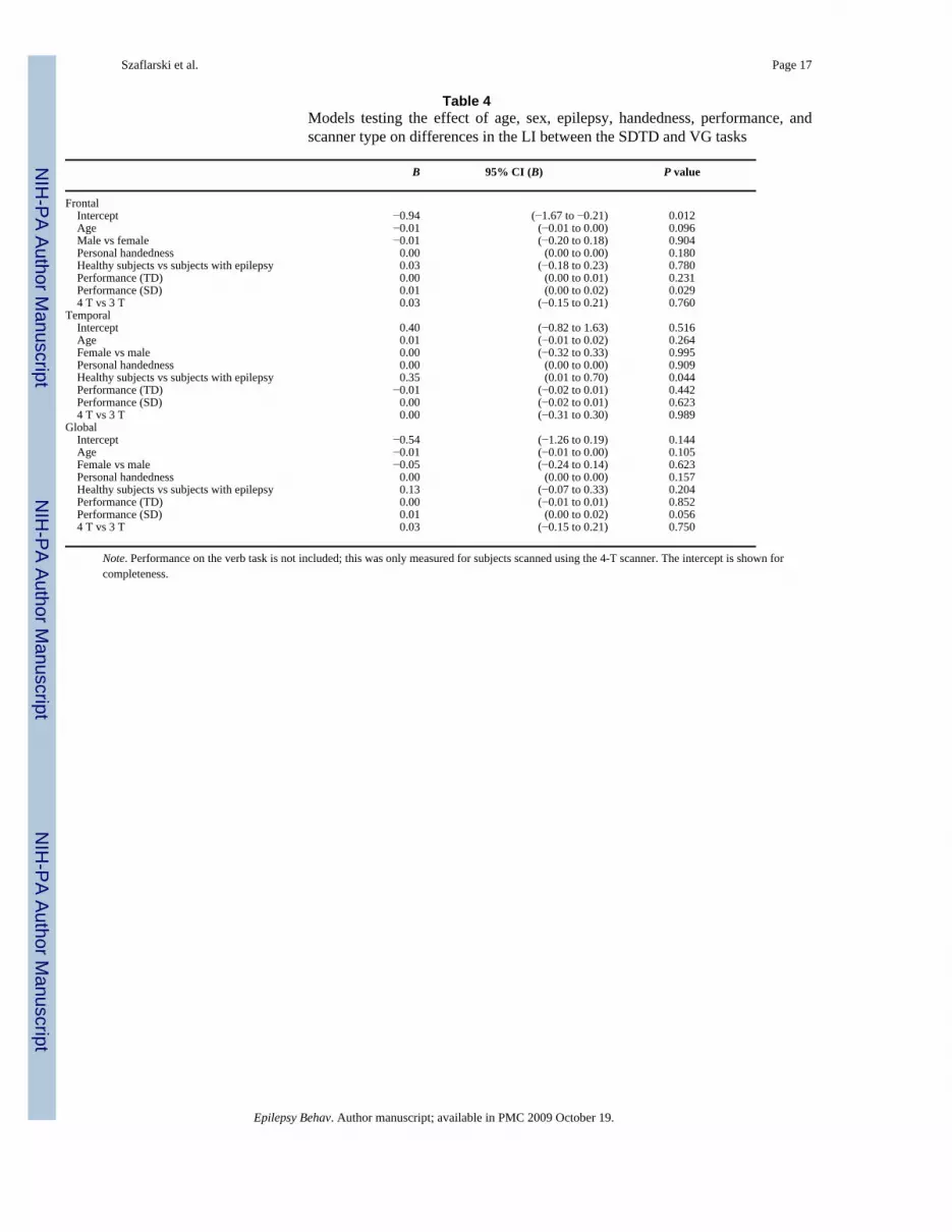
We noted significant correlations between the fMRI tasks for all three LIs (frontal, temporal,and global; all P ≤ 0.037) (Table 2). The correlation between language lateralization (globalROI) with the SDTD or VT task and IAP was strong (for both, Pearson's r ≥ 0.652, P < 0.001).In Table 3 are listed the clinical characteristics of subjects with epilepsy who underwent bothfMRI and the IAP. Of note is that four patients who had atypical language lateralization withthe IAP also had atypical language lateralization with fMRI. Further testing of the effects ofage, sex, epilepsy, handedness, performance, and scanner type on differences in the LI betweenthe SDTD and VG tasks revealed that these variables had either no effect or a very small effect(Table 4).
Next, we tested the contribution of the fMRI language tasks to the determination of LIs usingthe IAP as the “gold standard” for language lateralization. Unadjusted model testing revealedthat the SDTD and VG tasks combined explained approximately 58.4% in the variability ofthe IAP/language, and that only the SDTD task significantly contributed to this determination(in other words, the VG task did not add to the prediction from the SDTD task). Further analysisadjusted for other variables revealed that the effects of handedness and age were significant,whereas other variables tested did not contribute to the model; age, handedness, and SDTDcombined explained approximately 75.5% of the IAP/language variability (Table 5). We alsotested the effects of side of epilepsy onset on language lateralization (Table 6), and we foundthat there was a much higher chance of atypical language lateralization in patients with left butnot right hemispheric epilepsy (P = 0.013 for the SDTD task, P = 0.088 for the VG task). Thisrelationship was not seen when IAP results were analyzed, which may be the effect of the smallnumber of subjects who received this test.
Szaflarski et al. Page 6
Epilepsy Behav. Author manuscript; available in PMC 2009 October 19.
NIH
-PA Author Manuscript
NIH
-PA Author Manuscript
NIH
-PA Author Manuscript

4. DiscussionIn this study evaluating fMRI as a presurgical tool for language cortex localization, we firstestablished normal language activation patterns using two frequently used fMRI tasks: verbgeneration and semantic decision/tone decision. We then applied the results of languagelocalization in healthy controls to the presurgical evaluation of patients with epilepsy. BothfMRI tasks used in this study showed robust BOLD signal changes in the language cortices inhealthy controls (Fig. 1) and patients with epilepsy. The validity of language localization withfMRI tasks in epilepsy was later verified by comparing the fMRI results with the results of theIAP in patients with epilepsy (Fig. 2). Our findings confirm the clinical utility of the fMRItasks used in this study for localization of the eloquent cortex in presurgical evaluation, andare consistent with prior literature. Several studies have already documented the ability of fMRIto lateralize language in patients with epilepsy, and have shown a strong correlation betweenpresurgical fMRI performed using various language tasks and IAP [14,16,18,23,24,29]. Forexample, Binder and colleagues found a very strong correlation (Pearson's r = 0.96, P < 0.0001),and Desmond and colleagues observed a perfect (“100%”) correlation between the IAP andfMRI in the ability to lateralize language in patients with epilepsy [14,16]. Correct localizationand lateralization of language functions in patients undergoing surgical treatment of epilepsyis of utmost importance, as patients who undergo surgery in the language-dominant hemisphereare known to suffer from word finding deficits. The possibility of such deficits may be predictedby fMRI with the SDTD task [26,48]. These and our results confirm the utility of fMRI inpresurgical evaluation of patients with pharmacoresistant epilepsy and add to the growing bodyof evidence that fMRI can be used for presurgical language localization.
Some of the more recent studies used a combination of fMRI tasks for evaluation of languagefunctions in patients with epilepsy, and at least one study proposed using fMRI instead of theIAP [33,49]. Furthermore, one fMRI study using the SDTD task in patients with left temporallobe epilepsy has already shown 100% sensitivity and 73% specificity in predicting significantnaming decline after epilepsy surgery when language was lateralized to the left hemisphere,confirming the clinical utility of this fMRI task in presurgical evaluation of epilepsy surgerypatients [26]. We extended the results of the preceding studies by adding a comparison of thetwo most frequently used fMRI tasks and by demonstrating the excellent ability of both tasksin language localization and a strong correlation between fMRI and the IAP. In one of theprevious studies, Benson and colleagues reported a discrepancy in the ability of three languagetasks to lateralize language in patients with brain lesions; only the verb generation task showedgood language lateralizing capability and correlation with the IAP, whereas object naming andsingle-word reading tasks did not perform well at lateralizing language functions [29]. Bahnand colleagues reported that language lateralization with IAP and that with fMRI tasks agreedin all patients, but only the results of visual analysis were presented [13]. In contrast to theaforementioned studies, Galliard and colleagues used five different language fMRI tasks todetermine language lateralization in 26 predominantly right-handed patients with epilepsy withleft hemispheric seizure onset [33]. These authors reported significant agreement between theIAP and language lateralization with a panel of fMRI tasks, and they suggested that the panelmay improve designation of language laterality in patients with epilepsy when compared witha single task.
In view of the findings by various research groups, it is not surprising to find a strong correlationbetween each of the fMRI tasks and the IAP and between the fMRI tasks themselves. But,when we examined the contribution of each of the tasks to the correlation with the IAP, wefound that the additional contributions of the verb generation task to the estimation of languagelateralization by the SDTD task proved to be not significant. In other words, it appears thatperforming one task, preferably the SDTD task, would be suffcient for correct languagelateralization, provided the patient was able to cooperate with the requirements of the task.
Szaflarski et al. Page 7
Epilepsy Behav. Author manuscript; available in PMC 2009 October 19.
NIH
-PA Author Manuscript
NIH
-PA Author Manuscript
NIH
-PA Author Manuscript
These results have important clinical implications. The results of fMRI language localizationcan be analyzed independently and compared with each other later or, in the case when onestudy fails (e.g., because of equipment failure, excessive movement, or lack of activation), theresults of the second study would still be available, preventing the patient from undergoing asecond procedure (and, in the case of IAP, repeated exposure to radiation and contrast agents).Therefore, including two or more language tasks in the presurgical evaluation has the advantageof possibly streamlining presurgical evaluation and avoiding the need for test repetition.
In addition to the above findings, we noted that the SDTD task provided LIs that were moreleft-lateralized than the LIs generated with the VG task. This shift is likely related to thesemantic nature of the SDTD task and to subtracting from the active paradigm the tone decisioncomponent, which allows for control of attention/working memory, auditory processing, andmotor responses with this task [43]. This LI difference is also likely related, in part, to thedifferences in the activation patterns between the tasks that are due to various languagecomponents involved in the SDTD task versus the VG task. In contrast to the SDTD task, theVG task uses spontaneous verb generation in response to common nouns while finger tappingis used as a control [36,41]. Therefore, in this task, subtracting auditory and working memorycomponents of language processing is not possible. This may have contributed to thedifferences in LIs. Further, the VG task does not include intrascanner performance monitoring.Although the subjects performed very well during the mock run and on the postscan recall ofthe nouns outside the scanner immediately on completion of the fMRI procedure, we have notbeen able to assess the performance inside the scanner due to the covert nature of this task.This possibly could have affected the LI values generated with this task, but we do not thinkthat this is the case, as a recent study showed that language LIs generated with fMRI areperformance independent [50]. Nevertheless, despite the significant differences between thetasks and between the lateralization of the BOLD signal changes, the LIs were highlycorrelated, and the results of both fMRI tasks strongly agreed with the results of languageevaluation with the IAP and cortical stimulation mapping. This supports the notion that eitherboth fMRI tasks combined or each task independently can be used for language localizationin health and disease.
We noted differences between the LIs in patients with right and left hemispheric epilepsy onsetwith either task. The differences in the LIs obtained with the VG and SDTD tasks was betweenhealthy controls and patients with left hemispheric seizure onset (more symmetric languagedistribution). These findings suggest the possibility that the recurrent ictal and interictal activitycauses redistribution of language function in the developing brain to compensate for injury tothe traditional left-dominant language areas or connections to them. These findings are inconcordance with the study by Powell et al., who observed reorganization of functional tracts(i.e., changes in connectivity) in patients with left medial temporal but not right temporalseizure onset [51]. An alternative explanation is that the preexisting brain injury that leads tothe development of epilepsy causes a shift in language localization/lateralization. The latterpossibility is partially supported by a recent study indicating that early brain injury (definedas any insult, e.g., meningitis, febrile seizure, trauma) is associated with more symmetriclanguage distribution [27]. Further, the findings of differences in language lateralization inpatients with left hemispheric epilepsy are in agreement with an fMRI study that examined theeffects of left and right hemispheric epilepsy on language distribution using a word generationtask [28]. We also noted a trend toward more symmetric language distribution in patients withlonger duration of epilepsy. This trend was not significant, in contrast to previous studies[27,52], but it was consistent with our previous study that focused on the effects of epilepsyon language in children [30] and other studies examining this issue in adult patients withepilepsy [53,54]. Such a lack of significance may be the result of the relatively small sampleof patients with epilepsy included in our study or the enrollment in this study of subjects withvarious etiologies of epilepsy including stroke, tumor, and hippocampal sclerosis, as well as
Szaflarski et al. Page 8
Epilepsy Behav. Author manuscript; available in PMC 2009 October 19.
NIH
-PA Author Manuscript
NIH
-PA Author Manuscript
NIH
-PA Author Manuscript
subjects with normal anatomical MRIs (Table 1). However, a study that included a largenumber of patients with left medial temporal lobe epilepsy did not find such a relationship[53]. Hence, it is still somewhat unclear what factors affect language lateralization, and to whatdegree, in patients with epilepsy.
In summary, our data demonstrate that fMRI is a useful clinical tool for language lateralization.Here, we have identified several focus areas for future work. Of primary interest is the use offMRI to elucidate the effects of epilepsy on language, which will support research on thephysiological underpinning of language.
AcknowledgmentsThis study was supported by The Neuroscience Institute in Cincinnati, OH, USA; The National Institute of ChildHealth and Development (RO1-HD38578); and funds from the Cincinnati Epilepsy Center, Cincinnati, OH, USA.The data were presented in part at the 5th European Congress on Epileptology, Madrid, Spain, October 2002, and inpart at the Joint Annual Meeting of the American Epilepsy Society and the American Clinical NeurophysiologySociety, Washington, DC, USA, December 2005. Dr. Szaflarski and Dr. Privitera are members of The NeuroscienceInstitute. The authors thank Dr. Jennifer L. Cavitt, Dr. William T. Cahill, Dr. David M. Ficker, and Dr. Angela B.Morriss for referring their patients; Cindy Hughes, R.N., M.N., for help in subject recruitmen;t and Ms. Chris-ti Banksfor help in scanning of the subjects.
References1. Engel J Jr. Finally, a randomized, controlled trial of epilepsy surgery. N Engl J Med 2001;345:365–7.
[PubMed: 11484695]2. Kwan P, Brodie MJ. Early identification of refractory epilepsy. N Engl J Med 2000;342:314–9.
[PubMed: 10660394]3. Wiebe S, Blume WT, Girvin JP, Eliasziw M. A randomized, controlled trial of surgery for temporal-
lobe epilepsy. N Engl J Med 2001;345:311–8. [PubMed: 11484687]4. Jones-Gotman M, Sziklas V, Djordjevic J, et al. Etomidate speech and memory test (eSAM): a new
drug and improved intracarotid procedure. Neurology 2005;65:1723–9. [PubMed: 16344513]5. Loring DW, Meador KJ, Lee GP, et al. Cerebral language lateralization: evidence from intracarotid
amobarbital testing. Neuropsychologia 1990;28:831–8. [PubMed: 2247209]6. Rasmussen T, Milner B. The role of early left-brain injury in determining lateralization of cerebral
speech functions. Ann NY Acad Sci 1977;299:355–69. [PubMed: 101116]7. Wada J, Rasmussen T. Intracarotid injection of sodium amytal for the lateralization of cerebral speech
dominance. J Neurosurg 1960;17:226–82. [PubMed: 13826545]8. Wada JA, Clarke R, Hamm A. Cerebral hemispheric asymmetry in humans: cortical speech zones in
100 adults and 100 infant brains. Arch Neurol 1975;32:239–46. [PubMed: 1124988]9. Jayakar P, Bernal B, Santiago Medina L, Altman N. False lateralization of language cortex on functional
MRI after a cluster of focal seizures. Neurology 2002;58:490–2. [PubMed: 11839861]10. Kho KH, Leijten FS, Rutten GJ, Vermeulen J, Van Rijen P, Ramsey NF. Discrepant findings for
Wada test and functional magnetic resonance imaging with regard to language function: use ofelectro-cortical stimulation mapping to confirm results [case report]. J Neurosurg 2005;102:169–73.[PubMed: 15658111]
11. Hong SB, Kim KW, Seo DW, Kim SE, Na DG, Byun HS. Contralateral EEG slowing and amobarbitaldistribution in Wada test: an intracarotid SPECT study. Epilepsia 2000;41:207–12. [PubMed:10691118]
12. Baciu MV, Watson JM, McDermott KB, et al. Functional MRI reveals an interhemisphericdissociation of frontal and temporal language regions in a patient with focal epilepsy. Epilepsy Behav2003;4:776–80. [PubMed: 14698719]
13. Bahn MM, Lin W, Silbergeld DL, et al. Localization of language cortices by functional MR imagingcompared with intracarotid amobarbital hemispheric sedation. AJR Am J Roentgenol 1997;169:575–9. [PubMed: 9242780]
Szaflarski et al. Page 9
Epilepsy Behav. Author manuscript; available in PMC 2009 October 19.
NIH
-PA Author Manuscript
NIH
-PA Author Manuscript
NIH
-PA Author Manuscript

14. Binder JR, Swanson SJ, Hammeke TA, et al. Determination of language dominance using functionalMRI: a comparison with the Wada test. Neurology 1996;46:978–84. [PubMed: 8780076]
15. Cuenod CA, Bookheimer SY, Hertz-Pannier L, Zeffiro TA, Theodore WH, Le Bihan D. FunctionalMRI during word generation, using conventional equipment: a potential tool for language localizationin the clinical environment. Neurology 1995;45:1821–7. [PubMed: 7477975]
16. Desmond JE, Sum JM, Wagner AD, et al. Functional MRI measurement of language lateralizationin Wada-tested patients. Brain 1995;118(Pt 6):1411–9. [PubMed: 8595473]
17. Gaillard WD, Balsamo L, Xu B, et al. Language dominance in partial epilepsy patients identifiedwith an fMRI reading task. Neurology 2002;59:256–65. [PubMed: 12136067]
18. Hertz-Pannier L, Gaillard WD, Mott SH, et al. Noninvasive assessment of language dominance inchildren and adolescents with functional MRI: a preliminary study. Neurology 1997;48:1003–12.[PubMed: 9109891]
19. Lehericy S, Cohen L, Bazin B, et al. Functional MR evaluation of temporal and frontal languagedominance compared with the Wada test. Neurology 2000;54:1625–33. [PubMed: 10762504]
20. Liegeois F, Connelly A, Salmond CH, Gadian DG, Vargha-Khadem F, Baldeweg T. A direct test forlateralization of language activation using fMRI: comparison with invasive assessments in childrenwith epilepsy. Neuroimage 2002;17:1861–7. [PubMed: 12498760]
21. Spreer J, Arnold S, Quiske A, et al. Determination of hemisphere dominance for language: comparisonof frontal and temporal fMRI activation with intracarotid amytal testing. Neuroradiology2002;44:467–74. [PubMed: 12070719]
22. Woermann FG, Jokeit H, Luerding R, et al. Language lateralization by Wada test and fMRI in 100patients with epilepsy. Neurology 2003;61:699–701. [PubMed: 12963768]
23. Worthington C, Vincent DJ, Bryant AE, et al. Comparison of functional magnetic resonance imagingfor language localization and intracarotid speech amytal testing in presurgical evaluation forintractable epilepsy: preliminary results. Stereotact Funct Neurosurg 1997;69:197–201. [PubMed:9711754]
24. Yetkin FZ, Swanson S, Fischer M, et al. Functional MR of frontal lobe activation: comparison withWada language results. AJNR Am J Neuroradiol 1998;19:1095–8. [PubMed: 9672017]
25. Fernandez G, Specht K, Weis S, et al. Intrasubject reproducibility of presurgical languagelateralization and mapping using fMRI. Neurology 2003;60:969–75. [PubMed: 12654961]
26. Sabsevitz DS, Swanson SJ, Hammeke TA, et al. Use of preoperative functional neuroimaging topredict language deficits from epilepsy surgery. Neurology 2003;60:1788–92. [PubMed: 12796532]
27. Springer JA, Binder JR, Hammeke TA, et al. Language dominance in neurologically normal andepilepsy subjects: a functional MRI study. Brain 1999;122(Pt 11):2033–46. [PubMed: 10545389]
28. Adcock, J.; Smith, S.; Matthews, P. Human Brain Mapping. Brighton, UK: 2001. Comparison offMRI with the Wada test for lateralization of language in pre-surgical epilepsy patients.. 2001
29. Benson RR, FitzGerald DB, LeSueur LL, et al. Language dominance determined by whole brainfunctional MRI in patients with brain lesions. Neurology 1999;52:798–809. [PubMed: 10078731]
30. Yuan W, Szaflarski JP, Schmithorst VJ, et al. FMRI shows atypical language lateralization in pediatricepilepsy patients. Epilepsia 2006;47:593–600. [PubMed: 16529628]
31. Ries ML, Boop FA, Griebel ML, et al. Functional MRI and Wada determination of languagelateralization: a case of crossed dominance. Epilepsia 2004;45:85–9. [PubMed: 14692913]
32. Bookheimer SY, Zeffiro TA, Blaxton TA, Gaillard PW, Theodore WH. Activation of language cortexwith automatic speech tasks. Neurology 2000;55:1151–7. [PubMed: 11071493]
33. Gaillard WD, Balsamo L, Xu B, et al. fMRI language task panel improves determination of languagedominance. Neurology 2004;63:1403–8. [PubMed: 15505156]
34. Oldfield RC. The assessment and analysis of handedness: the Edinburgh Inventory. Neuropsychologia1971;9:97–113. [PubMed: 5146491]
35. Jacola LM, Schapiro MB, Schmithorst VJ, et al. Functional magnetic resonance imaging revealsatypical language organization in children following perinatal left middle cerebral artery stroke.Neuropediatrics 2006;37:46–52. [PubMed: 16541368]
36. Petersen SE, Fox PT, Posner MI, Mintun M, Raichle ME. Positron emission tomographic studies ofthe cortical anatomy of single-word processing. Nature 1988;331:585–9. [PubMed: 3277066]
Szaflarski et al. Page 10
Epilepsy Behav. Author manuscript; available in PMC 2009 October 19.
NIH
-PA Author Manuscript
NIH
-PA Author Manuscript
NIH
-PA Author Manuscript
37. Szaflarski JP, Holland SK, Schmithorst VJ, Byars AW. fMRI study of language lateralization inchildren and adults. Hum Brain Mapp 2006;27:202–12. [PubMed: 16035047]
38. Szaflarski JP, Schmithorst VJ, Altaye M, et al. A longitudinal functional magnetic resonance imagingstudy of language development in children 5 to 11 years old. Ann Neurol 2006;59:796–807. [PubMed:16498622]
39. Friedman A, Polson MC, Dafoe CG, Gaskill SJ. Dividing attention within and between hemispheres:testing a multiple resources approach to limited-capacity information processing. J Exp Psychol HumPercept Perform 1982;8:625–50. [PubMed: 6218226]
40. Kemper S, Herman RE, Lian CH. The costs of doing two things at once for young and older adults:talking while walking, finger tapping, and ignoring speech or noise. Psychol Aging 2003;18:181–92. [PubMed: 12825768]
41. Holland SK, Plante E, Weber Byars A, Strawsburg RH, Schmithorst VJ, Ball WS Jr. Normal fMRIbrain activation patterns in children performing a verb generation task. NeuroImage 2001;14:837–43. [PubMed: 11554802]
42. Chiu CY, Schmithorst VJ, Brown RD, Holland SK, Dunn S. Making memories: a cross-sectionalinvestigation of episodic memory encoding in childhood using FMRI. Dev Neuropsychol2006;29:321–40. [PubMed: 16515409]
43. Binder JR, Frost JA, Hammeke TA, Cox RW, Rao SM, Prieto T. Human brain language areasidentified by functional magnetic resonance imaging. J Neurosci 1997;17:353–62. [PubMed:8987760]
44. Szaflarski JP, Binder JR, Possing ET, McKiernan KA, Ward BD, Hammeke TA. Languagelateralization in left-handed and ambidextrous people: fMRI data. Neurology 2002;59:238–44.[PubMed: 12136064]
45. Schmithorst VJ, Dardzinski BJ, Holland SK. Simultaneous correction of ghost and geometricdistortion artifacts in EPI using a multiecho reference scan. IEEE Trans Med Imaging 2001;20:535–9. [PubMed: 11437113]
46. Thevenaz P, Unser M. A pyramid approach to sub-pixel registration based on intensity. IEEE TransImage Process 1998:7–41.
47. Talairach, J.; Tournoux, P. Co-planar stereotaxic atlas of the human brain. Thieme; New York: 1988.48. Langfitt JT, Rausch R. Word-finding deficits persist after left anterotemporal lobectomy. Arch Neurol
1996;53:72–6. [PubMed: 8599562]49. Medina LS, Bernal B, Dunoyer C, et al. Seizure disorders: functional mr imaging for diagnostic
evaluation and surgical treatment—prospective study. Radiology 2005;236:247–53. [PubMed:15987978]
50. Weber B, Wellmer J, Schur S, et al. Presurgical language fMRI in patients with drug-resistant epilepsy:effects of task performance. Epilepsia 2006;47:880–6. [PubMed: 16686653]
51. Powell HW, Parker GJ, Alexander DC, et al. Abnormalities of language networks in temporal lobeepilepsy. NeuroImage 2007;36:209–21. [PubMed: 17400477]
52. Saltzman J, Smith ML, Scott K. The impact of age at seizure onset on the likelihood of atypicallanguage representation in children with intractable epilepsy. Brain Cogn 2002;48:517–20. [PubMed:12030499]
53. Janszky J, Jokeit H, Heinemann D, Schulz R, Woermann FG, Ebner A. Epileptic activity influencesthe speech organization in medial temporal lobe epilepsy. Brain 2003;126(Pt 9):2043–51. [PubMed:12821521]
54. Brazdil M, Chlebus P, Mikl M, Pazourkova M, Krupa P, Rektor I. Reorganization of language-relatedneuronal networks in patients with left temporal lobe epilepsy: an fMRI study. Eur J Neurol2005;12:268–75. [PubMed: 15804243]
Szaflarski et al. Page 11
Epilepsy Behav. Author manuscript; available in PMC 2009 October 19.
NIH
-PA Author Manuscript
NIH
-PA Author Manuscript
NIH
-PA Author Manuscript
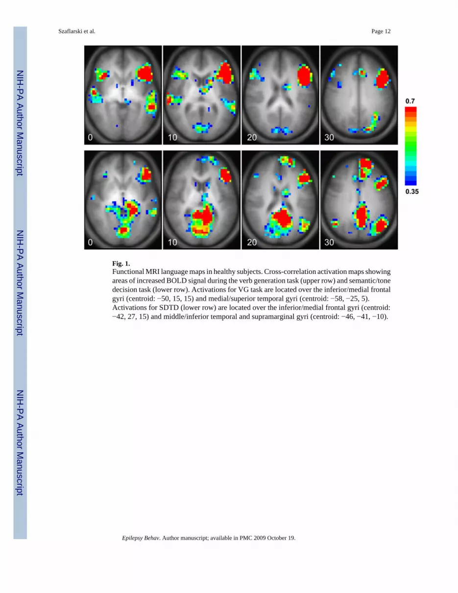
Fig. 1.Functional MRI language maps in healthy subjects. Cross-correlation activation maps showingareas of increased BOLD signal during the verb generation task (upper row) and semantic/tonedecision task (lower row). Activations for VG task are located over the inferior/medial frontalgyri (centroid: −50, 15, 15) and medial/superior temporal gyri (centroid: −58, −25, 5).Activations for SDTD (lower row) are located over the inferior/medial frontal gyri (centroid:−42, 27, 15) and middle/inferior temporal and supramarginal gyri (centroid: −46, −41, −10).
Szaflarski et al. Page 12
Epilepsy Behav. Author manuscript; available in PMC 2009 October 19.
NIH
-PA Author Manuscript
NIH
-PA Author Manuscript
NIH
-PA Author Manuscript

Fig. 2.Correlation between language lateralization with the IAP and the verb generation (left) andsemantic/tone decision tasks (right).
Szaflarski et al. Page 13
Epilepsy Behav. Author manuscript; available in PMC 2009 October 19.
NIH
-PA Author Manuscript
NIH
-PA Author Manuscript
NIH
-PA Author Manuscript

NIH
-PA Author Manuscript
NIH
-PA Author Manuscript
NIH
-PA Author Manuscript
Szaflarski et al. Page 14
Table 1Characteristics and primary measurements for study subjects
Healthy subjects (N = 49) Subjects with epilepsy (N = 36) All subjects (N = 85) P value
Age 39.5 (12.2) 37.9 (11.7) 38.8 (11.9) 0.544Male 37 (75.5) 22 (61.1) 59 (69.4) 0.155Female 12 (24.5) 14 (38.9) 26 (30.6)Personal handednessa 84.4 (38.2) 77.9 (50.3) 81.6 (43.5) 0.505Family handednessb 84.0 (18.3) 86.0 (23.6) 84.9 (20.6) 0.665Age at epilepsy onset 16.6 (15.2)Duration of epilepsy 21.1 (13.2)Left hemisphere epilepsy 0 (0.0) 18 (50.0) 18 (50.0)Right hemisphere epilepsy 0 (0.0) 18 (50.0) 18 (50.0)3-T scanner 24 (49.0) 13 (36.1) 37 (43.5) 0.2374-T scanner 25 (51.0) 23 (63.9) 48 (56.5)Performance, verbsc 98.8 (2.1) 83.1 (15.8) 91.3 (13.5) <0.001Performance, TDd 93.7 (6.1) 77.3 (17.0) 87.0 (14.3) <0.001Performance, SDe 81.0 (11.4) 71.7 (12.8) 77.3 (12.8) 0.001Frontal VG 0.42 (0.31) 0.45 (0.47) 0.43 (0.38) 0.396Temporal VG 0.11 (0.44) 0.18 (0.46) 0.14 (0.45) 0.135Global VG 0.35 (0.29) 0.38 (0.41) 0.36 (0.34) 0.180 Left 41 (83.7) 29 (80.6) 70 (82.4) Right 3 (6.1) 3 (8.3) 6 (7.1) 0.912 Symmetric 5 (10.2) 4 (11.1) 9 (10.6)Frontal SDTD 0.54 (0.36) 0.45 (0.55) 0.50 (0.45) 0.690Temporal SDTD 0.36 (0.46) 0.19 (0.58) 0.29 (0.52) 0.486Global SDTD 0.49 (0.30) 0.36 (0.54) 0.44 (0.42) 0.769 Left 47 (95.9) 24 (66.7) 71 (83.5) Right 1 (2.0) 7 (19.4) 8 (9.4) 0.002 Symmetric 1 (2.0) 5 (13.9) 6 (7.1)IAP 0.57 (0.49) Left 23 (85.2) Right 2 (7.4) Symmetric 2 (7.4)
Note. Data are reported as means and SD or frequency and proportions, as appropriate. The significance of the difference between subjects with andwithout epilepsy is given. Lateralization for VG and SDTD was determined from the global laterality index.
aFive subjects had values ≤-50, values for all other subjects were >50.
bAll subjects had a positive score; data were not available for one subject.
cAssessed only for subjects scanned at 4 T.
dNot assessed for six subjects.
eNot assessed for five subjects.
Epilepsy Behav. Author manuscript; available in PMC 2009 October 19.

NIH
-PA Author Manuscript
NIH
-PA Author Manuscript
NIH
-PA Author Manuscript
Szaflarski et al. Page 15Ta
ble
2C
orre
latio
n, d
iffer
ence
, and
lim
its o
f agr
eem
ent f
or L
Is m
easu
red
usin
g th
e V
G ta
sk, S
DTD
task
, and
IAP
Pear
son'
s rP
valu
eM
ean
diffe
renc
eL
ower
lim
it of
agre
emen
tU
pper
lim
it of
agre
emen
tC
orre
latio
nD
iffer
ence
Fron
tal (
SDTD
–VG
)0.
530
<0.0
010.
124
0.07
−0.7
40.
88Te
mpo
ral (
SDTD
–VG
)0.
227
0.03
70.
022
0.15
−1.0
61.
36G
loba
l (SD
TD–V
G)
0.49
5<0
.001
0.08
80.
07−0
.71
0.85
IAP–
VG
0.65
2<0
.001
0.00
20.
25−0
.52
1.03
IAP–
SDTD
0.73
5<0
.001
0.00
70.
22−0
.56
1.01
Not
e. F
or c
ompa
rison
of I
AP
and
fMR
I dat
a, th
e gl
obal
fMR
I mea
sure
has
bee
n us
ed. (
One
pat
ient
with
epi
leps
y di
d no
t hav
e va
lid V
G a
nd S
DTD
late
raliz
atio
n m
easu
res,
and
one
heal
thy
cont
rol
and
one
patie
nt w
ith e
pile
psy
did
not h
ave
a va
lid V
G m
easu
re. N
ine
subj
ects
with
epi
leps
y di
d no
t hav
e IA
P da
ta; t
hese
subj
ects
are
exc
lude
d fo
r ana
lyse
s rel
atin
g V
G a
nd S
DTD
to IA
P la
ngua
gela
tera
lizat
ion.
)
Epilepsy Behav. Author manuscript; available in PMC 2009 October 19.

NIH
-PA Author Manuscript
NIH
-PA Author Manuscript
NIH
-PA Author Manuscript
Szaflarski et al. Page 16Ta
ble
3C
linic
al in
form
atio
n fo
r the
pat
ient
s with
epi
leps
y w
ho u
nder
wen
t IA
P an
d fM
RI
Age
Age
of o
nset
Sex
EH
ISei
zure
ons
etSt
ruct
ural
MR
IE
tiolo
gy/r
isk
fact
orL
I
VG
SDT
DIA
P
124
14F
88R
MT
R M
TSFe
brile
seiz
ures
0.67
0.41
0.69
238
4F10
0R M
TB
ilate
ral h
ippo
cam
pal v
olum
elo
ss R
> L
; no
sign
al c
hang
eFe
brile
seiz
ures
0.54
0.78
0.77
330
11M
100R
tem
pora
lR
pos
terio
r tem
pora
l DN
ETD
NET
0.71
0.66
0.92
452
36M
85B
item
pora
lL
MTS
Men
ingi
tis0.
670.
450.
845
51<1
M10
0L M
Ts/
p L
late
ral t
empo
ral
rese
ctio
n/L
MTS
Perin
atal
insu
lt0.
160.
400.
46
651
5M33
L M
TL
MTS
Febr
ile se
izur
es−0
.83
−0.8
4−0
.23
717
16F
58R
MT
R te
mpo
ral t
ip ri
ng-e
nhan
cing
lesi
onLe
sion
0.40
0.20
0.92
839
5F10
0R te
mpo
ral
R h
emis
pher
ic a
troph
yM
enin
gitis
1.00
0.57
0.33
930
9F10
0R M
TR
MTS
Febr
ile se
izur
es0.
200.
210.
4610
3319
F67
L M
TN
orm
alN
one
0.54
0.03
0.27
1139
20F
−50L
MT
Nor
mal
Non
e−0
.33
−0.0
9−0
.54
1253
13M
100R
MT
R M
TSM
enin
gitis
0.14
0.41
0.77
1347
27M
100R
MT
R M
TSN
one
1.00
−0.0
30.
6914
212M
91R
tem
pora
lR
pos
terio
r med
ial l
esio
nLe
sion
0.60
0.96
1.00
1521
14F
52L
tem
pora
lN
orm
alN
one
0.75
0.84
0.84
1643
2M−8
9L te
mpo
ral
L la
tera
l ven
tricl
een
larg
emen
t/L M
TSN
one
−0.9
0−0
.68
−1.0
0
1735
1M10
0L M
TL
MTS
Prem
atur
e 11
wee
ks−0
.60
0.49
0.39
1820
15M
100L
par
ieto
tem
pora
lL
post
erio
r hip
poca
mpa
lD
NET
Lesi
on0.
940.
191.
00
1935
9F83
R M
TR
ant
erio
r tem
pora
l res
ectio
n/re
sidu
al h
ippo
cam
pus
rem
aini
ng
Febr
ile se
izur
es0.
810.
880.
77
2033
19F
83L
fron
tal
L fr
onta
l ara
chno
id c
yst
Cys
t; s/
p sh
untin
g−0
.50
−0.1
00.
3821
417F
100R
MT
R M
TSFe
brile
seiz
ures
0.15
0.28
1.00
2227
17M
100L
MT
Post
traum
atic
whi
te m
atte
rch
ange
sTB
I0.
700.
270.
77
2322
<1F
100R
fron
tote
mpo
ral
R c
ereb
ral h
emis
pher
icat
roph
yM
enin
gitis
and
stro
ke a
t 3 w
eeks
of a
ge0.
530.
120.
77
2446
40M
75L
MT
L M
TSTB
I−0
.29
0.1
0.08
2522
4F73
R te
mpo
ral
R M
TSTB
I0.
470.
541.
0026
4927
F10
0L M
TL
med
ial t
empo
ral g
liom
aLe
sion
0.83
0.85
0.47
2728
27F
−100
R M
TR
MTS
Thyr
oidi
tis-r
elat
ed e
ncep
halo
path
y−0
.24
−0.1
528
4136
F10
0L te
mpo
ral
L an
terio
r tem
pora
l cor
tical
dysp
lasi
aLe
sion
0.94
0.59
0.62
Not
e. N
= 2
8; o
ne p
atie
nt u
nder
wen
t the
fMR
I/VG
task
onl
y du
e to
diff
icul
ties w
ith u
nder
stan
ding
the
SDTD
task
; the
pat
ient
is a
non
-nat
ive
Engl
ish
spea
ker.
Patie
nts 1
−11
unde
rwen
t fM
RI a
t 3 T
,an
d th
e re
mai
ning
pat
ient
s, at
4 T
. Not
incl
uded
in th
e ta
ble
are
patie
nts w
ho d
id n
ot u
nder
go IA
P (N
= 1
0). O
f the
se 1
0 su
bjec
ts, 8
dec
ided
not
to u
nder
go fu
rther
eva
luat
ion
for e
pile
psy
surg
ery,
1su
bjec
t was
alle
rgic
to b
arbi
tura
tes (
the
deci
sion
rega
rdin
g su
rger
y w
as b
ased
on
fMR
I res
ults
), an
d 1
subj
ect u
nder
wen
t IA
P an
d fM
RI b
ut c
ould
not
hea
r the
pro
mpt
s for
lang
uage
task
s and
, hen
ce,
his f
MR
I dat
a w
ere
excl
uded
from
ana
lysi
s. M
T, m
esia
l tem
pora
l lob
e; M
TS, m
esia
l tem
pora
l scl
eros
is; D
NET
, dys
embr
yopl
astic
neu
roep
ithel
ial t
umor
; s/p
, sta
tus p
ost.
Epilepsy Behav. Author manuscript; available in PMC 2009 October 19.
NIH
-PA Author Manuscript
NIH
-PA Author Manuscript
NIH
-PA Author Manuscript
Szaflarski et al. Page 17
Table 4Models testing the effect of age, sex, epilepsy, handedness, performance, andscanner type on differences in the LI between the SDTD and VG tasks
B 95% CI (B) P value
Frontal Intercept −0.94 (−1.67 to −0.21) 0.012 Age −0.01 (−0.01 to 0.00) 0.096 Male vs female −0.01 (−0.20 to 0.18) 0.904 Personal handedness 0.00 (0.00 to 0.00) 0.180 Healthy subjects vs subjects with epilepsy 0.03 (−0.18 to 0.23) 0.780 Performance (TD) 0.00 (0.00 to 0.01) 0.231 Performance (SD) 0.01 (0.00 to 0.02) 0.029 4 T vs 3 T 0.03 (−0.15 to 0.21) 0.760Temporal Intercept 0.40 (−0.82 to 1.63) 0.516 Age 0.01 (−0.01 to 0.02) 0.264 Female vs male 0.00 (−0.32 to 0.33) 0.995 Personal handedness 0.00 (0.00 to 0.00) 0.909 Healthy subjects vs subjects with epilepsy 0.35 (0.01 to 0.70) 0.044 Performance (TD) −0.01 (−0.02 to 0.01) 0.442 Performance (SD) 0.00 (−0.02 to 0.01) 0.623 4 T vs 3 T 0.00 (−0.31 to 0.30) 0.989Global Intercept −0.54 (−1.26 to 0.19) 0.144 Age −0.01 (−0.01 to 0.00) 0.105 Female vs male −0.05 (−0.24 to 0.14) 0.623 Personal handedness 0.00 (0.00 to 0.00) 0.157 Healthy subjects vs subjects with epilepsy 0.13 (−0.07 to 0.33) 0.204 Performance (TD) 0.00 (−0.01 to 0.01) 0.852 Performance (SD) 0.01 (0.00 to 0.02) 0.056 4 T vs 3 T 0.03 (−0.15 to 0.21) 0.750
Note. Performance on the verb task is not included; this was only measured for subjects scanned using the 4-T scanner. The intercept is shown forcompleteness.
Epilepsy Behav. Author manuscript; available in PMC 2009 October 19.

NIH
-PA Author Manuscript
NIH
-PA Author Manuscript
NIH
-PA Author Manuscript
Szaflarski et al. Page 18
Table 5Models relating LI determined by SDTD and VG tasks to LI determined by IAP
B 95%CI (B) P value R2
Intercept 0.30 (0.14 to 0.47) 0.001 0.584SDTD 0.47 (0.15 to 0.78) 0.006VG 0.32 (−0.09 to 0.74) 0.121Intercept 0.35 (0.19 to 0.51) <0.001 0.539SDTD 0.63 (0.39 to 0.87) <0.001Intercept 0.61 (0.07 to 1.14) 0.030 0.7674 T vs 3 T 0.04 (−0.20 to 0.28) 0.642Female vs male −0.09 (−0.34 to 0.17) 0.481Personal handedness 0.00 (0.00 to 0.01) 0.004Age −0.01 (−0.02 to 0.00) 0.041Hemisphere −0.07 (−0.33 to 0.18) 0.562SDTD 0.26 (−0.04 to 0.56) 0.071VG 0.01 (−0.40 to 0.42) 0.932Intercept 0.50 (0.10 to 0.91) 0.026 0.755Personal handedness 0.01 (0.00 to 0.01) 0.001Age −0.01 (−0.02 to −0.00) 0.036SDTD 0.27 (0.01 to 0.52) 0.046
Note. Both unadjusted models and models adjusted for age, sex, handedness, scanner type, and hemisphere are shown.
Epilepsy Behav. Author manuscript; available in PMC 2009 October 19.

NIH
-PA Author Manuscript
NIH
-PA Author Manuscript
NIH
-PA Author Manuscript
Szaflarski et al. Page 19
Table 6Comparison of language lateralization between healthy controls, patients with lefthemisphere epilepsy, and patients with right hemisphere epilepsy
Controls Left hemisphere epilepsy Right hemisphere epilepsy P value comparinglateralization
between left and righthemisphere epilepsy
SDTD Left 47 (95.9) 9 (50.0) 15 (83.3) 0.013 Right 1 (2.0) 7 (38.9) 0 (0.0) Symmetric 1 (2.0) 2 (11.1) 3 (16.7)VG Left 41 (83.7) 12 (66.7) 17 (94.40 0.088 Right 3 (6.1) 3 (16.7) 0 (0.00 Symmetric 5 (10.2) 3 (16.7) 1 (5.6)IAP Left 10 (71.4) 13 (100.0) 0.113 Right 2 (14.3) 0 (0.0) Symmetric 2 (14.3) 0 (0.0)
Epilepsy Behav. Author manuscript; available in PMC 2009 October 19.
Related Documents