The effect of s human planta neuro Comprehens Char Departme Faculty short-term lower limb isch ar-flexors muscle force an omechanical mechanisms sive Abstract of Doctoral Diss by Paris Charalampidis rles University in Prague Czech Republic ent of Anatomy and Biomec y of Physical Education and December 2009 haemia on nd related s sertation chanics Sport

Welcome message from author
This document is posted to help you gain knowledge. Please leave a comment to let me know what you think about it! Share it to your friends and learn new things together.
Transcript
The effect of short
human plantar
neuromechanical mechanisms
Comprehensive Abstract of Doctoral D
Charles University in Prague
Department of
Faculty of Physical Education and Sport
The effect of short -term lower limb ischaemia on
human plantar -flexors muscle force and related
neuromechanical mechanisms
Comprehensive Abstract of Doctoral D issertation
by
Paris Charalampidis
Charles University in Prague
Czech Republic
Department of Anatomy and Biomechanics
Faculty of Physical Education and Sport
December 2009
term lower limb ischaemia on
flexors muscle force and related
neuromechanical mechanisms
issertation
Anatomy and Biomechanics
Faculty of Physical Education and Sport

2
Summary
The effect of short-term lower limb ischaemia on hu man plantar-flexors muscle
force and related neuromechanical mechanisms
Ischemic lower limb is a syndrome that causes neuromuscular dysfunction in
both acute and chronic periods. Only a short period of ischaemia can produce
disturbances affecting peripheral and spinal mechanism of human motor control. The
aim of this study was to assess the influence of short term ischaemia in lower limb on
plantar-flexors muscle force production, eliciting by H-reflex and M wave recruitment
curves and to determine the main site in the neuromuscular system affected by the
pathogenic condition of ischaemia.
Seventeen healthy adult volunteers participated in the study with their Informed
consent. The subjects lay prone on a physiotherapy table with both legs extended and
the right foot attached and secured to the force platform. Ischemia was induced by
blood pressure cuff placed around the right thigh 15 cm above the knee and inflated to a
pressure of 200 mmHg for 10 min. During the experiment, the plantar-flexors force, H
and M-responses of soleus muscle evoked by tibial nerve stimulation were measured at
rest, during 10 minutes of ischaemia, 10 and 20 minutes after the occlusion was
released. Obtained data were analyzed by means of one-way repeated measures
ANOVA with Tukey post hoc analysis (p<0.05).
Background EMG activity of the soleus muscle was not significantly different
between the four periods of experiment. However, thresholds, recruitment curves and
transmission across the synapses of Ia afferent were significantly altered during
ischemia. At the post-ischeamic period the plantar flexors force fall significantly
compare to pre-ischemic values and to ischaemia.
In conclusion our results show that ischaemia significantly reduce the mechanical
performance (force) of plantar-flexors muscle for at least 10min and induce metabolic
intracellular processes influencing excitability of both peripheral nerves and muscle
fibers. These changes resulted in alteration of neuromechanical coupling.
Keywords: H-reflex, Ischaemia, Nerve excitability, Synapse transmission, Plantar-
flexors muscle force, Neuromechanical coupling.

3
Introduction
Ischemic lower limb is a syndrome that causes neuromuscular dysfunction in
both acute (thrombosis, embolism, injury to large arteries) and chronic (diabetic
peripheral neuropathy) periods. Arterial occlusion caused by mechanical compression
induces ischemic conditions beneath and distal to the tourniquet cuff. Only a short
period of ischemia can produce intracellular metabolic disturbances affecting peripheral
and spinal mechanism of human motor control (Mogyoros et al., 1997; Grosskreutz
et.al., 1999; Hogan et al., 1999; Zakutansky et al., 2005). By this pathogenic condition
sensory and motor neuron excitability, neuromuscular transmission and skeletal muscle
contractile mechanism and thus forces capacity can be affected.
Previous electrophysiological H-reflex studies performed in healthy subjects (Lin
et al., 2002; Zakutansky et al., 2005) describes an increase in the excitability of the
cutaneous afferent and motor axons by decreasing the threshold for both types of axons
during ischaemia. This increase in peripheral excitability was accompanied by a
decrease in the efficiency of the Ia-fiber motoneuron synapse.
It is experimentally proved that arterial occlusion lasting up to 80minutes in adult
rat’s causes a gradual reduction in muscle twitch and titanic tension. After 50.7 (4.3) min
of ischaemia, the muscle stops functioning under direct stimulation. For this duration of
ischaemia the nerve function remained intact (Hatzipantelis et al., 2001). This implicates
significant alteration of function of the neuromuscular junction under acute ischaemia.
Recently, Clark et al. (2006a,b) studied a skeletal muscle contractile properties applying
periodic cessation of blood occlusion in humans. The muscle cross-sectional area
assesses by serial axial plane MRI scans and plantar flexor muscle measured by
custom-modified dynamometer did not significantly change when the knee was in the
flexed position.
To date, reports using excitation of sensory and motor axons to investigate
plantar-flexors (PFs) muscle force output changes after short term ischaemia and the
post-ischaemic reactions have not been published.

4
Aims
In conclusion, current knowledge about the consequences of pathophysiological
mechanisms induced by ischaemia on biomechanical properties of the lower limb
skeletal muscles is limited. Thus our aims were:
1) To assess the influence of short term ischaemia on the excitability parameters
of sensory and motor neurons.
2) To evaluate the affection of neuromuscular transmission on this pathogenic
condition of ischaemia.
3) To assess the plantar-flexors muscle force production, eliciting by H-reflex and
M wave recruitment curves under ischemic and post-ischaemic conditions.
4) To determine the main site in the neuromuscular system affected by the
pathogenic condition of short term ischaemia.
Methods
Seventeen healthy adult volunteers (11 male and 6 female; mean age 27.41yr
±1.06) participated in the study with their Informed consent to the nature and purpose of
the experiment. All experimental procedures were performed in accordance with the
Declaration of Helsinki, and the study had the approval of the Charles University Ethics
Committee. None of the subject had any history of vascular or other medical deficits
known to affect neuromuscular function.
Experiment procedures
All subjects underwent identical experimental protocol with constant internal
environment, temperature and body position. The subjects lay prone on a physiotherapy
table with both legs extended and the right foot (barefoot) attached (with respect to
muscle tone) and secured to the force platform. The position of the right ankle was
continually controlled by goniometer during the experiment (Knikou and Conway, 2001).

5
The force platform was calibrated in vertical position. During the experiment, the
plantar-flexor (PF) force, H and M-responses of soleus muscle evoked by tibial nerve
stimulation were measured at rest, during 10 minutes of ischaemia (starting after 6
minutes), 10 and 20 minutes after the occlusion was released.
Ischaemia
In order to induce ischemia by mechanical pressure, we used a conventional
sphygmomanometer. Before we start any assessment a blood pressure thigh cuff was
wrapped around the thigh, 15cm above the knee (mid-thigh level) of the investigated
right leg. Ischaemia was achieving, after the first series of measurements ended by
using manual inflation of the cuff to an occlusion pressure of 200mmHg (Krishnan et al.,
2006) and maintained for 10 minutes. Using occlusive pressure at 200mmHg we avoid
causing direct muscle and neural damage (Nitz et al., 1986; Schulte et al., 2008). Blood
occlusion was repeatedly checked by an auscultation (Korotkoff sounds) of the popliteal
artery under the tourniquets cuff. During post-ischaemic assessment procedures the
cuff remains (deflate) in the same position.
EMG recordings
To record the H-reflex and M-response, unipolar surface electrode (Ag/AgCl)
were taped over the right soleus and reference placed just above the external malleolus
and the ground placed between the stimulating and the active electrodes. To elicit the
H-reflex and M-wave recruitment curves, an anode covered with gauze and wetted in
saline was placed just above the patella and a point metal cathode (0.5cm in diameter)
was fixed over the posterior tibial nerve in the popliteal fossa (Capaday, 1997).
Constant voltage stimulation was provided by single rectangular pulses of 0.5ms
duration at a minimum interpulse interval of 10sec. The stimulating impulses were
gradually increased, from H reflex threshold to above the saturated state of the maximal
M-wave. In all four periods of measurements, changes in single pulse stimulus intensity
required to elicit H-reflex, M-wave and PF force amplitudes seen at threshold level,
6
were followed to compare axonal excitability. A decrease in single pulse intensity means
an increase in excitability and vice versa.
The EMG signal was amplified (GrassTelefactor) and digitalized at 16bit and
10kHz (Power1401 + Spike2 CED, UK) and continuously stored on PC hard disk for
further offline analysis.
Muscle force experimental data collection
Force platform (Kistler Instruments, Switzerland) with sampling frequency 5 kHz
was used to record the plantar-flexor force output produced during the determination of
H and M wave recruitment curves. Obtained mechanical responses were continuously
stored (BioWare software) on PC hard disk for further offline analysis.
Neuromechanical coupling
Another parameter that was measured and compare was the changes in a range
of neuromechanical coupling properties of skeletal muscle relative to threshold before,
during and after ischaemia. To assess the influence of short term ischaemia on the
neuromechanical parameters of excitation-contraction coupling and to determine the
difference in stimulus strength properties of contractile machinery, the thresholds
intensity required to produce a plantar flexors muscle twitch were measured. The
stimulus intensity for threshold measurement was delivered at the same time with the
thresholds required to produce an H-reflex and an M-wave. That means changes in
neuromechanical coupling were measured through the evaluation of the evoked
electromyographic and force output potentials. Those were assessed by the difference
in stimulus intensity required to evoke peak wave of plantar flexors muscle twitch seen
at threshold level during all periods of experiment. As a result of this procedure, a
decrease in stimulus intensity represented an increase in strength-duration properties of
neuromechanical coupling. The single pulse stimulus intensity required to elicit plantar
flexors muscle twitch evoked potential seen at threshold level, were followed to
compare axonal and skeletal muscle neuromechanical mechanisms.
7
Mmax - Force relation
Pre-ischaemic, ischaemic and post-ischaemic reactions of plantar-flexors muscle
force output related to the compound muscle fiber action potential (Mmax) were
measured. A single, supramaximal electrical stimulation was delivered to the tibial nerve
with stimulation intensity set at 15% above the level of maximal amplitude of M-wave to
ensure maximal muscle activation and were given more than 3 sec apart to avoid any
postactivation depression of the PFs muscle (Capaday, 1997; Klass et al., 2004).
During the measurement caution was taken also about the rate of stimulation, because
excessive rate in electrical stimulation may lead to block of neuromuscular transmission
(Jones, 1996). This was avoided by using adequate stimulation frequency. The M-wave
and the mechanical twitch in response to single and paired supramaximal stimuli were
recorded. The peak-to-peak amplitude of Mmax was assessed, as well as the maximal
peak twitch force of PFs muscle. Simultaneously measured EMG evoked potential
provides analysis of M-wave, which contains information about membrane properties of
the active MUs (Merletti et al., 1992).
Statistical analysis
To compare the experimental data of the measurements those were done before,
during ischaemia and two times in post-ischaemic period, conventional statistical
methods were used to calculate means and standard errors of the mean (SE). Obtained
data were analyzed by means of a one-way ANOVA with repeated measures. When
significant main effects were observed, Tukey test was used for post hoc analysis. A
probability of p<0.05 was chosen as the significant level in all analyses.
8
Results
Electromyographic and force evoked potential changes analysis allows the study
of different aspects of the neuromuscular system function under ischaemia. The
parameters chosen for further analysis were:
H-reflex and M-wave native traces
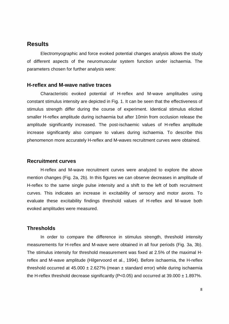
Characteristic evoked potential of H-reflex and M-wave amplitudes using
constant stimulus intensity are depicted in Fig. 1. It can be seen that the effectiveness of
stimulus strength differ during the course of experiment. Identical stimulus elicited
smaller H-reflex amplitude during ischaemia but after 10min from occlusion release the
amplitude significantly increased. The post-ischaemic values of H-reflex amplitude
increase significantly also compare to values during ischaemia. To describe this
phenomenon more accurately H-reflex and M-waves recruitment curves were obtained.
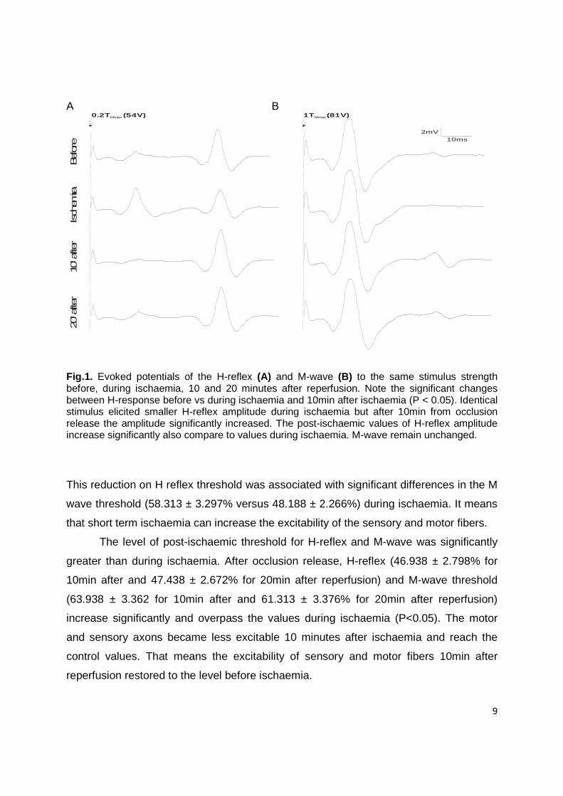
Recruitment curves
H-reflex and M-wave recruitment curves were analyzed to explore the above
mention changes (Fig. 2a, 2b). In this figures we can observe decreases in amplitude of
H-reflex to the same single pulse intensity and a shift to the left of both recruitment
curves. This indicates an increase in excitability of sensory and motor axons. To
evaluate these excitability findings threshold values of H-reflex and M-wave both
evoked amplitudes were measured.
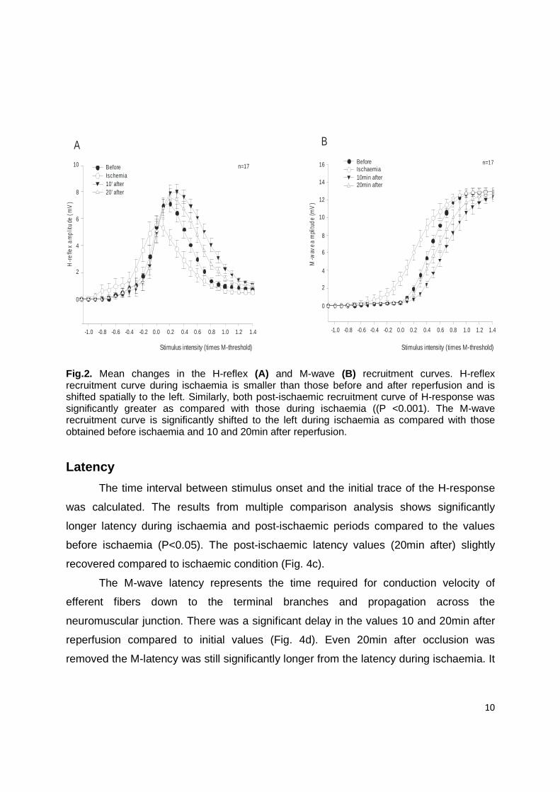
Thresholds
In order to compare the difference in stimulus strength, threshold intensity
measurements for H-reflex and M-wave were obtained in all four periods (Fig. 3a, 3b).
The stimulus intensity for threshold measurement was fixed at 2.5% of the maximal H-
reflex and M-wave amplitude (Hilgervoord et al., 1994). Before ischaemia, the H-reflex
threshold occurred at 45.000 ± 2.627% (mean ± standard error) while during ischaemia
the H-reflex threshold decrease significantly (P<0.05) and occurred at 39.000 ± 1.897%.
9
A B
10ms2mV
0.2T (54V)Mmax 1TMmax(81V)
Befor
eIsch
emia
10’ a
fter
20’ a
fter
Fig.1. Evoked potentials of the H-reflex (A) and M-wave (B) to the same stimulus strength before, during ischaemia, 10 and 20 minutes after reperfusion. Note the significant changes between H-response before vs during ischaemia and 10min after ischaemia (P < 0.05). Identical stimulus elicited smaller H-reflex amplitude during ischaemia but after 10min from occlusion release the amplitude significantly increased. The post-ischaemic values of H-reflex amplitude increase significantly also compare to values during ischaemia. M-wave remain unchanged.
This reduction on H reflex threshold was associated with significant differences in the M
wave threshold (58.313 ± 3.297% versus 48.188 ± 2.266%) during ischaemia. It means
that short term ischaemia can increase the excitability of the sensory and motor fibers.
The level of post-ischaemic threshold for H-reflex and M-wave was significantly
greater than during ischaemia. After occlusion release, H-reflex (46.938 ± 2.798% for
10min after and 47.438 ± 2.672% for 20min after reperfusion) and M-wave threshold
(63.938 ± 3.362 for 10min after and 61.313 ± 3.376% for 20min after reperfusion)
increase significantly and overpass the values during ischaemia (P<0.05). The motor
and sensory axons became less excitable 10 minutes after ischaemia and reach the
control values. That means the excitability of sensory and motor fibers 10min after
reperfusion restored to the level before ischaemia.
10
Stimulus intensity (times M-threshold)
1.4
n=17
0
2
4
6
8
10
-1.0 -0.8 -0.6 -0.4 -0.2 0.0 0.2 0.4 0.6 0.8 1.0 1.2
M-w
avea
mpli
tude
(mV
)0
2
4
6
8
10
12
14
16 BeforeIschaemia10min after20min after
B
Stimulus intensity (times M-threshold)
-1.0 -0.8 -0.6 -0.4 -0.2 0.0 0.2 0.4 0.6 0.8 1.0 1.2 1.4
BeforeIschemia10' after20' after
n=17
H-re
flex
ampl
itude
(mV
)
A
Fig.2. Mean changes in the H-reflex (A) and M-wave (B) recruitment curves. H-reflex recruitment curve during ischaemia is smaller than those before and after reperfusion and is shifted spatially to the left. Similarly, both post-ischaemic recruitment curve of H-response was significantly greater as compared with those during ischaemia ((P <0.001). The M-wave recruitment curve is significantly shifted to the left during ischaemia as compared with those obtained before ischaemia and 10 and 20min after reperfusion.
Latency
The time interval between stimulus onset and the initial trace of the H-response
was calculated. The results from multiple comparison analysis shows significantly
longer latency during ischaemia and post-ischaemic periods compared to the values
before ischaemia (P<0.05). The post-ischaemic latency values (20min after) slightly
recovered compared to ischaemic condition (Fig. 4c).
The M-wave latency represents the time required for conduction velocity of
efferent fibers down to the terminal branches and propagation across the
neuromuscular junction. There was a significant delay in the values 10 and 20min after
reperfusion compared to initial values (Fig. 4d). Even 20min after occlusion was
removed the M-latency was still significantly longer from the latency during ischaemia. It
11
is important to note, that the M-wave latency didn’t show any significant difference
during ischaemia.
H-reflex Threshold M-wave Threshold
Stim
ulus
Inte
nsity
(V
)
A
**
*
0
10
20
30
40
50
60
*
Stim
ulu
s In
ten
sity
(V
)
**
B
0
20
40
60
80
H-reflex amplitude at threshold level M-wave amplitudes at threshold level
Fig. 3. Threshold changes were measured using 0.5ms stimulus duration. Note that ischaemia produced significant hyperexcitability of sensory (A) and motor (B) axons. The decrease of threshold of sensory fibers between initial data and ischaemia was associated also with a change of threshold intensity for motor fibers. Measurements after reperfusion (after 10 and 20min) showed that H-reflex and M-wave thresholds significantly increase compare to the data during ischaemia (P < 0.05).
Characteristics of the H-reflex and M-wave maximal responses and
ratio
Background EMG activity of the soleus muscle was assessed in terms of
amplitude analysis and was not significantly different between the four periods of
experiment (Fig. 4f).
The maximal peak-to-peak amplitude of soleus (Hmax) and the Hmax/Mmax
ratio (Fig. 4e) during ischaemia was significantly decreased (P<0.01) compared to the
initial data. The values relative to the H-reflex and M-wave maximum amplitudes
parameters are summarized in Figure 4a, b.
On the other hand, the Hmax and the Hmax/Mmax ratio significantly increased in
both post-ischaemic periods as compared with the values during ischaemia (P<0.001).
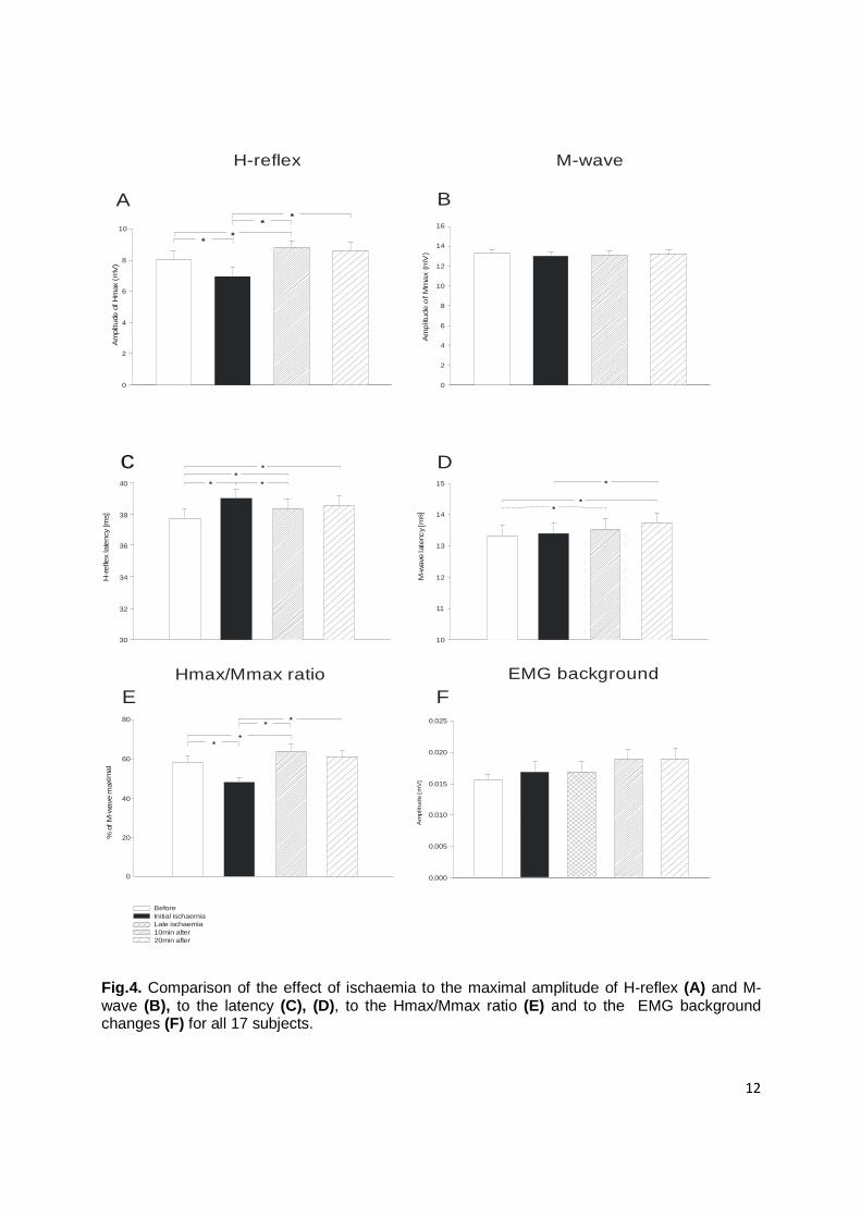
12
H-reflex M-wave
BeforeInitial ischaemiaLate ischaemia10min after20min after
M-w
ave
late
ncy
[ms]
D
**
*
10
11
12
13
14
15
H-refle
x la
tency
[ms]
30
32
34
36
38
40 * *
**
c
E
0.000
0.005
0.010
0.015
0.020
0.025
Am
plit
ude [m
V]
EMG background
**
**
0
2
4
6
8
10
Am
plit
ude
of H
max
(m
V)
0
2
4
6
8
10
12
14
16
Am
plit
ude o
f M
max (m
V)
A B
% o
f M
-wave
max
imal
*
*
*
*
0
20
40
60
80
Hmax/Mmax ratio
F
Fig.4. Comparison of the effect of ischaemia to the maximal amplitude of H-reflex (A) and M-wave (B), to the latency (C), (D), to the Hmax/Mmax ratio (E) and to the EMG background changes (F) for all 17 subjects.

13
A significant Hmax increase was also found between the period before ischaemia and
10min after reperfusion. The constant maximal amplitude of the M-wave in all four
phases of measurement means that the recording conditions were unchanged during
the course of the experiment and the mechanical responses were not altered from the
displacement (e.g. from contraction of underlying muscles) of the stimulating electrode
away from the tibial nerve during the experiment. Mmax represents the total activation
of the soleus motoneurone pool.
Hmax/Mmax- sensory transmission across the Ia-alpha motoneuron
The effectivity of the sensory to motoneuron transmission expressed as
Hmax/Mmax was affected significantly by acute ischemia of the lower limb.
However, no significant change of the Mmax was observed. That means no significant
differences or alteration of NM junction function were recorded during the course of the
experiment. From the above, it would be safe to conclude that all significant changes in
Hmax/Mmax ratio are due to a change in Hmax amplitude (Fig. 4e).
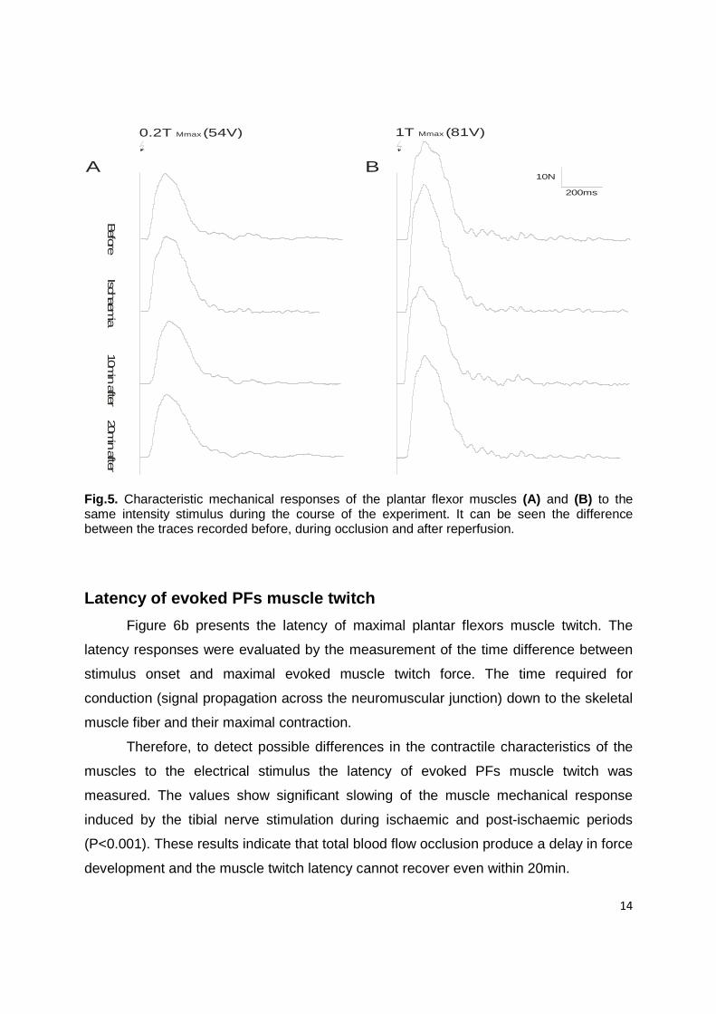
The plantar-flexors muscle mechanical output
A comparable series of recording made before, during and after ischaemia is
illustrated in Fig.5. Note that the plantar-flexor muscle force (N) output activated by the
same stimulus intensity changes during ischaemia and after reperfusion. The maximal
force of muscle twitch (Nmax) was significantly lower in both reperfusion intervals in
compare to values during ischaemia. Changes in developed force of plantar-flexor (PF)
were analyzed during each experimental period (Fig 6a). There was no significant
difference in force development between control period and during ischaemia.
At the first post-ischeamic measurements (10min after reperfusion) the force fall
significantly (69.065 ± 4.678 for) compare to control (75.389 ± 5.054) and to ischaemia
(79.292 ± 5.374) values (P<0.001). The PFs force remained significantly reduced 20min
after reperfusion (70.765 ± 4.794) compare to the force values during ischaemia. It
means that short term Ischaemia can produced marked changes in post-ischaemic
measures of plantar-flexor muscle force output (Nmax) and not even the reintroduction
and restoration of blood flow allowed the muscle to reach the control values.
14
200ms
10N
Before Ischaem
ia 10min after 20m
in after
A B
0.2T (54V)Mmax 1T (81V)Mmax
Fig.5. Characteristic mechanical responses of the plantar flexor muscles (A) and (B) to the same intensity stimulus during the course of the experiment. It can be seen the difference between the traces recorded before, during occlusion and after reperfusion.
Latency of evoked PFs muscle twitch
Figure 6b presents the latency of maximal plantar flexors muscle twitch. The
latency responses were evaluated by the measurement of the time difference between
stimulus onset and maximal evoked muscle twitch force. The time required for
conduction (signal propagation across the neuromuscular junction) down to the skeletal
muscle fiber and their maximal contraction.
Therefore, to detect possible differences in the contractile characteristics of the
muscles to the electrical stimulus the latency of evoked PFs muscle twitch was
measured. The values show significant slowing of the muscle mechanical response
induced by the tibial nerve stimulation during ischaemic and post-ischaemic periods
(P<0.001). These results indicate that total blood flow occlusion produce a delay in force
development and the muscle twitch latency cannot recover even within 20min.

15
0
20
40
60
80
100
BeforeIschaemia10min after20min after
La
ten
cy[m
s]
**
*
0
20
40
60
80
100
BeforeIschaemia10min after20min after
Fo
rce
[N
]
Fo
rce
[N]
*
**
A BNmax Latency of Nmax
-1 0 1 2
0
20
40
60
80
BeforeIschaemia10' after20' after
Stimulus intensity [times M threshold]
N=17
Nmax/Mmax
0
10
20
30
40
50
60
C DForce (N) threshold
**
*
Fig.6 . Comparison of Force evoked potential measurements during experiment. A. Maximal response of evoked twitch peak of PFs muscle mechanical reaction to electrical stimulation of tibial nerve at popliteal area. B. Changes in maximal muscle twitch reaction latency. C. Relation between stimulus intensity and PFs muscle twitch response. D. Recruitment curve of PFs force (N) displaying the increase of maximal amplitudes with increase of the stimulus intensity. The mean Nmax/Mmax values in the subjects rises at lower stimulus intensities relative to M-threshold and reaches a higher maximum.

16
The muscle twitch strength-duration properties
The full sequence of threshold measurements was obtained to define the
differences in threshold intensity required to produce a plantar flexors muscle twitch.
The influence of short term ischaemia on the excitability values of excitation-contraction
coupling was measured using 0.5ms stimulus duration. The initial (control) thresholds
stimulus intensity (V) values were obtained at rest (48.188 ± 2.590) before the effect of
short term ischaemia on different neuromechanical parameters were compared. The
ischaemia produced a decreased of the threshold significantly (P<0.05), reducing the
stimulus intensity required to evoke a particular response (42.375 ± 2.240), whereas
reperfusion returned the threshold near to the initial values. Results 10 and 20min after
the occlusion of thigh cuff was released (49.875 ± 2.805 and 50.625± 2.668) showed
that plantar-flexors muscle twitch evoked potential seen at threshold level significantly
increase compare to the data during ischaemia (P < 0.05).
According to these results the threshold of PFs muscle correlates with the
excitability results of H-reflex and M-wave amplitudes. Both results show significant
decrease in threshold for the sensory and motor fibers and skeletal muscle
neuromechanical coupling during ischaemia. Measurements after reperfusion showed
that stimulus intensity required evoking peak wave of plantar-flexors muscle twitch seen
at threshold level and M-wave and H-reflex threshold significantly increase compare to
the data during ischaemia (P < 0.05). These show that the excitability of sensory and
motor axons and the excitability of plantar-flexors muscle fibers returned to the baseline
(control values).
Plantar-flexors muscle force output related to Mmax
From the plantar-flexors force recruitment curve is obvious that the muscle twitch
was evoked before the M-wave was elicited and when only H response was present
(Fig. 15d). This implicates that activation of monosynaptic reflex loop produces
mechanical response of PFs muscles. These results indicate that peak twitch force was
evoked by stimulus lower than those evoking M-wave (M-threshold) and that the first
motor response activated at low stimulus strength by the reflex pathway.
17
The total activation of the soleus motorneuron pool produces maximal amplitude
of M-wave and also maximal amplitude of plantar-flexors force twitch, during
supramaximal stimulation of tibial nerve. That was verified by these results.
The maximal isometric activation of the PFs muscle was clearly correlated with
the Mmax response (CMUP) which represents the total activation of the soleus
motoneurone pool. The constant maximal amplitude of the M-wave in all four phases of
experiment ensures that the recording conditions were unchanged.
Discussion
Our results suggest that, there are more vulnerable to short-term lower limb
ischaemia structures involved in neuromuscular function. The main findings of the
present study are significant alterations of peripheral nerve excitability, affection of
synapse transmission and force-failure mechanisms known to exist at this level.
Pathophysiological changes on peripheral nerve exc itability
The neural structures were significantly affected by short term ischaemia on their
excitability parameters, efficiency of the transmission across the synapses of Ia afferent
terminals and latency.
Short-term ischemia of lower limb significantly increases excitability of afferent
and motor neuron axons by decreasing significantly H-reflex and M-wave thresholds
elicited from soleus muscle. These differences in excitability and threshold were
evaluated by the H-reflex and M-wave recruitment curves (Fig. 2a,b). The finding of
increased excitability of the afferent and motor fibers during ischaemia is partly in
agreement with that of Lin et al. (2002) and Zakutansky et al. (2005). Zakutansky and
colleagues also show that, H-wave threshold failed to return to the control level of
excitability, whereas the motor axons were fully recovered after 5 minutes of
reperfusion. However, this finding is in contrary to our evidence report, when the H-
reflex threshold was measured 10min after reperfusion without any significant
differences form the values obtained before ischaemia. The discrepant findings could be
due to differences in the electrical stimulus duration (Capaday, 1997) that was delivered
to the tibial nerve (in our study 0,5ms instead of 1ms) and due to different post-
18
ischaemic conditions under which the H-reflexes were measured. In the present study
the experimental data were obtained 10min after the occlusion was released and the
ischaemia of lower limb was induced for 10min (instead of 5min). These results indicate
that sensory Ia fibers are not more vulnerable to altered metabolic processes
(extracellular K+ accumulation) than motor axons.
The mechanisms located beyond the sensory and motor neurons alterations is
supported by the observation that membrane depolarization by applied currents
produces similar changes in excitability to those occurring in the first 10 min of
ischaemia (Baker and Bostock, 1989; Bostock et al.,1991b). Other studies have also
shown that during brief period of ischaemia, axonal excitability increase due to
membrane depolarization, caused by altered function of the electrogenic sodium pump
and extracellular K+ accumulation (Bostock et al., 1991a and 1994; Mogyoros et al.,
1997; Grosskreutz et al., 1999; Lin et al., 2002). Modulation of the H-reflex threshold
during ischaemia can probably result from altered function of ion channels and from
changes in metabolic processes (influenced by pH) due to dysfunction of the
electrogenic Na+ pump and subsequent K+ accumulation. The present findings of
peripheral nerve excitability changes correspond with the findings of previous
mentioned studies performed by different techniques and methods (e.g. long-lasting
depolarizing and hyperpolarizing currents, threshold tracking).They are also supported
with the theory that, ischaemia changes the accommodative properties of axons and
thus change the excitability of human nerve (Kugelberg, 1944; Bostock et al., 1991a).
This shows that the amplitude component of H-reflex can be one of the valuable
techniques to assess sensory and motor neurons excitability during ischaemia (Burke et
al., 1989).
Affection of synapse transmission
The increased excitability values of the sensory and motor fibers produced by
short-term ischemia were followed by a decrease of maximal amplitude of H-reflex and
Hmax/Mmax ratio. The decrease of Hmax/Mmax ratio was affected mainly by the
decrease of Hmax amplitude, because the maximal amplitude of M-wave (CMAP)
remains unchanged during the experiment. Also alteration of the Hmax/Mmax ratio
19
seems unlikely to be due to changes in neuromuscular junction because the maximal
amplitude and latency of M-wave remains unchanged. The majority of the changes in
H-reflex strength are attributed to changes in the amount of presynaptic inhibition
(Schieppati, 1987; Zehr, 2002). With respect to the results the decrease of Hmax/Mmax
ratio during ischaemia seems to be affected by the altered efficiency of the transmission
across the synapses of Ia afferent terminals. The decreased synapse transmission
(evaluated by decreased value of Hmax/Mmax ratio) can be due to increases in
presynaptic inhibition (Avela et al., 2001; Zakutansky et al., 2005) of group Ia-afferent
terminals projecting directly to the motoneurons (with matched level of background level
of motor activity). It is, however, not clear whether changes in Ca++ permeability or the
degree of the neuromechanical transmitter depletion can affect the efficiency of the
synaptic transmission (Komiyama et al., 1999).
One particularly interesting finding from the present study is the observation of an
increased latency in the H-wave (during ischaemia and at both post-ischaemic periods)
and M-wave (significant delay in the post-ischaemic values) indicates a slowing in nerve
conduction time required for signal propagation through the reflex arc.
Alteration of skeletal muscle function
We assess the plantar-flexors muscle force production under ischemic and post-
ischaemic conditions by eliciting the H-reflex and M-wave recruitment curves. The
results of the muscle property variables demonstrate that short term ischaemia can
produce marked changes in plantar-flexors muscle force output (Nmax), latency of the
muscle mechanical response and to neuromechanical parameters of PFs muscle.
Force output and latency during ischaemia
The maximal activation of the PFs muscle (Nmax) was clearly correlated with the
Mmax response which remains unchanged during the course of the experiment. Thus, it
seems that, the maximal muscle twitch was not functionally affected with fatigue-
induced reaction, on the other hand ischaemia create intracellular metabolic disruption.
The analysis of this mechanical responses induced by supramaximal electrical
stimulation of tibial nerve allows one to indirectly investigate the possible muscle

20
intracellular changes responsible for the reduced plantar flexors mechanical
performance after reperfusion. The complete elimination of blood flow in lower limb
certainly resulted in intracellular metabolic disruption (because of the rapid reduction in
oxidative phosphorylation) that contributed to the fall in force. The total ischemia
produces acidosis (alteration of pH), because of increased lactate production. As the
ischaemic time increases, acidosis causes dysfunction of the calcium pump, which is a
Ca–ATPase, and reduces the time of release of calcium from troponin C because the
amount of troponin C that binds up calcium is low. (Hatzipantelis et al., 2001; Clausen,
2003). The metabolic instability that was developed during 10min of ischaemia was
severe enough to significantly change the PFs muscle evoked twitch force for at least
10min. Even restoration of blood flow with oxygenated blood allowed the muscles to
reach the initial values.
The restoration of plantar flexors muscle force depends on the degree of
metabolic disturbance that occurred and the time allowed for recovery. However, even
when the metabolic disturbances are corrected during the recovery period, there can
remain substantial contractile dysfunction (Baker et al., 1993, Nagesser, 1992). These
dysfunction can be related to prolonged elevated (slow reabsorption from sarcoplasmic
reticulum) intracellular Ca2+ (Bruton et al., 1998). In addition, recovery of muscle
function after fatiguing contractions in whole muscle may depend in part on keeping
blood flow elevated and washing out metabolic waste products (Bogdanis et al., 1996).
Interestingly, the PFs muscle force was not significantly altered during
ischaemia. That finding is in contrary to the reports of Stainsby et al., 1990 and Hogan
et al., 1999 that the sudden and total reduction in blood flow result in a fall in force
production within seconds of the ischemic initiation. One possible explanation for the
discrepant findings could be difference in the conditions under which the muscle was
assessed and the type of muscle fibers.
The decrease in the muscle twitch force observed at both post-ischaemic results,
without any change in the M-wave, can be also explained by an alteration of the
excitation-contraction coupling and due to intracellular metabolic processes (Duchateau
and Hainaut, 1985; Bigland-Ritchie et al., 1986, Avela et al., 2001).
Another interesting finding of the present study was the observation of a

21
significant slowing of the time between stimulus onset and maximal evoked twitch force
during ischaemia and in post-ischaemic phases. This increased latency of maximal
force (Nmax) correlates absolutely with H-reflex latency alteration caused by ischaemia.
These changes can be due to some peripheral neurogenic or myopathic adaptations
resulted in slower electrical impulse propagation and related to impaired muscle
membrane excitability caused by the amount and rate of Ca2+ release from
sarcoplasmic reticulum (Dutka et al., 2005, Ortenblad et al., 2000). All physiological
mechanisms of strength-duration properties are driven by several factors but in general
can provide information on intracellular Ca2+ transients, muscle fiber type, and cross-
bridge changes (Westerblad et al., 1997; Ortenblad et al., 2000, Clark et al., 2006a).
Efficacy of ischaemia on neuromechanical threshold
The result of PFs neuromechanical threshold correlates with the excitability
changes of H-reflex and M-wave amplitudes. Both results show significant decrease in
threshold of sensory and motor axons related to decreased threshold of PFs muscle
and excitation of neuromechanical coupling during ischaemia. These increased
excitability values associated with the decrease of threshold. Both post-occlusive
measurements showed that stimulus intensity required to evoke peak wave of PFs
muscle, M-wave and H-reflex response significantly increase compare to the data
during ischaemia (P < 0.05). These show that the excitability of EMG and muscle force
evoked potentials returned to the baseline (before ischaemia).
Several factors accounting for a change in neuromechanichal threshold of
muscle fibers evoked by direct electrical stimulation (e.g. disturbance in E-C coupling,
as the E-C coupling could be affected by reduced sarcolemmal excitability, or reduced
rate of ATP utilization and regeneration). The action potential propagation on excitable
membranes of muscle fibers (sarcolemma and t-tubule) may be altered during
ischaemia because of the membrane Na+ pump dysfunction (Yensen et al. 2002;
Duchateau et al., 2002; Nielsen et al. 2004; Piitulainen et al., 2007).
There is another one parameter that must be kept in mind when PFs muscle
twitch force is analyzed. Ischaemia produces acidosis, because of increase lactate
22
production. It is known that acidosis decreases excitability in an excitable tissue and
vice versa. Muscles, however, can balance effect of intracellular acidosis because of
lactate and hydrogen production by efflux of potassium which can reach up to 8mM
which helps to preserve excitability of the muscle fiber during fatigue. (Hatzipantelis et
al, 2001, Sostaric et al, 2006). It is suspected that the reduced excitability contributes to
a reduction in calcium release by the sarcoplasmic reticulum and a consequent
decrease in the force of muscle contraction (Lindinger, 2006). That finding is in
agreement with our post-ischaemic results of PFs muscle force and neuromechanical
threshold. The post-ischaemic mechanical performance of PFs was significantly
reduced and in opposite the neuromechanical threshold was increased compare to the
values during ischaemia.
Effects of ischaemia on neuromuscular junction
Except the above mention neuromuscular mechanisms that can be impaired by
short-term ischaemia also neuromuscular junction can be affected. According to
Lundborg (1970) and Hatzipantelis et al (2001) the neuromuscular junction is the most
susceptible site of neuromuscular system to ischaemia. Hatzipantelis and colleagues
(2001) design an experimental animal model by applying 80minutes blood flow
occlusion on peripheral nerve of the rats. Their results indicates that under ischaemic
conditions the neuromuscular function probably stops because acidosis causes a
reduction in the time that calcium channels remain open, a reduction in the number of
synaptic vesicles and of their acetylcholine contents and reduction in the permeability of
muscle membrane to sodium and potassium. Contrary to their findings, we did not
observe from our results any effect of short-term ischaemia on the human
neuromuscular junction. Our assertion is based on the results of maximal amplitude of
M-wave (CMAP) and the values form M-wave latency before and during ischaemia.
Both responses remain unchanged. The present results indicate that neuromuscular
transmission during this experiment was unaffected. The fact is that little is known
23
about the influence of short-term ischaemia on the human neuromuscular junction and
further research is needed.
The observation of PFs muscle twitch response before M-wave action potential
was evoked is another one interesting findings from the current study. This is obvious
from the PFs muscle recruitment curves (Fig. 6d) elicited during all four phases of our
experiment. This mechanical response was recorded by the force platform and was
elicited by low intensity stimulation of the sensory fibers (only H-reflex was present). In
agreement with our finding are only the studies of Maffiuletti and colleagues (2000) and
Scaglioni and others (2003). Their studies were done in healthy human subjects but
without any intervention. They presenting investigations, showed the relative
contribution to the plantar-flexors torque of the soleus motor units activated by H and M
waves. The testing position of lower limb was at 900 of knee flexion. That position was
evaluated because it markedly reduces the mechanical contribution of gastrocnemii
muscles to the evoked twitch. That means they results interpreting only the twitch
evoked by soleus muscle and it doesn’t correspond to all PFs muscle group.
Epilogue
The aim of the present study was to determine the main site in the
neuromuscular system affected by the pathogenic condition of lower limb short-term
ischaemia. We identified the following variables that we felt may theoretically be
predictive of neuromuscular disturbances.
Excitability of peripheral nerves and muscle fibers are highly influenced by
metabolic intracellular processes (affected by pH). It is known that acidosis decreases
excitability in an excitable tissue and vice versa. Indeed, the results of the present study
appear to confirm the concept that the accumulation of K+ accompanies ischaemia and
has effect on excitability of nervous and muscle tissues. However, current extent of this
effect remains unknown.
There was extensive modulation of transmission across the synapses of Ia
afferent in the H-reflex pathway due to ischaemia. These altered synapse transmission
can be due to increases in presynaptic inhibition of group Ia-afferent terminals
24
projecting directly to the motoneurons. Also, during the experiment the human
neuromuscular junction was not affected by short-term ischemia. Assertion based on M-
wave values.
The short-term ischaemia induce intracellular metabolic disruptions that result to
significantly reduce mechanical performance (force) of plantar-flexors muscle for at
least 10min. This reduction of maximal evoked twitch force was accompanied with
increased latency of Nmax. The mechanical muscle twitch evoked by tibial nerve
stimulation at the early stage of H-reflex was recorded and observed during all four
phases of experiment. This action potential appears before the M-wave was elicited.
The mechanisms responsible for these results remain to be explored.
The further study of the above mention structures may provide important insights
employed into the performance of neuromuscular periphery in disease states.
References
Avela J, Kyrolainen H, Komi PV. Neuromuscular changes after long-lasting mechanically and electrically elicited fatique. Eur J Appl Physiol 2001;86:317-325.
Baker AJ, Kostov KG, Miller RG, Weiner MW. Slow force recovery after long-duration exercise: metabolic and activation factors in muscle fatigue. J. Appl. Physiol 1993;74:2294–2300.
Baker M, Bostock H . Depolarization changes the mechanism of accommodation in rat and human motor axons. J Physiol (Lond) 1989; 411:545–61.
Bigland-Ritchie B, Cafarelli E, and Vøllestad N.K. Fatigue of submaximal static contractions. Acta Physiol Scand 1986;128:137–148.
Bogdanis, GC, Nevill ME, Lakomy HKA, Graham CM, Lou is G. Effects of active recovery on power output during repeated maximal sprint cycling. Eur. J. Appl. Physiol 1996;74:461–469.
Bostock H, Baker M, Grafe P, Reid G. Changes in excitability and accommodation of human motor axons following brief periods of ischaemia. J Physiol 1991a; 441:513–535.
Bostock H, Baker M, Reid, G. Changes in excitability of human motor axons underlying post-ischaemic fasciculation’s: evidence for two stable states. J Physiol 1991b;441:537–557.
25
Bostock H, Burke D, Hales JP . Differences in behaviour of sensory and motor axons following release of ischaemia. Brain 1994;117:225–234.
Bruton JD, Lannergren J, Westerblad H . Mechanisms underlying the slow recovery of force after fatigue: importance of intracellular calcium. Acta. Physiol. Scand 1998;162: 285– 293.
Burke D, Adams RW, Skuse NF . The effects of voluntary contraction on the H-reflex of human limb muscles. Brain 1989;112: 417-433
Capaday C. Neurophysiological methods for studies of the motor system in freely moving human subjects. J Neurosci Methods 1997;74:201–218.
Clark BC, Fernhall B, Ploutz-Snyder LL. Adaptations in human neuromuscular function following prolonged unweighting: I. Skeletal muscle contractile properties and applied. J Appl Physiol 2006a;101:256–263.
Clark BC, Fernhall B, Ploutz-Snyder L.L. Adaptations in human neuromuscular function following prolonged unweighting: II. Neurological properties and motor imagery efficacy. J Appl Physiol 2006b;101:264–272.
Clausen T. Na+-K+ pump regulation and skeletal muscle contractility. Physiol Rev 2003;83:1269-1324.
Duchateau J, Hainaut K. Electrical and mechanical failures during sustained and intermittent contractions in humans. J Appl Physiol 1985;58:942–947.
Duchateau J, Balestra C, Carpentier A, Hainaut K. Reflex regulation during sustained and intermittent submaximal contractions in humans. J Physiol 2002;541: 959–967.
Dutka TL, Cole L, Lamb GD. Calcium-phosphate precipitation in the sarcoplasmic reticulum reduces action potential-mediated Ca2+ release in mammalian skeletal muscle. Am J Physiol Cell Physiol 2005;289: C1502– C1512.
Grosskreutz J, Lin C, Mogyoros I, Burke D. Changes in excitability indices of cutaneous afferents produced by ischaemia in human subjects. J Physiol 1999;518, 301–314.
Hatzipantelis KP, Natsis K, Albani M. Effect of acute limb ischaemia on neuromuscular function in rats. Eur J Surg 2001;167:831-838.
Hilgevoord AA, Koelman JH, Bour LJ, Ongerboer de Vi sser BW. Normalization of soleus H-reflex recruitment curves in controls and a population of spastic patients. Electroencephalogr clin Neurophysiol 1994;93:202– 8.
Hogan M C, Kohin S, Stary CM, Hepple RT. Rapid force recovery in contracting skeletal muscle after brief ischaemia is dependent on O2 availability. J Apppl Physiol 1999;87;2225-2229
26
Jones D. High- and low-frequency fatigue revisited. Acta Physiologica Scandinavia 1996;156:265-270.
Kiernan MC, Bostock H. Effect of membrane polarization and ischaemia on the excitability properties of human motor axons. Brain 2000;123:2542-2551.
Komiyama T, Kawai K, Fumoto M. The excitability of a motoneuron pool assessed by the H-reflex method is correlated with the susceptibility of Ia terminals to repetitive discharges in humans. Brain Research 1999;826:317-320.
Klass M, Guissard N, Duchateau J. Limiting mechanisms of force production after repetitive dynamic contractions in human triceps surae. J Appl Physiol 2004;96:15161521.
Knikou W, Conway B. Modulation of soleus H-reflex following ipsilateral mechanical loading of the sole of the foot in normal and complete spinal cord injured humans.Neuroscience Letter. 2001;303:107-110.
Krishnan AV, Phonn RKS, Pussell BA, CHarlesworth JA , Bostock H, Kiernan MC. Ischaemia induces paradoxical changes in axonal excitability in end-stage kidney disease. Brain 2006;129:1585-1592.
Kugelberg E. Accommodation in human nerves and its significance for symptoms in circulatory disturbances and tetany. Acta Physiologica Scandinavica 1944;8, suppl. 24: 1—103.
Lin Cindy SY, Chan Jane HL, Pierrot-Deseilligny E, Burke D. Excitability of human muscle afferents studied using threshold tracking of the H-reflex. J. Physiol 2002; 545:661-669.
Lindinger MI. Determinants of sarcolemmal and transverse tubular excitability in skeletal muscle: implications for high intensity exercise. Equine Comp Exerc Physiol 2006;2:209–217.
Lundborg G. Ischemic nerve injury: experimental studies on intraneural microvascular pathophysiology and nerve function in a limb subjected to temporary circulatory arrest. Scand J Plast Reconstr Surg 1970;6 (Suppl): 1–113.
Maffiuletti NA, Martin A, Van Hoecke J, Schieppati M. The relative contribution to the plantar-flexor torque of the soleus motor units activated by the H reflex and M response in humans. Neuroscience Letters 2000;288:127-130.
Merletti R., Knaflitz M, De Luca C . Electrically evoked myoelectric signals. Crti. Rev. Biomed. Eng 1992;19:293-340.
Mogyoros I, Kiernan MC, Burke D, Bostock H. Excitability changes in human sensory and motor axons during hyperventilation and ischaemia. Brain 1997;120:317–325.
27
Nagesser AS, van der Laarse WJ, Elzinga G. Metabolite changes with fatigue in different types of single muscle fibers of Xenopus laevis. J. Physiol. (Lond.) 1992;448:511–523.
Nielsen O, Ortenblad N, Lamb G, Stephenson D. Excitability of the T-tubular system in rat skeletal muscle: roles of K+ and Na+ gradients and Na+-K+ pump activity. J Physiol 2004;557:133-146.
Nitz AJ, Dobner JJ, Matulionis DH. Pneumatic tourniquet application and nerve integrity: motor function and electrophysiology. Exp Neurol 1986;94: 264–279.
Ortenblad N, Sjogaard G, Madsen K. Impaired sarcoplasmic reticulum Ca2+ release rate after fatiguing stimulation in rat skeletal muscle. J Appl Physiol 2000;89:210–217.
Piitulainen H, Komi P, Linnamo V, Avela J. Sarcolemmal excitability as investigated with M-waves after eccentric exercise in humans. JElektromyog Kinesiol 2007;18(4):672-81.
Schieppati M. The hoffmann reflex: a means of assessing spinal reflex excitability and its descending control in man. Progress in Neurobiology 1987;28:345-376.
Scaglioni G, Narici MV, Maffiuletti NA, Pensini M, Martin A . Effect of ageing on the electrical and mechanical properties of human soleus motor units activated by the H-reflex and M wave. J. Physiol 2003;548:649-661.
Shulte AC, Aschwanden M, Bilecen D. Calf muscles at blood oxygen level-dependent MR Imaging: Aging effects at postocclusive reactive hyperemia. Radiology 2008;247:482-489.
Sostaric SM, Skinner SL, Brown MJ, Sangkabutra T, M edved I, Medley T, Selig SE, Fairweather I, Rutar D, McKenna MJ. Alkalosis increases muscle K+ release, but lowers plasma [K+] and delays fatigue during dynamic forearm exercise. J Physiology 2006;570.1:185–205.
Stainsby WN, Brechue WF, O’Drobinak, DM, Barclay JK . Effects of ischemic and hypoxic hypoxia on V˙ O2 and lactic acid output during tetanic contractions. J. Appl. Physiol 1990;68:574–579.
Westerblad H, Lannergren J, Allen D.G. Slowed relaxation infatigued skeletal muscle fibers of Xenopus and mouse. Contribution of [Ca2+]i and cross-bridges. J Gen Physiol 1997;109:385–399.
Yensen G, Matar W, Renaud J-M. K+-induced twitch potentiation is not due to longer action potential. Am J Physiol 2002;283:C169-C177.
Zakutansky D, Kitano K, Wallace J, Koceja D. H-reflex and motor responses to acute ischemia in apparently healthy individuals. J Clin Neurophysiol 2005;22:210–215.
Zehr P. Considerations for use of the Hoffman reflex in exercise studies. Eur J Appl Physiol 2002;86:455–468.
Related Documents


![DOCTORAL THESIS [TESIS DOCTORAL] - UAM](https://static.cupdf.com/doc/110x72/62162d5b17f9b23eb24b8a87/doctoral-thesis-tesis-doctoral-uam.jpg)