BioMed Central Page 1 of 10 (page number not for citation purposes) BMC Biochemistry Open Access Research article Components of the ubiquitin-proteasome pathway compete for surfaces on Rad23 family proteins Amanda M Goh 1,5 , Kylie J Walters 2 , Suzanne Elsasser 3 , Rati Verma 4 , Raymond J Deshaies 4 , Daniel Finley 3 and Peter M Howley* 1 Address: 1 Department of Pathology, Harvard Medical School, Boston, Massachusetts, USA, 2 Department of Biochemistry, Molecular Biology and Biophysics, University of Minnesota, Minneapolis, Minnesota, USA, 3 Department of Cell Biology, Harvard Medical School, Boston, Massachusetts, USA, 4 Department of Biology, Howard Hughes Medical Institute, California Institute of Technology, Pasadena, California, USA and 5 Institute of Molecular and Cell Biology, Singapore Email: Amanda M Goh - [email protected]; Kylie J Walters - [email protected]; Suzanne Elsasser - [email protected]; Rati Verma - [email protected]; Raymond J Deshaies - [email protected]; Daniel Finley - [email protected]; Peter M Howley* - [email protected] * Corresponding author Abstract Background: The delivery of ubiquitinated proteins to the proteasome for degradation is a key step in the regulation of the ubiquitin-proteasome pathway, yet the mechanisms underlying this step are not understood in detail. The Rad23 family of proteins is known to bind ubiquitinated proteins through its two ubiquitin-associated (UBA) domains, and may participate in the delivery of ubiquitinated proteins to the proteasome through docking via the Rad23 ubiquitin-like (UBL) domain. Results: In this study, we investigate how the interaction between the UBL and UBA domains may modulate ubiquitin recognition and the delivery of ubiquitinated proteins to the proteasome by autoinhibition. We have explored a competitive binding model using specific mutations in the UBL domain. Disrupting the intramolecular UBL-UBA domain interactions in HHR23A indeed potentiates ubiquitin-binding. Additionally, the analogous surface on the Rad23 UBL domain overlaps with that required for interaction with both proteasomes and the ubiquitin ligase Ufd2. We have found that mutation of residues on this surface affects the ability of Rad23 to deliver ubiquitinated proteins to the proteasome. Conclusion: We conclude that the competition of ubiquitin-proteasome pathway components for surfaces on Rad23 is important for the role of the Rad23 family proteins in proteasomal targeting. Background Targeted protein degradation by the ubiquitin-proteas- ome pathway is a key means of regulating a wide variety of cellular processes, ranging from cell cycle progression [1] to antigen presentation [2]. In this pathway, an enzy- matic cascade covalently attaches ubiquitin to a lysine res- idue on substrate proteins. The subsequent conjugation of more ubiquitin moieties, each typically linked through Lys48 of ubiquitin, results in a polyubiquitin chain that directs substrate proteins to the proteasome where they are degraded (reviewed in [3]). The importance of this proteasome-targeting step was demonstrated by experi- Published: 30 January 2008 BMC Biochemistry 2008, 9:4 doi:10.1186/1471-2091-9-4 Received: 24 October 2007 Accepted: 30 January 2008 This article is available from: http://www.biomedcentral.com/1471-2091/9/4 © 2008 Goh et al; licensee BioMed Central Ltd. This is an Open Access article distributed under the terms of the Creative Commons Attribution License (http://creativecommons.org/licenses/by/2.0 ), which permits unrestricted use, distribution, and reproduction in any medium, provided the original work is properly cited.

Welcome message from author
This document is posted to help you gain knowledge. Please leave a comment to let me know what you think about it! Share it to your friends and learn new things together.
Transcript

BioMed CentralBMC Biochemistry
ss
Open AcceResearch articleComponents of the ubiquitin-proteasome pathway compete for surfaces on Rad23 family proteinsAmanda M Goh1,5, Kylie J Walters2, Suzanne Elsasser3, Rati Verma4, Raymond J Deshaies4, Daniel Finley3 and Peter M Howley*1Address: 1Department of Pathology, Harvard Medical School, Boston, Massachusetts, USA, 2Department of Biochemistry, Molecular Biology and Biophysics, University of Minnesota, Minneapolis, Minnesota, USA, 3Department of Cell Biology, Harvard Medical School, Boston, Massachusetts, USA, 4Department of Biology, Howard Hughes Medical Institute, California Institute of Technology, Pasadena, California, USA and 5Institute of Molecular and Cell Biology, Singapore
Email: Amanda M Goh - [email protected]; Kylie J Walters - [email protected]; Suzanne Elsasser - [email protected]; Rati Verma - [email protected]; Raymond J Deshaies - [email protected]; Daniel Finley - [email protected]; Peter M Howley* - [email protected]
* Corresponding author
AbstractBackground: The delivery of ubiquitinated proteins to the proteasome for degradation is a keystep in the regulation of the ubiquitin-proteasome pathway, yet the mechanisms underlying thisstep are not understood in detail. The Rad23 family of proteins is known to bind ubiquitinatedproteins through its two ubiquitin-associated (UBA) domains, and may participate in the deliveryof ubiquitinated proteins to the proteasome through docking via the Rad23 ubiquitin-like (UBL)domain.
Results: In this study, we investigate how the interaction between the UBL and UBA domains maymodulate ubiquitin recognition and the delivery of ubiquitinated proteins to the proteasome byautoinhibition. We have explored a competitive binding model using specific mutations in the UBLdomain. Disrupting the intramolecular UBL-UBA domain interactions in HHR23A indeedpotentiates ubiquitin-binding. Additionally, the analogous surface on the Rad23 UBL domainoverlaps with that required for interaction with both proteasomes and the ubiquitin ligase Ufd2.We have found that mutation of residues on this surface affects the ability of Rad23 to deliverubiquitinated proteins to the proteasome.
Conclusion: We conclude that the competition of ubiquitin-proteasome pathway components forsurfaces on Rad23 is important for the role of the Rad23 family proteins in proteasomal targeting.
BackgroundTargeted protein degradation by the ubiquitin-proteas-ome pathway is a key means of regulating a wide varietyof cellular processes, ranging from cell cycle progression[1] to antigen presentation [2]. In this pathway, an enzy-matic cascade covalently attaches ubiquitin to a lysine res-
idue on substrate proteins. The subsequent conjugation ofmore ubiquitin moieties, each typically linked throughLys48 of ubiquitin, results in a polyubiquitin chain thatdirects substrate proteins to the proteasome where theyare degraded (reviewed in [3]). The importance of thisproteasome-targeting step was demonstrated by experi-
Published: 30 January 2008
BMC Biochemistry 2008, 9:4 doi:10.1186/1471-2091-9-4
Received: 24 October 2007Accepted: 30 January 2008
This article is available from: http://www.biomedcentral.com/1471-2091/9/4
© 2008 Goh et al; licensee BioMed Central Ltd. This is an Open Access article distributed under the terms of the Creative Commons Attribution License (http://creativecommons.org/licenses/by/2.0), which permits unrestricted use, distribution, and reproduction in any medium, provided the original work is properly cited.
Page 1 of 10(page number not for citation purposes)
BMC Biochemistry 2008, 9:4 http://www.biomedcentral.com/1471-2091/9/4
ments showing that the artificial localization of proteinsto the proteasome is sufficient to cause their degradation[4].
Recent work indicates that ubiquitin receptors, whichbind ubiquitin but are not intrinsic subunits of the protea-some, facilitate the docking of ubiquitinated substrates tothe proteasome [5-8]. The best-studied of these receptorsare the UBL-UBA proteins and include three groups:Rad23/HHR23A/HHR23B, Dsk2/PLIC1/PLIC2, andDdi1. Rad23, for example, has been shown to play a rolein the targeting of the cyclin-dependent kinase inhibitorSic1 to the proteasome [5], and Ddi1 mediates degrada-tion of the Ho endonuclease [9] and of the F-box proteinUfo1 [10].
Each Rad23 family member has a ubiquitin-like (UBL)domain that binds proteasomes [11-13] as well as twoubiquitin-associated (UBA) domains that bind ubiquitin[14-17]. The structure of HHR23A has been solved byNMR spectroscopy [18,19], which revealed that the UBLand UBA domains interact intramolecularly in a highlydynamic manner, as each UBA domain competes for anoverlapping UBL domain surface [18]. The UBL domainof HHR23A has also been shown to bind to proteasomalsubunit S5a [12], and notably, the UBL surface bound byS5a overlaps significantly with that bound by the UBAdomains [18]. As with the UBL domain of HHR23A, theUBA domains also have multifunctional surfaces: specifi-cally, the UBL- and ubiquitin-binding surfaces overlap.The binding of S5a or ubiquitin to HHR23A thus disruptsthe intramolecular UBL-UBA interactions and drivesHHR23A into an open conformation [18,20]. We hypoth-esize that these conformational changes, governed byUBL-UBA interactions, are important for HHR23A func-tion.
To determine how UBL and UBA domain interactionscontribute to Rad23/HHR23A function in ubiquitin-mediated proteolysis, we identified mutations that dis-rupt UBL-UBA binding, then tested the ability of themutant proteins to bind components of the ubiquitin-proteasome pathway and to mediate delivery of a ubiqui-tinated substrate to the proteasome. Our results show thatthe interactions of the UBL and UBA domains with eachother and with other proteins are interdependent, andthat modulating proteasome-binding is important for therole of Rad23/HHR23 in proteasomal targeting.
ResultsIdentification of UBL mutations that reduce UBA-bindingTo identify UBL mutations that affect UBA-binding, weestablished an affinity column chromatography assayusing resin-bound UBA domains and mobile ligands. Todemonstrate that our assay can distinguish proteins based
on their relative affinities for the UBA domains, controlexperiments were performed with ubiquitin and SUMO.Both proteins are similar in size and structure to the UBLdomain but ubiquitin binds the UBA domains whereasSUMO does not [14,15,21]. Equal amounts of hemagglu-tinin-tagged SUMO (HA-SUMO), polyhistidine-taggedubiquitin (Ub-His), and FLAG-tagged wild-type HHR23AUBL (UBL-FLAG) were mixed and loaded onto the GST-HHR23A~ΔUBL column. HA-SUMO eluted from the col-umn first, followed by the wild-type UBL. Ub-His elutedfrom the column only when the salt concentration in therunning buffer was increased from 150 mM to 400 mM(Additional file 1A). Therefore, the order in which ubiqui-tin, UBL and SUMO eluted from the GST-HHR23A~ΔUBLcolumn corresponded to their relative ability to interactwith UBA domains. To confirm that the difference inretention time of each protein on the column is specifi-cally due to their respective abilities to bind the UBAdomains, we conducted a similar control experimentusing a glutathione-sepharose column charged with GSTonly. HA-SUMO, Ub-His and UBL-FLAG all eluted fromthe GST column simultaneously (Additional file 1B).
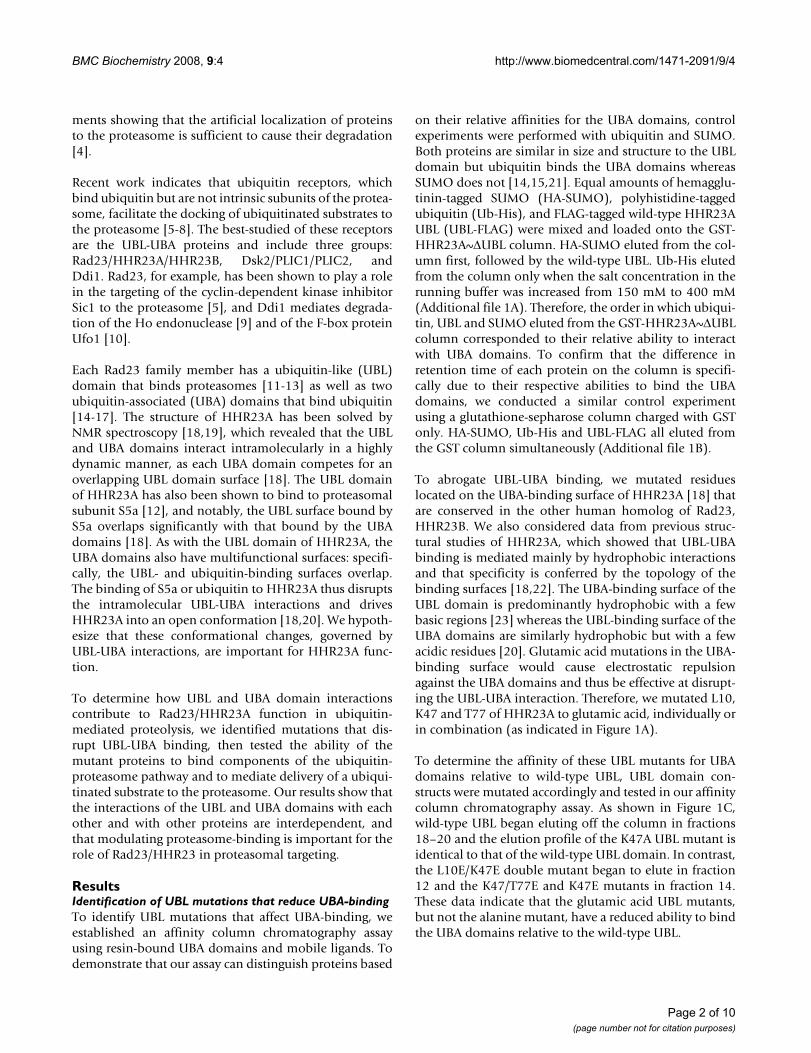
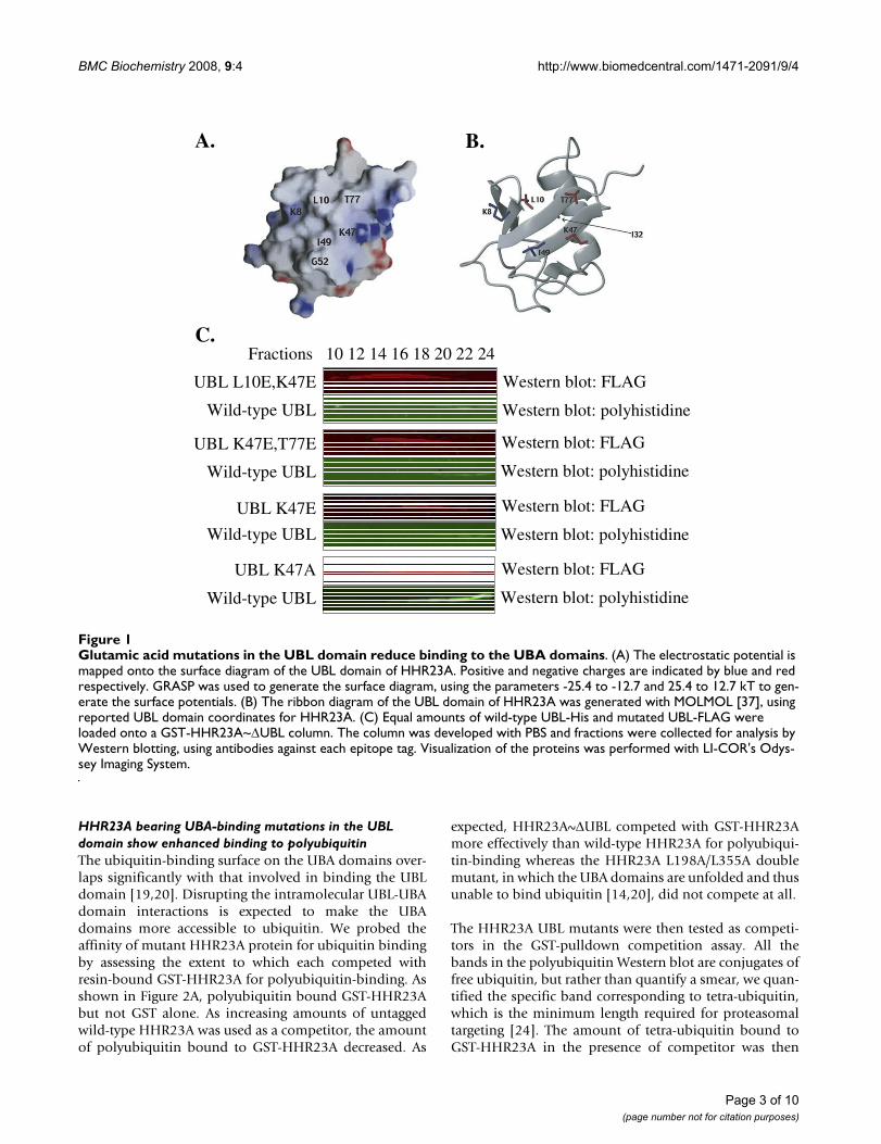
To abrogate UBL-UBA binding, we mutated residueslocated on the UBA-binding surface of HHR23A [18] thatare conserved in the other human homolog of Rad23,HHR23B. We also considered data from previous struc-tural studies of HHR23A, which showed that UBL-UBAbinding is mediated mainly by hydrophobic interactionsand that specificity is conferred by the topology of thebinding surfaces [18,22]. The UBA-binding surface of theUBL domain is predominantly hydrophobic with a fewbasic regions [23] whereas the UBL-binding surface of theUBA domains are similarly hydrophobic but with a fewacidic residues [20]. Glutamic acid mutations in the UBA-binding surface would cause electrostatic repulsionagainst the UBA domains and thus be effective at disrupt-ing the UBL-UBA interaction. Therefore, we mutated L10,K47 and T77 of HHR23A to glutamic acid, individually orin combination (as indicated in Figure 1A).
To determine the affinity of these UBL mutants for UBAdomains relative to wild-type UBL, UBL domain con-structs were mutated accordingly and tested in our affinitycolumn chromatography assay. As shown in Figure 1C,wild-type UBL began eluting off the column in fractions18–20 and the elution profile of the K47A UBL mutant isidentical to that of the wild-type UBL domain. In contrast,the L10E/K47E double mutant began to elute in fraction12 and the K47/T77E and K47E mutants in fraction 14.These data indicate that the glutamic acid UBL mutants,but not the alanine mutant, have a reduced ability to bindthe UBA domains relative to the wild-type UBL.
Page 2 of 10(page number not for citation purposes)
BMC Biochemistry 2008, 9:4 http://www.biomedcentral.com/1471-2091/9/4
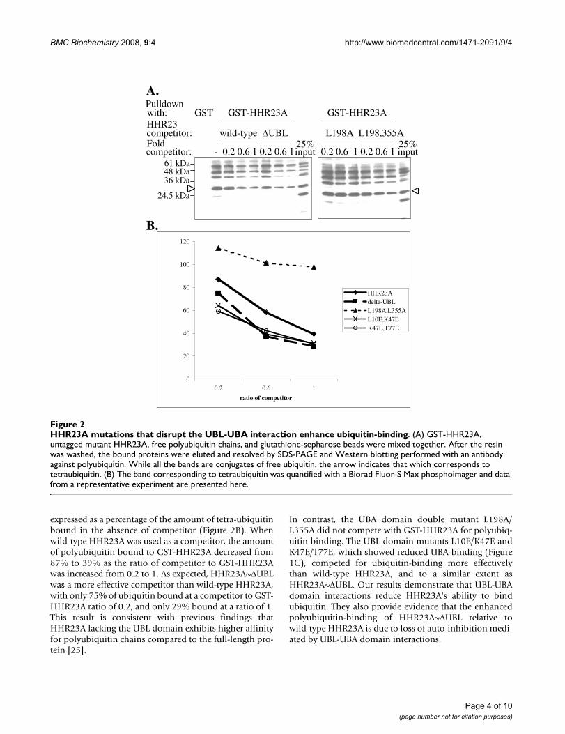
HHR23A bearing UBA-binding mutations in the UBL domain show enhanced binding to polyubiquitinThe ubiquitin-binding surface on the UBA domains over-laps significantly with that involved in binding the UBLdomain [19,20]. Disrupting the intramolecular UBL-UBAdomain interactions is expected to make the UBAdomains more accessible to ubiquitin. We probed theaffinity of mutant HHR23A protein for ubiquitin bindingby assessing the extent to which each competed withresin-bound GST-HHR23A for polyubiquitin-binding. Asshown in Figure 2A, polyubiquitin bound GST-HHR23Abut not GST alone. As increasing amounts of untaggedwild-type HHR23A was used as a competitor, the amountof polyubiquitin bound to GST-HHR23A decreased. As
expected, HHR23A~ΔUBL competed with GST-HHR23Amore effectively than wild-type HHR23A for polyubiqui-tin-binding whereas the HHR23A L198A/L355A doublemutant, in which the UBA domains are unfolded and thusunable to bind ubiquitin [14,20], did not compete at all.
The HHR23A UBL mutants were then tested as competi-tors in the GST-pulldown competition assay. All thebands in the polyubiquitin Western blot are conjugates offree ubiquitin, but rather than quantify a smear, we quan-tified the specific band corresponding to tetra-ubiquitin,which is the minimum length required for proteasomaltargeting [24]. The amount of tetra-ubiquitin bound toGST-HHR23A in the presence of competitor was then
Glutamic acid mutations in the UBL domain reduce binding to the UBA domainsFigure 1Glutamic acid mutations in the UBL domain reduce binding to the UBA domains. (A) The electrostatic potential is mapped onto the surface diagram of the UBL domain of HHR23A. Positive and negative charges are indicated by blue and red respectively. GRASP was used to generate the surface diagram, using the parameters -25.4 to -12.7 and 25.4 to 12.7 kT to gen-erate the surface potentials. (B) The ribbon diagram of the UBL domain of HHR23A was generated with MOLMOL [37], using reported UBL domain coordinates for HHR23A. (C) Equal amounts of wild-type UBL-His and mutated UBL-FLAG were loaded onto a GST-HHR23A~ΔUBL column. The column was developed with PBS and fractions were collected for analysis by Western blotting, using antibodies against each epitope tag. Visualization of the proteins was performed with LI-COR's Odys-sey Imaging System.
C.
A. B.
UBL K47E,T77E
Wild-type UBL
UBL K47E
Wild-type UBL
UBL K47A
Wild-type UBL
UBL L10E,K47E
Wild-type UBL
Fractions 10 12 14 16 18 20 22 24
Western blot: FLAG
Western blot: polyhistidine
Western blot: FLAG
Western blot: polyhistidine
Western blot: FLAG
Western blot: polyhistidine
Western blot: FLAG
Western blot: polyhistidine
Page 3 of 10(page number not for citation purposes)
BMC Biochemistry 2008, 9:4 http://www.biomedcentral.com/1471-2091/9/4
expressed as a percentage of the amount of tetra-ubiquitinbound in the absence of competitor (Figure 2B). Whenwild-type HHR23A was used as a competitor, the amountof polyubiquitin bound to GST-HHR23A decreased from87% to 39% as the ratio of competitor to GST-HHR23Awas increased from 0.2 to 1. As expected, HHR23A~ΔUBLwas a more effective competitor than wild-type HHR23A,with only 75% of ubiquitin bound at a competitor to GST-HHR23A ratio of 0.2, and only 29% bound at a ratio of 1.This result is consistent with previous findings thatHHR23A lacking the UBL domain exhibits higher affinityfor polyubiquitin chains compared to the full-length pro-tein [25].
In contrast, the UBA domain double mutant L198A/L355A did not compete with GST-HHR23A for polyubiq-uitin binding. The UBL domain mutants L10E/K47E andK47E/T77E, which showed reduced UBA-binding (Figure1C), competed for ubiquitin-binding more effectivelythan wild-type HHR23A, and to a similar extent asHHR23A~ΔUBL. Our results demonstrate that UBL-UBAdomain interactions reduce HHR23A's ability to bindubiquitin. They also provide evidence that the enhancedpolyubiquitin-binding of HHR23A~ΔUBL relative towild-type HHR23A is due to loss of auto-inhibition medi-ated by UBL-UBA domain interactions.
HHR23A mutations that disrupt the UBL-UBA interaction enhance ubiquitin-bindingFigure 2HHR23A mutations that disrupt the UBL-UBA interaction enhance ubiquitin-binding. (A) GST-HHR23A, untagged mutant HHR23A, free polyubiquitin chains, and glutathione-sepharose beads were mixed together. After the resin was washed, the bound proteins were eluted and resolved by SDS-PAGE and Western blotting performed with an antibody against polyubiquitin. While all the bands are conjugates of free ubiquitin, the arrow indicates that which corresponds to tetraubiquitin. (B) The band corresponding to tetraubiquitin was quantified with a Biorad Fluor-S Max phosphoimager and data from a representative experiment are presented here.
0
20
40
60
80
100
120
0.2 0.6 1
ratio of competitor
HHR23Adelta-UBLL198A,L355AL10E,K47EK47E,T77E
A.
B.
competitor: wild-type ∆UBL
with: GST GST-HHR23A GST-HHR23A
L198A L198,355A
61 kDa48 kDa36 kDa
24.5 kDa
- 0.2 0.6 1 0.2 0.6 125%
0.2 0.6 1 0.2 0.6 1competitor:Fold
input input25%
HHR23
Pulldown
Page 4 of 10(page number not for citation purposes)
BMC Biochemistry 2008, 9:4 http://www.biomedcentral.com/1471-2091/9/4
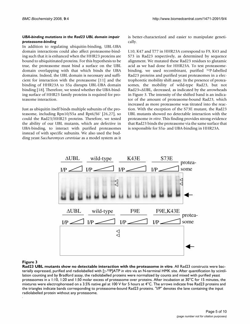
UBA-binding mutations in the Rad23 UBL domain impair proteasome-bindingIn addition to regulating ubiquitin-binding, UBL-UBAdomain interactions could also affect proteasome-bind-ing such that it is enhanced when the HHR23 proteins arebound to ubiquitinated proteins. For this hypothesis to betrue, the proteasome must bind a surface on the UBLdomain overlapping with that which binds the UBAdomains. Indeed, the UBL domain is necessary and suffi-cient for interaction with the proteasome [11] and thebinding of HHR23A to S5a disrupts UBL-UBA domainbinding [18]. Therefore, we tested whether the UBA-bind-ing surface of HHR23 family proteins is required for pro-teasome interaction.
Just as ubiquitin itself binds multiple subunits of the pro-teasome, including Rpn10/S5a and Rpt6/S6' [26,27], socould the Rad23/HHR23 proteins. Therefore, we testedthe ability of our UBL mutants, which are defective inUBA-binding, to interact with purified proteasomesinstead of with specific subunits. We also used the bud-ding yeast Saccharomyces cerevisiae as a model system as it
is better-characterized and easier to manipulate geneti-cally.
L10, K47 and T77 in HHR23A correspond to F9, K43 andS73 in Rad23 respectively, as determined by sequencealignment. We mutated these Rad23 residues to glutamicacid as we had done for HHR23A. To test proteasome-binding, we used recombinant, purified 32P-labelledRad23 proteins and purified yeast proteasomes in a elec-trophoretic mobility shift assay. In the presence of protea-somes, the mobility of wild-type Rad23, but notRad23~ΔUBL, decreased, as indicated by the arrowheadsin Figure 3. The intensity of the shifted band is an indica-tor of the amount of proteasome-bound Rad23, whichincreased as more proteasome was titrated into the reac-tion. With the exception of the S73E mutant, the Rad23UBL mutants showed no detectable interaction with theproteasome in vitro. This finding provides strong evidencethat Rad23 binds the proteasome via the same surface thatis responsible for S5a- and UBA-binding in HHR23A.
Rad23 UBL mutants show no detectable interaction with the proteasome in vitroFigure 3Rad23 UBL mutants show no detectable interaction with the proteasome in vitro. All Rad23 constructs were bac-terially expressed, purified and radiolabelled with [γ-32P]ATP in vitro via an N-terminal HMK site. After quantification by scintil-lation counting and by Bradford assay, the radiolabelled proteins were normalized by counts and mixed with purified yeast proteasomes in a 1:10, 1:20 and 1:50 molar excess of proteasome over proteins. After incubation at 30°C for 15 minutes, the mixtures were electrophoresed on a 3.5% native gel at 100 V for 5 hours at 4°C. The arrows indicate free Rad23 proteins and the triangles indicate bands corresponding to proteasome-bound Rad23 proteins. "I/P" denotes the lane containing the input radiolabelled protein without any proteasome.
I/P
�UBL wild-type F9E F9E,K43Eprotea-someI/P I/P I/P
I/P
�UBL wild-type K43E S73Eprotea-someI/P I/P I/P
Page 5 of 10(page number not for citation purposes)
BMC Biochemistry 2008, 9:4 http://www.biomedcentral.com/1471-2091/9/4
Mutation of the Rad23 UBL domain impairs interactions with Ufd2In addition to the proteasome, the Rad23/HHR23A UBLdomain binds other proteins involved in ubiquitin-medi-ated degradation, including the ubiquitin ligase Ufd2[6,28]. We thus tested whether our UBL mutants retaintheir ability to interact with Ufd2. We used bacteriallyexpressed and purified GST-Ufd2 and Rad23 proteins inGST-pulldown experiments, following which the boundRad23 proteins were detected by Western blotting forRad23. As expected, wild-type Rad23 bound to GST-Ufd2but not to GST alone, whereas Rad23ΔUBL did not bindto GST-Ufd2 (Figure 4). Interestingly, Ufd2-binding wassignificantly reduced by the mutation of F9, but not ofK43 or of S73. This finding suggests that hydrophobicinteractions govern Ufd2 binding to Rad23, and that thesurfaces of Rad23 bound by Ufd2 and the proteasome doindeed overlap partially.
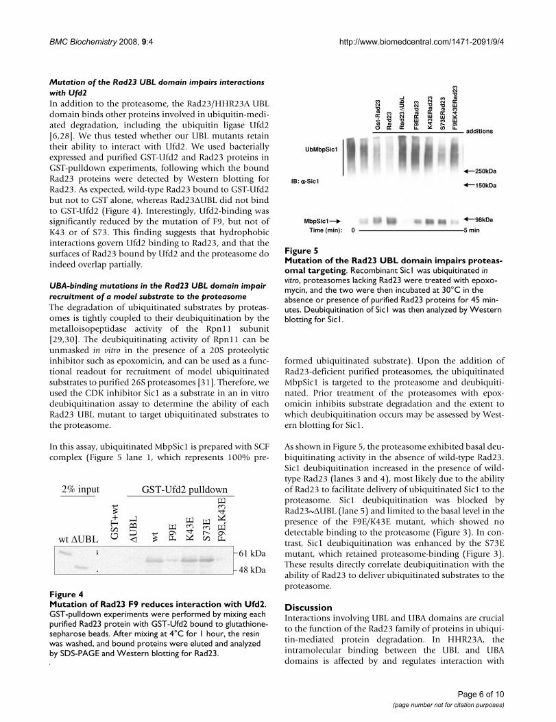
UBA-binding mutations in the Rad23 UBL domain impair recruitment of a model substrate to the proteasomeThe degradation of ubiquitinated substrates by proteas-omes is tightly coupled to their deubiquitination by themetalloisopeptidase activity of the Rpn11 subunit[29,30]. The deubiquitinating activity of Rpn11 can beunmasked in vitro in the presence of a 20S proteolyticinhibitor such as epoxomicin, and can be used as a func-tional readout for recruitment of model ubiquitinatedsubstrates to purified 26S proteasomes [31]. Therefore, weused the CDK inhibitor Sic1 as a substrate in an in vitrodeubiquitination assay to determine the ability of eachRad23 UBL mutant to target ubiquitinated substrates tothe proteasome.
In this assay, ubiquitinated MbpSic1 is prepared with SCFcomplex (Figure 5 lane 1, which represents 100% pre-
formed ubiquitinated substrate). Upon the addition ofRad23-deficient purified proteasomes, the ubiquitinatedMbpSic1 is targeted to the proteasome and deubiquiti-nated. Prior treatment of the proteasomes with epox-omicin inhibits substrate degradation and the extent towhich deubiquitination occurs may be assessed by West-ern blotting for Sic1.
As shown in Figure 5, the proteasome exhibited basal deu-biquitinating activity in the absence of wild-type Rad23.Sic1 deubiquitination increased in the presence of wild-type Rad23 (lanes 3 and 4), most likely due to the abilityof Rad23 to facilitate delivery of ubiquitinated Sic1 to theproteasome. Sic1 deubiquitination was blocked byRad23~ΔUBL (lane 5) and limited to the basal level in thepresence of the F9E/K43E mutant, which showed nodetectable binding to the proteasome (Figure 3). In con-trast, Sic1 deubiquitination was enhanced by the S73Emutant, which retained proteasome-binding (Figure 3).These results directly correlate deubiquitination with theability of Rad23 to deliver ubiquitinated substrates to theproteasome.
DiscussionInteractions involving UBL and UBA domains are crucialto the function of the Rad23 family of proteins in ubiqui-tin-mediated protein degradation. In HHR23A, theintramolecular binding between the UBL and UBAdomains is affected by and regulates interaction with
Mutation of the Rad23 UBL domain impairs proteasomal tar-getingFigure 5Mutation of the Rad23 UBL domain impairs proteas-omal targeting. Recombinant Sic1 was ubiquitinated in vitro, proteasomes lacking Rad23 were treated with epoxo-mycin, and the two were then incubated at 30°C in the absence or presence of purified Rad23 proteins for 45 min-utes. Deubiquitination of Sic1 was then analyzed by Western blotting for Sic1.
Time (min): 5 min
Rad
23
Gst
-Rad
23
F9E
Rad
23
K43
ER
ad23
S73
ER
ad23
F9E
K43
ER
ad23
UbMbpSic1
MbpSic1
additions
IB: αααα-Sic1
Rad
23�U
bL
0
250kDa
150kDa
98kDa
Mutation of Rad23 F9 reduces interaction with Ufd2Figure 4Mutation of Rad23 F9 reduces interaction with Ufd2. GST-pulldown experiments were performed by mixing each purified Rad23 protein with GST-Ufd2 bound to glutathione-sepharose beads. After mixing at 4°C for 1 hour, the resin was washed, and bound proteins were eluted and analyzed by SDS-PAGE and Western blotting for Rad23.
wt �UBL
2% input GST-Ufd2 pulldown
61 kDa
48 kDa
GS
T+
wt
�UB
L
wt
F9E
K43
E
S73
E
F9E
,K43
E
Page 6 of 10(page number not for citation purposes)
BMC Biochemistry 2008, 9:4 http://www.biomedcentral.com/1471-2091/9/4
other proteins [18]. We show that disruption of theintramolecular UBL-UBA binding facilitates HHR23Ainteraction with polyubiquitin. Our data support themodel that binding to either proteasomes or polyubiqui-tin disrupts the UBL-UBA interaction, causing HHR23A toadopt an open conformation that facilitates its interactionwith polyubiquitinated proteins or proteasomes respec-tively. This coupling of proteasome-binding and sub-strate-binding renders the Rad23 family more efficient indocking ubiquitinated substrates with the proteasome.Indeed, ubiquitin chains have been shown to enhanceRad23 binding to the proteasome in vivo [32]. Also, UBL-UBA domain interactions may enhance specificity byblocking interactions that are weaker than the intramo-lecular UBL-UBA interaction.
The UBA-binding surface of the Rad23/HHR23A UBLdomain also mediates binding to the proteasomal subunitS5a as well as to other proteins such as the ubiquitin ligaseUfd2. We had targeted residues on the edge of the UBA-binding surface in an attempt to minimize disruptingother interactions but our mutations also affected protea-some-binding and Ufd2-binding. Our results are consist-ent with a previous report suggesting that the bindingsurfaces on the UBL domain for Ufd2 and the proteasomemay overlap [28]. The difficulty of specifically disruptinginteraction with only one protein without affecting otherssuggests that these different binding surfaces overlap sig-nificantly on the UBL domain and that the various inter-actions may regulate one another. The use of the samesurface on Rad23/HHR23A for both proteasome- andUfd2-binding is consistent with a model in which Rad23first binds ubiquitin conjugates associated with Ufd2,then dissociates from Ufd2 and binds the proteasome, towhich the ubiquitin conjugates are thus delivered. Thismodel contrasts with one in which Rad23 bridges Ufd2and the proteasome by binding them simultaneously.
The UBL mutants tested constitute an allelic series, withthe S73E mutant behaving most similarly to the wild-typeUBL, following by K43E and then F9E. Despite the lack ofdetectable proteasome-binding in the native gel assay,which is based on fractionation of the interacting species,the UBL mutants possess sufficient residual affinity thatthey can still interact functionally with proteasomes, asevidenced by the deubiquitination of Sic1. Rad23ΔUBLdoes not interact with proteasomes, yet it inhibited deu-biquitination of Sic1. This dominant negative effect arisesfrom a sequestering mechanism, as Rad23ΔUBL can stillbind ubiquitin chains and thus interfere with recognitionby intrinsic ubiquitin receptors and other shuttling fac-tors. In contrast, Sic1 was deubiquitinated in the presenceof the UBL mutants in a manner that is consistent witheach mutant's relative ability to bind proteasomes. Thesedata indicate that the capacity of Rad23 to bind proteas-
omes is essential for the ability of Rad23 family proteinsto target ubiquitinated substrates to the proteasome. Thismodel contrasts one in which ubiquitin chains bind theproteasome directly.
Interestingly, HHR23A does not dimerize [18] whereasRad23 does so in a manner that involves the C-terminalUBA domain [33,34]. Rad23 can also heterodimerize withother UBL-UBA proteins such as Ddi1 [33,34] whileHHR23A interacts with hPLIC2 [35]. Therefore, intermo-lecular UBL-UBA domain interactions may also play a reg-ulatory role in the recruitment of ubiquitinated substratesto the proteasome.
ConclusionIn conclusion, both the intramolecular as well as intermo-lecular interactions of the UBL and UBA domains areimportant for the function of Rad23 family proteins in theubiquitin-proteasome pathway. In this study we haveshown that physical interactions between the UBL andUBA domains couple proteasome binding via the UBLdomain with ubiquitin-binding via the UBA domain, thuspriming Rad23/HHR23A for its role in shuttling ubiquiti-nated substrates to the proteasome.
MethodsPlasmidsFor bacterial expression, proteins were cloned into eitherpGEX-6p-1 (Pharmacia) or pET-23a (Novagen). Whereapplicable, the FLAG tag was inserted by QuikChange-XLmutagenesis (Stratagene). We used pGEX2TK-based plas-mids to express Rad23 and Rad23ΔUBL proteins for radi-olabelling [7]. HHR23A (GenBank:P54725), HHR23AUBL mutants, HHR23B (GenBank:P54727), and Rad23(GenBank:P32628) UBL mutants were cloned into"pGEX-6pK," in which the heart muscle kinase recogni-tion site present in pGEX-2TK was inserted upstream ofthe pGEX-6p-1 multiple cloning site by QuikChange-XLmutagenesis (Stratagene). The GST-Ufd2-myc plasmidhas been previously described [28].
Protein expression and purificationProtein expression was induced in BL21(DE3) bacteriawith 0.4 mM isopropyl β-D-thiogalactoside (IPTG) for 4hours at 37°C. For GST-fusion protein purification, bacte-ria were lysed by sonication in PBS containing 2 mM DTT,protease inhibitor cocktail (Roche), and 1% Triton X-100.The proteins were purified on glutathione-sepharosebeads and either eluted with 10 mM reduced glutathionein 50 mM Tris-HCl pH 8.0 or cleaved from the resin-bound GST with PreScission protease (pGEX-6p1 pro-teins) or thrombin (pGEX-2TK proteins) according to themanufacturer's instructions. For the purification of poly-histidine-tagged proteins, the bacteria were lysed in 25mM sodium phosphate pH 8, 300 mM NaCl, 10 mM imi-
Page 7 of 10(page number not for citation purposes)
BMC Biochemistry 2008, 9:4 http://www.biomedcentral.com/1471-2091/9/4
dazole, 2 mM DTT and protease inhibitor cocktail(Roche). The polyhistidine-tagged proteins were bound toNi-NTA resin (QIAGEN), washed with 50 mM sodiumphosphate pH 6.0, 300 mM NaCl and 20 mM imidazole.The proteins were then eluted with 25 mM Tris-HCl pH 8,300 mM NaCl, 2 mM DTT and 0.25 M imidazole. Purifiedproteins were dialyzed overnight against PBS, 10% glyc-erol and 2 mM DTT. Protein purity was verified by SDS-PAGE analysis and Coomassie Blue staining.
Free polyubiquitin chains were synthesized in vitro byincubating 100 nM E1 (Boston Biochem), 10 μM E2-25K(Boston Biochem), and 40 μg ubiquitin (Sigma) with 25mM Tris-HCl pH 7.6, 50 mM NaCl, 0.1 mM DTT, 5 mMMgCl2, 4 mM ATP (Sigma), 0.03 mg/ml creatine kinase, 5mM creatine phosphate, and 0.3 unit/ml pyrophos-phatase, at 30°C for 2 hours.
Affinity column chromatographyGlutathione sepharose was saturated with purified GST-HHR23A~ΔUBL. The charged resin was then loaded intoa 10 ml HR10/10 (Pharmacia) column. 1 μg each of wild-type UBL-His and mutated UBL-FLAG were mixed andloaded onto the GST-HHR23A~ΔUBL column, followingwhich the column was developed in PBS. 0.5 ml fractionswere collected, resolved by SDS-PAGE and the proteinswere detected by Western blotting. Antibodies usedincluded anti-FLAG M2 monoclonal antibody (Sigma),anti-HA 12CA5 monoclonal antibody (produced in ourlab), and anti-polyhistidine affinity-purified rabbit poly-clonal antibody (Rockland). Use of the Odyssey ImagingSystem (LI-COR Biotechnology) enabled each protein tobe visualized simultaneously but distinctly.
Competition assay to assess polyubiquitin-bindingGST-HHR23A, untagged mutant variants of GST-HHR23A, free polyubiquitin chains and glutathione-sepharose beads were mixed for 1–2 hours at 4°C in bind-ing buffer (20 mM Tris-HCl pH 7.2, 150 mM NaCl, 2 mMEDTA, 2% (v/v) glycerol, 1 mM DTT) [14] supplementedwith 1% BSA. The resin was washed with binding buffersupplemented with 0.5% NP40. Proteins were elutedfrom the glutathione-sepharose resin and analyzed byWestern blotting for polyubiquitin (FK2 monoclonalantibody, Affiniti Research Products). The band corre-sponding to tetra-ubiquitin was quantified with a BioradFluor-S Max phosphoimager. Total binding was definedas the amount of polyubiquitin bound to the GST-HHR23A in the absence of competitor.
Native gel assays for proteasome-bindingProteasomes were purified from S. cerevisiae as previouslydescribed [7]. All Rad23/HHR23 proteins were bacteriallyexpressed, purified on glutathione-sepharose beads, andlabelled in vitro with [γ-32P]ATP (NEN) and heart muscle
kinase according to the manufacturer's instructions (Phar-macia Biotech). The radiolabelled proteins were cleavedfrom the resin with thrombin or PreScission protease,then quantified by scintillation counting and by Bradfordassay. The specific activities of the proteins were normal-ized and mixed with proteasomes in a 1:50, 1:20, and1:10 molar excess of proteasome over proteins. After incu-bation at 30°C for 15 minutes, the mixtures were resolvedby 3.5% native PAGE essentially as previously described[7,36], though electrophoresis was carried out for 5 hoursand the proteins visualized by autoradiography.
GST-pulldown assay for Ufd2-bindingGlutathione sepharose was saturated with purified GST-Ufd2. Purified Rad23 proteins were then added togetherwith 50 mM HEPES pH 7.5, 150 mM NaCl, 5 mM EDTA,2% Triton X-100, 0.2 mg/ml BSA, and protease inhibitorcocktail (Roche). After mixing at 4°C for 60 minutes, theresin was washed with PBS and 0.1% Tween-20. The resin-bound Rad23 proteins were detected by Western blottingusing an antibody against Rad23 (a gift from KiranMadura).
In vitro deubiquitination assayThe in vitro deubiquitination assay was performed as pre-viously described [5]. Essentially, proteasomes were puri-fied from Rad23-deficient S. cerevisiae and preincubatedwith 100 μM epoxomicin for 45 minutes at 30°C toinhibit the protease activity of the proteasome. Ubiquiti-nated MbpSic1 was then added to the proteasome and itsdeubiquitination was analyzed by Western blotting forSic1. Rad23 was added to the deubiquitination assaywhere indicated.
AbbreviationsUBL, ubiquitin-like; UBA, ubiquitin-associated; His, poly-histidine; Ub, ubiquitin; HHR23, Human Homolog ofRad23; S. cerevisiae, Saccharomyces cerevisiae.
Authors' contributionsAMG participated in the design of the study, carried outexperiments and drafted the manuscript. KJW created themodels of the UBL domains, participated in the design ofthe study and helped to revise the manuscript. SE partici-pated in the design of the study, contributed reagents, andhelped to revise the manuscript. RV performed the in vitrodeubiquitination studies and helped to revise the manu-script. RJD and DF participated in the design of the studyand helped to revise the manuscript. PMH participated inthe design and coordination of the study and helped torevise the manuscript. All authors read and approved thefinal manuscript.
Page 8 of 10(page number not for citation purposes)
BMC Biochemistry 2008, 9:4 http://www.biomedcentral.com/1471-2091/9/4
Additional material
AcknowledgementsWe are grateful to Shahram Misaghi for assistance with the affinity column chromatography experiments, to Eric Cooper and Michal-Ruth Schweiger for helpful discussions, and to Yang Kang for help with the model of the HHR23A UBL domain. We thank Hai Rao for the GST-Ufd2-myc plasmid and Kiran Madura for the Rad23 antibody. We also thank Jennie Croyle and Melven Chok for assistance during manuscript preparation. This work was funded by grants from the National Institutes of Health CA0970004 (KJW), GM43601 (DF) and CA64888 (PMH) as well as by a scholarship from the Agency of Science, Technology and Research of Singapore (AMG).
References1. Murray AW: Recycling the cell cycle: cyclins revisited. Cell
2004, 116:221-234.2. Rock KL, York IA, Goldberg AL: Post-proteasomal antigen
processing for major histocompatibility complex class I pres-entation. Nat Immunol 2004, 5:670-677.
3. Pickart CM, Fushman D: Polyubiquitin chains: polymeric pro-tein signals. Curr Opin Chem Biol 2004, 8:610-616.
4. Janse DM, Crosas B, Finley D, Church GM: Localization to theproteasome is sufficient for degradation. J Biol Chem 2004,279:21415-21420.
5. Verma R, Oania R, Graumann J, Deshaies RJ: Multiubiquitin chainreceptors define a layer of substrate selectivity in the ubiqui-tin-proteasome system. Cell 2004, 118:99-110.
6. Richly H, Rape M, Braun S, Rumpf S, Hoege C, Jentsch S: A series ofubiquitin binding factors connects CDC48/p97 to substratemultiubiquitylation and proteasomal targeting. Cell 2005,120:73-84.
7. Elsasser S, Chandler-Militello D, Muller B, Hanna J, Finley D: Rad23and Rpn10 serve as alternative ubiquitin receptors for theproteasome. J Biol Chem 2004, 279:26817-26822.
8. Chen L, Madura K: Rad23 promotes the targeting of proteo-lytic substrates to the proteasome. Mol Cell Biol 2002,22:4902-4913.
9. Kaplun L, Tzirkin R, Bakhrat A, Shabek N, Ivantsiv Y, Raveh D: TheDNA damage-inducible UbL-UbA protein Ddi1 participatesin Mec1-mediated degradation of Ho endonuclease. Mol CellBiol 2005, 25:5355-5362.
10. Ivantsiv Y, Kaplun L, Tzirkin-Goldin R, Shabek N, Raveh D: Uniquerole for the UbL-UbA protein Ddi1 in turnover of SCFUfo1complexes. Mol Cell Biol 2006, 26:1579-1588.
11. Elsasser S, Gali RR, Schwickart M, Larsen CN, Leggett DS, Muller B,Feng MT, Tubing F, Dittmar GA, Finley D: Proteasome subunitRpn1 binds ubiquitin-like protein domains. Nat Cell Biol 2002,4:725-730.
12. Hiyama H, Yokoi M, Masutani C, Sugasawa K, Maekawa T, Tanaka K,Hoeijmakers JH, Hanaoka F: Interaction of hHR23 with S5a. Theubiquitin-like domain of hHR23 mediates interaction withS5a subunit of 26 S proteasome. J Biol Chem 1999,274:28019-28025.
13. Schauber C, Chen L, Tongaonkar P, Vega I, Lambertson D, Potts W,Madura K: Rad23 links DNA repair to the ubiquitin/proteas-ome pathway. Nature 1998, 391:715-718.
14. Bertolaet BL, Clarke DJ, Wolff M, Watson MH, Henze M, Divita G,Reed SI: UBA domains of DNA damage-inducible proteinsinteract with ubiquitin. Nat Struct Biol 2001, 8:417-422.
15. Chen L, Shinde U, Ortolan TG, Madura K: Ubiquitin-associated(UBA) domains in Rad23 bind ubiquitin and promote inhibi-tion of multi-ubiquitin chain assembly. EMBO Rep 2001,2:933-938.
16. Rao H, Sastry A: Recognition of specific ubiquitin conjugates isimportant for the proteolytic functions of the ubiquitin-asso-ciated domain proteins Dsk2 and Rad23. J Biol Chem 2002,277:11691-11695.
17. Wilkinson CR, Seeger M, Hartmann-Petersen R, Stone M, Wallace M,Semple C, Gordon C: Proteins containing the UBA domain areable to bind to multi-ubiquitin chains. Nat Cell Biol 2001,3:939-943.
18. Walters KJ, Lech PJ, Goh AM, Wang Q, Howley PM: DNA-repairprotein hHR23a alters its protein structure upon bindingproteasomal subunit S5a. Proc Natl Acad Sci U S A 2003,100:12694-12699.
19. Ryu KS, Lee KJ, Bae SH, Kim BK, Kim KA, Choi BS: Binding surfacemapping of intra- and interdomain interactions amonghHR23B, ubiquitin, and polyubiquitin binding site 2 of S5a. JBiol Chem 2003, 278:36621-36627.
20. Wang Q, Goh AM, Howley PM, Walters KJ: Ubiquitin recognitionby the DNA repair protein hHR23a. Biochemistry 2003,42:13529-13535.
21. Raasi S, Orlov I, Fleming KG, Pickart CM: Binding of polyubiquitinchains to ubiquitin-associated (UBA) domains of HHR23A. JMol Biol 2004, 341:1367-1379.
22. Mueller TD, Kamionka M, Feigon J: Specificity of the interactionbetween ubiquitin-associated domains and ubiquitin. J BiolChem 2004, 279:11926-11936.
23. Walters KJ, Goh AM, Wang Q, Wagner G, Howley PM: Ubiquitinfamily proteins and their relationship to the proteasome: astructural perspective. Biochim Biophys Acta 2004, 1695:73-87.
24. Thrower JS, Hoffman L, Rechsteiner M, Pickart CM: Recognition ofthe polyubiquitin proteolytic signal. Embo J 2000, 19:94-102.
25. Raasi S, Pickart CM: Rad23 ubiquitin-associated domains(UBA) inhibit 26 S proteasome-catalyzed proteolysis bysequestering lysine 48-linked polyubiquitin chains. J Biol Chem2003, 278:8951-8959.
26. Deveraux Q, Ustrell V, Pickart C, Rechsteiner M: A 26 S proteasesubunit that binds ubiquitin conjugates. J Biol Chem 1994,269:7059-7061.
27. Lam YA, Lawson TG, Velayutham M, Zweier JL, Pickart CM: A pro-teasomal ATPase subunit recognizes the polyubiquitin deg-radation signal. Nature 2002, 416:763-767.
28. Kim I, Mi K, Rao H: Multiple interactions of rad23 suggest amechanism for ubiquitylated substrate delivery important inproteolysis. Mol Biol Cell 2004, 15:3357-3365.
29. Verma R, Aravind L, Oania R, McDonald WH, Yates JR 3rd, KooninEV, Deshaies RJ: Role of Rpn11 metalloprotease in deubiquiti-nation and degradation by the 26S proteasome. Science 2002,298:611-615.
30. Yao T, Cohen RE: A cryptic protease couples deubiquitinationand degradation by the proteasome. Nature 2002,419:403-407.
31. Verma R, Deshaies RJ: Assaying degradation and deubiquitina-tion of a ubiquitinated substrate by purified 26S proteas-omes. Methods Enzymol 2005, 398:391-399.
32. Ghaboosi N, Deshaies RJ: A conditional yeast E1 mutant blocksthe ubiquitin-proteasome pathway and reveals a role forubiquitin conjugates in targeting Rad23 to the proteasome.Mol Biol Cell 2007, 18:1953-1963.
33. Bertolaet BL, Clarke DJ, Wolff M, Watson MH, Henze M, Divita G,Reed SI: UBA domains mediate protein-protein interactionsbetween two DNA damage-inducible proteins. J Mol Biol 2001,313:955-963.
Additional file 1Retention of proteins on the GST-HHR23A~ΔUBL column corre-sponds to their ability to interact with UBA domains. (A) Purified GST-HHR23A~ΔUBL protein was used to saturate glutathione-sepharose resin, which was in turn used to pack an HR10/10 column. Equal amounts of HA-tagged SUMO (HA-SUMO), polyhistidine-tagged ubiq-uitin (Ub-His) and FLAG-tagged UBL (UBL-FLAG) were loaded simul-taneously onto the column and the column was resolved in PBS. Fractions were collected and analyzed by Western blotting using epitope-specific antibodies. (B) Purified GST was bound to glutathione-sepharose resin in excess, which was then used to pack an HR10/10 column. Equal amounts of HA-SUMO, Ub-His and UBL-FLAG were mixed, loaded onto the col-umn and analyzed as described above.Click here for file[http://www.biomedcentral.com/content/supplementary/1471-2091-9-4-S1.PPT]
Page 9 of 10(page number not for citation purposes)
http://www.ncbi.nlm.nih.gov/entrez/query.fcgi?cmd=Retrieve&db=PubMed&dopt=Abstract&list_uids=9490418
http://www.ncbi.nlm.nih.gov/entrez/query.fcgi?cmd=Retrieve&db=PubMed&dopt=Abstract&list_uids=9490418
http://www.ncbi.nlm.nih.gov/entrez/query.fcgi?cmd=Retrieve&db=PubMed&dopt=Abstract&list_uids=8125911
BMC Biochemistry 2008, 9:4 http://www.biomedcentral.com/1471-2091/9/4
Publish with BioMed Central and every scientist can read your work free of charge
"BioMed Central will be the most significant development for disseminating the results of biomedical research in our lifetime."
Sir Paul Nurse, Cancer Research UK
Your research papers will be:
available free of charge to the entire biomedical community
peer reviewed and published immediately upon acceptance
cited in PubMed and archived on PubMed Central
yours — you keep the copyright
Submit your manuscript here:http://www.biomedcentral.com/info/publishing_adv.asp
BioMedcentral
34. Kang Y, Vossler RA, Diaz-Martinez LA, Winter NS, Clarke DJ, Wal-ters KJ: UBL/UBA ubiquitin receptor proteins bind a com-mon tetraubiquitin chain. J Mol Biol 2006, 356:1027-1035.
35. Kang Y, Zhang N, Koepp DM, Walters KJ: Ubiquitin receptor pro-teins hHR23a and hPLIC2 interact. J Mol Biol 2007,365:1093-1101.
36. Elsasser S, Finley D: Delivery of ubiquitinated substrates to pro-tein-unfolding machines. Nat Cell Biol 2005, 7:742-749.
37. Koradi R, Billeter M, Wuthrich K: MOLMOL: a program for dis-play and analysis of macromolecular structures. J Mol Graph1996, 14:51-5, 29-32.
Page 10 of 10(page number not for citation purposes)
http://www.ncbi.nlm.nih.gov/entrez/query.fcgi?cmd=Retrieve&db=PubMed&dopt=Abstract&list_uids=8744573
Related Documents