COMPLICATIONS OF SINUSITIS ATIN BINDAL 11M2321

Complications of sinusitis
Jul 16, 2015
Welcome message from author
This document is posted to help you gain knowledge. Please leave a comment to let me know what you think about it! Share it to your friends and learn new things together.
Transcript
COMPLICATIONS OF
SINUSITIS
ATIN BINDAL11M2321

Complications of sinusitis
As long as infection is confined only to the sinus mucosa,it is called sinusitis. Complications are said to arise when infection spreads into or beyond the bony wall of the sinus.

A- Local Mucocele/PyoceleMucous retention cystOsteomyelitis- frontal bone and maxila
B- Orbital Preseptal inflammatory oedema of lidsSubperiosteal abscessOrbital cellulitisOrbital abscessSuperior orbital fissure syndromeOrbital apex syndrome
C- Intacranial MeningitisExtradural abscessSubdural abscessBrain abscessCavernous sinus thrombosis
D- Descendinginfections
E- Focal infections
TYPES :
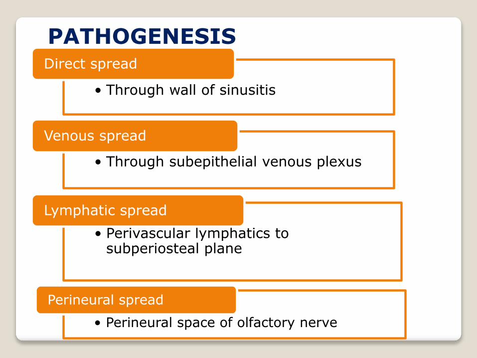
• Perineural space of olfactory nerve
Perineural spread
PATHOGENESIS
• Through wall of sinusitis
Direct spread
• Through subepithelial venous plexus
Venous spread
• Perivascular lymphatics to subperiosteal plane
Lymphatic spread
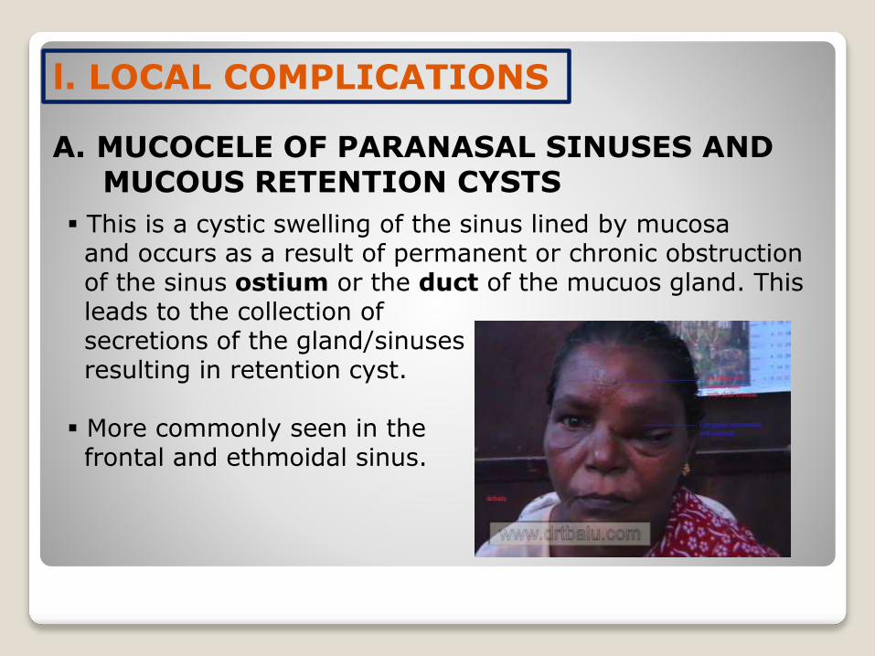
l. LOCAL COMPLICATIONS
A. MUCOCELE OF PARANASAL SINUSES AND MUCOUS RETENTION CYSTS
This is a cystic swelling of the sinus lined by mucosa and occurs as a result of permanent or chronic obstruction of the sinus ostium or the duct of the mucuos gland. This leads to the collection ofsecretions of the gland/sinuses resulting in retention cyst.
More commonly seen in the frontal and ethmoidal sinus.

Frontal sinus mucocele:Usually presents in the superomedial quadrant of the orbitCommonly occurs as a result of obstruction to the frontal
ostium due to chronic disease of the frontal recess or dueto postsurgical/traumatic fibrosis causing blockage.
Clinical Features:
Symptoms:•Supraorbital swelling usually above and lateral to medial canthus.•Diplopia may be present due to proptosis.•Headache is usually confined to the frontal region, mild type.
Signs :•Proptosis•Swelling is usually cystic and non-tender and egg shell cracking
may be elicited.

Investigations:•X-ray PNS- show cloudiness of the affected frontalsinus with loss of scalloping.
•CT scan of the osteomeatal complex and PNS.•Diagnostic nasal endoscopy.
Treatment:•Endoscopic sinus surgery with frontal recess clearance and uncapping of the mucocele is the treatment of choice.•Alternatively external frontoethmo-diectomy(Lynch-Howarth operation) or osteoplastic flap operation by bicoronal incision may be done.•In case of pyocele a course of antibiotics should be given prior to surgery.
B. OSTEOMYELITIS
• Osteomyelitis is infection of bone marrow and should be differentiated from osteitis which is infection of compact bone. It involves either maxilla or frontal bone.
1. Osteomyelitis of maxilla
• More often seen in infants and children because of presence of spongy bone in the anterior wall of the maxilla.
• Clinical features: Erythema, swelling of cheek, lower lid oedema, purulent nasal discharge and fever.
• Subperiosteal abscess followed by fistulae may form in infraorbitalregion,alveolus, or in zygoma.
• Sequestration of bone may occur.
• Treatment: Large doses of antibiotics, drainage of any abscess and sequestra removal.
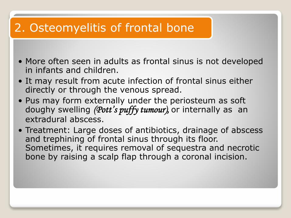
2. Osteomyelitis of frontal bone
• More often seen in adults as frontal sinus is not developed in infants and children.
• It may result from acute infection of frontal sinus either directly or through the venous spread.
• Pus may form externally under the periosteum as soft doughy swelling (Pott’s puffy tumour), or internally as an extradural abscess.
• Treatment: Large doses of antibiotics, drainage of abscess and trephining of frontal sinus through its floor. Sometimes, it requires removal of sequestra and necrotic bone by raising a scalp flap through a coronal incision.
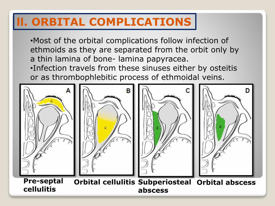
ll. ORBITAL COMPLICATIONS
•Most of the orbital complications follow infection of ethmoids as they are separated from the orbit only by a thin lamina of bone- lamina papyracea.•Infection travels from these sinuses either by osteitisor as thrombophlebitic process of ethmoidal veins.
Pre-septalcellulitis
Orbital cellulitis Subperiostealabscess
Orbital abscess
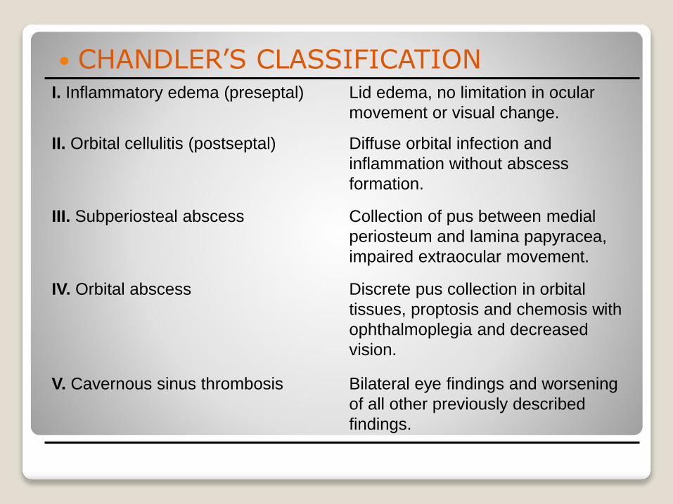
I. Inflammatory edema (preseptal) Lid edema, no limitation in ocular
movement or visual change.
II. Orbital cellulitis (postseptal) Diffuse orbital infection and
inflammation without abscess
formation.
III. Subperiosteal abscess Collection of pus between medial
periosteum and lamina papyracea,
impaired extraocular movement.
IV. Orbital abscess Discrete pus collection in orbital
tissues, proptosis and chemosis with
ophthalmoplegia and decreased
vision.
V. Cavernous sinus thrombosis Bilateral eye findings and worsening
of all other previously described
findings.
CHANDLER’S CLASSIFICATION

1. Preseptal inflammatory oedema of lids
• This is only reactionary. No erythema or tenderness.
• Eyeball movements and vision are normal.
• Generally, upper lid is swollen in frontal, lower lid in maxillary and both the lids in ethmoidal sinusitis.

Pus collects outside the bone under the periosteum.
A subperiosteal abscess from ethmoids forms on the medial wall of the orbit and displaces the eyeball forward, downward and laterally.
From frontal sinuses, abscess is situated just
above and behind the medial canthus and
displaces the eye ball downwards and laterally.
From the maxillary sinus, abscess forms in
the floor of the orbit and displaces the
eyeball upwards and forwards.
2. Subperiosteal abscess
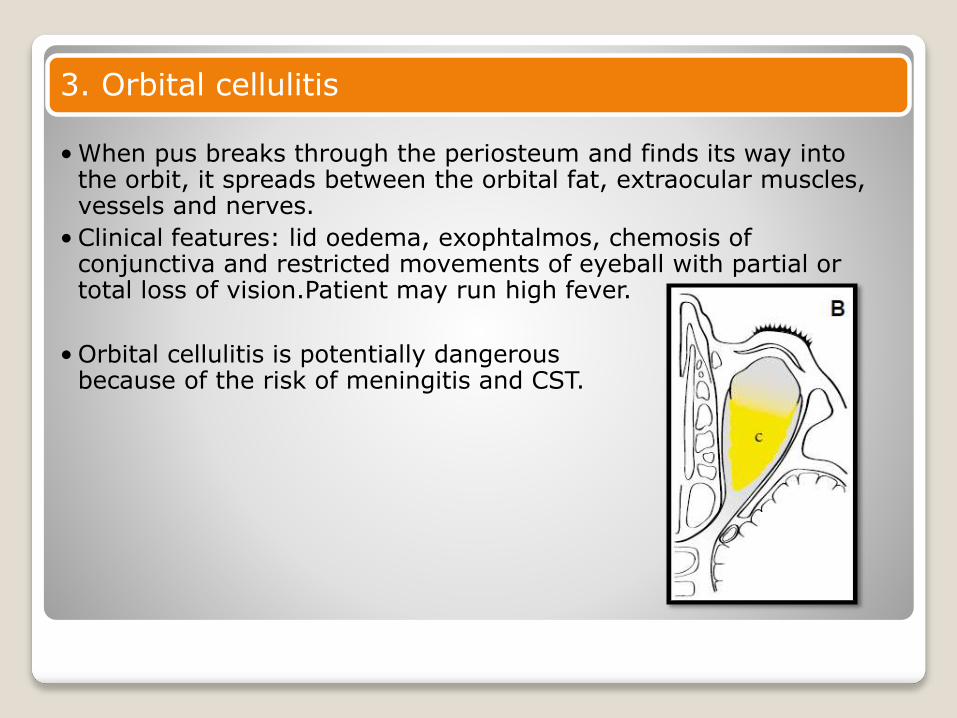
3. Orbital cellulitis
• When pus breaks through the periosteum and finds its way into the orbit, it spreads between the orbital fat, extraocular muscles, vessels and nerves.
• Clinical features: lid oedema, exophtalmos, chemosis of conjunctiva and restricted movements of eyeball with partial or total loss of vision.Patient may run high fever.
• Orbital cellulitis is potentially dangerous because of the risk of meningitis and CST.

Intraorbital abscess usually forms along lamina papyracea or the floor of the frontal sinus.
Clinical features: same as above.
Diagnosis: CT scan or ultrasound of the orbit.
Treatment: i.v. antibiotics and drainage of the
abscess and that of the sinus (ethmoidectomy
or trephination of frontal sinus).
4. Orbital abscess

5. Superior orbital fissure syndrome
• Infection of sphenoid sinus can rarely affect structures of superior orbital fissure.
•Symptoms: Deep orbital pain, frontal headache and progressive paralysis of CN VI, III and IV, in that order.
6. Orbital apex syndrome
• It is superior orbital fissure syndrome with additional involvement of the optic nerve and maxillary division of the tigeminal (V ).2
lll. INTRACRANIAL COMPICATIONS
Frontal, ethmoid and sphenoid sinuses are closely related to anterior cranial fossa and infection from these can cause following complications:
1) Meningitis and encephalitis2) Extradural abscess3) Subdural abscess4) Brai abscess5) Cavernous sinus thrombosis
Cavernous sinus thrombosis :Aetiology: Infection of paranasal sinuses and orbital
complications from these sinus infections can
cause thrombophlebitis of the cavernous sinus.
Clinical features: Abrupt onset with chills and rigors.
Swollen eyelids with chemosis and proptosis of eyeball.
CN III, IV and VI get involved individually and sequentially causing total ophthalmoplegia.
Pupil becomes dilated and fixed.
Congestion of optic disc with diminution of vision.
Sensation in the distribution of V is diminished.
Treatment: i.v. antibiotics and attention to the focus of infection,
drainage of infected ethmoid or sphenoid sinus.
1

ORBITAL CELLULITIS CAVERNOU SINUS THROMBOSIS
SOURCE Commonly ethmoidsinuses
Nose, sinuses, orbit, ear or pharynx
ONSET Slow; starts with oedema of eyelids the inner canthuschemosis proptosis
•Abrupt with high fever and chills with near signs of toxaemia•Oedema of eyelids,chemosis and proptosis
CRANIAL NERVE INVOLVEMENT
Involved concurrently with complete ophthalmoplegia
Involved individuallyand sequentially
LATERALITY Often involves one eye Involves both eyes
DIFFERENCES BETWEEN ORBITAL CELLULITIS AND CAVERNOUS SINUS THROMBOSIS

lV. DESCENDING INFECTIONS
In suppurative sinusitis, discharge constantly flows into the pharynx and can cause or aggravate:
1) Otitis media2) Pharyngitis and tonsillitis: hypertrophy of lateral lymphoid
bands behind the posterior pillars (lateral pharyngitis) is indicative of chronic sinusitis.
3) Persistent laryngitis and tracheobronchitis
V. FOCAL INFECTIONS
Sinusitis may act as focus of infection is conditions like: Polyarthritis, tenosynovitis, fibrositis and certain skin diseases.

THANKYOU
Related Documents


![Recurrent Sinusitis and Periorbital Cellulitis Secondary ...medcraveonline.com/MOJI/MOJI-01-00027.pdfdehiscence of lamina papyracea in 1% of patients [4]. RS orbital complications](https://static.cupdf.com/doc/110x72/5e601c2f6b0bcf66d0055845/recurrent-sinusitis-and-periorbital-cellulitis-secondary-dehiscence-of-lamina.jpg)


