DEPARTMENT OF ORTHOPAEDICS J.J.M. MEDICAL COLLEGE, DAVANGERE. Moderators : Presented by : Dr.Prabhu B Dr. Rakshith Kumar K Professor and Unit Head. P.G. in Orthopaedics Dr. Prasanna Anaberu Professor Seminar on COMPLICATIONS OF FRACTURES Date : 20.07.2016

Welcome message from author
This document is posted to help you gain knowledge. Please leave a comment to let me know what you think about it! Share it to your friends and learn new things together.
Transcript
DEPARTMENT OF ORTHOPAEDICS
J.J.M. MEDICAL COLLEGE, DAVANGERE.
Moderators : Presented by :
Dr.Prabhu B Dr. Rakshith Kumar K
Professor and Unit Head. P.G. in Orthopaedics
Dr. Prasanna Anaberu
Professor
Seminar on
COMPLICATIONS OF FRACTURES
Date : 20.07.2016

INTRODUCTION
Fracture is a disrupting event, more so, if it develops
complications. In a great majority of fractures, union proceeds
according to expectation, function of injured part is gradually
restored and little, if any, permanent disability remains.
• Complications inevitably occur in a proportion of them some
slight, some severe, some catastrophic.
• Complications of fractures can be lethal for life or limb ; can
make the patient disabled and can make working capacity loss.
Awareness of possibilities of complications and prompt and proper
management can overt most of them.
CLASSIFICATION
1) Immediate : (Occur at the time of injury)
Systemic : Hypovolaemic shock Local : Injury to major vessels, muscles and tendons, peripheral nerves,
joints and viscera.
2) Early : (occur in initial few days after injury) Systemic : Hypovolaemic shock
ARDS and Fat embolism syndrome. DVT and pulmonary embolism Aseptic me Tetanus and gas gangrene
traumatic fever Septicaemia in open fractures Crush syndrome
Local : infection Compartment syndrome Setting in of myositis ossificans
3) Late : (Occur long time after injury) Delayed union and non union Malunion and cross union
Others : Avascular necrosis reflex sympathetic dystrophy VIC myositis ossificans Osteomyelitis Shortening joint stiffness osteoarthritis Frictional tendinitis rupture of tendon Delayed nerve complications Growth disturbances Implant failure

HAEMORRHAGIC (HYPOVOLEMIC SHOCK)
• Shock is defined as ‘Inadequate perfusion and oxygenation of tissues due to failure of circulatory system which leads to hypoxia which may damage vital organs.
• In shock due to ineffective circulating blood volume or because of abnormal partitioning of cardiac output, capillary blood flow in tissues is reduced to levels below the minimum requirements for oxidative metabolism.
• Occurs in fractures of long bones, pelvic fractures, multisystem injuries following RTAS, burns surgery and dehydration.
• Commonest cause of death following fractures.
Classification : - Hypovolemic - Cardiogenic - Septic - Neurogenic
- Anaphylactic.
Source of haemorrhage :
a) External → In compound fractures, pelvic fractures etc.
b) Internal → In blunt injury abdomen, chest, femur and
pelvic fractures etc.
Blood loss :
• Femoral shaft fractures range 500 – 2000 ml.
• Pelvic fractures → range 1000 – 2500 ml.
• Tibial shaft fractures → range 500 – 1500 ml.
Clinical features :
• Hypotension, Tachycardia
• Oliguria, Clouded sensorium
• Cool, clammy, moist skin.
• Increased respiratory rate, Low volume rapid / thready pulse.
• Sweating, Sunken eyeballs, Tongue – pale and dry
• Patient gradually becomes drowsy, BP falls and renal output
decreases ; patient may become unconscious and die.
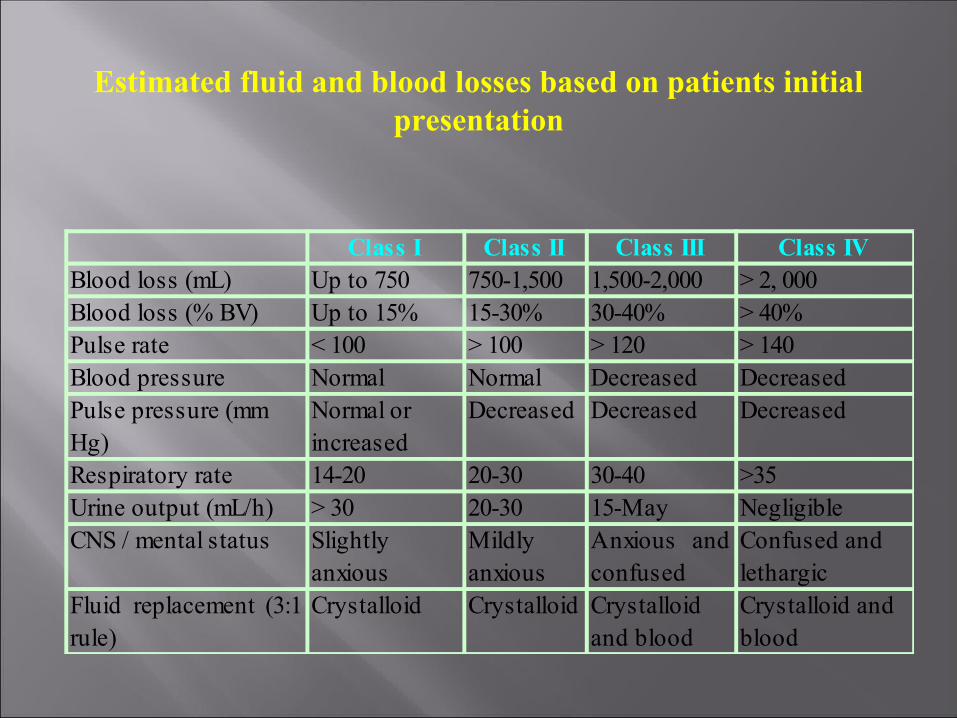
Estimated fluid and blood losses based on patients initial presentation
Class I Class II Class III Class IVBlood loss (mL) Up to 750 750-1,500 1,500-2,000 > 2, 000Blood loss (% BV) Up to 15% 15-30% 30-40% > 40%Pulse rate < 100 > 100 > 120 > 140Blood pressure Normal Normal Decreased DecreasedPulse pressure (mm Hg)
Normal or increased
Decreased Decreased Decreased
Respiratory rate 14-20 20-30 30-40 >35Urine output (mL/h) > 30 20-30 15-May NegligibleCNS / mental status Slightly
anxiousMildly anxious
Anxious andconfused
Confused and lethargic
Fluid replacement (3:1rule)
Crystalloid Crystalloid Crystalloid and blood
Crystalloid and blood

MANAGEMENT :
General principles : Goal is to maintain arterial pressure and to ensure adequate perfusion and O2 delivery to vital organs.
• Patients should be treated in I.C.U., Electro cardiographic monitoring should be done for rythm disturbances, Arterial line for measurement of arterial pressures, Pulse oximeter for fluctuations in arterial oxygenation, Frequent measurements of arterial blood gases, serum electrolyte, complete blood counts should be done.
Emergency management : 1. Head low position 2. Respiratory support
3. Initial Fluid Resuscitation – crystalloids and colloids
4. Blood transfusion therapy : Haemaccel can be used if blood not available
5. Urine output measurement -24 hrs urine output should be maintained with fluid input / output charts and renal function should be assessed.
6. Drugs : Analgesics, Antibiotics and Corticosteroids, acidosis corrected by 7.5% sodium bicarbonate
7. Bleeding site should be searched in thorax, abdomen and pelvis, continuing bleeding may need surgical interventions.
8. Management of fractured limb –Wound debridement and Immobilization

FAT EMBOLISM SYNDROME
• A complex alteration of homeostasis that occurs as an infrequent complication of fractures of long bones and pelvis and manifests clinically as acute respiratory insufficiency.
• Important cause of ARDS and death
• Characterised by - embolisation of marrow fat globules (stearin in children and olein in adults) from long bones and pelvis into pulmonary micro-circulation or other micro-vascular beds like brain.
Causes : Fracture of long bones and pelvis Multisystem injuries like chest and abdomen Following reaming of medullary canals of long bones Following reaming and cementation in joint replacement surgeries Following bone infarcts in hemoglobinopathies, massive soft tissue injury Severe burns, Liposuction, Neoplasms, Severe Infections (chronic O.M), Blood transfusion, Renal transplant and Collagen disease.
Pathogenesis : source of embolic fat is bone marrow.
a) Mechanical Theory b) Biochemical Theory
Sevitt’s Classification :
1. Subclinical FES : Occurs in all long bone fractures of lower extremity and pelvic fractures.
• Decrease Pao2, decrease Hb%, Decreased platelets
• No clinical signs and symptoms of respiratory insufficiency.
2. Non-fulminant FES :
• Signs and symptoms evident
• Respiration insufficiency, cerebral dysfunction and petechiae appear clinically.
• Typical radiological and haemotological changes seen.
3. Fulminant FES : Rare, Appears within hours of injury, so no time for rashes to appear. Characterized by respiratory failure (severe), Altered mental status and Convulsions. Patient is comatosed within hours and throws repeated seizures. Patient collapses and death supervenes.
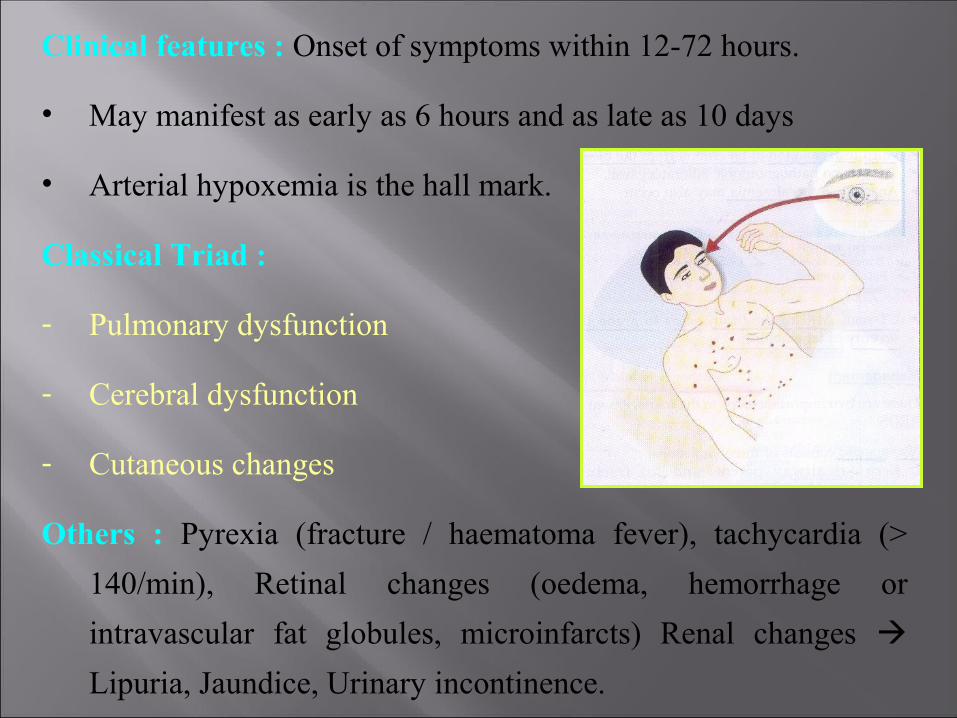
Clinical features : Onset of symptoms within 12-72 hours.
• May manifest as early as 6 hours and as late as 10 days
• Arterial hypoxemia is the hall mark.
Classical Triad :
- Pulmonary dysfunction
- Cerebral dysfunction
- Cutaneous changes
Others : Pyrexia (fracture / haematoma fever), tachycardia (>
140/min), Retinal changes (oedema, hemorrhage or
intravascular fat globules, microinfarcts) Renal changes
Lipuria, Jaundice, Urinary incontinence.
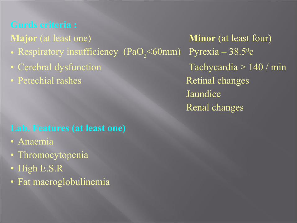
Gurds criteria :Major (at least one) Minor (at least four)
• Respiratory insufficiency (PaO2<60mm) Pyrexia – 38.50c
• Cerebral dysfunction Tachycardia > 140 / min• Petechial rashes Retinal changes Jaundice Renal changes
Lab. Features (at least one)• Anaemia• Thromocytopenia• High E.S.R• Fat macroglobulinemia
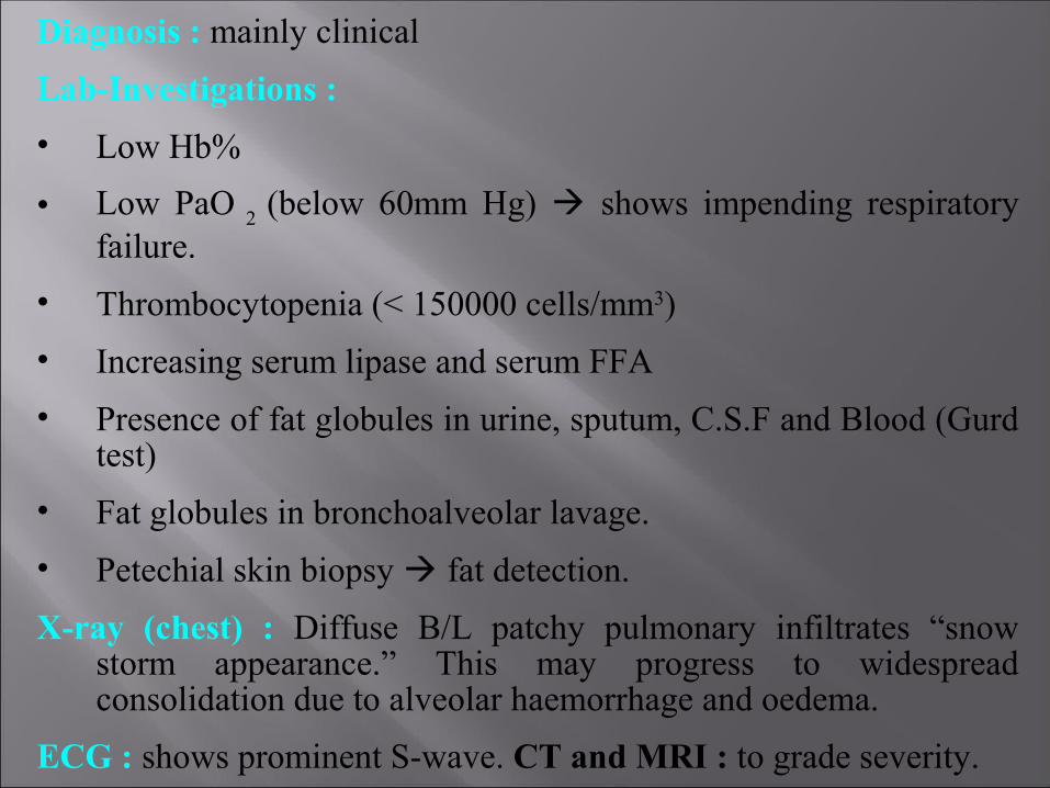
Diagnosis : mainly clinical
Lab-Investigations :
• Low Hb%
• Low PaO 2 (below 60mm Hg) shows impending respiratory failure.
• Thrombocytopenia (< 150000 cells/mm3)
• Increasing serum lipase and serum FFA
• Presence of fat globules in urine, sputum, C.S.F and Blood (Gurd test)
• Fat globules in bronchoalveolar lavage.
• Petechial skin biopsy fat detection.
X-ray (chest) : Diffuse B/L patchy pulmonary infiltrates “snow storm appearance.” This may progress to widespread consolidation due to alveolar haemorrhage and oedema.
ECG : shows prominent S-wave. CT and MRI : to grade severity.
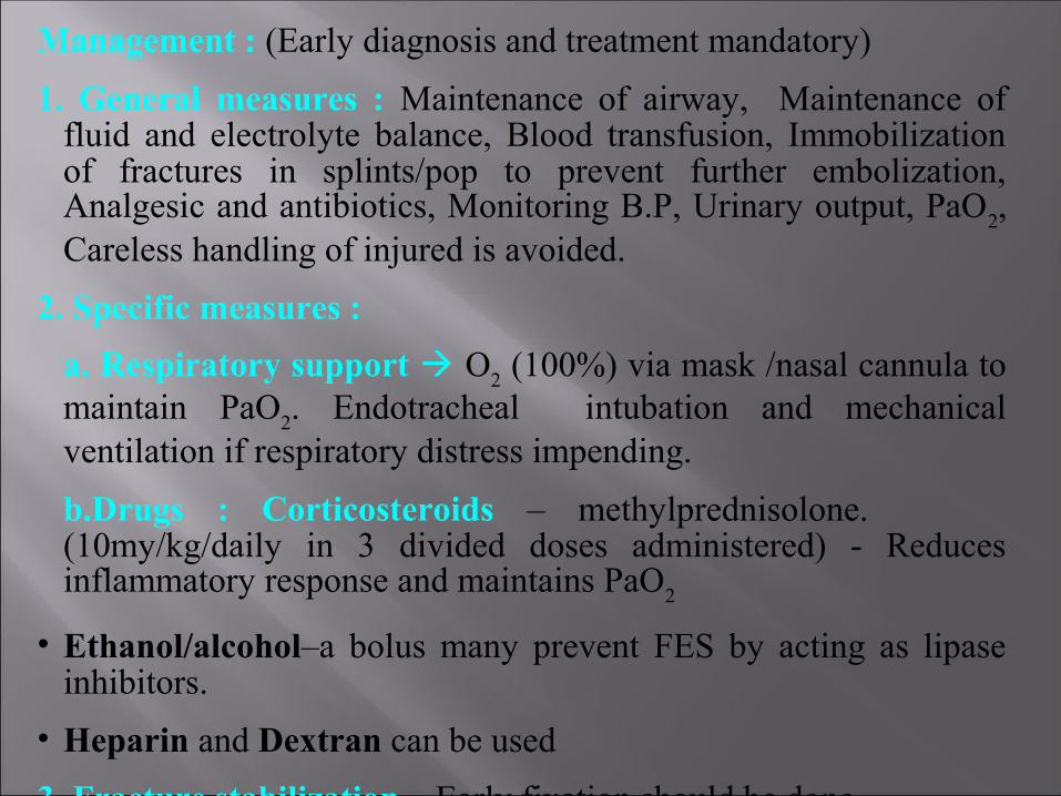
Management : (Early diagnosis and treatment mandatory)
1. General measures : Maintenance of airway, Maintenance of fluid and electrolyte balance, Blood transfusion, Immobilization of fractures in splints/pop to prevent further embolization, Analgesic and antibiotics, Monitoring B.P, Urinary output, PaO2, Careless handling of injured is avoided.
2. Specific measures :
a. Respiratory support O2 (100%) via mask /nasal cannula to maintain PaO2. Endotracheal intubation and mechanical ventilation if respiratory distress impending.
b.Drugs : Corticosteroids – methylprednisolone. (10my/kg/daily in 3 divided doses administered) - Reduces inflammatory response and maintains PaO2
• Ethanol/alcohol–a bolus many prevent FES by acting as lipase inhibitors.
• Heparin and Dextran can be used
3. Fracture stabilization – Early fixation should be done.
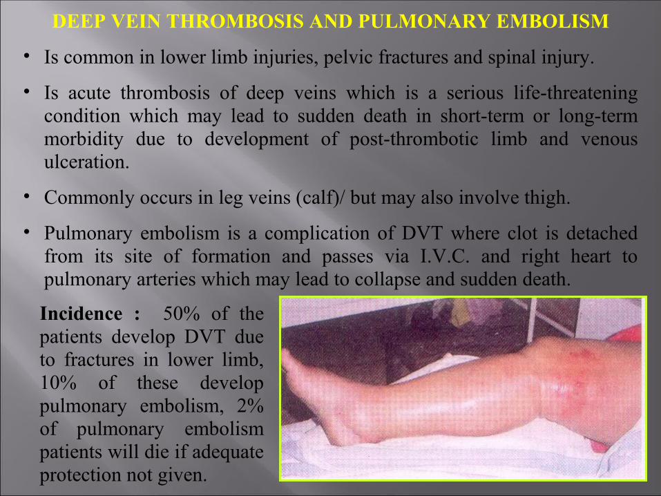
DEEP VEIN THROMBOSIS AND PULMONARY EMBOLISM
• Is common in lower limb injuries, pelvic fractures and spinal injury.
• Is acute thrombosis of deep veins which is a serious life-threatening condition which may lead to sudden death in short-term or long-term morbidity due to development of post-thrombotic limb and venous ulceration.
• Commonly occurs in leg veins (calf)/ but may also involve thigh.
• Pulmonary embolism is a complication of DVT where clot is detached from its site of formation and passes via I.V.C. and right heart to pulmonary arteries which may lead to collapse and sudden death.
Incidence : 50% of the patients develop DVT due to fractures in lower limb, 10% of these develop pulmonary embolism, 2% of pulmonary embolism patients will die if adequate protection not given.
Factors : The most common cause of DVT in lower limbs is following hospital admission (immobilization) for treatment of medical or surgical condition which leads to venous stasis which results in thrombosis of veins.
Clinical feature :• DVT can be recognized as early as 48 hours of injury. • Pulmonary embolism usually develops 4-5days after injury. • First complaint is oedema, erythema and dilated veins of the leg. • Dull aching or nagging pain in calf muscles. • Superficial blebs in the skin, skin red and warm. • Low grade fever with increased pulse rate is characteristic.
• Phlegmasia alba dolens (while leg) occurs when thrombus extends from calf to ileofemoral vein.
• Phlegmasia coerula (blue leg) with loss of superficial tissues of the toes.
• Patient complains of swelling, difficulty in standing, walking and cramps in the leg.
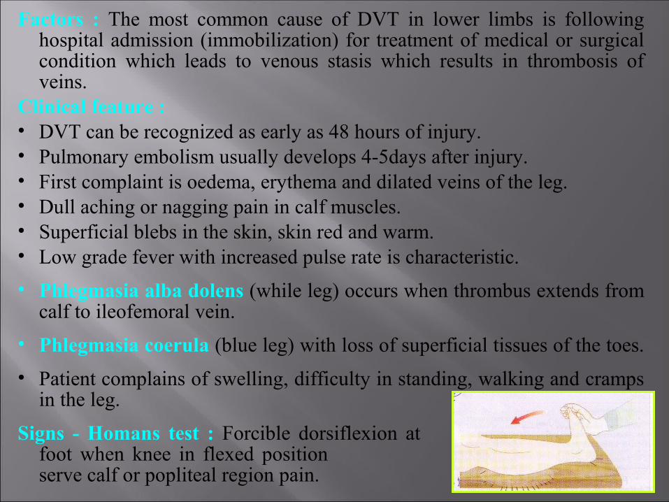
Signs - Homans test : Forcible dorsiflexion at foot when knee in flexed position produces serve calf or popliteal region pain.
Investigations (DVT) :
1. Contrast venography
2. Ultrasound flowmeter using Doppler effect is utilized for detecting blood flow. Recently colour flow ultrasonography is being used.
3. MRI for imaging of veins.
4. Radioactive iodine.
Diagnosis of pulmonary embolism :
Clinical feature : (Minor cases) - Transient shortness of breath, Sharp localized chest pain aggravated by inspiration, Haemoptysis.
(Major cases) - Severe shortness of breath, Hypotension, Syncope
- Peripheral circulatory failure, Tachypnea
Test : Pulmonary Angiography, Perfusion lung scan
• Electrocardiography to exclude M.I. or pericarditis.
• X – rays - Show blunting of costo-phrenic angle
- Elevated hemi-diaphragm of side of embolus.
• C.T. scan, M.R.I.
Treatment :
1. Bed rest, elevation of limb and elastic bandaging.
2. A loading dose of 10,000-15,000 IU of heparin is administered intravenously. Then further anti-coagulant therapy is regulated with prothrombin time. Duration of heparin treatment is 7-10 days (minimum 5 days) to reduce risk of pulmonary embolism and encourage thrombus to resolve.
3. Warfarin (dose – 10mg twice a day) is started 2-3 days before heparin is withdrawn because of slow onset of action. (Given for 6-12 months)
4. Use of subcutaneous injections of low-molecular weight heparin is an alternative method.
5. Surgery is indicated when DVT leads to severe impairment in blood supply. Venous bypass relieves venous obstruction.
TETANUS
• Is a potentially fatal disease that is preventable by appropriate
immunization.
• Occurs in all age groups, caused by clostridium tetani bacillus
which is anaerobic, gram-positive spore forming bacilli found
in faecal matter of animals and humans.
Factors favouring its development - Deep wounds without
exposure to air, Wounds containing ischaemic tissues, Wounds
infected with other organisms, No immunization, Improper
sterilization
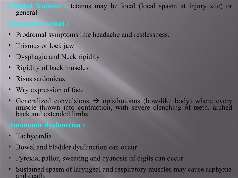
Clinical features : tetanus may be local (local spasm at injury site) or general
In general variant :
• Prodromal symptoms like headache and restlessness.
• Trismus or lock jaw
• Dysphagia and Neck rigidity
• Rigidity of back muscles
• Risus sardonicus
• Wry expression of face
• Generalized convulsions opisthotonus (bow-like body) where every muscle thrown into contraction, with severe clenching of teeth, arched back and extended limbs.
Autonomic dysfunction :
• Tachycardia
• Bowel and bladder dysfunction can occur
• Pyrexia, pallor, sweating and cyanosis of digits can occur.
• Sustained spasm of laryngeal and respiratory muscles may cause asphyxia and death.
Management :1) General measures 2) Specific measures :a) Mild cases – (Only toxic rigidity but no spasm or dysphagia). • Managed by heavy sedation to avoid spasms and convulsions. • Drugs : benzodiazepines, chlorpromazine and morphine used. • Urinary catheterization for emptying bladder. • Laxatives for emptying G.I.T. • Skin care to prevent sores. b) Seriously ill cases : (Have dysphagia and reflex spasms) • Nasogastric tube for feeding purposes and for drugs. • Traheostomy if breathing difficult.
• During this period adequate nutrition, care of bladder and bowel and prevention of sores should be done.
c) Dangerously ill cases : have major cyanotic convulsions • Sedatives used • Patients are paralysed with muscle relaxants • Positive pressure ventilation given till they recover
During this period adequate nutrition, care of bladder and bowel and prevention of sores should be done.
Prophylaxis :
• Infants and children are immunized with tetanus toxoid,
diphtheria and pertusis (DPT) vaccine with three doses at 6,
10, 14 weeks of age. A booster is given at 18 months, 5 years
(DT) and after 10 (TT) years to achieve active immunity.
• Prevention of tetanus after injury : Wound cleaned with
removal of soil, foreign body and necrotic tissue. Tetanus
antitoxin (ATS) is given (250) units.

GAS GANGRENE
Gangrene : Is macroscopic death of tissue with superadded putrefaction.
Gas Gangrene : Is a highly fatal spreading infection of superticial and deep fascia which results in myonecrosis.
Aetiology : Caused by Clostridial species like Cl. perfringes (Cl. Welchii) – commonest, Cl. Septicum, i.e., histolyticum, Cl, noryi etc. Cl. welchii – is a non-sporing, non-motiles gram-positive anaerobic bacillus.
• Occurs as a complication to open injuries, war injuries, RTAs.
• Prime site is a dirty wound with dead muscle that has been closed without adequate treatment.
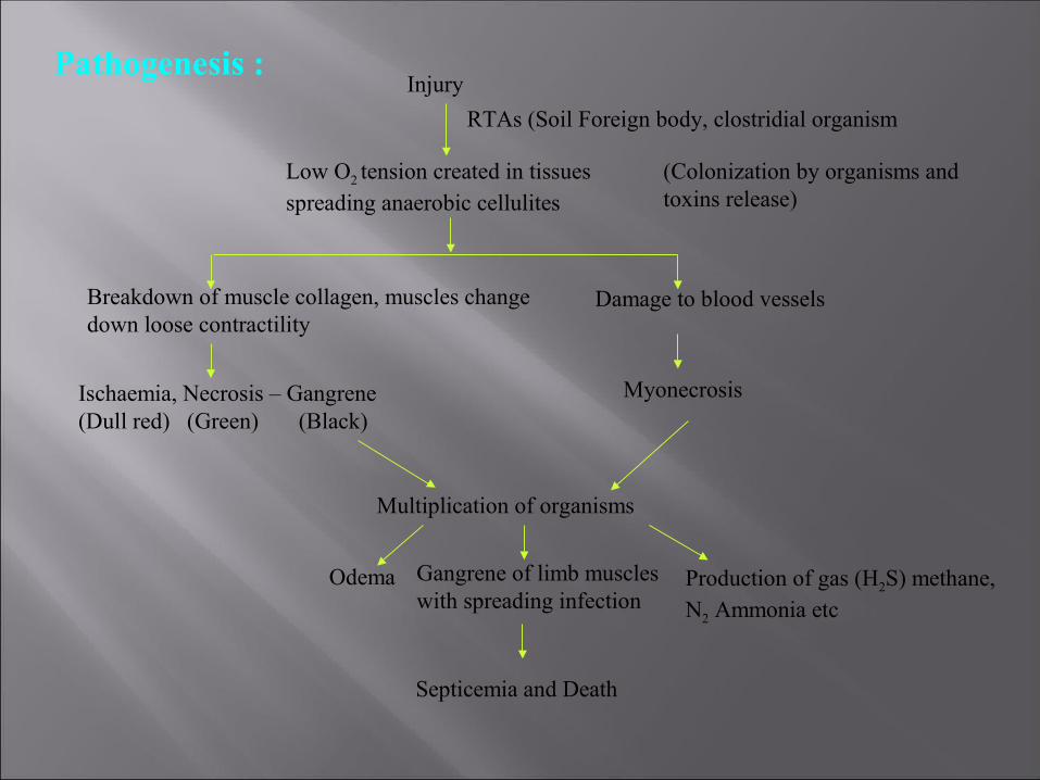
Injury
Low O2 tension created in tissues spreading anaerobic cellulites
RTAs (Soil Foreign body, clostridial organism
Breakdown of muscle collagen, muscles change down loose contractility
Damage to blood vessels
Ischaemia, Necrosis – Gangrene (Dull red) (Green) (Black)
Myonecrosis
(Colonization by organisms and toxins release)
Multiplication of organisms
Odema Production of gas (H2S) methane, N2 Ammonia etc
Gangrene of limb muscles with spreading infection
Septicemia and Death
Pathogenesis :
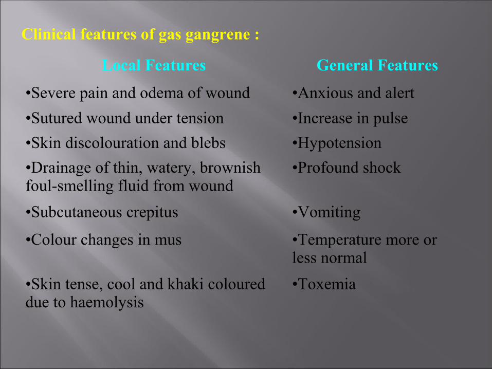
Clinical features of gas gangrene :
Local Features General Features
•Severe pain and odema of wound •Anxious and alert
•Sutured wound under tension •Increase in pulse
•Skin discolouration and blebs •Hypotension
•Drainage of thin, watery, brownish foul-smelling fluid from wound
•Profound shock
•Subcutaneous crepitus •Vomiting
•Colour changes in mus •Temperature more or less normal
•Skin tense, cool and khaki coloured due to haemolysis
•Toxemia
Diagnosis : Clinical suspicion, Gram staining to find organism, X-ray C.T, MRI of involved area.
Management :
Supportive : Fluid and blood transfusion, Respiratory support etc
Surgical excision : Early surgical excision of all necrotic tissues, surgical decompression with multiple incision, fasciotomy, drainage of fascial compartment, repeated irrigation of wounds.
• Heavy dose of penicillin (30 laks units every 3rd hourly and cephalosporins and aminoglycosides started).
• Hyperbaric O2 patient placed at three times atmospheric pressure (HBO inhibits alpha toxin production).
• Amputation (open) done in late diagnosed cases to save life.
Prophylaxis :
• All dead, necrotic tissue, foreign body, bone process removed.
• Through wound toilet with H2O2, saline, wound irrigated with antiseptics.
• When in doubt of cleanliness, do not suture the wound.
• Prophylactic use of penicillin (10-20 lakhs units) 4-6 hrly for 7 days and other broad spectrum antibiotics.
• Avoid tourniquet use.
CRUSH SYNDROME(TRAUMATIC RHABDOMYOLYSIS)
• Refers to sequela of prolonged continuous pressure on muscle tissue.
• Crush syndrome refers to systemic manifestation associated with crush injuries like hyperkalemia, myoglobinemia and anuric renal failure.
• Causes – Earthquake, bombings, train accidents prolonged use of military anti shock trousers, vehicle accidents, gangrenes, prolonged tourniquet application and acute compartment syndrome.
Clinical features : Oedema, Hematoma, Limb pulseless and red.Muscle power may be lost, Hypovolemia, Anaemia, Hypotension Tachycardia, Compartment syndrome.
• Electrolyte imbalance : Hyperkalemia, Hypocalcemia, Metabolic acidosis, Phosphorus and magnesium imbalance
• Renal output decreases and low output uraemia with acidosis develops.
• If renal secretion returns within 1 week, patient survives.
Management :
1. Systemic : begins within an hour.
• Aggressive fluid resuscitation (1500 ml/hour crystalloid infusion to maintain urine output of at least 100 ml/hour).
• Osmotic diuretic – i.v. mannitol – 1 gm/kg given
• Inj. Bicarbonate should be given to keep urine alkaline.
• Maintenance of electrolyte imbalance.
• Platelet extracts (FFP) to counteract coagulation abnormalities.
• Administration of XO inhibitors to prevent free toxic radical formation.
• In case of ARF – renal dialysis mandatory.
2. Local : Conservative management of limb, Fasciotomy for compartment syndrome, Severe crushed limbs – amputations done.
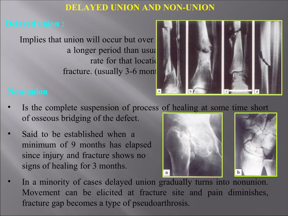
DELAYED UNION AND NON-UNION
Delayed union :
Implies that union will occur but over a longer period than usual average
rate for that location and type of fracture. (usually 3-6 months)
Non-union
• Is the complete suspension of process of healing at some time short of osseous bridging of the defect.
• Said to be established when a minimum of 9 months has elapsed since injury and fracture shows no signs of healing for 3 months.
• In a minority of cases delayed union gradually turns into nonunion. Movement can be elicited at fracture site and pain diminishes, fracture gap becomes a type of pseudoarthrosis.
Causes of delayed union / non union 1. Cause related to patient :
• Old age• Poor nutritional status• Nicotine and alcohol consumption• Associated illness – malignancies, osteomalacia • Metabolic disorders. – hyperparathyroidism
2. Causes related to fracture : • Distraction of fracture site –
- Pull of fragments eg. Fracture patella, Fracture olecranon- Gravity eg. Fracture shaft humerus
• Soft tissue interposition - Eg. Fracture shaft femur and humerus • Bone loss at fracture site – fracture tibia and ulna. (open type).• Infection• Loss of blood supply – eg. Fracture neck femur.• Damage of surrounding muscles. • Pathological fractures, Intact fellow bone
3. Causes related to treatment • Inadequate reduction eg. Fracture of congenital bones (shafts).• Inadequate immobilization eg. Fracture neck femur. • Distraction during treatment • Improperly applied fixation devices.
Sites : fracture neck of femur, talus, scaphoid, fifth metatarsal
Tibia is the most frequent site because of frequency of severe open tibial fractures.
Diagnosis of non-union :
C/F: - sense of weakness or instability in extremity
H/o developing deformity, Difficulty in walking
On examination : -No tenderness, Crepitus absent, Abnormal mobility (+)
X-Ray : Wooly appearance of bone ends
• Obliteration of medullary canal near bone fragments.
• Clear and wide fracture line
• Minimal / absent internal or external callus, Sclerosis of bone ends
Delayed union : Fracture site tender, X-ray shows callus formation but not upto expectations.
Management (non-union) - Three principles :
a) Good reduction
b) Bone grafting
c) Stabilization of fragments by external or internal fixation devices.
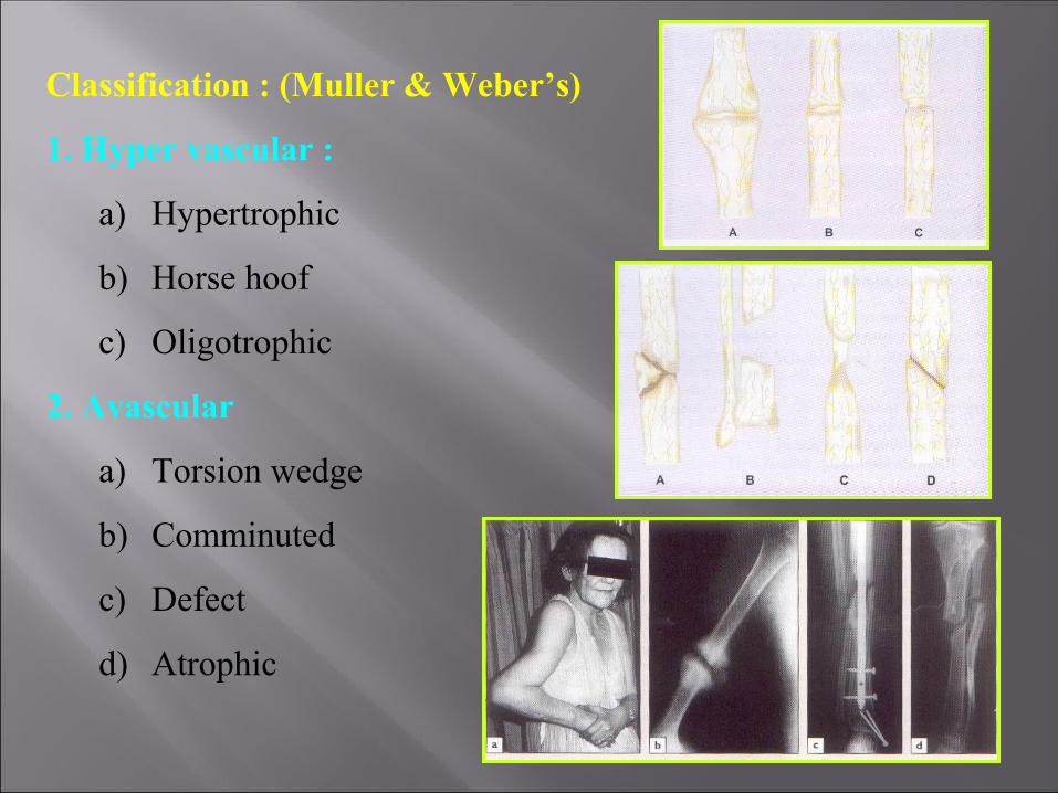
Classification : (Muller & Weber’s)
1. Hyper vascular :
a) Hypertrophic
b) Horse hoof
c) Oligotrophic
2. Avascular
a) Torsion wedge
b) Comminuted
c) Defect
d) Atrophic
Treatment of infected cases
1. Classical method : first wound debridement with clearing of infected, necrosed material done. Then infection controlled by antibiotics. Once infection controlled, split thickness grafting done to cover the wound. Then fracture fixed internally with bone grafting takes longer time.
2. Active method : internal fixation bone grafting continuous irrigation with saline and antibiotics – skin / muscle pedicle graft. Duration of treatment less
3. Illizarovs technique : best modality simultaneously angulatory, rotatory and translational deformities can be corrected, shortening and segmental bone loss can be made up and infection can be controlled.
Management of delayed union : Prolonging immobilization with treatment of obvious completes union, Electrical and electromagnetic stimulation used by percutaneous implantation of electrodes, Percutaneous injection of aspirated bone marrow accelerates union Reinforcement with cancellous bone grafting in considerably delayed union.
MALUNION
• Is fracture healing with fragments in non-anatomical position or unacceptable position. Fracture united with angulation, rotation, loss of end-end apposition or overlap and shortening.
Reasons : Inaccurate reduction, Infective immobilization during healing, Gradual collapse of comminuted or osteoporotic bone, Improper fixation of fracture, redisplacement after reduction within cast, growth disturbances due to epiphyseal cartilage injury.
Classification :
1. (length malunion) (overriding) : results in shortening and rarely lengthening.
2. (rotatory malunion) : results in external or internal rotation deformities.
3. (angulatory malunion) : results in varus or valgus deformities.
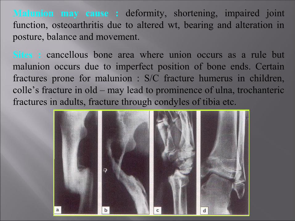
Malunion may cause : deformity, shortening, impaired joint function, osteoarthritis due to altered wt, bearing and alteration in posture, balance and movement.
Sites : cancellous bone area where union occurs as a rule but malunion occurs due to imperfect position of bone ends. Certain fractures prone for malunion : S/C fracture humerus in children, colle’s fracture in old – may lead to prominence of ulna, trochanteric fractures in adults, fracture through condyles of tibia etc.
Management : There are four characteristics that determine acceptability of fracture reduction - Alignment, Rotation, Restoration of normal length and Actual position of fragments. Small angular deformities and shortening of limb usually gets corrected in children due to excellent remodeling but it is not so in adult. Slight malunion is generally accepted but malunion producing significant deformity needs correction.
Methods : - Osteoclysis
- Redoing fracture surgically
- Corrective and Compensatory osteotomy
- Excision of protruding bone
Cross-union : When radius and ulna fractures are joined to each other by a bridge of callus, it is called cross-union. It is likely to develop in cases where fractures are at same level. It results in complete limitation of forearm rotations.
Treatment : If cross union is in mid-pronation it is left as it is as this position is best for function.
If it occurs in excessive pronation or supination cross union is undone, malalignment corrected and fracture internally fixed.
REFLEX SYMPATHETIC DYSTROPHY OR SUDECK’S OSTEODYSTROPHY
• Is a group of disorder in which there is dysfunction in the normal “injury and repair process” leading to an exaggerated response to a noxious stimulus.
• Characterized by pain, swelling, stiffness, discoloration and hyperhydrosis, osteoporosis and trophic changes which are out of proportion to the inciting event.
• Incidence : 7-35% in prospective studies of colle’s fracture, 1-2% after various fractures, 2-5% after peripheral nerve injury
Precipitating factors :
Trauma
Surgery
Peripheral nerve lesion
Stroke.
Classification :
1) Minor causalgia : Follows injury to purely sensory nerve, Burning pain
2) Minor traumatic dystrophy : Follows minor crush injuries, sprains and fractures.
3) Shoulder hand syndrome : Characterized by pain and stiffness in upper extremity as a result of proximal injury to neck, chest shoulder or due to a visceral lesion like heart attack, stroke or gastric ulcers.
• Traumatic causes may be fractures of shoulder, clavicle and ribs.
4. Major traumatic dystrophy : (crush injuries or colles #), Pain and deformity intense.
5. Major Causalgia : Most devastating but rare. Injury to a major mixed nerve in proximal extremity (median and sciatic commonly).
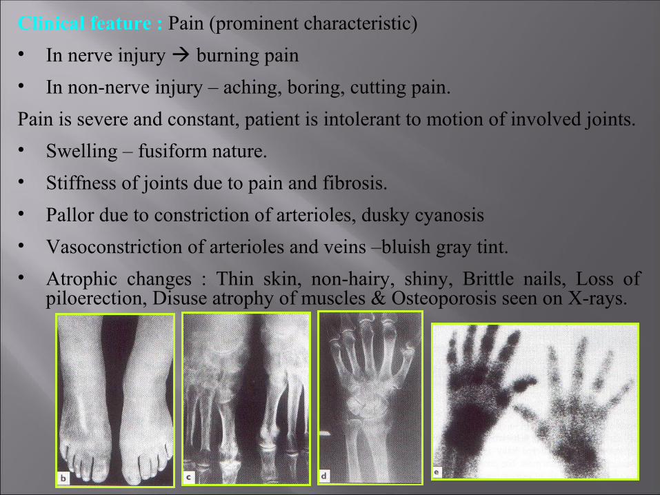
Clinical feature : Pain (prominent characteristic)
• In nerve injury burning pain
• In non-nerve injury – aching, boring, cutting pain.
Pain is severe and constant, patient is intolerant to motion of involved joints.
• Swelling – fusiform nature.
• Stiffness of joints due to pain and fibrosis.
• Pallor due to constriction of arterioles, dusky cyanosis
• Vasoconstriction of arterioles and veins –bluish gray tint.
• Atrophic changes : Thin skin, non-hairy, shiny, Brittle nails, Loss of piloerection, Disuse atrophy of muscles & Osteoporosis seen on X-rays.

Diagnosis :
• Physical examination : Reveals discoloration of skin, oedema, subcutaneous thickening, temperature differences between affected and unaffected parts.
o Light touch, pin-prick and temperature evaluation can elicit and illustrate morbidity.
o Muscle testing can demonstrate joint stiffness, weakness and allodynia
• X-rays: Osteopenia (diffuse and spotty), Visible after 2-4 weeks.
• Bone scans : Show increased uptake.
• Modified sympathetic blockage testing.
Treatment : Early diagnosis and initial immobilization.
• Physiotherapy
• Sympathetic block using sympatholytic drugs
• Sympathectomy : Includes removal of T2 and T3 ganglia in upper limb and L2-L3 in lower limb.
MYOSITIS OSSIFICANS (TRAUMATIC)
Is a reactive lesion occurring in soft tissues and sometimes in periosteum which is characterized by fibrous and cartilaginous proliferation and by metaplastic changes.
• Word is misnomer as no inflammation in muscle occurs.
• Haematoma is a perquisite for this condition which under stripped periosteum is invaded by osteoblasts and becomes ossified.
• A calcified mass appears near a joint in tissues which causes restriction of movements.
• More common in children and young adults as periosteum is loosely attached.
Causes : 1) Trauma - Simple blow or repeated minor trauma
2) Dislocation and avulsion injuries cause violent stripping of periosteum – more prone.
3) Massage – commonest cause
4) Passive stretching of joints after injuries.
Muscles commonly involved : Brachialis anticus, Quadratus femoris, Adductor muscles of thigh.
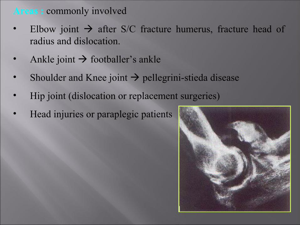
Areas : commonly involved
• Elbow joint after S/C fracture humerus, fracture head of radius and dislocation.
• Ankle joint footballer’s ankle
• Shoulder and Knee joint pellegrini-stieda disease
• Hip joint (dislocation or replacement surgeries)
• Head injuries or paraplegic patients
Clinical features :
1. Acute : Pain, swelling and restriction of movements
• x-ray normal, Bone scan may show increased activity.
2. Chronic : (next 2-3 weeks)
• Pain and Swelling subsides
• X-ray shows fluffy calcification in soft tissue
• By 8 weeks– x ray – well defined margins with trabeculated pattern seen.
• An indurated, circumscribed, hard mass palpable over anterior aspect of elbow limiting joint movements.

Management :
Acute cases : (Conservative) Massage following injury is contraindicated. Immobilize parts by splint for 3-4 wks to prevent stretching and movement to prevent haematoma formation. Drugs – NSAIDS and calcitonin can be used. Physiotherapy – active encouraged, passive stretching avoided.
Chronic cases : (established) First active exercises encouraged preventing passive stretching to prevent further bleeding. 6-12 mths later, when man is mature and quite, excision of bony mass advocated with indometacin or radiotherapy (7 Gy units) to prevent recurrence.
Prophylaxis : when risks are high, indomethacin (2x50mg daily for 7 days) can be given.
NERVE INJURIES : More common in fractures of humerus, around elbow injuries or knee.
• May be due to trauma or sometimes iatrogenic (during surgery, pop application, skeletal or skin traction etc.)
• In closed injuries → - nerve seldom severed, Spontaneous recovery in 90% cases in 4 months, In open fracture : nerve lesion mostly complete.
Incidence : Radial nerve (45%), Ulnar nerve (30%), Median nerve (15%), Peroneal nerve and lumbosacral plexus (3%), Tibial nerve – rare.
Mechanism : Nerve may be damaged by fracture fragments, entrapment between fracture fragments during reduction, direct injury by sharp object etc. In late stages, nerve may be trapped in callus or fibrous tissue.
Consequences : damage to a particular nerve may result in neuroproxia, axonotemesiss or neurotmesis. May result in variable degree of motor and sensory loss along the distribution of nerves.
Classification : (Seddon’s)
1. Neuropraxia
2. Axonotemesis
3. Neurotmesis
Majority are neuropraxia or axonotemesis. Common nerve
injuries Injury Deformity
Axillary nerve Dislocation of shoulder Deltoid paralysis
Radial nerve Fracture shaft humerus Wrist drop and finger drop.
Ulnar and median nerve
S/C fracture humerus Ulnar claw, total claw hand
Post interrosseus nerve Monteggias fracture dislocation
Finger drop.
Sciatic nerve Dislocation hip Flail limb
Lateral popliteal nerve Fracture neck of fibula Foot drop
Management :
1. Conservative :
• Splinting of limbs
• Passive movements of joints to prevent contractures.
• Physiotherapy : exercises, stimulation etc.
Operative management : if fracture needs open reduction, nerve should be explored. Generally open fractures with neurotmesis need surgery.
• Primary repair → done within 6-8 hrs after injury if wound is clean.
• Delayed primary repair → between 7 and 18 days after injury if wound contaminated.
• Secondary repair → 18 days after injury, if injury seen late, failure of conservative treatment, cases with signs of nerve irritation etc.
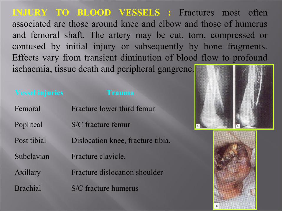
INJURY TO BLOOD VESSELS : Fractures most often associated are those around knee and elbow and those of humerus and femoral shaft. The artery may be cut, torn, compressed or contused by initial injury or subsequently by bone fragments. Effects vary from transient diminution of blood flow to profound ischaemia, tissue death and peripheral gangrene.
Vessel injuries Trauma
Femoral Fracture lower third femur
Popliteal S/C fracture femur
Post tibial Dislocation knee, fracture tibia.
Subclavian Fracture clavicle.
Axillary Fracture dislocation shoulder
Brachial S/C fracture humerus

Clinical features :
Signs of fracture site : Increasing swelling and Massive external bleeding.
Sings in limb distal to fracture : Pain, Pulselessness, Pallor, Paraesthesia and Paralysis.
Diagnosis : If vascular injury suspected, angiogram done immediately.
• Doppler study can be done– most reliable
Treatment : Aims at arresting bleeding by direct pressure and restoring blood volume.
• All bandages and splints should be removed.
• Fracture reduction should be done is early as possible and circulation assessed.
• If after reduction, no improvement vessel exploration done.
• A torn vessel can be sutured or a segment may be replaced by vein graft.
• If vessel thrombosed, endarterectomy done.
• If vessel repair undertaken, stable internal fixation is imperative.
• Anticoagulants like heparin used to irrigate the vessel.
INJURY TO VISCERA
Fractures around the trunk are often complicated by injuries to
adjacent viscera.
These are : →
• Rupture of uretura or bladder and perforation of rectal wall in
pelvic fractures. Penetration of lung with lift threatening
pneumothorax following rib fractures. Rupture of spleen,
kidney or liver may follow local trauma, abdominal
compression or crushing.
• Such injuries need accurate diagnosis, prompt and proper
management.
AVASCULAR NECROSIS : Rare but severe complication of certain fractures. Occurs when blood supply to a segment of bone affected. Common sites of fracture which are liable to undergo this complication are fracture neck of femur, scaphoid waist fracture (proximal pole), fracture neck talus (body) and dislocation of lunate (whole bone necrosed).
• Loss of blood supply to major bone fragment impairs healing. This defective healing makes bone weak and susceptible to external forces which leads to collapse and late osteo-arthritic changes.
• Clinical features – usually asymptomatic in early stages. In late stages, pain, limp and slight loss of movements. In very late case, osteoarthritis features are seen.
• In early stages, AVN can be detected by bone scanning (cold area) radioisotope study whereas in later stages (1-3 mths). X-ray shows dense changes in bone, collapse and osteoarthritic changes.
• Management : early stages – generally no treatment, protective braces used. Late stages – delayed wt. Bearing, revascularization by bone grafts (AVN femoral head), excision of avascular segment (AVN scaphoid), total joint replacement or arthodesis if patient is disabled.
SHORTENING
Is generally a sequel of malunion, Occurs in transverse fractures which are off end (complete lateral displacement or translation with loss of bony contact at fracture site) and often in spiral or oblique displaced fractures, Also occurs from marked angulation and Injury to growth plate
In children : Bone growth accelerated in injured limb as a result of epiphyses being stimulated by increased blood supply. Any discrepancy in limb length is usually quickly made up.
In adults : Shortening in lower limb is seen most frequently after fractures of tibial shaft and femoral shaft fractures. Shortening of 1.5cm easily tolerated being compensated for by tilting of pelvis, shortening in excess of this should be dealt by alteration of foot wear, leg-lengthening / operations etc. When shortening is due to severe persistent angulation of fracture, corrective osteotomy indicated. In upper limb, shortening seldom causes any problem.
OSTEOMYELITIS
Is a suppurative process of the bone caused by pyogenic organisms or simply a pyogenic infection of cancellous area of the bone. Is a serious complication of open fractures and internal fixation of fractures. Caused by bacterial, fungal, viral or protozoal infections. S. areus most common organism. Can be classified as acute (< 2 wks) sub-acute (2-3 wks) and chronic (> 3 wks) duration. Route of spread hematogenous.
Clinical features : fever, sweating, chills and rigors, ↑ temperature, ↑ pulse rate, anemia, local swelling, limitation of movement and signs of dehydration and shock. When well established, purulent discharge seen which is foul smelling.
Management : Risk of this complication must be kept in mind in open fractures and when fixation is planned treatment delay must be avoided. Antibiotics should be started according to culture reports and given for usually 4 wks. If, after internal fixation, there is evidence of systemic manifestation, infected material including hematoma removed before pus forms by opening wound and irrigating it. If infection not controlled at early stages, drainage should be established and regular dressing should be done.
In established cases : saucerisation of area with radical dissection of infected base and open packing is done. Implantation of irrigation tubes impregnated with gentamycin or other antibiotics.
JOINT STIFFNESS : common complication
Factors :
1) Intra-articular causes : a) Intra-articular adhesions
b) Mechanical restrictions
c) Osteoarthritis
2) Peri-articular causes
3) Stiffness from causes remote from a joint
Management : Heat therapy and exercises, Surgery indicated to excise an extra-articular block, to lengthen contracted muscles and joint replacement if secondary O.A.
• Avoidance : Accurate reduction • Splinting minimum numbers of joints. • Urgent mobilization of unsplinted joints. • Elevation to reduce oedema. • Splinting of fracture for short time. • When fractures involve joint early movement done. • Physiotherapy and early motion required in internally fixed fractures.
DELAYED NEUROLOGICAL DISTURBANCE :
• Nerve palsy developing long time after fracture has healed.
• Tardy ulnar nerve palsy : Striking example, Causes are S/C fracture humerus or monteggia fracture dislocation, Develops over years, Deformity (cubitus valgus) present, Treatment is by early transposition of ulnar nerve.
• Median nerve palsy : Generally after colle’s fracture which results in, Carpal tunnel syndrome, Treatment is by decompression.
DELAYED TENDON RUPTURE :
• Seen most common in wrist after colle’s fracture
• Rupture of extensor pollius longus tendon some weeks or months after fracture, Results from gradual fraying of tendon as it rubs against healing fracture or by fibrotic interferences with its arterial supply.
• Loss of extension of terminal joint of thumb.
• Treatment : transposition and suture of tendon of extensor indicis to distal segmental of EPL.
REFERENCES
Cambell s operative orthopedics
Rockwood and greens fractures in adults
Apley s system of orthopedics and fractures
Mercer s textbook of orthopedics and trauma
internet

Related Documents