This article was published in the above mentioned Springer issue. The material, including all portions thereof, is protected by copyright; all rights are held exclusively by Springer Science + Business Media. The material is for personal use only; commercial use is not permitted. Unauthorized reproduction, transfer and/or use may be a violation of criminal as well as civil law. ISSN 0178-7888, Volume 26, Number 1

Welcome message from author
This document is posted to help you gain knowledge. Please leave a comment to let me know what you think about it! Share it to your friends and learn new things together.
Transcript
This article was published in the above mentioned Springer issue.The material, including all portions thereof, is protected by copyright;all rights are held exclusively by Springer Science + Business Media.
The material is for personal use only;commercial use is not permitted.
Unauthorized reproduction, transfer and/or usemay be a violation of criminal as well as civil law.
ISSN 0178-7888, Volume 26, Number 1
ORIGINAL PAPER
Complex etiology and pathology of mycotoxic nephropathyin South African pigs
Stoycho D. Stoev & Stefan Denev & Mike F. Dutton &
Patrick B. Njobeh & Joseph S. Mosonik &
Paul A. Steenkamp & Iordan Petkov
Received: 25 August 2009 /Revised: 21 October 2009 /Accepted: 22 October 2009 /Published online: 17 November 2009# Society for Mycotoxin Research and Springer 2009
Abstract Spontaneous nephropathy in pigs seen in SouthAfrica was found to have multi-mycotoxic etiologyinvolving several mycotoxins such as ochratoxin A(OTA), penicillic acid (PA) and fumonisin B1 (FB1) inaddition to a not yet identified mycotoxin. Contaminationlevels of OTA were comparatively low (67–75 μg/kg) incontrast to high contamination levels of FB1 (5,289–5,021 μg/kg) and PA (149–251 μg/kg). A heavy contam-ination with Gibberella fujikuroi var. moniliformis andPenicillium aurantiogriseum complex (mainly P. poloni-cum) was observed in the fed forages in contrast to the lightcontamination with Aspergillus ochraceus, P. verrucosum
and P. citrinum. The pathomorphological picture of thisnephropathy was found to differ from the classicaldescription of mycotoxic porcine nephropathy as originallymade in Scandinavia by the extensive vascular changes.
Keywords Ochratoxin A . Penicillic acid . Citrinin .
Fumonisin B1.Mycotoxins . Mycotoxic nephropathy . Pigs
AbbreviationsAFs AflatoxinsBEN Balkan endemic nephropathyCIT CitrininCA Cyclopiazonic acidDAS DiacetoxyscirpenolDON DeoxynivalenolERY ErythroskyrinFB1 Fumonisin B1FB2 Fumonisin B2GLI GliotoxinISL IslanditoxinKA Kojic acidLUT LuteoskyirinMPN Mycotoxic porcine nephropathyMCN Mycotoxic chicken nephropathyMON MoniliforminMTT 3-(4,5-dimethylthiazol-2-yl)-2,
5-diphenyltetrazolium bromideOTA Ochratoxin AOTC Ochratoxin COOS Oosporein (isoosporin)PenA Penitrem APAT PatulinPA Penicillic acidppb μg/kgT-2 T-2 toxin
S. D. Stoev (*)Department of General and Clinical Pathology,Faculty of Veterinary Medicine, Trakia University,Students Campus,6000 Stara Zagora, Bulgariae-mail: [email protected]
S. DenevDepartment of Microbiology, Faculty of Agriculture,Trakia University,Students Campus,6000 Stara Zagora, Bulgaria
M. F. Dutton : P. B. Njobeh : J. S. MosonikFood, Environment and Health Research Group,Faculty of Health Science, University of Johannesburg,Doornfontein 2028, PO Box 17011, Gauteng, South Africa
P. A. SteenkampCouncil for Scientific and Industrial Research, Biosciences,Ardeer Road, Private Bag x2,Modderfontein 1645, South Africa
I. PetkovState Veterinary Service,590 Vermeulen Street,Pretoria 0001, South Africa
Mycotox Res (2010) 26:31–46DOI 10.1007/s12550-009-0038-7
Author's personal copy
UM Unknown metaboliteXA XanthomegninZEA Zearalenone.
Introduction
The mycotoxic nephropathy (MN) is a renal disordercaused by alimentary ingestion of secondary fungalmetabolites possessing nephrotoxic properties, whichcontaminate feeds and foods made mainly from cerealsor fibrous plants, and kept in storehouse conditions withincreased humidity. Since its discovery, the disease hasbeen named in different ways: nephrosis provoked bymoulds, chronic interstitial fibrosis of kidneys, chronicinterstitial nephritis, ochratoxicosis, mycotoxic nephropathy,etc. Some varieties were found with the manifestation of thedisease, especially with the clinicomorphological picture,which in many cases is influenced by the combinednephrotoxic effect of several nephrotoxic mycotoxins (Stoevet al. 1998a, 2001, 2002a; Stoev 2008b) as well as by somesecondary bacterial infections as a result of pronouncedimmunosuppression in the affected animals (Stoev et al.2000a, b; Oswald et al. 2003, 2005).
Among the mycotoxins, mainly ochratoxin A (OTA),citrinin (CIT) and fumonisin B1 (FB1) are reported to havenephrotoxic effects and to be responsible for nephropathyproblems in various countries (Voss et al. 2001; Stoev2008a, b; Stoev et al. 1998a). Fumonisins are produced byFusarium verticillioides (F. moniliforme), F. proliferatumand other Fusarium species contaminating mainly corn(Dutton and Kinsey 1995), whereas OTA is mainlyproduced by Aspergillus ochraceus and Penicillium viridi-catum, which are encountered in feeds/foods stored for along period of time. Penicillic acid (PA) is anothermycotoxin produced by P. aurantiogriseum strains andsome other fungi such as those of the Aspergillus ochraceusgroup, which are the major producers of OTA in thewarmer climatic zones (Stoev et al. 1998a; Stoev 2008a).The same mycotoxin was found to be also involved inmycotoxic nephropathy in animals (Stoev et al. 2001,2002a). CIT is usually produced by P. citrinum, which canalso be considered as a storage fungus. However, the datafor the combined exposure to OTA, CIT, FB1 and PA,which might spontaneously occur under field conditions(Stoev et al. 2002a; Stoev 2008b), are very scarce andincomplete.
The amount of OTA in South African feeds (70–80 μg/kg) from farms with nephropathy problems was signifi-cantly less than that presented as the explanation for Danishmycotoxic porcine nephropathy (MPN) (Krogh 1976;Krogh et al. 1974). It seems, therefore, that South African
porcine nephropathy may have a multitoxic aetiologybecause it cannot be explained by the concentration ofOTA alone, and the conventional explanation appropriatefor northern latitudes does not seem to apply.
On the other hand, the mixtures of mycotoxins usuallyhave at least an additive, if not synergistic, toxic effect. Themultiple mycotoxins contamination in various feedspresents new concerns since the data on the effects ofsimultaneous exposure is still very scarce. There are only afew in vitro or in vivo studies which can help us to predictthe outcome (Klaric et al. 2007; Kubena et al. 1997; Stoev2008a, b). In addition, some studies with pigs and chicksclearly showed an increase in the toxicity of OTAwhen it isingested together with PA (Micco et al. 1991; Stoev et al.2001, 2004). A similar synergistic toxic effect betweenOTA and PA is reported in mice (Sansing et al. 1976;Shepherd et al. 1981). Some studies suggested an importantrole of the pancreatic enzyme carboxypeptidase A in thepartial detoxification of OTA in the small intestine in rats(Doster and Sinnhuber 1972; Suzuki et al. 1977). Parker etal. (1982) showed that PA inhibits carboxypeptidaseactivity both in vitro and in vivo, and such inhibition maysignificantly impair the primary detoxification of OTA inthe intestinal tract and so be partly responsible for theenhanced toxicity of OTA when ingested together with PA.The hepatobiliary excretory dysfunction, which can beinduced by PA (Chan and Hayes 1981), may also result indecreased hepatobiliary excretion of OTA. Such synergismbetween OTA and other mycotoxins (such as PA, CIT orFB1) under field conditions may be responsible for thespontaneous MPN in South Africa, which is associated withrelatively low contamination levels of OTA in feed. Asimilar nephropathy caused by the same combination ofmycotoxins has been recently observed in Bulgaria (Stoevet al. 2009b). The low contamination levels of OTA inforage fed in South Africa suggest a possible synergisticinteraction between OTA and other mycotoxins (producedby the same ochratoxinogenic or other fungi), enhancingthe nephrotoxicity of OTA and/or having an additionalnephrotoxic effect, which remains to be proved.
Having in mind the proven synergistic effects betweenvarious mycotoxins, the purpose of this study was to carryout extensive field toxicological and mycological inves-tigations in order to assess the probability of involvement ofall possible mycotoxins in the etiology of MPN seen insome South African farms, as well as to identify the mainproducers of the same mycotoxins. The risk assessment foranimals and humans potentially exposed to multi-mycotoxins suffers from a lack of adequate information,because synergistic or antagonistic interaction betweenvarious mycotoxins are often seen, but not taken intoaccount when assessing the possible cause of variousspontaneous cases of nephropathy.
32 Mycotox Res (2010) 26:31–46 Author's personal copy
Materials and methods
Sampling
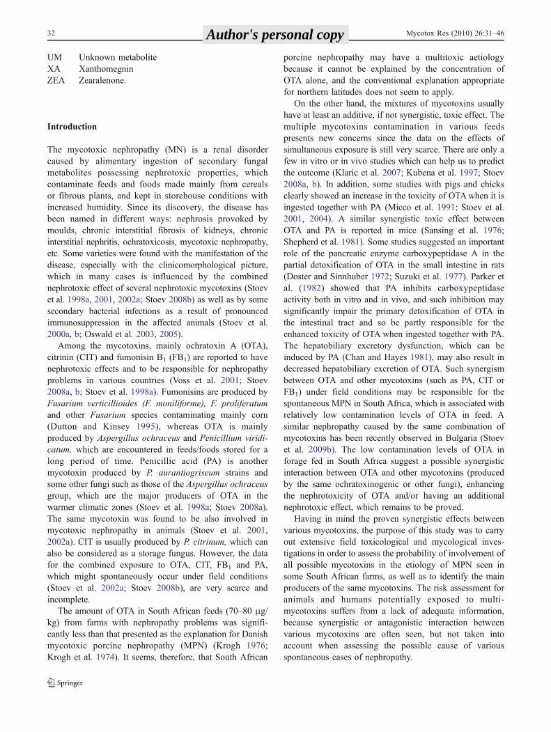
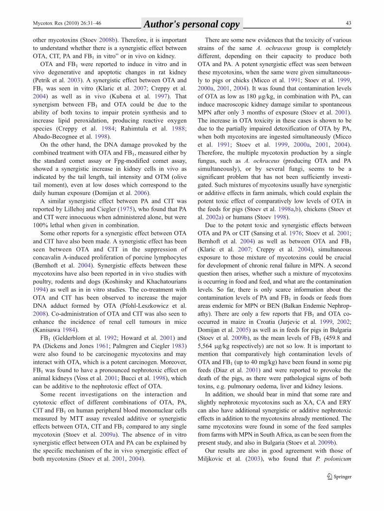
Twenty-four feed samples were collected in years 2007 and2008 (a total of 48 feed samples) from various pig farms inSouth Africa (Baviaanspoort Abattoir, R&R LTD Abatoir,Porkus Abattoir) having nephropathy problems (enlargedand mottled or pale appearance of kidneys) at slaughtertime (Fig. 1). Samples were taken from several locations inthe food storehouses (top, middle and bottom) afterthorough mixing, and subsequently the joint sample wasalso mixed. Most of the samples came directly from thefarms with nephropathy problems and may represent theactual toxic feed. Other feed samples came from someprivate houses in South Africa (Limpopo province) havingnephropathy problems (enlarged or mottled appearance ofkidneys) at slaughter time. Some of the feed samples wouldnot be directly representative of the nephrotoxic feed,because sampling was always retrospective. The feedsamples were then frozen at −20°C until analysis.
Ten blood samples were also taken in 2007 and 2008 (atotal of 20 serum samples) at slaughter time from 20 pigshaving nephropathy problems and originating from three ofthe same farms. After coagulation of the blood, the serumwas separated and the serum samples were then frozen at−20°C until toxicological analysis.
Histological examination
Materials for histological examination were taken from 40kidneys originating from the same farms and showing“mottled”, “enlarged and mottled” or “pale” appearance ofkidneys at slaughter time in the years 2007 and 2008.Kidney samples were fixed in 10% neutral buffered
formalin. Fixed tissues were embedded in paraffin wax,sectioned at 6 µm and stained with hematoxylin-eosin.Periodic acid-Schiff (PAS) stain was also used for provingof lipoproteid, glycoproteid or mucoproteid substances andespecially for proving the thickening of basement tubularmembranes (with lipoprotein structure). Some materialswere stained according to Weigert with iron haematoxylinfor proving the presence or absence of fibrin in various cystformations. Kidney and liver sections from the same pigswere also silver-stained specifically for evidence of lepto-spirosis.
Mycotoxin extraction from feed samples and clean-upprocedures
A multi-mycotoxin extraction method (multi-mycotoxinscreen), which is similar to that of Patterson and Roberts(1979), was used to analyse the feed samples from farms orprivate houses with nephropathy problems. In the samemethod, two extracts can be generated in one step: a neutralfraction (containing mainly: aflatoxins-B1, B2, G1, G2 andM1; trichothecenes-T-2 toxin, diacetoxyscirpenol, deoxyni-valenol, fusarenon X, nivalenol and their acetyl derivatives;zearalenone; patulin; sterigmatocystin; unknown metabo-lite) and an acid fraction (containing mainly: citrinin,ochratoxin A, kojic acid, cyclopiazonic acid, penicillicacid). Most of the mycotoxins of interest (ochratoxin A,citrinin, penicillic acid, penitrem A, deoxynivalenol andzearalenone) were extracted via the same method. In thiscase, 25 g of milled sample was weighed into a conicalflask and 100 ml acetonitrile/H2O (9:1, v/v) was added andplaced on a mechanical shaker for 1 h. The content wasfiltered through a Whatman no 2 V filter paper into aseparation funnel and the filtrate was defatted twice with25 ml iso-octane. Thirty ml saturated Na2CO3 solutiondiluted to 50 ml was added into the defatted filtrate and theextraction was performed (three times) with 25 ml ofdichloromethane through a bed of anhydrous Na2SO4 into around-bottom flask. The supernatant aqueous layer wasretained for the acid phase, whereas the dichloromethaneextract was dried by rotary evaporation, reconstituted with2 ml acetonitrile and transferred into a previously soakeddialysis tube with a knot tied at one end to form a sac. Afteremptying the extract into the dialysis, a knot was tied on theother end and then placed into a boiling tube containing40 ml 30% acetone, sealed with parafilm and placed on ashaker overnight. The overnight dialysate was then trans-ferred to a separation funnel and extracted three times withdichloromethane and passed through a bed of Na2SO4
anhydrous as already described. The extract (neutralfraction) was then reconstituted with 2 ml dichloromethaneand the content put into a 0.5-ml screw-cap vial, dried bypassing through a stream of N2 gas and stored at 4°C for
Fig. 1 Macroscopic appearance of kidneys with mycotoxic porcinenephropathy (MPN). Enlargement and mottled appearance of group 2kidneys in pigs aged 6–8 months
Mycotox Res (2010) 26:31–46 33 Author's personal copy
further analyses. Regarding the acid phase (acid fraction),50 ml 1 M H2SO4 was added gently and shaken, andallowed to stand until the effervescence has subsided. Theextraction was performed with 25 ml dichloromethane (×3times) through a bed of Na2SO4 anhydrous into a round-bottom flask. The filtrate was dried by rotary evaporation.The content was then reconstituted with 2 ml dichloro-methane, put into a 0.5-ml screw-cap vial and dried bypassing through a stream of N2 gas and stored at 4°C forfurther analyses.
Extraction and clean-up of FB1 was performed by aseparate procedure, because fumonisins are only soluble inlower alcohols and acetonitrile and therefore require anadditional extraction step compared to other mycotoxins.The extraction and clean-up of fumonisins was performedaccording to Hinojo et al. (2006) using a SAX column. Tengrams of milled sample was weighed and extracted with20 ml CH3OH/H2O (60:40, v/v) (after shaking for 1 h)through a Whatman no. 2 V filter paper. Ten ml of thefiltrate was cleaned by passing it through a SAX cartridgepreviously conditioned with 5 ml each of CH3OH andCH3OH/H2O (3:1, v/v). The cartridge was washed with8 ml of CH3OH/H2O (3:1, v/v) and 3 ml of CH3OH. Flowrate was maintained at 1 ml/min. The content containing FB(P-fraction) was then flushed with 10 ml of 1% acetic acidin CH3OH into a screw-cap vial, evaporated by passingthrough a stream of N2 gas and stored at 4°C until analysed.
TLC analysis of feed extracts
TLC (thin layer chromatography) analysis was performedaccording to Patterson and Roberts (1979). A two-dimensional thin layer chromatographic technique was usedas 20 ml of the neutral and acid fractions (dissolved in anappropriate solvent-mainly dichloromethane) obtained fromeach feed sample were spotted on TLC plates (about 1 cmfrom the edge of a silica gel TLC plate) and dried in a warmstream of air. The spotted plates were then developed inTLC tanks using two solvents (mainly CEI and TEF formost of the mycotoxins; CM2 and BWA for FB1 or MON,CtE and ChE for ZEA, etc)1 in two-dimensional directionsand were dried after each development. The solvents movethe toxins to the solvent front. The same procedure wasfollowed for the standard mycotoxins. The plates werevisualized under ultraviolet (UV) light at 254 and 365 nm
for the presence of any fluorescent or absorbing spot andwere compared with the standard plates for each analysedmycotoxin. The identity of the mycotoxins was confirmedas the plates were sprayed with a specific reagent for aparticular mycotoxin, such as p-anisaldehyde for FB1, NH3
vapour for PA, diazotised benzidine for ZEA, chromotropicacid for DON and other trichotecenes, Pauly’s reagent forKA or PAT, Ehrlich’s reagent for CA, etc. The RF value foreach spot was calculated and compared with the RF. valueof a standard for each mycotoxin.
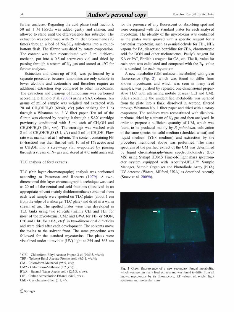
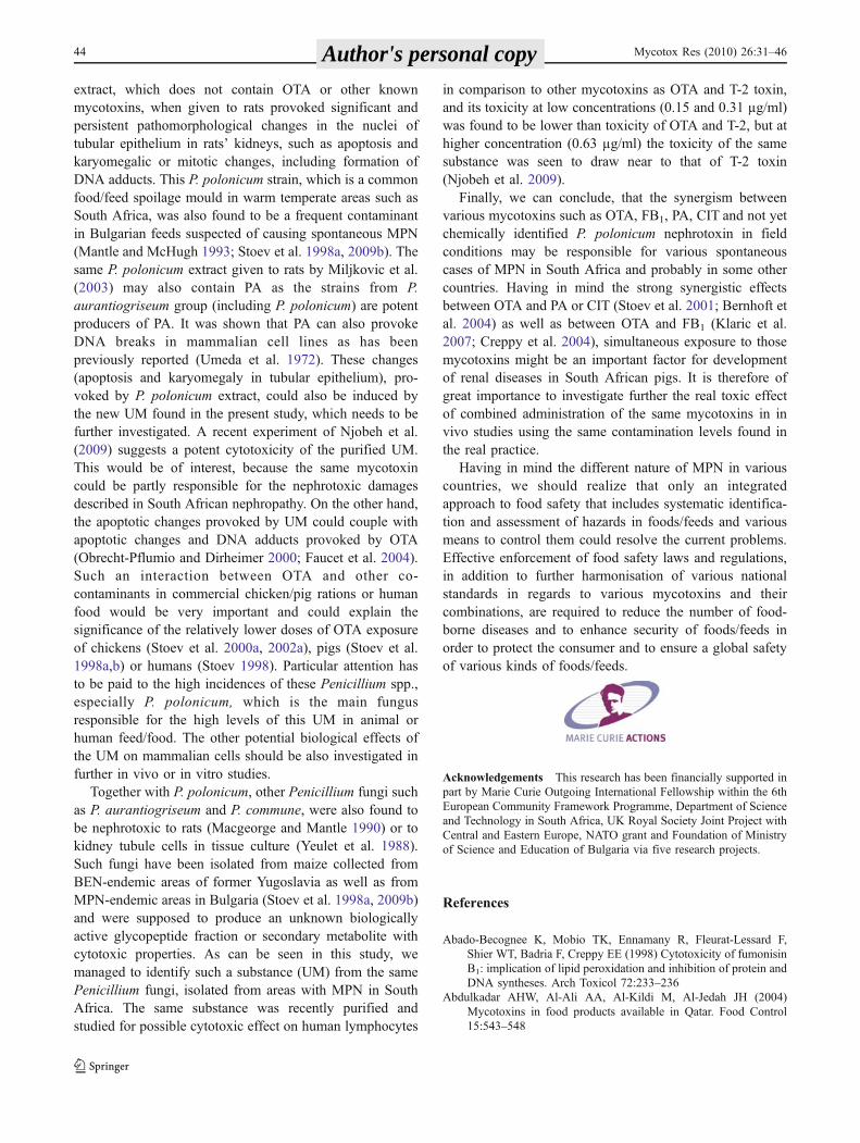
A new metabolite (UM-unknown metabolite) with greenfluorescence (Fig. 2), which was found to differ fromknown mycotoxins and which was seen in many feedsamples, was purified by repeated one-dimensional prepar-ative TLC with alternating mobile phases (CEI and CM).Silica containing the unidentified metabolite was scrapedfrom the plate into a flask, dissolved in acetone, filteredthrough Whatman No. 1 filter paper and dried with a rotoryevaporator. The residues were reconstituted with dichloro-methane, dried by a stream of N2 gas and then analysed. Inorder to prepare a sufficient quantity of UM, which wasfound to be produced mainly by P. polonicum, cultivationof the same species on solid medium (shredded wheat) andliquid medium (YES medium) and extraction by theprocedure mentioned above was performed. The massspectrum of the purified extract of the UM was determinedby liquid chromatography/mass spectrophotometry (LC/MS) using Synapt HDMS Time-of-Flight mass spectrom-eter system equipped with Acquity-UPLC™ SampleManager, Sample Organizer and Photodiode Array (PDA)UV detector (Waters, Milford, USA) as described recently(Stoev et al. 2009b).
1 CEI - Chloroform-Ethyl Acetate-Propan-2-ol (90:5:5, v/v/v);TEF - Toluene-Ethyl Acetate-Formic Acid (6:3:1, v/v/v);CM - Chloroform-Methanol (95:5, v/v);CM2 - Chloroform-Methanol (3:2 ,v/v);BWA - Butanol-Water-Acetic acid (12:5:3, v/v/v);CtE - Carbon tetrachloride-Ethanol (98:2, v/v);ChE - Cyclohexane-Ether (3:1, v/v)
Fig. 2 Green fluorescence of a new secondary fungal metabolite,which was seen in many feed extracts and was found to differ from allknown mycotoxins by its fluorescence, RF values, ultraviolet lightspectrum and molecular mass
34 Mycotox Res (2010) 26:31–46 Author's personal copy
HPLC analysis of feed extracts and serum samples
All mycotoxins of interest found by TLC in the feedsamples were then quantified using HPLC. HPLC analysiswas performed using a Shimadzu system (Kyoto, Japan),consisting of liquid chromatograph LC 20A fitted todegasser DGU 20A3, auto sampler (injection) SIL 20A,communications bus module CBM 20A, column oven CTO20A, photodiode array detector SPD M20A and fluores-cence detector RF 10AXL, all connected to a gigabytecomputer with Intel Core DUO with Microsoft XP. ForHPLC analysis of PA, CIT, OTA, PenA and DON,photodiode array (PDA) detector SPD M20A with WatersSymetry C18 column (250 mm long, 4.6 mm internaldiameter) were used with appropriate mobile phase, flowrate and running time for each mycotoxin, as described byKokkonen et al. (2005) or Abdulkadar et al. (2004) withsome modifications. Reagents and HPLC conditions for thesame mycotoxins were: mobile phase—acetonitrile-water(50:50, v/v); flow rate—0.8 ml/min; injection volume—10 µl per sample; running time—30 min; column temper-ature—30°C. The differences in HPLC conditions for DONwere: mobile phase—acetonitrile/water (15;85, v/v); flowrate—1 ml/min, running time—20 min. The detection ofthe mycotoxins was accomplished at the maximum wave-length of each mycotoxin as PDA setting was usuallybetween 190 and 331 nm. The identification of themycotoxins was based on the comparison of the UV-spectra and the retention times of the detected peaks withthose of the standard substances. Mycotoxins were quanti-fied using peak area and external standard calibration. Thecalibration curves for all investigated mycotoxins werefound to be appropriate for their validation. The detectionlimits ranged usually from 0.02–0.03 µg/kg for OTA up to10–15 µg/kg for DON and FB1. Selected feed samples(free of mycotoxins) were spiked with known levels ofmycotoxins standards and were processed in a similarmanner as the other samples in order to establish recoveryrates for various mycotoxins. Generally, the recovery of theinvestigated mycotoxins was found to be above 60%, butmost often between 85 and 95%, whereas for FB1 it wasabove 90%. Data received in this study were not adjustedbased on the recovery rates obtained.
For performing of FB1 analysis, RF-10AXL fluores-cence detector (FD) was used with the same column andrespective wavelength (excitation and emission), mobilephase, flow rate and running time. The settings of the FDwere: wavelength of 420 nm excitation and 500 nmemission. Reagents and HPLC conditions were: mobilephase—acetonitrile-water in the ratio 60:40; flow rate—1 ml/min; injection volume—10 µl per sample; runningtime—30 min; column temperature—30°C. Derivatizationreagent used for FB1 was naphthalene dicarboxyaldehyde
(NDA), and HPLC was performed according to Bennettand Richard (1994). Residues (sample and standards) weredissolved with 1 ml methanol. Into 100 µl of sample ormycotoxin standard dissolved with methanol in the HPLCvial, the following reagents were added in sequence andmixed after each addition: 100 µl 0.05 M sodium boratebuffer (pH 9.5), 50 µl sodium cyanide and 50 µl NDA. Thiswas followed by incubation in a water bath set at 50°C for20 min, after which the samples were cooled and dilutedwith 700 µl 0.05 M phosphate buffer (pH 7)/acetonitrile(40:60, v/v).
Extraction and HPLC analyses of DON from serumsamples were performed according to Bily et al. (2004), andthose of CIT according to Phillips et al. (1980). PA in serumwas extracted and analysed according to Hanna et al.(1981). OTA in serum was extracted and analysed accord-ing to Boudra and Morgavi (2006). The same methodsusually concern only the free fractions of the mycotoxinsinvestigated, but disregard the conjugates.
Fungal screening and identification
Fungal screening and identification of mycotoxins inanalysed feed samples was performed by the followingmycological analytical procedures: fungal isolation onpotato dextrose agar (PDA) and Ohio Agricultural andExperimental Station agar (OAEA), sub-culturing on PDA,malt extract agar (MEA) and Czapek yeast extract agar(CYA), and macro- and microscopic identification. Thefinal step involved: (1) DNA extraction, (1) PCR (poly-merase chain reaction) amplification, (3) purification ofPCR product, (4) product quantification, and (5) DNAsequence for a confirmation of various species of fungi.
A serial dilution technique was employed for each feedsample as 1 g of the sample was diluted in 10 ml ringersolution and vortexed, and subsequently 1 ml of thissuspension was transferred to 10 ml Ringer solution andvortexed, etc. One ml of each suspension was thenaseptically inoculated on PDA and OAEA in Petri dishesand incubated at 25°C for 7–14 days. The culture methodemployed is that suggested by Klich (2002). From the 4thto the 7th day, plates were screened for different types offungal colonies, and counted using a colony counter. Afterincubation, the number of fungal colonies per gram ofsample was calculated and expressed in colony formingunits per gram (cfu/g). For the identification of fungalspecies, CYA, MEA and PDA were used. The hyphae andconidia from each colony representing each fungal specieswere transfered aseptically onto three spots on PDA andincubated at 30°C for 7–14 days for further identification.Determination of each species of fungi was done using thekeys of Klich and Pitt (1988) and Klich (2002) forAspergillus spp., Pitt and Hocking (1997) for Penicillium
Mycotox Res (2010) 26:31–46 35 Author's personal copy
and Nelson et al. (1983) for Fusarium spp. This was doneby observing both macroscopic characteristics of thecolonies on various media used as well as the microscopicmorphology and measurements of the conidiophores (afterstaining mycelia with 0.1% fuchsin dissolved in lactic acidor with lactophenol blue solution). The isolates were storedat 4°C for further uses and PCR analysis.
When the morphological characteristics of individualfungal species were not sufficient for clear identification,further analysis was performed. All isolated fungi werefurther identified via PCR analysis. The mycelia for PCRanalysis were scraped and transferred into a 0.5-ml sterilescrew-cap vial containing 200 µl of Ringer solution, freeze-dried and stored at −40°C until analysed. Fresh mycelia ofsome fungi were also used for PCR analysis in some cases.The technique involving the comparison of nucleic acidprofiles of individual fungal species was employed using anautomated sequencer. DNA extraction, PCR amplification,purification and quantification of PCR product, and DNAsequencing and analysis were performed using similartechnologies to those described by Samson et al. (2004)and Geiser et al. (2004) with some modifications, usingInvisorb Spin Plant Mini Kit (Invitek, Berlin) for DNAextractions from plant material (fresh, frozen or driedmaterials) in addition to MSB® Spin PCRapace for ultrafastpurification and concentration of PCR-fragments (Invitek).This PCR process is covered by U.S. Patents 4,683,195 and4,683,202 owned by Hoffmann-LaRoche. Samples werefurther analysed on an ABI PRISM 3700 Genetic analyzer(AB; Applied Biosystems, Nieuwerkerk a/d Yssel, TheNetherlands). The forward and reverse sequences of thePCR products were assembled with a DYEamic ETTerminator Cycle Sequencing Kit (Amersham Bioscience,Roosendaal, Netherlands) using the programmes SeqManand EditSeq from the LaserGene package (DNAStar,Madison, WI). The PCR analysis was performed in InqabaBiotec in Pretoria (South Africa) as an Inqaba Finch serverwas used for DNA sequencing and identification of fungi.
Determination of mycotoxinogenic potentials of isolates
Fungal species were further analysed for their capability toproduce various mycotoxins. The isolates were individuallycultured on YES agar in Petri dishes and incubated at 25°Cfor 2–3 weeks according to the method of Singh et al.(1991). A TLC technique was then employed whereby 5 gof isolate including the medium was plugged and dissolvedin 10 ml of dichloromethane. This solution was furtherfiltered, and the filtrate put in a screw-cap vial and driedunder a stream of N2 gas then stored at 4°C until analysed.Two-dimensional TLC (Patterson and Roberts 1979) wasemployed for the detection of mycotoxins. The identity ofthe mycotoxins was confirmed as the plates were sprayed
with a specific reagent for a particular mycotoxin asdescribed above and the RF value for each spot wascalculated and compared with the RF. value of a standardfor each mycotoxin.
Chemicals and mycotoxins
The standards of all analysed mycotoxins were obtainedfrom Sigma Bioscience (St Louis, USA) or Merck, exceptfor penicillic acid (PA) which was obtained from A.G.Scientific (San Diego, CA, USA).
All solvents for HPLC analysis were HPLC grade andwere obtained from Merck, Darmstadt, Germany. Allchemicals for PCR analysis were purchased from Invitek.
Statistical analysis
The Student's t test was used to calculate the mean valuesand standard error of the mean of various parameters asappropriate.
Results
This nephropathy (“mottled”, “enlarged and mottled” or“pale” appearance of kidneys) was established during themeat inspection at slaughter time (Fig. 1). As a whole, thefrequency and duration of the observed nephropathy indifferent batches of slaughtered pigs varied significantly.According to their macroscopic appearance, kidneys couldbe grouped into three different groups: mottled, enlargedand mottled (marbled) or enlarged and pale (Table 1). Thesegroups can be considered to illustrate the progressive stagesof development of the disease.
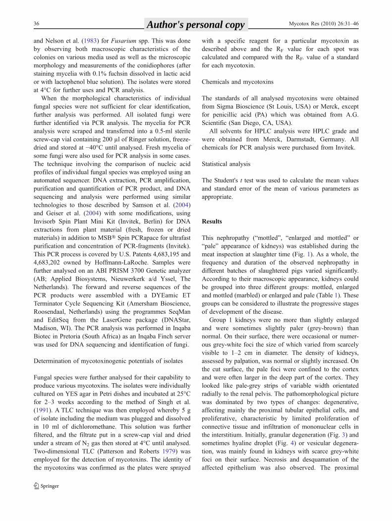
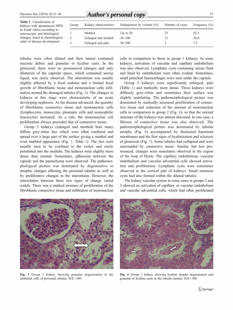
Group 1 kidneys were no more than slightly enlargedand were sometimes slightly paler (grey-brown) thannormal. On their surface, there were occasional or numer-ous grey-white foci the size of which varied from scarcelyvisible to 1–2 cm in diameter. The density of kidneys,assessed by palpation, was normal or slightly increased. Onthe cut surface, the pale foci were confined to the cortexand were often larger in the deep part of the cortex. Theylooked like pale-grey strips of variable width orientatedradially to the renal pelvis. The pathomorphological picturewas dominated by two types of changes: degenerative,affecting mainly the proximal tubular epithelial cells, andproliferative, characteristic by limited proliferation ofconnective tissue and infiltration of mononuclear cells inthe interstitium. Initially, granular degeneration (Fig. 3) andsometimes hyaline droplet (Fig. 4) or vesicular degenera-tion, was mainly found in kidneys with scarce grey-whitefoci on their surface. Necrosis and desquamation of theaffected epithelium was also observed. The proximal
36 Mycotox Res (2010) 26:31–46 Author's personal copy
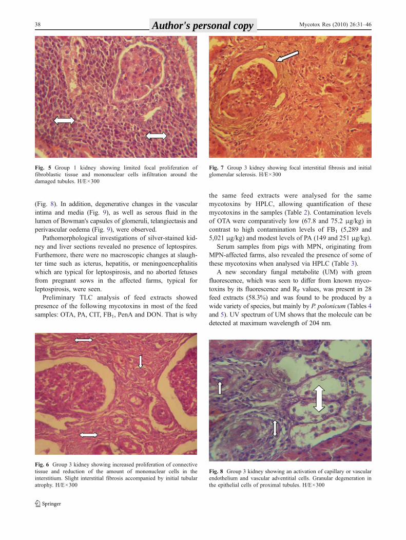
tubules were often dilated and their lumen containednecrotic debris and granular or hyaline casts. In theglomeruli, there were no pronounced changes and onlydilatation of the capsular space, which contained serousliquid, was rarely observed. The interstitium was usuallyslightly affected by a focal oedema and a limited focalgrowth of fibroblastic tissue and mononuclear cells infil-tration around the damaged tubules (Fig. 5). The changes inkidneys at this stage were characteristic of an acutedeveloping nephrosis. As the disease advanced, the quantityof fibroblastic connective tissue and mononuclear cells(lymphocytes, monocytes, plasmatic cells and eosinophilicleucocytes) increased. As a rule, the mononuclear cellproliferation always preceded that of connective tissue.
Group 2 kidneys (enlarged and mottled) bore manydiffuse grey-white foci which were often confluent andspread over a large part of the surface giving a mottled andeven marbled appearance (Fig. 1, Table 1). The foci wereusually seen to be confined to the cortex and rarelypenetrated into the medulla. The kidneys were slightly moredense than normal. Sometimes, adhesions between thecapsule and the parenchyma were observed. The pathomor-phological picture was dominated by degenerative oratrophic changes affecting the proximal tubules as well asby proliferative changes in the interstitium. However, theinterrelation between these two types of change variedwidely. There was a marked increase of proliferation of thefibroblastic connective tissue and infiltration of mononuclear
cells in comparison to those in group 1 kidneys. In somekidneys, activation of vascular and capillary endotheliumwas also observed. Lymphatic cysts containing serous fluidand lined by endothelium were often evident. Sometimes,small petechial haemorrhages were seen under the capsule.
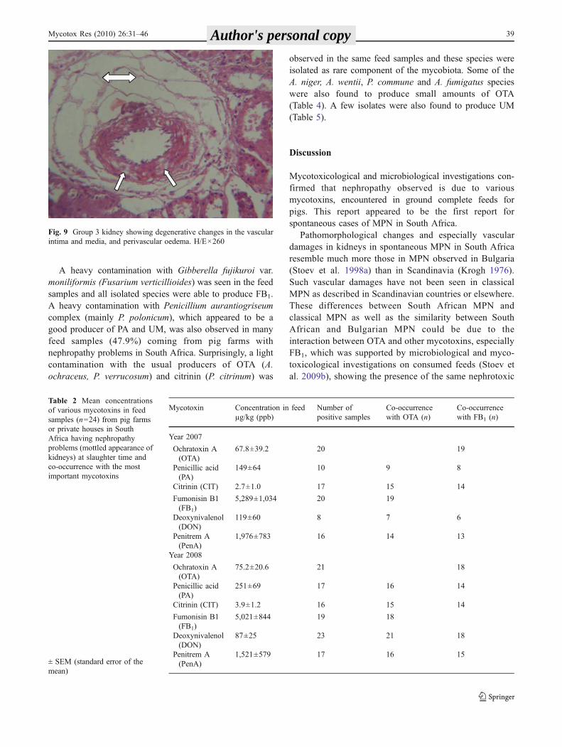
Group 3 kidneys were significantly enlarged, pale(Table 1) and markedly more dense. These kidneys werediffusely grey-white and sometimes their surface wasslightly undulating. The pathomorphological picture wasdominated by markedly increased proliferation of connec-tive tissue and reduction of the amount of mononuclearcells in comparison to group 2 (Fig. 6), so that the normalstructure of the kidneys was almost obscured. In one case, afibrosis of connective tissue was also observed. Thepathomorphological picture was dominated by tubularatrophy (Fig. 6) accompanied by thickened basementmembranes and the first signs of hyalinization and sclerosisof glomeruli (Fig. 7). Some tubules had collapsed and weresurrounded by connective tissue. Similar, but less pro-nounced, changes were sometimes observed in the regionof the loop of Henle. The capillary endothelium, vascularendothelium and vascular adventitial cells showed activa-tion and proliferation. Lymphatic cysts were sometimesobserved in the cortical part of kidneys. Small retentioncysts had also formed within the dilated tubules.
The kidney vascular system in some cases in groups 2 and3 showed an activation of capillary or vascular endotheliumand vascular adventitial cells, which had often proliferated
Fig. 4 Group 1 kidney showing hyaline droplet degeneration andgranular or hyaline casts in the tubular lumina. H/E×300
Fig. 3 Group 1 kidney showing granular degeneration in theepithelial cells of proximal tubules. H/E×300
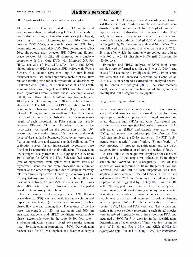
Group Kidney characteristics Enlargement by volume (%) Number of cases Frequency (%)
1 Mottled Up to 20 25 62.5
2 Enlarged and mottled 20–100 12 30.0
3 Enlarged and pale 50–200 3 7.5
Table 1 Classification ofkidneys with spontaneous MPNin South Africa according tomacroscopic and histologicalchanges, listed in chronologicalorder of disease development
Mycotox Res (2010) 26:31–46 37 Author's personal copy
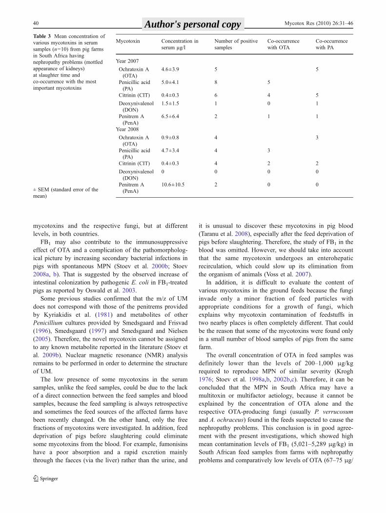
(Fig. 8). In addition, degenerative changes in the vascularintima and media (Fig. 9), as well as serous fluid in thelumen of Bowman's capsules of glomeruli, telangiectasis andperivascular oedema (Fig. 9), were observed.
Pathomorphological investigations of silver-stained kid-ney and liver sections revealed no presence of leptospires.Furthemore, there were no macroscopic changes at slaugh-ter time such as icterus, hepatitis, or meningoencephalitiswhich are typical for leptospirosis, and no aborted fetusesfrom pregnant sows in the affected farms, typical forleptospirosis, were seen.
Preliminary TLC analysis of feed extracts showedpresence of the following mycotoxins in most of the feedsamples: OTA, PA, CIT, FB1, PenA and DON. That is why
the same feed extracts were analysed for the samemycotoxins by HPLC, allowing quantification of thesemycotoxins in the samples (Table 2). Contamination levelsof OTA were comparatively low (67.8 and 75.2 μg/kg) incontrast to high contamination levels of FB1 (5,289 and5,021 μg/kg) and modest levels of PA (149 and 251 μg/kg).
Serum samples from pigs with MPN, originating fromMPN-affected farms, also revealed the presence of some ofthese mycotoxins when analysed via HPLC (Table 3).
A new secondary fungal metabolite (UM) with greenfluorescence, which was seen to differ from known myco-toxins by its fluorescence and RF values, was present in 28feed extracts (58.3%) and was found to be produced by awide variety of species, but mainly by P. polonicum (Tables 4and 5). UV spectrum of UM shows that the molecule can bedetected at maximum wavelength of 204 nm.
Fig. 8 Group 3 kidney showing an activation of capillary or vascularendothelium and vascular adventitial cells. Granular degeneration inthe epithelial cells of proximal tubules. H/E×300
Fig. 7 Group 3 kidney showing focal interstitial fibrosis and initialglomerular sclerosis. H/E×300
Fig. 6 Group 3 kidney showing increased proliferation of connectivetissue and reduction of the amount of mononuclear cells in theinterstitium. Slight interstitial fibrosis accompanied by initial tubularatrophy. H/E×300
Fig. 5 Group 1 kidney showing limited focal proliferation offibroblastic tissue and mononuclear cells infiltration around thedamaged tubules. H/E×300
38 Mycotox Res (2010) 26:31–46 Author's personal copy
A heavy contamination with Gibberella fujikuroi var.moniliformis (Fusarium verticillioides) was seen in the feedsamples and all isolated species were able to produce FB1.A heavy contamination with Penicillium aurantiogriseumcomplex (mainly P. polonicum), which appeared to be agood producer of PA and UM, was also observed in manyfeed samples (47.9%) coming from pig farms withnephropathy problems in South Africa. Surprisingly, a lightcontamination with the usual producers of OTA (A.ochraceus, P. verrucosum) and citrinin (P. citrinum) was
observed in the same feed samples and these species wereisolated as rare component of the mycobiota. Some of theA. niger, A. wentii, P. commune and A. fumigatus specieswere also found to produce small amounts of OTA(Table 4). A few isolates were also found to produce UM(Table 5).
Discussion
Mycotoxicological and microbiological investigations con-firmed that nephropathy observed is due to variousmycotoxins, encountered in ground complete feeds forpigs. This report appeared to be the first report forspontaneous cases of MPN in South Africa.
Pathomorphological changes and especially vasculardamages in kidneys in spontaneous MPN in South Africaresemble much more those in MPN observed in Bulgaria(Stoev et al. 1998a) than in Scandinavia (Krogh 1976).Such vascular damages have not been seen in classicalMPN as described in Scandinavian countries or elsewhere.These differences between South African MPN andclassical MPN as well as the similarity between SouthAfrican and Bulgarian MPN could be due to theinteraction between OTA and other mycotoxins, especiallyFB1, which was supported by microbiological and myco-toxicological investigations on consumed feeds (Stoev etal. 2009b), showing the presence of the same nephrotoxic
Fig. 9 Group 3 kidney showing degenerative changes in the vascularintima and media, and perivascular oedema. H/E×260
Mycotoxin Concentration in feedµg/kg (ppb)
Number ofpositive samples
Co-occurrencewith OTA (n)
Co-occurrencewith FB1 (n)
Year 2007
Ochratoxin A(OTA)
67.8±39.2 20 19
Penicillic acid(PA)
149±64 10 9 8
Citrinin (CIT) 2.7±1.0 17 15 14
Fumonisin B1(FB1)
5,289±1,034 20 19
Deoxynivalenol(DON)
119±60 8 7 6
Penitrem A(PenA)
1,976±783 16 14 13
Year 2008
Ochratoxin A(OTA)
75.2±20.6 21 18
Penicillic acid(PA)
251±69 17 16 14
Citrinin (CIT) 3.9±1.2 16 15 14
Fumonisin B1(FB1)
5,021±844 19 18
Deoxynivalenol(DON)
87±25 23 21 18
Penitrem A(PenA)
1,521±579 17 16 15
Table 2 Mean concentrationsof various mycotoxins in feedsamples (n=24) from pig farmsor private houses in SouthAfrica having nephropathyproblems (mottled appearance ofkidneys) at slaughter time andco-occurrence with the mostimportant mycotoxins
± SEM (standard error of themean)
Mycotox Res (2010) 26:31–46 39 Author's personal copy
mycotoxins and the respective fungi, but at differentlevels, in both countries.
FB1 may also contribute to the immunosuppressiveeffect of OTA and a complication of the pathomorpholog-ical picture by increasing secondary bacterial infections inpigs with spontaneous MPN (Stoev et al. 2000b; Stoev2008a, b). That is suggested by the observed increase ofintestinal colonization by pathogenic E. coli in FB1-treatedpigs as reported by Oswald et al. 2003.
Some previous studies confirmed that the m/z of UMdoes not correspond with those of the penitrems providedby Kyriakidis et al. (1981) and metabolites of otherPenicillium cultures provided by Smedsgaard and Frisvad(1996), Smedsgaard (1997) and Smedsgaard and Nielsen(2005). Therefore, the novel mycotoxin cannot be assignedto any known metabolite reported in the literature (Stoev etal. 2009b). Nuclear magnetic resonance (NMR) analysisremains to be performed in order to determine the structureof UM.
The low presence of some mycotoxins in the serumsamples, unlike the feed samples, could be due to the lackof a direct connection between the feed samples and bloodsamples, because the feed sampling is always retrospectiveand sometimes the feed sources of the affected farms havebeen recently changed. On the other hand, only the freefractions of mycotoxins were investigated. In addition, feeddeprivation of pigs before slaughtering could eliminatesome mycotoxins from the blood. For example, fumonisinshave a poor absorption and a rapid excretion mainlythrough the faeces (via the liver) rather than the urine, and
it is unusual to discover these mycotoxins in pig blood(Taranu et al. 2008), especially after the feed deprivation ofpigs before slaughtering. Therefore, the study of FB1 in theblood was omitted. However, we should take into accountthat the same mycotoxin undergoes an enterohepaticrecirculation, which could slow up its elimination fromthe organism of animals (Voss et al. 2007).
In addition, it is difficult to evaluate the content ofvarious mycotoxins in the ground feeds because the fungiinvade only a minor fraction of feed particles withappropriate conditions for a growth of fungi, whichexplains why mycotoxin contamination of feedstuffs intwo nearby places is often completely different. That couldbe the reason that some of the mycotoxins were found onlyin a small number of blood samples of pigs from the samefarm.
The overall concentration of OTA in feed samples wasdefinitely lower than the levels of 200–1,000 μg/kgrequired to reproduce MPN of similar severity (Krogh1976; Stoev et al. 1998a,b, 2002b,c). Therefore, it can beconcluded that the MPN in South Africa may have amultitoxin or multifactor aetiology, because it cannot beexplained by the concentration of OTA alone and therespective OTA-producing fungi (usually P. verrucosumand A. ochraceus) found in the feeds suspected to cause thenephropathy problems. This conclusion is in good agree-ment with the present investigations, which showed highmean contamination levels of FB1 (5,021–5,289 μg/kg) inSouth African feed samples from farms with nephropathyproblems and comparatively low levels of OTA (67–75 μg/
Mycotoxin Concentration inserum µg/l
Number of positivesamples
Co-occurrencewith OTA
Co-occurrencewith PA
Year 2007
Ochratoxin A(OTA)
4.6±3.9 5 5
Penicillic acid(PA)
5.0±4.1 8 5
Citrinin (CIT) 0.4±0.3 6 4 5
Deoxynivalenol(DON)
1.5±1.5 1 0 1
Penitrem A(PenA)
6.5±6.4 2 1 1
Year 2008
Ochratoxin A(OTA)
0.9±0.8 4 3
Penicillic acid(PA)
4.7±3.4 4 3
Citrinin (CIT) 0.4±0.3 4 2 2
Deoxynivalenol(DON)
0 0 0 0
Penitrem A(PenA)
10.6±10.5 2 0 0
Table 3 Mean concentration ofvarious mycotoxins in serumsamples (n=10) from pig farmsin South Africa havingnephropathy problems (mottledappearance of kidneys)at slaughter time andco-occurrence with the mostimportant mycotoxins
± SEM (standard error of themean)
40 Mycotox Res (2010) 26:31–46 Author's personal copy
kg), CIT (2.7–3.9 μg/kg) and PA (149–251 μg/kg) in thesame feeds. It is also important to mention that the percentof feed contaminated with the mycotoxins involved wasconstantly high, above 70%. A similar multimycotoxinetiology was also found for Bulgarian MPN as the samemixture of mycotoxins (188–376 μg/kg OTA with 100%
positives; 54–120 μg/kg CIT with 92–96% positives; 838–904 μg/kg PA with 88–92% positives; 3,254–5,564 μg/kgFB1 with 92–96% positives) was established in Bulgarianfeed samples from pig farms with nephropathy problems(Stoev 2008b). The levels of OTA and PA, considered to bethe main mycotoxins involved in this nephropathy, were 2–
Table 4 Percentage of contaminated feed samples (24 samples for 2007 and 24 samples for 2008) with Fusarium, Penicillium and Aspergillusfungi, level of fungal contamination, number of strains isolated and number of toxinogenic strains isolated as well as kind of mycotoxins producedby each fungal strain and co-occurrence of mycotoxins with OTA and FB1 in various feed samples from pig farms or private houses in SouthAfrica having nephropathy problems (enlarged and mottled appearance of kidneys) at slaughter time
Strain isolated Percentage ofcontaminatedsamples (%)
Range of fungalcontamination(cpu/g)
Number ofstrainsisolated
Number oftoxinogenicstrains
Kind of mycotoxinsproduced (number ofspecies)
Fusarium species
Gibberella fujikuroi(var. moniliformis)
62.5 2×10³–8×10⁶ 30 30 FB1(15), FB2(4)b, ZEA(12)b,
MON(4), UM(1)
F. verticillioides(F. moniliforme)
2 4×10⁶ 1 1 FB1(1), ZEA(1)b
F. oxysporum 2 3×10⁴ 1 1 ZEA(1)
F. graminearum 4.2 3×10³–4×10⁵ 2 2 ZEA(2), DON(2), T-2 (1), DAS(1)
Penicillium species
P. viridicatum(P. verrucosum)
2 5×10³ 1 1 OTA(1), CIT(1)a, PA(1)a
XA(1)a, UM(1)a
P. commune 6.2 4×10³–5×10⁶ 3 2 OTA(2), UM (2)a
P. citrinum 6.2 3×10³–5×10⁵ 3 3 CIT(3), KA(1), UM(1)
P. expansum 2 3×10⁵ 1 1 CIT(1), PAT(1), UM(1)
P. aurantiogriseum 8.3 3×10³–4×10⁶ 4 2 PA(2), UM(1)
P. polonicum 47.9 5×10³–6×10⁶ 23 18 PA (16), UM (18)
P. freii 2 4×10³ 1 1 PA(1), UM(1)
P. chrysogenum 4.2 3×10³–4×10⁶ 2 1 PA(2), UM(1)
P. crustosum 6.2 3×10³–8×10⁵ 3 3 PA(3), PenA(2), UM(2)
P. simplicissimum 2 3×10⁴ 1 1 PA(1)
P. islandicum 2 5×10³ 1 1 ERY(1), LUT(1), ISL(1), UM(1)
Aspergillus species
A. ochraceus 4.2 2×10³–3×10⁴ 2 1 OTA(1), PA(1)a
A. niger 25 4×10³–3×10⁶ 12 5 OTA(5), OTC(1)a
A. wentii 2 3×10⁴ 1 1 OTA(1), KA(1)a
A. candidus 18.7 4×10³–8×10⁵ 9 7 CIT(1), KA(1), UM(7)
A. fumigatus 45.8 3×10³–7×10⁶ 22 22 OTA(3), GLI(2), KA(2), UM(17)a
A. flavus 52 2×10³–4×10⁶ 25 25 Afs (21), CA (12), KA(14), UM(12)
A. oryzae 2 4×10⁴ 1 1 CA(1), KA(1)
A. restrictus 2 4×10⁴ 1 1 UM(1)
Eurotiumamstelodami
8.3 4×10³–2×10⁵ 4 1 UM(1)
AFs Aflatoxins, CIT citrinin, CA cyclopiazonic acid, DAS diacetoxyscirpenol, DON deoxynivalenol, ERY erythroskyrin, FB1 fumonisin B1, FB2
fumonisin B2, GLI gliotoxin, ISL islandiotoxin, KA kojic acid, LUT luteoskyirin, MON moniliformin, UM unknown metabolite, OTA ochratoxinA, OTC ochratoxin C, PenA penitrem A, PAT patulin, PA penicillic acid, T-2 T-2 toxin, XA xanthomegnin, ZEA zearalenonea Co-occurrence with OTAbCo-occurrence with FB1
Table 4 Percentage of contaminated feed samples (24 samples for2007 and 24 samples for 2008) with Fusarium, Penicillium andAspergillus fungi, level of fungal contamination, number of strainsisolated and number of toxinogenic strains isolated as well as kind of
mycotoxins produced by each fungal strain and co-occurrence ofmycotoxins with OTA and FB1 in various feed samples from pigfarms or private houses in South Africa having nephropathy problems(enlarged and mottled appearance of kidneys) at slaughter time
Mycotox Res (2010) 26:31–46 41 Author's personal copy
3 times higher as should be anticipated from the higherseverity and spread of the same nephropathy in Bulgariaunlike in South Africa (Stoev et al. 2009b).
A possible model for the South African nephropathycould be the renal disease that was previously investigatedin depth in Denmark in the 1970s and shown conclusivelyto be caused by nephrotoxins of fungal origin (mainly OTAand CIT produced by P. verrucosum growing on poorlystored feed or grain). However, in the South Africannephropathy, similar to the Bulgarian nephropathy, theincidence of OTA measured in the diet and in the serumwas much less than should be necessary (according toextensive experimentation in Denmark) to fully account forthe renal changes (Stoev et al. 1998a,b). A classificationwas made of the kidney damage in pigs in Bulgaria.According to the different macroscopic and microscopicchanges as well as according to the different stages of theprogress of this nephropathy, the kidneys were classified
into five separated groups: “mottled kidneys”, “enlargedand marbled kidneys”, “enlarged pale kidneys”, “cystickidneys” and “fibrotic kidneys” (Stoev et al. 1998a). Thepathology in the last two groups of kidneys is notcharacteristic for nephropathy in South Africa as was seenin the present study. These differences in macroscopical ormicroscopical picture of the kidneys were considered to bedue to the differences in the length of the time of exposureto the mouldy feed (Stoev et al. 1998a) as well as to thedifferences in the amount or combination of nephrotoxicmycotoxins ingested by each pig.
It seems that some differences between classic Danishporcine nephropathy and South African or Bulgariannephropathy in pigs (e.g. vascular and edematous changes)as described recently by Stoev et al. (1998a) are probably aresult of the effects of other nephrotoxic metabolites, suchas FB1 and PA in addition to the toxic effect of OTA, ormay be attributed to synergistic effects between OTA and
Table 5 Percentage of contaminated feed samples (24 samples for 2007 and 24 samples for 2008) with various other fungi, level of fungalcontamination, number of strains isolated and number of toxinogenic strains isolated as well as kind of mycotoxins produced by each fungal strainin various feed samples from pig farms or private houses in South Africa having nephropathy problems (enlarged and mottled appearance ofkidneys) at slaughter time
Strain isolated Percentage ofcontaminated samples(%)
Range of fungalcontamination (cpu/g)
Number ofstrains isolated
Number oftoxinogenicstrains
Kind of mycotoxins produced(number of species)
Acremoniumstrictum
12.5 4×10³–5×10⁵ 6 1 OOS (1)
Pichiaguilliermondii
2 3×10⁴ 1 – –
Pichia caribbica 14.5 3×10³–5×10⁴ 7 – –
Paecilomycesvariotii
8.3 3×10³–4×10⁴ 4 – –
Paecilomycessinensis
4.2 3×10³–2×10⁵ 2 1 UM (1)
Cladosp.cladosporioides
4.2 3×10³–4×10⁴ 2 – –
Talaromycesemersonii
4.2 3×10³–3×10⁴ 2 – –
Cordicepsbassiana
2 4×10⁴ 1 – –
Mucorracemosus
4.2 3×10³–2×10⁵ 2 1 UM (1)
Mucorcircinelloides
10.4 3×10³–2×10⁵ 5 2 UM (2)
Candidaalbicans
4.2 3×10³–5×10⁴ 2 – –
Candidatropicalis
6.2 4×10³–7×10⁴ 3 – –
Geomithiapallida
4.2 5×10³–8×10⁴ 2 1 UM (1)
Sacharomycescerevisiae
2 7×10⁴ 1 – –
Rhodotorulamucilaginosa
2 5×10⁴ 1 – –
OOS Oosporein (isoospoin)
Table 5 Percentage of contaminated feed samples (24 samples for2007 and 24 samples for 2008) with various other fungi, level offungal contamination, number of strains isolated and number oftoxinogenic strains isolated as well as kind of mycotoxins produced by
each fungal strain in various feed samples from pig farms or privatehouses in South Africa having nephropathy problems (enlarged andmottled appearance of kidneys) at slaughter time
42 Mycotox Res (2010) 26:31–46 Author's personal copy
other mycotoxins (Stoev 2008b). Therefore, it is importantto understand whether there is a synergistic effect betweenOTA, CIT, PA and FB1 in vitro” or in vivo on kidney.
OTA and FB1 were reported to induce in vitro and invivo degenerative and apoptotic changes in rat kidney(Petrik et al. 2003). A synergistic effect between OTA andFB1 was seen in vitro (Klaric et al. 2007; Creppy et al.2004) as well as in vivo (Kubena et al. 1997). Thatsynergism between FB1 and OTA could be due to theability of both toxins to impair protein synthesis and toincrease lipid peroxidation, producing reactive oxygenspecies (Creppy et al. 1984; Rahimtula et al. 1988;Abado-Becognee et al. 1998).
On the other hand, the DNA damage provoked by thecombined treatment with OTA and FB1, measured either bythe standard comet assay or Fpg-modified comet assay,showed a synergistic increase in kidney cells in vivo asindicated by the tail length, tail intensity and OTM (olivetail moment), even at low doses which correspond to thedaily human exposure (Domijan et al. 2006).
A similar synergistic effect between PA and CIT wasreported by Lillehoj and Ciegler (1975), who found that PAand CIT were innocuous when administered alone, but were100% lethal when given in combination.
Some other reports for a synergistic effect between OTAand CIT have also been made. A synergistic effect has beenseen between OTA and CIT in the suppression ofconcavalin A-induced proliferation of porcine lymphocytes(Bernhoft et al. 2004). Synergistic effects between thesemycotoxins have also been reported in in vivo studies withpoultry, rodents and dogs (Koshinsky and Khachatourians1994) as well as in in vitro studies. The co-treatment withOTA and CIT has been observed to increase the majorDNA adduct formed by OTA (Pfohl-Leszkowicz et al.2008). Co-administration of OTA and CIT was also seen toenhance the incidence of renal cell tumours in mice(Kanisawa 1984).
FB1 (Gelderblom et al. 1992; Howard et al. 2001) andPA (Dickens and Jones 1961; Palmgren and Ciegler 1983)were also found to be carcinogenic mycotoxins and mayinteract with OTA, which is a potent carcinogen. Moreover,FB1 was found to have a pronounced nephrotoxic effect onanimal kidneys (Voss et al. 2001; Bucci et al. 1998), whichcan be additive to the nephrotoxic effect of OTA.
Some recent investigations on the interaction andcytotoxic effect of different combinations of OTA, PA,CIT and FB1 on human peripheral blood mononuclear cellsmeasured by MTT assay revealed additive or synergisticeffects between OTA, CIT and FB1 compared to any singlemycotoxin (Stoev et al. 2009a). The absence of in vitrosynergistic effect between OTA and PA can be explained bythe specific mechanism of the in vivo synergistic effect ofboth mycotoxins (Stoev et al. 2001, 2004).
There are some new evidences that the toxicity of variousstrains of the same A. ochraceus group is completelydifferent, depending on their capacity to produce bothOTA and PA. A potent synergistic effect was seen betweenthese mycotoxins, when the same were given simultaneous-ly to pigs or chicks (Micco et al. 1991; Stoev et al. 1999,2000a, 2001, 2004). It was found that contamination levelsof OTA as low as 180 µg/kg, in combination with PA, caninduce macroscopic kidney damage similar to spontaneousMPN after only 3 months of exposure (Stoev et al. 2001).The increase in OTA toxicity in these cases is shown to bedue to the partially impaired detoxification of OTA by PA,when both mycotoxins are ingested simultaneously (Miccoet al. 1991; Stoev et al. 1999, 2000a, 2001, 2004).Therefore, the multiple mycotoxin production by a singlefungus, such as A. ochraceus (producing OTA and PAsimultaneously), or by several fungi, seems to be asignificant problem that has not been sufficiently investi-gated. Such mixtures of mycotoxins usually have synergisticor additive effects in farm animals, which could explain thepotent toxic effect of comparatively low levels of OTA inthe feeds for pigs (Stoev et al. 1998a,b), chickens (Stoev etal. 2002a) or humans (Stoev 1998).
Due to the potent toxic and synergistic effects betweenOTA and PA or CIT (Sansing et al. 1976; Stoev et al. 2001;Bernhoft et al. 2004) as well as between OTA and FB1
(Klaric et al. 2007; Creppy et al. 2004), simultaneousexposure to those mixture of mycotoxins could be crucialfor development of chronic renal failure in MPN. A secondquestion then arises, whether such a mixture of mycotoxinsis occurring in food and feed, and what are the contaminationlevels. So far, there is only scarce information about thecontamination levels of PA and FB1 in foods or feeds fromareas endemic for MPN or BEN (Balkan Endemic Nephrop-athy). There are only a few reports that FB1 and OTA co-occurred in maize in Croatia (Jurjevic et al. 1999, 2002;Domijan et al. 2005) as well as in feeds for pigs in Bulgaria(Stoev et al. 2009b), as the mean levels of FB1 (459.8 and5,564 μg/kg respectively) are not so low. It is important tomention that comparatively high contamination levels ofOTA and FB1 (up to 40 mg/kg) have been found in some pigfeeds (Diaz et al. 2001) and were reported to provoke thedeath of the pigs, as there were pathological signs of bothtoxins, e.g. pulmonary oedema, liver and kidney lesions.
In addition, we should bear in mind that some rare andslightly nephrotoxic mycotoxins such as XA, CA and ERYcan also have additional synergistic or additive nephrotoxiceffects in addition to the mycotoxins already mentioned. Thesame mycotoxins were found in some of the feed samplesfrom farms withMPN in South Africa, as can be seen from thepresent study, and also in Bulgaria (Stoev et al. 2009b).
Our results are also in good agreement with those ofMiljkovic et al. (2003), who found that P. polonicum
Mycotox Res (2010) 26:31–46 43 Author's personal copy
extract, which does not contain OTA or other knownmycotoxins, when given to rats provoked significant andpersistent pathomorphological changes in the nuclei oftubular epithelium in rats’ kidneys, such as apoptosis andkaryomegalic or mitotic changes, including formation ofDNA adducts. This P. polonicum strain, which is a commonfood/feed spoilage mould in warm temperate areas such asSouth Africa, was also found to be a frequent contaminantin Bulgarian feeds suspected of causing spontaneous MPN(Mantle and McHugh 1993; Stoev et al. 1998a, 2009b). Thesame P. polonicum extract given to rats by Miljkovic et al.(2003) may also contain PA as the strains from P.aurantiogriseum group (including P. polonicum) are potentproducers of PA. It was shown that PA can also provokeDNA breaks in mammalian cell lines as has beenpreviously reported (Umeda et al. 1972). These changes(apoptosis and karyomegaly in tubular epithelium), pro-voked by P. polonicum extract, could also be induced bythe new UM found in the present study, which needs to befurther investigated. A recent experiment of Njobeh et al.(2009) suggests a potent cytotoxicity of the purified UM.This would be of interest, because the same mycotoxincould be partly responsible for the nephrotoxic damagesdescribed in South African nephropathy. On the other hand,the apoptotic changes provoked by UM could couple withapoptotic changes and DNA adducts provoked by OTA(Obrecht-Pflumio and Dirheimer 2000; Faucet et al. 2004).Such an interaction between OTA and other co-contaminants in commercial chicken/pig rations or humanfood would be very important and could explain thesignificance of the relatively lower doses of OTA exposureof chickens (Stoev et al. 2000a, 2002a), pigs (Stoev et al.1998a,b) or humans (Stoev 1998). Particular attention hasto be paid to the high incidences of these Penicillium spp.,especially P. polonicum, which is the main fungusresponsible for the high levels of this UM in animal orhuman feed/food. The other potential biological effects ofthe UM on mammalian cells should be also investigated infurther in vivo or in vitro studies.
Together with P. polonicum, other Penicillium fungi suchas P. aurantiogriseum and P. commune, were also found tobe nephrotoxic to rats (Macgeorge and Mantle 1990) or tokidney tubule cells in tissue culture (Yeulet et al. 1988).Such fungi have been isolated from maize collected fromBEN-endemic areas of former Yugoslavia as well as fromMPN-endemic areas in Bulgaria (Stoev et al. 1998a, 2009b)and were supposed to produce an unknown biologicallyactive glycopeptide fraction or secondary metabolite withcytotoxic properties. As can be seen in this study, wemanaged to identify such a substance (UM) from the samePenicillium fungi, isolated from areas with MPN in SouthAfrica. The same substance was recently purified andstudied for possible cytotoxic effect on human lymphocytes
in comparison to other mycotoxins as OTA and T-2 toxin,and its toxicity at low concentrations (0.15 and 0.31 µg/ml)was found to be lower than toxicity of OTA and T-2, but athigher concentration (0.63 µg/ml) the toxicity of the samesubstance was seen to draw near to that of T-2 toxin(Njobeh et al. 2009).
Finally, we can conclude, that the synergism betweenvarious mycotoxins such as OTA, FB1, PA, CIT and not yetchemically identified P. polonicum nephrotoxin in fieldconditions may be responsible for various spontaneouscases of MPN in South Africa and probably in some othercountries. Having in mind the strong synergistic effectsbetween OTA and PA or CIT (Stoev et al. 2001; Bernhoft etal. 2004) as well as between OTA and FB1 (Klaric et al.2007; Creppy et al. 2004), simultaneous exposure to thosemycotoxins might be an important factor for developmentof renal diseases in South African pigs. It is therefore ofgreat importance to investigate further the real toxic effectof combined administration of the same mycotoxins in invivo studies using the same contamination levels found inthe real practice.
Having in mind the different nature of MPN in variouscountries, we should realize that only an integratedapproach to food safety that includes systematic identifica-tion and assessment of hazards in foods/feeds and variousmeans to control them could resolve the current problems.Effective enforcement of food safety laws and regulations,in addition to further harmonisation of various nationalstandards in regards to various mycotoxins and theircombinations, are required to reduce the number of food-borne diseases and to enhance security of foods/feeds inorder to protect the consumer and to ensure a global safetyof various kinds of foods/feeds.
Acknowledgements This research has been financially supported inpart by Marie Curie Outgoing International Fellowship within the 6thEuropean Community Framework Programme, Department of Scienceand Technology in South Africa, UK Royal Society Joint Project withCentral and Eastern Europe, NATO grant and Foundation of Ministryof Science and Education of Bulgaria via five research projects.
References
Abado-Becognee K, Mobio TK, Ennamany R, Fleurat-Lessard F,Shier WT, Badria F, Creppy EE (1998) Cytotoxicity of fumonisinB1: implication of lipid peroxidation and inhibition of protein andDNA syntheses. Arch Toxicol 72:233–236
Abdulkadar AHW, Al-Ali AA, Al-Kildi M, Al-Jedah JH (2004)Mycotoxins in food products available in Qatar. Food Control15:543–548
44 Mycotox Res (2010) 26:31–46 Author's personal copy
Bennett GA, Richard JL (1994) Liquid chromatographic method foranalysis of the naphthalene dicarboxaldehyde derivative offumonisins. J AOAC Int 77(2):501–506
Bernhoft A, Keblys M, Morrison E, Larsen HJS, Flåøyen A (2004)Combined effects of selected Penicillium mycotoxins on in vitroproliferation of porcine lymphocytes. Mycopathologia 158:441–450
Bily AC, Reid LM, Savard ME, Reddy R, Blackwell BA, CampbellCM, Krantis A, Durst T, Philogene BJR, Arnason JT, Regnault-Roger C (2004) Analysis of Fusarium graminearum mycotoxinsin different biological matrices by LC/MS. Mycopathologia157:117–126
Boudra H, Morgavi DP (2006) Development and validation of aHPLC method for the quantitation of ochratoxins in plasma andraw milk. J Chromatogr B 843:295–301
Bucci TJ, Howard PC, Tolleson WH, Laborde JB, Hansen DK (1998)Renal effects of fumonisin mycotoxins in animals. Toxicol Pathol26:190–194
Chan PK, Hayes AW (1981) Effect of penicillic acid on biliaryexcretion of indocyanine green in the mouse and rat. J ToxicolEnviron Health 7:169–179
Creppy EE, Röschenthaler R, Dirheimer G (1984) Inhibition ofprotein synthesis in mice by ochratoxin A and its prevention byphenylalanine. Food Chem Toxicol 22:883–886
Creppy EE, Chiarappa P, Baudrimont I, Borracci P, Moukha S,Carratù MR (2004) Synergistic effects of fumonisin B1 andochratoxin A: are in vitro cytotoxicity data predictive of in vivoacute toxicity. Toxicology 201:115–123
Diaz CT, Sogbe E, Ascanio E, Hernandez M (2001) Ochratoxin A andfumonisin B1 natural interaction in pigs. Clinical and patholog-ical studies. Rev Cient Fac Cien V:314–321
Dickens F, Jones HE (1961) Carcinogenic activity of a series ofreactive lactones and related substances. Br J Cancer 15:85–100
Domijan A, Peraica M, Jurjevic Z, Ivic D, Cvjetkovic B (2005)Fumonisin B1, fumonisin B2, zearalenone and ochratoxin Acontamination of maize in Croatia. Food Addit Contam 22:677–680
Domijan A, Zeljezic D, Kopjar N, Peraica M (2006) Standard andFpg-modifed comet assay in kidney cells of ochratoxin A-andfumonisin B1-treated rats. Toxicology 222:53–59
Doster RC, Sinnhuber RO (1972) Compаrative rates of hydrolysis ofochratoxin A and B in vitro. Food Cosmet Toxicol 10:389–394
Dutton MF, Kinsey A (1995) Incidence of mycotoxins and fungi infeedstuffs in Natal in 1995. Mycopathologia 131:31–36
Faucet V, Pfohl-Leszkowicz A, Dai J, Castegnaro M, Manderville RA(2004) Evidence for covalent DNA adduction by ochratoxin Afollowing chronic exposure to rat and subacute exposure to pig.Chem Res Toxicol 17:1289–1296
Geiser DM, Jimenez-Gasco M, Kang S, Makalowska I, VeerrarghavanN, Ward TJ, Zhang N, Kuldau GA, O’Donnell K (2004)Fusarium-ID v. 1.0: A DNA sequence database for identifyingFusarium. Eur J Plant Pathol 110:473–479
Gelderblom WCA, Marasas WFO, Farber E (1992) The cancer initiatingpotential of the fumonisin Bmycotoxins. Carcinogenesis 13:433–437
Hanna GD, Phillips TD, Kubena LF, Cysewski SJ, Ivie GW,Heidelbaugh ND, Witzel DA, Hayes AW (1981) High pressureliquid chromatographic determination of penicillic acid inchicken tissues. Poultry Sci 60:2246–2252
Hinojo MJ, Medina A, Valle-Algarra FM, Gimeno-Adelantado JV,Jimenez M, Mateo R (2006) Fumonisin production in rice culturesof Fusarium verticillioides under different incubation conditionsusing an optimized analytical method. Food Microbiol 23:119–127
Howard PC, Warbritton A, Voss KA, Lorenzen RJ, Thurman JD,Kovach RM, Bucci TJ (2001) Compensatory regeneration as amechanism for renal tubule carcinogenesis of fumonisin B1 inF344/N/Nctr BR rat. Environ Health Persp 109:309–314
Jurjevic Z, Solfrizzo M, Cvjetkovic B, Avantaggiato G, Visconti A(1999) Ochratoxin A and fumonisins (B1 and B2) in maize from
Balkan nephropathy endemic and non endemic areas of Croatia.Mycotoxin Res 15:67–80
Jurjevic L, Solfrizzo M, Cvjetkovic B, De Girolamo A, Visconti A(2002) Occurrence of beauvericin in corn from Croatia. FoodTechnol Biotechnol 40:91–94
Kanisawa M (1984) Synergistic effect of citrinin on hepatorenalcarcinogenesis of ochratoxin A in mice. Dev Food Sci 7:245–254
Klaric MS, Rumora L, Ljubanovic D, Pepeljnjak S (2007) Cytotox-icity and apoptosis induced by fumonisin B1, beauvericin andochratoxin A in porcine kidney PK15 cells: effects of individualand combined treatment. Arch Toxicol 82:247–255
Klich MA (2002) Introduction; economic and medical importance ofAspergillus. In: Identification of common Aspergillus species.Centraalbureau voor Schimmelculture, Utrecht, pp 1–16
Klich MA, Pitt JI (1988) A laboratory guide to common Aspergillusspecies and their teleomorphs. CSIRO Division of Food Research,North Ryde NSW
Kokkonen M, Jestoi M, Rizzo A (2005) The effect of substrate onmycotoxin production of selected Penicillium strains. Int J FoodMicrobiol 99:207–214
Koshinsky HA, Khachatourians GG (1994) Mycotoxicoses: The effectsof mycotoxin combinations. In: Hui YH, Gorham JR, Murrell KD,Cliver DO (eds) Foodborne disease handbook. Diseases caused byviruses, parasites, and fungi, vol. 2. University of Saskatchewan,Saskatoon, pp 463–520
Krogh P (1976) Mycotoxic nephropathy. In: Advances in veterinaryscience and comparative medicine, vol. 20. Academic, New York,pp 147–170
Krogh P, Axelsen NH, Elling F, Gyrd-Hansen N, Hald B, Hyldgaard-Jensen J, Larsen AE, Madsen A, Mortensen HP, Moller T, PetersonOK, Ravnskov U, Rostgaard M, Aalund O (1974) Experimentalporcine nephropathy: changes of renal function and structureinduced by ochratoxin A-contaminated feed. Acta Pathol MicrobScand Sect A Suppl 246:1–21
Kubena LF, Edrington TS, Harvey RB, Phillips TD, Sarr AB,Rottinghaus GE (1997) Individual and combined effects offumonisin B1 present in Fusarium moniliforme culture materialand diacetoxyscirpenol or ochratoxin A in turkey poults. PoultrySci 76:256–264
Kyriakidis N, Waight ES, Day JB, Mantle PG (1981) Novelmetabolites from Penicillium crustosum, including penitrem E,a tremorgenic mycotoxin. Appl Environ Microbiol 42:61–62
Lillehoj EB, Ciegler A (1975) Mycotoxin synergism. In: Schlessinger D(ed) Microbiology. American Society ofMicrobiology, Washington,pp 344–358
Macgeorge KM, Mantle PG (1990) Nephrotoxicity of Penicilliumaurantiogriseum and P. commune from an endemic nephropathyarea of Yugoslavia. Mycopathologia 112:139–145
Mantle PG, McHugh KM (1993) Nephrotoxic fungi in foods fromnephropathy households in Bulgaria. Mycol Res 97:205–212
Micco C, Miraglia M, Onori R, Libanori A, Brera C, Mantovani A,Macri C (1991) Effect of combined exposure to ochratoxin A andpenicillic acid on residues and toxicity in broilers. Rev Soc ItalSci Aliment 20:101–108
Miljkovic A, Pfohl-Leszkowicz A, Dobrota M, Mantle PG (2003)Comparative responses to mode of oral administration and doseof ochratoxin A or nephrotoxic extract of Penicillium polonicumin rats. Exp Toxicol Pathol 54:305–312
Nelson PE, Toussoun TA, Marasas WFO (1983) Fusarium species: anillustrated manual for identification. The Pennsylvania StateUniversity Press, Pennsylvania
Njobeh PB, Dutton MF, Chuturgoon AA, Koch SH, Steenkamp PA,Stoev SD (2009) Identification of a novel metabolite and itscytotoxic effect on human lymphocyte cells in comparison toother mycotoxins. Int J Biol Chem Sci 3(3):524–531
Mycotox Res (2010) 26:31–46 45 Author's personal copy
Obrecht-Pflumio S, Dirheimer G (2000) In vitro DNA and dGMPadducts formation caused by ochratoxin A. Chem Biol Interact127:29–44
Oswald IP, Desautels C, Laffitte J, Fournout S, Peres SY, Odin M, LeBars P, Le Bars J, Fairbrother JM (2003) Mycotoxin fumonisinB-1 increases intestinal colonization by pathogenic Escherichiacoli in pigs. Appl Environ Microbiol 69:5870–5874
Oswald IP, Marin DE, Bouhet S, Pinton P, Taranu I, Accensi F (2005)Immunotoxicological risk of mycotoxins for domestic animals.Food Addit Contam 22:354–360
Palmgren MS, Ciegler A (1983) Toxicity and carcinogenicity offungal lactones: patulin and penicillic acid. In: Keeler RF, Tu AT(eds) Handbook of natural toxins, vol. 1. Plant and fungal toxins.Marcel Dekker, New York, pp 325–341
Parker R, Phillips T, Russell KL, LH HND (1982) Inhibition ofpancreatic carboxypeptidase A: a possible mechanism of inter-action between penicillic acid and ochratoxin A. J Environ SciHealth B17:77–91
Patterson DSP, Roberts BA (1979) Mycotoxins in animal feedstuffs:Sensitive thin layer chromatographic detection of aflatoxin,ochratoxin A, sterigmatocystin, zearalenone and T2 toxin. JAssoc Off Anal Chem 62:1265–1267
Petrik J, Zanic´-Grubišic T, Barišic K, Pepeljnjak S, Radic B,Fereničic Z, Cepelak I (2003) Apoptosis and oxidative stressinduced by ochratoxin A in rat kidney. Arch Toxicol 77:685–693
Pfohl-Leszkowicz A, Molinié A, Tozlovanu M, Manderville RA(2008) Combined toxic effects of ochratoxin A and citrinin, invitro and in vivo. In: Siantar DP, Trucksess MW, Scott PM,Herman EM (eds) Food contaminants, mycotoxins and foodallergen. ACS Symposium series 1001. Oxford University Press,Oxford, pp 56–80
Phillips RD, Hayes AW, Berndt WO (1980) High-performance liquidchromatographic analysis of the mycotoxin citrinin and itsapplication to biological fluids. J Chromatogr A 190:419–427
Pitt JI, Hocking AD (1997) Primary keys and miscellaneous fungi. In:Fungi and food spoilage, 2nd edn. Blackie, London, pp 59-171
Rahimtula AD, Bereziat JC, Bussachini-Griot V, Bartsch H (1988)Lipid peroxidation as possible cause of ochratoxin A toxicity.Biochem Pharmacol 37:4469–4477
Samson RA, Seifert KA, Kuijpers AFA, Houbraken JAP, Frisvad JC(2004) Phylogentic analysis of Penicillium subgenus Penicilliumusing partial β-tubulin sequences. Stud Mycol 49:175–200
Sansing GA, Lillehoj EB, Detroy RW, Müller MA (1976) Synergistictoxic effects of citrinin, ochratoxin A and penicillic acid in mice.Toxicon 14:213–220
Shepherd EC, Phillips TD, Joiner GN, Kubena LF, Heidelbaugh ND(1981) Ochratoxin A and penicillic acid interaction in mice. JEnviron Sci Health B 16:557–573
Singh K, Frisvad JC, Thrane U, Mathur SB (1991) An illustratedmanual on identification of some seed-borne Aspergilli, Fusaria,Penicillia and their mycotoxins. Institute of Seed Pathology forDeveloping Countries, Hellerup, Denmark, pp 8–12
Smedsgaard J (1997) Terverticillate penicillia studied by directelectrospray mass spectrometric profiling of crude extracts. II.Database and identification. Biochem Syst Ecol 25:65–71
Smedsgaard J, Frisvad JC (1996) Using direct electrospray massspectrometry in taxonomy and secondary metabolite profiling ofcrude fungal extracts. J Microbiol Methods 25:5–17
Smedsgaard J, Nielsen J (2005) Metabolite profiling of fungi andyeast: from phenotype to metabolome by MS and informatics. JExp Bot 56:273–286
Stoev SD (1998) The role of ochratoxin A as a possible cause ofBalkan Endemic Nephropathy and its risk evaluation. Vet HumToxicol 40:352–360
Stoev SD (2008a) Mycotoxic nephropathies in farm animals-diagnostics,risk assessment and prevеntive measures. In: Oswald I, Taranu I,
Pandalai SG (eds) Effect of mycotoxins in farm animals, chapter 8.Transworld Research Network 37/661 (2), Fort PO, Trivandrum-695 023, Kerala, India, pp 155-195
Stoev SD (2008b) Complex etiology, prophylaxis and hygiene controlin mycotoxic nephropathies in farm animals and humans. Int JMol Sci 9:578–605
Stoev SD, Hald B, Mantle P (1998a) Porcine nephropathy in Bulgaria:a progressive syndrome of complex of uncertain (mycotoxin)etiology. Vet Rec 142:190–194
Stoev SD, Stoeva J, Anguelov G, Hald B, Creppy EE, Radic B(1998b) Haematological, biochemical and toxicological inves-tigations in spontaneous cases with different frequency of porcinenephropathy in Bulgaria. J Vet Med Ser A 45:229–236
Stoev SD, Anguelov G, Pavlov D, Pirovski L (1999) Some antidotesand paraclinical investigations in experimental intoxication withochratoxin A and penicillic acid in chicks. Vet Arh 69:179–189
Stoev SD, Anguelov G, Ivanov I, Pavlov D (2000a) Influence ofochratoxin A and an extract of artichoke on the vaccinal immunityand health in broiler chicks. Exp Toxicol Pathol 52:43–55
Stoev SD, Goundasheva D, Mirtcheva T, Mantle P (2000b) Suscep-tibility to secondary bacterial infections in growing pigs as anearly response in ochratoxicosis. Exp Toxicol Pathol 52:287–296
Stoev SD, Vitanov S, Anguelov G, Petkova-Bocharova T, Creppy EE(2001) Experimental mycotoxic nephropathy in pigs provoked bya mouldy diet containing ochratoxin A and penicillic acid. VetRes Commun 25:205–223
Stoev SD, Daskalov H, Radic B, Domijan A, Peraica M (2002a)Spontaneous mycotoxic nephropathy in Bulgarian chickens withunclarified mycotoxin aetiology. Vet Res 33:83–94
Stoev SD, Paskalev M, MacDonald S, Mantle PG (2002b) Experi-mental one year ochratoxin A toxicosis in pigs. Exp ToxicolPathol 53:481–487
Stoev SD, Djuvinov D, Mirtcheva T, Pavlov D, Mantle P (2002c)Studies on some feed additives giving partial protection againstochratoxin A toxicity in chicks. Toxicol Lett 135:33–50
Stoev SD, Stefanov M, Denev S, Radic B, Domijan A, Peraica M(2004) Experimental mycotoxicosis in chickens induced byochratoxin A and penicillic acid and intervention by naturalplant extracts. Vet Res Commun 28:727–746
Stoev SD, Dutton MF, Nkosi B (2009a) Cytotoxic effect ofmycotoxins ochratoxin A, citrinin, penicillic acid, fumonisin B1and their combinations on human peripheral blood mononuclearcells as measured by MTT assay. Open Toxinol J 2:1–8
Stoev SD, Dutton MF, Njobeh PB, Mosonik JS, Steenkamp PA(2009b) Mycotoxic nephropathy in Bulgarian pigs and chickens:complex etiology and similarity to Balkan Endemic Nephropathy,Food Addit Contam (in press)
Suzuki S, Satoh T, Yamazaki M (1977) The pharmacokinetics ofochratoxin A in rats. Jpn J Pharmacol 27:735–744
Taranu I, Marin DE, Bouhet S, Oswald IP (2008) Effect of fumonisinon the pig. In: Oswald I, Taranu I, Pandalai SG (eds) Mycotoxinsin farm animals. Chapter 5. Transworld Research Network 37/661 (2), Fort PO Trivandrum-695 023, Kerala, India, pp 91-111
Umeda M, Yamamoto T, Saito M (1972) DNA-strand breakage ofHeLa cells induced by several mycotoxins. Jpn J Exp Med42:527–539
Voss KA, Riley RT, Norred WP, Bacon CW, Meredith FI, Howard PC(2001) An overview of rodent toxicities: liver and kidney effectsof fumonisins and Fusarium moniliforme. Environ HealthPerspect 109:259–266
Voss KA, Smith GW, Haschek WM (2007) Fumonisins: Toxicoki-netics, mechanism of action and toxicity. Anim Feed Sci Technol137:299–325
Yeulet SE, Mantle PG, Rudge MS, Greig JB (1988) Nephrotoxicity ofPenicillium aurantiogriseum, a possible factor in the aetiology ofBalkan Endemic Nephropathy. Mycopathologia 102:21–30
46 Mycotox Res (2010) 26:31–46 Author's personal copy
Related Documents


![Nephrotoxicity of immune checkpoint inhibitors beyond … · nephropathy), pauci-immune glomerulonephritis [10], and two cases of IgA nephropathy [11–16]. The etiology of the reported](https://static.cupdf.com/doc/110x72/5ff8de2a19cd7b1cc8218c7c/nephrotoxicity-of-immune-checkpoint-inhibitors-beyond-nephropathy-pauci-immune.jpg)
