Complete genome sequences of avian paramyxovirus serotype 6 prototype strain Hong Kong and a recent novel strain from Italy: evidence for the existence of subgroups within the serotype Sa Xiao a , Madhuri Subbiah a , Sachin Kumar a , Roberta De Nardi b , Calogero Terregino b , Peter L. Collins c , and Siba K. Samal a a Department of Veterinary Medicine, University of Maryland, College Park, Maryland, USA b OIE/FAO and National Reference Laboratory for Newcastle Disease and Avian Influenza, Istituto Zooprofilattico Sperimentale delle Venezie, Viale dell'Universita 10, Legnaro (PD), Italy c Laboratory of infectious diseases, National Institute of Allergy and Infectious Diseases, National Institute of Health, Bethesda, USA Abstract Complete genome sequences were determined for two strains of avian paramyxovirus serotype 6 (APMV-6): the prototype Hong Kong (HK) strain and a more recent isolate from Italy (IT4524-2). The genome length of strain HK is 16236 nucleotide (nt), which is the same as for the other two APMV-6 strains (FE and TW) that have been reported to date, whereas that of strain IT4524-2 is 16230 nt. The length difference in strain IT4524-2 is due to a 6-nt deletion in the downstream untranslated region of the F gene. All of these viruses follow the “rule of six”. Each genome consists of seven genes in the order of 3’N-P-M-F-SH-HN-L5’, which differs from other APMV serotypes in containing an additional gene encoding the small hydrophobic (SH) protein. Sequence comparisons revealed that strain IT4524-2 shares an unexpectedly low level of genome nt sequence identity (70%) and aggregate predicted amino acid (aa) sequence identity (79%) with other three strains, which in contrast are more closely related to each other with nt sequence 94– 98% nt identity and 90–100% aggregate aa identity. Sequence analysis of the F-SH-HN genome region of two other recent Italian isolates showed that they fall in the HK/FE/TW group. The predicted signal peptide of IT4524-2 F protein lacks the N-terminal first 10 aa that are present in the other five strains. Also, the F protein cleavage site of strain IT4524-2, REP R↓L, has two dibasic aa (arginine, R) compared to the monobasic F protein cleavage site of PEP R↓L in the other strains. Reciprocal cross-hemagglutination inhibition (HI) assays using post infection chicken sera indicated that strain IT4524-2 is antigenically related to the other APMV-6 strains, but with 4- to 8-fold lower HI tiers for the test sera between strain IT4524-2 and the other APMV-6 strains. Taken together, our results indicated that the APMV-6 strains represents a single serotype with two subgroups that differ substantially based on nt and aa sequences and can be distinguished by HI assay. © 2010 Elsevier B.V. All rights reserved. Author for correspondence: Siba K Samal, Virginia-Maryland Regional College of Veterinary Medicine, University of Maryland, College Park, MD 20742. Phone: (301) 314-6813. Fax: (301) 314-6855. [email protected]. Publisher's Disclaimer: This is a PDF file of an unedited manuscript that has been accepted for publication. As a service to our customers we are providing this early version of the manuscript. The manuscript will undergo copyediting, typesetting, and review of the resulting proof before it is published in its final citable form. Please note that during the production process errors may be discovered which could affect the content, and all legal disclaimers that apply to the journal pertain. NIH Public Access Author Manuscript Virus Res. Author manuscript; available in PMC 2011 June 1. Published in final edited form as: Virus Res. 2010 June ; 150(1-2): 61–72. doi:10.1016/j.virusres.2010.02.015. NIH-PA Author Manuscript NIH-PA Author Manuscript NIH-PA Author Manuscript

Welcome message from author
This document is posted to help you gain knowledge. Please leave a comment to let me know what you think about it! Share it to your friends and learn new things together.
Transcript

Complete genome sequences of avian paramyxovirus serotype 6prototype strain Hong Kong and a recent novel strain from Italy:evidence for the existence of subgroups within the serotype
Sa Xiaoa, Madhuri Subbiaha, Sachin Kumara, Roberta De Nardib, Calogero Terreginob,Peter L. Collinsc, and Siba K. SamalaaDepartment of Veterinary Medicine, University of Maryland, College Park, Maryland, USAbOIE/FAO and National Reference Laboratory for Newcastle Disease and Avian Influenza, IstitutoZooprofilattico Sperimentale delle Venezie, Viale dell'Universita 10, Legnaro (PD), ItalycLaboratory of infectious diseases, National Institute of Allergy and Infectious Diseases, NationalInstitute of Health, Bethesda, USA
AbstractComplete genome sequences were determined for two strains of avian paramyxovirus serotype 6(APMV-6): the prototype Hong Kong (HK) strain and a more recent isolate from Italy (IT4524-2).The genome length of strain HK is 16236 nucleotide (nt), which is the same as for the other twoAPMV-6 strains (FE and TW) that have been reported to date, whereas that of strain IT4524-2 is16230 nt. The length difference in strain IT4524-2 is due to a 6-nt deletion in the downstreamuntranslated region of the F gene. All of these viruses follow the “rule of six”. Each genomeconsists of seven genes in the order of 3’N-P-M-F-SH-HN-L5’, which differs from other APMVserotypes in containing an additional gene encoding the small hydrophobic (SH) protein. Sequencecomparisons revealed that strain IT4524-2 shares an unexpectedly low level of genome ntsequence identity (70%) and aggregate predicted amino acid (aa) sequence identity (79%) withother three strains, which in contrast are more closely related to each other with nt sequence 94–98% nt identity and 90–100% aggregate aa identity. Sequence analysis of the F-SH-HN genomeregion of two other recent Italian isolates showed that they fall in the HK/FE/TW group. Thepredicted signal peptide of IT4524-2 F protein lacks the N-terminal first 10 aa that are present inthe other five strains. Also, the F protein cleavage site of strain IT4524-2, REPR↓L, has twodibasic aa (arginine, R) compared to the monobasic F protein cleavage site of PEPR↓L in the otherstrains. Reciprocal cross-hemagglutination inhibition (HI) assays using post infection chicken seraindicated that strain IT4524-2 is antigenically related to the other APMV-6 strains, but with 4- to8-fold lower HI tiers for the test sera between strain IT4524-2 and the other APMV-6 strains.Taken together, our results indicated that the APMV-6 strains represents a single serotype withtwo subgroups that differ substantially based on nt and aa sequences and can be distinguished byHI assay.
© 2010 Elsevier B.V. All rights reserved.Author for correspondence: Siba K Samal, Virginia-Maryland Regional College of Veterinary Medicine, University of Maryland,College Park, MD 20742. Phone: (301) 314-6813. Fax: (301) 314-6855. [email protected]'s Disclaimer: This is a PDF file of an unedited manuscript that has been accepted for publication. As a service to ourcustomers we are providing this early version of the manuscript. The manuscript will undergo copyediting, typesetting, and review ofthe resulting proof before it is published in its final citable form. Please note that during the production process errors may bediscovered which could affect the content, and all legal disclaimers that apply to the journal pertain.
NIH Public AccessAuthor ManuscriptVirus Res. Author manuscript; available in PMC 2011 June 1.
Published in final edited form as:Virus Res. 2010 June ; 150(1-2): 61–72. doi:10.1016/j.virusres.2010.02.015.
NIH
-PA Author Manuscript
NIH
-PA Author Manuscript
NIH
-PA Author Manuscript
KeywordsAvian paramyxovirus; APMV-6; strain Italy; genome sequence
1. IntroductionParamyxoviruses are pleomorphic, enveloped viruses containing a negative-sense, single-stranded RNA genome. These viruses have been isolated from a great variety of mammalianand avian species around the world, and in some cases from fish and reptiles (Lamb &Parks, 2007). Paramyxoviruses are classified under the family Paramyxoviridae, whichincludes two subfamilies, Paramyxovirinae and Pneumovirinae. The subfamilyParamyxovirinae is further divided into five genera: Morbillivirus (including measles[MeV] and canine distemper [CDV] viruses), Rubulavirus (including simian virus 5 [SV5,now also known as parainfluenza virus 5], mumps virus [MuV], and human parainfluenzavirus [HPIV-2]), Respirovirus (including Sendai virus [SeV] and HPIV-1), Henipavirus(comprising Hendra virus [HeV] and Nipah virus [NiV]) and Avulavirus (comprising avianparamyxovirus [APMV] serotype 1, also known as Newcastle disease virus [NDV], andAPMV serotypes 2 to 9). The subfamily Pneumovirinae comprises two genera:Pneumovirus (including human and bovine respiratory syncytial viruses [HRSV andBRSV]), and Metapneumovirus (comprising human and avian metapneumoviruses [HMPVand AMPV]) (Lamb et al., 2005).
The genomes of paramyxoviruses range from 13 to 19 kb and contain 6–10 genes that codefor up to 12 different proteins. For the members of subfamily Paramyxovirinae, efficientgenome replication depends on the total genome nucleotide (nt) length being an evenmultiple of six, known as ‘rule of six’ (Kolakofsky et al., 1998). The genome termini consistof extragenic regions, called the 3′-leader and 5′-trailer: the 3’leader region contains thegenome promoter, and the trailer encodes the 3’ end of the antigenome containing theantigenome promoter. Each gene starts with a conserved gene start (GS) sequence and endswith a conserved gene end (GE) sequence. Transcription begins at the 3′-leader region andproceeds in a sequential manner by a start–stop mechanism that is guided by short,conserved gene-start (GS) and gene-end (GE) signals that flank each gene (Lamb & Parks,2007). The genes are separated by non-coding intergenic sequences (IGS) that are conservedin length and sequence among the different gene junctions for some genera (Respirovirus,Morbillivirus, and Henipavirus) and are non-conserved in sequence or length for others(Rubulavirus, Avulavirus, Pneumovirus, and Metapneumovirus). All members of familyParamyxoviridae encode a nucleocapsid protein (N), a phosphoprotein (P), a matrix protein(M), a fusion protein (F), a haemagglutinin-neuraminidase (HN) or glycoprotein (G), and alarge polymerase protein (L). Most members of subfamily Paramyxovirinae encode twoadditional proteins, V and W (or I, in case of genus Rubulavirus), from alternative openreading frames (ORFs) in the P gene that are accessed by RNA editing. In addition,members of the family Pneumovirinae and genus Rubulavirus contain a small genedesignated SH, which encodes a small hydrophobic protein (SH).
Paramyxoviruses isolated from avian species fall into two distinct groups based on gene mapand antigenic and sequence relationships: the APMVs of genus Avulavirus, and the avianmetapneumoviruses of genus Metapneumovirus. The APMVs have been divided into ninedifferent serotypes based on haemagglutination inhibition (HI) and neuraminadase inhibition(NI) assays (Alexander, 2003). NDV is an economically important disease of poultry and isthe most studied member of the genus Avulavirus. Very little information is available aboutthe molecular and biological characteristic and pathogenicity of APMV-2 through 9.APMV-2, −3, −6 and −7 have been associated with disease in domestic poultry (Zhang et
Xiao et al. Page 2
Virus Res. Author manuscript; available in PMC 2011 June 1.
NIH
-PA Author Manuscript
NIH
-PA Author Manuscript
NIH
-PA Author Manuscript
al., 2007; Alexander & Collins, 1982; Warke et al., 2008a; Saif et al., 1997). APMV-3 andAPMV-5 has been implicated in a severe pulmonary disease of birds (Jung et al., 2009;Nerome et al., 1978). The pathogenicity of the remaining APMV serotypes is not known.The APMV-6 virus causes mild respiratory disease and is associated with a drop in eggproduction in turkeys (Alexander, 1997). Experimental pathogenesis studies showed thatAPMV-6 was avirulent in chickens (Chang et al., 2001; Warke et al, 2008b). Recently,complete genome sequences have been determined for representative strains of all of theAPMV serotypes except APMV-5: APMV-2 (Subbiah et al., 2008), APMV-3 (Kumar et al.,2008), APMV-4 (Nayak et al., 2008; Jeon et al., 2008), APMV-6 (Chang et al., 2001),APMV-7 (Xiao et al., 2009), APMV-8 (Paldurai et al., 2009) and APMV-9 (Samuel et al.,2009). This has substantially increased our understanding of the members of genusAvulavirus, but further studies are necessary to characterize the extent of diversity within theserotypes.
APMV-6 strain duck/HongKong/18/199/77 was first isolated from a domestic duck in HongKong (HK) in 1977 and is considered the prototype for the entire serotype (Shortridge et al.,1980). APMV-6 strain duck/Taiwan/Y1/99 (TW) was isolated from a domestic duck inTaiwan in 1999 (Chang et al., 2001). APMV-6 strain goose/FarEast/4440/2003 (FE) wasisolated from a goose in the Far East of Russia in 2003 (GenBank accession No. EF569970).APMV-6 strains continue to be isolated from wild birds around the world (Warke et al.,2008a; Stanislawek et al., 2002). To date, the complete genome sequences of APMV-6strains TW and FE have been determined.
APMV-6 is an atypical member of the genus Avulavirus in having a genome organization,3’Leader-NP-P/V-M-F-SH-HN-L-5’Trailer, that includes an SH gene between F and HNgenes, (Chang et al., 2001), which is not found in the other APMV serotypes sequenced todate. The biological function of the APMV-6 SH protein is not known. However, the SHproteins of SV5 and MuV appears to play an essential role in blocking the TNF-alpha-mediated apoptosis pathway (Wilson et al., 2006). The SH protein of HRSV has ion channelactivity in planar lipid bilayers (Gan et al., 2008). HRSV from which the SH gene wasdeleted was fully viable in cell culture, but was slightly attenuated in mice and chimpanzees(Bukreyev et al., 1997 and Whitehead et al., 1999).
As noted, APMV-6 strains have been isolated from a wide range of avian species fromdifferent parts of the world. But little is known about the serological and geneticrelationships among these strains. This information is important for understanding virusevolution and epidemiology and for the development of vaccines against these viruses. As afirst step towards understanding the serological and genetic relationships among APMV-6strains, we report the complete genome sequences of the APMV-6 prototype strain HK aswell as a strain, duck/Italy/4524-2/07 (IT4524-2), that was isolated in Italy in 2007. We alsodetermined sequences for the F, SH and HN genes of two more Italian strains, IT4526 andIT6895-1, that also were isolated in 2007, and compared the phylogenetic relatedness of allknown APMV-6 strains to representative strains of the other APMV serotypes as well asmembers of other genera of family Paramyxoviridae. The results suggested that APMV-6contains two subgroups that can be distinguished by sequence and antigenic comparisons.
2. Materials and Methods2.1. Virus and cells
APMV-6/duck/Italy/4524-2/07 (IT4524-2), APMV-6 /duck/Italy/4526/07 (IT4526), andAPMV-6/teal/Italy/6895-1/07 (IT6895-1) were kindly provided by Dr. Ilaria Capua (IstitutoZooprofilattico Sperimentale delle Venezie, Padova, Italy). APMV-6 /duck/HongKong/18/199/77 (HK) was received from the National Veterinary Services Laboratory, Ames,
Xiao et al. Page 3
Virus Res. Author manuscript; available in PMC 2011 June 1.
NIH
-PA Author Manuscript
NIH
-PA Author Manuscript
NIH
-PA Author Manuscript

Iowa, USA. The viruses were propagated in 9-day-old embryonated specific pathogen free(SPF) chicken eggs. Infected allantoic fluid was harvested 3 days post-inoculation. The titerof the virus was determined by hemagglutination (HA) assay using 1% chicken RBC atroom temperature. The following established cell lines: DF-1 chicken embryo fibroblast,African green monkey kidney (Vero), baby hamster kidney (BHK-21), Madin Darby BovineKidney (MDBK), Madin Darby Canine Kidney (MDCK), and human cervical carcinoma(HEp-2), were were grown in Dulbecco’s minimum essential medium (DMEM) containing10% fetal bovine serum (FBS) and incubated at 37°C under 5% CO2. The cells wereinfected with 10−2 dilution of 28 HA units of egg-grown virus, with or withoutsupplementation with 1 µg/ml trypsin (Invitrogen, USA) in serum-free DMEM. The infectedcells were observed daily for 7 days for cytopathic effect (CPE) and HA activity of the cellculture supernatant.
2.2. Serological analysisAntisera against APMV-6 strains HK and IT4524-2 were prepared by single infection of 2-weeks-old chickens via the intraocular (IO) and intranasal (IN) routes, mimicking naturalinfection. Briefly, three two-week old chickens of each group were infected with each virus(108 HAU) at separate time to avoid cross-infection. Two weeks after infection the chickenswere bleed and sera were collected. The sera were heat-inactivated at 56 °C for 30 minutesand stored at −20 °C. HN-specific antibody titers in the serum samples were determined byHI assay using chicken erythrocytes as previously described (Alexander, 1997).
2.3. Virus RNA isolation and sequence analysisViral RNA was isolated from the allantoic fluid collected from virus-infected embryonatedchicken eggs using RNeasy kit (QIAGEN, USA) according to the manufacturer'sinstructions. Most of the APMV-6 genome except the 3’ and 5’ termini was amplified intocDNA using (i) primers designed from the published APMV-6 strains TW (GenBank no.NC003043 and FE (GenBank no. EF569970), and (ii) consensus primers designed using thepublished APMVs sequences. All primers were commercially synthesized from IntegratedDNA Technologies Inc, USA. Briefly, the first-strand cDNA was synthesized from viralRNA by Superscript II kit (Invitrogen) using random hexamers according to manufacturer’sinstructions. PCR was performed using virus specific or consensus primers and Taqpolymerase (Invitrogen). The PCR fragments were cloned into TOPO TA cloning kit(Invitrogen) and the clones were sequenced using vector primers. In addition, selected PCRproducts were purified by agarose gel electrophoresis and sequenced directly. The DNAsequencing was carried out using BigDye® Terminator v3.1 cycle sequencing kit (AppliedBiosystems Inc, USA) in ABI 3130xl genetic analyzer. Every nt in the genome wassequenced at least three times and once directly from RT-PCR product without cloning, thusensuring a consensus sequence.
2.4. Determination of the sequences of genome terminiThe sequences of genome termini were determined by rapid amplification of cDNA ends(RACE) as previous described (Xiao et al., 2009). Briefly, for the 3’ end, viral genomicRNA was ligated with an adaptor 1 (5’-GAAGAGAAGGTGGAAATGGC GTTTTGG-3’,5’-phosphorylated; 3’-blocked), (Li et al., 2005). The cDNAs were synthesized usingadaptor-2 which is complementary to adaptor-1 (5’-CCAAAACGCCATTTCCACCTTCTCTTC-3’). The PCR was performed with adaptor-2 and a viral N-specific reverse primer.The sequence of the trailer region was determined using a L-specific forward primerForward15773 (5’-GTAAGGAGACTAGTACCTCTGCTAGATAAGG-3’) for strain HK,and L 15193 forward primer (5’-CGCTATTACATCATGTGCTGTC-3’) for strainIT4524-2 for cDNA synthesis. The cDNA was subsequently poly dATP tailed using T4terminal deoxy nucleotidyl transferase according to the manufacturer’s protocol
Xiao et al. Page 4
Virus Res. Author manuscript; available in PMC 2011 June 1.
NIH
-PA Author Manuscript
NIH
-PA Author Manuscript
NIH
-PA Author Manuscript
(Invitrogen). The PCR was performed using specific forward primer which was used for RTand an oligo (dT) reverse primer (5’-ACCACGCGTATCGATGTCGACTTTTTTTTTTTTTTTV-3’) using the poly adenylatedcDNA as template. The PCR products were cloned into the TOPO TA cloning vector andsequenced, and were also directly sequenced to ensure a consensus sequence.
2.5. Alignment and phylogenetic tree analysesSequence compilation and prediction of ORFs were carried out using the SeqMan andEditSeq programs in the Lasergene 6 (DNASTAR) software package (www.dnastar.com).The search for matching protein sequences in GenBank was done using the blastp programof the same package. Phylogenetic and molecular evolutionary analyses were conducted byusing MEGA version 4.0 (Tamura et al., 2007).
2.6. Database accession numbersThe complete genome sequence of APMV-6 strains IT4524-2 and HK and nt sequences ofF, SH and HN genes of strains IT4526 and IT6895-1, have been deposited in GenBankunder accession no. GQ406232, EU622637, GQ406233 and GQ406234, respectively.Accession numbers for other Avulavirus sequences used in this study are given below:APMV-1 (NDV strain LaSota), AF077761; APMV-2, EU338414 (strain Yucaipa); APMV-3(strain Netherland, Net) EU403085; APMV-4, EU877976 (strain Korea, KR) and FJ177514(strain Hong Kong, HK); APMV-6, NC_003043 (strain TW), EF569970 (strain FE);APMV-7, FJ231524; APMV-8, FJ215863 (strain Delaware, Del) and FJ215864 (strainWakuya, Wak) and APMV-9, EU910942.
3. Results3.1. Growth characteristic of APMV-6 strains
APMV-6 prototype strain HK and the more-recently isolated strains IT4524-2, IT4526, andIT6895-1 each replicated to a titer of 258 HAU/ml in 9-day old embryonated chicken eggs.Since the growth characteristics and in vitro host spectrum of these strains were not known,we evaluated their replication, with and without added trypsin (1 µg/ml), in six establishedcell lines that each represent a different species of origin: chicken embryo fibroblasts(DF-1), African green monkey kidney (Vero) cells, baby hamster kidney (BHK-21) cells,Madin Darby Bovine Kidney (MDBK) cells, Madin Darby Canine Kidney (MDCK) cells,and human cervical carcinoma (HEp-2) cells. The production of virus was measured by HAassay (Table 1). Strains HK and IT4524-2 were able to replicate in Vero, MDCK, andMDBK cells in the absence of trypsin, with MDBK cells yielding the highest titers. Theinclusion of trypsin resulted in a marginal (two-fold) increase in yield in MDCK and MDBKcells. These two APMV-6 strains also replicated in HEp-2, DF-1, and BHK-21 cells, butrequired trypsin and yielded lower titers. Strain IT4526 had the same pattern except thatreplication in Vero cells required trypsin whereas the yield in MDCK and MDBK cells wasnot increased with trypsin. The fourth strain, IT6895-1, replicated in only two cell lines andto lower final titers: MDBK cells, where trypsin was not required and did not increase theyield, and DF-1 cells, where trypsin was required. Thus, each of the four APMV-6 strainswas able to replicate in at least one cell line without added trypsin, with some strainsreplicating in two or three lines without added trypsin. Inclusion of trypsin in these caseshad either a marginal effect or no effect, indicating that replication indeed was substantiallytrypsin-independent. On the other hand, each of the four strains was strictly dependent ontrypsin in at least three, and as many as five, of the six cell lines. The cell lines in whichtrypsin was required were also the ones that were less permissive. The viral cytopathiceffects (CPE) that were observed involved rounding and detachment of cells, but there wasno evidence of syncytia formation in any of the cell lines in the absence or presence of
Xiao et al. Page 5
Virus Res. Author manuscript; available in PMC 2011 June 1.
NIH
-PA Author Manuscript
NIH
-PA Author Manuscript
NIH
-PA Author Manuscript
trypsin. Furthermore, these viruses failed to produce visible plaques under methylcelluloseoverlay in all six cell lines. The mean death times (MDT) of the four viruses in 9-day oldembryonated chicken eggs were more than 168 h, indicating that they are avirulent forchickens and that, at least by this test, there was no evident difference in virulence amongthese four strains.
3.2. Serological relationship among APMV-6 strainsThe serological relationship among the four APMV-6 strains was evaluated by HI assayusing convalescent antisera that were raised against strain IT4524-2 or HK, by a singleinfection of chickens by the IN/IO route, mimicking natural infection. The HI assay showedthat each of the antisera reacted with all four of the APMV-6 strains (Fig. 1), as would beexpected. However, the four strains could be segregated into two groups based on reactivity.For example, against the HK strain, the HI titer of the homologous HK-specific antiserumwas 8-fold higher than that of the heterologous IT4524-2-specific antiserum. Conversely,against the IT4524-2 strain, the HI titer of the homologous IT4524-2-specific antiserum was4-fold higher than that of the heterologous HK-specific antiserum. Against the remainingtwo strains, IT4526 and IT6895-1, the HI titer of the HK-specific antiserum was 2- and 8-fold higher, respectively, than that of the IT4524-2-specific antiserum. These resultssuggested that there was a low level of antigenic variation among APMV-6 strains, with theHK, IT4526, and IT6895-1 strains representing one subgroup and the IT4524-2 strainrepresenting a second subgroup.
3.3. Determination of genome sequences of APMV-6 strains HK and IT4524-2 and partialgenome sequences for strains IT4526 and IT6895-1
We determined the complete genome sequences of APMV-6 strains HK and IT4524-2. Inaddition, for strains IT4526 and IT6895-1, we sequenced the region of the genome encodingthe three surface glycoproteins (F-SH-HN). The genome of prototype strain HK was foundto be 16,236 nt in length, which is the same as was previously reported for APMV-6 strainsTW (Chang et al., 2001) and FE (GenBank no. EF569970). However, the genome of strainIT4524-2 was found to be 16,230 nt in length, which is 6 nt shorter than those of strains HK,TW and FE. The nt lengths of the genomes of strains HK and IT4524-2 are even multiplesof six, as is the case with the previously reported sequences for the TW and FE strains.Thus, these viruses conform to the “rule of six”, which is a characteristic of all othermembers of subfamily Paramyxovirinae (Kolakofsky et al., 1998). As was the case with thepreviously reported TW and FE strains, APMV-6 strains HK and IT4524-2 have the geneorder 3’leader-N-P-M-F-SH-HN-L-trailer5’. The partial sequence analysis of the IT4526and IT6895-1 strains also indicated the presence of the SH gene. Thus, APMV-6 has thesame gene order as the other APMV serotypes except that all of the APMV-6 strains thathave been analyzed have in addition the SH gene located between the F and HN genes.
The complete genome of the prototype strain HK has 94% nt sequence identity with thepreviously-reported TW and FE strains, with an aggregate amino acid identity of 95%(Table 3). The TW and FE strains were previously found to be very closely related, with a ntsequence identity of 98% and an aggregate aa identity of 99%, However, these strains haveonly 70% nt and 79% aggregate aa sequence identity with strain IT4524-2. Thus, whereasthese two strains have only 71% nt and 80% aggregate aa sequence identity with strainIT4524-2. Thus, whereas strains HK, TW, and FE are very closely related, strain IT4524-2is somewhat distinct. This is consistent with the finding noted above that strain IT4524-2was distinct antigenically. Alignment of the complete nt sequences of strain IT4524-2 withthose of strains HK, TW, and FE showed that there were areas of substantial nt sequencedifferences that were distributed unevenly throughout the genome (Fig. 2). The regions ofextensive divergence (less than 50% sequence identity) between strain IT4524-2 and the
Xiao et al. Page 6
Virus Res. Author manuscript; available in PMC 2011 June 1.
NIH
-PA Author Manuscript
NIH
-PA Author Manuscript
NIH
-PA Author Manuscript
other APMV-6 strains were in the gene junction regions, including untranslated (UTR) genesequences and IGS. The sequences of the ORFs that encode viral proteins had higher ntsequence identities (more than 75%) except for the SH gene (about 60%) and segmentswithin the P gene. This comparison also revealed that the difference in length between strainIT4524-2 versus strains HK, TW, and FE is due to a 6-nt deletion in the downstream (3’relative to mRNA) UTR of the F gene. An alignment of this UTR using the availablesequences is shown in Fig. 3, and illustrates the extensive sequence divergence inuntranslated regions of strain IT4524-2 compared to the other strains which, in contrast,form a closely related group.
The 3’-leader sequences of APMV-6 strains HK and IT4524-2 consist of 55 nt, a length thatis conserved among almost all the members of the subfamily Paramyxovirinae. The ntsequences of the leader region of strains HK and IT4524-2 differ at 13 out of 55 positions.In contrast, the sequence of the HK leader region differs from those of strains TW and FE atonly four positions, and the sequences of these latter two strains are identical. Comparisonof the leader sequences of all the four strains of APMV-6 showed that the 3’-terminal 22 ntwere identical among the four strains (Fig. 4A). The lengths of the trailer regions ofAPMV-6 strains IT4524-2 and HK are 54 nt, which is same as strains TW and FE. The 5’-terminal 20 nt of the trailer region are identical among these four APMV-6 strains, but strainIT4524-2 differed from the other three strains at 25 of the remaining 34 nt (Fig. 4B). Thetrailer regions of strains HK and TW trailer regions are 100% identical and differ from thatof strain FE at only one position.
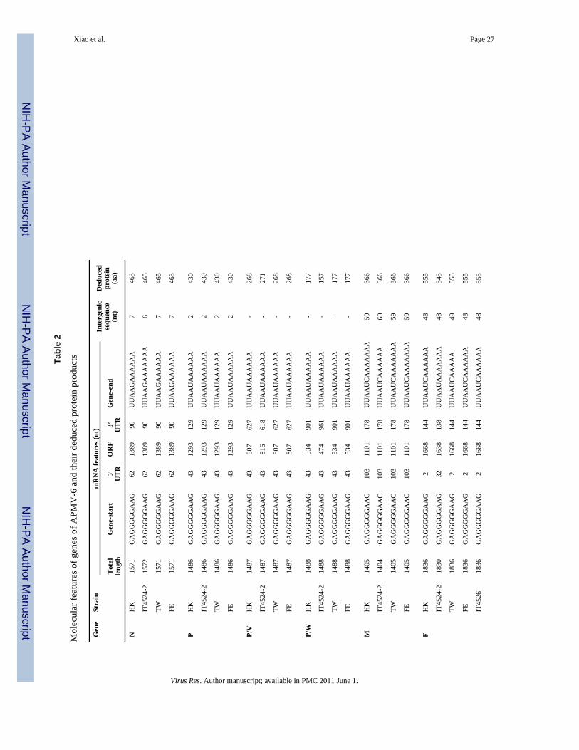
The proposed gene-start (GS) signal sequence is highly conserved among APMV-6 strains(Table 2). The GS sequences of the P, F, HN and L genes of all four strains are 10 nt inlength and have the identical sequence GAGGGGGAAG (positive-sense). The GS sequenceof the M gene differed at a single position (underlined): GAGGGGGAAC. However, the GSof SH gene contained an apparent insertion of an additional G residue in the central run of Gresidues, which contains five C residues for the other genes but 6 for SH (underlined)GAGGGGGGAAG. For each gene, the sequence of the GS signal is identical among all ofthe APMV-6 strains for which sequences are available. The proposed gene-end (GE) signalsequences of the APMV-6 genes are also highly conserved (Table 2). The GE sequences ofthe N and SH genes conform to the sequence UUAAGA5–7, whereas those of the P, HN andL genes including that of the strain IT4524-2 F gene have a single nt difference (underlined):UUAAUA6. The GE sequences of the M and F genes are “UUAAUCA5–7, which contain anapparent insertion of a single nt (underlined) except that of the strain IT4524-2 F gene, butotherwise are identical to the GE signals of the P, HN, and L genes. For each gene (exceptfor the strain IT4524-2 F gene), the sequence of the GE signal is most identical among all ofthe APMV-6 strains for which sequence is available.
For each APMV-6 strain, the IGS vary in length (from 2 to 63 nt, Table 2) and are notconserved from one gene junction to the next. Between APMV-6 strains, the IGS length ateach junction is conserved except that there is a single nt difference in several cases due tovariation in the proposed GE signal. For example, the IGS between the N and P genes ofstrain HK is 7 nt in length and its proposed GE signal is 11 nt long. In contrast, the IGSbetween the N and P genes of strain IT4524-2 is 6 nt in length because its proposed GEsignal is 12 nt long. Thus, in these cases, the difference in IGS length lies in whether thefinal A residue of the GE signal indeed is part of the signal (as we have assigned it) or partof the following IGS. The nt sequences of the IGS of strain IT4524-2 had less than 50% ntidentity with the corresponding IGS of the other APMV-6 strains. In contrast, among theseother strains, the level of nt sequence identity between corresponding IGS was greater than95% (data not shown).
Xiao et al. Page 7
Virus Res. Author manuscript; available in PMC 2011 June 1.
NIH
-PA Author Manuscript
NIH
-PA Author Manuscript
NIH
-PA Author Manuscript
3.4. The nucleocapsid protein (N) geneThe N gene of APMV-6 strains HK, IT4524-2, TW and FE is 1,389 nt in length and encodesa N protein of 465 aa (Table 2). The N protein of all the four strains contains an identicalsequence segment, 324-FAPGNYPLMYSYAMG-338, that conforms to a motif (F-X4-Y-X3-Φ-S-Φ-A-M-G, where X is any aa and Φ is any aromatic aa) that has been identified inother members of the subfamily Paramyxovirinae and is thought to be responsible for N-Nself assembly during genomic RNA binding (Yu et al., 1998;Morgan, 1991). In the case ofAPMV-6, the aa at positions 325 (alanine, A), 326 (proline, P), and 333 (tyrosine, Y) in themotif are conserved in all avulaviruses sequenced to date. The complete N protein of strainHK has 100% aa sequence identity with that of strains TW and FE, and a somewhat lowerlevel of identity (94%) with that of strain IT4524-2 (Table 3).
3.5. The phosphoprotein (P) geneThe P gene of APMV-6 strains HK, IT4524-2, TW and FE is 1,293 nt long and encodes a Pprotein of 430 aa. The P protein of strain HK has an aa sequence identity of 94% and 93%with that of strain TW and FE, respectively, and a lower level of identity (62%) with that ofstrain IT4524-2 (Table 3). The P gene of strains HK, TW and FE contain a putative editingsite AAAAAAGGG (negative-sense) at nt positions 461–472 of the P gene. The addition ofa single G residue to the encoded mRNA would produce a V mRNA encoding a 268-aa Vprotein. In contrast, the putative editing site of strain IT4524-2 is at nt position 450–458 inthe P gene. In addition, the V protein of strain IT4524-2 extends for 3 aa longer at the C-terminus than for the other strains, resulting in a V protein that is 3 aa longer other strains(271 aa, compared to 268 aa) (Fig. 5A). For all four strains, the V protein domain containsthe conserved cysteine rich motif that is characteristic of most members of subfamilyParamyxovirinae. The carboxyl domain of the V protein of strain HK (aa positions 158 to268 in the V protein) has 95% aa sequence identity with the corresponding domain of strainsTW and FE, respectively, and a lower level of identity (50%) with that of strain IT4524-2.The addition of two G residues to the encoded mRNA in the P gene editing site wouldproduce a W mRNA encoding a 177 aa W protein in the strains HK, TW and FE. The WmRNA of strain IT4524-2 encodes a shorter, 157 aa, W protein. Interestingly, the carboxyldomain of the W protein of strain IT4524-2 is only 4 aa which is 16 aa shorter than those ofother three strains (Fig. 5B).
3.6. The matrix protein (M) geneThe M gene of APMV-6 strains HK, IT4524-2, TW and FE is 1,101 nt long and encodes aM protein of 366 aa. The M proteins of the four APMV-6 strains contain the sequence FPIIat aa positions 22 to 25 that conforms to a “late motif” pattern (α-P-Φ-Φ, where α representsan aliphatic aa and Φ represents an aromatic aa), which is involved in virus assembly andbudding in SV5 (Schmitt et al., 2005). The M protein of strain HK has an aa sequenceidentity of 99% with that of strains TW and FE, and a somewhat lower level of identity(92%) with strain IT4524-2 (Table 3).
3.7. The fusion protein (F) geneThe F gene of APMV-6 strains HK, IT4526, IT6895-1, TW and FE is 1,836 nt long (ORF,1,668 nt) and encodes an F protein of 555 aa. In contrast, the F gene of strain IT4524-2 is1,830 nt long (ORF, 1,638 nt) and encodes an F protein of 545 aa. The F protein of strainHK has a high level of aa sequence identity with the F proteins of strains TW (99%), FE(99%), IT4526 (98%), and IT6895-1 (98%), and a lower level of aa sequence identity (86%)with that of strain IT4524-2 (Table 3).
Xiao et al. Page 8
Virus Res. Author manuscript; available in PMC 2011 June 1.
NIH
-PA Author Manuscript
NIH
-PA Author Manuscript
NIH
-PA Author Manuscript
Amino acid sequence alignment showed that the F protein of strain IT4524-2 is 10 aa shorterat the N-terminus than those of the other strains (Fig. 6A), which accounts for the differencein length between these F proteins. This is due to a difference in the position of the ATGtranslational start codon in the respective ORFs: in strain IT4524-2, the start codon islocated 33 nt downstream of the GS signal, whereas the start codon in each of the otherstrains is located 3 nt downstream of the GS signal (Fig. 6B). The signal peptide of the Fprotein of strain IT4524-2, as predicted by the Signal3.0 server (Bendtsen et al., 2004), is 19aa in length, with cleavage occurring between positions 19 and 20 (T↓L). In contrast, thesignal peptides of the other APMV-6 strain F proteins are 29 aa in length and are predictedto be cleaved between aa positions 29 and 30 (Fig. 6A). The aa sequence of the signalpeptide of strain IT4524-2 has only a low level of sequence identity (26%) with those of theother strains, which amongst themselves share a higher level of identity (90–100%). The 29-aa signal peptide of APMV-6 strains HK, TW, FE, IT4526 and IT6895-1 has a polar N-terminus (n region), hydrophobic core (h region) and C-terminus (c-region) (Martoglio &Dobberstein, 1998). In contrast, the signal peptide of strain IT4524-2 does not contain the n-region because it is 10 aa shorter at the n-region. It is not known whether this has functionalconsequences for translocation into the endoplasmic reticulum.
In the case of NDV, the cleavage sequence of the F protein is a critical factor for viral entryand pathogenesis. For APMV-6 strains TW, FE, IT4526, IT6895-1, and the prototype strainHK, the aa sequences spanning the cleavage site and adjacent upstream end of the F1subunit are identical and contain a monobasic aa residue (arginine, R): PAPEPR↓L. Incontrast, for strain IT4524-2, there are several aa differences including the presence of asecond arginine residue at position −4 relative to the cleavage site (REPR↓L, Fig. 7).Despite the additional basic residue in strain IT4524-2, none of the cleavage sitesdetermined to date for APMV-6 strains conforms to the preferred furin cleavage site (RX-R/K-R↓). In addition, the residue immediately following the cleavage site in each of theAPMV-6 strains is leucine, whereas the presence of phenylalanine at this position isconsistent with intracellular cleavage (Morrison et al., 1993). In comparison, virulent NDVstrains have four basic aa followed by a flanking phenylalanine (RRQKR↓F) at the cleavagesite, whereas avirulent NDV strains contain two basic aa followed by a flanking leucineresidue (RQGR↓L) at the cleavage site (Lamb & Parks, 2007).
3.8. The small hydrophobic protein (SH) geneThe SH gene of all of these APMV-6 strains is 429 nt long and encodes an SH protein of142 aa. The SH protein is predicted to be a transmembrane protein. The function of theAPMV-6 SH protein is not known. The SH protein of strain HK has an aa sequence identityof 90% to 92% with those of strains TW, FE, IT4526, and IT6895-1, and a lower level ofidentity (55%) with strain IT4524-2 (Table 3).
3.9. The hemagglutinin-neuraminidase (HN) geneThe HN gene of all of these APMV-6 strains is 1,842 nt long and encodes an HN protein of613 aa. The HN protein is predicted to be a type II integral membrane protein. The sequencemotif NRKSCS that has been identified in other paramyxoviruses including otheravulaviruses and is thought to be involved in sialic acid binding (Mirza et al., 1994) ispresent in avulavirus (Xiao et al., 2009). This motif also is present at aa positions 240 to 245in the HN proteins of strains HK, IT4524-2, IT4526, IT6895-1 and FE, whereas thecorresponding sequence in strain TW has a single aa difference (underlined): NRKSCN. TheHN protein of strain HK has 97% aa sequence identity with all of these strains, except for81% identity with that of strain IT4524-2 (Table 3).
Xiao et al. Page 9
Virus Res. Author manuscript; available in PMC 2011 June 1.
NIH
-PA Author Manuscript
NIH
-PA Author Manuscript
NIH
-PA Author Manuscript

3.10. The large polymerase protein (L) geneThe L gene of the APMV-6 strains HK, IT4524-2, TW and FE is 6,726 nt long and encodesa L protein of 2,241 aa. The motif QGDNQ present in L protein domain III of othernonsegmented negative strand RNA viruses is widely conserved and is thought to beinvolved in L protein transcriptional activity (Malur et al., 2002). This motif also is presentat aa positions 776 to 780 in the L protein of APMV-6 strains HK, IT4524-2 and FE,whereas in strain TW the corresponding sequence has a single difference (underlined):QGDEQ. The L protein of strain HK has an aa sequence identity of 98% with those ofstrains TW and FE, and 86% with that of strain IT4524-2 (Table 3).
3.11. Evolutionary relatedness and genetic classification of the APMV-6 strainsPhylogenetic trees were constructed based on aa sequence alignments of the F and HNproteins of the six APMV-6 strains for which sequences are available versus the cognateproteins of representatives of the other APMV serotypes, except APMV-5 for whichsequences have not been reported (Fig. 8). The phylogenetic analysis clearly indicated thatthe APMV-6 strains analyzed in the present study, IT4524-2, IT4526, IT6895-1 and HK, aremore closely related to the APMV-6 strains TW and FE than to other APMV serotypes, aswould be expected. Furthermore, APMV-6 strains IT4526, IT6895-1, HK, TW, and FE aremore closely related to each other than to strain IT4524-2 which is more distinct and appearsto form a separate clade within APMV-6.
We also constructed a phylogenetic tree for the six APMV-6 strains based on a classificationsystem for NDV that uses common genetic markers in the nt sequence of the F gene toclassify NDV strains (Miller et al, 2009; Czegledi et al., 2006; Kim et al., 2007) (Fig. 9).This analysis indicated that the six APMV-6 strains could be divided into two classes: classII, which contains strain IT4524-2, and class I, which contains the other 5 strains. Thecomparative genetic distance between strain IT4524-2 and other APMV-6 strains wassignificant, with a range distance value of 29–31% (distance matrix not shown). Within classI, prototype strain HK has a range distance of 4–5% to other strains, compared to the valueof 1–2% between the other strains (Fig. 9). This classification system should be helpful ingrouping future APMV-6 isolates.
5. DiscussionAPMVs constitute the genus Avulavirus and are divided into nine serotypes based on HI andNI assays. Among these serotypes, APMV-1 (NDV) is well characterized because it causessevere disease in poultry worldwide. A great deal of information is available on theantigenic and genetic relationships among APMV-1 strains isolated from different parts ofthe world. Recently, we and others have reported complete genome sequences forrepresentative strains of APMV-2, 3, 4, 6, 7, 8 and 9. However, very little is known aboutthe antigenic and genetic relationships among strains within serotypes 2 through 9. In thisstudy, we have determined complete consensus genome sequences for the prototypeAPMV-6 strain HK, which was isolated from a domestic duck in Hong Kong in 1977, andstrain IT4524-2, which was isolated from a duck in Italy in 2007. In addition, we determinedconsensus sequences for the F-SH-HN region of the genomes of two additional strainsisolated in Italy in 2007, strains IT4526 and IT6895-1. The antigenic relationships amongthese four strains were evaluated using an HI assay, which is the principal standard fordistinguishing between APMV serotypes. In addition, we evaluated the genetic relationshipsof these four strains with two other APMV-6 strains, TW and FE, for which completegenome sequences had previously been reported. This information will have implications forstudies in pathogenesis and epidemiology and for the development of vaccines againstAPMV-6.
Xiao et al. Page 10
Virus Res. Author manuscript; available in PMC 2011 June 1.
NIH
-PA Author Manuscript
NIH
-PA Author Manuscript
NIH
-PA Author Manuscript

To evaluate the antigenic relationship among the four APMV-6 strains described in thepresent paper, we raised chicken antisera against strains HK and IT4524-2 separately byrespiratory infection, mimicking a natural route of infection. Since serological responsestend to broaden over time and with repeated antigenic exposure, we (i) limited theimmunization to a single infection, and (ii) collected serum samples at an early time point(14 days post infection). Our results showed the HI titer of the HK antiserum was 8-, 2-, and8-fold higher against the homologous strain HK and strains IT4526 and IT6895-1,respectively, than was the HI titer of the IT4524-2-specific antiserum. Conversely, the HItiter of the IT4524-2-specific serum was 4-fold higher against the homologous strainIT4524-2 than that of the HK-specific antiserum. These results indicated an antigenicdimorphism that would be consistent with the existence of two antigenic subgroups withinAPMV-6, with strains HK, IT4526 and IT6895-1 belonging to one antigenic subgroup andstrain IT4524-2 belonging to the second antigenic subgroup. The TW and FE strains werenot evaluated in this assay, but their high degree of sequence relatedness to strains HK,IT4526 and IT6895-1, and divergence from strain IT4524-2, predicts that they would begrouped with the former three strains. In this regard, it will be of interest to evaluateadditional APMV-6 strains and further evaluate these strains by neutralization assay.
The genome lengths of strains HK, TW and FE are 16236 nt compared to 16230 nt for strainIT4524-2. Among APMV-1 (NDV) strains, there are three genome sizes: 1) 15186 nt inearly (>1930s) isolated strains, 2) 15192 nt in late (>1960s) isolated strains (due to a six ntinsertion in the upstream of the N gene), and 3) 15198 nt (12 nt insertion in the P gene ORF)(Czegledi et al., 2006). These different genome sizes of NDV strains did not relate to theviral virulence, but seem to be related to the time (year) of virus isolation, with the genomesbecoming progressively longer (Miller et al., 2009; Czegledi et al., 2006). However, theconverse appeared to be the case for APMV-6, since strains HK, TW and FE were isolatedin 1977, 1999 and 2003, respectively, whereas the shorter strain IT4524-2 was isolated in2007. In any event, the genome lengths of strains HK, TW, FE and IT4524-2 follow the“rule of six”, indicating that this rule is a requirement for virus replication. The six ntdifference for strain IT4524-2 was in the 3’ UTR of F mRNA. Interestingly, the 3’UTR ofstrain IT4524-2 F mRNA showed high level of nt sequence variation (37 to 40% nt identity)when compared with the corresponding sequence in other available APMV-6 strains. TheUTRs of other viral genes also showed highly divergence. The UTRs of strain IT4524-2genes shows a low level nt identities (37 to 56%), compared with those of other strains ashigh level identities (85 to 100%). The length of viral UTR can play an important role invirus replication and pathogenesis in paramyxoviruses. (Yan et al., 2009; Evans et al.,1990). Therefore, the difference in length of sequence of 3’UTR of IT4524-2 F mRNAmight play a role of in virus replication and pathogenicity.
The genome of strain HK shared 94% nt identity with those of strains TW and FE, which inturn were 98% identical. In contrast, the genomes of strain IT4524-2 shared 70% ntsequence identity with strains HK, TW, and FE. With regard to the aggregate aa sequence,strain HK was 95% identical to strains TW and FE, which in turn were 97% identical. Incontrast, strain IT4524-2 shared 79% aggregate aa identity with strains HK, TW, and FE.Thus, there was sequence dimorphism between these strains. Sequence analysis of the F-SH-HN genome region of strains IT4526 and IT6895-1 indicated that they were highly related tostrains HK, TW, and FE. Taken together, this provided evidence for two subgroups, onecontaining strains HK, TW, FE, IT4526, and IT6895-1, and the other containing strainIT4524-2. This sequence dimorphism was completely consistent with the antigenicdimorphism noted above, and provides support for the existence of two APMV-6 subgroups.This subgroup difference could not be attributed to the time, place, or host of isolation, sinceboth subgroups contained at least one strain isolated from ducks in Italy in 2007. It isnoteworthy that the extent of sequence divergence between the two proposed subgroups of
Xiao et al. Page 11
Virus Res. Author manuscript; available in PMC 2011 June 1.
NIH
-PA Author Manuscript
NIH
-PA Author Manuscript
NIH
-PA Author Manuscript

APMV-6 is greater than that between the two subgroups of HRSV, which share 81% ntsequence identity, or HMPV, which share 80% nt sequence identity (Biacchesi et al., 2003,Johnson et al., 1987). For all three viruses, the nt sequence divergence was the greatest inregions that do not encode protein: for example, the IGS between strains HK and IT4524-2were less than 50% identical, similar to the value of 48% identity for the IGS between thetwo HPMV subgroups and 42% identity between the two HRSV subgroups (Biacchesi et al.,2003). When compared with the other serotypes of APMV, the genome of four APMV-6strains had a nt sequence identity of 39% to 47% with APMV-1, 2, 3, 4, 7, 8, and 9. Thus,the divergence of nt sequence between the two proposed APMV-6 subgroups wassubstantially less than between either APMV-6 subgroup and the other APMV serotypes, aswould be expected.
The APMV-6 is the only known virus in genus Avulavirus that encodes the additional SHprotein, which is 142 aa in length for all six APMV-6 strains. Similar SH proteins also arepresent in the other paramyxoviruses: SV5 (44 aa), MuV (57 aa), HRSV (64 aa), humanmetapneumovirus (179 aa), and J-virus (179 aa) (Lamb & Parks, 2007). The SH protein ofAPMV-6 strain HK shares only 13 to 22% aa sequence identity with the SH protein of theseother paramyxoviruses (analyzed by Lasergene 6 with Clustal V). Although the function ofthe APMV-6 SH protein is not known, the SH protein of HRSV is not necessary for virusviability (Whitehead et al., 1999) and the SH protein of SV5 might be involved in apoptosis(Wilson et al., 2006). Comparison of aa sequence relatedness of cognate proteins betweenstrain IT4542-2 and other APMV-6 strains showed values of 81 to 94% aa identity, exceptfor the P and SH proteins which are more divergent (62 and 55% aa identity, respectively).The extent of variability in the APMV-6 P proteins contrasts with that of the P proteins ofthe two subgroups of HMPV and HRSV, which are more highly conserved (85 and 90% aaidentity, respectively) (Biacchesi et al., 2003). It is noteworthy that the C-terminal domainof the V protein has only 50% identity between strains HK and IT4524-2, and the completeV protein has only 54% identity. By analogy with other paramyxoviruses, the V protein isthought to play a role in blocking host type I interferon response. The finding that thispathogenesis factor is one of the more divergent proteins is surprising and raises thepossibility that this may have consequences for pathogenesis. The SH proteins of the twosubgroups of HMPV and HRSV are more highly conserved (59 and 72% aa identity,respectively) than for the two subgroups of APMV-6 (55% identity), which is consistentwith the idea that there is greater divergence between the two APMV-6 subgroups thanbetween those of HRSV and HMPV.
One interesting difference between APMV-6 strain IT4524-2 and other APMV-6 strains wasobserved in the F protein. The F protein of strain IT4524-2 has a signal peptide of 19 aacompared to a signal peptide of 29 aa in other APMV-6 strains. Signal peptides are usually15 to more than 50 aa residues in length and contain a hydrophilic, positively charged N-terminal region (n region), a central hydrophobic domain of 7–10 residues (h region) and aC-terminal region (c region) containing the cleavage site for signal peptidase. The signalpeptide is cleaved during the translocation of the secretory proteins across the lumen of theendoplasmic reticulum (Martoglio & Dobberstein, 1998). The n region of signal peptide ofviral glycoprotein (like APMV F protein) was essential for protein processing andmaturation, and the n region mutated virus was defective for viral infectivity in Foamy virus(Lindemann et al., 2001) and lymphocytic choriomeningitis virus (Schrempf et al., 2007).The signal peptide of the APMV-6 strain IT4524-2 F protein contains h and c regions, butlacks the n region. It will be of interest to investigate whether this has implications forprocessing and maturation compared to other APMV-6 strains.
Another difference between strain IT4524-2 and other APMV-6 strains was observed in thecleavage site of F protein. The aa sequence at the cleavage site of F protein plays a major
Xiao et al. Page 12
Virus Res. Author manuscript; available in PMC 2011 June 1.
NIH
-PA Author Manuscript
NIH
-PA Author Manuscript
NIH
-PA Author Manuscript

role in NDV pathogenesis (Lamb and Parks, 2007). Virulent NDV strains have a multiplebasic aa cleavage site R-X-K/R-R↓F, which is cleaved intracellularly by ubiquitous cellularfurin-like proteases, and a phenylalanine (F) residue at the beginning of the F1 subunit,which also may play a role in facilitating cleavage (Morrison et al., 1993). The avirulentNDV strains have one or a few basic residues at the cleavage site and do not conform to thefurin motif, and have a leucine (L) residue at the first position of F1 subunit. Interestingly,however, the putative cleavage sites of other APMV serotypes showed that the cleavage sitesequences of some serotypes are not necessarily predictive of the protease activationphenotype (Xiao et al., 2009). The putative F protein cleavage site (PEPR↓L) of theAPMV-6 strains HK, IT4526, IT6895-1, TW and FE contains a single basic residue whilethat of strain IT4524-2 contains dibasic residues. However, none of the sites conform to thefurin cleavage site. Therefore it was surprising to find that each of these strains replicated ina trypsin-independent manner in at least one of the cell types that were tested and, in thesecases, the addition of trypsin did not substantially increase replication. On the other hand,each of the strains required trypsin for replication in one or more of the tested cell types.Thus, there was unexpected diversity in the ability of various cell lines to cleave the variousstrains. In addition, in light of the fact that strains HK, IT4526, and IT6895-1 have the samecleavage site sequence, it was surprising that they did not exhibit exactly the same cleavagephenotype in all of the six tested cell lines. For example, strain IT6895-1 was trypsin-independent in only one cell line whereas strain HK was trypsin-independent in three lines.It may be that other residues in the F protein that differ between these viruses contributed tothe cleavage phenotype. Strain IT4524-2, which had two basic residues in the cleavagesequence, had approximately the same cleavage as strain HK that contained only one basicresidue, indicating that this difference did not significantly affect cleavability. On the basisof cleavage phenotype, it would not be possible to predict whether these strains would bevirulent or not in vivo, since the cleavage phenotype varied with the cell line that was tested.However, evaluation of MDT in chicken eggs provided evidence of an avirulent phenotypefor each of the strains.
In conclusion, complete genome sequences were determined for APMV-6 strains HK andIT4524-2. Comparison of the nt and predicted protein aa sequences among APMV-6 strainsshowed that the divergence between strain IT4524-2 and other APMV-6 strains wassubstantially greater overall than that between the two HMPV or the two HRSV subgroups.This grouping based on sequence relatedness was consistent with the antigenic analysis.This indicated that these APMV-6 strains represent two APMV-6 subgroups. We proposethat the prototype strain HK and strains TW, FE, IT4526 and IT6895-1 represent onesubgroup, and strain IT4524-2 represents a second subgroup. Given the rather substantialdifferences in the nt and aa sequences between the two APMV-6 subgroups, it wassomewhat surprising that the extent of antigenic difference by HI test was modest. Onepossible explanation might be that the APMV-6 antigenic site in the HN protein that isinvolved in HI assay is more highly conserved between the two subgroups than the overallgenome and aggregate amino acid sequences. Additional antigenic and genetic analysisinvolving additional APMV-6 strains is needed to further define the antigenic and geneticvariability of APMV-6.
AcknowledgmentsWe thank Drs. Ilaria Capua and Isabella Monne at IZSV, Italy for helpful support and advice. We also thankAnandan Paldurai, Flavia Dias and Dianel Rockemann for their excellent technical assistance and help. “Thisresearch was supported by NIAID contract no.N01A060009 (85% support) and NIAID, NIH Intramural ResearchProgram (15% support). The views expressed herein do not necessarily reflect the official policies of theDepartment of Health and Human Services; nor does mention of trade names, commercial practices, ororganizations imply endorsement by the U.S. Government”.
Xiao et al. Page 13
Virus Res. Author manuscript; available in PMC 2011 June 1.
NIH
-PA Author Manuscript
NIH
-PA Author Manuscript
NIH
-PA Author Manuscript

ReferencesAlexander, DJ. Newcastle disease and other avian Paramyxoviridae infections. In: Calnek, BW.,
editor. Diseases of Poultry. Ames: Iowa State University Press; 1997. p. 541-569.Alexander, DJ. Avian Paramyxoviruses 2–9. In: Saif, YM., editor. Diseases of poultry. 11th edn.
Ames: Iowa State University Press; 2003. p. 88-92.Alexander DJ, Collins MS. Pathogenecity of PMV-3/Parakeet/Netherland/449 /75 for chickens. Avian
Pathol 1982;11:179–185. [PubMed: 18770182]Bendtsen JD, Nielsen H, von Heijne G, Brunak S. Improved prediction of signal peptides: Signal 3.0. J
Mol Biol 2004;340:783–795. [PubMed: 15223320]Biacchesi S, Skiadopoulos MH, Boivin G, Hanson CT, Murphy BR, Collins PL, Buchholz UJ. Genetic
diversity between human metapneumovirus subgroups. Virology 2003;315:1–9. [PubMed:14592754]
Brudno M, Do CB, Cooper GM, Kim MF, Davydov E, Green ED, Sidow A, Batzoglou S. LAGANand Multi-LAGAN: efficient tools for large-scale multiple alignment of genomic DNA. GenomeRes 2003;13:721–731. [PubMed: 12654723]
Bukreyev A, Whitehead SS, Murphy BR, Collins PL. Recombinant respiratory syncytial virus fromwhich the entire SH gene has been deleted grows efficiently in cell culture and exhibits site-specificattenuation in the respiratory tract of the mouse. J Virol 1997;71:8973–8982. [PubMed: 9371553]
Chang PC, Hsieh ML, Shien JH, Graham DA, Lee MS, Shieh HK. Complete nucleotide sequence ofavian paramyxovirus type 6 isolated from ducks. J Gen Virol 2001;82:2157–2168. [PubMed:11514725]
Czeglédi A, Ujvári D, Somogyi E, Wehmann E, Werner O, Lomniczi B. Third genome size categoryof avian paramyxovirus serotype 1 (Newcastle disease virus) and evolutionary implications. VirusRes 2006;120:36–48. [PubMed: 16766077]
Evans SA, Belsham G, Barrett T. The role of the 5' nontranslated regions of the fusion protein mRNAof canine distemper virus and rinderpest virus. Virology 1990;177:317–323. [PubMed: 2353458]
Gan SW, Ng L, Lin X, Gong X, Torres J. Structure and ion channel activity of the human respiratorysyncytial virus (hRSV) small hydrophobic protein transmembrane domain. Protein Sci2008;17:813–820. [PubMed: 18369195]
Jeon WJ, Lee EK, Kwon JH, Choi KS. Full-length genome sequence of avain paramyxovirus type 4isolated from a mallard duck. Virus Genes 2008;37:342–350. [PubMed: 18770019]
Johnson PR Jr, Olmsted RA, Prince GA, Murphy BR, Alling DW, Walsh EE, Collins PL. Antigenicrelatedness between glycoproteins of human respiratory syncytial virus subgroups A and B:evaluation of the contributions of F and G glycoproteins to immunity. J Virol 1987;61:3163–3166.[PubMed: 3305988]
Jung A, Grund C, Müller I, Rautenschlein S. Avian paramyxovirus serotype 3 infection inNeopsephotus, Cyanoramphus, and Neophema species. J Avian Med Surg 2009;23:205–208.[PubMed: 19999764]
Kim LM, King DJ, Curry PE, Suarez DL, Swayne DE, Stallknecht DE, Slemons RD, Pedersen JC,Senne DA, Winker K, Afonso CL. Phylogenetic diversity among low-virulence newcastle diseaseviruses from waterfowl and shorebirds and comparison of genotype distributions to those ofpoultry-origin isolates. J Virol 2007;81:12641–12653. [PubMed: 17855536]
Kolakofsky D, Pelet T, Garcin D, Hausmann S, Curran J, Roux L. Paramyxovirus RNA synthesis andthe requirement for hexamer genome length: the rule of six revisited. J Virol 1998;72:891–899.[PubMed: 9444980]
Kumar S, Nayak B, Collins PL, Samal SK. Complete genome sequence of avian paramyxovirus type 3reveals an unusually long trailer region. Virus Res 2008;137:189–197. [PubMed: 18691616]
Lamb, RA.; Collins, PL.; Kolakofsky, D.; Melero, JA.; Nagai, Y.; Oldstone, MBA.; Pringle, CR.;Rima, BK. Fauquet, CM., editor. Virus Taxonomy. The Classification and Nomenclature ofViruses. The Eighth Report of the International Committee in Taxonomy of Viruses; 2005. FamilyParamyxoviridae.
Xiao et al. Page 14
Virus Res. Author manuscript; available in PMC 2011 June 1.
NIH
-PA Author Manuscript
NIH
-PA Author Manuscript
NIH
-PA Author Manuscript
Lamb, RA.; Parks, GD. Paramyxoviridae: the viruses and their replication, Fields Virology. 5th edn..Knipe, DM.; Howley, PM., editors. Philadelphia: Lippincott Williams and Wilkins; 2007. p.1449-1496.
Li Z, Yu M, Zhang H, Wang HY, Wang LF. Improved rapid amplification of cDNA ends (RACE) formapping both the 5' and 3' terminal sequences of paramyxovirus genomes. J Virol Meth2005;130:154–156.
Lindemann D, Pietschmann T, Picard-Maureau M, Berg A, Heinkelein M, Thurow J, Knaus P,Zentgraf H, Rethwilm A. A particle-associated glycoprotein signal peptide essential for virusmaturation and infectivity. J. Virol 2001;75:5762–5771. [PubMed: 11390578]
Lipkind M, Shihmanter E. Antigenic relationships between avian paramyxoviruses. I. Quantitativecharacteristics based on hemagglutination and neuraminidase inhibition tests. Arch Virol1986;89:89–111. [PubMed: 2424406]
Malur AG, Gupta NK, De Bishnu P, Banerjee AK. Analysis of the mutations in the active site of theRNA-dependent RNA polymerase of human parainfluenza virus type 3 (HPIV3). Gene Expr2002;10:93–100. [PubMed: 12064576]
Martoglio B, Dobberstein B. Signal sequences: more than just greasy peptides. Trends in Cell Biol1998;8:410–415. [PubMed: 9789330]
Miller PJ, Decanini EL, Afonso CL. Newcastle disease: Evolution of genotypes and the relateddiagnostic challenges. Infect Genet 2009;10:26–35.
Mirza AM, Deng R, Iorio RM. Site-directed mutagenesis of a conserved hexapeptide in theparamyxovirus hemagglutinin–neuraminidase glycoprotein: effects on antigenic structure andfunction. J. Virol 1994;68:5093–5099. [PubMed: 8035509]
Morgan, EM. Evolutionary relationships of paramyxovirus nucleocapsid-associated proteins. In:Kingsbury, DW., editor. The Paramyxoviruses. New York: Plenum Press; 1991. p. 163-179.
Morrison T, McQuain C, Sergel T, McGtnnes L, Reitter J. The role of the amino terminus of F 1 of theNewcastle disease virus fusion protein in cleavage and fusion. Virology 1993;193:997–1000.[PubMed: 8460504]
Nayak B, Kumar S, Collins PL, Samal SK. Molecular characterization and complete genome sequenceof avian paramyxovirus type 4 prototype strain duck/Hong Kong/D3/75. Virol. J 2008;5:124.[PubMed: 18937854]
Nerome K, Nakayama M, Ishida M, Fukumi H. Isolation of a new avian paramyxovirus frombudgerigar (Melopsittacus undulatus). J. Gen. Virol 1978;38:293–301. [PubMed: 627875]
Paldurai A, Subbiah M, Kumar S, Collins PL, Samal SK. Complete genome sequences of avianparamyxovirus type 8 strains goose/Delaware/1053/76 and pintail/Wakuya/20/78. Virus Res2009;142:144–153. [PubMed: 19341613]
Redmann T, Zeydanli MM, Herbst W, Kaleta EF. Isolation of a paramyxovirus-3 from turkeys withrespiratory tract disease in Germany. Dtsch. Tierarztl. Wochenschr 1991;98:138–141. [PubMed:1829671]
Robinson CM, Shariati F, Zaitshik J, Gillaspy AF, Dyer DW, Chodosh J. Human adenovirus type 19:genomic and bioinformatics analysis of a keratoconjunctivitis isolate. Virus Res 2009;139:122–126. [PubMed: 19000724]
Saif YM, Mohan R, Ward L, Senne DA, Panigrahy B, Dearth RN. Natural and experimental infectionof turkeys with avian paramyxovirus-7. Avian Dis 1997;41:326–329. [PubMed: 9201395]
Sakai K, Mizutani T, Fukushi S, Saijo M, Endoh D, Kurane I, Takehara K, Morikawa S. An improvedprocedure for rapid determination of viral RNA sequences of avian RNA viruses. Arch Virol2007;152:1763–1765. [PubMed: 17541697]
Samuel AS, Kumar S, Madhuri S, Collins PL, Samal SK. Complete sequence of the genome of avianparamyxovirus type 9 and comparison with other paramyxoviruses. Virus Res 2009;142:10–18.[PubMed: 19185593]
Schmitt AP, Leser GP, Morita E, Sundquist WI, Lamb RA. Evidence for a new viral late-domain coresequence, FPIV, necessary for budding of a paramyxovirus. J. Virol 2005;79:2988–2997.[PubMed: 15709019]
Xiao et al. Page 15
Virus Res. Author manuscript; available in PMC 2011 June 1.
NIH
-PA Author Manuscript
NIH
-PA Author Manuscript
NIH
-PA Author Manuscript

Schrempf S, Froeschke M, Giroglou T, von Laer D, Dobberstein B. Signal peptide requirements forlymphocytic choriomeningitis virus glycoprotein C maturation and virus infectivity. J Virol2007;81:12515–12524. [PubMed: 17804515]
Shortridge KF, Alexander DJ, Collins MS. Isolation and properties of viruses from poultry in HongKong which represent a new (sixth) distinct group of avian paramyxoviruses. J Gen Virol1980;49:255–262. [PubMed: 7441204]
Stanislawek WL, Wilks CR, Meers J, Horner GW, Alexander DJ, Manvell RJ, Kattenbelt JA, GouldAR. Avian paramyxoviruses and influenza viruses isolated from mallard ducks (Anasplatyrhynchos) in New Zealand. Arch Virol 2002;147:1287–1302. [PubMed: 12111409]
Steward M, Samson A, C, Errington W, Emmerson PT. The Newcastle disease virus V protein bindszinc. Arch Virol 1995;140:1321–1328. [PubMed: 7646364]
Subbiah M, Xiao S, Collins PL, Samal SK. Complete sequence of the genome of avian paramyxovirustype 2 (strain Yucaipa) and comparison with other paramyxoviruses. Virus Res 2008;137:40–48.[PubMed: 18603323]
Tamura K, Dudley J, Nei M, Kumar S. MEGA4: Molecular Evolutionary Genetics Analysis (MEGA)software version 4.0. Molecular Biology and Evolution 2007;24:1596–1599. [PubMed: 17488738]
Whitehead SS, Bukreyev A, Teng MN, Firestone CY, St Claire M, Elkins WR, Collins PL, MurphyBR. Recombinant respiratory syncytial virus bearing a deletion of either the NS2 or SH gene isattenuated in chimpanzees. J Virol 1999;73:3438–4342. [PubMed: 10074199]
Warke A, Appleby L, Mundt E. Prevalence of antibodies to different avian paramyxoviruses incommercial poultry in the United States. Avian Dis 2008a;52:694–697. [PubMed: 19166066]
Warke A, Stallknecht D, Williams SM, Pritchard N, Mundt E. Comparative study on the pathogenicityand immunogenicity of wild bird isolates of avian paramyxovirus 2, 4, and 6 in chickens. AvianPathol 2008b;37:429–434. [PubMed: 18622861]
Wilson RL, Fuentes SM, Wang P, Taddeo EC, Klatt A, Henderson AJ, He B. Function of smallhydrophobic proteins of paramyxovirus. J Virol 2006;80:1700–1709. [PubMed: 16439527]
Xiao S, Paldurai A, Nayak B, Subbiah M, Collins PL, Samal SK. Complete genome sequence of avianparamyxovirus type 7 (strain Tennessee) and comparison with other paramyxoviruses. Virus Res2009;145:80–91. [PubMed: 19540277]
Yan Y, Rout SN, Kim SH, Samal SK. Role of untranslated regions of the hemagglutinin-neuraminidase gene in replication and pathogenicity of newcastle disease virus. J Virol2009;83:5943–5946. [PubMed: 19321607]
Yu M, Hansson E, Shiell B, Michalski W, Eaton BT, Wang LF. Sequence analysis of the Hendra virusnucleoprotein gene: comparison with other members of the subfamily Paramyxovirinae. J. Gen.Virol 1998;79:1775–1780. [PubMed: 9680142]
Zhang GZ, Zhao JX, Wang M. Serological survey on prevalence of antibodies to avian paramyxovirusserotype 2 in China. Avian Dis 2007;51:137–139. [PubMed: 17461281]
Xiao et al. Page 16
Virus Res. Author manuscript; available in PMC 2011 June 1.
NIH
-PA Author Manuscript
NIH
-PA Author Manuscript
NIH
-PA Author Manuscript
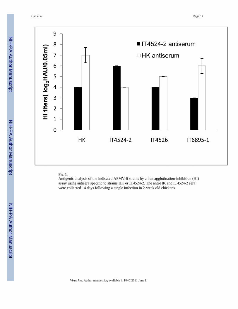
Fig. 1.Antigenic analysis of the indicated APMV-6 strains by a hemagglutination-inhibition (HI)assay using antisera specific to strains HK or IT4524-2. The anti-HK and IT4524-2 serawere collected 14 days following a single infection in 2-week old chickens.
Xiao et al. Page 17
Virus Res. Author manuscript; available in PMC 2011 June 1.
NIH
-PA Author Manuscript
NIH
-PA Author Manuscript
NIH
-PA Author Manuscript

Fig. 2.Nucleotide sequence alignment of the F mRNA 3’ UTRs of the six APMV-6 strains forwhich sequences are available. The sequences for strains IT4524-2 and HK are provided infull because they have little sequence identity. The other strain sequences are listed below,with nt identity to strain HK is indicated by dots. The unfilled rectangle on the leftrepresents the stop codon of the F gene ORF, and the solid rectangle on the right representsthe GE transcription signal of the F gene. The sequences are positive-sense, and thesequence numbers refer to the position in the UTR.
Xiao et al. Page 18
Virus Res. Author manuscript; available in PMC 2011 June 1.
NIH
-PA Author Manuscript
NIH
-PA Author Manuscript
NIH
-PA Author Manuscript
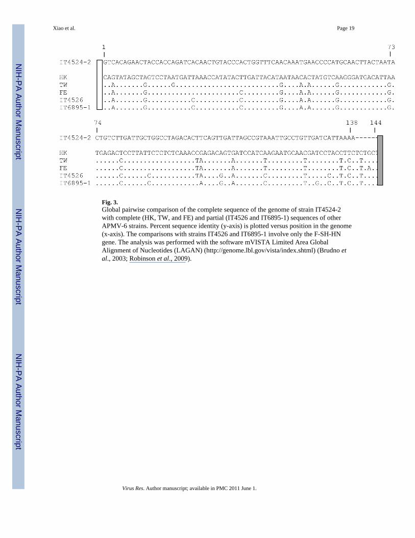
Fig. 3.Global pairwise comparison of the complete sequence of the genome of strain IT4524-2with complete (HK, TW, and FE) and partial (IT4526 and IT6895-1) sequences of otherAPMV-6 strains. Percent sequence identity (y-axis) is plotted versus position in the genome(x-axis). The comparisons with strains IT4526 and IT6895-1 involve only the F-SH-HNgene. The analysis was performed with the software mVISTA Limited Area GlobalAlignment of Nucleotides (LAGAN) (http://genome.lbl.gov/vista/index.shtml) (Brudno etal., 2003; Robinson et al., 2009).
Xiao et al. Page 19
Virus Res. Author manuscript; available in PMC 2011 June 1.
NIH
-PA Author Manuscript
NIH
-PA Author Manuscript
NIH
-PA Author Manuscript
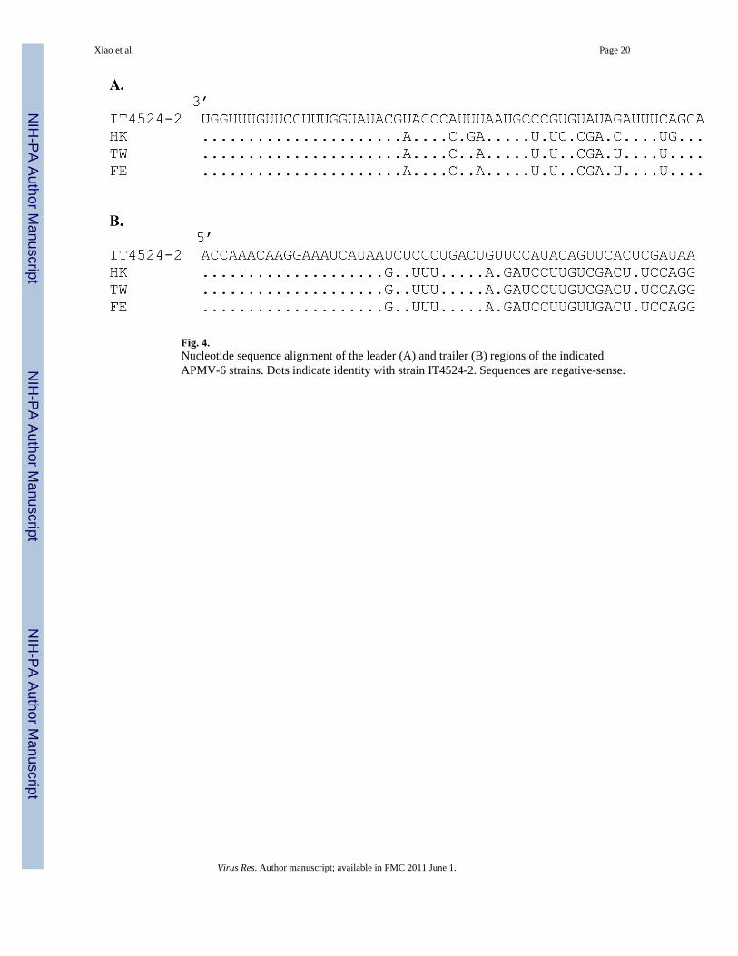
Fig. 4.Nucleotide sequence alignment of the leader (A) and trailer (B) regions of the indicatedAPMV-6 strains. Dots indicate identity with strain IT4524-2. Sequences are negative-sense.
Xiao et al. Page 20
Virus Res. Author manuscript; available in PMC 2011 June 1.
NIH
-PA Author Manuscript
NIH
-PA Author Manuscript
NIH
-PA Author Manuscript

Fig. 5.Amino acid sequence alignment of the C-terminal domain of the V proteins (A) and Wproteins (B) of the indicated APMV-6 strains. Conserved cysteine (C) residues areunderlined, dots indicate identity with strain IT4524-2, and gaps are indicated by dashes.The sequences are numbered according to the complete V and W proteins.
Xiao et al. Page 21
Virus Res. Author manuscript; available in PMC 2011 June 1.
NIH
-PA Author Manuscript
NIH
-PA Author Manuscript
NIH
-PA Author Manuscript

Fig. 6.Sequence alignments of the N-terminal region of the F protein (A) and 5’ end of the FmRNA (B) of the six APMV-6 strains. This illustrates an N-terminal truncation in the signalpeptide of strain IT4524-2 compared to the others due to a difference in the position of thetranslational start site. (A) Amino acid sequence alignment of the N-terminal region of the Fproteins of the indicated APMV-6 strains. The polar N-terminus (n region), hydrophobiccore (h region, underlined) and C-terminus (c-region) of the signal peptide are indicated.The arrow indicates the predicted cleavage site of signal peptidase. Amino acid identityrelative to strain IT4524-2 (top sequence) is indicated by dots; gaps in the IT4524-2sequence compared to the others are indicated with dashes. (B) Nucleotide sequencealignment of the upstream end of the F mRNA of the indicated APMV-6 strains. Thesequence is positive-sense, the GS signal is italicized, the remainder of the 5’ UTR iscapitalized, and the ATG translational start sites are underlined. Nucleotide identity relativeto strain IT4524-2 (top sequence) is indicated by dots.
Xiao et al. Page 22
Virus Res. Author manuscript; available in PMC 2011 June 1.
NIH
-PA Author Manuscript
NIH
-PA Author Manuscript
NIH
-PA Author Manuscript

Fig. 7.Alignment of the F protein cleavage site sequence of APMV-6 strain IT4524-2 with those ofother APMVs. Basic aa were underlined and the cleavage position was indicated.
Xiao et al. Page 23
Virus Res. Author manuscript; available in PMC 2011 June 1.
NIH
-PA Author Manuscript
NIH
-PA Author Manuscript
NIH
-PA Author Manuscript

Fig. 8.Phylogenetic trees based on the aa sequences of the F (A) and HN (B) proteins of the sixstrains of APMV-6 and representative strains of APMV serotypes 1–4 and 7–9. The treeswere constructed by bootstrap analysis (1,000 replicates) using the neighbor-joining of thePoisson-corrected values for aa differences in the MEGA 4.0 phylogenetic analysis program(Tamura et al., 2007). All positions containing alignment gaps and missing data wereeliminated only in pairwise sequence comparisons. The scale bar shows the number ofsubstitutions per site. Bootstrap values are shown at the nodes.
Xiao et al. Page 24
Virus Res. Author manuscript; available in PMC 2011 June 1.
NIH
-PA Author Manuscript
NIH
-PA Author Manuscript
NIH
-PA Author Manuscript

Fig. 9.Phylogenetic tree based on the nt sequence of the F genes of the indicated APMV-6 strains,and proposed scheme for classification of APMV-6 strains. The unrooted tree wasconstructed by bootstrap analysis (1,000 replications) using the neighbor-joining of theKimura-2-parameter method for nt differences in the MEGA 4.0 phylogenetic analysisprogram. All positions containing alignment gaps and missing data were eliminated only inpairwise sequence comparisons. Scale bar shows number of base substitutions per site.Bootstrap values are shown at the nodes. The percent genetic distances of complete F geneswere computed by same program. The matrix of genetic distance among APMV-6 strains isnot shown.
Xiao et al. Page 25
Virus Res. Author manuscript; available in PMC 2011 June 1.
NIH
-PA Author Manuscript
NIH
-PA Author Manuscript
NIH
-PA Author Manuscript

NIH
-PA Author Manuscript
NIH
-PA Author Manuscript
NIH
-PA Author Manuscript
Xiao et al. Page 26
Tabl
e 1
Rep
licat
ion
of A
PMV
-6 st
rain
s in
cell
lines
Stra
inT
ryps
in(1
µg/m
l)H
A ti
ter
(HA
U/m
l)
HE
p-2
DF-
1V
ero
MD
CK
MD
BK
BH
K21
HK
−−
−20
8016
0−
+80
8020
160
320
40
IT45
24-2
−−
−40
4016
0−
+80
160
4080
320
40
IT45
26−
−−
−80
80−
+40
4040
8080
20
IT68
95-1
−−
−−
−80
−
+−
20−
−80
−
Virus Res. Author manuscript; available in PMC 2011 June 1.
NIH
-PA Author Manuscript
NIH
-PA Author Manuscript
NIH
-PA Author Manuscript
Xiao et al. Page 27
Tabl
e 2
Mol
ecul
ar fe
atur
es o
f gen
es o
f APM
V-6
and
thei
r ded
uced
pro
tein
pro
duct
s
Gen
eSt
rain
mR
NA
feat
ures
(nt)
Inte
rgen
icse
quen
ce(n
t)
Ded
uced
prot
ein
(aa)
Tot
alle
ngth
Gen
e-st
art
5′ UT
RO
RF
3′ UT
RG
ene-
end
NH
K15
71G
AG
GG
GG
AA
G62
1389
90U
UA
AG
AA
AA
AA
746
5
IT45
24-2
1572
GA
GG
GG
GA
AG
6213
8990
UU
AA
GA
AA
AA
AA
646
5
TW15
71G
AG
GG
GG
AA
G62
1389
90U
UA
AG
AA
AA
AA
746
5
FE15
71G
AG
GG
GG
AA
G62
1389
90U
UA
AG
AA
AA
AA
746
5
PH
K14
86G
AG
GG
GG
AA
G43
1293
129
UU
AA
UA
AA
AA
A2
430
IT45
24-2
1486
GA
GG
GG
GA
AG
4312
9312
9U
UA
AU
AA
AA
AA
243
0
TW14
86G
AG
GG
GG
AA
G43
1293
129
UU
AA
UA
AA
AA
A2
430
FE14
86G
AG
GG
GG
AA
G43
1293
129
UU
AA
UA
AA
AA
A2
430
P/V
HK
1487
GA
GG
GG
GA
AG
4380
762
7U
UA
AU
AA
AA
AA
-26
8
IT45
24-2
1487
GA
GG
GG
GA
AG
4381
661
8U
UA
AU
AA
AA
AA
-27
1
TW14
87G
AG
GG
GG
AA
G43
807
627
UU
AA
UA
AA
AA
A-
268
FE14
87G
AG
GG
GG
AA
G43
807
627
UU
AA
UA
AA
AA
A-
268
P/W
HK
1488
GA
GG
GG
GA
AG
4353
490
1U
UA
AU
AA
AA
AA
-17
7
IT45
24-2
1488
GA
GG
GG
GA
AG
4347
496
1U
UA
AU
AA
AA
AA
-15
7
TW14
88G
AG
GG
GG
AA
G43
534
901
UU
AA
UA
AA
AA
A-
177
FE14
88G
AG
GG
GG
AA
G43
534
901
UU
AA
UA
AA
AA
A-
177
MH
K14
05G
AG
GG
GG
AA
C10
311
0117
8U
UA
AU
CA
AA
AA
AA
5936
6
IT45
24-2
1404
GA
GG
GG
GA
AC
103
1101
178
UU
AA
UC
AA
AA
AA
6036
6
TW14
05G
AG
GG
GG
AA
C10
311
0117
8U
UA
AU
CA
AA
AA
AA
5936
6
FE14
05G
AG
GG
GG
AA
C10
311
0117
8U
UA
AU
CA
AA
AA
AA
5936
6
FH
K18
36G
AG
GG
GG
AA
G2
1668
144
UU
AA
UC
AA
AA
AA
4855
5
IT45
24-2
1830
GA
GG
GG
GA
AG
3216
3813
8U
UA
AU
AA
AA
AA
A48
545
TW18
36G
AG
GG
GG
AA
G2
1668
144
UU
AA
UC
AA
AA
A49
555
FE18
36G
AG
GG
GG
AA
G2
1668
144
UU
AA
UC
AA
AA
AA
4855
5
IT45
2618
36G
AG
GG
GG
AA
G2
1668
144
UU
AA
UC
AA
AA
AA
4855
5
Virus Res. Author manuscript; available in PMC 2011 June 1.

NIH
-PA Author Manuscript
NIH
-PA Author Manuscript
NIH
-PA Author Manuscript
Xiao et al. Page 28
Gen
eSt
rain
mR
NA
feat
ures
(nt)
Inte
rgen
icse
quen
ce(n
t)
Ded
uced
prot
ein
(aa)
Tot
alle
ngth
Gen
e-st
art
5′ UT
RO
RF
3′ UT
RG
ene-
end
IT68
95-1
1836
GA
GG
GG
GA
AG
216
6814
4U
UA
AU
CA
AA
AA
A48
555
SHH
K57
4G
AG
GG
GG
GA
AG
6142
962
UU
AA
GA
AA
AA
A28
142
IT45
24-2
574
GA
GG
GG
GG
AA
G61
429
62U
UA
AG
AA
AA
AA
2814
2
TW57
4G
AG
GG
GG
GA
AG
6142
962
UU
AA
GA
AA
AA
2914
2
FE57
4G
AG
GG
GG
GA
AG
6142
962
UU
AA
GA
AA
AA
A28
142
IT45
2657
4G
AG
GG
GG
GA
AG
6142
962
UU
AA
GA
AA
AA
2914
2
IT68
95-1
574
GA
GG
GG
GG
AA
G61
429
62U
UA
AG
AA
AA
AA
2814
2
HN
HK
2031
GA
GG
GG
GA
AG
4018
4212
8U
UA
AU
AA
AA
AA
6361
3
IT45
24-2
2031
GA
GG
GG
GA
AG
1842
128
UU
AA
UA
AA
AA
A63
613
TW20
31G
AG
GG
GG
AA
G40
1842
128
UU
AA
UA
AA
AA
A63
613
FE20
31G
AG
GG
GG
AA
G40
1842
128
UU
AA
UA
AA
AA
A63
613
IT45
2620
31G
AG
GG
GG
AA
G40
1842
128
UU
AA
UA
AA
AA
A63
613
IT68
95-1
2031
GA
GG
GG
GA
AG
4018
4212
8U
UA
AU
AA
AA
AA
6361
3
LH
K70
17G
AG
GG
GG
AA
G10
267
2616
8U
UA
AU
AA
AA
AA
-22
41
IT45
24-2
7017
GA
GG
GG
GA
AG
102
6726
168
UU
AA
UA
AA
AA
A-
2241
TW70
17G
AG
GG
GG
AA
G10
267
2616
8U
UA
AU
AA
AA
AA
-22
41
FE70
17G
AG
GG
GG
AA
G10
267
2616
8U
UA
AU
AA
AA
AA
-22
41
Virus Res. Author manuscript; available in PMC 2011 June 1.

NIH
-PA Author Manuscript
NIH
-PA Author Manuscript
NIH
-PA Author Manuscript
Xiao et al. Page 29
Tabl
e 3
Perc
ent i
dent
ities
of A
PMV
-6 v
iral p
rote
ins
Stra
inN
PM
L
HK
TW
FEH
KT
WFE
HK
TW
FEH
KT
WFE
IT45
24-2
94.4
94.4
94.4
61.6
62.3
62.1
91.5
91.3
91.3
86.0
86.1
86.2
HK
100
100
94.2
93.3
99.2
99.2
98.0
98.1
TW10
098
.610
099
.3
Stra
inF
SHH
N
IT45
26IT
6895
-1H
KT
WFE
IT45
26IT
6895
-1H
KT
WFE
IT45
26IT
6895
-1H
KT
WFE
IT45
24-2
85.5
85.3
85.5
85.7
85.7
55.6
56.6
54.9
55.6
55.6
81.4
80.9
81.1
80.6
80.9
IT45
2699
.398
.498
.999
.399
.390
.897
.998
.698
.797
.297
.998
.2
IT68
95-1
98.4
98.9
99.3
90.1
97.2
98.6
96.9
97.6
98.2
HK
99.1
99.1
91.5
90.8
97.1
97.4
TW99
.697
.998
.4
Ana
lyze
d by
Las
erge
ne 6
with
clu
stal
W.
Virus Res. Author manuscript; available in PMC 2011 June 1.
Related Documents