Complementary Roles of Slow-Wave Sleep and Rapid Eye Movement Sleep in Emotional Memory Consolidation Scott A. Cairney 1,2 , Simon J. Durrant 3 , Rebecca Power 2 and Penelope A. Lewis 2 1 Department of Psychology, University of York, York YO10 5DD, UK, 2 School of Psychological Sciences, University of Manchester, Manchester M13 9PL, UK and 3 School of Psychology, University of Lincoln, Lincoln LN6 0BG, UK Address correspondence to Dr Scott A. Cairney, Department of Psychology, University of York, York YO10 5DD, UK. Email: [email protected] Although rapid eye movement sleep (REM) is regularly implicated in emotional memory consolidation, the role of slow-wave sleep (SWS) in this process is largely uncharacterized. In the present study, we in- vestigated the relative impacts of nocturnal SWS and REM upon the consolidation of emotional memories using functional magnetic res- onance imaging (fMRI) and polysomnography (PSG). Participants encoded emotionally positive, negative, and neutral images (remote memories) before a night of PSG-monitored sleep. Twenty-four hours later, they encoded a second set of images (recent memories) immediately before a recognition test in an MRI scanner. SWS pre- dicted superior memory for remote negative images and a reduction in right hippocampal responses during the recollection of these items. REM, however, predicted an overnight increase in hippocam- pal–neocortical connectivity associated with negative remote memory. These findings provide physiological support for sequential views of sleep-dependent memory processing, demonstrating that SWS and REM serve distinct but complementary functions in conso- lidation. Furthermore, these findings extend those ideas to emotional memory by showing that, once selectively reorganized away from the hippocampus during SWS, emotionally aversive representations undergo a comparably targeted process during subsequent REM. Keywords: hippocampus, neocortex, reorganization, sleep Introduction In recent years, a surge of experimental evidence has led to the general consensus that memory consolidation processes are supported by sleep (Maquet 2001; Gais and Born 2004; Stick- gold 2005; Gais et al. 2006; Tucker et al. 2006; Walker 2008; Diekelmann and Born 2010; Cairney et al. 2011). The active systems model of consolidation suggests that newly formed memories are actively reorganized during slow-wave sleep (SWS), such that the retrieval of consolidated representations no longer depends upon the hippocampus, but instead draws more directly upon neocortical networks (Born et al. 2006, Born 2010; Diekelmann et al. 2009, 2011; Born 2010;; Born and Wilhelm 2012; Rasch and Born 2013). Despite support for this model at behavioral (Plihal and Born 1997, 1999; Marshall et al. 2004, 2006; Peigneux et al. 2004) and functional levels (Takashima et al. 2006), the precise mechanisms underpinning sleep-specific memory processes remain a matter of intense debate, with some suggesting that overnight consolidation may actually depend on the sequential influences of both SWS and rapid eye movement sleep (REM) (Giuditta et al. 1995; Ambrosini and Giuditta 2001; Walker and Stickgold 2010). Several studies have suggested that emotional memories are preferentially consolidated during sleep (Wagner et al. 2001, 2006; Hu et al. 2006; Payne et al. 2008), and that the brain regions supporting such memories undergo distinct changes overnight, when compared with their neutral counterparts (Sterpenich et al. 2007, 2009; Lewis et al. 2011; Payne and Kensinger 2011). Although some work has implicated REM in emotional memory consolidation (Wagner et al. 2001; Nishida et al. 2009; Payne et al. 2012; Groch et al. 2013), other studies have failed to demonstrate such a relationship (Baran et al. 2012), and research addressing the role of SWS in this process is particularly limited (Groch et al. 2011). Moreover, since recent work has suggested that highly salient memories are se- lectively supported by slow oscillation activity (Wilhelm et al. 2011), it is possible that SWS may facilitate the consolidation of inherently salient emotional information. The current study aimed to examine the roles of both SWS and REM in the overnight consolidation of emotional mem- ories. We used a picture-learning task, in which participants encoded emotionally positive, negative, and neutral images before a night of polysomnographically monitored sleep (remote memories). Twenty-four hours later, participants encoded a second equivalent set of images (recent memories) immediately before a recognition test with functional magnetic resonance imaging (fMRI). In previous work, Takashima et al. (2006) revealed a SWS-related reduction of hippocampal responses during the retrieval of emotionally neutral images. Drawing upon both this finding and work suggesting that SWS supports the targeted consolidation of highly salient infor- mation (Wilhelm et al. 2011; Born and Wilhelm 2012), we pre- dicted that SWS would be associated with a reduction in hippocampal responses during the retrieval of emotionally negative images, reflecting a selective systems-level reorganiz- ation of these intrinsically relevant representations. In keeping with sequential views of consolidation (Giuditta et al. 1995; Ambrosini and Giuditta 2001; Walker and Stickgold 2010), we expected that REM would offer complementary support to the consolidation processes in SWS, and that this would be appar- ent from REM-related changes in the responses of areas where activity had been mediated by SWS. Materials and Methods Participants Twenty-two (12 males) healthy participants aged 19–28 years (mean age = 22.47 years, SD ± 2.37) were recruited on a voluntary basis. Six participants were excluded from the study for reasons including: poor task performance (3), insufficient overnight sleep (2), and failing the prescan medical (1). Therefore, we present data from the remaining 16 participants (10 males) aged 19–28 years (mean age = 22.67 years, SD ± 2.47). As evaluated with prestudy screening questionnaires and tele- phone interviews, participants had no history of neurological, depress- ive, or sleep disorders, and reported a regular sleep pattern over the © The Author 2014. Published by Oxford University Press. All rights reserved. For Permissions, please e-mail: [email protected] Cerebral Cortex June 2015;25:1565–1575 doi:10.1093/cercor/bht349 Advance Access publication January 9, 2014 at University of York on May 25, 2015 http://cercor.oxfordjournals.org/ Downloaded from

Welcome message from author
This document is posted to help you gain knowledge. Please leave a comment to let me know what you think about it! Share it to your friends and learn new things together.
Transcript
Complementary Roles of Slow-Wave Sleep and Rapid Eye Movement Sleepin Emotional Memory Consolidation
Scott A. Cairney1,2, Simon J. Durrant3, Rebecca Power2 and Penelope A. Lewis2
1Department of Psychology, University of York, York YO10 5DD, UK, 2School of Psychological Sciences, University ofManchester, Manchester M13 9PL, UK and 3School of Psychology, University of Lincoln, Lincoln LN6 0BG, UK
Address correspondence to Dr Scott A. Cairney, Department of Psychology, University of York, York YO10 5DD, UK.Email: [email protected]
Although rapid eye movement sleep (REM) is regularly implicated inemotional memory consolidation, the role of slow-wave sleep (SWS)in this process is largely uncharacterized. In the present study, we in-vestigated the relative impacts of nocturnal SWS and REM upon theconsolidation of emotional memories using functional magnetic res-onance imaging (fMRI) and polysomnography (PSG). Participantsencoded emotionally positive, negative, and neutral images (remotememories) before a night of PSG-monitored sleep. Twenty-four hourslater, they encoded a second set of images (recent memories)immediately before a recognition test in an MRI scanner. SWS pre-dicted superior memory for remote negative images and a reductionin right hippocampal responses during the recollection of theseitems. REM, however, predicted an overnight increase in hippocam-pal–neocortical connectivity associated with negative remotememory. These findings provide physiological support for sequentialviews of sleep-dependent memory processing, demonstrating thatSWS and REM serve distinct but complementary functions in conso-lidation. Furthermore, these findings extend those ideas to emotionalmemory by showing that, once selectively reorganized away fromthe hippocampus during SWS, emotionally aversive representationsundergo a comparably targeted process during subsequent REM.
Keywords: hippocampus, neocortex, reorganization, sleep
Introduction
In recent years, a surge of experimental evidence has led to thegeneral consensus that memory consolidation processes aresupported by sleep (Maquet 2001; Gais and Born 2004; Stick-gold 2005; Gais et al. 2006; Tucker et al. 2006; Walker 2008;Diekelmann and Born 2010; Cairney et al. 2011). The activesystems model of consolidation suggests that newly formedmemories are actively reorganized during slow-wave sleep(SWS), such that the retrieval of consolidated representationsno longer depends upon the hippocampus, but instead drawsmore directly upon neocortical networks (Born et al. 2006,Born 2010; Diekelmann et al. 2009, 2011; Born 2010; ; Bornand Wilhelm 2012; Rasch and Born 2013). Despite support forthis model at behavioral (Plihal and Born 1997, 1999; Marshallet al. 2004, 2006; Peigneux et al. 2004) and functional levels(Takashima et al. 2006), the precise mechanisms underpinningsleep-specific memory processes remain a matter of intensedebate, with some suggesting that overnight consolidationmay actually depend on the sequential influences of both SWSand rapid eye movement sleep (REM) (Giuditta et al. 1995;Ambrosini and Giuditta 2001; Walker and Stickgold 2010).
Several studies have suggested that emotional memories arepreferentially consolidated during sleep (Wagner et al. 2001,2006; Hu et al. 2006; Payne et al. 2008), and that the brain
regions supporting such memories undergo distinct changesovernight, when compared with their neutral counterparts(Sterpenich et al. 2007, 2009; Lewis et al. 2011; Payne andKensinger 2011). Although some work has implicated REM inemotional memory consolidation (Wagner et al. 2001; Nishidaet al. 2009; Payne et al. 2012; Groch et al. 2013), other studieshave failed to demonstrate such a relationship (Baran et al.2012), and research addressing the role of SWS in this processis particularly limited (Groch et al. 2011). Moreover, sincerecent work has suggested that highly salient memories are se-lectively supported by slow oscillation activity (Wilhelm et al.2011), it is possible that SWS may facilitate the consolidation ofinherently salient emotional information.
The current study aimed to examine the roles of both SWSand REM in the overnight consolidation of emotional mem-ories. We used a picture-learning task, in which participantsencoded emotionally positive, negative, and neutral imagesbefore a night of polysomnographically monitored sleep(remote memories). Twenty-four hours later, participantsencoded a second equivalent set of images (recent memories)immediately before a recognition test with functional magneticresonance imaging (fMRI). In previous work, Takashima et al.(2006) revealed a SWS-related reduction of hippocampalresponses during the retrieval of emotionally neutral images.Drawing upon both this finding and work suggesting that SWSsupports the targeted consolidation of highly salient infor-mation (Wilhelm et al. 2011; Born and Wilhelm 2012), we pre-dicted that SWS would be associated with a reduction inhippocampal responses during the retrieval of emotionallynegative images, reflecting a selective systems-level reorganiz-ation of these intrinsically relevant representations. In keepingwith sequential views of consolidation (Giuditta et al. 1995;Ambrosini and Giuditta 2001; Walker and Stickgold 2010), weexpected that REM would offer complementary support to theconsolidation processes in SWS, and that this would be appar-ent from REM-related changes in the responses of areas whereactivity had been mediated by SWS.
Materials and Methods
ParticipantsTwenty-two (12 males) healthy participants aged 19–28 years (meanage = 22.47 years, SD ± 2.37) were recruited on a voluntary basis. Sixparticipants were excluded from the study for reasons including: poortask performance (3), insufficient overnight sleep (2), and failing theprescan medical (1). Therefore, we present data from the remaining 16participants (10 males) aged 19–28 years (mean age = 22.67 years, SD± 2.47). As evaluated with prestudy screening questionnaires and tele-phone interviews, participants had no history of neurological, depress-ive, or sleep disorders, and reported a regular sleep pattern over the
© The Author 2014. Published by Oxford University Press. All rights reserved.For Permissions, please e-mail: [email protected]
Cerebral Cortex June 2015;25:1565–1575doi:10.1093/cercor/bht349Advance Access publication January 9, 2014
at University of Y
ork on May 25, 2015
http://cercor.oxfordjournals.org/D
ownloaded from
month preceding the study. Aside from females using the contraceptivepill, participants were also free from any form of medication. Partici-pants followed a standardized sleep schedule for 3 days prior to theexperiment during which they went to bed before 11 PM and rose thefollowing morning at 7 AM. To ensure compliance, participants re-corded lights out and wake times on each day of the scheduled period,together with subjective estimations of hours slept throughout thenight and information relating to any acute sleep disturbances, inlaboratory-issued sleep diaries. All participants were native Englishspeakers and were right handed, as indicated by a score of 80% orhigher on the Edinburgh Handedness Inventory (Oldfield 1971).Written informed consent was acquired from all participants in accord-ance with the research ethics committees of the University of Manche-ster and the University of Liverpool. Participants were paid £50 fortheir time and were required to remain alcohol and caffeine free from24 h prior to the study start and throughout the study period. Beforeany scanning took place, participants were assessed by a qualified radi-ographer to ensure their safety within an MR environment. Participantswere asked not to nap on experiment days, and the Stanford SleepinessScale (Hoddes et al. 1973) was used to check for differences in alert-ness between the experimental sessions. Because time spent in SWSpredicts homeostatic sleep pressure, it was also important to ensurethat any observed relationship between SWS and memory consolida-tion was not simply the result of varying levels of sleep pressure.Therefore, using an approach adopted in previous work (Durrant et al.2011, 2013), we examined correlations between the amount of SWSobtained by participants overnight and their response times (RTs)during subsequent memory testing, with an absence of relationshipbetween these measures indicating that SWS-related effects could notbe attributed to participant differences in sleep pressure.
StimuliTwo hundred seventy images were selected from the InternationalAffective Picture System (IAPS) (Lang et al. 2005) for use in this study.IAPS images range from everyday scenes to images of injury, violence,and contaminated foods, and each are rated on 9-point scales foremotional valence (1 = negative; 5 = neutral; 9 = positive) and arousal(1 = calm; 9 = exciting). Images were selected on the basis of theirvalence rating and placed into 1 of 3 sets: “positive,” “negative,” or“neutral,” which each contained 90 images. Thus, the mean valencerating of each image set was significantly different (positive: 7.55 [SD± 0.36], negative: 2.40 [SD ± 0.34], neutral: 5.03 [SD ± 0.19], F2,267 =6355.03; P < 0.0001). Although the mean arousal rating of the neutral
image set was significantly different from that of the emotional imagesets (positive: 5.35 [SD ± 0.80], negative: 5.39 [SD ± 0.48], neutral: 3.10[SD ± 0.52], F2,267 = 405.40; P < 0.0001), the difference in arousal ratingbetween positive and negative image sets alone was not significantt(89) = 0.34; P = 0.73.
One hundred eighty images (60 positive, 60 negative, 60 neutral)were used for the encoding and retrieval phase, while the remaining90 images (30 positive, 30 negative, 30 neutral) were used as foils inthe retrieval phase alone. Of those images used for encoding, halfwere presented 24 h prior to retrieval and referred to as “remote”images while the other half, known as “recent” images, were presented30 min prior to retrieval. The order in which these subsets were used(i.e., as remote images or recent images) was counterbalanced acrossparticipants.
Experimental ProtocolAll participants undertook 2 experimental sessions, separated by aperiod of 24 (±1.5) h. The first took place at the Manchester Sleep Lab-oratory, University of Manchester, while the second took place at theMagnetic Resonance and Image Analysis Research Centre (MARIARC),University of Liverpool. Before arriving for the first study session, par-ticipants were explicitly informed of all the experimental procedures,including the memory tests. The experimental design is summarized inFigure 1.
Session 1 (Remote Encoding)The first experimental session began at 2 PM (±30 min) and lasted ∼25min. Here, participants were presented with 90 photographic imageson a computer screen (30 positive, 30 negative, and 30 neutral) inrandom order twice, with a short interval (<1 min) between encodingruns. At the beginning of the encoding task, participants saw a blackscreen with a white crosshair in the center for 2000 ms. They werethen presented with the first image for 3000 ms, which was followedby the black screen and white crosshair for another 2000 ms until thenext image was presented. This pattern continued for each remainingimage until the end of the task. In 1 encoding run, using the corre-sponding keys on the keyboard, participants were asked to rate theimages in terms of their emotional valence on a scale of 1–7 (1 = highlynegative, 4 = neutral, and 7 = highly positive), while in the other, par-ticipants were asked to rate the same images for emotional arousal,again on a scale of 1–7 (1 = nonarousing or calm and 7 = highly arous-ing or exciting). Participants were asked to respond as quickly and
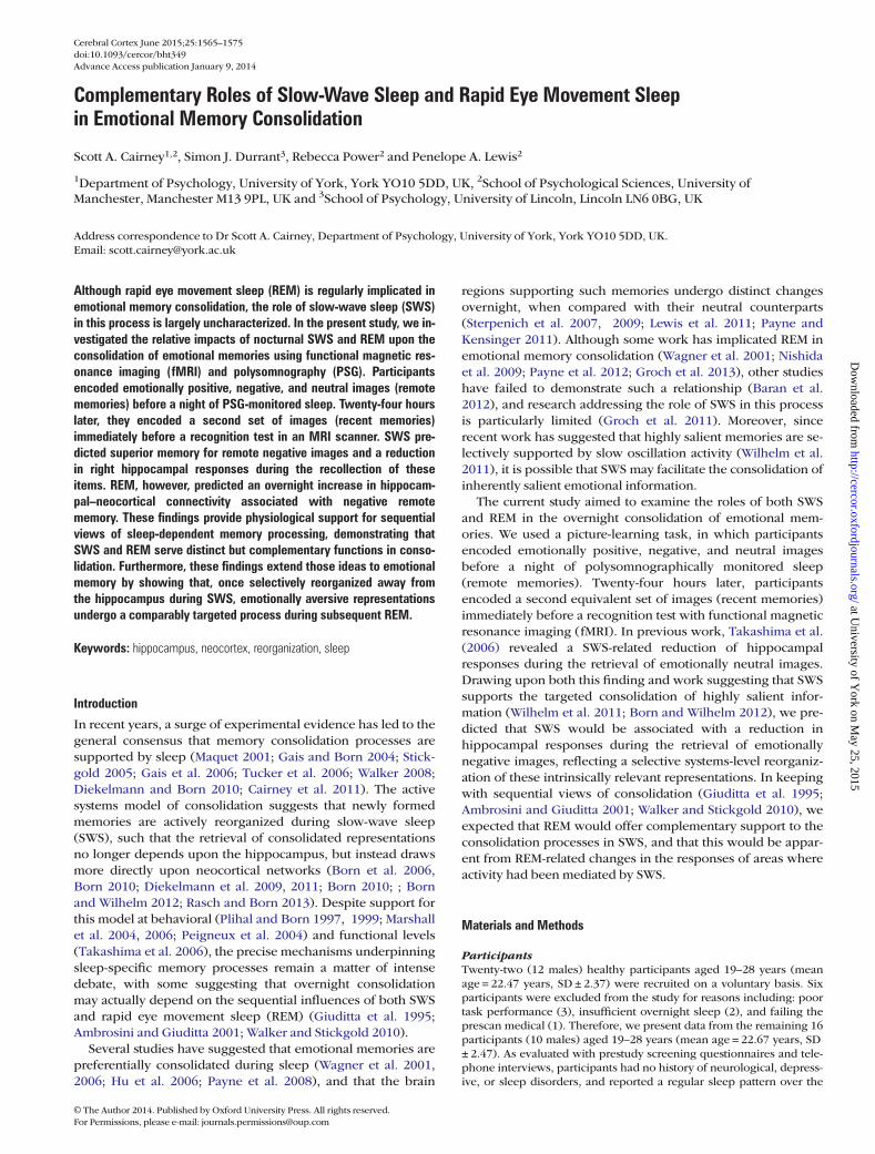
Figure 1. Study procedures. In the first session, participants encoded 90 (30 positive, 30 negative, and 30 neutral) images and then returned in the evening for overnight sleepwith PSG monitoring. The next day, participants encoded another 90 images before completing a recognition task for all previously learned images in the MRI scanner.
1566 Sleep and Emotional Memory Consolidation • Cairney et al.
at University of Y
ork on May 25, 2015
http://cercor.oxfordjournals.org/D
ownloaded from
accurately as possible, and were advised that they could make theirresponses during either the presentation of the image or the sub-sequent crosshair. The order of rating type (i.e., valence or arousalrating in the first or second encoding run) was counterbalanced acrossparticipants. On completing session 1, participants were free to goabout their usual daily activities.
Overnight SleepParticipants returned to the Manchester Sleep Laboratory at 10 PM,where they were connected to a polysomnography (PSG) unit beforebeing left to sleep in a darkened bedroom until 8 AM the nextmorning. After waking, participants were disconnected from the PSGunit and given a 2-h break before traveling to the MRI scanner for thesecond experimental session.
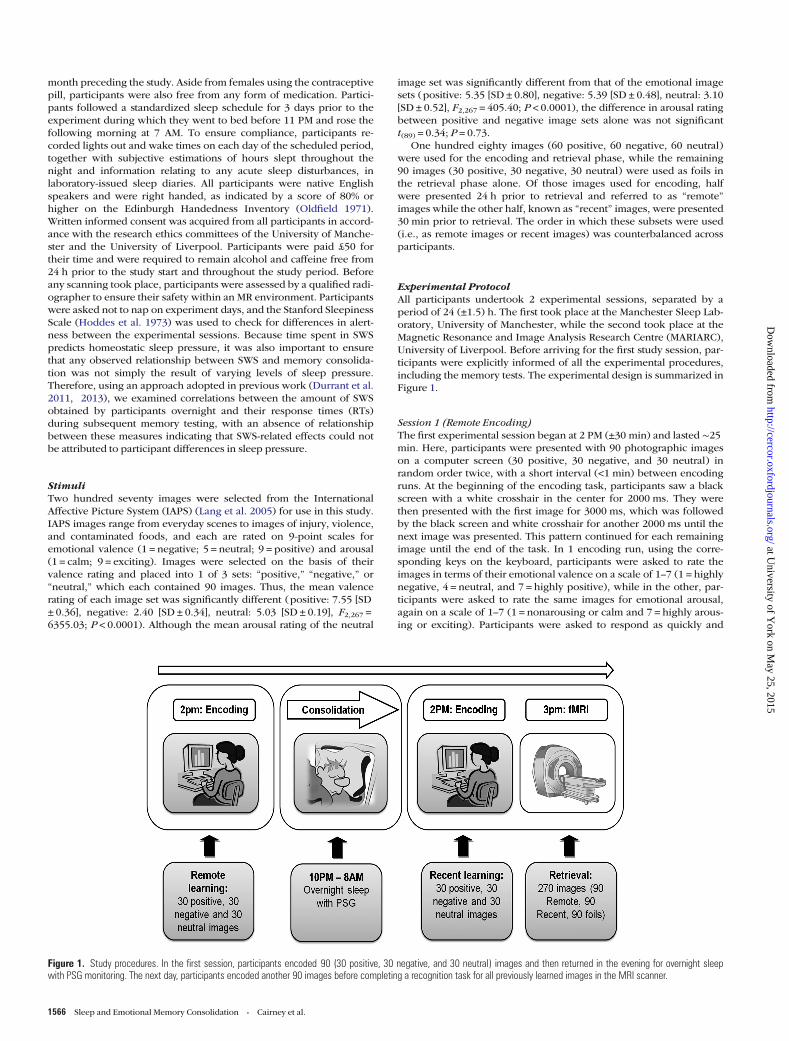
Session 2 (Recent Encoding and Retrieval)At 2 PM (±1 h) the next day, participants repeated the procedures ofsession 1 but with a new batch of 90 images. Approximately 30 minafter completing encoding (3pm ± 1 h), participants began the retrievalphase, which took place inside the MRI scanner. The retrieval test wasin the form of a recognition task, which encompassed all previouslyencoded remote and recent images and an additional 90 unseen foils(30 positive, 30 negative, and 30 neutral), making a total of 270images. The retrieval phase was divided into 2 scanning sessions, witha short (<1 min) break placed between each. Thus, both sessions con-sisted of 90 encoded images and 45 foils presented in a randomizedorder. At the beginning of the retrieval task, participants saw a blackscreen with a white crosshair in the center for 2000 ms. They werethen presented with the first image for 1000 ms, which was followedby the black screen and white crosshair for another 2000 ms until thenext image was presented. This pattern continued for each remainingimage until the end of the task. For each presented image, participantswere required to make a remember/know/new judgment (Tulving1985) using a 3-button keypad which they held in their right hand. A“remember” (R) response indicated that participants could consciouslyrecollect seeing the image in one of the previous encoding sessions.A “know” (K) response indicated that participants knew that they hadseen the image at encoding, but could not retrieve any contextual detailsabout its occurrence. Finally, a “new” (N) response indicated that partici-pants believed they had not seen the image in either encoding session.Participants were asked to respond as quickly and accurately as possible,and were advised that they could make their responses during either thepresentation of the image or the subsequent crosshair. The encodingand retrieval tasks are illustrated in Figure 2.
In keeping with similar work (Takashima et al. 2006), all remoteand recent images were seen only twice, once in the encoding sessionand once in the retrieval test. This methodology ensures that memorybias cannot arise from unbalanced stimuli exposure, as would resultfrom an immediate retrieval test in session 1. However, it also meantwe were unable to obtain a measure of immediate memory for theremote images, and must therefore assume that these were encoded inan identical manner to their recent counterparts.
Equipment
Experimental TaskThe experimental tasks were created using Cogent 2000 developed atthe functional imaging laboratory (FIL), University College London.The tasks were written and implemented using MatLab© version 6.5on a Toshiba Satellite Pro laptop with a 13” screen. During the encod-ing sessions, participant responses were recorded using the laptopkeyboard, while retrieval session responses were recorded with aserial multibutton box attached to a Domino 2 microcontroller fromMi-cromint© with a temporal resolution of ∼1 ms. The order of buttonsused in the retrieval task was counterbalanced across participants.
PolysomnographySleep monitoring was carried out using an Embla© N7000 PSG system,with silver–silver chloride (Ag–AgCl) electrodes attached using EC2©electrogel after the scalp was cleaned using NuPrep© exfoliatingagent. Scalp electrodes were attached according to the international10–20 system at 6 standardized locations: central (C3 and C4), frontal(F3 and F4), and occipital (O1 and O2), and each was referenced to thecontralateral mastoid (A1 or A2). Left and right electrooculagram, men-talis, and bilateral submentalis electromyogram, and a ground elec-trode were also attached. All electrodes were verified to have aconnection impedance of <5 kΩ. All signals were digitally sampled at arate of 200 Hz.
fMRI Data AcquisitionEvent-related fMRI data were attained using a Siemens MAGNETOMTrio 3 T Allegra MR scanner with an 8-channel head coil. Stimuli wereprojected onto a screen at the rear of the magnet bore and comfortablyviewed with an angled mirror attached to the head coil. Blood oxygenlevel–dependent (BOLD) signal was recorded using T2*-weighted fMRIimages obtained with a gradient echo-planar sequence (EPI). Fiftyoblique transaxial slices tilted at 15° were acquired in an ascending
Figure 2. Encoding and retrieval tasks. At encoding, participants were required to rate each image in terms of its emotional valence (unpleasant–pleasant) and emotional arousal(calm–exciting). At retrieval, participants were required to indicate if they recognized the image with recollection (R response) or familiarity (K response), or if they thought that theimage was new (N response).
Cerebral Cortex June 2015, V 25 N 6 1567
at University of Y
ork on May 25, 2015
http://cercor.oxfordjournals.org/D
ownloaded from
sequence with a voxel size of 3 × 3 × 2.8 mm3 including an interslicegap of 40%, matrix size of 64 × 64, TR of 2960 ms, TE of 30 ms, and aflip angle of 80°. A T1-weighted structural image was also acquired inthe same session for each participant using a 3D IR/GR sequence witha matrix size of 224 × 256 × 176, cubic isovoxels of 1 mm3, TR of 2040ms, TE of 5.57 ms, and a flip angle of 8°.
Data AnalysisAlthough our experimental task provided an index of both recollection(R responses) and familiarity (K responses) (Tulving 1985), we wereparticularly interested in recollection as this has previously shownemotion-specific sensitivity to sleep (Sterpenich et al. 2007), and wetherefore made this the primary focus of our investigation. To ensure ahigh proportion of correct R responses, we used a deep encoding task,with participants viewing all images twice, rating them once forvalence and once for arousal. A high proportion of R responses isnecessarily associated with a low proportion of K responses, as the 2are mutually exclusive (Yonelinas 2002). One-sample t-tests revealedthat, while recollection performance (R hits − R false alarms) wasabove chance for both remote and recent images, irrespective ofemotional valence (all P < 0.0001), familiarity performance (K hits – Kfalse alarms) was not (all P > 0.2). When R and K responses were com-bined, memory performance remained above chance for both remoteand recent images across all emotional valences (all P < 0.0001). Unlessotherwise stated, our analyses were therefore focused on recollection.
BehaviorThe sensitivity index (d′) [Normalized (hits/(hits +misses)) –Normalized(false alarms/(false alarms + correct rejections))] was calculated forremote and recent images and used as a measure of memory perform-ance. A combination of 2 retention types and 3 emotional valences pro-duced a 2 × 3 repeated-measures ANOVA design with factors “RetentionType” (remote/recent) and “Valence” (positive/negative/neutral).
To further examine the time course of memory consolidation, wecreated a difference measure, which we refer to as “behavioral consoli-dation,” by subtracting recent from remote memory performance (d′)for positive, negative, and neutral images. The resulting behavioralconsolidation scores provide indices of the emotional memory pro-cesses taking place across the 24-h retention delay. These were used toexamine relationships between sleep parameters and behavioralchange. All behavioral data were analyzed with SPSS version 16.0, anda two-tailed P < 0.05 was considered significant.
Sleep ScoringPSG sleep recordings were independently scored by 2 experiencedsleep researchers using RemLogic© 1.1 software. Sleep data were cate-gorized into 30-s epochs and scored on the referenced central electro-des (C3–A2 and C4–A1) according to the standardized sleep scoringcriteria of Rechtschaffen and Kales (1968), with sleep stages 3 and 4grouped together as SWS. Scored data were then partitioned in termsof the percentage of total sleep time spent in stage 1 sleep (S1), stage 2sleep (S2), SWS, and REM. To examine how SWS and REM, which havebeen previously implicated in the respective consolidation of neutraland emotional declarative memory (Wagner et al. 2001; Hu et al. 2006;Born and Wilhelm 2012), impacted upon subsequent remote memoryperformance, we separately correlated these sleep parameters with ourscores of behavioral consolidation for positive, negative, and neutralimages.
Functional Magnetic Resonance ImagingFunctional imaging data were processed using the Statistical Para-metric Mapping 8 software (SPM8; Wellcome Trust Centre for Neuroi-maging, London, UK, http://www.fil.ion.ucl.ac.uk/spm). To allow forT1 equilibration effects, the first 3 volumes of each retrieval sessionwere discarded from our analysis. Single-subject EPI volumes were rea-ligned to correct for minor motion artifacts before slice-timing correc-tion, referenced to the middle slice, was carried out. Images were thentransformed into standard stereotaxic space, corresponding to theMontreal Neurological Institute (MNI) canonical brain. This was
accomplished by registering the mean EPI volume (acquired duringrealignment) to SPM8’s EPI template and applying the subsequenttransform to all image volumes. A spherical Gaussian smoothingkernel with a full-width half-maximum of 8 mm was then applied tothe normalized data to attenuate variations in intersubject localization.
Data analysis was conducted with a 2-level, random effects generallinear model (GLM) (Friston et al. 1995). To minimize the impact of gues-sing, we focused on successful recollection (correct R responses) alone. Atthe first level, our designmatrix contained 27 regressors. Thesewere remoterecollection (remote-positive-R, remote-negative-R, remote-neutral-R), recentrecollection (recent-positive-R, recent-negative-R, recent-neutral-R), remotefamiliarity (remote-positive-K, remote-negative-K, remote-neutral-K), recentfamiliarity (recent-positive-K, recent-negative-K, recent-neutral-K), correctrejections (new-positive-N, new-negative-N, new-neutral-N), misses(remote-positive-N, remote-negative-N, remote-neutral-N, recent-positive-N,recent-negative-N, recent-neutral-N), and false alarms (new-positive-R, new-negative-R, new-neutral-R, new-positive-K, new-negative-K, new-neutral-K).Each regressor was convolved with a canonical hemodynamic responsefunction. Movement parameters were included as 6 nonconvolved regres-sors of no interest. To ensure that our fMRI analysis contained sufficient stat-istical power, participants with fewer than 10 successful recollection trialsfor each image combination of retention type (remote/recent) and emotionalvalence (positive/negative/neutral) were excluded from the study.Three participants were excluded on these grounds. For the 16 partici-pants included in our analysis, mean frequencies of successful remoteand recent recollection trials were as follows: remote positive: 26.06[SD ± 2.86], remote negative: 26.13 [SD ± 3.01], remote neutral: 25.63[SD ± 3.77], recent positive: 25.31 [SD ± 4.38], recent negative: 26.50[SD ± 3.14], recent neutral: 25.31 [SD ± 3.59].
Linear t-contrasts were calculated at the first level to examine over-night changes in recollection-related brain responses for positiveimages [positive remote hits < > positive recent hits], negative images[negative remote hits < > negative recent hits], and neutral images[neutral remote hits < > neutral recent hits]. To assess group effects, theresulting contrast images were carried forward to separate second-levelone-sample t-tests. In order to examine how postlearning SWS or REMinfluenced overnight changes in recollection-related activity, the con-trast images acquired from our first-level analyses were also carriedforward to 2 additional sets of second-level one-sample t-tests whereSWS was included as a covariate in one set, and REM as a covariate inthe other set. Accordingly, activation changes represented a SWS- orREM-dependent modification in the neural architecture of emotionallypositive, negative, or neutral memory recollection.
Data were thresholded at a whole-brain-corrected significance levelof P < 0.05. This was achieved by using an uncorrected voxel-levelthreshold of P < 0.001 in combination with a cluster extent threshold ofk = 16 voxels, as determined by a Monte-Carlo Simulation with 1000iterations (Slotnick et al. 2003). Because the hippocampus has beenpreviously implicated in sleep-dependent changes to the associativeneural trace of declarative memory (Takashima et al. 2006; Gais et al.2007; Sterpenich et al. 2007), we were specifically interested inresponses within this structure. We therefore conducted additionalvoxel-wise bilateral region of interest (ROI) analyses to examineresponses in the hippocampus in more detail [P < 0.05 family-wiseerror (FWE) corrected; k = 5 voxels]. These were carried out using hip-pocampal masks from the automated anatomical labeling templates(Tzourio-Mazoyer et al. 2002), as implemented within the Wake ForestUniversity (WFU) pickatlas software version 3.0 (Maldjian et al. 2003).
Functional ConnectivityWe used a psychophysiological interaction (PPI) to examine SWS- orREM-related changes in connectivity between areas of the hippo-campus that had already shown strong sensitivity to SWS in local func-tional activations and other regions of the brain. Accordingly, the peakhippocampal coordinates from the previous ROI analysis that hadyielded the strongest SWS-related change in hippocampal activity[Negative Remote hits > Negative Recent hits × SWS] were used tocreate a 6-mm-radius volume of interest in the right hippocampus (x, y,z (mm): 27, −10, −17) of each participant, from which the time serieswas extracted. The deconvolved time series were then multiplied bycontrasts for the effect of remote recollection for positive images
1568 Sleep and Emotional Memory Consolidation • Cairney et al.
at University of Y
ork on May 25, 2015
http://cercor.oxfordjournals.org/D
ownloaded from
[positive remote hits > positive recent hits], negative images [negativeremote hits > negative recent hits], and neutral images [neutral remotehits > neutral recent hits], forming 3 PPIs. As such, the resulting first-level GLM analyses contained a psychological factor (remote > recentrecollection), a physiological factor (the deconvolved time series), andthe interaction of these (psychological × physiological) as regressors.The 6 movement parameters were also included as nonconvolved re-gressors of no interest. Linear t-contrasts were calculated at the firstlevel to assess how recollection-related patterns of connectivitybetween the right hippocampus and other regions of the brainchanged over sleep for positive images [positive remote hits<> positiverecent hits], negative images [negative remote hits < > negative recenthits], and neutral images [neutral remote hits < > neutral recent hits].The resulting interaction contrast maps were then taken forward toseparate second-level one-sample t-tests for an examination of groupeffects. To investigate how postlearning SWS or REM influenced over-night changes in hippocampal connectivity, the first-level contrastmaps were also carried forward to 2 additional sets of second-level,one-sample t-tests, where one set included SWS as a covariate and theother set included REM as a covariate. Activation changes therefore,represented a SWS- or a REM-related alteration of hippocampal con-nectivity associated with emotionally positive, negative, or neutralmemory recollection. As with our localized analyses, our connectivityanalyses were thresholded at a whole-brain corrected significance levelof P < 0.05, achieved by combining an uncorrected voxel-levelthreshold of P < 0.001 with a cluster extent threshold of k = 16 voxels,as determined by a Monte-Carlo Simulation with 1000 iterations (Slot-nick et al. 2003).
Results
Sleep Patterns and AlertnessSleep diaries revealed that participants had adhered to ourstandardized sleep schedule throughout the 3 days prior to theexperiment and had not suffered any major acute sleep dis-turbance. The mean number of hours slept on the night pre-ceding the first experimental session was 7.84 [SD ± 0.85], andthe mean estimation of hours sleep obtained in a typical nightwas 7.47 [SD ± 0.50]. The difference between these 2 measureswas not significant (t(15) = 1.54; P = 0.15), confirming that par-ticipants’ prestudy sleep had not deviated from their usualpractices. Mean alertness scores, as obtained with the StanfordSleepiness Scale, showed no significant difference betweensession 1 (2.25 [SD ± 0.77]) and session 2 (2.00 [SD ± 0.52])(t(15) = 1.00; P = 0.33). In addition, no significant correlationwas found between the SWS obtained overnight and RTs forremote or recent images in the subsequent retrieval test, irre-spective of image valence (remote positive: r =−0.13; P = 0.64,remote negative: r =−0.20; P = 0.46, remote neutral: r =−0.13;P = 0.64, recent positive: r =−0.14; P = 0.62, recent negative:r =−0.22; P = 0.41, recent neutral: r =−0.12; P = 0.66). Takentogether with the data obtained from participant sleep diaries,these results indicate that any sleep-related effects were unli-kely to have occurred as a result of participant differences insleep pressure.
Subjective Ratings of Valence and ArousalThe image ratings obtained at encoding were also examined toensure that subjective ratings of valence and arousal were inkeeping with those used to initially categorize images as posi-tive, negative, or neutral (Lang et al. 2005). The mean valenceratings were significantly different for each image set (positive:5.60 [SD ± 0.35], negative: 2.04 [SD ± 0.54], neutral: 3.94 [SD ±0.40], F2,118 = 1045.78; P < 0.0001), as were mean arousal
ratings (positive: 4.69 [SD ± 0.71], negative: 5.30 [SD ± 0.78],neutral: 2.34 [SD ± 0.89], F2,118 = 233.15; P < 0.0001). Partici-pant ratings also revealed that negative images were associatedwith a significantly greater degree of arousal than positiveimages (t(59) = 4.44; P < 0.0001).
Experimental FindingsAs described in the Materials and Methods, familiarity perform-ance (K responses) for both remote and recent images did notdiffer from chance, irrespective of emotional valence (all P >0.2), indicating that recognition performance was driven by re-collection (R responses). Therefore, unless otherwise stated,the results described in this section are exclusively focused onrecollection.
Memory PerformanceA 2 × 3 repeated-measures ANOVA, with factors “RetentionType” (remote/recent) and “Valence” (positive/negative/neutral) showed equivalent memory performance (d′) forimages encoded 24 h (remote) or 30 min (recent) before retrie-val (F1,15 = 0.02; P = 0.89). Memory performance was also unaf-fected by image valence (F2,30 = 0.42; P = 0.66), and there wasno interaction between factors (F2,30 = 0.07; P = 0.94). TheANOVAwas repeated with R and K responses combined. Again,this revealed no main effect of Retention Type (F1,15 = 0.12; P =0.74), no main effect of Valence (F2,30 = 0.88; P = 0.43), and nointeraction between factors (F2,30 = 0.02; P = 0.98). Behavioraldata are available in Table 1.
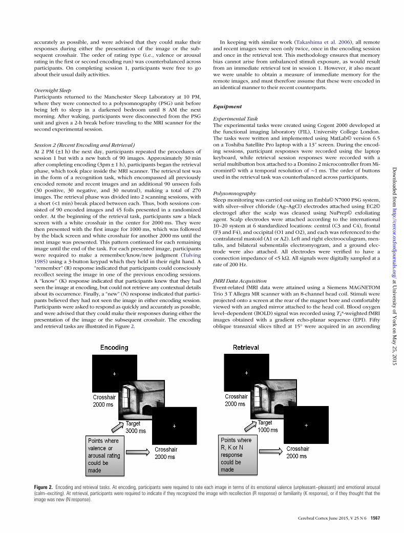
Sleep ParametersPSG data revealed that participants spent the following percen-tages of total sleep time in each stage of sleep (mean):S1 = 14.31 [SD ± 8.87], S2 = 44.44 [SD ± 8.83], SWS = 20.26 [SD± 4.05], REM = 20.98 [SD ± 5.01]. SWS predicted the behavioralconsolidation of negative memories (r = 0.57; P = 0.02),suggesting that the offline processing of such memories mayhave been mediated by this sleep stage (see Fig. 3A). In con-trast, REM was associated with less behavioral consolidation ofpositive memories (r = −0.58; P = 0.018), suggesting that mne-monic processes unique to this sleep stage may have inhibitedthe stabilization of emotionally positive material (see Fig. 3B).Care should be taken when drawing interpretations from thesecorrelations; however, as one-sample t-tests revealed no group-level effect of behavioral consolidation (remote d′ – recent d′)for positive, negative, or neutral images (all P > 0.8), and nosignificant difference in this measure was found between va-lences (F2,30 = 0.07; P = 0.94).
We found no evidence of a predictive relationship betweenSWS and the behavioral consolidation of positive (r = 0.04; P =0.88) or neutral (r =−0.15; P = 0.58) memories, or betweenREM and the behavioral consolidation of negative (r =−0.25; P= 0.35) or neutral (r = 0.24; P = 0.37) memories, and no signifi-cant correlation was found between the percentages of totalsleep time spent in SWS and REM (r =−0.22; P = 0.41).
Functional ImagingTo determine how SWS and REM influenced the consolidationof emotionally positive, negative, and neutral memories, we in-cluded these sleep parameters as covariates in 2 separatewhole-brain fMRI analyses [remote hits < > recent hits]. Foremotionally negative images, SWS mediated a reduction ofrecollection-related activity in the right hippocampus (see
Cerebral Cortex June 2015, V 25 N 6 1569
at University of Y
ork on May 25, 2015
http://cercor.oxfordjournals.org/D
ownloaded from
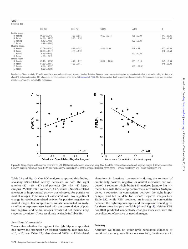
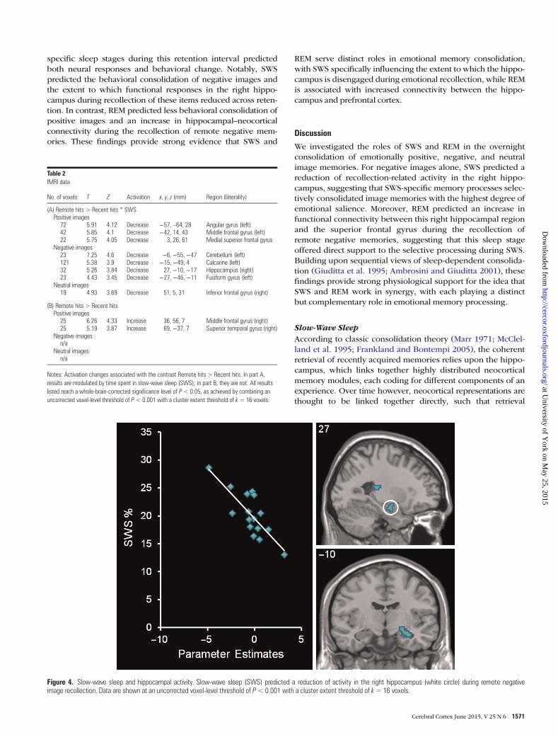
Table 2A and Fig. 4). Our ROI analyses supported this finding,revealing SWS-related activity decreases in both the rightanterior (27, −10, −17) and posterior (30, −28, −8) hippo-campus (P < 0.05 FWE corrected; k > 5 voxels). No SWS-relatedalteration in hippocampal activity was observed for positive orneutral images. REM was not associated with any significantchange in recollection-related activity for positive, negative, orneutral images. For completeness, we also conducted an analy-sis of brain responses associated with the consolidation of posi-tive, negative, and neutral images, which did not include sleepstages as covariates. These results are available in Table 2B.
Functional ConnectivityTo examine whether the region of the right hippocampus thathad shown the strongest SWS-related functional response (27,−10, −17, see Table 2A) also showed SWS- or REM-related
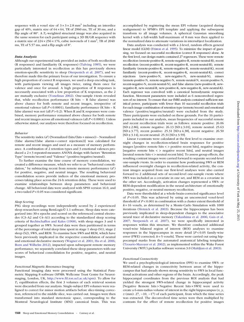
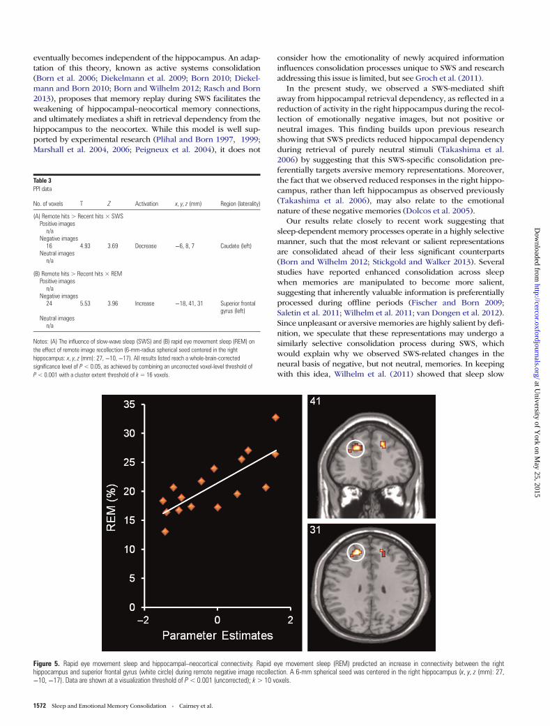
alterations in functional connectivity during the retrieval ofemotionally positive, negative, or neutral memories, we con-ducted 2 separate whole-brain PPI analyses [remote hits < >recent hits] with these sleep parameters as covariates. SWS pre-dicted a reduction in connectivity between the right hippo-campus and left caudate for remote negative images (seeTable 3A), while REM predicted an increase in connectivitybetween the right hippocampus and the superior frontal gyrusfor these same images (see Table 3B and Fig. 5). Neither SWSnor REM predicted connectivity changes associated with theconsolidation of positive or neutral images.
Summary
Although we found no group-level behavioral evidence ofemotional memory consolidation across 24 h, the time spent in
Table 1Behavioral data
Hits (%) Miss (%) CR (%) FA (%) d′
Positive imagesR: Remote 86.88 (±9.54) 4.38 (±5.54) 85.00 (±9.74) 3.96 (±3.89) 3.47 (±0.46)R: Recent 84.38 (±14.59) 3.96 (±2.78) 3.44 (±0.36)K: Remote 6.46 (±7.65) 8.33 (±8.34)K: Recent 9.17 (±13.69)
Negative imagesR: Remote 87.08 (±10.03) 5.21 (±5.57) 88.33 (10.54) 4.38 (4.34) 3.37 (±0.45)R: Recent 88.33 (±10.47) 4.58 (±4.19) 3.40 (±0.53)K: Remote 5.42 (±7.59) 5.00 (±7.50)K: Recent 4.38 (±7.67)
Neutral imagesR: Remote 85.42 (±12.58) 4.79 (±4.71) 85.42 (±12.64) 3.13 (±3.10) 3.45 (±0.44)R: Recent 84.38 (±11.97) 4.38 (±4.51) 3.48 (±0.38)K: Remote 6.88 (±10.92) 9.17 (±12.02)K: Recent 8.33 (±12.05)
Recollection (R) and familiarity (K) performance for remote and recent images (mean ± standard deviation). Because images were not categorized as belonging to the first or second encoding session, falsealarm (FA) and correct rejection (CR) values relate to both remote and recent items (Takashima et al. 2006). FAs that received an R or K response are shown separately. Because our analysis was focused onrecollection, d’ was only calculated for R responses.
Figure 3. Sleep stages and behavioral consolidation (d′). (A) Correlation between slow-wave sleep (SWS) and the behavioral consolidation of negative images. (B) Inverse correlationbetween rapid eye movement sleep (REM) and the behavioral consolidation of positive images. Behavioral consolidation= remote recollection (d′) – recent recollection (d′).
1570 Sleep and Emotional Memory Consolidation • Cairney et al.
at University of Y
ork on May 25, 2015
http://cercor.oxfordjournals.org/D
ownloaded from
specific sleep stages during this retention interval predictedboth neural responses and behavioral change. Notably, SWSpredicted the behavioral consolidation of negative images andthe extent to which functional responses in the right hippo-campus during recollection of these items reduced across reten-tion. In contrast, REM predicted less behavioral consolidation ofpositive images and an increase in hippocampal–neocorticalconnectivity during the recollection of remote negative mem-ories. These findings provide strong evidence that SWS and
REM serve distinct roles in emotional memory consolidation,with SWS specifically influencing the extent to which the hippo-campus is disengaged during emotional recollection, while REMis associated with increased connectivity between the hippo-campus and prefrontal cortex.
Discussion
We investigated the roles of SWS and REM in the overnightconsolidation of emotionally positive, negative, and neutralimage memories. For negative images alone, SWS predicted areduction of recollection-related activity in the right hippo-campus, suggesting that SWS-specific memory processes selec-tively consolidated image memories with the highest degree ofemotional salience. Moreover, REM predicted an increase infunctional connectivity between this right hippocampal regionand the superior frontal gyrus during the recollection ofremote negative memories, suggesting that this sleep stageoffered direct support to the selective processing during SWS.Building upon sequential views of sleep-dependent consolida-tion (Giuditta et al. 1995; Ambrosini and Giuditta 2001), thesefindings provide strong physiological support for the idea thatSWS and REM work in synergy, with each playing a distinctbut complementary role in emotional memory processing.
Slow-Wave SleepAccording to classic consolidation theory (Marr 1971; McClel-land et al. 1995; Frankland and Bontempi 2005), the coherentretrieval of recently acquired memories relies upon the hippo-campus, which links together highly distributed neocorticalmemory modules, each coding for different components of anexperience. Over time however, neocortical representations arethought to be linked together directly, such that retrieval
Table 2fMRI data
No. of voxels T Z Activation x, y, z (mm) Region (laterality)
(A) Remote hits > Recent hits * SWSPositive images72 5.91 4.12 Decrease −57, –64, 28 Angular gyrus (left)42 5.85 4.1 Decrease −42, 14, 43 Middle frontal gyrus (left)22 5.75 4.05 Decrease 3, 26, 61 Medial superior frontal gyrus
Negative images23 7.25 4.6 Decrease −6, −55, −47 Cerebellum (left)121 5.38 3.9 Decrease −15, −49, 4 Calcarine (left)32 5.26 3.84 Decrease 27, −10, −17 Hippocampus (right)23 4.43 3.45 Decrease −27, −46, −11 Fusiform gyrus (left)
Neutral images19 4.93 3.69 Decrease 51, 5, 31 Inferior frontal gyrus (right)
(B) Remote hits > Recent hitsPositive images25 6.26 4.33 Increase 36, 56, 7 Middle frontal gyrus (right)25 5.19 3.87 Increase 69, −37, 7 Superior temporal gyrus (right)
Negative imagesn/a
Neutral imagesn/a
Notes: Activation changes associated with the contrast Remote hits > Recent hits. In part A,results are modulated by time spent in slow-wave sleep (SWS); in part B, they are not. All resultslisted reach a whole-brain-corrected significance level of P< 0.05, as achieved by combining anuncorrected voxel-level threshold of P< 0.001 with a cluster extent threshold of k= 16 voxels.
Figure 4. Slow-wave sleep and hippocampal activity. Slow-wave sleep (SWS) predicted a reduction of activity in the right hippocampus (white circle) during remote negativeimage recollection. Data are shown at an uncorrected voxel-level threshold of P<0.001 with a cluster extent threshold of k=16 voxels.
Cerebral Cortex June 2015, V 25 N 6 1571
at University of Y
ork on May 25, 2015
http://cercor.oxfordjournals.org/D
ownloaded from
eventually becomes independent of the hippocampus. An adap-tation of this theory, known as active systems consolidation(Born et al. 2006; Diekelmann et al. 2009; Born 2010; Diekel-mann and Born 2010; Born and Wilhelm 2012; Rasch and Born2013), proposes that memory replay during SWS facilitates theweakening of hippocampal–neocortical memory connections,and ultimately mediates a shift in retrieval dependency from thehippocampus to the neocortex. While this model is well sup-ported by experimental research (Plihal and Born 1997, 1999;Marshall et al. 2004, 2006; Peigneux et al. 2004), it does not
consider how the emotionality of newly acquired informationinfluences consolidation processes unique to SWS and researchaddressing this issue is limited, but see Groch et al. (2011).
In the present study, we observed a SWS-mediated shiftaway from hippocampal retrieval dependency, as reflected in areduction of activity in the right hippocampus during the recol-lection of emotionally negative images, but not positive orneutral images. This finding builds upon previous researchshowing that SWS predicts reduced hippocampal dependencyduring retrieval of purely neutral stimuli (Takashima et al.2006) by suggesting that this SWS-specific consolidation pre-ferentially targets aversive memory representations. Moreover,the fact that we observed reduced responses in the right hippo-campus, rather than left hippocampus as observed previously(Takashima et al. 2006), may also relate to the emotionalnature of these negative memories (Dolcos et al. 2005).
Our results relate closely to recent work suggesting thatsleep-dependent memory processes operate in a highly selectivemanner, such that the most relevant or salient representationsare consolidated ahead of their less significant counterparts(Born and Wilhelm 2012; Stickgold and Walker 2013). Severalstudies have reported enhanced consolidation across sleepwhen memories are manipulated to become more salient,suggesting that inherently valuable information is preferentiallyprocessed during offline periods (Fischer and Born 2009;Saletin et al. 2011; Wilhelm et al. 2011; van Dongen et al. 2012).Since unpleasant or aversive memories are highly salient by defi-nition, we speculate that these representations may undergo asimilarly selective consolidation process during SWS, whichwould explain why we observed SWS-related changes in theneural basis of negative, but not neutral, memories. In keepingwith this idea, Wilhelm et al. (2011) showed that sleep slow
Table 3PPI data
No. of voxels T Z Activation x, y, z (mm) Region (laterality)
(A) Remote hits > Recent hits × SWSPositive images
n/aNegative images
16 4.93 3.69 Decrease −6, 8, 7 Caudate (left)Neutral images
n/a
(B) Remote hits > Recent hits × REMPositive images
n/aNegative images
24 5.53 3.96 Increase −18, 41, 31 Superior frontalgyrus (left)
Neutral imagesn/a
Notes: (A) The influence of slow-wave sleep (SWS) and (B) rapid eye movement sleep (REM) onthe effect of remote image recollection (6-mm-radius spherical seed centered in the righthippocampus: x, y, z (mm): 27, −10, −17). All results listed reach a whole-brain-correctedsignificance level of P< 0.05, as achieved by combining an uncorrected voxel-level threshold ofP< 0.001 with a cluster extent threshold of k= 16 voxels.
Figure 5. Rapid eye movement sleep and hippocampal–neocortical connectivity. Rapid eye movement sleep (REM) predicted an increase in connectivity between the righthippocampus and superior frontal gyrus (white circle) during remote negative image recollection. A 6-mm spherical seed was centered in the right hippocampus (x, y, z (mm): 27,−10, −17). Data are shown at a visualization threshold of P< 0.001 (uncorrected); k> 10 voxels.
1572 Sleep and Emotional Memory Consolidation • Cairney et al.
at University of Y
ork on May 25, 2015
http://cercor.oxfordjournals.org/D
ownloaded from
oscillation activity increased after the salience of newly formedmemories was augmented, and the extent of this increase pre-dicted retrieval performance in a subsequent test. If this iscorrect however, then emotionally positive memories should bepreferentially consolidated during SWS, just like their negativecounterparts, but we found no evidence for this. One possibleexplanation relates to emotional arousal; because participantsrated negative images as significantly more arousing than posi-tive images at encoding, it is reasonable to assume that negativeimages were also most emotionally salient.
Rapid Eye Movement SleepResearch linking overnight memory benefits to time spent inboth SWS and REM has supported the idea that these sleepstages play complementary roles in optimizing the potential ef-ficacy of newly acquired information (Stickgold et al. 2000), anidea which is otherwise known as the sequential hypothesis(Giuditta et al. 1995; Ambrosini and Giuditta 2001). Thishypothesis states that, after the consolidation processes of SWShave unfolded, representations are subject to an additionalmnemonic process during REM, which further facilitates theirstorage. Our observation that REM predicts no functionalchange in localized neural responses but instead predicts in-creased connectivity in the hippocampal area where responseswere modulated by SWS provides physiological support forthis idea. Because these results were only observed withinemotionally negative items, they also suggest that both SWSand REM target emotionally salient representations. This com-bined preferential processing could help to explain whyemotional material is often selectively protected across a nightof sleep (Hu et al. 2006; Payne et al. 2008).
Limbic and forebrain levels of the neurotransmitter acetyl-choline are markedly elevated during REM, and are reportedlydouble the levels observed during quiet wakefulness (Marrosuet al. 1995). Because acetylcholine appears to co-regulate theeffects of emotion on consolidation (Introini-Collison et al.1996; Power and McGaugh 2002), it has been suggested thatREM-related increases in cholinergic activity may promoteemotional plasticity (Walker and van der Helm 2009). Further-more, theta oscillations, which prevail during REM and coordi-nate communication between the hippocampus and prefrontalcortex (Jones and Wilson 2005), have been suggested as apotential mechanism for the formation of new neural connec-tions. Interestingly, recent work has revealed REM-related in-creases in creative insight (Cai et al. 2009), presumablythrough the formation of such connections, a finding that is ingood keeping with our observation that REM predicts increasedconnectivity. Having said this, it is important to note that thebrain responses observed during retrieval do not necessarilycorrespond to activity patterns in specific stages of sleep. Forexample, while our data revealed a REM-related increase in hip-pocampal–neocortical connectivity during retrieval, communi-cation between these structures is actually reduced during REM(Axmacher et al. 2008).
In their recent adaptation of the sequential hypothesis,Walker and Stickgold (2010) proposed that REM supports theintegration of recently acquired memories, strengthenedduring the preceding SWS, into rich and generalized networksof related knowledge. As effective integration can presumablybe indexed by modifications in the associative neural trace atretrieval (Lewis and Durrant 2011), our observed REM-related
increase in hippocampal connectivity could potentially reflecta form of targeted integration. The experimental task adoptedin this study however provides no behavioral metric of inte-gration, and it is unclear why such integrative memory processeswould depend on hippocampal connections to prefrontalregions, particularly when anterior temporal areas are mostcommonly associated with semantic knowledge (Binney et al.2010; Lambon Ralph et al. 2012). Accordingly, this suggestedrole for REM in selective mnemonic integration is highly specu-lative and represents just one potential interpretation of ourdata. Future studies should examine this in more detail with atask that specifically tests for integration.
SWS was associated with the behavioral consolidation ofnegative images in the present study, but REM showed no suchrelationship, instead predicting less behavioral consolidationof positive images. This pattern fails to support the view thatSWS and REM facilitate affective consolidation processes in asequential manner, and is at odds with research linking REM tosuperior retention of emotional memories (Wagner et al. 2001;Nishida et al. 2009; Payne et al. 2012; Groch et al. 2013). Thatsaid, others have also failed to observe such a relationship(Baran et al. 2012), suggesting that these behavioral effects arefragile and may only emerge under very specific experimentalconditions. Furthermore, although a number of reports haveexamined how emotionally negative memories are influencedby postlearning sleep (Hu et al. 2006; Payne et al. 2008; Lewiset al. 2011; Payne and Kensinger 2011), few have addressedthe role of sleep in processing positive emotional represen-tations (Sterpenich et al. 2007, 2009) and, to our knowl-edge, no prior study has revealed a specific link betweenREM and positive memory. Accordingly, while REM appearsto affect emotional memory consolidation, the precisenature of these effects may depend on both the valence ofthe representation in question and the conditions of learn-ing and retrieval. It is important to note, however, that indi-vidual differences in habitual sleep patterns may haveaffected the relative proportions of time spent in differentsleep stages by each participant, meaning that habitualinfluences cannot be fully excluded when considering theresults of this study.
Funding
This work was supported by the Engineering and PhysicalSciences Research Council, Unilever, and a Biotechnology andBiological Sciences Research Council New Investigator award(BB/F003048/1 to P.A.L.).
NotesThe authors thank the staff at the Magnetic Resonance and ImageAnalysis Research Centre (MARIARC), University of Liverpool, for tech-nical assistance and to Andrew Mayes, Björn Rasch, and James Cousinsfor fruitful discussions and critical reading of the manuscript. Theauthors are also grateful to 3 anonymous reviewers for their invaluablecomments and suggestions. Conflict of Interest: None declared.
ReferencesAmbrosini MV, Giuditta A. 2001. Learning and sleep: the sequential
hypothesis. Sleep Med Rev. 5:477–490.
Cerebral Cortex June 2015, V 25 N 6 1573
at University of Y
ork on May 25, 2015
http://cercor.oxfordjournals.org/D
ownloaded from
Axmacher N, Helmstaedter C, Elger CE, Fell J. 2008. Enhancement ofneocortical-medial temporal EEG correlations during non-REMsleep. Neural Plast. 2008. Article ID 563028.
Baran B, Pace-Schott EF, Ericson C, Spencer RMC. 2012. Processing ofemotional reactivity and emotional memory over sleep. J Neurosci.32:1035–1042.
Binney RJ, Embleton KV, Jefferies E, Parker GJM, Lambon Ralph MA.2010. The ventral and inferolateral aspects of the anterior temporallobe are crucial in semantic memory: evidence from a novel directcomparison of distortion-corrected fMRI, rTMS, and semantic de-mentia. Cereb Cortex. 20:2728–2738.
Born J. 2010. Slow-wave sleep and the consolidation of long-termmemory. World J Biol Psychiatry. 11:16–21.
Born J, Rasch B, Gais S. 2006. Sleep to remember. Neuroscientist. 12:410–424.
Born J, Wilhelm I. 2012. System consolidation of memory during sleep.Psychol Res. 76:192–203.
Cai DJ, Mednick SA, Harrison EM, Kanady JC, Mednick SC. 2009. REM,not incubation, improves creativity by priming associative networks.Proc Natl Acad Sci USA. 106:10130–10134.
Cairney SA, Durrant SJ, Musgrove H, Lewis PA. 2011. Sleep andenvironmental context: interactive effects for memory. Exp BrainRes. 214:83–92.
Diekelmann S, Born J. 2010. The memory function of sleep. Nat RevNeurosci. 11:114–126.
Diekelmann S, Buchel C, Born J, Rasch B. 2011. Labile or stable: op-posing consequences for memory when reactivated during wakingand sleep. Nat Neurosci. 14:381–386.
Diekelmann S, Wilhelm I, Born J. 2009. The whats and whens of sleep-dependent memory consolidation. Sleep Med Rev. 13:309–321.
Dolcos F, LaBar KS, Cabeza R. 2005. Remembering one year later: role ofthe amygdala and the medial temporal lobe memory system in retriev-ing emotional memories. Proc Natl Acad Sci USA. 102:2626–2631.
Durrant SJ, Cairney SA, Lewis PA. 2013. Overnight consolidation aidsthe transfer of statistical knowledge from the medial temporal lobeto the striatum. Cereb Cortex. 23:2467–2478.
Durrant SJ, Taylor C, Cairney S, Lewis PA. 2011. Sleep-dependent con-solidation of statistical learning. Neuropsychologia. 49:1322–1331.
Fischer S, Born J. 2009. Anticipated reward enhances offline learningduring sleep. J Exp Psychol Learn Mem Cogn. 35:1586–1593.
Frankland PW, Bontempi B. 2005. The organization of recent andremote memories. Nat Rev Neurosci. 6:119–130.
Friston KJ, Holmes AP, Worsley KJ, Poline JP, Frith CD, FrackowiakRSJ. 1995. Statistical parametric maps in functional imaging: ageneral linear approach. Hum Brain Mapp. 1:153–171.
Gais S, Albouy G, Boly M, Dang-Vu TT, Darsaud A, Desseilles M, RauchsG, Schabus M, Sterpenich V, Vandewalle G et al. 2007. Sleep trans-forms the cerebral trace of declarative memories. Proc Natl Acad SciUSA. 104:18778–18783.
Gais S, Born J. 2004. Declarative memory consolidation: mechanismsacting during human sleep. Learn Mem. 11:679–685.
Gais S, Lucas B, Born J. 2006. Sleep after learning aids memory recall.Learn Mem. 13:259–262.
Giuditta A, Ambrosini MV, Montagnese P, Mandile P, Cotugno M,Grassi Zucconi G, Vescia S. 1995. The sequential hypothesis of thefunction of sleep. Behav Brain Res. 69:157–166.
Groch S, Wilhelm I, Diekelmann S, Born J. 2013. The role of REM sleepin the processing of emotional memories: evidence from behaviorand event-related potentials. Neurobiol Learn Mem. 99:1–9.
Groch S, Wilhelm I, Diekelmann S, Sayk F, Gais S, Born J. 2011. Contri-bution of norepinephrine to emotional memory consolidationduring sleep. Psychoneuroendocrinology. 36:1342–1350.
Hoddes E, Zarcone V, Smythe H, Phillips R, Dement WC. 1973. Quantifi-cation of sleepiness: a new approach. Psychophysiology. 10:431–436.
Hu P, Stylos-Allan M, Walker MP. 2006. Sleep facilitates consolidationof emotional declarative memory. Psychol Sci. 17:891–898.
Introini-Collison IB, Dalmaz C, McGaugh JL. 1996. Amygdalaβ-noradrenergic influences on memory storage involve cholinergicactivation. Neurobiol Learn Mem. 65:57–64.
Jones MW, Wilson MA. 2005. Theta rhythms coordinate hippocampal-prefrontal interactions in a spatial memory task. PLoS Biol. 3:e402.
Lambon Ralph MA, Ehsan S, Baker GA, Rogers TT. 2012. Semanticmemory is impaired in patients with unilateral anterior temporallobe resection for temporal lobe epilepsy. Brain. 135:242–258.
Lang PJ, Bradley MM, Cuthbert BN. 2005. International affective picturesystem (IAPS): affective ratings of pictures and instruction manual.Technical Report A-6. University of Florida, Gainesville, FL.
Lewis PA, Cairney S, Manning L, Critchley HD. 2011. The impact ofovernight consolidation upon memory for emotional and neutralencoding contexts. Neuropsychologia. 49:2619–2629.
Lewis PA, Durrant SJ. 2011. Overlapping memory replay during sleepbuilds cognitive schemata. Trends Cogn Sci. 15:343–351.
Maldjian JA, Laurienti PJ, Kraft RA, Burdette JH. 2003. An automatedmethod for neuroanatomic and cytoarchitectonic atlas-basedinterrogation of fMRI data sets. Neuroimage. 19:1233–1239.
Maquet P. 2001. The role of sleep in learning and memory. Science.294:1048–1052.
Marr D. 1971. Simple memory: a theory for archicortex. Philos Trans RSoc Lond B Biol Sci. 262:23–81.
Marrosu F, Portas C, Mascia MS, Casu MA, Fa M, Giagheddu M, Impera-to A, Gessa GL. 1995. Microdialysis measurement of cortical andhippocampal acetylcholine release during sleep-wake cycle infreely moving cats. Brain Res. 671:329–332.
Marshall L, Helgadottir H, Molle M, Born J. 2006. Boosting slow oscil-lations during sleep potentiates memory. Nature. 444:610–613.
Marshall L, Molle M, Hallschmid M, Born J. 2004. Transcranial directcurrent stimulation during sleep improves declarative memory. JNeurosci. 24:9985–9992.
McClelland JL, McNaughton BL, O’Reilly RC. 1995. Why there arecomplementary learning systems in the hippocampus and neo-cortex: insights from the successes and failures of connectionistmodels of learning and memory. Psychol Rev. 102:419–457.
Nishida M, Pearsall J, Buckner RL, Walker MP. 2009. REM sleep, pre-frontal theta, and the consolidation of human emotional memory.Cereb Cortex. 19:1158–1166.
Oldfield RC. 1971. The assessment and analysis of handedness: theEdinburgh inventory. Neuropsychologia. 9:97–113.
Payne JD, Chambers AM, Kensinger EA. 2012. Sleep promotes lastingchanges in selective memory for emotional scenes. Front IntegrNeurosci. 6:108.
Payne JD, Kensinger EA. 2011. Sleep leads to changes in the emotion-al memory trace: evidence from fMRI. J Cogn Neurosci.23:1285–1297.
Payne JD, Stickgold R, Swanberg K, Kensinger EA. 2008. Sleep prefer-entially enhances memory for emotional components of scenes.Psychol Sci. 19:781–788.
Peigneux P, Laureys S, Fuchs S, Collette F, Perrin F, Reggers J, PhillipsC, Degueldre C, Del Fiore G, Aerts J et al. 2004. Are spatial mem-ories strengthened in the human hippocampus during slow wavesleep? Neuron. 44:535–545.
Plihal W, Born J. 1997. Effects of early and late nocturnal sleep on de-clarative and procedural memory. J Cogn Neurosci. 9:534–547.
Plihal W, Born J. 1999. Effects of early and late nocturnal sleep onpriming and spatial memory. Psychophysiology. 36:571–582.
Power AE, McGaugh JL. 2002. Cholinergic activation of the basolateralamygdala regulates unlearned freezing behavior in rats. Behav BrainRes. 134:307–315.
Rasch B, Born J. 2013. About sleep’s role in memory. Physiol Rev. 93:681–766.
Rechtschaffen A, Kales A. 1968. A manual of standardized terminology,techniques and scoring system for sleep stages of human subjects.Bethesda (MD): US Department of Health.
Saletin JM, Goldstein AN, Walker MP. 2011. The role of sleep indirected forgetting and remembering of human memories. CerebCortex. 21:2534–2541.
Slotnick SD, Moo LR, Segal JB, Hart J. 2003. Distinct prefrontal cortexactivity associated with item memory and source memory for visualshapes. Brain Res Cogn Brain Res. 17:75–82.
Sterpenich V, Albouy G, Boly M, Vandewalle G, Darsaud A, Balteau E,Dang-Vu TT, Desseilles M, D’Argembeau A, Gais S et al. 2007.Sleep-related hippocampo-cortical interplay during emotionalmemory recollection. PLoS Biol. 5:e282.
1574 Sleep and Emotional Memory Consolidation • Cairney et al.
at University of Y
ork on May 25, 2015
http://cercor.oxfordjournals.org/D
ownloaded from
Sterpenich V, Albouy G, Darsaud A, Schmidt C, Vandewalle G, DangVu TT, Desseilles M, Phillips C, Degueldre C, Balteau E et al. 2009.Sleep promotes the neural reorganization of remote emotionalmemory. J Neurosci. 29:5143–5152.
Stickgold R. 2005. Sleep-dependent memory consolidation. Nature. 437:1272–1278.
Stickgold R, Walker MP. 2013. Sleep-dependent memory triage: evolvinggeneralization through selective processing. Nat Neurosci. 16:139–145.
Stickgold R, Whidbee D, Schirmer B, Patel V, Hobson JA. 2000. Visualdiscrimination task improvement: a multi-step process occurringduring sleep. J Cogn Neurosci. 12:246–254.
Takashima A, Petersson KM, Rutters F, Tendolkar I, Jensen O, ZwartsMJ, McNaughton BL, Fernandez G. 2006. Declarative memory con-solidation in humans: a prospective functional magnetic resonanceimaging study. Proc Natl Acad Sci USA. 103:756–761.
Tucker MA, Hirota Y, Wamsley EJ, Lau H, Chaklader A, Fishbein W. 2006.A daytime nap containing solely non-REM sleep enhances declarativebut not procedural memory. Neurobiol Learn Mem. 86:241–247.
Tulving E. 1985. Memory and consciousness. Can Psychol. 26:1–12.Tzourio-Mazoyer N, Landeau B, Papathanassiou D, Crivello F, Etard
O, Delcroix N, Mazoyer B, Joliot M. 2002. Automated anatomicallabeling of activations in SPM using a macroscopic anatomical
parcellation of the MNI MRI single-subject brain. Neuroimage.15:273–289.
van Dongen EV, Thielen JW, Takashima A, Barth M, Fernández G.2012. Sleep supports selective retention of associative memoriesbased on relevance for future utilization. PLoS ONE. 7:e43426.
Wagner U, Gais S, Born J. 2001. Emotional memory formation is en-hanced across sleep intervals with high amounts of rapid eye move-ment sleep. Learn Mem. 8:112–119.
Wagner U, Hallschmid M, Rasch B, Born J. 2006. Brief sleep after learn-ing keeps emotional memories alive for years. Biol Psychiatry.60:788–790.
Walker MP. 2008. Sleep-dependent memory processing. Harv Rev Psy-chiatry. 16:287–298.
Walker MP, Stickgold R. 2010. Overnight alchemy: sleep-dependentmemory evolution. Nat Rev Neurosci. 11:218.
Walker MP, van der Helm E. 2009. Overnight therapy? The role ofsleep in emotional brain processing. Psychol Bull. 135:731–748.
Wilhelm I, Diekelmann S, Mozlow I, Ayoub A, Mölle M, Born J. 2011.Sleep selectively enhances memory expected to be of future relevance.J Neurosci. 31:1563–1569.
Yonelinas AP. 2002. The nature of recollection and familiarity: a reviewof 30 years of research. J Mem Lang. 46:441–517.
Cerebral Cortex June 2015, V 25 N 6 1575
at University of Y
ork on May 25, 2015
http://cercor.oxfordjournals.org/D
ownloaded from
Related Documents